Embed Size (px)
Citation preview

9:30
9:45
10:15
10:30 Keith Nugent University of Melbourne Science Industry and Synchrotrons
11:00 Stuart Thickett The University of Sydney Functional Patterned Surface Coatings Prepated by the Dewetting of Thin Polymer Films
11:20 Vijay Bhatia The University of Technology Sydney Thin Films of AuCuAl Shape Memory Alloy for Use in Plasmonic Nano-Actuators
11:40 Ehsan Jazaeri Deakin University
Fabrication and Characterization of Carbon Nanofibre by Pyrolysis of Freeze-Dried Celllulose Nanofibre
12:00 Brendon MacDonald University of Melbourne Solution-Processed Nanocrystal Solar Cells
12:20
13:10 James Chon Swinburne University
Surface plasmon resonance mediated optical properties in gold nanorods and its application to 5-dimensional optical storage
13:40 Karen Jarvis University of South AustraliaAmine modification of quartz particles via plasma polymerization for water contaminant removal
14:00 Jiangbo Zhao Macquarie UniversityTuning the Luminescence Lifetime by Sensitizer Yb3+ Concentration in Upconversion Nanocrystals
14:20 Rajesh Ganesan Macquarie University
Time resolved spectroscopic and kinetic studies of argon VUV luminescence in a windowless dielectric barrier discharge
14:40 Heather Catchpoole National Measurements Institute Multi-technique approach for the size characteriSation of nanoparticles
15:00
15:30 Patrick Parkinson Australian National UniversityTime-resolved spectroscopy of III-V semiconductor nanowires
15:50 Adam Burke University of New South WalesUnderstanding how electron density affects spin splitting in 1D systems
16:30
18:00
Lunch
ANN Early Career SymposiumMacquarie Park Conference Centre
21-22nd November 2011Co Chairs Deb Kane(Macq Uni) Adam Micolich (UNSW) and Jaret Lee (ANU)
Invited Talk
Afternoon Tea
Poster SessionBBQ
Monday 21st NovemberRegistrationMorning Tea
Welcome/ IntroductionsInvited Talk

9:00Petar Atanackovic Chris Escott Silanna
Perspectives on research and development: the long and short of it
9:30Laurens Willems van Beveren University of Melbourne
Overlapping-gate architecture for silicon Hall bar MOSFET devices in the low density and high magnetic field regime
9:50 LaReine Yeoh University of New South Wales
The Study of Low-Dimensional Semiconductor Nanostructures at milli-Kelvin Temperatures and High Magnetic Fields
10:10 Kok Wai Chan University of New South WalesSingle-electron shuttle based on a silicon quantum dot
10:30
11:00 Ela Eroglu University of Western AustraliaBiosynthesis of Palladium Nanoparticles by Green Microalgae
11:20 Thomas Barclay Flinders UniveristyThe Rational Design of Diazenyl amphiphiles for self-assembly into nanotubes within aqueous systems
11:40 Shulei Chou University of Wollongong
Rapid Synthesis of Li4Ti5O12 Microspheres Composed of Nanoflakes as Anode Materials for Lithium-ion Battery
12:00 Sanly Liu University of New South Wales
Reduced Pseudomonas aeruginosa proliferation and biofilm formation on zinc oxide/silica coated glass coupons
12:20
13:20 Angel Tan University of South AustraliaHybrid Nanomaterials that Mimic the ‘Food Effect’ to Enhance Oral Drug Absorption
13:40 Vipul Agarwal University of Western Australia
In vitro evaluation of electrospun pluronic F-127 dimethacrylate copolymer towards wound healing in burn injuries
14:00 Zi Gu University of Queensland
Enhanced Efficacy of Anti-restenotic Drug by Intercalation into Inorganic Layered Double Hydroxide Nanoparticle
14:20 Benjamin Gully University of Western AustraliaGraphene and graphene oxide (GO) as nucleating agents for protein crystalisation.
14:40
15:00
Tuesday 22nd November
Afternoon Tea
Invited Talk
Lunch
Morning Tea
Closing

Mojtaba Abtahi University of SydneyPreparation of Toughened PLA Nanocomposites with Nanoclay and Nano Powder Rubber
Nikki Amos University of Sydney
Nano-structured carbon dioxide sorbents and nickel-based catalysts for use in the selective generation of hydrogen from biomass
Michael Bradshaw The University of Western AustraliaMagnetically induced preferential migration of keratinocytes in vitro
Thomas Chaffraix Deakin University
Toughening of a carbon-fibre composite using electrospun poly(hydroxyether of bisphenol A) nanofibrous membranes through inverse phase separation and inter-domain etherification
Cameron Evans The University of Western AustraliaMultimodal Nanoparticles for Intracellular Delivery of a Calcium Channel Blocker
Diwei Ho The University of Western AustraliaIn-vitro evaluation of designer RADA16 nanofiber scaffolds on skin cells for wound healing
Dominic Ho The University of Western AustraliaIron Oxide Based Conductive Nanowires via Magnetic Field Induced Self-Assembly
Nian Jaing Australian National UniversityResearch on GaAs/AlxGa1-xAs/GaAs core-shell structure
Jaret Lee Australian National UniversityFabrication of GaAs-AlGaAs Nanowire Solar Cell Devices
Xiaokong Liu University of South Australia
Room-Temperature Fabrication of Lanthanide Phosphate Nanocrystals with Well-Controlled Morphologies by Layer-by-Layer Adsorption and Reaction Method
Jie Lu Macquarie UniversityFunctionalization and Time-gated Luminescence Bioimging application of Polystyrene Nanoparticles
Saquib Peerzade The University of Western AustraliaEffect of chain formation in magnetic nanoparticles on the stability of transverse relaxation rates.
Prakash Prasai Australian National UniversityElectrical Properties of Single InP Nanowires using Focused Ion Beam Contacting Technique
Dhruv Saxena Australian National UniversityReflection and confinement of guided modes in a nanowire
Roslyn Tedja University of New South Wales Stabilisation of TiO2 Nanoparticles via CCTP and Thiolene Chemistry Modification: Biological Impact
Andrew Telford University of Sydney New polymer coatings for biomedical applications
Rahi Versani The University of Western Australia Clustering and chaining of magnetic nanomaterials
Zhenyu(Wayne) Wan University of New South Wales
Nanocrystalline Silicon in Silicon Carbide Matrix: Fabrication, Characterization and Application in Solar Cells
Hao Wang Australian National UniversityFabrication of single GaAs nanowire photodetectors by focused ion beam
Meilina Widyawati The University of SydneyDevelopment of highly stable CaO-SiC sorbent for carbon dioxide capture
Hilda Wiogo The University of New South Wales
Aggregation Stability Study of Functionalised Magnetite Nanoparticles in Biological Media Containing Serum
Haolan Xu University of South Australia
Pristine Emulsion interfaces induced self-polymerization for the synthesis of polydopamine hollow capsules
Posters
INVITED TALK
Science, Industry and Synchrotrons
Keith A Nugent
ARC Centre of Excellence for Coherent X-ray Science (CXS), The University of Melbourne, Vic. Australia
Australian Synchrotron, Clayton, Vic. Australia
It is fair to say that, while I have been primarily employed at the University of Melbourne for over 25 years, I have had a rather diverse career ranging from Head of a larger university department, a Director of a public company, Federation Fellow and Centre of Excellence Director, through to my current efforts in securing a future for the Australian Synchrotron. A commitment to science has run through all of these roles, along with a view that there is interest and challenge in a whole range of things.
I will endeavour to extract some lessons from these experiences.

INVITED TALK
Surface plasmon resonance mediated optical properties in gold nanorods and its application to 5-dimensional optical storage
James W. M. Chon
Centre for Micro-Photonics, Faculty of Engineering and Industrial Sciences,
Swinburne University of Technology, P. O. Box 218, Hawthorn, 3122, VIC Australia
[email protected] Metallic nanorods exhibit fascinating optical properties due to the surface plasmons – collective oscillation of the electron cloud at particle surface. They exhibit two principle absorption bands which correspond to surface plasmon resonances (SPR) along the longitudinal and transverse directions of the rods. The longitudinal SPR band can be tuned spectrally with the aspect ratio of the rod, and in polarization with its orientation, making it a perfect material for optical addressing in these two virtual dimensions1. Furthermore, these nanorods have extremely high linear and non-linear absorption cross sections that can be fully beneficial to any application where high photothermal energy conversion is needed. We studied the SPR mediated photothermal properties of gold nanorods and its application to high density optical storage, in which all of the above properties could be utilized to demonstrate five- dimensional recording and readout at 1.1 Tbits/cm3 data density (equivalent to 1.6 TB/disk2-5.
In this talk, I will present how the plasmonic photothermal properties of gold nanorods can be beneficial to high density optical storage schemes of the future. I will also present my experience with industry linkage that was associated with this work, which led to patent production of the work.
1. James W. M. Chon, Craig Bullen, Peter Zijlstra, Min Gu, Adv. Funct. Mater., 17, 875–880
(2007) 2. Peter Zijlstra, James W. M. Chon and Min Gu, Phys. Chem. Chem. Phys., in-press, (2009) 3. James W. M. Chon, et. al., Jpn. J. Appl. Phys., 17, 875–880 (2007) 4. Kyongsik Choi, Peter Zijlstra, James W. M. Chon and Min Gu, Adv. Funct. Mater., 18, 2237-
2245 (2008) 5. Peter Zijlstra, James W. M. Chon & Min Gu, Nature 459, 410-413 (2009)

INVITED TALK
Perspectives on research and development: the long and short of it
Petar Atanackovic, Chris Escott,
Device Engineer at Silanna
Abstract: Attitudes and approaches towards research and development in an industrial environment starkly contrast to academia. The different prerogatives demand different skills as an engineer and scientist. These differences are highlighted through discussion of solar cell research carried out at Silanna.
Functional Patterned Surface Coatings Prepared by the Dewetting of Thin Polymer Films
Dr Stuart C. Thickett School of Chemistry
The University of Sydney, NSW, 2006 [email protected]
We are interested in the creation of functional polymeric surface coatings designed for specific applications to solve real-world problems. These coatings must be able to be fabricated in a simple, inexpensive and scalable manner, with the ability to coat irregularly shaped substrates. To satisfy these requirements we utilise the method of polymer thin film dewetting1,2 to create novel surface coatings, which have demonstrated application in diverse research areas.
Dewetting is the process whereby an unstable liquid film breaks apart upon a solid substrate due to unfavourable intermolecular forces at the liquid-solid interface. In addition to simple liquids, dewetting also occurs with polymer films when annealed above their glass transition temperature Tg. Unstable polymer films (with thickness of the order of 100 nm) prepared on solid substrates by spin coating break apart via the nucleation of holes within the film which grow in time, eventually transforming into a series of isolated polymer droplets.1,3,4 This process produces coatings with simultaneous chemical and topographical functionality, and the formed pattern can be trapped at any stage of the dewetting process by simply cooling the substrate. The dewetting process can be controlled by varying parameters such as film thickness, polymer molecular weight and annealing conditions.
In this presentation, two distinct applications of materials prepared by polymer film dewetting will be presented. Firstly, we have shown5 that patterned surface coatings prepared by this approach mimic the water capture mechanism of the Stenocara6 beetle, where atmospheric water condenses on these materials in an enhanced manner. Using polymers of different hydrophobicities, materials consisting of hydrophilic ‘bumps’ on a water-repellent background are readily formed. This unique surface patterning with significant hydrophobic contrast facilitates enhanced water collection compared to flat hydrophilic films, demonstrating significant promise for localized water collection.
Currently, we are developing surface coatings that will enhance the biocompatibility of implants such as vascular stents. Over 40 % of patients with a cardiovascular condition require the implantation of a stent,7 however rejection and failure is commonplace due to inflammatory response of endothelial cells in contact with the implant. To minimize this inflammatory response, I we are creating patterned coatings by dewetting that consist of regions of polymers that enhance extracellular matrix protein adsorption.8 It is hypothesized that these coatings will promote healthy cell adhesion on an implanted device, minimizing the likelihood of rejection.
(1) Reiter, G. Langmuir 1993, 9, 1344. (2) Seemann, R.; Herminghaus, S.; Jacobs, K. J. Phys. Condensed Matter 2001, 13, 4925. (3) de Gennes, P. G. Rev. Mod. Phys. 1985, 57, 827. (4) Neto, C.; Jacobs, K. Physica A 2004, 339, 66. (5) Thickett, S. C.; Neto, C.; Harris, A. T. Adv. Mater. 2011, DOI: 10.1002/adma.201100290. (6) Parker, A. R.; Lawrence, C. R. Nature 2001, 414, 33. (7) Chew, D. P.; Amerena, J. V.; Coverdale, S. G.; Rankin, J. M.; Astley, C. M.; Soman, A.; Brieger, D. B. Med. J. Aust.
2008, 188, 691. (8) Neto, C. Phys. Chem. Chem. Phys. 2007, 9, 149.
Thin Films of AuCuAl Shape Memory Alloy for Use in Plasmonic Nano-Actuators
Vijay Bhatia1, Gordon Thorogood2, Annette Dowd1 and Michael B. Cortie1
1Institute for Nanoscale Technology, University of Technology Sydney, PO Box 123, Broadway NSW 2007, Australia.
2Institute of Materials Engineering, Australian Nuclear Science and Technology Organisation, PMB 1, Menai NSW, 2234, Australia
The beta-phase shape memory alloy (SMA) with a composition in the vicinity of Au7Cu5Al4 has been shown to be relatively resistant to aging and martensite stabilization compared to copper-based SMAs 1, 2. However, although Au7Cu5Al4 has been studied in the bulk form, there has been no attempt yet to prepare thin film actuators of it. In contrast, thin films of the better known TiNi SMA have been extensively studied for use in thin film actuators, however their use in nano-sized actuators has been limited due to the oxidation of films of less than 100 nm thickness3, 4. The Au7Cu5Al4 SMA is relatively resistant to oxidation due to its high gold content and may therefore be a better candidate for use in nanoscale SMA actuators. Another advantage of this alloy is that its dielectric properties suggest that it can support a surface plasmon in the visible spectrum. This has the potential to enable a range of interesting new functionalities in which the shape memory effect and plasmonics are combined.
Here we describe the synthesis and characterisation of films of Au7Cu5Al4 and related alloys produced by magnetron sputtering. The microstructure of the films was controlled by varying the Al content, while keeping the Au:Cu ratio fixed. In this way, the microstructure could be controlled to produce alpha, beta or gamma phase according to position on the pseudobinary transect, however it is only the beta structured intermetallic phase that has the SMA property. The films were characterised by XRD, SEM, TEM, resistance measurements, x-ray reflectometry and SPM. These techniques showed that films of correct crystal structure and composition were produced and that they exhibited the reversible austenite to martensite phase transition required of a SMA. These properties are key in the development of a SMA opto-mechanical nano-actuator.
1. F. C. Levey, M. B. Cortie & L. A. Cornish, Displacive transformations in Au - 18 wt% Cu - 6 wt% Al. Metall. Mater. Trans. A., vol.31, 2000, pp.1917-1923.
2. S. Urbano, A. Manca, S. Besseghini & G. Airoldi, Martensite ageing effects in Au7Cu5Al4. Scripta Materialia, vol.52, 2005, pp.317-321.
3. D. Wan & K. Komvopoulos, Thickness effect on thermally induced phase transformations in sputtered titanium-nickel shape-memory films. Journal of Materials Research, vol.20, 2005, pp.1606-1612.
4. Y. Q. Fu, S. Zhang, M. J. Wu, W. M. Huang, H. J. Du, J. K. Luo, A. J. Flewitt & W. I. Milne, On the lower thickness boundary of sputtered TiNi films for shape memory application. Thin Solid Films, vol.515, 2006, pp.80-86.
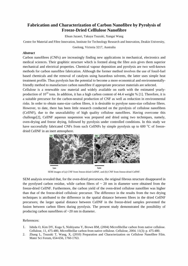
Fabrication and Characterization of Carbon Nanofibre by Pyrolysis of Freeze-Dried Celllulose Nanofibre
Ehsan Jazaeri, Takuya Tsuzuki, Xungai Wang
Centre for Material and Fibre Innovation, Institute for Technology Research and Innovation, Deakin University,
Geelong, Victoria 3217, Australia
Abstract Carbon nanofibres (CNFs) are increasingly finding new applications in mechanical, electronics and medical sciences. Their graphitic structure which is formed along the fibre axis gives them superior mechanical and electrical properties. Chemical vapour deposition and pyrolysis are two well-known methods for carbon nanofibre fabrication. Although the former method involves the use of fossil-fuel based chemicals and the removal of catalysts using hazardous solvents, the latter uses simple heat treatment profile. Thus pyrolysis has the potential to become a more economical and environmentally-friendly method to manufacture carbon nanofibre if appropriate precursor materials are selected. Cellulose is a renewable raw material and widely available on earth with the estimated yearly-production of 1010 tons. In addition, it has a high carbon content of 44.4 weight % [1]. Therefore, it is a suitable precursor for the carbon-neutral production of CNF as well as reduction in environmental risks. In order to obtain nano-size carbon fibres, it is desirable to pyrolyse nano-size cellulose fibres. However, to date, there has been little research conducted on the pyrolysis of cellulose nanofibres (CellNF), due to the unavailability of high quality cellulose nanofibres. Having overcome this challenge[2], CellNF aqueous suspension was prepared and dried using two techniques, namely, oven-drying and freeze drying, followed by pyrolysis under controlled conditions. In this study we have successfully fabricated CNFs from such CellNFs by simple pyrolysis up to 600 oC of freeze-dried CellNF in an inert atmosphere.
SEM images of (a) CNF from freeze-dried CellNF, and (b) CNF from freeze-dried CellNF.
SEM analysis revealed that, for the oven-dried precursors, the original fibrous structure disappeared in the pyrolysed carbon residue, while carbon fibres of ~ 20 nm in diameter were obtained from the freeze-dried CellNF. Furthermore, the carbon yield of the oven-dried cellulose nanofibre was higher than that of the freeze-dried cellulosic precursor. The difference in the results from the two drying techniques is attributed to the difference in the spatial distance between fibres in the dried CellNF precursors; the larger spatial distance between CellNF in the freeze-dried samples prevented the fusion between carbon fibres during pyrolysis. The present study demonstrated the possibility of producing carbon nanofibres of ~20 nm in diameter. References:
1. Ishida O, Kim DY, Kuga S, Nishiyama Y, Brown RM, (2004) Microfibrillar carbon from native cellulose. Cellulose, 11, 475-480, Microfibrillar carbon from native cellulose. Cellulose, 2004. 11(3): p. 475-480.
2. Zhang L, Tsuzuki T, Wang, X, (2010) Preparation and Characterization on Cellulose Nanofibre Film. Mater Sci Forum, 654-656, 1760-1763.
Solution-Processed Nanocrystal Solar Cells
Brandon MacDonald1,2, Jacek Jasieniak1, Scott Watkins1, Paul Mulvaney2
1CSIRO Material Science and Engineering, Bayview Ave. Clayton, VIC, 3168 2School of Chemistry and Bio21 Institute, The University of Melbourne, Parkville,
VIC, 3010
Colloidal nanocrystals are promising materials for solar cell applications as they
combine the excellent electronic properties of inorganic semiconductors with low-cost
solution processing. However, to become a commercially viable technology it will be
necessary for cell efficiencies to exceed 10%. Accomplishing this will require a
deeper understanding of the material properties and processing techniques used in
these nanoscale systems.
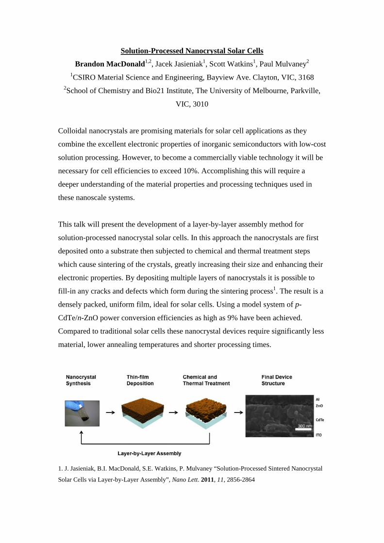
This talk will present the development of a layer-by-layer assembly method for
solution-processed nanocrystal solar cells. In this approach the nanocrystals are first
deposited onto a substrate then subjected to chemical and thermal treatment steps
which cause sintering of the crystals, greatly increasing their size and enhancing their
electronic properties. By depositing multiple layers of nanocrystals it is possible to
fill-in any cracks and defects which form during the sintering process1. The result is a
densely packed, uniform film, ideal for solar cells. Using a model system of p-
CdTe/n-ZnO power conversion efficiencies as high as 9% have been achieved.
Compared to traditional solar cells these nanocrystal devices require significantly less
material, lower annealing temperatures and shorter processing times.
1. J. Jasieniak, B.I. MacDonald, S.E. Watkins, P. Mulvaney “Solution-Processed Sintered Nanocrystal
Solar Cells via Layer-by-Layer Assembly”, Nano Lett. 2011, 11, 2856-2864
Amine modification of quartz particles via plasma polymerization for water contaminant removal
Karyn L. Jarvis1 and Peter Majewski2
1. Mawson Institute, University of South Australia, Mawson Lakes, SA 5095, 2. School of Advanced Manufacturing and Mechanical Engineering, University of South
Australia, Mawson Lakes, SA 5095
Access to clean drinking water is essential to human life. Although a number of effective and reliable purification techniques are available, the majority are energy intensive and thus are normally only used in highly populated areas of developed countries. The availability of clean drinking water in rural areas, disaster zones, and developing countries is of major concern. Simple and cheap decentralized water treatment can be used to supply clean drinking water to these areas on demand. An inductively coupled radio frequency plasma reactor with a rotating chamber was used to modify the surface of quartz particles to aid in the development of a decentralized water treatment system. Plasma polymerization was undertaken using allylamine and ethyelendiamine to produce amine terminated surfaces. The addition of amine functional groups produce surfaces with higher isoelectric points which are predominately positively charged in solution thus enabling the removal of negatively charged contaminants via electrostatic attraction. Humic acid is a negatively charged contaminant which is commonly found in drinking water and originates from the biodegradation of organic matter producing undesirable colour, taste and odour.
Polymerization time, RF power and monomer flow for both allylamine and ethylenediamine monomers were varied for produce optimal surface chemistry for humic acid removal. The effect of these parameters on surface chemistry was determined using XPS, ToF-SIMS and electrokinetic analysis. The coated particles were immersed into solutions of varying humic acid concentrations with pH and adsorption time varied. The mass of humic acid removed was determined via a UV-vis spectrophotometer at λ=254 nm. Ethylenediamine coated particles had a higher surface concentration of nitrogen but removed significantly less humic acid from solution than allylamine coated particles. Such behaviour is expected to be sue to the saturated hydrocarbon structure of ethylenediamine which results in a less stable film in solution. Plasma polymerization has shown to be a successful method for the modification of quartz particles to facilitate humic acid removal from solution. Further investigation of the modification of low cost adsorbents via plasma polymerization with additional monomers and contaminants will further the development of an effective decentralized water treatment system.
Tuning the Luminescence Lifetime by Sensitizer Yb3+ Concentration in Upconversion Nanocrystals
Jiangbo Zhaoa, Yiqing Lua, James A. Pipera, Judith M. Dawesa, Ewa M. Goldysa, Dayong Jina a. Advanced Cytometry Laboratories, MQ BioFocus Research Centre, Macquarie University, North Ryde 2109, NSW, Australia
1. Introduction Recent rapid advances of non-invasive fluorescent imaging techniques have enabled sensitive and quantitative measurements of single biomolecule as well as more complex biological systems, such as living cells and whole animals. Lanthanide-doped upconversion nanocrystals used as bioprobes represent the next frontier of fluorescence imaging, with several unique features including non-blinking, non-bleaching single particle sensitivity (1). Moreover, following the pulsed ~976 nm excitation, the upconversion visible emission has exceptionally long luminescence lifetime in hundreds of microseconds, which may offer opportunities in time-resolved fluorescence sensing. In this time domain, it is highly desirable to engineer nanocrystals at tunable lifetimes to probe multiple targets in a single test. However, it remains a challenging for a simple technique to tune the lifetime over a large dynamic range without sacrificing single nanoparticle luminescence intensity (2). We focus here on investigation of upconversion sensitizer Yb concentration to manipulate the luminescence decay lifetimes, which shows differentiable lifetimes to yield the multiplexed lifetime coded bioprobes without the spectral interference.
2. Methods We employed a modified user-friendly solvothermal method to synthesize different upconversion nanocrystals with varying Yb3+ and Tm3+ dopant combinations (3). Our TEM (Fig. 1a) and XRD characterization results demonstrate promising monodisperse property and good crystalline quality, separately. All samples exhibit blue, red, and IR emissions spectra. The distinguishable lifetimes are quantified by purpose-built upconversion luminescence microscopy.
3. Results and Discussion
Fig. 1. (a) TEM image of one typical NaYF4:Yb,Tm nanocrystals. (b) Summary of lifetimes for varied Yb3+ (10%, 20%, 30% and
40%) upconversion nanocrystals with unchanged Tm3+ concentration of 1%.
This Fig. 1b shows increasing Yb3+ concentration from 10% to 40% can significantly decrease the blue luminescence lifetime by a factor of more than 3, over a large dynamic range from 504 µs to 163 µs. Taking both the Red and IR lifetime decrease into the consideration, this phenomenon is attributed to the reduced average distance from the sensitizer to activator during the cooperative upconversion energy transfer process. Our results suggest controllable lifetimes can be achieved by fine-tuning donor and acceptor concentrations, as an effective and simply method to produce lifetime-coded nanocrystals for multiplexing sensing platforms.
4. Conclusions Dopant-dependent lifetimes can be differentiated over a large dynamic range. Such engineered upconversion luminescence properties are highly desirable for multiplexed biosensing.
5. References 1. S. W. Wu et al., P Natl Acad Sci USA 106, 10917 (Jul 7, 2009). 2. J. Zhao, J. A. Piper, J. M. Dawes, D. Jin, E. M. Goldys, in IQEC/CLEO Pacific Rim 2011. (Sydney, 2011), vol. 978-0-9775657-7-1,
pp. 384-386. 3. Z. Q. Li, Y. Zhang, S. Jiang, Adv Mater 20, 4765 (Dec 17, 2008).
Time resolved spectroscopic and kinetic studies of argon VUV luminescence
in a windowless dielectric barrier discharge
Rajesh Ganesan, Deborah Kane and Robert Carman Department of Physics and Astronomy, Macquarie University, Sydney, Australia
Rare-gas and rare-gas halogen mixtures excited by a dielectric barrier discharge (DBD) produce UV
and vacuum ultraviolet (VUV) excimer radiation. The VUV output produced by argon excimer lamps
covering the wavelength range 110-130nm has found significant interest for the surface activation of
semiconductors and polymers for the photochemical matting of coatings. The VUV sources are in
fact an interesting tool for research and technologic development in many fields of material science,
chemistry, and biochemistry. By reason of its high photon energy around 9.85 eV, the argon excimer
radiation is potentially able to modify high density polymeric surfaces. The experimental work
reported here is devoted to the temporal analysis of the VUV emission from a windowless dielectric
barrier discharge[1] in pure argon from 50- 800 mbar. The studies were carried out to gain insight
into the underlying kinetic processes relating to the first and second continuum emission bands of
the Ar2* excimer. Pulsed excitation using bi-polar voltage pulses with 2% duty-cycle and 32 kHz
repetition frequency were employed to achieve a uniform discharge with well-controlled electrical
breakdown characteristics. By comprehensively measuring the rising and decay time constants for
~50 individual wavelengths within the first and second continuum of argon covering the range
λ=107nm-140nm, the dominant collisional and radiative rates, kinetic rates relating to Ar2* excimer
production and loss have been obtained. The variation of time constants as a function of wavelength
and gas pressure have been determined. The VUV emission curves at the transition phase between
the first and second continuum have been analysed in detail.
References:
[1] Carman et al., J.Phys.D:Applied Physics, 44, 25205 (2010).
MULTI-TECHNIQUE APPROACH FOR THE SIZE CHARACTERISATION OF NANOPARTICLES
Heather J. Catchpoole, Victoria A. Coleman, Åsa K. Jämting, Maitreyee Roy, Jan Herrmann
National Measurement Institute, Department of Innovation, Industry, Science and Research, PO Box 264,
Lindfield NSW 2070, AUSTRALIA ABSTRACT
Engineered Nanoparticles (ENPs) have applications in many areas including health care, energy generation and
environmental remediation. As the number of applications for ENPs grow, it is likely that their use will become subject
to regulation. One of the significant challenges for regulation is the accurate characterisation of the physical and
chemical properties of ENPs such as particle size, surface area, number concentration, state of
agglomeration/aggregation, surface charge and chemical composition. Accurate knowledge of dimensional properties
such as particle size is of fundamental importance for the understanding and control of the characteristics of nano-
materials, as size is often strongly correlated with other properties that determine the material’s interaction with
biological systems.
A variety of methods have been developed to measure the dimensional properties of nanoparticle systems. Whilst
“particle size” appears to be a straightforward parameter, in reality particle systems can be quite complex, comprising of
broad or multimodal size distributions, often with shape inhomogeneity, which cannot be represented by a single
number. Ensemble techniques such as dynamic light scattering (DLS), average over a large number of particles and
provide a good statistical representation of the sample, but may not be suitable for measuring samples composed of
wide distributions of particle size or shape. Single-particle resolution techniques such as scanning and transmission
electron microscopy (SEM and TEM) can provide detailed information on particle size and shape but may be of limited
statistical relevance due to the relatively small number of particles that can be practically examined. Separation and
classification techniques such as differential centrifugal sedimentation and field flow fractionation overcome some of
these statistical limitations by sequentially presenting the detection and measurement systems with narrow fractions of
the particle size distribution.
Established measurement techniques are increasingly being complemented by novel methods such as microchannel
resonator particle analysis, an ultra high-resolution mass sensor, which can provide single-particle resolution at
moderate throughputs to obtain statistically representative information on sample characteristics. When comparing
results from different particle characterisation techniques it is important to recognise that they may be based on different
measurands. For example, DLS determines the average hydrodynamic diameter of a particle ensemble, while TEM
determines particle dimensions based on the projection of the particle outline onto a two-dimensional image plane.
In this study we use both mono- and multi-modal dispersions of well-characterised nanoparticle materials to compare
and contrast traditional and novel methods for the determination of particle size distribution.
Time-resolved spectroscopy of III-V semiconductor
nanowires
P. PARKINSON1,*, H. J. JOYCE2, L.M HERZ2, M.B. JOHNSTON2, Q. GAO1, H.H. TAN1 AND C. JAGADISH1
1Deparment of Electronic Materials Engineering, Research School of Physics and Engineering, The Australian National University, Canberra, ACT 0200, Australia
2Department of Physics, University of Oxford, Oxford, OX1 3PU, United Kingdom e-mail: [email protected]
Semiconductor nanowires represent an emerging class of 1D and quasi-1D materials, that have many interesting properties for optoelectronic applications. In particular, the large surface-area to volume ratio provides opportunities for stress relief allowing axial and radial heterostructures of materials with different lattice constants to be prepared, as well as imparting important changes to the optoelectronic properties of the material. III-V nanowires grown using the VLS process in an MOCVD reactor, (and especially GaAs and InP nanowires) are particularly well studied for their application in nanoelectronics.
In this presentation, the use of ultrafast (femto- to nanosecond) time-resolved techniques to understand the optical and electronic dynamics of III-V nanowires will be discussed. Specifically, terahertz time-domain spectroscopy [1,2] and time-resolved photoluminescence [3,4] will be shown to be excellent techniques for investigating electronic dynamics. A rapid determination of carrier lifetime, carrier mobility and surface trap density will be presented, using room-temperature, non-contact terahertz time-domain spectroscopy.
Using optical-pump, terahertz-probe spectroscopy, the effect upon carrier lifetime and mobility of a) surface passivation of GaAs nanowires using an AlGaAs shell and b) using an improved two-temperature core-growth process will be presented. In particular, a near doubling in carrier mobility from 1200cm2/Vs to 2250cm2/Vs for two-temperature growth demonstrates the high-quality of III-V nanowire achievable via VLS MOCVD growth.
References
[1] Nano Letters, P. Parkinson et al. 2009, 9(9):3349 [2] Nano Letters, P. Parkinson et al. 2007, 7(7):2162 [3] Prog. Quantum Electron., H. J. Joyce et al. 2011, 35:23 [4] J. Phys. Chem. Lett, P. Parkinson et al. 2010, 1:2788
Understanding how electron density affects spin splitting in 1D systems
A. M. Burke1, O. Klochan1, J. C. H. Chen1, I. Farrer2, D. A. Ritchie2, A. R. Hamilton1, and A. P. Micolich1
1School of Physics, University of New South Wales, Sydney NSW 2052, Australia
2Cavendish Laboratory, JJ Thomson Avenue, Cambridge CB3 0HE, UK Since the discovery of the first transistor in 1947, enormous efforts have been made toward furthering our understanding of the physics of solid state devices. Over the past few decades, and mainly driven by the push toward better computers, the critical dimensions of devices have been reduced into the sub-micron range. As a result, quantum effects within these systems have become increasingly accessible and are therefore interesting phenomena to study. Within a conducting solid, electrons can move in three dimensions as an electron gas. By layering semiconductor materials in an ordered and atomically precise manner, transport can be restricted in one dimension to a scale comparable to the electron’s wavelength resulting in a two dimensional electron gas (2DEG). The GaAs/AlGaAs system is an excellent example, where a 2DEG forms due to the difference in band-gaps creating a narrow quantum well at the heterostructure’s interface. Many interesting phenomena have been studied in 2D systems including the integer and fractional quantum Hall effects, both of which led to Nobel prizes in Physics. Electron transport can be further limited to one dimension (1D) and this can be achieved by nanofabrication of semiconductors, or in nanomaterials such as carbon nanotubes and semiconductor nanowires. We have studied a 1D electronic device known as the quantum point contact (QPC), which consists of a pair of nanoscale metal gates [1] patterned on the surface of a GaAs/AlGaAs heterostructure using electron beam lithography (EBL). By applying a negative voltage Vg to the metal gates we deplete the underlying 2DEG electrostatically and confine transport to a 1D channel. At low temperature, the electrical conductance of the QPC as a function of Vg is quantized in units of Go=2e2/h, where e is the electron charge and h is Planck’s constant, taking a staircase appearance [2]. This quantization reflects mode-matching of the electron wavelength to the width of the QPC, which is adjusted by Vg. An interesting exception to the quantized plateaus is a widely reported plateau observed at 0.7Go. Although it is clear that this feature arises from electron-electron interactions within the constriction, an exact mechanism for its origin has not been established. An important parameter in studying electron-electrons interactions in a semiconductor is the Landé g factor, g*, which is the constant of proportionality between the Zeeman splitting of the spin-up and spin-down levels and the applied magnetic field (i.e. ∆E = g*µBB, where ∆E is the energy splitting, µB is the Bohr magneton and B is the applied magnetic field). Free electrons have g*=2, but interactions both between electrons and between electrons and nuclei can alter this value. Since the electron-electron interactions are often separation dependent, a study of how g* evolves with the electron density in the device could provide useful information. Although g* has been widely studied within 1D systems, devices are usually measured at a fixed density set by the doping in the semiconductor heterostructure used. Although it is possible to measure devices on different heterostructures to vary the density, this changes other parameters including the exact device geometry and dopant profiles within the material, and is thus non-optimal. In our study, we incorporate a second metal gate that covers the entire QPC region. A thin layer of insulating polyimide is used to separate the density controlling gate from the QPC. Biasing this gate grants control of the 2D electron density for the source and drain reservoirs of the quantum constriction. This allows us to study how g* varies with density in a single device, providing interesting new insight into the physics behind the 0.7 plateau. References: [1] T. J. Thornton et al., 1986 Phys. Rev. Lett. 56, 1198 [2] K.-F. Berggren and M. Pepper, 2002 Physics World, 15, 37

TUESDAY
22ND November 2011
Overlapping-gate architecture for silicon Hall bar MOSFET devices in the low density and high magnetic field regime Laurens H. Willems van Beveren1*, Kuan Y. Tan2, Nai-Shyan Lai2, Oleh Klochan3, Andrew S. Dzurak2, and Alex R. Hamilton3
1 ARC Centre of Excellence for Quantum Computation and Communication Technology (CQC2T), School of Physics, The University of Melbourne, Melbourne 3010, Australia 2 ARC Centre of Excellence for Quantum Computation and Communication Technology (CQC2T), School of Electrical Engineering and Telecommunications, The University of New South Wales, Sydney 2052, Australia 3 School of Physics, The University of New South Wales, Sydney 2052, Australia
* [email protected] A common issue in low temperature measurements of enhancement-mode metal-oxide-semiconductor (MOS) field-effect transistors (FETs) in the low electron density regime is the high contact resistance dominating the device impedance. In that case a voltage bias applied across the source and drain contact of a Hall bar MOSFET will mostly fall across the contacts (and not across the channel) and therefore magneto-transport measurements become challenging. However, from a physical point of view, the study of MOSFET nanostructures in the low electron density regime is very interesting (impurity limited mobility1, carrier interactions2,3 and spin-dependent transport4) and it is therefore important to come up with solutions that work around the problem of a high contact resistance in such devices. In this work5, the authors report the fabrication and study of silicon Hall bar MOSFET devices in which an overlapping-gate architecture allows 4-terminal measurements of low electron density 2D systems, while maintaining a high electron density at the ohmic contacts. Comparison with conventional devices using a single gate, show that measurements can be performed at much lower electron densities and higher channel resistances, despite a reduced peak mobility. We also observe a voltage threshold shift which is attributed to negative oxide charge, injected during the electron-beam lithography processing. Hall bar data obtained at magnetic fields up to 15 T show that Landau level filling factors of 2 can readily be achieved6 in the low electron density regime. Here, either the spin or valley degeneracy is lifted. [1] A. Gold, Phys. Rev. B 38, 10798 (1988). [2] S. D. Sarma, Phys. Rev. Lett. 83, 164 (1999). [3] B. Spivak, S. V. Kravchenko, S. A. Kivelson, and X. P. A. Gao, Rev. Mod. Phys. 82, 1743 (2010). [4] L. H. Willems van Beveren, H. Huebl, D. R. McCamey, T. Duty, A. J. Ferguson, R. G. Clark, and M. S. Brandt, Appl. Phys. Lett. 93, 072102 (2008). [5] L. H. Willems van Beveren, K. Y. Tan, N. S. Lai, A. S. Dzurak, and A. R. Hamilton, Appl. Phys. Lett. 97, 152102 (2010). [6] L. H. Willems van Beveren, K. Y. Tan, N. S. Lai, O. Klochan, A. S. Dzurak, and A. R. Hamilton, Proceedings of the 5th International Conference on Advanced Materials and Nanotechnology, Wellington, New Zealand (AMN-5), In Press (2011).
Australian Nanotechnology Network Early Career Symposium 2011
The Study of Low-Dimensional Semiconductor Nanostructures at milli-Kelvin Temperatures and High Magnetic Fields
L.A. Yeoh1, A. Srinivasan1, O. Klochan1, A.R. Hamilton1, D.A. Ritchie2
1. School of Physics, University of New South Wales, Sydney NSW 2052, Australia 2. Cavendish Laboratory, University of Cambridge, Cambridge CB3 0HE, U.K.
The emerging field of Spintronics (spin-electronics) aims to harness the property of a particle’s ‘spin’, which can be used as an additional degree of freedom, on top of charge, to represent and store information. Devices such as spin-based transistors may one day form the building blocks of future quantum information processing technologies [1]. One way to manipulate the spin of an electron electrically rather than magnetically is by controlling the spin-orbit interaction in semiconductor nanostructures. However there is a lot still to be learnt about spin-orbit interactions in semiconductors.
Our investigations focus on the Gallium-Arsenide material system, which is used for high speed transistors and in the photonics industry. In particular we study devices that use positively charged holes, rather than negatively charged electrons, as the spin-orbit coupling is much stronger [2,3]. However fabricating very small GaAs hole devices is a formidable challenge. In my talk I will describe a new fabrication technique that can be used to make extremely small GaAs nanostructures. This technique was developed at the University of Cambridge to create stable, very shallow 2D devices where the electron gas is 50nm below the surface and hosting an extra insulating layer [4]. I will also briefly describe how this work can be potentially extended into creating hole based devices.
We have also developed some new measurement tools specifically to study spin-orbit effects in semiconductor nanostructures. In particular we can probe the effects of spin-orbit interactions by measuring the electrical properties of the device at low temperatures when a magnetic field is applied in different orientations. To do this the sample needs to be oriented in different directions with respect to magnetic field, within the tight constraints of a dilution refrigerator operating at 0.1 degrees above absolute zero. We have designed and implemented a rotation stage to achieve this, based upon a piezoelectric rotator which allows for in-situ orientation of the sample in a magnetic field up to 15 Tesla [5]. This system is being used to study spin-orbit effects in hole quantum wires [6] and hole quantum dots [7].
[1] S.A. Wolf et al., Science 294, 1488 (2001). [2] R. Winkler, Spin-orbit coupling effects in two-dimensional electron and hole systems (Springer
Tracts in Modern Physics, Vol. 191, Springer, Berlin, 2003). [3] R. Danneau et al., Phys. Rev. Lett. 97, 026403 (2006). [4] W. Y. Mak et al., App. Phys. Lett. 97, 242107 (2010) [5] L. A. Yeoh et al., Rev. Sci. Instrum. 81, 113905 (2010) [6] A. Srinivasan et al., "Measurement of out-of-plane hole g-factor in a GaAs-AlGaAs heterostructure",
Poster Presentation Tu-P-63 at EP2DS19, Tallahassee, Florida, USA (2011). [7] O. Klochan et al., Phys. Rev. Lett. 107, 076805 (2011)
Single-electron shuttle based on a silicon quantum dot aK. W. Chan, bM. Mottonen, cA. Kemppinen, aN. S. Lai, aK. Y. Tan, aW. H. Lim, and aA. S. Dzurak aSchool of Electrical Engineering and Telecommunications, The University of New South Wales, Sydney 2052, Australia bDepartment of Applied Physics/COMP, AALTO University, P.O. Box 14100, FI-00076 AALTO, Finland cLow Temperature Laboratory, AALTO University, P.O. Box 13500, FI-00076 AALTO, Finland Centre for Metrology and Accreditation (MIKES), P.O. Box 9, FI-02151 Espoo, Finland We report on single-electron shuttling experiments1 with a silicon quantum dot2. An electron layer is accumulated at the Si/SiO2 interface below an aluminum top gate with two additional barrier gates used to deplete the electron gas locally and to define the quantum dot. Directional single-electron shuttling from the source and to the drain lead is achieved by applying a dc source-drain bias while driving the barrier gates with an ac voltage at frequency f. Current plateaus at integer levels of ef are observed up to f = 240 MHz. The observed results are explained by a sequential tunneling model which suggests that the electron gas may be heated substantially by the ac driving voltage. Future device optimization is expected to make these quantum dots serious candidates for a metrological current standard. 1K. W. Chan, M. Mottonen, A. Kemppinen, N. S. Lai, K. Y. Tan, W. H. Lim, A. S. Dzurak, Applied Physics Letters 98, 063121 (2011). 2W. H. Lim et al., Applied Physics Letters 95, 242102 (2009).
Biosynthesis of Palladium Nanoparticles by Green Microalgae
Ela Eroglua,b,*, Jianli Zoua, Jeremy A. Shawc, Steven M. Smithb, Colin L. Rastona , K. Swaminathan Iyera
a Centre for Strategic Nano-Fabrication, School of Biomedical, Biomolecular and Chemical Science, The University of Western Australia, Crawley, Perth, WA 6009, Australia b ARC Centre for Plant Energy Biology, The University of Western Australia, Crawley, Perth, WA 6009, Australia c Centre for Microscopy, Characterisation and Analysis, The University of Western Australia, Crawley, Perth, WA 6009, Australia *Fax: +61-8-6488-1005; Tel: +61-8-6488-4470; E-mail: [email protected]
Palladium (Pd) nanoparticles have recently been receiving significant interest, since they can be used as an efficient catalyst for C-C forming cross-coupling reactions [1] or for hydrogenation / dehydrogenation reactions [2]. They can also perform as an effective hydrogen-gas sensor due to their high surface to volume ratio, lightweight and small size [3]. In order to scale-up the palladium production process, it is essential to develop more practical methods in comparison with the current technology which mostly follows the synthetic routes. For this reason, a microalgae-based Pd bioproduction system has been studied in the current study.
The synthesis of crystalline Pd nanoparticles has been investigated by the interaction of green microalgae (Chlorella vulgaris) with sodium palladium chloride (Na2PdCl4) solution at room temperature. Chlorophyll content has been used to validate the viability of the cell cultures. Growth studies showed that Na2PdCl4 salt content below 50 mg/L is nontoxic for C. vulgaris. Viable cells were observed to synthesize larger palladium nanoparticles than non-viable cells, probably due to the continuous production of reducing agents by the algal cells. The release of metabolites such as organic acids (i.e., acetic and citric acid) and hydrogen might promote the reduction of palladium chloride into crystalline Pd nanoparticles. Transmission electron microscopy (TEM), elemental mapping via energy filtered TEM (EFTEM) and electron energy-loss spectroscopy (EELS), and electron-diffraction-pattern techniques were used to reveal the presence of palladium nanoparticles. Preliminary results showed that this bio-nano process can serve as a good candidate for a low cost, energy-efficient, and environmentally-friendly method for the fabrication of palladium nanoparticles. Further process optimization studies are still ongoing, and more detailed results will be presented during the Symposium.
References [1] Yang et al., Inorg. Chem., 2008, 47 (8): 3292–97; [2] Ohde et al., J. Am. Chem. Soc., 2002, 124: 4540–41; [3] Jeon et al., Nanotechnology, 2008, 19: 495501.
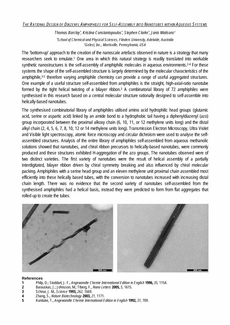
THE RATIONAL DESIGN OF DIAZENYL AMPHIPHILES FOR SELF-ASSEMBLY INTO NANOTUBES WITHIN AQUEOUS SYSTEMS
Thomas Barclay*, Kristina Constantopoulos*, Stephen Clarke*, Janis Matisons†
*School of Chemical and Physical Sciences, Flinders University, Adelaide, Australia †Gelest, Inc., Morrisville, Pennsylvania, USA
The ‘bottom-up’ approach to the creation of the nanoscale artefacts observed in nature is a strategy that many researchers seek to emulate.1 One area in which this natural strategy is readily translated into workable synthetic nanostructures is the self-assembly of amphiphilic molecules in aqueous environments.2-4 For these systems the shape of the self-assembled structure is largely determined by the molecular characteristics of the amphiphile,3-5 therefore varying amphiphile chemistry can provide a range of useful aggregated structures. One example of a useful structure self-assembled from amphiphiles is the straight, high-axial-ratio nanotube formed by the tight helical twisting of a bilayer ribbon.3 A combinatorial library of 72 amphiphiles were synthesised in this research based on a central molecular structure rationally designed to self-assemble into helically-based nanotubes.
The synthesised combinatorial library of amphiphiles utilised amino acid hydrophilic head groups (glutamic acid, serine or aspartic acid) linked by an amide bond to a hydrophobic tail having a diphenyldiazenyl (azo) group incorporated between the proximal alkoxy chain (6, 10, 11, or 12 methylene units long) and the distal alkyl chain (2, 4, 5, 6, 7, 8, 10, 12 or 14 methylene units long). Transmission Electron Microscopy, Ultra Violet and Visible light spectroscopy, atomic force microscopy and circular dichroism were used to analyse the self-assembled structures. Analysis of the entire library of amphiphiles self-assembled from aqueous methanolic solutions showed that nanotubes, and chiral ribbon precursors to helically-based nanotubes, were commonly produced and these structures exhibited H-aggregation of the azo groups. The nanotubes observed were of two distinct varieties. The first variety of nanotubes were the result of helical assembly of a partially interdigitated, bilayer ribbon driven by chiral symmetry breaking and also influenced by chiral molecular packing. Amphiphiles with a serine head group and an eleven methylene unit proximal chain assembled most efficiently into these helically based tubes, with the conversion to nanotubes increased with increasing distal chain length. There was no evidence that the second variety of nanotubes self-assembled from the synthesised amphiphiles had a helical basis, instead they were predicted to form from flat aggregates that rolled up to create the tubes.
References 1 Philp, D.; Stoddart, J. F., Angewandte Chemie International Edition in English 1996, 35, 1154. 2 Barauskas, J.; Johnsson, M.; Tiberg, F., Nano Letters 2005, 5, 1615. 3 Schnur, J. M., Science 1993, 262, 1669. 4 Zhang, S., Nature Biotechnology 2003, 21, 1171. 5 Kunitake, T., Angewandte Chemie International Edition in English 1992, 31, 709.
1
Rapid Synthesis of Li4Ti5O12 Microspheres Composed of Nanoflakes as Anode Materials for Lithium-ion Battery
Shu-Lei Chou,a,b* Jia-Zhao Wang,a,b Hua-Kun Liu,a,b and Shi-Xue Dou a
a Institute for Superconducting and Electronic Materials and b ARC Centre of Excellence for Electromaterials Science Australia, University of Wollongong, Wollongong, NSW 2522 Australia
*Corresponding Author: [email protected] Abstract
Transport is one of the largest sources of greenhouse gas emissions and fossil-fuel consumption. To reduce emissions of carbon dioxide and conquer the greater and greater scarcity of fossil fuels, one of the most effective ways is to use electrical vehicles (EVs) or hybrid electrical vehicles (HEVs). Lithium-ion batteries have now shown that they have a promising future in the coming era of EVs/HEVs. However, the current lithium-ion battery is handicapped by several critical disadvantages for EV/HEV applications, including short cycling life, low power density, and safety hazards. Spinel lithium titanate, Li4Ti5O12, has attracted great interest as anode material for rechargeable Li-ion batteries because it can offer a great improvement in safety due to its high and flat Li insertion voltage at about 1.55 V vs. Li/Li+, which prevents the growth of lithium dendrites and the decomposition of electrolyte, as well as providing long cycle life and high rate capability.1-6
In most of previous works, Li4Ti5O12 powders were fabricated via either high temperature (800–1000 °C) and/or great time consuming (12-24 h) methods.1-5 These methods require large energy consumption. Recently, Tarascon’s group reported the synthesis of nanocrystalline Li4Ti5O12 by the solution-combustion method in less than one minute, which showed great enhancement in rate capability.10 However, the morphology of the product is not easy to control within such a short time. The microwave solid-state synthesis method was also used to reduce the time for preparing Li4Ti5O12 nanocrystallites, which showed relatively good performance.11 However, the morphology of the product is also not easy to control in solid-state reaction. The present combination of microwave-assisted hydrothermal and microwave post-annealing method, on the other hand, is a great time saving method to produce nanomaterials with controlled morphologies.
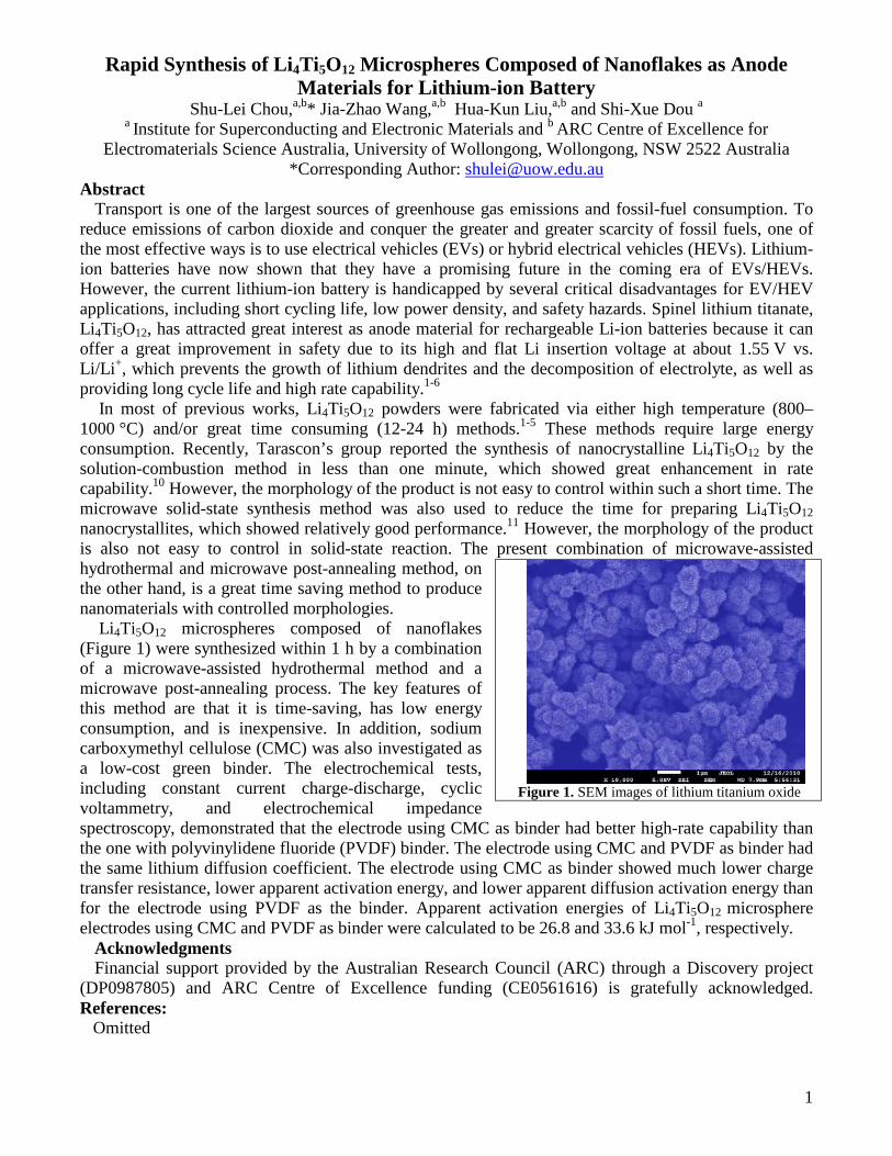
Li4Ti5O12 microspheres composed of nanoflakes (Figure 1) were synthesized within 1 h by a combination of a microwave-assisted hydrothermal method and a microwave post-annealing process. The key features of this method are that it is time-saving, has low energy consumption, and is inexpensive. In addition, sodium carboxymethyl cellulose (CMC) was also investigated as a low-cost green binder. The electrochemical tests, including constant current charge-discharge, cyclic voltammetry, and electrochemical impedance spectroscopy, demonstrated that the electrode using CMC as binder had better high-rate capability than the one with polyvinylidene fluoride (PVDF) binder. The electrode using CMC and PVDF as binder had the same lithium diffusion coefficient. The electrode using CMC as binder showed much lower charge transfer resistance, lower apparent activation energy, and lower apparent diffusion activation energy than for the electrode using PVDF as the binder. Apparent activation energies of Li4Ti5O12 microsphere electrodes using CMC and PVDF as binder were calculated to be 26.8 and 33.6 kJ mol-1, respectively.
Acknowledgments Financial support provided by the Australian Research Council (ARC) through a Discovery project
(DP0987805) and ARC Centre of Excellence funding (CE0561616) is gratefully acknowledged. References: Omitted
Figure 1. SEM images of lithium titanium oxide
Reduced Pseudomonas aeruginosa proliferation and biofilm formation on zinc oxide/silica coated glass coupons
Sanly Liu1, Nicolas Barraud2, Cindy Gunawan1, May Lim1, Rose Amal1 1ARC Centre of Excellence for Functional Nanomaterials, School of Chemical Engineering, The
University of New South Wales, Sydney NSW 2052 2Centre for Marine Bio-Innovation, School of Biotechnology and Biomolecular Sciences, The University
of New South Wales, Sydney NSW 2052 Abstract
Nanotechnology based water purification system offer the possibility of an efficient
removal of pollutants and microbes in water treatment. Recent applications of
nanotechnology include the functionalisation of surfaces with antibacterial properties by
coating, impregnation, or embedding nanomaterials. Here, we have used zinc oxide and
silica thin film prepared using a sol gel technique, to determine the antibacterial efficacy
against Pseudomonas aeruginosa, a soil and water borne pathogenic bacteria. An
important aspect of the use of ZnO as antibacterial agent is the fact that it does not
require UV light activation, as in the case of TiO2 semiconductor. The structural
characteristics of the coating was characterised by X-ray diffraction (XRD), scanning
electron microscopy (SEM), and atomic force microscopy (AFM). Biofilm formation
was quantified in 12-well flat-bottomed polystyrene microtitre plates using a crystal
violet biofilm assay. This study has shown that the zinc oxide and silica coating on the
glass coupons can retard or partially inhibit biofilm formation from P. aeruginosa. In
addition, reduction of biomass in the planktonic phase was also observed for the coated
sample compared to the control.
ANN Early Career Symposium - Abstract
1
Hybrid Nanomaterials that Mimic the ‘Food Effect’ to Enhance Oral Drug Absorption
Angel Tan, Clive A. Prestidge Ian Wark Research Institute, University of South Australia
Abstract Lipid-based formulations are developed to mimic the positive food (or post-prandial) effect in which co-administration of poorly-water soluble drugs with fatty food can increase their bioavailability. Lipid-based carriers are attractive formulation strategies but their therapeutic applications are limited by the inability to establish stable solid dosage forms, inadequate mechanistic understanding of the delivery performance and lack of suitable in vitro tests that are predictive of their in vivo performance.
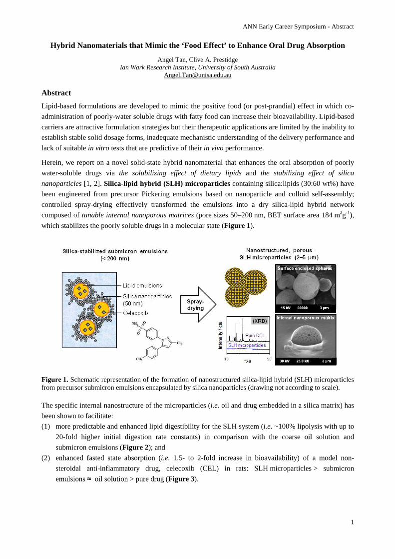
Herein, we report on a novel solid-state hybrid nanomaterial that enhances the oral absorption of poorly water-soluble drugs via the solubilizing effect of dietary lipids and the stabilizing effect of silica nanoparticles [1, 2]. Silica-lipid hybrid (SLH) microparticles containing silica:lipids (30:60 wt%) have been engineered from precursor Pickering emulsions based on nanoparticle and colloid self-assembly; controlled spray-drying effectively transformed the emulsions into a dry silica-lipid hybrid network composed of tunable internal nanoporous matrices (pore sizes 50–200 nm, BET surface area 184 m2g-1), which stabilizes the poorly soluble drugs in a molecular state (Figure 1).
Figure 1. Schematic representation of the formation of nanostructured silica-lipid hybrid (SLH) microparticles from precursor submicron emulsions encapsulated by silica nanoparticles (drawing not according to scale).
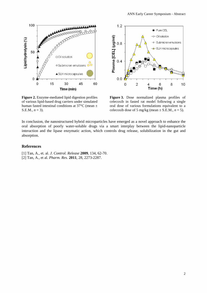
The specific internal nanostructure of the microparticles (i.e. oil and drug embedded in a silica matrix) has been shown to facilitate: (1) more predictable and enhanced lipid digestibility for the SLH system (i.e. ~100% lipolysis with up to
20-fold higher initial digestion rate constants) in comparison with the coarse oil solution and submicron emulsions (Figure 2); and
(2) enhanced fasted state absorption (i.e. 1.5- to 2-fold increase in bioavailability) of a model non-steroidal anti-inflammatory drug, celecoxib (CEL) in rats: SLH microparticles > submicron emulsions ≈ oil solution > pure drug (Figure 3).
ANN Early Career Symposium - Abstract
2
Figure 2. Enzyme-mediated lipid digestion profiles of various lipid-based drug carriers under simulated human fasted intestinal conditions at 37°C (mean ± S.E.M., n = 3).
Figure 3. Dose normalized plasma profiles of celecoxib in fasted rat model following a single oral dose of various formulations equivalent to a celecoxib dose of 5 mg/kg (mean ± S.E.M., n = 5).
In conclusion, the nanostructured hybrid microparticles have emerged as a novel approach to enhance the oral absorption of poorly water-soluble drugs via a smart interplay between the lipid-nanoparticle interaction and the lipase enzymatic action, which controls drug release, solubilization in the gut and absorption.
References [1] Tan, A., et. al. J. Control. Release 2009, 134, 62-70. [2] Tan, A., et al. Pharm. Res. 2011, 28, 2273-2287.
In vitro valuation of Mannose-6-phosphate on migratory behaviour of NIH/3T3 fibroblasts cell line
Vipul Agarwala, Ela Eroglua, Fiona Woodb,c, Mark Fearc and K. Swaminathan Iyera* a School of Biomedical, Biomolecular and Chemical Science, University of Western Australia,
Perth, Western Australia, Australia b School of Surgery, University of Western Australia,
Perth, Western Australia, Australia c The McComb Foundation,
Perth, Western Australia, Australia
Keywords: Pluronic F-127, FDMA, electrospinning, hydrogels and wound healing
Contact information: a: School of Biomedical, Biomolecular and Chemical Science, University of Western Australia, M313,35 Stirling Highway, Crawley, Perth, WA 6009 Australia; * Corresponding author: [email protected] , Phone: +61 (08) 6488 4470
Tissue engineering using nanotechnology has emerged as a promising alternative to treat skin injuries. Tissue engineering is the application of principles and methods of engineering and life sciences to the development of biological substitutes to restore, maintain, or improve tissue function. The approach herein involves the use of scaffolds and templates along with biological cues. An ideal scaffold would mimic the functions of native skin, protect the injury from loss of fluid and proteins, enable the removal of exudates, inhibiting exogenous microorganism invasion, and improve the aesthetic appearance of the wound site. Besides these, many tissues, such as nerve, muscle, tendon, ligament, blood vessel, bone and teeth, have tubular or fibrous bundle architectures and anisotropic properties. Current strategies in the treatment of burn injuries emphasised on healing and not towards subsequent scar reduction. Mannose-6-phosphate (M6P) is a natural sugar analogue known to reduce scarring.
In this presentation we will describe the use of gelatin as scaffolds for regenerative therapy and the impact of incorporating mannose-6-phosphate in vitro. Gelatine hydrogel was further crosslinked with/ out M6P to provide stability and characterised under scanning electron microscope (SEM). These polymer scaffolds were studied for their biocompatibility to promote wound healing in burn injuries. Toxicity studies were carried out as function of cell proliferation on both keratinocytes (HaCaT) and fibroblasts (NIH/3T3) cell lines. Finally, migratory behaviour of the cells will be studied using live cell imaging to evaluate the effectiveness of the polymer scaffold towards acceleration in healing process.
Enhanced Efficacy of Anti-restenotic Drug by Intercalation into Inorganic Layered Double Hydroxide Nanoparticle
Zi Gua, Zhi Ping Xua, Barbara E. Rolfeb, Anita C. Thomasc, Julie H. Campbellb, and Gao Qing Lua
aARC Centre of Excellence for Functional Nanomaterials,
bCentre for Research in Vascular Biology, Australian Institute of Bioengineering and Nanotechnology, The University of Queensland, Brisbane, QLD 4072, Australia cBristol Heart Institute, University of Bristol, Bristol, BS2 8HW, United Kingdom
Restenosis, a re-blocking of arteries, has been the major limitation of surgical treatment to remove atherosclerotic plaque. Identification of effective anti-restenotic strategies is a high priority in cardiovascular research, and smooth muscle cells (SMCs) are a key target for intervention. Although a number of drugs are effective in experimental models of restenosis, most have had limited success in clinical trials. Our laboratory has identified layered double hydroxides (LDHs), a class of anionic clay materials, as vehicles for improved intracellular drug delivery. The present study investigated the efficacy of LDH nanoparticles for delivery of an anti-restenotic drug (low molecular weight heparin, LMWH) both in vitro and in vivo. LMWH was intercalated into LDH nanoparticle interlayers by the co-precipitation method, which confirmed by powder X-ray diffraction (XRD) and transmission electron microscopy (TEM). Release studies conducted under physiological conditions revealed the sustained release of LMWH from LMWH-LDH. Cytotoxicity assays showed LDH concentrations up to 50 μg/mL were non-toxic for SMCs. Intercalation to LDH nanoparticles enhanced the uptake of FITC-LMWH into cultured rat vascular SMCs more than ten fold. The ability of LMWH to inhibit SMC proliferation and migration was also enhanced, possibly due to prolonged inhibition of ERK1/2 activation. Further investigations using LysoTracker Red showed that SMC internalization of FITC-LMWH-intercalated LDH is via the endocytic pathway. While unconjugated LMWH appeared to be rapidly degraded within the endosomes, at later time-points (48 hours) the LMWH-LDH conjugate appeared to diffuse throughout the cytoplasm, possibly reflecting the unique capacity of LDH to facilitate LMWH escape from endosomal/lysosomal compartments. To target deliver LMWH-LDH, the nanocomposite was coupled with antibody to cross-linked fibrin (1D2). Administration of Q-dot-labelled 1D2-LMWH-LDH to rats immediately following balloon catheter injury of carotid arteries showed that the nanoconjugate specifically targeted the site of injury. Moreover, the preliminary data shows that treatment with 1D2-LMWH-LDH reduced the neointimal response to arterial injury in comparison with that in control groups treated with LMWH, LMWH-LDH alone or conjugated with an irrelevant antibody. In summary, these studies demonstrate (1) LMWH can be stored in LDH interlayers and released from LMWH-LDH nanohybrids in a sustained manner, and (2) both in vitro and in vivo, the potential of an inorganic LDH nanoparticle-based drug delivery system for anti-restenotic therapy.
Graphene and graphene oxide (GO) as nucleating agents for protein crystalisation. Benjamin S. Gully1,2, K. Swaminathan Iyer2, Charles S. Bond1. 1School of Biomedical Biomolecular and Chemical Sciences., 2Centre for Strategic Nanofabrication, The University of Western Australia, X-ray crystallography provides atomic resolution structural data, allowing comprehensive understanding of protein structure and function. Generation of suitably diffracting crystals is a multi-parametric process and remains the current bottleneck in structural genomics. Therefore, a clear rationale exists for development of methodologies to improve probability of obtaining crystals, and improving reproducibility or quality. Crystallisation is a first order phase transition proceeding via the formation of a homogeneous or heterogeneous nuclei. Homogeneous nucleation, the method typically employed, involves identification of conditions amenable to spontaneous nucleation and subsequent crystal growth. This requires many trials, time, costly protein and can be an extremely inefficient process. Heterogeneous nucleation operates via a surface or cavity interaction inducing nuclei formation via imposed local super saturations lowering the free energy barrier to drive nuclei formation. The research herein investigated the ability of graphene and graphene oxide (GO) to act as heterogeneous nucleating agents in protein crystallisation due to the high surface area to volume ratio, bi-directionality and documented use for immobilisation of proteins. Upon the addition of both graphene and GO improvements were observed in all crystallisation trials. In the presence of nucleating agents, crystalline products were observed in novel conditions in every trial undertaken relative to the control. The improvements observed include, the number of; clear drops decreased, ordered precipitates increased and the number of crystalline yielding conditions increased. The nucleating agents were additionally able to facilitate nucleation from metastable crystal trials with low protein concentrations. Suggesting use as universally applicable nucleating agents in trials where no crystals have been grown via conventional methods. Generation of nuclei is a major obstacle in crystal growth, graphene and GO have proven to be efficient nucleating agents in the application to protein crystallization.

POSTERS
Preparation of Toughened PLA Nanocomposites with Nanoclay and Nano Powder Rubber
Mojtaba Abtahi*, Avinash Baji, Yiu-Wing Mai
Centre for Advanced Materials Technology (CAMT) &
School of Aerospace, Mechanical and Mechatronic Engineering J07 University of Sydney, Sydney, NSW 2006, Australia
*e-mail: [email protected]
Poly lactide (PLA) as a biodegradable alternative for petrochemical based plastics has received much recent attention by both academia and industry. However, the brittle nature of PLA has restricted its application to those cases which do not require high fracture toughness. The objective of this research was to investigate the possibility of formulating high performance PLA composites using nano-rubber and nano-clays simultaneously. This was conducted by identifying the appropriate types of nano-clays and nano powder rubber separately. This paper demonstrates the effect of filler type on tensile strength, Young’s modulus, fracture strain and thermal properties of PLA-based nanocomposites. Two types of organically modified nanoclays (i.e., Cloisite 30B and Cloisite 93A) and one type of nano powder rubber (VP-501) were melt-blended with PLA in weight ratios 0, 2, 4 and 6 wt%. The tensile strength was not changed significantly for the nanocomposites compared to the neat polymer, while the Young’s modulus was increased. Fracture strain was increased in nanocomposites filled with Closite 93A but was decreased in those incorporated with Closite 30B. These results were attributed to the plasticizing effect of the organic surfactant in the modified nanoclays. According to the TEM images more stacked multilayers and agglomerates were observed in the PLA nanocomposites based on Closite 30B. X-ray diffraction (XRD) provided further evidence showing higher degree of exfoliation in PLA nanocomposites based on Closite 93A. Differential Scanning Calorimetry (DSC) analyses showed that the nature of clay affected the glass transition temperature (Tg) and melting temperature (Tm). Adding just 2 wt% of nano-rubber in PLA was sufficient to improve the ductility compared to neat PLA. In the PLA composite filled with 6 wt% rubber, SEM results showed extensive cavitations of rubber particles which is a prelude to large toughness due to the enhanced matrix plasticity at the expense of sharp decrease in Young’s modulus.
Keyword: Polylactide (PLA), Nanocomposites, Physical Properties, Mechanical Properties
Nano-structured carbon dioxide sorbents and nickel-based catalysts for
use in the selective generation of hydrogen from biomass Nikki Amos1, Dimosthenis Trimis2, Andrew Harris1
1 School of Chemical and Biomolecular Engineering, The University of Sydney, 2006, NSW,
Australia.2 Institute of Thermal Engineering, Technische Universität Bergakademie, D-09596,
Freiberg, Germany
The development of new viable and sustainable processes for hydrogen (H2) production may lead to a decreased dependence on fossil-fuel-derived energy sources and, ultimately, bring about a hydrogen energy economy. Hydrogen gas production from the pyrolysis-gasification of renewable biomass feedstocks, using in-situ carbon dioxide (CO2) capture, is a promising process that has been investigated extensively. Previous literature studies into calcium oxide (CaO) as a CO2 sorbent have revealed both its high potential for CO2 uptake capacity and its limitations in terms of decay in activity, due to sintering effects, over multiple carbonation and regeneration cycles. To overcome these limitations, a number of research groups have reported that dispersion of nanoparticulate calcium oxide on/through support materials can enhance the reactivity, thermal stability and longevity of the sorbent. Therefore, mesoporous alumina and calcium aluminate materials have been synthesised in this study to use as supports for calcium oxide sorbent for CO2-sorption enhanced steam methane reforming (SMR) and water-gas shift (WGS) reactions which are model reactions for biomass gasification. The design criteria for these supports were a high surface area, a pore size large enough to accommodate calcium carbonate (CaCO3) formation and resistance to pore coalescence effects. The nanocasting technique using a soft-templating method and evaporation-induced self-assembly was employed to synthesise these mesoporous alumina and calcium aluminate supports. Pluronic P123 was used as a structure-directing agent and ensured that the materials had a high surface area of more than 200 m2/g and 2D hexagonal structure with pore sizes between 6-9 nm. These materials exhibit a high thermal stability and, in the case of mesoporous alumina, mesoporosity is retained up to 900°C, despite a shift to the gamma-alumina phase. Hierarchically ordered alumina supports were also synthesised via the same method using both Pluronic P123 and polyurethane foam or polystyrene beads (~220 nm in diameter) as co-templates. In addition, mesoporous nickel aluminate catalysts, for use in sorption-enhanced SMR and WGS catalytic experiments, were also synthesised in this study. Preliminary testing of the CO2 uptake capacity and thermal stability of CaO/calcium aluminate sorbents have shown that they remain active for at least ten carbonation and regeneration cycles, using a typical SMR operating temperature of 560°C as the carbonation temperature and a relatively high regeneration temperature of 900°C. Based on the good CO2 uptake/ regeneration activity of the CaO/calcium aluminate system these designed sorbents and catalysts were tested for their activity in sorption-enhanced steam methane reforming and water-gas shift reactions.
Magnetically induced preferential migration of keratinocytes in vitro
Michael Bradshawa,b, Diwei Hoa,b, Tristan Clemonsa,c, Mark Fearb, Fiona Woodb and Swaminathan Iyera
a School of Biomedical, Biomolecular and Chemical Sciences, The University of Western Australia, Crawley, WA, 6009, Australia.
b The McComb Foundation, Burn Injury Research Unit, School of Surgery, The University of Western Australia, Crawley, WA, 6009, Australia
c Experimental and Regenerative Neurosciences, School of Animal Biology, The University of Western Australia, Crawley, WA, 6009, Australia.
We have developed a method for influencing cell migration using cell-internalised polymeric nanoparticles and an external static-magnetic field.
Rhodamine B dye-modified nanospheres of poly(glycidyl methacrylate) (PGMA) were used to encapsulate magnetite (Fe3O4) nanoparticles. These nanospheres were coated with polyethylenimine (PEI) to facilitate cell internalisation.
Keratinocytes (HaCaT) were used as a cell model to simulate the re-epithelialisation process that occurs as a critical stage in skin wound healing. In vivo; keratinocytes are enticed to migrate directionally, by a process called chemotaxis, which involves the movement of cells through a chemical gradient of a molecular stimulus or chemoattractant.
Numerous cytokines and growth factors can function in this role, and recently a new class of small proteins called chemokines has been described that appears to be specifically dedicated to this function.
Using a 0.2 tesla neodymium rare earth magnet (30 x 10 x 6mm), the keratinocyte’s migration was examined after a 24 hour time period and then imaged with fluorescence and brightfield microscopy. The effect of the magnet caused the cells to preferentially migrate towards the magnet. This has implications in many biomedical fields, with the ability to exercise control over cells in vivo, a highly desirable outcome.
Toughening of a carbon-fibre composite using electrospun poly(hydroxyether of bisphenol A) nanofibrous membranes through inverse phase separation and
inter-domain etherification
Thomas Chaffraix, Kevin Magniez, Bronwyn Fox
Poly(hydroxyether of bisphenol A); nanofibres; Fracture toughness; Delamination; Electro-spinning Keywords
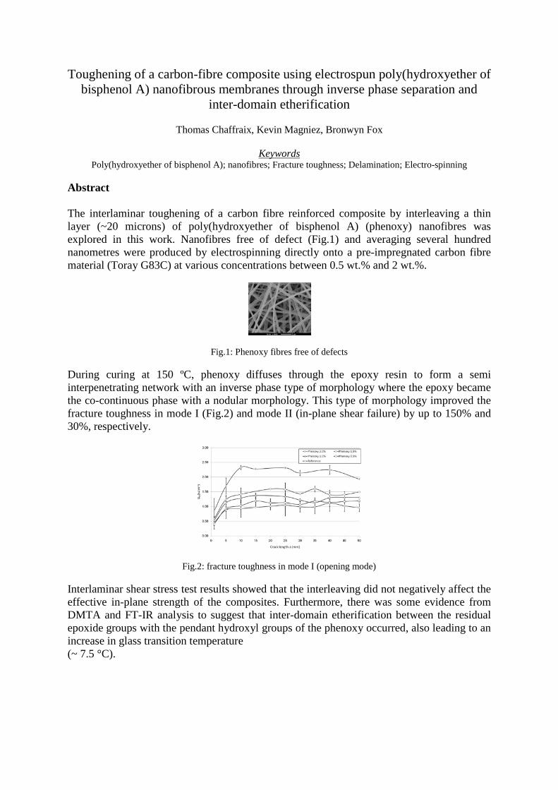
Abstract The interlaminar toughening of a carbon fibre reinforced composite by interleaving a thin layer (~20 microns) of poly(hydroxyether of bisphenol A) (phenoxy) nanofibres was explored in this work. Nanofibres free of defect (Fig.1) and averaging several hundred nanometres were produced by electrospinning directly onto a pre-impregnated carbon fibre material (Toray G83C) at various concentrations between 0.5 wt.% and 2 wt.%.
Fig.1: Phenoxy fibres free of defects During curing at 150 ºC, phenoxy diffuses through the epoxy resin to form a semi interpenetrating network with an inverse phase type of morphology where the epoxy became the co-continuous phase with a nodular morphology. This type of morphology improved the fracture toughness in mode I (Fig.2) and mode II (in-plane shear failure) by up to 150% and 30%, respectively.
Fig.2: fracture toughness in mode I (opening mode) Interlaminar shear stress test results showed that the interleaving did not negatively affect the effective in-plane strength of the composites. Furthermore, there was some evidence from DMTA and FT-IR analysis to suggest that inter-domain etherification between the residual epoxide groups with the pendant hydroxyl groups of the phenoxy occurred, also leading to an increase in glass transition temperature (~ 7.5 °C).
Multimodal Nanoparticles for Intracellular Delivery of a Calcium Channel Blocker
Cameron W. Evans†,‡, Helena M. Viola†, Diwei Ho†, Livia C. Hool†, Sarah A. Dunlop‡, Melinda Fitzgerald‡, and K. Swaminathan Iyer† † School of Biomedical, Biomolecular and Chemical Sciences, The University of Western Australia, Crawley WA 6009, Australia. ‡ Experimental and Regenerative Neurosciences, School of Animal Biology, The University of Western Australia, Crawley WA 6009, Australia.
Protecting vulnerable neurons and glia from damage that occurs secondary to neurotrauma is one of several requirements for successful therapy. Deregulation of calcium ion homeostasis is associated with secondary degeneration and release of excitatory amino acids, initiating cascades that lead to increased mitochondrial production of reactive oxygen species and cell death. The calcium channel blocker lomerizine has been shown to prevent calcium influx in vitro and protect against secondary injury effects in vivo. But in these latter studies, large, regular doses are required, much of which is excreted. The drug is soluble in acidic media but virtually insoluble under physiological conditions; bioavailability is therefore limited. In this work, lomerizine is encapsulated in polymer nanospheres containing iron oxide nanoparticles and rhodamine B fluorescent dye. The protective effect of lomerizine, that is, preventing calcium influx, is demonstrated by measuring intracellular calcium concentration in PC12 cells exposed to glutamate. The release of lomerizine likely occurs when nanospheres are contained in endosomes. The results show that targeted intracellular delivery of lomerizine using labeled nanoparticles may be a feasible approach to deliver sustained doses of lomerizine to CNS injury sites. This multifunctional approach could be combined with the delivery of other therapies that promote recovery after neurotrauma.
In-vitro evaluation of designer RADA16 nanofiber scaffolds on skin cells for wound healing
Diwei Hoa,b, Michael Bradshawa,b, Mark Fearb, Fiona Woodb and Swaminathan Iyera
a School of Biomedical, Biomolecular and Chemical Sciences, The University of Western Australia, Crawley, WA, 6009, Australia.
b The McComb Foundation, Burn Injury Research Unit, School of Surgery, The University of Western Australia, Crawley, WA, 6009, Australia.
Skin injuries not only cause physical damage and harm but also have traumatic psychological and negative social consequences, especially when there is scarring or disfigurement. Treatments aim to increase the rate of recovery to minimize scarring and to restore functionality of the skin.
RADA16 belongs to a class of self-assembling peptides that forms nanofibers through hydrophobic interactions in ionic solutions at physiological pH. The nanofibers self-organise to form a nanoscaffold with structural similarities to the extracellular matrix. RADA16, originally developed for use as a three-dimensional tissue culture scaffold, has shown promise to be a useful material for therapeutic purposes.
In this study, we have added two different types of functional motifs (collagen type I and fibronectin) to the original RADA16 peptide sequence to enhance biological interactions with skin cells such as keratinocytes and fibroblasts. Collagen type I and fibronectin are proteins found within the extracellular matrix and have been known to assist the migration of skin cells.
We investigated the effects of the RADA16 peptides (RADA16, RADA16-collagen type I and RADA16-fibronectin) on the migratory and proliferative rate of skin cells. With time-lapse imaging, we analysed the rate of migration of both keratinocytes and fibroblasts on the three different nanoscaffolds. The proliferation rates of the cells on these nanoscaffolds were determined with a MTS cell proliferation assay. The results and implications of the study will be further discussed in the poster.
Iron Oxide Based Conductive Nanowires via Magnetic Field Induced Self-Assembly
Dominic Ho1,2, Saquib Peerzade1, Stuart Hodgetts2, Alan R Harvey2, Iyer Swaminathan1
1 School of Biomedical, Biomolecular and Chemical Sciences, The University of Western Australia, Crawley, WA, 6009, Australia 2 School of Anatomy and Human Biology, The University of Western Australia, Crawley, WA, 6009, Australia
Recently, there has been much interest in using magnetic nanoparticles in a diverse range of research applications. These range from medical diagnostics and experimental cancer treatments to bio-imaging and information storage. Ordered structures of such magnetic nanoparticles have been obtained from a stable colloidal solution utilising techniques such as controlled evaporation and spin coating. In addition, these techniques allow for the controlled generation of structures with varying architectures and properties. Techniques such as capillary forces, template synthesis, shape selective and size selective interactions have been investigated for the directed self-assembly of nanoparticles. Of these approaches, the template synthesis appears to be simple and effective. To date, polymers have been used as suitable templates e.g. block co-polymers. A distinct advantage of using magnetic nanomaterials is that magnetic forces applied are able to overcome the mutual electrostatic attraction of the nanoparticles, allowing for the simple fabrication of 1D structures in aqueous solution. In our present research, we have fabricated wire like structures by linking Fe3O4 superparamagnetic nanoparticles with Polyethyleneimine (PEI) and a conductive polymer Poly(3,4-ethylenedioxythiophene) poly(styrenesulfonate) (PEDOT:PSS). The addition of the respective polymers links the particles permanently and conserves their shape. Furthermore, the deposition of these structures onto a solid substrate in the presence of a magnetic field results in the formation of parallel patterns which remain attached to the substrate and are resistant to changes in the applied magnetic field.
Research on GaAs/AlxGa1-xAs/GaAs core-shell structure
Nian Jiang, Qiang Gao, Patrick Parkinson, Hark Hoe Tan, Chennupati Jagadish
Department of Electronic Materials Engineering, Research School of Physics and Engineering, The Australian National University, Canberra, ACT 0200, Australia
Abstract: Among III-V semiconductors, the GaAs material system is used extensively in electronics and optoelectronics industries, and accordingly GaAs nanowires are prime candidates for electrically and optically active nanowire devices such as lasers, solar cells and photodetectors. Due to the high density of surface defect states which act as non-radiative exciton and carrier traps, the performance of core only GaAs nanowires and based optoelectronic devices is largely limited. This problem can be overcome by cladding the GaAs nanowire in a shell of wider band-gap material. It is well known that an AlGaAs layer with a larger band-gap and negligible lattice mismatch grown over the GaAs nanowires can reduce the surface defect density remarkably, resulting in a significant improvement of the photoluminescence efficiency and the photo carrier lifetimes. [1]
Good control of the shell growth conditions is essential to achieve high electronic quality nanowires. Any variation in aluminium concentration in the shell, induced by the much shorter diffusion length of Al species, tends to broaden the photoluminescence emission peak. [2] In addition, stronger Al-C bond can lead to a higher carbon impurity level in the shell, which allows C species diffusing to the GaAs core [3]. Thus, a high quality AlGaAs layer is critical in realising the high performance of the nanowires. A GaAs cap outside of the AlGaAs layer is also essential to prevent the Al from oxidation.
In our research, GaAs/AlxGa1-xAs/GaAs core-shell structure nanowires were grown by vapour-liquid-solid (VLS) mechanism using metal-organic-chemical-vapor-deposition (MOCVD). The GaAs core was nucleated for 1min at 450oC followed by 45min growth at 375oC, which eliminates stacking defaults and lead to pure zinc-blend structure. The results show that the profile of the core-shell structure is dependent on the Al%, the shell growth temperature and shell growth time. It is observed that the irregular growth around the tips increased with increasing Al% and shell growth time. The high shell growth shell growth temperature can suppress the irregular growth efficiently. A bulging part underneath the Au alloy particle was observed in GaAs/AlxGa1-xAs/GaAs (x≥50%) in vapour during shell growth while no such extra growth occurred with 26% Al in vapour. The unexpected extra growth for GaAs/AlxGa1-xAs/GaAs (x=50%) can be eliminated with the shell growth temperature at 750oC for 1.5min. All the GaAs/AlxGa1-xAs/GaAs (x=50%) nanowires show much stronger PL than the bare GaAs nanowires while the lifetime increased marginally with shell growth temperature but dramatically with decreased shell growth time.
References: 1. Parkinson, P., et al., carrier lifetime and mobility enhancement in nearly defect-free core-shell
nanowires measured using time-resolved Terahertz Spectroscopy. Nano Letters, 2010. XX(X ): p. -.
2. Paek, J.H., et al., Structural and optical properties of a catalyst-free GaAs/AlGaAs core-shell nano/microwire grown on (1 1 1)Si substrate. Physica E-Low-Dimensional Systems & Nanostructures, 2010. 42(10): p. 2722-2726.
3. van Deelen, J., et al., Influence of growth parameters on the composition and impurity levels of intrinsically carbon doped AlxGa1-xAs. Journal of Crystal Growth, 2005. 284(1-2): p. 28-38.
Fabrication of GaAs/AlGaAs Core-shell Nanowire Solar Cells
Jaret Lee, Kaushal Vora*, Lan Fu, Fouad Karouta*, Hoe Tan, Chennupati Jagadish
Department of Electronic Materials Engineering,
*Australian National Fabrication Facilities, ACT Node
Research School of Physics and Engineering, Australian National University, Canberra, ACT 0200
GaAs nanowires, grown by vapour-liquid-solid process using metal-organic chemical vapour deposition, have emerged as promising materials for high-efficiency solar cells. By including new physical principles offered from nanotechnology, these devices are expected to attain high performance due to enhanced light absorption and improved carrier collection. Particular attention has been given to synthesizing core-shell structures, which allow the diffusion length for the minority carriers to be shorter than the optical absorption length. This greatly reduces the demand for using high-quality materials leading to lower fabrication cost. In this study, we present the fabrication procedure for planarising and making low-resistance ohmic contact to GaAs-AlGaAs core-shell, nanowires. Various filling materials (PMMA, PDMS, BCB, and Spin-on-glass) were investigated for their suitability in planarising ensemble nanowire arrays. Surface roughness and optical transmission measurements were performed. Reactive ion etching was used as an effective method to etch back the planarising materials and uncover the nanowire tops for uniform top-contact deposition. Finished devices contacted by indium tin oxide films and Au finger electrodes were tested. I-V and photo-response measurements were carried out to confirm the photovoltaic behaviour from these devices.
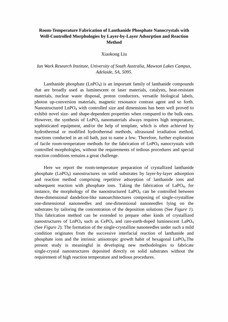
Room-Temperature Fabrication of Lanthanide Phosphate Nanocrystals with Well-Controlled Morphologies by Layer-by-Layer Adsorption and Reaction
Method
Xiaokong Liu
Ian Wark Research Institute, University of South Australia, Mawson Lakes Campus, Adelaide, SA, 5095.
Lanthanide phosphate (LnPO4) is an important family of lanthanide compounds
that are broadly used as luminescent or laser materials, catalysts, heat-resistant materials, nuclear waste disposal, proton conductors, versatile biological labels, photon up-conversion materials, magnetic resonance contrast agent and so forth. Nanostructured LnPO4 with controlled size and dimensions has been well proved to exhibit novel size- and shape-dependent properties when compared to the bulk ones. However, the synthesis of LnPO4 nanomaterials always requires high temperature, sophisticated equipment, and/or the help of template, which is often achieved by hydrothermal or modified hydrothermal methods, ultrasound irradiation method, reactions conducted in an oil bath, just to name a few. Therefore, further exploration of facile room-temperature methods for the fabrication of LnPO4 nanocrystals with controlled morphologies, without the requirements of tedious procedures and special reaction conditions remains a great challenge.
Here we report the room-temperature preparation of crystallized lanthanide
phosphate (LnPO4) nanostructures on solid substrates by layer-by-layer adsorption and reaction method comprising repetitive adsorption of lanthanide ions and subsequent reaction with phosphate ions. Taking the fabrication of LaPO4, for instance, the morphology of the nanostructured LaPO4 can be controlled between three-dimensional dandelion-like nanoarchitectures composing of single-crystalline one-dimensional nanoneedles and one-dimensional nanoneedles lying on the substrates by tailoring the concentration of the deposition solutions (See Figure 1). This fabrication method can be extended to prepare other kinds of crystallized nanostructures of LnPO4 such as CePO4 and rare-earth-doped luminescent LaPO4 (See Figure 2). The formation of the single-crystalline nanoneedles under such a mild condition originates from the successive interfacial reaction of lanthanide and phosphate ions and the intrinsic anisotropic growth habit of hexagonal LnPO4.The present study is meaningful in developing new methodologies to fabricate single-crystal nanostructures deposited directly on solid substrates without the requirement of high reaction temperature and tedious procedures.
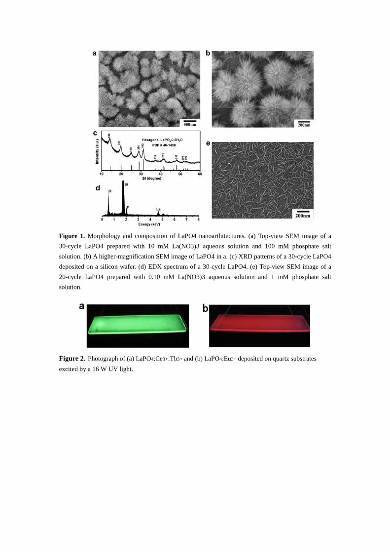
Figure 1. Morphology and composition of LaPO4 nanoarthitectures. (a) Top-view SEM image of a 30-cycle LaPO4 prepared with 10 mM La(NO3)3 aqueous solution and 100 mM phosphate salt solution. (b) A higher-magnification SEM image of LaPO4 in a. (c) XRD patterns of a 30-cycle LaPO4 deposited on a silicon wafer. (d) EDX spectrum of a 30-cycle LaPO4. (e) Top-view SEM image of a 20-cycle LaPO4 prepared with 0.10 mM La(NO3)3 aqueous solution and 1 mM phosphate salt solution.
Figure 2. Photograph of (a) LaPO4:Ce3+:Tb3+ and (b) LaPO4:Eu3+ deposited on quartz substrates excited by a 16 W UV light.
Functionalization and Time-gated Luminescence Bioimging application of Polystyrene Nanoparticles
Jie Lu, Jiangbo Zhao, Yiqing Lu, Dayong Jin
Advanced Cytometry Laboratories, MQ Biofocus Research Centre, Macquarie University, Sydney, Australia
Background: Nanoparticle is emerging as a sensitive tool in the broad areas of biosensing. Encapsulating
molecular dyes into silica or polymer nano-matrix represents one of the most popular methods to produce
bright, photo-stable and luminescence tunable nanoparticles (1). However, there remains unclear how much
quantitatively these nanoparticles could enhance the immunofluorescence labelling for cell imaging.
Methods: We employed the EDC conjugation method (2) to couple the streptiavidin protein onto the surface
of two europium containing polystyrene nanoparticles (40nm and 200nm, Invitrogen F20880 and F20881).
The successful conjugation was confirmed by both zeta potential and gel electrophoresis analysis. We
applied these streptavidin conjugated nanoparticles to analyse the CD34 surface antigen level on the
transfected HEK 293 cells, which primarily labelled by biotinylated anti-CD34 antibody. All labelled cells
were analysed by the automated time-gated luminescence scanning cytometry (3). These imaging results
were used to compare molecular complex (BHHCT-Eu) labelling results.
Results: Refer to Fig. 1, the time-resolved microscopy (4) method provides a background-free condition
permitting direct quantification of labelling brightness. Both nanoparticle (40 nm, 200 nm) - labelled cells
show enhanced red luminescence than molecular complex labelling by a factor of 3, and 16 respectively.
Fig. 1. The CD34 transfected HEK 293 cells labelled by streptiAvidin conjugated BHHCT-Eu (left),
40 nm (middle), and 200 nm europium containing polystrene nanoparticles.
Conclusion: This result also suggests a novel method using both time-gated luminescence scanning
cytometry and nanoparticles for direct cell surface antigen quantification, which is important for cell
phenotyping and disease progressive monitoring and diagnosis.
1. Jiang HF, Wang GL, Zhang WZ, Liu XY, Ye ZQ, Jin DY, Yuan JL, Liu ZG. Preparation and Time-Resolved Luminescence Bioassay Application of Multicolor Luminescent Lanthanide Nanoparticles. Journal of Fluorescence 2010;20(1):321-328.
2. Deen C, Claassen E, Gerritse K, Zegers ND, Boersma WJA. A novel carbodiimide coupling method for synthetic peptides:: Enhanced anti-peptide antibody responses. Journal of Immunological Methods 1990;129(1):119-125.
3. Lu YQ, Jin DY, Leif RC, Deng W, Piper JA, Yuan JL, Duan YS, Huo YJ. Automated Detection of Rare-Event Pathogens Through Time-Gated Luminescence Scanning Microscopy. Cytometry Part A 2011;79A(5):349-355.
4. Jin D, Piper JA. Time-gated luminescence microscopy allowing direct visual inspection of lanthanide-stained microorganisms in background-free condition. Analytical Chemistry 2011;83 (6):2294-2300.
Effect of chain formation in aqueous suspensions of magnetic nanoparticles on the stability of proton transverse relaxation rates.
Saquib A. Peerzade1,2, Rahi R. Varsani1,2, Swaminathan Iyer1, Robert C. Woodward2, Michael J. House2, Tim St. Pierre2.
1School of Biomolecular and Chemical Sciences, The University of Western Australia, Crawley, WA, 6009, Australia
2School of Physics, The University of Western Australia, Crawley, WA, 6009, Australia
Magnetic nanoparticles are being intensively studied for a wide variety of biomedical applications. These applications include their use as contrast agents in magnetic resonance imaging (MRI), for cancer treatment via magnetic hyperthermia, as magnetically targeted drug delivery vehicles, and for applications in cell separation and purification.
Owing to their high magnetic susceptibility, magnetic nanoparticles have high relaxivity, a measure of how they affect proton relaxation, and hence are used in MRI as contrast agents. In targeted drug delivery the magnetic particles are coated with drugs and then magnetic fields are used to localize the drugs to a specific part of the body. Both these applications require the use of a magnetic field in vivo. However, it is known that some magnetite nanoparticles form linear chains in the presence of magnetic fields. The length and thickness of the chain depends upon the size of nanoparticles, method of synthesis of magnetic nanoparticles, magnetic susceptibility of the particles, concentration, strength of magnetic field and the length of time the particles have been in the applied magnetic field. Recently, variations in the proton transverse relaxation rate with time have been observed in our laboratory for several magnetic nanoparticle suspensions. These variations are potentially related to chain formation, which could be a concern for their use as MRI contrast agents. Contrast enhancement in MRI depends upon the relaxation rates and changing relaxation rates due to chain formation would change the image contrast over the time. Moreover, chain formation may cause blocking of blood vessels or increase the chance that the particles will be identified as foreign objects by the reticuloendothelial system.
Although relaxation rates for many contrast agents have been reported, the time of exposure to the magnetic field after which these relaxation rates were measured is generally unknown. In this project we have studied the change in relaxation rates as a function of time for different magnetic nanoparticle systems and prepared an ideal non-chaining sample that has a stable (time independent) proton transverse relaxation rate. The ideal sample will overcome many limitations of current magnetic nanoparticle suspensions. With a non-chaining sample, MRI contrast from the magnetic nanoparticles will be stable with time making quantitative assessment of images possible. The absence of chain formation will also potentially increase the circulation time inside the body; a critical requirement for targeted contrast agents and targeted drug delivery.
Electrical Properties of Single InP Nanowires using Focused Ion Beam Contacting Technique
Prakash Raj Prasai, Lan Fu, Patrick Parkinson, Michael Gao, Vidya Ramesh, Suriati Paiman ,Jie Tian*, Fouad Karouta*, Hark Hoe Tan,
Chennupati Jagadish
Department of Electronic Materials Engineering,
*Australian National Fabrication Facilities, ACT Node
Research School of Physics and Engineering, Australian National University, Canberra, ACT 0200
Nanowires exhibit unique electrical and optical properties which make them promising materials as the building blocks of future nanoscale devices and integrated circuits. To realise these devices, the ability to make reliable contacts to individual nanowires is crucial. In the present work, we use focused ion beam (FIB) to contact single nanowires. FIB is a powerful technique for milling, depositing, and patterning materials with nanoscale resolution. InP nanowires were grown by Au-assisted vapour-liquid-solid (VLS) method using metal-organic chemical vapour deposition (MOCVD). Optical lithography and Au metal-liftoff was carried out to define the contact pads on SiO2/Si substrates. The InP nanowire arrays were transferred onto these substrates using a simple sonication approach. By selecting nanowires which lie across the Au contact pads, Pt deposition by FIB was applied to securely bond the nanowires for electrical measurements. The measurements show a linear I-V response, indicating Ohmic contact formation for the nanowires studied. The nanowire resistance also demonstrates the expected proportionality to L/r2 (where L and r are the length and radius of the nanowires, respectively) behaviour. An increase of photocurrent upon light illumination was also observed with increasing excitation intensity. The results show that FIB contacting method may be a suitable technique for making contacts to nanowires for device application or for the study of their electrical and transport properties.
Reflection and confinement of guided modes in a nanowire Dhruv Saxena, Hoe Tan, Chennupati Jagadish
Department of Electronic Materials Engineering, Research School of Physics and Engineering, Australian National University, Canberra, ACT 0200, Australia III-V semiconductor nanowires are promising structures for developing lasers because the nanowire not only
serves as the gain medium, but also serves as a waveguide with its ends acting like a mirror due to the
dielectric mismatch, similar to a Fabry-Perot cavity. However due to the small diameters, the modes
supported in a nanowire are evanescent, with some of the field decaying away from the nanowire surface.
Furthermore light from the ends of the nanowire suffers diffraction losses. In order to fabricate a nanowire
laser we require both high optical confinement within the nanowire and high reflectivity at the ends. In this
study we investigate the reflection and confinement of guided modes in a nanowire through finite-difference
time-domain (FDTD) simulations. In particular, we model GaAs nanowire and calculate the reflectivity of low
order modes from the nanowire end facets. The dependence of reflectivity on nanowire diameter and index of
surrounding medium is examined. The confinement of these modes to the nanowire as a function of nanowire
diameter is also calculated. These preliminary calculations provide us with an indication for the nanowire
dimensions that are required to achieve lasing.
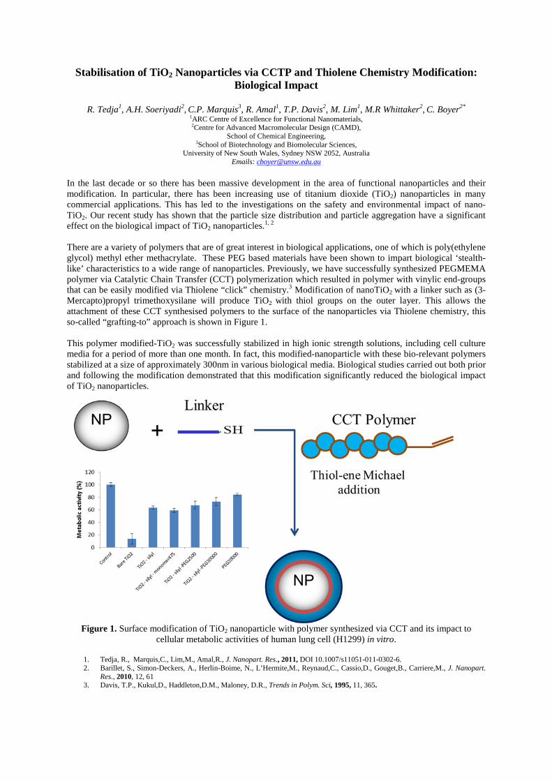
Stabilisation of TiO2 Nanoparticles via CCTP and Thiolene Chemistry Modification: Biological Impact
R. Tedja1, A.H. Soeriyadi2, C.P. Marquis3, R. Amal1, T.P. Davis2, M. Lim1, M.R Whittaker2, C. Boyer2*
1ARC Centre of Excellence for Functional Nanomaterials, 2Centre for Advanced Macromolecular Design (CAMD),
School of Chemical Engineering, 3School of Biotechnology and Biomolecular Sciences,
University of New South Wales, Sydney NSW 2052, Australia Emails: [email protected]
In the last decade or so there has been massive development in the area of functional nanoparticles and their modification. In particular, there has been increasing use of titanium dioxide (TiO2) nanoparticles in many commercial applications. This has led to the investigations on the safety and environmental impact of nano-TiO2. Our recent study has shown that the particle size distribution and particle aggregation have a significant effect on the biological impact of TiO2 nanoparticles.1, 2 There are a variety of polymers that are of great interest in biological applications, one of which is poly(ethylene glycol) methyl ether methacrylate. These PEG based materials have been shown to impart biological ‘stealth-like’ characteristics to a wide range of nanoparticles. Previously, we have successfully synthesized PEGMEMA polymer via Catalytic Chain Transfer (CCT) polymerization which resulted in polymer with vinylic end-groups that can be easily modified via Thiolene “click” chemistry.3 Modification of nanoTiO2 with a linker such as (3-Mercapto)propyl trimethoxysilane will produce TiO2 with thiol groups on the outer layer. This allows the attachment of these CCT synthesised polymers to the surface of the nanoparticles via Thiolene chemistry, this so-called “grafting-to” approach is shown in Figure 1. This polymer modified-TiO2 was successfully stabilized in high ionic strength solutions, including cell culture media for a period of more than one month. In fact, this modified-nanoparticle with these bio-relevant polymers stabilized at a size of approximately 300nm in various biological media. Biological studies carried out both prior and following the modification demonstrated that this modification significantly reduced the biological impact of TiO2 nanoparticles.
Figure 1. Surface modification of TiO2 nanoparticle with polymer synthesized via CCT and its impact to
cellular metabolic activities of human lung cell (H1299) in vitro.
1. Tedja, R., Marquis,C., Lim,M., Amal,R., J. Nanopart. Res., 2011, DOI 10.1007/s11051-011-0302-6. 2. Barillet, S., Simon-Deckers, A., Herlin-Boime, N., L’Hermite,M., Reynaud,C., Cassio,D., Gouget,B., Carriere,M., J. Nanopart.
Res., 2010, 12, 61 3. Davis, T.P., Kukul,D., Haddleton,D.M., Maloney, D.R., Trends in Polym. Sci, 1995, 11, 365.

New polymer coatings for biomedical applicationsAndrew M. Telford1,2, Chiara Neto1, Laurence Meagher3, Michael James4
1 School of Chemistry, The University of Sydney, NSW 2006 Australia 2 CSIRO Future Manufacturing Flagship, Clayton 3169, Australia 3 CSIRO Molecular Science, Clayton South, VIC 3168 Australia
4 Bragg Institute (ANSTO), Menai, NSW 2234 Australia The implementation of artificial parts in human bodies has become more and more widely spread. The main issue that has yet to be overcome when interfacing a foreign body with a living tissue is the inflammatory reaction that the artificial material triggers, leading to the rejection of the device itself (foreign body response).1 The very first step of such response is the coverage of the unknown surface with proteins that both isolate it from the living environment and direct the following cell attachment. Antifouling materials play a critical role in preventing the foreign body response by preventing this protein adhesion. Such coatings are referred to as “bio-inert” and are the simplest way to enhance the biocompatibility of the material. We discovered that poly(vinylpyrrolidone) (PNVP) could be cast in films and thermally cross-linked to become insoluble in water environments. These coatings are easy to prepare on surfaces with any shape. We have thoroughly characterized the cross-linking process on the nanometric scale of the polymer films, and demonstrated that such coatings are extremely efficient in repelling different types of proteins commonly found in human blood.2 A more complex strategy to reduce the foreign body response is to design coatings that provide an active interaction with the living tissue and promote a “healthy” response and integration of the device. Such surfaces are referred to as “bio-active”.3 An example is a coating patterned with specific proteins that can guide cell adhesion in selected areas. The cell response will depend on their density and distribution on the surface. We have developed a method to obtain chemically and topographically patterned surfaces for the immobilization of biomolecules (such as proteins) and cells, based on polymer thin film dewetting and surface grafting (Figure 1). Dewetting is the process by which an unstable thin liquid film (such as a polymer over its glass transition temperature) spontaneously breaks up, resulting in the formation of holes. The duration of dewetting controls the dimensions of the holes, from tens of nanometers to tens of microns. By tuning the thickness of the layers and the molecular weight of the polymers we can vary the type of pattern that is obtained.4,5 Our system comprises of two layers: the bottom layer is a protein adhesive polystyrene, while the top layer is an engineered polymer bearing bromine-esters in its side chains that can act as atom transfer radical polymerization (ATRP) initiators. The top layer of the pattern is dewetted to form a pattern of holes, and selectively grafted with highly protein repellent polymer brushes (poly(ethylene glycol) (PEG))
using ATRP. The process conditions were finely tuned to achieve grafting in less than one hour in alcohol/water media at room temperature. The densely packed PEG chains can greatly improve the selectivity of protein adhesion in the pattern holes. Cells can then adhere selectively on the protein patches.
Figure 1. Schematic of pattern formation upon dewetting. The bottom layer is protein adhesive; the top layer bears ATRP initiators. The top layer dewets forming holes that expose the underlying polymer. It is then grafted with protein repellent PEG chains. Proteins (green) adhere only inside the holes on the adhesive polymer, and a microscopic protein pattern is obtained. Cells (red) can attach to the protein-coated domains. The coating techniques used in our work could be used to achieve conformal coatings on non flat surfaces, which open exciting prospects in biomedical applications. The use of living radical polymerization for the grafting of the brush allows for post-functionalization of the chain ends, which can be used to introduce biological tags to tailor the properties of the coatings, in applications such as tissue engineering.
(1) Anderson, J. M.; Rodriguez, A.; Chang, D. T. Semin. Immunol. 2008, 20, 86. (2) Telford, A. M.; James, M.; Meagher, L.; Neto, C. ACS Appl. Mater. Interfaces 2010, 2, 2399. (3) Williams, D. F. Biomaterials 2008, 29, 2941. (4) Neto, C. Phys. Chem. Chem. Phys. 2007, 9, 149. (5) Seemann, R.; Herminghaus, S.; Jacobs, K. Phys. Rev. Lett. 2001, 86, 5534.
Magnetic field induced chains of magnetic nanoparticles: Correlating structure with magnetic properties
Rahi R. Varsani1,2,3,*, Saquib A. Peerzade2,3, K. Swaminathan Iyer2, Robert C. Woodward3,
Michael J. House3, Timothy G. St. Pierre3 and Martin Saunders1
1 Centre for Microscopy, Characterisation and Analysis, The University of Western Australia, Crawley, WA 6009, Australia
2 School of Biomedical, Biomolecular and Chemical Sciences, The University of Western Australia, Crawley, WA 6009, Australia
3 School of Physics, The University of Western Australia, Crawley, WA 6009, Australia * Corresponding author. Email: [email protected]
In the 1970s, de Gennes and Pincus [1] predicted the formation of magnetic nanoparticle chains under the influence of a magnetic field. The formation of chains is significant as it changes the magnetic properties of the magnetic nanomaterial, affecting use in applications such as MRI contrast enhancement and drug delivery. Investigating the structure of the chains at the nanoscale is far from trivial, as standard TEM preparation would result in unwanted solvent drying effects. The first TEM evidence of chains eliminating solvent effects was obtained 30 years later, when Butter et al. [2] used cryogenic techniques to freeze the chains in a thin-film and observe them on a TEM.
We have extended this work by correlating the change in magnetic properties of the nanomaterial with the formation of chains, which gives a better insight into how this might impact real-world applications. Furthermore, we have developed a more representative method of visualising chains formed in a larger volume of material as opposed to the thin-film technique used by Butter et al. [2].
[1] de Gennes, P. & Pincus, P. Pair correlations in a ferromagnetic colloid. Phys. Kondens. Mater. 11, 189-198 (1970).
[2] Butter, K., Bomans, P., Frederik, P., Vroege, G. & Philipse, A. Direct observation of dipolar chains in iron ferrofluids by cryogenic electron microscopy. Nature Materials 2, 88-91 (2003).
Nanocrystalline Silicon in Silicon Carbide Matrix: Fabrication, Characterization and Application in Solar Cells
Zhenyu Wan, Shujuan Huang, Marin Green and Gavin Conibeer
School of Photovoltaic and Renewable Energy Engineering, University of NSW
Silicon nano-crystals (Si NCs) embedded in dielectric material are considering as promising candidate
materials to fabricate “all-Si” tandem photovoltaic solar cells approaching the solar conversion
efficiency up to 40%. In this thesis, Si NC embedded in silicon carbide (SiC) material was
investigated thoroughly from the perspectives of structural, optical and electrical properties. Three
principal thin film structures were deposited using a sputtering and subsequently annealed: silicon rich
carbide (SRC) single layers, SRC/SiC multilayers and SRC/Si3N4 hybrid multilayers.
For SRC single layer research, significant concentration of Si NCs and SiC NCs were observed after
high temperature annealing at 1100oC. Furnace annealing and rapid thermal annealing (RTA) were
both carried out with different annealing time duration (1 hour and 1 minute respectively). After
annealing, structural characterisation revealed different Si and SiC crystallinity in the two samples. In
RTA, the annealing time at the temperature for SiC NC formation was very limited due to the rapid
profile ramp-up. As a consequence, fewer SiC NCs were observed in RTA samples. Detailed
discussions of crystallization mechanism, precipitation are included and diffusion coefficients were
calculated and compared on the basis of the two different annealing processes.
Photovoltaic devices (hetero-junctions and homo-junctions) were also fabricated based on this
structure (Boron in P type and Antimony in N type). Electrical characterisation demonstrated an
enhancement of photon absorption at shorter wavelength. This can be explained as the
implementation of Si NCs in the hetero-junction device which effectively tuned the band-gap of the
semiconductor material in emitter layer. However, the homo-junction device shunting effects as well
as high contact resistance observed in these could be associated with doping problem which may have
arisen from unsuccessful Si NCs confinement.
Hybrid structure devices consisting of SRC and Si3N4 were deposited to solve the structural Si NC
confining problem. An ultra-thin Si3N4 layer (less than 1nm) was successfully implemented into the
structure to confine the Si NC growth due to Si3N4 demonstrating a very low diffusion coefficient of
Si. Both structural and optical properties revealed that an optimum confinement was achieved with a
mere 0.8nm thick Si3N4 barrier layer. Electrical characterisation (temperature dependent IV
measurement) indicated the main carrier transport mechanism in the structure was thermal hopping
instead of tunnelling through the barriers.
Fabrication of single GaAs nanowire photodetectors by focused ion beam
Hao Wang*, Patrick Parkinson, Jie Tian, Qiang Gao, Kaushal Vora, Prakash Prasai, Animesh Basak, Lan Fu, Fouad Karouta, Hark Hoe Tan, and Chennupati Jagadish
Department of Electronic Materials Engineering, Research School of Physics and Engineering, The Australian National University, Canberra, ACT 0200, Australia
*Email [email protected]
Abstract:
Metal-semiconducotr-metal (M-S-M) Schottky structure has been employed to fabricate devices for
several decades, such as InGaAs photodectors (PDs). On such device a Schottky barrier forms at the
metal/semiconductor interface and this barrier plays a crucial role for the performance of the PDs.
Single GaAs NW PDs are fabricated by using the focused ion beam (FIB) facility. These PDs are
based on the back-to-back contact structure with Pt as the metal. Depending on the FIB fabrication
parameters, either Ohmic or Schottky contacts could be obtained. Current-voltage (I-V) characteri-
zation of the M-S-M structures showed an almost symmetrical instead of rectifying I-V characteris-
tic for the Schottky detector. It is found that the Schottky barrier height is very sensitive to carrier
generation and transport and can be controlled by the choice of metal, electrode spacing, doping etc.
The ability to control the Schottky barrier height provides the flexibility to control the response of
the PD.
For Australian Nanotechnology Network Early Career Symposium。
Development of highly stable CaO-SiC sorbent for carbon dioxide capture
Meilina Widyawati, Tamara L. Church and Andrew T. Harris*
Laboratory for Sustainable Technology, School of Chemical and Biomolecular Engineering, The University of Sydney, NSW 2006, Australia.
Phone: +61 2 9351 2926, Fax: +61 2 9351 2854 *email: [email protected]
Abstract Calcium oxide (CaO) is a superior sorbent that captures carbon dioxide (CO2)1 to form calcium carbonate (CaCO3). This carbonation reaction plays important role in the production of hydrogen (H2) a future cleaner fuel from biomass gasification, as it drives the water-gas shift reaction toward higher H2 yield.2 The CaCO3 can be calcined to regenerate CaO, thus the CaO is utilisable for carbonation and calcination cycles in biomass gasifier.2 Despite its superiority and regenerability, the decay of CaO reactivity due to sintering remains a challenge to its stability over multiple cycles. Others have reported strategies to improve the reactivity of CaO, including by incorporating it onto support materials like alumina3, 4 or silica.5 Herewith we report a novel strategy to incorporate CaO onto a mesoporous silicon carbide (SiC) support, which was chosen for its excellent thermal and mechanical properties.6 The SiC support was produced from polycarbomethylsilane (PCMS) using mesoporous SBA-15 silica as a template.7, 8 The resulting material, silicon-carbide-supported calcium oxide (CaO-SiC), demonstrated high stability over 40 cycles of carbonation and calcination in a thermogravimetric analyser (TGA). This was as indicated by weight increases of 9.6, 11.2, and 11.5% due to CO2 absorption during the first, second, and 40th cycles, respectively. Further study revealed that the performances of CaO-SiC sorbents, in terms of the both reactivity and stability, were influenced by parameters like the CaO loading, and the molarity and temperature during impregnation of the SiC with a calcium acetate solution, Ca(OAc)2 (aq). Acknowledgement This research was sponsored by The University of Sydney through ‘University of Sydney World Scholar’ program and by E.ON AG.
References 1 H. Gupta and L.-S. Fan, Ind. Eng. Chem. Res., 2002, 41, 4035-4042. 2 N. H. Florin and A. T. Harris, Int. J. Hydrogen Energy, 2007, 32, 4119-4134. 3 Z.-S. Li, N.-S. Cai and Y.-Y. Huang, Ind. Eng. Chem. Res., 2006, 45, 1911-1917. 4 C. S. Martavaltzi and A. A. Lemonidou, Microporous Mesoporous Mater., 2008, 110, 119-127. 5 C.-H. Huang, K.-P. Chang, C.-T. Yu, P.-C. Chiang and C.-F. Wang, Chem. Eng. J., 2010, 161,
129-135. 6 M. J. Ledoux and C. Pham-Huu, CATTECH, 2001, 5, 226-246. 7 Y.-F. Shi, Y. Meng, D.-H. Chen, S.-J. Cheng, P. Chen, H.-F. Yang, Y. Wan and D.-Y. Zhao,
Adv. Funct. Mater., 2006, 16, 561-567. 8 P. Krawiec, C. Schrage, E. Kockrick and S. Kaskel, Chem. Mater., 2008, 20, 5421-5433.
Aggregation Stability Study of Functionalised Magnetite Nanoparticles in Biological Media
Containing Serum Hilda T. R. Wiogo1, May Lim1, Volga Bulmus2, and Rose Amal1
1ARC Centre of Excellence for Functional Nanomaterials, School of Chemical Engineering, The University of New South Wales, Sydney, NSW 2052, Australia
2Department of Chemical Engineering, İzmir Institute of Technology, Gülbahçe Urla 35430, Turkey Email: [email protected], [email protected], [email protected]
Abstract Size stability of magnetite nanoparticles (MNP) having different surface functional
groups in biological media, RPMI-1640, had been successfully achieved for at least
16 h, by addition of fetal bovine serum (FBS). The stability was obtained due to the
formation of protein corona around the particles, which provides sufficient steric
hindrance to prevent aggregation of the particles. Its formation is determined by
functional group present on the surface of MNP. This study was performed on MNP
synthesised by co-precipitation method, which was functionalised with either
positively charged polyethylenimine (PEI) or negatively charged polymethacrylic acid
(PMAA). The bare and functionalised MNPs were treated with FBS by a simple
mixing of particles with different amount of FBS in RPMI-1640 with the aid of
ultrasonication. The amount of FBS required for stability of MNP depends on the
surface functional groups present, where amine functionalised MNP found to require
the lowest amount of FBS and carboxylic acid functionalised MNP required the
highest amount of FBS. This is due to the different amount of protein adsorption onto
different particles surfaces, where positive charged particles was shown to attract
highest amount of serum protein, while the negative charge particles has the least
protein adsorbed. This study provides insight into stabilisation by serum protein for
different functionalised MNP, which would allow the design of biocompatible and
stable MNP in biological media, which further broaden their potential for various
biomedical applications.

Pristine Emulsion interfaces induced self-polymerization for the synthesis of polydopamine hollow capsules
Haolan Xu, Dayang Wang
Ian Wark Research Institute, University of South Australian. SA 5095, Australia Email: [email protected]
Oil-in-water emulsions have been widely employed as reaction media to sculpture various organic and inorganic materials into macroporous structures. But they have been rarely used to template formation of hollow capsules. The main reason should be that in an oil-in-water emulsion, precursors transform into targeted materials usually by reaction with water or active species in water, so there is no preference for the oil/water interface over the bulk aqueous phase to dictate the targeted materials to grow on the oil droplets. Selective growth and efficient accumulation of targeted materials at the oil/water interface is prerequisite for individual emulsion droplets to template formation of hollow capsules. To achieve that, one has to employ water-in-oil emulsions as templates, in which targeted materials are selectively formed at the oil/water interfaces only when their hydrophobic precursors in bulk organic phase interact with water or reactive species dissolved in the water droplets.1 Recently, Xu et al have used cyclohexane-in-water emulsion droplets, stabilized by non-ionic polyglycol, as templates to synthesize hollow nickel capsules and they rationalized the formation of the hollow capsules as a result of a strong coordination between the polyglycol coating and nickel ions.2 Zoldesi and Imhof have used surfactant-free dimethyldiethoxysilane(DMDES)-in-water emulsion droplets as templates to produce hollow silica capsules.3 Later on, Zoldesi et al have indicated a noticeable influence of the types of surfactants used to stabilize DMDES oil droplets on the wall thickness of the hollow silica capsules obtained in DMDES oil-in-water emulsions; anionic surfactant, sodium dodecylsulfate (SDS) significantly reduced the silica wall thickness whereas non-ionic surfactant, Triton X-100 caused little variation.4 Possibly owing to the complicated chemical composition of the silane oil droplets and other additive used, the underlying mechanism of hollow silica capsules has been little discussed in these studies. In order to gain in-depth insight to the template effect of oil-in-water emulsions on formation of hollow capsules, herein we employed 3,4-dihydroxyphenylethylamine (dopamine) as model monomer and studied how the surface chemistry nature of alkane-in-water emulsions, surfactant-free or surfactant-stabilized, affect self-polymerization of dopamine and the location and efficiency of the formation of polydopamine (PDA) in the emulsions and in turn the PDA structures obtained thereafter.
It has been clearly documented that oil-in-water emulsions can be generated in the absence of any surfactant – known as pristine emulsions – as a result of enrichment of OH ions at the oil/water interfaces.5-
7 The surfaces of the oil droplets, stabilized by OH ions, are therefore negatively charged and their pH are much higher than that in bulk water. For instance, when the pH in bulk water is 9, the zeta potential of hexadecane oil droplets is -105 mV, the surface charge density of hexadecane/water interfaces in the absence of surfactants can be as high as -4.9 C cm-2 and the pH at the interfaces can be as high as 14.6 Up to date, however, this peculiar surface chemistry character of the oil droplets in pristine oil-in-water emulsions has been rarely used to template formation of nanostructured materials and taken into account to rationalize the template effect of oil-in-water emulsions. Herein, we should for the first time deliberately use the basic interfacial environment of pristine oil-in-water emulsions to guide preferential material growth and accumulation to create hollow capsules. The advantage of this method is that the reaction system is simple and versatile, only oil, water and OH- are involved in the formation of emulsion template. In principle, all the hydrophobic oil can be stabilized solely OH ions and server as template for PDA growth. Furthermore, because of the simple interfacial chemical milieu of pristine emulsion, it provides an ideal model to study the template effect of interfaces.
We demonstrated a pronounced dependence of the structures of PDA, obtained via self-polymerization in alkane-in-water emulsions, on the surface chemistry nature of the alkane droplets. Self-polymerization of dopamine in pristine alkane-in-water emulsions leads to hollow PDA capsules (Figure 1) as the OH- enriched surfaces of the alkane droplets have a significantly high pH to guide efficient growth and accumulation of PDA on the droplet surfaces. In contrast, there is no preferential self-polymerization of dopamine on surfactant-stabilized alkane droplets, so the interstitial spaces between the alkane droplets
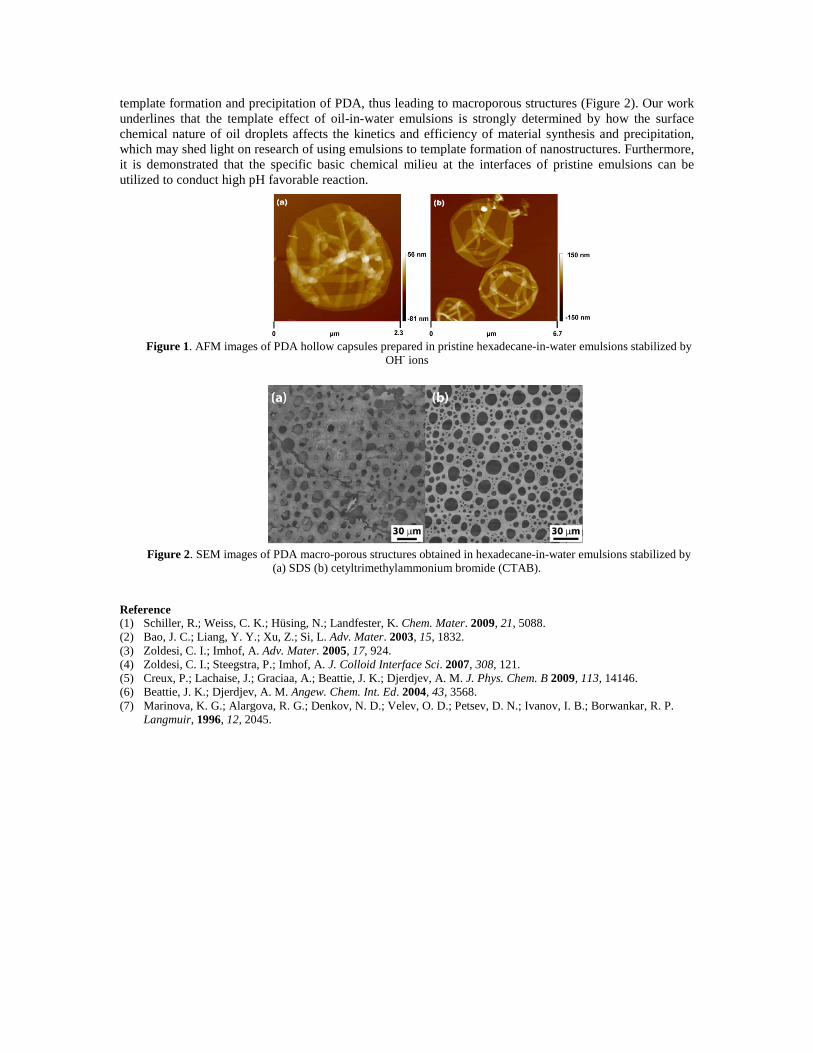
template formation and precipitation of PDA, thus leading to macroporous structures (Figure 2). Our work underlines that the template effect of oil-in-water emulsions is strongly determined by how the surface chemical nature of oil droplets affects the kinetics and efficiency of material synthesis and precipitation, which may shed light on research of using emulsions to template formation of nanostructures. Furthermore, it is demonstrated that the specific basic chemical milieu at the interfaces of pristine emulsions can be utilized to conduct high pH favorable reaction.
Figure 1. AFM images of PDA hollow capsules prepared in pristine hexadecane-in-water emulsions stabilized by
OH- ions
Figure 2. SEM images of PDA macro-porous structures obtained in hexadecane-in-water emulsions stabilized by
(a) SDS (b) cetyltrimethylammonium bromide (CTAB).
Reference (1) Schiller, R.; Weiss, C. K.; Hüsing, N.; Landfester, K. Chem. Mater. 2009, 21, 5088. (2) Bao, J. C.; Liang, Y. Y.; Xu, Z.; Si, L. Adv. Mater. 2003, 15, 1832. (3) Zoldesi, C. I.; Imhof, A. Adv. Mater. 2005, 17, 924. (4) Zoldesi, C. I.; Steegstra, P.; Imhof, A. J. Colloid Interface Sci. 2007, 308, 121. (5) Creux, P.; Lachaise, J.; Graciaa, A.; Beattie, J. K.; Djerdjev, A. M. J. Phys. Chem. B 2009, 113, 14146. (6) Beattie, J. K.; Djerdjev, A. M. Angew. Chem. Int. Ed. 2004, 43, 3568. (7) Marinova, K. G.; Alargova, R. G.; Denkov, N. D.; Velev, O. D.; Petsev, D. N.; Ivanov, I. B.; Borwankar, R. P.
Langmuir, 1996, 12, 2045.