Embed Size (px)
Citation preview
Annese, V.F. , Mezzina, G., Gallo, V.L., Scarola, V. and De Venuto,
D. (2017) Wearable Platform for Automatic Recognition of Parkinson
Disease by Muscular Implication Monitoring. In: 7th IEEE International
Workshop on Advances in Sensors and Interfaces (IWASI), Vieste, Italy,
15-16 June 2017, pp. 150-154. ISBN
9781509067077 (doi:10.1109/IWASI.2017.7974236)
This is the author’s final accepted version.
There may be differences between this version and the published version.
You are advised to consult the publisher’s version if you wish to cite from
it.
http://eprints.gla.ac.uk/146738/
Deposited on: 28 August 2017
Enlighten – Research publications by members of the University of Glasgow
http://eprints.gla.ac.uk33640
Wearable Platform for Automatic Recognition of ParkinsonDisease by Muscular Implication Monitoring
V.F. Annese2, G. Mezzina!, V.L. Gallo!, V. Scarola!, D. De Venuto!1 Dept. ofElectrical and Information Engineering (DEI) Politecnico di Bari, Italy
2 School ofEngineering, University ofGlasgow, Glasgow G12 8LT, U.K
Abstract - The need for diagnostic tools for the characterizationofprogressive movement disorders - as the Parkinson Disease (PD)- aiming to early detect and monitor the pathology is getting moreand more impelling. The parallel request ofwearable and wirelesssolutions, for the real-time monitoring in a non-controlledenvironment, has led to the implementation of a Quantitative GaitAnalysis platform for the extraction of muscular implicationsfeatures in ordinary motor action, such as gait.The here proposed platform is used for the quantification of PDsymptoms. Addressing the wearable trend, the proposedarchitecture is able to define the real-time modulation of themuscular indexes by using 8 EMG wireless nodes positioned onlower limbs. The implemented system "translates" the acquisitionin a I-bit signal, exploiting a dynamic thresholding algorithm. Theresulting I-bit signals are used both to define muscular indexesboth to drastically reduce the amount of data to be analyzed,preserving at the same time the muscular information. The overallarchitecture has been funy implemented on Altera Cyclone VFPGA. The system has been tested on 4 subjects: 2 affected by PDand 2 healthy subjects (control group). The experimental resultshighlight the validity of the proposed solution in Diseaserecognition and the outcomes match the clinical literature results.
Keywords-EMG, Gait, FPGA, Parkinson Disease, WearableDiagnostic
I. INTRODUCTION
Parkinson disease (PD) is a progressive neurological diseasecharacterized by bradykinesia (slowness) or akinesia (absentmovement), tremor, rigidity and postural instability [1]. TheCenters for Disease Control and Prevention (CDC) rated themotor complications from PD as the 14th top cause of death inthe United States [2]. Typically, these movement disorders areassociated with a slow short-stepped, shuffling gait pattern. Forthis aim, analysis of the gait in response to medication, visualcues, attentional strategies, provide insight into the nature ofthemotor control deficit in Parkinson disease and the efficacy ofcurrent therapeutic interventions. Currently, the UnifiedParkinson Disease Rating Scale [3] jointly with theHoehn&Yahr (H&Y) [4] one, are used to assess the severity ofgait and mobility complications, as well as thepresence/absence of characteristic motor signs in term ofindependence and quality oflife. Despite the widespread use ofthis scales, these metrics of judgment suffer of strongsubjectivity from neurologists and caregivers, since the scoreassignment are left to visual inspection approaches. Then,subjectively extracted information are the current trends indaily clinician's diagnostic work and therapeutic decisions.With the aim of releasing the assessment of a serious diseasesuch as Parkinson's from this kind of information, a new rising
978-1-5090-6707-7/17/$31.00 ©2017 IEEE
branch of bio-medical application was born, namedQuantitative Gait Analysis (QGA).The QGA is a systematicstudy of human walking in terms of kinematics, and spatiotemporal parameters useful for the characterization of humanmovement. With the above-stated definition, QGA in clinicalassessment can guide physicians to optimal decision making. Insuch cases, QGA can provide objective and progressive dataabout the gait deviation and functional deficits [5]. The QGAsolutions can be divided in: (i) wearable systems (WS) and (ii)non-wearable ones (NWS). Among the NWSs, most of thesystems utilizes a combination of commercial video recordingsystem and inertial sensors for gait monitoring in PD [6, 7], orcomplex hybrid combinations of cameras and Kinect system[8]. These systems adopt image-processing approaches that arenot suitable for low computational and real-timeimplementation. Other NWSs use floor sensors [9] to reachuseful evaluation about the force exerted by the subject's feeton the floor when he/she walks. The major limitation ofNWSsis that these solutions typically require the use of controlledresearch environment, in which the sensors are located andcapture data on the gait. For NWS applications, the subject istypically asked to walk on a clearly marked walkway [10].Differently, WSs make it possible to analyze data outside thelaboratory and capture information about the human gait duringthe person's everyday activities. Wearable systems use anetwork of sensor (e.g. accelerometers, gyroscopic,magnetometers, active markers) located on the user body (e.g.feet, knees, leg, arms and waist) [11, 12]. The above-mentionedmethods despite of their sensing capability, provide only apartial knowledge of the disease since the PD involves adeterioration of deep muscular activity before it becomesclearly visible through visual inspection techniques. Analyzingthe muscular involvements represents, instead, a deepknowledge of motor activity allowing precise quantification ofthe progression ofthe disease. In this work, we propose a novelQGA wearable and wireless platform for PD early recognition,fully based on electromyography (EMG). The proposedarchitecture is able to define the real-time modulation ofan "adhoc" calculated muscular index, to characterize the gait. Thesystem describes the muscular activity, acquired by 8commercial EMG wireless electrodes, exploiting their single bitcorrespondent signals and, thus, define on them, some muscularindexes. The monitoring ofthese quantitative deep myoelectricparameters allows discriminating also an early stage of PD, aswell as highlighting the difference between Controls anddiseased people. Finally, if used in synchronized mode withEEG signals, the system is able to understand the presence of
150
(1)
an involuntary movement, which can be an unbalancing event(e.g. inducing fall) or muscular dyskinesia [13, 14]. The hereproposed QGA platform has been tested on PD patients (n=2)and controls (n=2), provided in-vivo measures, in order tohighlight the approach validity in the quantification of diseasesymptoms. The paper is organized as follows: Sec. II recalls abasic medical knowledge, extracts the typical clinical protocolfor the gait assessment, and describes the algorithm for theEMG - I-bit translation and the muscular indexes evaluation.Sec. III outlines the implementation on FPGA. Sec. N isdedicated to the experimental results. Sec. V concludes thepaper.
II. THE QGA PLATFORMA. Clinical Description and Gait Analisys Protocol
The early symptom of Parkinson's disease is thedisappearance of the anticipatory postural reflexes, which areusually activated when a disturbance, however slight, of theposture occurs. For this reason, the probability to reach posturalinstability trends increases in PD patients. Indeed, the PD is anextrapyramidal system impairment, which is the motion controlsystem [15]. Therefore, when a lesion affects this system, inparticular the nigrostriatal circuits, induces decrement in themovement control capability, plastic rigidity and lack of motorautomatisms. For instance, missing accessory movements, suchas the oscillation of the upper limbs in the course of walking,lead the patient to lose the control and then generatesabnormality in the gait patterns. These gait drifts are typicallylinked to abnormal trunk's postures ofthe parkinsonian patients.Among them, the Pisa Syndrome (PS) - a sustained lateralbending ofthe trunk (at least 10°) - is a real clinical enigma, andits management remains a challenge [15, 16]. However, the lackof consistent diagnostic criteria leads to significant differencesin frequency reports [17]. All of these cardinal features of theParkinson's disease are strictly linked to the motor symptoms ofthe disease itself: In this work, the standardized clinical taskadopted during the system validation is extracted from theUnified Parkinson Disease Rating Scale (UPDRS) Part III andN guidelines [3]. In order to validate the proposed QGAplatform ability in gait analysis and PD status recognition,subjects are asked to perform the protocolled task named 10meter walk. The protocol asks to the subjects under test to walkfor 10 meters distance, 10 times with a comfortable walkingspeed [3]. This procedure is clinically defined to be suitably usedas diagnostic procedure (sec.: III10 "March", ill.ll "Freezing",111.13 ''Postural Assessment", IIl14 "Bradykinesia" - UPDRS,Nand N.3 " Motor Fluctuations") [3, 4].
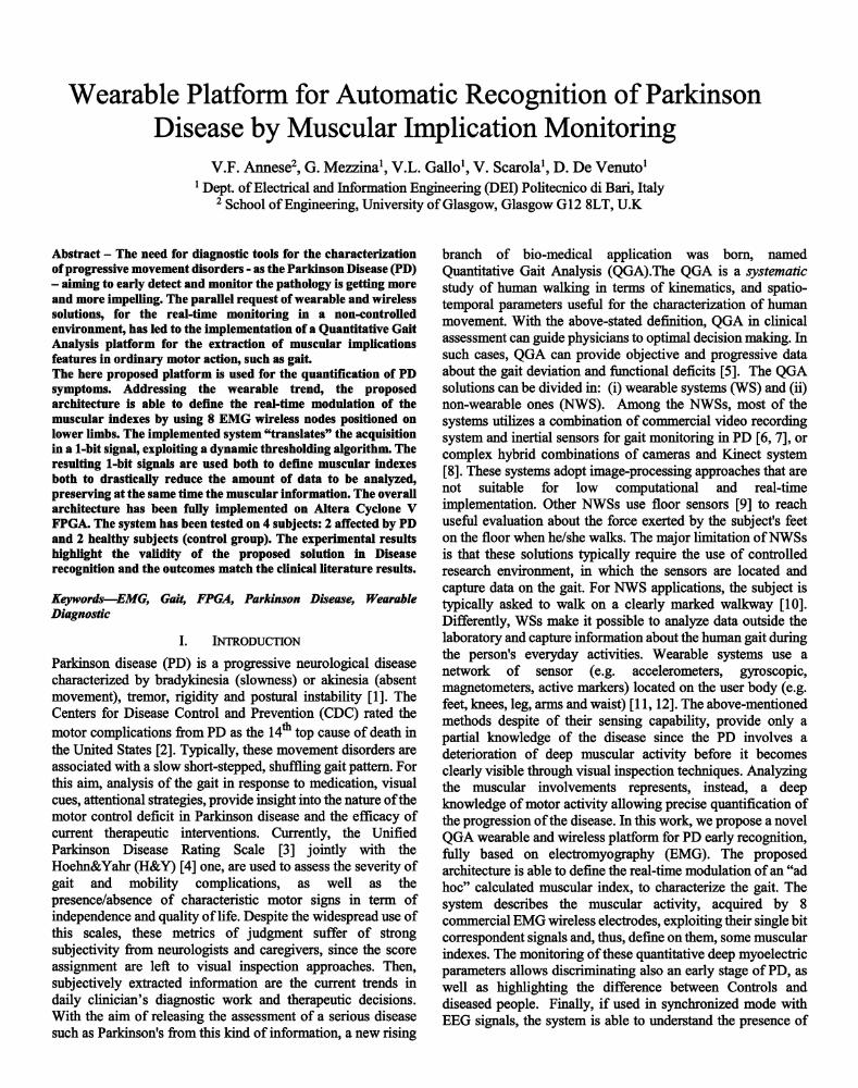
B. The Implemented SolutionThe system, outlined in Fig.l, uses 8 EMG electrodes
positioned on the lower limbs. In particular, the musclemonitored by the surface EMG are: Gastrocnemius, Tibialis,Rectus and Biceps Femoralis of both the legs. The acquiredsignals undergo to a low-pass filtering that allows only the usefulfrequency EMG spectrum: about 250Hz [18]. At the FPGAplatform front-end, the data are sampled at 500Hz with 16-bitresolution [19, 20]. The FPGA is then dedicated to the signalprocessing stage. As shown in Figure 1, the wireless body area
Fig. 1. Architecture ofthe implmented QGA system.
network sends data to the FPGA platform, which operates witha first block oftriggering (Fig.1 - ith ''Trigger System").This block aims to create a unique correspondence between a16-bit EMG sample and a Boolean value, as soon as themagnitude of the EMG represent an activation condition.Mathematically this correspondence is evaluable as in (1):
(
X l ,l XS,l): : -+ (Yl ... Ys)
Xl,16 XS,16
where the matrix XeR16,8represents the 16-bit samples presentat the same time on the 8 EMG channels. The first column ofXis the EMG sample dedicated to the first muscle. The Triggersystem converts them into a I-D vector yeR8. The generic Yielement represent the Boolean status of the first column of X.The Boolean status starts when the detection of the contractionoccurs, i.e. when the magnitude of the signal level overcomesthe learned baseline. This block is realized exploiting thedynamic thresholding algorithm proposed in our previous works[19, 21]. As widely stated in [19, 21], the EMG is stored in Msample shift-registers, which realizes 1 sec lenght acquisition(about 500 samples). The average of the samples in the M shiftregister contributes to define the threshold. Similarly, the last Nsamples (~250ms ofacquisition, and then N=128) ofthe Mones(withN~M) are used to define a local average. The local averageis compared with the threshold. The I-bit EMG signal goes '1'whenever the local average is larger than the dynamic threshold.Similarly to [19, 21], four co-contraction signals are alsogenerated. They consist in a square waveform that goes'1' whenboth agonist-antagonist I-bit signals are both high. Once the 1bit signal is generated downstream the ith ''Trigger system", 8dedicated computing blocks (one for each muscle) extract themuscular indexes, in order to recognize the cardinal PDabnormality in walking pattern. These blocks compose the"Indexes Extraction Unit" (Fig. 1) and are realized with countersdriven by the system clock. The Indexes Extraction Unitgenerates a total of32 parameters from the 8 trigger signals and4 co-contraction ones. They are: (i) 4 agonist-antagonist musclesco-contractions, (ii) 4 numbers of co-contractions during asecond of acquisition (512 samples), (iii) 8 contractions, (iv) 8relaxation times and (v) 8 step duty cycles. The first twomuscular indexes, allow compiling the UPDRS-III sectionrelated to instability. The last three parameters (contraction,relaxation and duty cycle) contribute to UPDRS Section III andN. They allow the evaluation of the bradykinesia degree (i.e.slowness or abnormal muscular hyperactivity) and the PisaSyndrome implications. In addition, they allow to objectively
151
assessing motor fluctuations In long period and the drugtreatment impact.
III. THE QGA SYSTEM LEVEL DESIGNThis proposed QGA platform algorithm has been
implemented by using VHDL coding in Quartus II softwareenvironment. The adopted hardware board is Altera Cyclone VSE 5CSEMA5F31 C6N FPGA. The design plans 8 input biosignals and 32 outputs. The inputs, coming from signalconditioning circuits and level shifter [22]. The 32 outputsvalues are functionally distributed on the available GPIO pinsand, at the same time, made available in real time on four 7segment dedicated display. The output parameters are: (i) 4 cocontraction time values defined by 11 bit (2ms time resolution 500sps) (ii) 4 co-contraction/s defined by 3 bit (lco-contr.lsresolution), (iii) 8 contractions and 8 relaxation times of 11 bit(iv) 8 duty cycles with 7 bit (1 % resolution). The global systemclock is set by an embedded PLL to 8.19209MHz driven by a50MHz oscillator. A 500Hz clock manages both the ADCsampling rate for the input EMG data and the increment of thededicated muscular counters. All the 8 EMG branches operatein parallel on FPGA [22]. Two global signals have been used asasynchronous Reset and Enable.
A. Dynamic Thresholding FPGA ImplementationThe Trigger System implementation is proposed unaltered
w.r.t. [22]. The acquired EMG samples are squared and then areused to feed two VHDL based finite state machines (FSM)designed to realize the global average, or threshold and the localone. These two FSM uses 2 dedicated RAM (512 samples and128 samples for the Threshold and the local average,respectively) in a FIFO functionality to store the new arrivedEMG data sample at the first address, and to pop out the lastsample, previously inserted. This last sample is subtracted andthe new sample is added to refresh the sum, before to divide thevalue for the register length. For instance, the sum obtained bythe Threshold FSM is divided by (512)10, while the LocalAverage FSM is divided by (128)10 The FSMs overwrite theRAM word with the new data. Finally, a 64-bit comparatorevaluates the Local Average w.r.t. the Threshold magnitude.The comparator provides a I-bit EMG Trigger, used for themuscular computing.
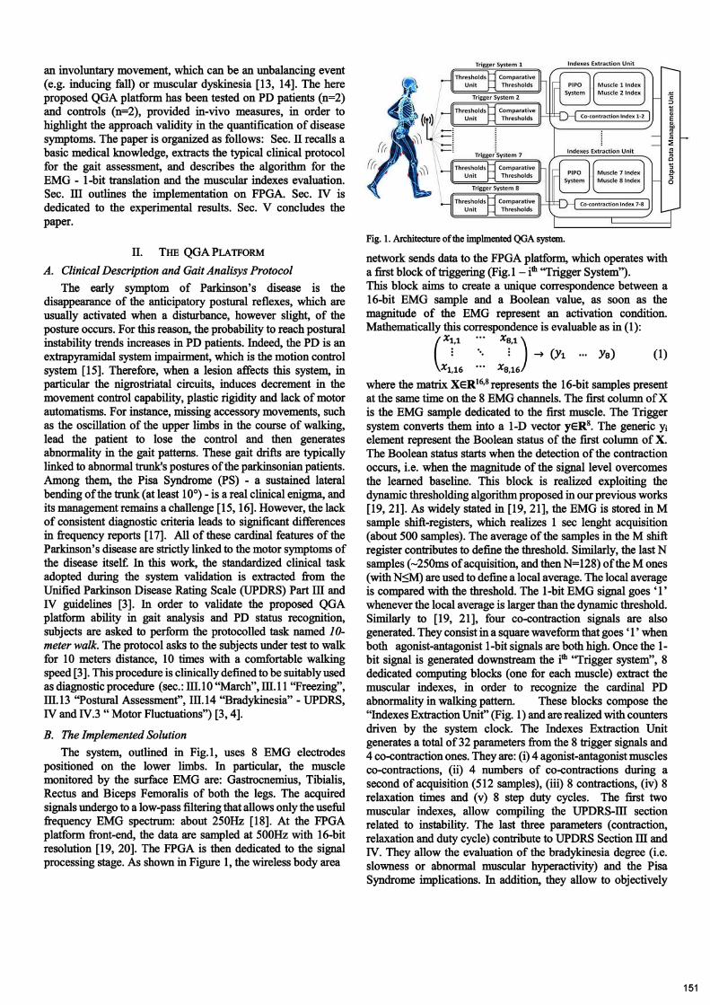
B. The MIs Computing Branch on FPGAFig. 2 schematizes the operation process of a single muscle
dedicated computing implementation. Eight similar branchesare present in the architecture, one for each monitored muscle.It operates serially with the Trigger System block, analyzing theTrigger I-bit signal. As shown in Fig.2, when the Triggergoes '1' a counter, named Contra. Counter is enabled tocount and, thus, starts increasing its value by (2)10 (due to thesampling rate), every time a CLK_500Hz positive edgeoccurs. Relaxation Counter operates with similarmodality, but is fed by the Trigger'. When the step isover, both the counters makes available the reached valueupstream a canalization system made up by the Parallel InputParallel Output (PIPO) register.
Fig. 2. Functional block diagram ofa single Muscular Index extraction unit
Indeed, the Contra Time loop counter is not reset (becausethe Trigger signal work as a count enable), allowing the valueto be available in parallel with Relax Time. The progressivebit parallel sum realizes the Step Time. When the secondTrigger positive edge arrives all the extracted indexes arefirst stored in a sequence of parallel DFF which constitute theParallel In (PI) and then canalized to the output (PO) by using2 pulses driven by the 1 bit Reg EN signal. Furthermore, adelayed version (two 8MHz Clk pulses) of Reg EN is usedto resets the Contra and Relaxation Counters,allowing the data transfer before the asynchronous reset. Afterthe Reg EN pulse on PO section, all the useful values(Contra Time and Step Time) are simultaneouslystatically present downstream from the PO, while the circuitupstream the PI section, is now reset and able to acquire. Inother words, this approach isolates the counting section,generating a static calculation section for the duty cycle (DC).In the DC section, the acquired Contra Time is firstmultiplied for (100)10 and then divided by the Step Time.The quotient in output represents the integer value of the DC(7-bit representation). The remaining of the divider block isdefined as a binary subtraction between Step Time andContra Time. It is also multiplied with (100)10 and, thus,divided for the Step Time. Ifthe quotient is higher than (50)10the DC is increased by one, otherwise it is left unaltered. Thisprocess halves the maximum error in DC assessment from 1%to 0.5%. Agonist and antagonist muscle triggers togethercontribute, through an AND gate, to generate the square cocontraction waveform. Then, similarly toContra. /Relaxation Counters, the co-contractionstime is evaluated (CoCon Time) and returns its value whenthe step - in which the co-contraction is contained - end.
IV. RESULTSThis section is dedicated to the evaluation of the
implemented QGA solution responses during a clinical walkingtest. A quantitative comparison between the PD parameters andControls ones is here proposed, aiming to emphasize the system
152
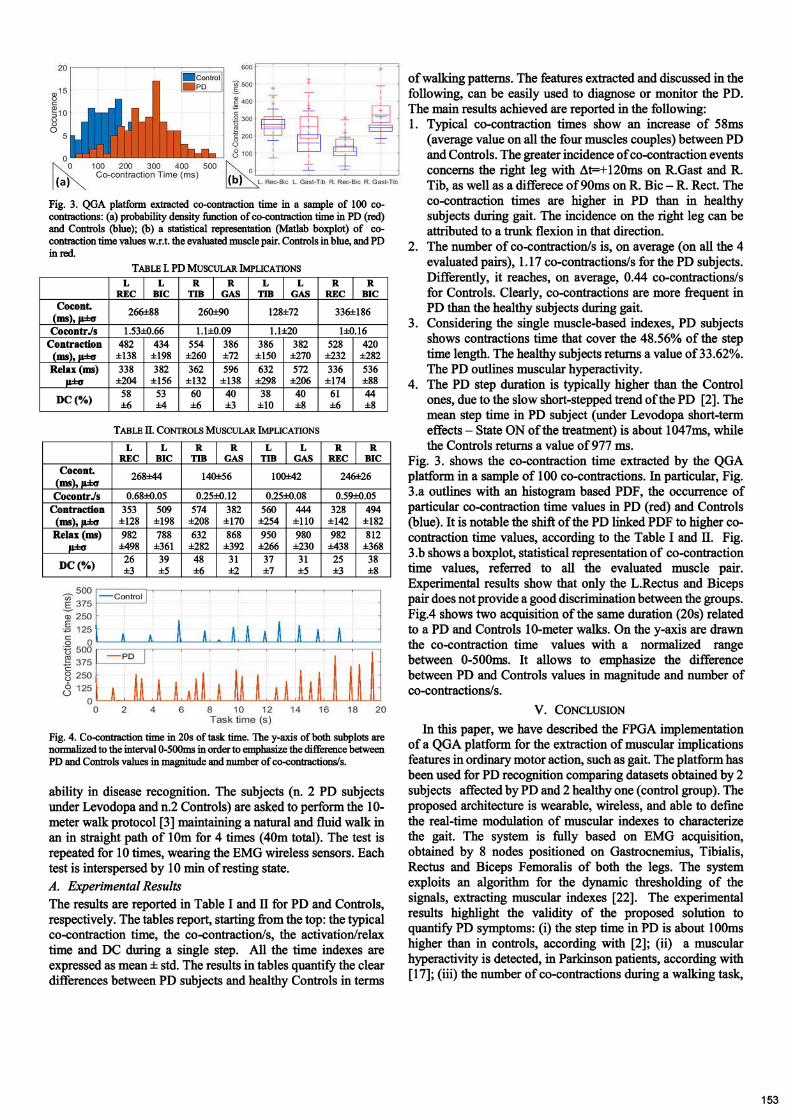
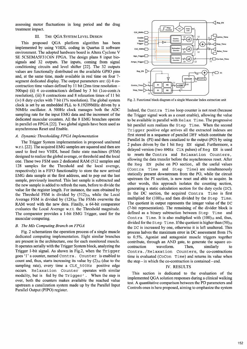
Fig. 3. QGA platform extracted co-contraction time in a sample of 100 cocontractions: (a) probability density function ofco-contraction time in PO (red)and Controls (blue); (b) a statistical representation (Matlab boxplot) of cocontraction time values w.r.t. the evaluated muscle pair. Controls in blue, and POin red.
TABLE I. PO MUSCULAR IMPLICATIONSL L R R L L R R
REC DIC TIB GAS TIB GAS REC DICCocont. 266±88 260:1:90 128±72 336±186(ms), p:I:o
CocontrJs 1.53±O.66 1.1±0.09 1.1±20 l±O.16Contraction 482 434 554 386 386 382 528 420
(ms), p.:I:o ±138 ±198 ±260 ±72 ±150 ±270 ±232 ±282Relax (ms) 338 382 362 596 632 572 336 536
p:I:o ±204 ±156 ±132 ±138 ±298 ±206 ±174 ±88
DC(%) 58 53 60 40 38 40 61 44±6 ±4 ±6 ±3 ±10 ±8 ±6 ±8
TABLE TI. CONTROLS MUSCULAR IMPLICATIONS
L L R R L L R RREC DIC TIB GAS TIB GAS REC DIC
Cocont. 268±44 140±56 100±42 246±26(ms), p.:I:oCocontrJs 0.68±O.05 0.25±O.12 0.25±O.08 0.59±O.05
Contraction 353 509 574 382 560 444 328 494(ms), p.:I:o ±128 ±198 ±208 ±170 ±254 ±110 ±142 ±182
Relax (ms) 982 788 632 868 950 980 982 812p:I:o ±498 ±361 ±282 ±392 ±266 ±230 ±438 ±368
DC(%) 26 39 48 31 37 31 25 38±3 ±5 ±6 ±2 ±7 ±5 ±3 ±8
Fig. 4. Co-contraction time in 20s of task time. The y-axis ofboth subplots arenormalized to the interval 0-50Oms in order to emphasize the difference betweenPO and Controls values in magnitude and number ofco-contractionsls.
ability in disease recognition. The subjects (n. 2 PD subjectsunder Levodopa and n.2 Controls) are asked to perform the 10meter walk protocol [3] maintaining a natural and fluid walk inan in straight path of 10m for 4 times (40m total). The test isrepeated for 10 times, wearing the EMG wireless sensors. Eachtest is interspersed by 10 min ofresting state.A. Experimental ResultsThe results are reported in Table I and II for PD and Controls,respectively. The tables report, starting from the top: the typicalco-contraction time, the co-contraction/s, the activation/relaxtime and DC during a single step. All the time indexes areexpressed as mean ± std. The results in tables quantify the cleardifferences between PD subjects and healthy Controls in terms
ofwalking patterns. The features extracted and discussed in thefollowing, can be easily used to diagnose or monitor the PD.The main results achieved are reported in the following:1. Typical co-contraction times show an increase of 58ms
(average value on all the four muscles couples) between PDand Controls. The greater incidence ofco-contraction eventsconcerns the right leg with ~t=+120ms on R.Gast and R.Tib, as well as a differece of90ms on R. Bic - R. Rect. Theco-contraction times are higher in PD than in healthysubjects during gait. The incidence on the right leg can beattributed to a trunk flexion in that direction.
2. The number of co-contraction/s is, on average (on all the 4evaluated pairs), 1.17 co-contractions/s for the PD subjects.Differently, it reaches, on average, 0.44 co-contractions/sfor Controls. Clearly, co-contractions are more frequent inPD than the healthy subjects during gait.
3. Considering the single muscle-based indexes, PD subjectsshows contractions time that cover the 48.56% of the steptime length. The healthy subjects returns a value of33.62%.The PD outlines muscular hyperactivity.
4. The PD step duration is typically higher than the Controlones, due to the slow short-stepped trend ofthe PD [2]. Themean step time in PD subject (under Levodopa short-termeffects - State ON ofthe treatment) is about 1047ms, whilethe Controls returns a value of 977 IDS.
Fig. 3. shows the co-contraction time extracted by the QGAplatform in a sample of 100 co-contractions. In particular, Fig.3.a outlines with an histogram based PDF, the occurrence ofparticular co-contraction time values in PD (red) and Controls(blue). It is notable the shift of the PD linked PDF to higher cocontraction time values, according to the Table I and II. Fig.3.b shows a boxplot, statistical representation of co-contractiontime values, referred to all the evaluated muscle pair.Experimental results show that only the L.Rectus and Bicepspair does not provide a good discrimination between the groups.Fig.4 shows two acquisition of the same duration (20s) relatedto a PD and Controls 10-meter walks. On the y-axis are drawnthe co-contraction time values with a normalized rangebetween 0-500ms. It allows to emphasize the differencebetween PD and Controls values in magnitude and number ofco-contractions/so
v. CONCLUSION
In this paper, we have described the FPGA implementationof a QGA platform for the extraction of muscular implicationsfeatures in ordinary motor action, such as gait. The platform hasbeen used for PD recognition comparing datasets obtained by 2subjects affected by PD and 2 healthy one (control group). Theproposed architecture is wearable, wireless, and able to definethe real-time modulation of muscular indexes to characterizethe gait. The system is fully based on EMG acquisition,obtained by 8 nodes positioned on Gastrocnemius, Tibialis,Rectus and Biceps Femoralis of both the legs. The systemexploits an algorithm for the dynamic thresholding of thesignals, extracting muscular indexes [22]. The experimentalresults highlight the validity of the proposed solution toquantify PD symptoms: (i) the step time in PD is about lOOmshigher than in controls, according with [2]; (ii) a muscularhyperactivity is detected, in Parkinson patients, according with[17]; (iii) the number of co-contractions during a walking task,
153
is higher in PD than in Control group [1]. The solution aims tobe a diagnostic tool for the early detection ofthe disease, as wellas a useful clinical support tool for monitoring the therapyimpact. Future perspectives include the design and fabricationof an Application Specific Integrated Circuit (ASIC) [23-30],the use of biocompatible and flexible electronics in order toincrease the wearability degree of the system [31, 32] and theoptimization of the wireless network [33-36].
REFERENCES[1] de Lau LM, Breteler MM (June 2006). "Epidemiology of Parkinson's
disease". Lancet Neurol. 5 (6): 525-35.[2] Online Website: https://www.cdc.gov/[3] Movement Disorder Society Task Force on Rating Scales for Parkinson's
Disease. "The Unified Parkinson's Disease Rating Scale (UPDRS): statusand recommendations." Movement disorders: official journal of theMovement Disorder Society 18.7 (2003): 738.
[4] Goetz, Christopher G., et al. "Movement Disorder Society Task Forcereport on the Hoehn and Yahr staging scale: status and recommendationsthe Movement Disorder Society Task Force on rating scales forParkinson's disease." Movement disorders 19.9 (2004): 1020-1028.
[5] Chen M., Huang B. Intelligent shoes for abnormal gait detection.Proceedings ofthe International Conference on Robotics and Automation;Pasadena, CA, USA. 19-23 May 2008; pp. 2019-2024.
[6] Sofuwa, Olumide, et al. "Quantitative gait analysis in Parkinson's disease:comparison with a healthy control group." Archives ofphysical medicineand rehabilitation 86.5 (2005): 1007-1013.
[7] BTS Bioengineering (accessed on 1/05/2017). Online:http://www.btsbioengineering.com/products/integrated-solutions/btsgaitlab/
[8] Stone, Erik E., and Marjorie Skubic. "Passive in-home measurement ofstride-to-stride gait variability comparing vision and Kinect sensing."Engineering in Medicine and Biology Society, EMBC, 2011.
[9] Middleton, Lee, et al. "A floor sensor system for gait recognition."Automatic Identification Advanced Technologies, 2005. Fourth IEEEWorkshop on. IEEE, 2005.
[10] Muro-de-Ia-Herran, Alvaro, Begonya Garcia-Zapimin, and AmaiaMendez-Zorrilla. "Gait analysis methods: An overview of wearable andnon-wearable systems, highlighting clinical applications." Sensors 14.2(2014): 3362-3394.
[11] Klucken J, Barth J, Kugler P, Schlachetzki J, Henze T, Marxreiter F, et al.(2013) Unbiased and Mobile Gait Analysis Detects Motor Impairment inParkinson's Disease. PLoS ONE 8(2): e56956.
[12] Griffiths, R. I., et al. Automated assessment of bradykinesia anddyskinesia in Parkinson's disease. J Parkinsons Dis 2 (1): 47-55. doi:10.3233. JPD-2012-11071, 2012.
[13] De Tommaso, M., Vecchio, E., Ricci, K., Montemurno, A., De Venuto,D., Annese, V.F. "Combined EEG/EMG evaluation during a novel dualtask paradigm for gait analysis." (2015) Proceedings - 2015 6th IEEEInternational Workshop on Advances in Sensors and Interfaces, IWASI2015, art. no. 7184949,pp. 181-186. DOl: 10.1109/IWASI.2015.7184949
[14] De Venuto, D., Annese, V.F., Ruta, M., Di Sciascio, E., SangiovanniVincentelli, A.L. "Designing a Cyber-Physical System for Fall Preventionby Cortico-Muscular Coupling Detection." (2016) IEEE Design and Test,33 (3), art. no. 7273831, pp. 66-76. 001: 10.1109/MDAT.2015.2480707
[15] Ekbom K., Lindholm H., Ljungberg L. New dystonic syndromeassociated with butyrophenone therapy. Zeitschrift fUr Neurologie.1972;202(2):94-103. doi: 10.1007/BF00316159.
[16] Doherty K. M., van de Warrenburg B. P., Peralta M. C., et al. Posturaldeformities in Parkinson's disease. The Lancet Neurology.2011;10(6):538-549. doi: 10.1016/s1474-4422(11)70067-9.
[17] Castrioto A., Piscicelli C., Perennou D., Krack P., Debft B. Thepathogenesis of Pisa syndrome in Parkinson's disease. MovementDisorders. 2014;29(9):1100-1107. doi: 10.1002/mds.25925.
[18] Basmajian, John V., and Carlo J. De Luca. Muscles alive: their functionsrevealed by electromyography. Williams & Wilkins, 1985. pp. 90-93
[19] Annese, V.F., De Venuto, D. ''The truth machine of involuntarymovement: FPGA based cortico-muscular analysis for fall prevention"(2016) 2015 IEEE International Symposium on Signal Processing andInformation Technology, ISSPIT 2015, art. no. 7394398, pp. 553-558.DOl: 10.1109/ISSPIT.2015.7394398.
[20] Annese, V.F., De Venuto, D. "FPGA based architecture for fall-riskassessment during gait monitoring by synchronous EEG/EMG." (2015)Proceedings - 2015 6th IEEE International Workshop on Advances in
Sensors and Interfaces, IWASI 2015, art. no. 7184953, pp. 116-121. DOl:10.1109/IWASI.2015.7184953
[21] V. F. Annese, M. Crepaldi, D. Demarchi and D. De Venuto, "A digitalprocessor architecture for combined EEG/EMG falling risk prediction,"2016 Design, Automation & Test in Europe Conference & Exhibition(DATE), Dresden, 2016, pp. 714-719.
[22] D. De Venuto, D. T. Castro, Y. Ponomarev and E. Stikvoort, "Low power12-bit SAR ADC for autonomous wireless sensors network interface,"2009 3rd International Workshop on Advances in sensors and Interfaces,Trani, 2009, pp. 115-120. doi: 10.1109/IWASI.2009.5184780
[23] De Venuto, D., Annese, V.F., Mezzina, G. "An Embedded SystemRemotely Driving Mechanical Devices by P300 Brain Activity".Proceedings of the 2017 Design, Automation and Test in EuropeConference and Exhibition, DATE 2017. ISBN: 978-3-9815370-8-6.
[24] De Venuto, D., Annese, V.F., Mezzina, G. "Remote Neuro-CognitiveImpairment Sensing based on P300 Spatio-Temporal Monitoring". IEEESensors Journal, PP (99), art. no. 7562544. DOl:10.1109/JSEN.2016.2606553.2016.
[25] Annese, V.F., Mezzina, G., De Venuto, D. ''Towards mobile health care:Neurocognitive impairment monitoring by BCI-based game".Proceedings of IEEE Sensors, art. no. 7808745. DOl:10.1109/ICSENS.2016.7808745. 2017.
[26] De Venuto, D., Annese, V.F., Mezzina, G., Ruta, M., Sciascio, E.D."Brain-computer interface using P300: A gaming approach forneurocognitive impairment diagnosis". 2016 IEEE International HighLevel Design Validation and Test Workshop, HLDVT 2016, art. no.7748261, pp. 93-99. DOl: 10.1109/HLDVT.2016.7748261. 2016.
[27] Annese, V.F., De Venuto, D. "Gait analysis for fall prediction using EMGtriggered movement related potentials". Proceedings - 2015 10th IEEEInternational Conference on Design and Technology of IntegratedSystems in Nanoscale Era, DTIS 2015, art. no. 7127386. DOl:10.1109/DTIS.2015.7127386.2015.
[28] Annese, V.F., De Venuto, D. "Fall-risk assessment by combinedmovement related potentials and co-contraction index monitoring". IEEEBiomedical Circuits and Systems Conference: Engineering for HealthyMinds and Able Bodies, BioCAS 2015 - Proceedings, art. no. 7348366.DOl: 10.1109/BioCAS.2015.7348366. 2015.
[29] De Venuto, D., Annese, V.F., Sangiovanni-Vincentelli, A.L. ''Theultimate loT application: A cyber-physical system for ambient assistedliving". Proceedings - IEEE International Symposium on Circuits andSystems, 2016-July, art. no. 7538979, pp. 2042-2045. DOl:10.1109/ISCAS.2016.7538979.2016.
[30] De Venuto, D., Annese, V.F., Defazio, G., Gallo, V.L., Mezzina, G. "GaitAnalysis and Quantitative Drug Effect Evaluation in Parkinson Diseaseby Jointly EEG-EMG Monitoring". Proceedings - 2017 12th IEEEInternational Conference on Design and Technology of IntegratedSystems in Nanoscale Era, DTIS 2017.
[31] Annese, V.F., De Venuto, D., Martin, C., Cumming, D.R.S."Biodegradable pressure sensor for health-care". 2014 21st IEEEInternational Conference on Electronics, Circuits and Systems, ICECS2014, art. no. 7050056, pp. 598-601. DOl:10.1109/ICECS.2014.7050056.2014.
[32] Annese, V.F., Martin, C., Cumming, D.R.S., De Venuto, D. ''Wirelesscapsule technology: Remotely powered improved high-sensitivebarometric endoradiosonde". Proceedings - IEEE InternationalSymposium on Circuits and Systems, 2016-July, art. no. 7527504, pp.1370-1373. DOl: 10.1109/ISCAS.2016.7527504. 2016.
[33] Annese, V.F., De Venuto, D. "On-line shelf-life prediction in perishablegoods chain through the integration of WSN technology with a 1st orderkinetic model". 2015 IEEE 15th International Conference onEnvironment and Electrical Engineering, EEEIC 2015 - ConferenceProceedings, art. no. 7165232, pp. 605-610. DOl:10.1109/EEEIC.2015.7165232.2015.
[34] Annese, V.F., Biccario, G.E., Cipriani, S., De Venuto, D. "Organolepticproperties remote sensing and life-time prediction along the perishablesgoods supply-chain". Proceedings of the International Conference onSensing Technology, ICST, 2014-January, pp. 130-135. 2014.
[35] De Venuto, D., Annese, V. F., de Tommaso, M., Vecchio, E., andVincentelli, A. S. "Combining EEG and EMG signals in a wireless systemfor preventing fall in neurodegenerative diseases." Ambient AssistedLiving. Springer International Publishing. pp 317-327. 2015.
[36] Biccario, G.E., Annese, V.F., Cipriani, S., De Venuto, D. "WSN-basednear real-time environmental monitoring for shelf life prediction throughdata processing to improve food safety and certification" ICINCO 2014 Proceedings of the 11th International Conference on Informatics inControl, Automation and Robotics, 1, pp. 777-782. 2014.
154