Embed Size (px)
Citation preview


NIVEDI - Annual Report 2016-17 i
Annual Report2016-17
ICAR-National Institute of Veterinary Epidemiology and Disease Informatics (NIVEDI)
Post Box No. 6450, Yelahanka, Bengaluru-560064, Karnataka, IndiaPh: +91 80 23093100; 23093110; 23093111, Fax: +91 80 23093222
Email: [email protected] Website: www.nivedi.res.in

NIVEDI - Annual Report 2016-17ii
Editors:
Dr. P.P. Sengupta Dr. V. BalamuruganDr. G. GovindarajDr. Mohd. Mudassar Chanda Dr. Jagadish HiremathDr. Yogishadhya RDr. Awadhesh Prajapati
Cover Front Page: Glimpses of activities and achievements in 2016-17Cover Back Page: Pie Diagram showing distribution of Culicoides flies in Bannergatta National Park, Bengaluru, Karnataka
© ICAR-National Institute of Veterinary Epidemiology and Disease Informatics (NIVEDI)All rights reserved
Printed at:CNU Graphic PrintersMalleshwaram, Bengaluru - 560 003Mobile : 9880 888 399

NIVEDI - Annual Report 2016-17 iii
Contents Page No.Executive Summary 1
About NIVEDI 4Institute Research Projects
Development of Geographic Information System (GIS) enabled Early Warning System (EWS) for detection of Avian influenza infection using Remote Sensing (RS)
7
Identification of ecological risk factors for occurrence of Anthrax in India 8Molecular epidemiology of MRSA, MR-CoNS and ESBL producing Gram-negative bacteriain Animals including their environment
8
Epidemiology of Haemorrhagic Septicemia in livestock vis-à-vis Foot and Mouth Disease in India 9Epidemiology of Haemorrhagic Septicaemia in India 10Epidemiology and impact analysis of sheep and goat pox 11Disease severity pattern and risk factors identification for PPR in sheep and goats in India 12Epidemiology of porcine reproductive and respiratory syndrome in India 12Epidemiological study of surra and fasciolosis in animals 13Retrospective Epidemiological studies on HPAI with reference to spatio-temporal pattern and the probable associated risk factors identification
14
Institute Service ProjectsNational Animal Disease Referral Expert System (NADRES) 17Maintenance and updating of livestock serum repository 18Seroepidemiology of Brucellosis 20Seroepidemiology of Infectious Bovine Rhinotracheitis (IBR) in India 22
Externally Funded ProjectsICAR Projects Outreach Programme on Zoonotic Diseases 25All India Network programme on Bluetongue 26National Innovations on Climate Resilient Agriculture (NICRA) - Livestock disease Surveillance in relation to weather data and emergence of new pathogens
27
All India network project on Gastro Intestinal Parasitism (GIP) 28ICAR-Extramural ProjectsEvaluation of vaccine effectiveness and identification of the factors that affect field level vaccine efficacy of the vaccines against diseases under control program
29
Understanding the epidemiology of Culicoides borne diseases (CBD’s) in wild and domestic ruminants 30Impact assessment of PPR vaccine technology in India 31DBT Network Projects on BrucellosisDBT Network Project on Brucellosis: Project Monitoring Unit (PMU) 31DBT Network Project: Brucellosis Epidemiology ( BE-1) 32DBT Network Project: Brucellosis Diagnostics (BD-2) 33DBT ProjectExternal validation of diagnostic assays for detection of anti brucella antibodies developed under the DBT-network project on brucellosis
33
DBT-Programe for NER on Advanced Animal Disease Diagnosis and Management Consortium (ADMaC)Surveillance and molecular analysis of MRSA, MR-CoNS, VRE, ESBL and Carbapenemase Producing Gram-Negative bacteria in farm animals, the animal handlers and livestock products in India
34
Sero-epidemiological study of brucellosis in livestock in North East states of India using ELISA and Fluorescent Polarization Assays
35

NIVEDI - Annual Report 2016-17iv
Contents Page No.Epidemiological study of Classical swine fever (CSF), Porcine reproductive and respiratory syndrome (PRRS) and Porcine torqueteno (TTV) in North East (NE) region of India
36
Development of Infectious Disease Information System (IDIS) and Risk assessment models for Transboundary animal diseases (TAD) & other emerging livestock diseases in NE region of India
37
DBT-Programme for NE Region -Twinning ProjectsSero-Surveillance, molecular characterization and epidemiology of pox viral infections in animals from North Eastern Region of India
38
Aetio-pathology and molecular epidemiology of bacterial and viral diseases associated with the respiratory problems of yak in the North Eastern states of India
38
Serosurveillance and molecular epidemiology of Bovine Herpes Virus 1 (BoHV1) infection in bovines of North Eastern states of Mizoram, Meghalaya and Tripura
39
Serosurveillance, isolation and molecular characterization of bluetongue virus in sheep and goats of Tripura and Assam states
40
NFDB ProjectNational Surveillance Programme for Aquatic Animal Diseases (NSPAAD) 41Grant –In-Aid Project from DADFBrucellosis Control programme 41International collaborative project- DBT-BBSRC, UK Development of diagnostic system, reference collection and molecular epidemiology studies for important arboviral pathogens of livestock in India
42
AICRP on ADMAS 44Tribal Sub-Plan 45
Mera Gaon Mera Gaurav 46Swachh Bharat Abhiyan 47
PublicationPeer reviewed Journals 51Presentation in Conferences/Symposia/Seminars/other Fora 54Book/Book Chapters/Technical bulletins/Training Manuals/SOP’s/Popular Articles 60
Capacity Building and Human Resource Development Training/Refresher Course/Summer/Winter school/Seminars/Conferences/Symposia/Workshops/Programmes organized
62
Training/Refresher Course/Summer/Winter school/Seminars/Conferences/Symposia/Workshops/Programmes Participated
63
Awards/Fellowship/Recognition 69Miscellaneous
Quinquennial Review Team 73Research Advisory Committee 74Institute Research Committee 74Institutional Animal Ethics Committee 75Institute Management Committee 75Distinguished Visitors 76Staff position during 2016-2017 77Budget 79
NIVEDI activities 83

NIVEDI - Annual Report 2016-17 v
ACKNOWLEDGEMENT
The Director and staff of ICAR-National Institute of Veterinary Epidemiology and Disease Informatics (NIVEDI) express their whole-hearted gratitude to Dr. Trilochan Mohapatra, Secretary, DARE and Director General, ICAR for his constant guidance and support.
ICAR-NIVEDI family is also thankful to Dr. J.K. Jena, Deputy Director General (Animal Science) (Acting) Dr. H. Rahman, Former Deputy Director General (Animal Science) and Dr. Ashok Kumar, Assistant Director General (Animal Health) for cooperation, encouragement and generous support.
Our sincere thanks are also due to the Directors and Heads of ICAR institutes located in Bengaluru, viz., ICAR-NIANP, ICAR-NBAIR, ICAR-IVRI, ICAR-NBSSLUP, ICAR-NDRI, ICAR-IIHR, KVAFSU, Karnataka Veterinary Council, Director I-AIM, Yelahanka, Bengaluru and also other institutes and organisations for their vital logistics support and co-operation from time to time.
The institute conveys sincere thanks to all the principal investigators of AICRP on ADMAS and related State Animal Husbandry Departments and Universities for their valuable inputs and cooperation. At the last, I sincerely thank all the staff members of ICAR-NIVEDI for their timely cooperations.
‘Jai Kisan Jai Vigyan’Jai Hind!
( Parimal Roy ) Director

NIVEDI - Annual Report 2016-17vi

NIVEDI - Annual Report 2016-17 1
EXECUTIVE SUMMARYDuring the year 2016-17, three new external projects have been initiated. This year, as per the NADRES report, Foot rot, Hemorrhagic Septicemia (HS), Black Quarter (BQ), Enterotoxaemia (ET), Glanders and Anthrax were recorded as the predominant bacterial diseases whereas Peste des Petits Ruminants (PPR), Capripox, Bluetongue (BT), Rabies and Classical Swine Fever (CSF) were reported as major viral diseases, and Fasciolosis, Amphistomosis, Anaplasmosis, Babesiosis, Coccidiosis and Trypanosomosis were found most frequently reported parasitic diseases. The monthly forewarning information were sent to DADF and other state animal husbandry departments two months in advance to take up suitable preventive measures.
During this period, 32 blood samples, 16 nasal swabs and 30 tissue samples from heart, liver, spleen, bone marrow from large and small ruminants belonging to Odisha, Madhya Pradesh and Karnataka were collected during outbreak investigation and further screened for presence of Pasteurella multocida by conventional methods like post mortem lesion and polymerase chain reaction (PCR) assay. A total of 8 samples were found positive by PCR. N-terminal gene of P. multocida encoding for NanB-Nt protein (~94 kDa) was expressed and characterized by SDS-PAGE and further it will be evaluated for its diagnostic potential. Two whole genome sequence, of P. multocida strain and M. haemolytica isolated from Yak were done and reported first time from India. A total of 157 Yak nasal samples were analysed and M. haemolytica (17.9%), P. multocida (3.9%) and H Somnil (2.5%) were detected. A multiplex PCR was optimized for detection of Mannheimia haemolytica and P. multocida in bovine clinical samples. Study on epidemiology of HS in livestock vis-à-vis Foot and Mouth Disease (FMD) in India revealed the high cumulative outbreaks of HS than FMD in Gujarat and FMD than HS in West Bengal. In both the states a decreasing pattern was noticed for both the diseases. In Gujarat, HS showed high case fatality rate (CFR) than FMD. In FMD vaccine effectiveness study, it was found that age & breed significantly affect the vaccine effectiveness of FMD vaccine in Karnataka.
A total 113 clinical environmental samples were collected from Odisha for screening of Bacillus
antharacis 14 samples from cattle, elephant and pigs showed positive by standard laboratory technique. Further, risk factor analysis showed Kolar, Tumkur, Hassan, Mysuru, Chamarajanagara, Raichur, Belagavi, Gadag, Koppal and parts of Mandya were at high risk for anthrax.
A multi-epitope recombinant protein antigen of Brucella abortus was designed, synthesized cloned in the vector and expressed in addition to expression of individual immuno dominant proteins BP26, BLS, SodC, VirB12 etc. Further these recombinant proteins are to be evaluated in single or cocktail as diagnostic antigen in ELISA for bovine brucellosis. During this period, 4767 random serum samples received from 18 AICRP centers were screened for brucellosis. Among livestock species screened, high prevalence was observed in pig (40.19%) followed by sheep (20.82%), cattle (2.73%) goat (2.18%) and buffalo (1.11%). An overall prevalence of 7.69% was observed among all species. During milk surveillance for brucellosiss, 1986 milk samples from Kolar and Chickballapur were screened by Milk Ring Test (MRT) which showed 5.06% and 6.36% prevalence respectively. In house developed Protein G ELISA kit was validated against commercial Priocheck BrucellaAb 2.0 iELISA kit and it showed 95 % agreement which clearly indicated high sensitivity and specificity of in house developed kit. Fluorescent Polarization Assay (FPA) for sero-monitoring of brucellosis in livestock was developed and validated and was transferred to ADMaC core Lab-I to generate the geo-epidemiological data on brucelloisis in North Eastern States.
The extended spectrum beta lactamase producing multi drug resistant Escherichia coli isolated from subclinical mastitis milk was identified and characterized. The multidrug resistant Proteus mirabilis carrying multiple efflux pumps detected in apparently healthy pig fecal sample. Escherichia coli strain SCM-21 and Proteus mirabilis strain NIVEDI3-PG74 whole genome shotgun sequencing was done. The study concludes that the environmental variables are very crucial to study the epidemiology of pathogen and disease prediction to implement timely control measures to the present scenario of global climate change. Out of 96 human clinical isolates four were found positive for E.coli.

NIVEDI - Annual Report 2016-172
On virulence gene analysis, 50 E. coli isolates showed positive traT and six for cnf1 gene.
Out of 119 human samples of PUO cases 41.17% shown sero positivity for leptospirosis representatively of major of Leptospira serovars like Hurstbridge, Tarassovi, Javanica, Bataviae Pyrogenes, Shermani, Icterohaemorrhagiae, Kaup, Hardjo, etc., Out of a total of 1116 cattle samples, 617 showed seropositivity in MAT at 1:100 titre with 18 reference leptospira serovars. Out of 43 rat samples from Nagpur, 79.06% showed positive by MAT with serovars like Grippotyphosa, Hurstbridge, Australis, Tarassovi, Shermani, Bataviae, Bankinang, Canicola, Djasiman. Out of 297 human serum samples from Kerala, Karnataka and Dadra and Nagar Haveli, 55 samples were positive for IgG antibody in of Toxoplasma gondi.
The outbreak data of Avian Influenza (AI) from 2006 to 2015 were collected and analyzed with related risk factors namely Normalized Difference Vegetation Index (NDVI), Normalized Difference Water Index (NDWI), Normalized Difference Moisture Index (NDMI) and Land Surface Temperature (LST), rainfall and other Anthropometric variable like distance from highways and rivers water-bodies and Poultry population density was found Nadia, Malda, Dakshin Dinajpur, and Purulia in WB and Nandurbar in Maharashtra. Spatial heat map of AI outbreaks in eastern and north eastern states from 2008-10 plotted. Economic impact of AI outbreaks to hatcheries, commercial poultry farms/duck farms, traditional duck farmers, tourist boat industry in Kerala during 2014 was carried out.
The economic loss due to pox in sheep and goat was estimated based on the data derived from secondary sources, and certain assumptions. It revealed that, the total loss estimated due to sheep and goat pox was INR 480.72 crore, 961.34 crore and INR 1442.05 crore at assumed 1%, 2% and 3% annual incidence levels, respectively. The PPR clinical score card was used for assessing the severity of disease pattern in sheep and goats like very mild, mild, moderate, severe, and very severe. Further, it was revealed that the outbreaks of the diseases remains in mild to severe form and mild to moderate form in the places nearby regularly vaccinated whereas the severe form of the disease occurred in the places where the vaccination was not conducted. In another survey in Karnataka it was observed that incidence was 8.31%. The estimated mortality loss, cost of treatment, distress sale and opportunity cost of labor among the infected flock
was INR 3231, INR 108.2, INR 3040, and INR 15.7 per animal, respectively. A questionnaire has been developed to assess the risk factors for occurrence of Porcine Reproductive and Respiratory Syndrome (PRRS) using different variables. A total of 62 sero-prevalence samples were screened for PRRSV by RT-PCR, out of which 22 samples were found positive. Clinical samples received from different regions of India were screened by PCR and out of the total of 61 clinical samples, 3/20 from Sikkim, 1/2 from Kerala, 1/2 from Telangana, 3/10 from Karnataka, 2/27 from Madhya Pradesh were found positive for Torque Teno Virus (TTSuV) infection.
Out of 2008 serum samples from NER states screened for the prevalence of CSFV antibodies and showed 20.54% prevalence in Assam, 37.13% in Mizoram, 21.3% in Meghalaya, 44.07% in Manipur, 36.11% in Sikkim and 10.57% in Tripura. Meta analysis of ten CSFV study data from seven states of NER with sample of 1323, the seoprevalance of CSFV was estimated as 31% and same for BT was estimated as 35% when Meta analysis study was done with samples size of 460.
Three multiplex PCR were developed for detection of BTV serotypes 5 & 9, 3, 13 & 16 and 10 & 24 were developed, optimized and specificity was confirmed by nucleotide sequencing. Blood samples from different parts of states were collected and isolation of virus was done. The predominant isolates were belonged to BTV 1, 2, 3, 4, 5, 9, 16, 23, and 24. A total of 411 serum samples from small ruminants were received from the NE states like Assam, Mizoram, Manipur, Meghalaya, Nagaland and Sikkim. On screening for the presence of BT antibody, 88 samples found positive by C-ELISA kit. Highest prevalence found in Manipur where as the lowest was found in Assam. In another study 8000 Culicoides specimens from 31 sites near to wildlife sanctuaries were entrapped using CDC light traps with UV light for a period of eight months. Among them 13 major Culicoides species were found. The selected Culicoides species were DNA bar coded and BTV was detected in C.oxystoma and C.imicola samples. Results of study indicate that there is a circulation of Culicoides species in forest habitats and presence of BTV in some Culicoides species.
During this period, 9923 bovine sera samples from different states of India screened for the IBR antibodies using Avidin-Biotin ELISA and Chattisgarh showed highest prevalence rate with 50.54% whereas Nagaland showed least with 6.80%. A total of 16 isolates Bovine

NIVEDI - Annual Report 2016-17 3
Herpes Virus (BoHV) were received from Karnataka, Orissa, West Bengal and UP and maintained in virus repository of NIVEDI. In another study 2418 bovine serum samples NE states were screened for the presence of IBR antibodies by avidin biotin ELISA and an overall 33.79% positive was found. 59 bovine blood samples were screened by PCR for presence of BOHV-1 and 10 samples were found positive.
A total of 2077 Lymnaea spp. snails were collected from 25 water bodies covering 11 districts of Karnataka and screened for infection of F. gigantica by PCR targeting ITS 2 region. It was found that 5.1% of snails were found to be positive for F. gigantica. Deccan plateau of Karnataka showed highest prevalence with 5.79% followed by 5.22% in Western Ghats and 3.95% in Coastal region. Districts wise, Ramanagara showed highest infection rate with7.8% and Dakshina Kannada least infection (3.09%). Seasonal prevalence of infection was found to be more in winter (6.22%) followed by the rainy (4.61%) and summer (4.35%) seasons
respectively. In another study, 968 bovine samples from Tamil Nadu, Madhya Pradesh, Karnataka, Bihar and Andaman & Nicobar isoland, Himachal Pradesh, Uttar Pradesh, Sikkim, Odisha, Assam, Mizoram and Puducherry were screened for the prevalence of Trypanosoma evansi antibodies using recombinant VSG based indirect ELISA and 732 (75.62%) animals showed positive. Puducherry showed highest (94.5%), whereas Odisha showed lowest (25.58%), prevalence.
During 2016-17 a total of 16824 serum samples were received from different states, were catalogued and aliquoted. The sera samples were screened for brucellosis, IBR, CSF, BT and trypanosomosis.
Three MoU has been made between ICAR-NIVEDI and Other institutes (Department of Biosciences and Bioengineering, IIT, Guwahati, College of Veterinary Sciences & Animal Husbandry, Bhubaneswar, Odisha, OUAT and SKUAST-Kashmir FVSc & AH) for carrying out collaborative research.

NIVEDI - Annual Report 2016-174
ABOUT ICAR-NIVEDIICAR-NIVEDI had its humble beginning as AICRP on ADMAS in 1987, upgraded to PD-ADMAS in 2000 and finally in the year 2013 the institute was rechristened as ICAR-National Institute of Veterinary Epidemiology and Disease Informatics (ICAR-NIVEDI). The coordinating units of AICRP-ADMAS continued to grow in number from 4 co-ordinating units during 1987 to 32 at present. ICAR-NIVEDI is a pioneering institute working with the mandate of R&D in the field of veterinary epidemiology and disease informatics. Its role is significant in developing disease models, risk analysis, animal disease forecasting & forewarning, need based diagnostics and analysis of disease economic impact. The institute has developed various technologies and patented few products which are being utilized by different stakeholders of the country. The role of this institute in the eradication of Rinderpest from India and development of National Animal Disease Referral Expert System (NADRES), interactive software for animal disease forecasting are noteworthy. The institute has been conducting plethora of training programmes related to epidemiology, economic impact, research methodologies, sampling frame and disease diagnosis for various stakeholders associated with animal health as part of capacity building in the area. The efforts of ICAR-NIVEDI have been appreciated and recognized by various organizations by conferring international and national awards and fellowships.
ICAR-NIVEDI plays a significant role by delivering many innovative solutions and services in the form of improved diagnostic techniques, animal disease forecasting and forewarning models, animal disease economic impact analysis and capacity building in animal disease epidemiology in the country. The institute is working on the following vision, mission, focus and mandates :
VisionAchieving freedom from animal diseases, animal welfare, food and nutritional security through healthy foods of animal origin, poverty alleviation and economic growth of rural India.MissionCapacity building in frontier areas of Veterinary Epidemiology dynamics of animal diseases including zoonoses and animal healthcare intelligence.Focus
� Improving disease monitoring and surveillance through development of pen side diagnostics
� Risk assessment for occurrence of economically important animal diseases
� Adapting strategies to improve animal disease data quality
� Understanding the threat from animal diseases in the background of climate change and globalization
� Developing early warning system and disease modelling/ forecasting
� Understanding economic impacts of animal diseases and the management strategies
� Promoting innovations and improving human resource capacity
Mandate of ICAR-NIVEDI � Epidemiology, informatics and economics of
animal disease including zoonoses � Surveillance, forecasting and forewarning
for management of animal diseases including zoonoses
� Repository and capacity development Mandate of AICRP on ADMAS
� Strengthening of National Livestock Serum Repository
� Effective updating of NADRES with active disease data climatic and non-climatic risk factors
� Surveillance of diseases/pathogens of companion, lab and wild animals
� Analysis on economic losses due to animal diseases and the control measures adopted for their management
� Sero-monitoring of animal diseases based on sample frame
� Investigation of endemic, emerging and reemerging animal disease outbreaks using innovative technologies
NIVEDI - Annual Report 2016-17 5
INSTITUTE RESEARCH PROJECTS

NIVEDI - Annual Report 2016-176

NIVEDI - Annual Report 2016-17 7
IPC: ANSCNIVEDISIL201500500068 Project ID: IXX12238
Development of Geographic Information System (GIS) enabled Early Warning System for Avian Influenza (AI)
infection using Remote Sensing (RS)
K P Suresh, M M Chanda, R Sridevi (NIVEDI) and S Nagarajan (NISHAD)
The aim was to study the spatial epidemiology of a disease remote sensing data and mapping. The disease outbreak data for AI was collected for the time period of 2006 to 2015. The variables of AI considered for the study are remote sensing variables. Normalized Difference Vegetation Index (NDVI), Normalized Difference Water Index (NDWI), Normalized Difference Moisture Index (NDMI) and Land Surface Temperature (LST), Meteorological variables, Anthropometric environmental variables and Management risk variables. Risk variables such as NDVI, LST, rainfall, Air temperature and other anthropogenic variables like distance from major cities, railways and highways, distance from nearest water-bodies like lakes and rivers, etc, Poultry density and Population density were generated using remote sensing. The Poisson Regression model was used in ArcMap for risk map generation. It was observed that environmental variables and their interactions were significantly associated with incidence of AI.
The high risk of disease occurrence is predicted in Jalgaon, Murshidabad and Birbhum regions and was represented in Red colour and followed by Nadia, Maldah, Dakshin Dinajpur, Nandurbar and Purulia represented in Rose colour (Fig. 1). Movement of AI outbreaks based on the year of occurrence is shown in the Fig. 2
Fig. 1. Risk map for Avian influenza in India
Fig. 2. Movement of AI outbreaks based on the year of occurrence in India

NIVEDI - Annual Report 2016-178
IPC: ANSCNIVEDISIL201500600069 Project ID: IXXI12456
Identification of ecological risk factors for occurrence of Anthrax in India
M M Chanda, D Hemadri, P P Sengupta, K P Suresh, R Sridevi and S B Shivachandra
Analysis of Anthrax outbreak data was conducted to develop risk map for Karnataka using remotely sensed variables (Fig. 3). The analysis revealed importance of elevation, day time land surface temperature and night time land surface temperature. The analysis identified the Kolar, Tumkur, Hassan, parts of Mandya, Mysore, Chamarajanagara, Raichur, Belgaum, Gadag and Koppal Districts as high risk areas. The risk map was validated using different accuracy and validation statistics (Kappa: 0.8233 ± 0.066, Sensitivity: 0.9908 ± 0.0105, Specificity: 0.9931 ± 0.0095). The risk areas identified will be useful for planning vaccination in high risk areas. The district level predictive model was also developed which can be used to predict outbreaks in different districts.
Suspected anthrax outbreaks that occurred in Karnataka was investigated. The clinical materials were collected from dead/live animals as well as from environment for diagnosis of anthrax. A total of 14 samples were collected from different outbreaks in Karnataka and two isolates of Bacillus anthracis were obtained. All the clinical specimens were processed as per standard conventional and molecular methods. The bacterial isolates of Bacillus anthracis were confirmed by growth characteristics, staining, Phage lysis and specific PCR assays. Fig. 3. Risk map for Anthrax in Karnataka
showing high risk and low risk areas
IPC: ANSCNIVEDISIL201100200021 Project ID: IXX08329
Molecular epidemiology of MRSA, MR-CoNS and ESBL producing Gram-negative bacteria in animals including their environment
B R Shome and R Shome
Screening of MRSA/MRCoNS in livestock and handlers: Out of a total of cattle milk samples (n=123), nasal (n=50), extramammary site (n=46), wound (n=7), animal handlers (n=17) and environment (n=16) collected from various cattle farms of Karnataka, duplex PCR identified Staphylococci spp. in various sources as milk (n=201), nasal (n=62), extramammary site (n=56),
wound (n=8), animal handler (n=23) and environment (n=20). MecA gene was detected in cattle milk (n=22), nasal (n=1), extramammary site (n=3), wound (n=2), animal handlers (n=7) and environment (n=0). A total of 32 sheep nasal swabs collected from Kolar, duplex PCR identified 35 Staphylococcus spp but none of the isolates were positive for mecA gene. Multiplex PCR for

NIVEDI - Annual Report 2016-17 9
species level identification identified majority of mecA positive isolates as S.epidermidis (n=12), followed by S.aureus (n=10), S.haemolyticus (n=7), S.chromogenes (n=1) and S.scuiri (n=1). SCC mec typing showed S. aureus, S. epidermidis, S.heamolyticus, S.chromogenes, as Type V among cattle and type V S.epidermidis (2) in cattle handlers.
Screening of ESBL/MBL/AmpC in livestock and handlers: Out of a total fecal samples (n=35), milk (n=34), extramammary site (n=15), wound (n=3), animal handlers hand swabs (n=6), cattle environment (n=6) (Karnataka), 74 Gram negative isolates were obtained viz., milk (n=16), fecal (n=33), extramammary site (n=15), animal handlers (n=4) and environment (n=6). Antimicrobial resistance genes were detected in 19 isolates by PCR viz., 13 from fecal (n=3 shv, n=1 ampC, n=5 mbl, n=2 shv +mbl n=2 ctxm1), 3 from milk (n=2 shv, n=1 shv+ampC), 2 from animal handlers (n=2 ctxm1) and environment (n=1 mbl).In goat fecal swabs (n=4) and sheep fecal (n=36) from Kolar, mPCR for E.coli identified in sheep (n=36 E.coli) in goat (n=5 E.coli). Antibiotic resistance genes showed for tem
(n=2), ctxm1+ampC(n=1), ampC(n=1), mbl(n=1) and tem+ampC(n=1). Where as in goat three E.coli isolates were positive for tem genes by PCR.
Antimicrobial resistance in human clinical samples (Hospital set up): Among human clinical isolates (n=96, 70 E.coli and 26 non E.coli) obtained from KIMS hospital, Bengaluru, plasmid replicon typing (n=63 E.coli) was done to investigate the presence of 18 replicons using three multiplex PCR assays, showing positive for four different genes in four isolates. PCR-based replicon typing approach, a novel method was carried out to describe the dissemination and evolution of resistance plasmids. Virulence gene analyses (n=63), showed 50 E.coli isolates positive for traT and 6 positive for cnf1genes respectively. MLST of E.coli isolates (n=9) showed ST 1079 to be predominant. Among human clinical isolates (n=100) obtained from Ramaiah hospital, Bengaluru 45 isolates showed combination of various antibiotic resistance genes while 33 isolates showed positive for genes viz., tem (n=1), ctxm1 (n=20), ctxm IV (n=3), ampC (n=6), mbl (n=3).
IPC: ANSCNIVEDISIL201201000034 Project ID: IXX09422
Epidemiology of Haemorrahgic Septicemia in livestock vis-à-vis Foot and Mouth Disease in India
P Krishnamoorthy, B R Shome and G Govindaraj
Village level time series data on HS and FMD outbreaks occurred in Gujarat (2006-17) and West Bengal (2009-16), were collected. The geo coordinates were also collected for outbreak villages and mapped using QGIS software version 2.10, and cluster analysis was carried using EpiInfo software version 7 (CDC, Atlanta, USA). The results revealed high cumulative outbreaks of HS (129) than FMD (70) in Gujarat and FMD (511) than HS (123) in West Bengal whereas trend analysis showed decreasing pattern for both diseases in these states. In Gujarat HS occurrence was high in monsoon season whereas FMD during winter season, but in West Bengal, both disease outbreaks were high in south west monsoon. Agroclimatic zone wise analysis revealed more number of HS in North Gujarat zone (36) and undulating red and laterite zone (81), FMD in North Saurastra zone (16) and gangetic alluvial zone (226) in Gujarat and West Bengal, respectively indicating variation in HS and FMD outbreaks occurring zones. District wise analysis revealed HS and FMD outbreaks reported were high in Rajkot (12) and Jamnagar (7)
in Gujarat; Bankura (41) and Hooghly (69) in West Bengal, respectively. The prevalence and mortality rate per 103 population and case fatality rate (CFR) are presented in Table 1. The prevalence rate was high for FMD and mortality rate, CFR was high for HS in both the states indicating the importance of HS in death cases and FMD in diagnosed cases. The HS and FMD outbreaks were mapped for Gujarat (Fig.4a) and West Bengal (Fig. 4b). Cluster analysis showed important clusters for HS in Rajkot and FMD in Ahmedabad districts in Gujarat; HS in Purulia and FMD in North 24 Paraganas, Hooghly districts in West Bengal. Further, cluster analysis of HS and FMD outbreaks showed occurrence of no overlapping clusters in Gujarat and West Bengal (Fig. 4c&d), indicating no co-occurrence of HS and FMD in these states. Thus, the spatial and temporal variation in HS and FMD occurrence in these states indicated the need for vaccination in the specified districts at appropriate time for effective control of HS and FMD in animals. No co-occurrence of the HS & FMD during screening data.

NIVEDI - Annual Report 2016-1710
Table 1 : Prevalence, Mortality and Case fatality rate of HS and FMD in Gujarat and West Bengal states
State NamePrevalence rate per 103
PopulationMortality rate per 103
Population Case Fatality Rate (%)
HS FMD HS FMD HS FMD
Gujarat 0.22 0.37 0.075 0.008 34.48 2.18
West Bengal 0.04 0.83 0.02 0.003 42.48 0.36
a b
c d
Fig. 4. Haemorrahgic septicemia and Foot and Mouth disease outbreaks mapped for Gujarat (a) and West Bengal (b) and depicting case cluster map of these diseases in
Gujarat (c) and West Bengal (d) indicating the separate clusters.
IPC: ANSCNIVEDISIL201500300066 Project ID: IXX12176
Epidemiology of haemorrhagic septicaemia [HS] in IndiaS B Shivachandra, M M Chanda, J Hiremath, P Krishnamoorthy and R Yogishardhya
Haemorrhagic septicaemia outbreaks occurs regularly in different parts of the country. In Karnataka, the HS outbreaks are regularly reported since 1987 and from 2011 onwards there is slight reduction in the outbreaks. The analysis of HS outbreaks for Karnataka resulted in development of risk map and predictive model. In Andhra Pradesh and Telangana, the outbreaks were more severe (Fig 5) from the year 1992 until 2006. However, there was reduction in number of HS outbreaks from the year 2007 onwards and there were very few outbreaks in the year 2016. The reduction in number of outbreaks may be due to effective and planned vaccination in the Andhra Pradesh and Telangana.
During a reporting period, a total of 32 blood samples, 16 nasal swabs and more than 30 tissue samples (heart, liver, spleen, bone marrow) from sheep/goat/cattle/buffaloes belonging to Odisha, Madhya Pradesh and Karnataka were screened for presence of P. multocida by conventional methods as well as PM specific PCR
assay. A total of 8 samples were found positive for P. multocida serogroup A in PM-PCR assay and serogroup A specific PCR assay with amplification of ~460 bp and ~564 bp products respectively. Rest all the samples were found negative for P. multocida. A cloned N-terminal gene (~2061 bp) encoding for NanB-Nt protein (~94 kDa) of P. multocida was over-expressed and purified from recombinant E. coli BL21-CodonPlus(DE3)-RIPL cells by affinity chromatography using Ni-NTA superflow cartridges. Further, a purified rNanB-Nt protein along with native antigens would be evaluated for its diagnostic efficacy using indirect-ELISA.
Estimating HS vaccine effectiveness (VE) and identifying the factors that influence HS VE is important for HS control in endemic areas. To achieve this, a questionnaire was prepared and field testing is in progress. A separate questionnaire was also prepared to estimate the field level VE of HS vaccine as perceived by the veterinarians.

NIVEDI - Annual Report 2016-17 11
Fig. 5. Map showing Haemorrhagic septicaemia outbreaks (1987 - 2016) in Andhra Pradesh, Telangana and Karnataka.
IPC: ANSCNIVEDISIL201201800042 Project ID: IXX09665
Epidemiology and impact analysis of sheep and goat poxG B Manjunathareddy, V Balamurugan, D Hemadri and G Govindaraj
The secondary data analysis for last 10 years, the highest number of outbreaks were reported during the year 2006 followed by 2000 and 2005. Among the different states, south India states reported more number of outbreaks. The number of outbreaks, attacks and mortality followed similar trends over a period of time. The sheep and goat pox disease outbreaks were more recorded during December to May followed by decline in the number of outbreaks. The spatial distribution showed more number of outbreaks in southern India than rest of India.
During active surveillance, the disease outbreaks were recorded in unvaccinated flocks. The clinical signs, postmortem and microscopic lesions observed were characteristic of sheep and goat pox. The capripox
viruses were isolated from the clinical samples and confirmed by P32 gene based PCR and sequencing of partial and full length P32 gene. The phylogenetic analysis revealed 94.6% to 100 % homology with all the other Indian capripox virus isolates at nucleotide as well as amino acid levels (Fig 6).
The economic loss due to pox in sheep and goat was estimated based on the data derived from secondary sources, expert opinion, field investigation results and past reviews. The results revealed that, the total loss estimated due to sheep and goat pox was INR 480.72 crore, 961.34 crore and INR 1442.05 crore at the assumed 1% , 2% and 3% annual incidence levels, respectively.
Fig. 6. Phylogenetic analysis of capripox virus isolates from outbreak based on the complete nucleotide sequence of P32 gene. The phylogenetic tree was constructed by the neighbour joining algorithm using Lasergene 5.0 software. The
percentages of bootstrap scores (500 replicates) are indicated on the branches. The scale bar represents genetic distance.

NIVEDI - Annual Report 2016-1712
IPC: ANSCNIVEDISIL201500200065 Project ID: IXX09659
Disease severity pattern and risk factors identification for PPR in sheep and goats in India
V Balamurugan, G Govindaraj, G B Manjunathareddy and R Yogisharadhya
A declining trend was observed in PPR outbreaks during the past five years due to implementation of strategic mass vaccination of sheep and goats in states like Karnataka, Andhra Pradesh and Chhattisgarh. The disease occurrence and severity of the clinical disease have progressively and substantially declined in areas under regular vaccination mostly under PPR_CP and partly under ASCAD programme. Further, changing pattern of the disease in term of severity levels was also observed. In India, decreased number of outbreaks in the recent part might be due to the effectiveness of vaccination programme. Hence, in the present scenario, the studies need to be undertaken with the objective to know the effect of vaccination on the occurrence of PPR in sheep and goats and also to analysis the severity of the disease pattern in different geographical locations under both vaccination and non-vaccinated area in different states. Further, there is a need for
identification of associated risk factors associate with the incidence of PPR in sheep and goats. The PPR clinical score card was used for assessing the severity of the disease pattern during PPR outbreaks in sheep and goats in the pattern of very mild, mild, moderate, severe, and very severe.
By using score card, the severity of the disease pattern during PPR outbreaks in sheep and goats in the selected states of India (vaccinated and unvaccinated area) was carried out which revealed the outbreaks were mild to severe form of the disease. Generally, mild or moderate form of the disease was observed in the places or nearby regularly vaccinated place of the villages, whereas the severe form of the disease occurred in the places where the vaccination was not taken place. Further, schedules were also developed to identify risk factors as well disease severity in the field level.
IPC: ANSCNIVEDISIL201500400067 Project ID: IXX12421
Epidemiology of Porcine Reproductive and Respiratory Syndrome in India
J Hiremath, D Hemadri, K P Suresh, S S Patil, G Govindaraj and M M Chanda
The pig population in India is 10.3 million and pork constitutes 7% of the country’s animal protein sources. The major share of pork meat consumption is with north eastern (NE) India and pig is a important livestock that plays major role in livelihood of the farmers. There are number of infectious diseases that threaten pig industry in NE India, of all, Porcine Reproductive and Respiratory Syndrome Virus (PRRSV) is highly infectious, economically important and emerging viral disease which has been reported first time in India in 2013 and later resurfaced in 2015. The economic loss due to PRRSV in US is reported to be $664 million
in 2011 and loss in India needs to be estimated. For effective control and prevention of PRRSV in NE India, it is critical to understand the epidemiological factors that lead to PRRSV outbreak in this part of the country.
Currently the disease is officially reported from north east India but status of the disease in rest of India is not well studied. The pilot tested questionnaire for identification of risk factors is used and data collection is in progress. To assess the risk of getting PRRS from northeastern India to rest of India, we have developed a questionnaire by literature review, interaction with
NIVEDI - Annual Report 2016-17 13
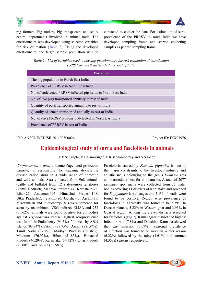
pig farmers, Pig traders, Pig transporters and state/central departments involved in animal trade. The questionnaire was developed using selected variables for risk estimation (Table 2). Using the developed questionnaire, the target sample population will be
contacted to collect the data. For estimation of sero-prevalence of the PRRSV in south India we have developed sampling frame and started collecting samples as per the sampling frame.
Table 2 : List of variables used to develop questionnaire for risk estimation of introduction PRRS from northeastern India to rest of India
Variables
The pig population in North East India
Prevalence of PRRSV in North East India
No. of undetected PRRSV-infected pig herds in North East India
No. of live pigs transported annually to rest of India
Quantity of pork transported annually to rest of India
Quantity of semen transported annually to rest of India
No. of days PRRSV remains undetected in North East India
Prevalence of PRRSV in rest of India
IPC: ANSCNIVEDISIL201100500024 Project ID: IXX07976
Epidemiological study of surra and fascioliosis in animalsP P Sengupta, V Balamurugan, P Krishnamoorthy and S S Jacob
Trypanosoma evansi, a haemo flagellated protozoan parasite, is responsible for causing devastating disease called surra in a wide range of domestic and wild animals. Sera collected from 968 animals (cattle and buffalo) from 12 states/union territories (Tamil Nadu-80, Madhya Pradesh-44, Karnataka-72, Bihar-27, Andaman-195, Himachal Pradesh-108, Uttar Pradesh-23, Sikkim-80, Odisha-43, Assam-35, Mizoram-78 and Puducherry-183) were screened for surra by recombinant VSG indirect ELISA and 732 (75.62%) animals were found positive for antibodies against Trypanosoma evansi. Highest seroprevalence was found in Puducherry (94.5%) followed by A&N islands (93.84%), Sikkim (88.75%), Assam (88. 57%), Tamil Nadu (87.5%), Madhya Pradesh (86.36%), Mizoram (76.92%), Bihar (51.85%), Himachal Pradesh (46.29%), Karnataka (34.72%), Uttar Pradesh (26.08%) and Odisha (25.58%).
Fasciolosis caused by Fasciola gigantica is one of the major constraints to the livestock industry and aquatic snails belonging to the genus Lymnaea acts as intermediate host for this parasite. A total of 2077 Lymnaea spp. snails were collected from 25 water bodies covering 11 districts of Karnataka and screened for F. gigantica larval stages and 5.1% of snails were found to be positive. Region wise prevalence of fasciolosis in Karnataka was found to be 5.79% in Deccan plateau, 5.22% in Western ghat and 3.95% in Coastal region. Among the eleven districts screened for fasciolosis (Fig.7), Ramanagara district had highest infection rate (7.8%) and Dakshina Kannada showed the least infection (3.09%). Seasonal prevalence of infection was found to be more in winter season (6.22%) followed by the rainy (4.61%) and summer (4.35%) seasons respectively.

NIVEDI - Annual Report 2016-1714
Fig.7. Prevalence of fasciolosis in Lymnaeid snails in Karnataka
IPC: ANSCNIVEDISIL201300300046 Project ID: IXX10616
Retrospective Epidemiological studies on HPAI with reference to spatio-temporal pattern and the probable associated
risk factors identificationR Sridevi, G Govindaraj, P Krishnamoorthy, K P Suresh and A A Raut
Brief Summary: Data on various factors like poultry population density, human population density, wetlands, waterbodies, ramsar convention sites, collected and analysed.Spatial heat map of AI outbreaks in eastern and north eastern states from 2008-2010 plotted (Fig. 8). Economic impact analysis of avian influenza outbreaks occurred in Kerala on various sectors hatcheries, commercial poultry farms/ducks farms, traditional duck farmers, tourist boat industry, human
health in 2014 was done. Pre-tested questionnaires were employed to collect the primary data from farmers, hatcherres, boat owners, retail sale markets of meat shop owners,etc. Probable factors for avian influenza outbreaks at Kerala–Kuttanad region identified were high duck population density, vast backwater, presence of branches of 4 major rivers, presence of vast paddy fields, close contact between the host (ducks) and the wetland environment, and lack of biosecurity measures.
Illustration:
Fig. 8. Heat map of Avian influenza outbreaks from 2008-2010 in Assam, West Bengal and Tripura

NIVEDI - Annual Report 2016-17 15
INSTITUTE SERVICE PROJECTS

NIVEDI - Annual Report 2016-1716

NIVEDI - Annual Report 2016-17 17
IPC: ANSCNIVEDISIL201100100020 Project ID: IXX08329
National Animal Disease Referral Expert System (NADRES)D Hemadri, K P Suresh, S S Patil and J Hiremath
The NADRES database contains information on major livestock diseases of the country along with their associated risk factors. During the period under report, about 82440 entries related livestock diseases in the country were made. Details of the most reported bacterial, viral and parasitic diseases during the calendar year 2016 shown in Fig. 9. The red line indicates number of states reporting the said diseases. Thus from the figure it can be inferred that though largely reported
some diseases are restricted to smaller geographic regions. Among the bacterial diseases, foot rot, HS, BQ, ET, Glanders and anthrax were the most reported in that order. Similarly, PPR, Capripox, BT, Rabies and swine fever were the most reported viral diseases. In addition, fasciolasis, amphistomiasis, anaplasmosis, babesiosis, coccidiosis and trypanosomiasis were most reported parasitic diseases.
*FMD not included for analysis as it is taken up by Directorate of FMD
a) b)
c)
Fig. 9. Major Bacterial (a), Viral (b) and Parasitic disease (c) in India during 2016
The NADRES database contains information on major livestock diseases of the country along with their associated risk factors. Using these databases, (which are updated regularly) the probability of occurrence of disease outbreaks at the district level was forecasted 2 months in advance. The results are available to any user on interactive basis at the website (www.nadres.res.in). The forewarning information were also sent
as monthly bulletin to DADF and other directors of animal husbandry departments for taking up suitable preventive measures. During the period, forewarning for 13 of the livestock diseases widely reported in 29 states and 7 union-territories of the country were prepared and circulated. (Table 3) Time series analysis of major livestock diseases also carried out to the disease trend in India (Fig. 10 & Fig. 11)

NIVEDI - Annual Report 2016-1718
Table 3: The total number of predicted district- diseases for the period between April 2016 and March 2017.
Sl.No Diseases Month-wise OB No. of districts predicted ( April 2016-March 2017) April May June July Aug Sept Oct Nov Dec Jan Feb March1 Anthrax 68 57 64 58 60 58 66 62 62 63 41 482 Babesiosis 43 42 50 42 46 38 37 37 38 38 58 543 Black Quarter 187 152 183 154 143 108 84 108 108 109 152 1834 Bluetongue 3 9 14 23 25 24 20 22 22 22 3 05 Entertoxemia 42 41 33 29 33 36 41 37 41 42 37 366 Fascioliasis 68 60 64 72 51 54 36 59 55 55 37 477 FMD 148 161 152 166 227 263 250 242 265 266 181 1388 HS 172 192 213 213 176 138 124 156 136 137 142 1669 PPR 53 48 48 48 51 59 60 62 63 63 69 6410 S&G Pox 51 50 50 49 49 54 57 54 53 57 63 5211 Swine Fever 19 23 33 29 28 24 27 25 24 24 32 3412 Theileriosis 35 37 35 32 49 40 34 40 38 39 34 6513 Trypanosomiasis 44 36 45 63 64 41 26 104 41 42 64 63 GRAND TOTAL 933 908 984 978 1002 937 862 1008 946 957 913 950
Fig. 10. Graphs showing the time series analysis of FMD in India
Fig. 11. Graph showing the time series analysis of Black quarter in India
IPC: ANSCNIVEDISIL201100300022 Project ID: IXX08279
Maintenance and updating of livestock serum repositoryD Hemadri, K P Suresh and S S Patil
As part of the annual survey conducted by the AICRP on ADMAS, central unit at NIVEDI designs and sends sampling plan every year to each of the centers of AICRP on ADMAS. The serum samples so collected, as per the
plan, are sent to NIVEDI for screening against various livestock diseases mainly, Brucellosis (Bovine, Caprine, Swine), Infectious Bovine Rhinotracheitis (IBR), Classical Swine Fever, Bluetongue, Leptospirosis

NIVEDI - Annual Report 2016-17 19
etc. The serum bank at ICAR-NIVEDI, arranges for screening (aliquoting, labeling, distributing) against the said diseases and catalogues the serum along with the results. The serum bank also sends the results to sender. During the period under report a total of 16824 serum samples were received. From different states (Fig.12). Depending on suitability, all the samples
were aliquoted and distributed for screening against Brucella, IBR, Classical swine fever, Bluetongue and trypanosomiasis. The screening against bluetongue for the year 2016-17 could not be done due to non-availability of kits, however, screening of 943 samples of 2015-16 showed that 134 of these were positive.
CamelPig
SheepBuffalo
GoatCattle
0
200
400
600
Statewise distribution of serum samples
Camel Pig Sheep Buffalo Goat Cattle
Fig. 12. State wise distribution of serum samples collected from different states of India during 2016-17.
Species-wise distribution of samplesSpecies-wise distribution of samples shows that large quantity of serum received belong to cattle and buffaloes as these are two predominant species of livestock in the country (Fig. 13).
2495 335
6997
4134
15061183
Buffalo Camel Cattle Goat Pig Sheep
Fig. 13. Species wise distribution of samples collected from different states of India during 2016-17
Bovine BrucellosisA total of 9500 serum samples from cattle/buffalo samples were received as part of the annual survey conducted by the AICRP on ADMAS. Of which 1401 samples were screened only 9 were positive for bovine brucellosis.
Ovine BrucellosisA total of 1224 serum samples from sheep samples were received as part of the annual survey conducted
by the AICRP on ADMAS. Of which results of 41 samples were received at NLSR, which showed only 2 samples positive for ovine brucellosis.
Caprine BrucellosisA total of 4134 serum samples from goat samples were received as part of the annual survey conducted by the AICRP on ADMAS. Of which results 339 samples were received at NLSR which showed 6 samples as positive for caprine brucellosis.

NIVEDI - Annual Report 2016-1720
Swine BrucellosisA total 1506 pig serum samples were received as part of the annual survey conducted by the AICRP on ADMAS. Of which, results of 288 samples were received at NLSR which showed all the samples as negative for caprine brucellosis.
Infectious Bovine RhinotracheitisA total of 9500 serum samples from cattle/buffalo samples were received as part of the annual survey conducted by the AICRP on ADMAS. Of which results 4565 samples were received at NLSR which showed 1408 samples as positive for anti BoHV-1 antibodies.
Annual survey on classical swine fever A total 1506 pig serum samples were received as part of the annual survey conducted by the AICRP on ADMAS, of which large number (n=1461) of serum
samples belonged to seven states of the north east India. Considering the insignificant number of serum samples received from rest of India and the availability of the testing kits, it was decided to screen the samples from the north eastern states. In addition it is also to be noted that north eastern states harbor nearly half of the total pig population in India.
CSF in the North Eastern StatesA total of 1140 out of 1461 pig serum samples from seven states of north east were screened for the presence of anti-CSFV antibodies using a commercially available kit. These serum samples collected as part of the AICRP annual survey. CSF prevalence of more than 40% was found in Manipur, Mizoram, Nagaland and Sikkim. The significance of the values found in Sikkim needs further investigation as the state ranks low on livestock population.
IPC: ANSCNIVEDISIL201300200045 Project ID: IXX10708
Sero-epidemiology of brucellosisR Shome, B R Shome and M Nagalingam
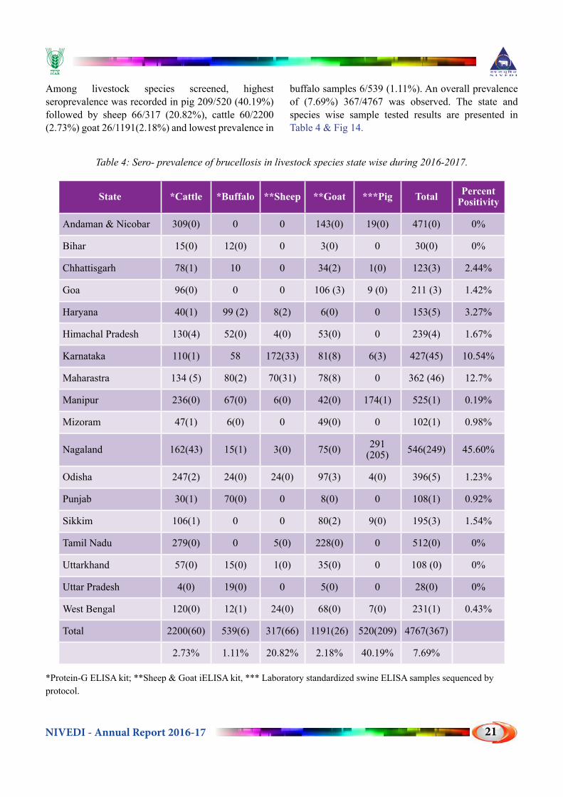
During the period, a total of 4767 random serum samples {cattle (2200), buffalo (539), sheep (317), goat (1191) and swine (520)} received from 18 AICRP centers were screened for brucellosis by using Protein-G iELISA kit for bovine brucellosis, Sheep & Goat iELISA kit for sheep and goat and laboratory standardized swine protocol for swine brucellosis (Table 4). Highest
seroprevalence was recorded in Nagaland 45.6% (249/546); followed by Maharashtra12.7% (46/362) and Karnataka10.54% (45/427). States like Mizoram, Punjab, West Bengal and Manipur have shown < 1% and A&N, island Bihar, Tamil Nadu, Uttarakhand and Uttar Pradesh have shown no seroprevalence for brucellosis.
471
30123
211153
239
427362
525
102
546
396
108195
512
10828
231
0 0 3 3 5 445 46
1 1
249
5 1 3 0 0 0 1
Total number of samples screened in the year 2016-2017
Total Number of samples screened Positive
Fig. 14. Graph representing the total number of samples screened in the year 2016-17
NIVEDI - Annual Report 2016-17 21
Among livestock species screened, highest seroprevalence was recorded in pig 209/520 (40.19%) followed by sheep 66/317 (20.82%), cattle 60/2200 (2.73%) goat 26/1191(2.18%) and lowest prevalence in
buffalo samples 6/539 (1.11%). An overall prevalence of (7.69%) 367/4767 was observed. The state and species wise sample tested results are presented in Table 4 & Fig 14.
Table 4: Sero- prevalence of brucellosis in livestock species state wise during 2016-2017.
State *Cattle *Buffalo **Sheep **Goat ***Pig Total Percent Positivity
Andaman & Nicobar 309(0) 0 0 143(0) 19(0) 471(0) 0%
Bihar 15(0) 12(0) 0 3(0) 0 30(0) 0%
Chhattisgarh 78(1) 10 0 34(2) 1(0) 123(3) 2.44%
Goa 96(0) 0 0 106 (3) 9 (0) 211 (3) 1.42%
Haryana 40(1) 99 (2) 8(2) 6(0) 0 153(5) 3.27%
Himachal Pradesh 130(4) 52(0) 4(0) 53(0) 0 239(4) 1.67%
Karnataka 110(1) 58 172(33) 81(8) 6(3) 427(45) 10.54%
Maharastra 134 (5) 80(2) 70(31) 78(8) 0 362 (46) 12.7%
Manipur 236(0) 67(0) 6(0) 42(0) 174(1) 525(1) 0.19%
Mizoram 47(1) 6(0) 0 49(0) 0 102(1) 0.98%
Nagaland 162(43) 15(1) 3(0) 75(0) 291(205) 546(249) 45.60%
Odisha 247(2) 24(0) 24(0) 97(3) 4(0) 396(5) 1.23%
Punjab 30(1) 70(0) 0 8(0) 0 108(1) 0.92%
Sikkim 106(1) 0 0 80(2) 9(0) 195(3) 1.54%
Tamil Nadu 279(0) 0 5(0) 228(0) 0 512(0) 0%
Uttarkhand 57(0) 15(0) 1(0) 35(0) 0 108 (0) 0%
Uttar Pradesh 4(0) 19(0) 0 5(0) 0 28(0) 0%
West Bengal 120(0) 12(1) 24(0) 68(0) 7(0) 231(1) 0.43%
Total 2200(60) 539(6) 317(66) 1191(26) 520(209) 4767(367)
2.73% 1.11% 20.82% 2.18% 40.19% 7.69%
*Protein-G ELISA kit; **Sheep & Goat iELISA kit, *** Laboratory standardized swine ELISA samples sequenced by protocol.

NIVEDI - Annual Report 2016-1722
IPC: ANSCNIVEDISIL201200800032 Project ID: IXX10709
Seroepidemiology of Infectious Bovine Rhinotracheitis in IndiaS S Patil and D Hemadri
Infectious Bovine Rhinotracheitis (IBR) is a highly contagious, infectious respiratory disease that is caused by Bovine Herpesvirus-1 (BoHV-1). Disease outbreaks can result in severe production losses, abortion and delayed inter calving periods.
A total of 9923 sera samples from different states of India were tested (Table 5), out of which 2702 samples were found to be positive for the presence of IBR antibodies using Avidin-Biotin ELISA. The highest prevalence rate of 50.54 was observed in Chattisgarh and the lowest prevalence was found to be 6.80 in Nagaland.
Bovine Herpesvirus 1(BoHV-1) Repository: A total of 16 isolates of BoHV-1 were revived and maintained in the laboratory and they are-IBRV-1, ADMAS-1, BoHV-1:258/08, BoHV-1:685/10, BoHV-1:688/10, BoHV-1:707/10, BoHV-1:723/10, BoHV-1:741/10, BoHV-1:743/10, BoHV-1:744/10, BoHV-1:745/10, BoHV-1:746/10, BoHV-1:774/11, BoHV-1:775/11, BoHV-1:776/11, BoHV-1:777/11 from Karnataka, Orissa, West Bengal and UP.
PCR analysis of Bovine clinical samples: A total of 69 samples (nasal swabs, vaginal swabs, blood in EDTA) received from Tripura (20 Blood in EDTA, 5 vaginal swabs, 4 nasal swabs), Kerala (one tissue samples from Elephant calf), Mizoram (39 blood in EDTA) were subjected for DNA extraction and tested by PCR using primers specific for gB region of BoHV-1. Ten samples were found to be positive for gB293 (IBR) amplicon. A total of 11 bovine samples ( one each tissue from Amreli-Gujarat_buffaloes and Vizag-AP-Bison, 9 blood samples from Amreli-Gujarat-Buffaloes) were screened for MCF (OvHV-2) sequences using tegument and gB specific primers of OvHV-2. All were found positive (except one blood samples from Gujarat) for tegument and gB sequences. A total of 12 IBR Avidin Biotin-ELISA kits were supplied to different Disease Diagnostic Laboratories across the country (A & N island, Andhra Pradesh, Haryana, Kerala, Maharshtra, Punjab, UP).
Table 5: Sero-prevalence of IBR in different states of India during 2016-17
STATE Total no sample
IBR positive
Percent Positive
Andaman & Nicobar 600 278 46.33
Assam 856 157 18.34
Bihar 27 6 22.22
Chattisgarh 632 93 14.72
Goa 96 19 19.79
Gujarat 320 91 28.44
Haryana 135 65 48.15
Himachal Pradesh 774 140 18.09
Jammu and Kashmir 222 25 11.26
Karnataka 883 339 38.39
Kerala 630 144 22.86
Kolkata 132 73 55.30
Maharashtra 215 68 31.63
Manipur 752 218 28.99
Meghalaya 109 16 14.68
Mizoram 80 37 46.25
Nagaland 324 111 34.26
Orissa 415 134 32.29
Punjab 405 127 31.36
Rajasthan 275 30 10.91
Sikkim 582 227 39.00
Tamil Nadu 668 120 17.96
Telangana 332 122 36.75
Uttar Pradesh 151 11 7.28
Uttarakhand 308 51 16.56
Total 9923 2702 27.23

NIVEDI - Annual Report 2016-17 23
EXTERNALLY FUNDED PROJECTS

NIVEDI - Annual Report 2016-1724

NIVEDI - Annual Report 2016-17 25
IPC: ANSCNIVEDIISOP200900500017 Project ID: OXX02232
Outreach Programme on Zoonotic DiseasesV Balamurugan, P P Sengupta, S B Shivchandra, R Sridevi and M M Chanda
For surveillance of leptospirosis in livestock and human from different areas, samples from Karnataka (Bidar and Doddabalapur), Andhra Pradesh (Tirupati), Tamil Nadu (Chennai, Kanchipuram), Uttarakhand (Kashipur), Punjab, Gujarat, Kerala and Sikkim were tested in MAT and ELISA. A total of 1149 livestock (Cattle-1116, Bear -23 and Goat-10), and 562 human serum samples were tested along with 43 rat samples from Nagpur in the MAT at 1:100 titre with 18 reference leptospira serovars in MAT of which 640 livestock (Cattle-617, Bear-13 and Goat-10), 207 human and 34 rat samples were showed positive reactivity for leptospira serovars group specific antibodies.
The seropositivity of 41.17 % (49/119) leptospirosis was observed when testing the human samples collected from pyrexia of unknown origin (PUO) cases with sero prevalence of major of Leptospira serovars representing serogroups specific antibodies against serovars Hurstbridge, Tarassovi, Javanica, Bataviae/ Pyrogenes, Shermani, Icterohaemorrhagiae, Kaup, Hardjo, etc., whereas the seropositivity of 65 % (13/20) was observed in the case of human with neurological disorders with serogroups specific antibodies against serovars Hardjo/Pyrogenes, Shermani/ Kaup /Djasiman Hebdomadis/ Tarassovi /Hurstbridge /Bataviae, Australis/Bankinang/Canicola/ Djasiman.
Samples received from human with history suspected for leptospirosis with case definition, showed the seropositivity of 78.9 % (101/128) with seroprevalence of major Leptospira serovars representing serogroups specific antibodies against serovars Pyrogenes / Tarassovi / Hebdomadis, Hurstbridge, Kaup, Shermani/ Hebdomadis, Australis/Bankinang/ Pomona. In the risk groups of veterinarians, the seropositivity of 14.97% (44/295) leptospirosis was observed with seroprevalence of major Leptospira serovars representing serogroups specific antibodies against serovars Pyrogenes, Javanica,
Tarassovi, Icterohaemorrhagiae, Hebdomadis, Canicola, Hardjo, Australis/ Bankinang. However high sero positivity of 64.75% (191/295) was observed when sample tested with in panbio human IgM ELISA kit.
Samples from the reservoir host (rat) from Nagpur showing seropositity of 79.06% with prevalence of serovars against Grippotyphosa /Hurstbridge, Australis, Tarassovi/ Shermani/ Bataviae, Bankinang/Canicola/ Djasiman. Further, on testing of 323 random purposive livestock (Cattle, Buffaloes, sheep and goats) serum samples from Surat district in Gujarat, 179 samples were showed positive reactivity in MAT representing high seropositivity (55.41%) with serogroups specific antibodies against serovars, Panama, Hebdomadis/ Bataviae, Pyrogenes, Pomona/ Hurstbridge/ Tarassovi, Grippotyphosa/ Icterohaemorrhagiae/ Canicola, Hardjo/ Shermani /Kaup.
On testing 373 bovine serum samples with history of abortion and reproductive disorders, 263 samples showed positive reactivity in MAT, indicating the 70.51 % seropositivity against serovars Hardjo, Canicola, Hebdomadis, Icterohaemorrhagiae, Pyrogenes, Hurstbridge, Javanica, Panama, Copenhageni, etc., in cattle associated with abortion and reproductive disorders. Moreover, seropositivity of 29.22 % only was observed when subject the samples in Leptospira Bovine Hardjo ELISA kit. This study supports that cattle have a role as reservoir in maintaining Hebdomadis, Icterohaemorrhagiae, Hurstbridge, Shermani, Pomona, etc., in addition to Leptospira Hardjo serovar in cattle dairy farms of India and warrants need of an intensive surveillance programme, prevention and control strategies including implementation of vaccination and mitigating measures to reduce the incidence of leptospirosis in cattle farms.
Toxoplasmosis is a well-known zoonotic disease in human and it mainly causes abortion and reproductive disorders in human, sheep and other animals. As a

NIVEDI - Annual Report 2016-1726
preliminary study, to generate the data, during the period 2016-17, a total of 297 human serum samples (Kerala n=184 and Karnataka n=66; Dadra and Nagar Haveli n=47) were screened for toxoplasmosis by using commercial diagnostic kit (Toxoplasma IgG & IgM, DIESSE Diagnostica Senese, Italy Enzywell) as per manufacture’s protocols. Out of 297 human serum samples, 55 samples (Kerala n=19, Karnataka-n=25 and Dadra and Nagar Haveli n=11) were showed positive by toxoplasma IgG, further 10 samples (male-3 and female-7) out of 19 from Kerala also showed positive reaction by IgM ELISA with overall seropositivity of 18.51% with 16.88 % (26/154) and 20.28 % (29/143) in male and female, respectively. The seroposititvity of 31.85 % was observed with history of PUO cases (28/93=30.10%) and neurological disorders (8/20=40%) whereas in the risk groups veterinarians seropositivity of 10.32 % (19/184) was observed with respect to toxoplasmosis specific antibodies.
Anthrax: A total number of (113) clinical/environmental sample from Odisha state were screened for presence of Bacillus anthracis of which 14 samples (Cattle-7 ; Elephant 2 and Soil-5) showed positive when applied standard bacterial techniques along with Grams Staining and confirmatory test by using Protective antigen (PA) and capsular specific PCR confirming anthrax infection. The details of samples and its test results are given in Table 6
Table 6: Details of samples received from Odisha and its test results for Bacillus anthracis
Host Sample TypeTotal No.
of Samples tested
No. of samples Positive
Cattle Spleen, liver, lung, kidney, heart
05 -
Heart Blood 01 -
CattleBlood 37 04
Soil 06 -
Bone 04 01
Dried Meat/beef 07 01
Soil + Bone 05 01
Elephant Blood 10 02
Soil 06 01
Tissue 01 -
Muscle 01 -
Bone 02
Sheep Blood 07 -
Goat Blood 01 -
Soil 05 01
Blood 01 01
Nasal swab with Blood 01 -
Zebra Lung 01 -
Spleen 01 -
Blood 01 -
Environmental sample -Soil
10 02
Total 113 14
IPC: ANSCNIVEDISOP201200600030 Project ID: OXX01504
All India Network Programme on BluetongueD Hemadri, M M Chanda and K P Suresh
Compared to the previous year, there were relatively fewer number of bluetongue outbreaks reported from the states of Karnataka and Andhra Pradesh. During the period 2016-17 (Fig.15), eighty five outbreaks were reported from all over India, of which, large number of these were reported from Telangana. Noteworthy information about the disease is the first time reporting of bluetongue outbreaks from the state of Odisha. Initial serotyping results indicated the involvement of serotypes 24 along with serotype 1.
With the availability of different serotypes, the work on development of multiplex PCR was continued during the period. Multiplex PCR for the detection of serotypes 5 & 9, 3, 13 & 16 and 10 & 24 were optimized and specificity of the so obtained PCR was confirmed by nucleotide sequencing. Partial nucleotide sequencing indicated higher levels of genetic homogeneity with in the each serotypes (BTV1, 2, 24, 16, 4) tested.

NIVEDI - Annual Report 2016-17 27
Fig.15. Bluetongue outbreaks during April 2016-December 2016
IPC: ANSCNIVEDICOP201500100064 Project ID: OXX02963
National Innovations on Climate Resilient Agriculture - Livestock disease surveillance in relation to weather data and emergence of new pathogens
B R Shome, K P Suresh, P Krishnamoorthy, G B Manjunath Reddy, S S Patil, G Govindaraj, R Yogisharadhya and A Prajapati
During period (2016-17), disease incidence data reported in West Bengal, Kerala & Rajasthan were linked to the remote sensing and meteorological parameters subjecting it to Poisson regression model to establish the climate-disease relationship model. Risk Maps were developed for West Bengal, Kerala & Rajsthan states, which are useful for resource allocation like manpower, material, money and effective vaccination program. In West Bengal, the high risk of parasitic disease occurrence was predicted (Fig. 16) in Malda and Diamond Harbour regions in red. and mode safe
risk was predicated in Krishnanagar in pink and The high risk of bacterial disease occurrence was predicted in Purulia, Bankura and Murshidabad whereas for viral diseases, the high risk of occurrence was predicted in Kandi, Raiganj, Jangipur and Alipurduar region of West Bengal.
In Kerala, the high risk of disease occurrence for parasitic diseases was found in Ernakulum and Malappuram followed by Thirssur, (Fig. 17). In Rajasthan, the high risk of disease occurrence for parasitic diseases was found in Udaipur and Sikar district.
Fig. 16. Risk Map for parasitic diseases in West Bengal Fig. 17. Risk Map for parasitic diseases in Kerala

NIVEDI - Annual Report 2016-1728
Emergence of new pathogens: The extended spectrum beta lactamase producing multi drug resistant Escherichia coli isolated from subclinical mastitis milk was identified and characterized. The multidrug resistant Proteus mirabilis carrying multiple efflux pumps detected in apparently healthy pig fecal sample. Escherichia coli strain SCM-21 and Proteus mirabilis
strain NIVEDI3-PG74 whole genome shotgun sequencing was done.
The study concludes that the environmental variables are very crucial to study the epidemiology of pathogen and disease prediction to implement timely control measures to the present screening of global climate change.
IPC: ANSCNIVEDICOP201600800077 Project Code: OXX03488
All India network project on GIPP P Sengupta, K P Suresh and Siju Susan Jacob
Haemonchosis caused by Haemonchus contortus is a predominant, highly pathogenic and economically important disease of sheep and goats. These parasites are common blood feeders that cause anaemia and reduced productivity and can lead to death in heavily infected animals. In order to assess the risk areas for haemonchosis in Rajasthan, disease data on haemonchosis (EPG) was received from CSWRI, Avikanagar unit. The data related to four districts viz., Tonk, Pali, Sikar and Bhilwara including 6 talukas and 11 villages. Some other parameters like LST (land surface temperature), NDVI (normalized difference vegetation index), distance from river/highways etc.
were incorporated in the data. The disease prediction models were developed using logistic regression analysis considering few influencing parameters viz., LST, LST (1 month lag), rainfall and flock size (Fig. 18). The overall accuracy of the model found to be 67.5% which is quite satisfactory. Further accuracy can be achieved by increasing randomness, representativeness and independence by proper sampling. Risk maps were prepared using Poisson regression model employing ArcGIS software. The parameters showed highly significant with a low profile of standard error.
Fig. 18. Risk map for EPG in Rajasthan in correlation with rainfall and LST

NIVEDI - Annual Report 2016-17 29
IPC: ANSCNIVEDISOL201600200071 Project Code: OXX03660
Evaluation of vaccine effectiveness and identification of the factor that affect field level vaccine efficacy of the vaccine against diseases under
control programJ Hiremath, R Shome, D Hemadri, K P Suresh, V Balamurugan, S S Patil,
M M Chanda and GB Manjunathareddy
The objective of the project was to evaluate vaccine effectiveness and to identify the factors that affect the effectiveness of vaccines against FMD. A questionnaire was used to collect the survey data on vaccine and vaccination. In addition, pre and post-vaccination serum samples were also collected from selected districts of Karnataka to measure the antibody titre values using LPB ELISA kit (Life Technologies [India] Pvt. Ltd). The pre and post-vaccination serum samples were collected from the identified cattle from selected districts (Hassan, Chikkaballapur, Mandya and Udupi) of Karnataka which were subjected to estimation of Liquid Phase Blocking (LPB) ELISA antibody titre for FMD serotype O.
The comparison was done between pre and post-vaccination serum LPB ELISA titer (>1.8 is protective
titre as per the LPB FMD ELISA Kit from Life Technologies) in population of cattle irrespective of age and breed. A 67% (89/132) of the cattle showed the titre of more than 1.8 before vaccination and number has increased to 82% (108/132) after 45 to 60 days of post vaccination. Whereas 33% (43/132) of the cattle showed the titre of less than 1.8 before vaccination and number has decreased to 18% (24/132) after 45 to 60 days post vaccination. Further, 45% (23/51) of the cattle < 3 years old showed the titre of >1.8 before vaccination and number has increased to 59% (30/51) after 45 to 60 days of post vaccination (Fig.19A). Whereas the 81% (66/81) of the cattle (>3years) were having titre of >1.8 before vaccination and number has increased to 93% (75/81) after 45 to 60days post vaccination (Fig. 19B). The data analysis for other districts is in progress.
Fig. 19. Pre and Post vaccination serum LPB ELISA antibody titer in cattle. A <3years, B >3years (*p<0.05)

NIVEDI - Annual Report 2016-1730
IPC: ANSCNIVEDISOL201600300072 Project Code: OXX02913
Understanding the epidemiology of Culicoides borne diseases in wild and domestic ruminants
M M Chanda, D Hemadri, P P Sengupta, J Hiremath and S B Shivachandra
Culicoides species are responsible for transmitting many viral diseases (e.g. Bluetongue) in domestic and wild ruminants. There is a risk of spill over of the pathogens transmitted by Culicoides from wild to domestic and vice-versa. Control strategies require research on different aspects of vector, virus and host involved in complex epidemiology of Culicoides borne diseases. In the present study, different wild life sanctuaries and national parks of Karnataka were selected for collection of the Culicoides using light traps. The sites near to wild life sanctuaries with domestic livestock population were also selected to compare the Culicoides species composition and possible spill over from wild to domestic and vice-versa. We also selected three sites from Tamil Nadu in the study. Monthly collections were made by using CDC light traps with UV light for a period of eight months. The traps were placed overnight in thirty one
sites during which more than 8000 Culicoides were collected. The major species found in these sites were: C. imicola, C. oxystoma, C. fulvus, C. brevitarsis, C. huffi, C. arakawae, C. peregrinus, C. palpifer, C. anophelis, C. palpisimilis, C. inoxius and C. actoni (Fig. 20) The selected culicoides species were DNA bar coded and voucher specimens prepared. The work on host preference was carried out to identify the host bitten by the Culicoides species which is important in transmission of diseases. Bluetongue virus (BTV) was detected in C.oxystoma and C.imicola samples. Results of present study indicate that there is a circulation of Culicoides species in forest habitats and presence of BTV in some Culicoides species. thus, more studies are required to get deeper insights into the epidemiology of Culicoides borne diseases in forest habitat, so that control strategies can be planned to prevent spill over of the disease.
Fig. 20. Culicoides species distribution in Bannerghatta biological park

NIVEDI - Annual Report 2016-17 31
IPC: ANSCNIVEDISOL201600400073 Project ID: OXX03634
Impact assessment of PPR vaccine technology in IndiaG Govindaraj, V Balamurugan, GB Manjunathareddy and R Yogisharadhaya
Peste des petits ruminants (PPR) is one of the highly contagious and economically important viral diseases of small ruminants, especially in sheep and goats. The mortality and morbidity rates are as high as 90% and 100%. The annual loss due to PPR in the small ruminants in India was estimated at 1611 crores at 10% annual incidence level. Considering the devastating nature of the disease and threat to the smallholders livelihood, an effective live attenuated vaccine was developed in India. The developed vaccine has been widely used in National Control Programme on PPR (PPR-CP) since 2010-11. A multi-stage random sampling procedure was followed for undertaking primary survey to assess the field level disease lossess. During the period under report, the survey was completed in Karnataka state and hence the results are presented for Karnataka. The total number of farms surveyed were 350 and the samples were distributed among the identified districts in proportion to the number of households rearing sheep and goat (Fig. 21). The pooled results revealed that among the surveyed villages PPR incidence level was 8.31% in sheep and goats. The estimated per animal mortality loss, cost of treatment, distress sale
and opportunity cost of labour among the infected flocks was INR 3231, INR 108.2, INR 3040, and INR 15.7 respectively.
Fig.21: Represents the districts surveyed in Karnataka during 2016-17
IPC: ANSCNIVEDISOP201201600039 IPC code: OXX02733
DBT - Network Project on Brucellosis : Project Monitoring Unit (PMU)
H Rahman and G B Manjunatha ReddyBrucellosis is an economically important bacterial disease affecting both animals and human. The DBT-Network Project on Brucellosis is a multi-intuitional project sponsored by Department of Biotechnology, Ministry of Science and Technology, GoI. The project has different subunits on brucellosis epidemiology (6), Brucellosis diagnostics (2), Brucellosis vaccine (2), Brucellosis repository (1) and Brucellosis bioinformatics (1), with overall monitoring of project entrusted to Project Monitoring Unit (PMU) at ICAR-NIVEDI, Bengaluru. PMU is involved in co-ordinating different activities of all the subunits under DBT Network Project on Brucellosis. Monitoring the research activities of different centres by means
of monthly and quarterly reports, also submitting the compiled reports on monthly, quarterly and annual basis to DBT. PMU coordinated the midterm review meet on 25th and 26th July, 2016 at MKU, Madurai and Tamil Nadu. The regular updating and maintenance of DBT-Brucellosis website in collaboration with MKU. PMU actively coordinated and participated for successful completion of international brucellosis conference organized at NAAS complex, New Delhi from 17th to 19th December, 2016. PMU co-ordinated in sending the serum samples, bacterial cultures, Brucella antigens procurement, DNA between the different subunits. PMU also coordinated the validation of different Brucella diagnostic kits developed under the

NIVEDI - Annual Report 2016-1732
project. The 4th annual review meeting was organised in collaboration with DBT at DBT headquarters on
6th February, 2017 to review the research progress of different units.
IPC: ANSCNIVEDISOP201201600040 Project ID: OXX02578
Brucellosis Epidemiology (BE-1)R Shome, BR Shome and Nagalingam M
A total of 3610 cattle (3221) and buffaloes (389) sera were tested, positivity was recorded with apparent and true prevalence overall, 6.31% (228/3610) were 6.1% and 1.2% for cattle and 8.2 % and 6.9% for buffaloes, respectively. Among 24 farms prevalence screened seropositivity in the farms varied from 0 to >12% in which 3 farms had seropositivity of greater than 12% and lowest seroprevalence rate of 0-3% in majority (13) of the farms. Multivariate logistic regression model identified overall seven risk factors such as sex, absence of separate sheds, nonvaccination against brucellosis, disposing manure in the pits, cleaning the shed without disinfectants, cleaning the animal shed twice in a week and obtaining monthly veterinary services in the farms have been significantly correlated to brucellosis (Fig. 22).
The results of 1084 serum samples, (719 from seven sheep flocks and 365 from five goat flocks) tested showed seropositivity in 26 out of 365 (7.12%) in goats and 58 out of 719 (8.06%) in sheep. Multivariate logistic regression model in caprines indicated that female sex and procurement of animals from livestock fairs were considered risk factor for brucellosis. Similarly, in ovines female sex, orchitis, extensive rearing, disposal of aborted foetus in water bodies and access to stray animals in the farm were considered.
Milk surveillance for brucellosis was carried out in 2 districts of Karnataka (Kolar and Chikkaballapur) through Kolar Milk Union Limited (KOMUL), Kolar by Milk Ring Test (MRT). A total of 960 milk samples from Kolar [Bulk Milk Cooler Centre (BMC=79); Dairy Co-operative Societies (DCS=766) and Sub Centres (SC=115)] and 1026 milk samples from Chikkaballapur
[(BMC=78; DCS = 849 and SC= 99) were screened by MRT at Kolar and Chinthamani milk union laboratories, respectively. Of these, 5.06% and 2.08% milk samples from Kolar BMC and DCS were positive for anti Brucella antibodies. Similarly 5.12%, 6.36% and 5.05% from Chikkaballapur BMC, DCS and SC, respectively were recorded positive by MRT. Very high prevalence of brucellosis was recorded in Chikkaballapur district where 63 villages were identified positive for brucellosis out of 1521 villages. Whereas, only 20 villages were identified positive for brucellosis out of 1809 villages of Kolar district. The annual disease mapping of brucellosis through milk co operative societies as per OIE 2016 facilitate vaccination in risk areas.
Fig. 22. Univariate analysis of 15 potential risk factors in dairy farms

NIVEDI - Annual Report 2016-17 33
IPC: ANSCNIVEDISOP201201700041 Project ID: OXX02384
Brucellosis Diagnostics (BD-2)M Nagalingam, V Balamurugan, R Shome and GB Manjunathareddy
The project was initiated in an attempt to develop serological test for diagnosing bovine brucellosis using recombinant proteins in order to avoid use of lipopolysaccharide (LPS) of Brucella species which causes cross reactivity with organism like Yersinia enterocolitica : O9 that share common features of the LPS thereby reducing the specificity of the test. In 2016-17, a multi-epitope recombinant protein antigen was designed by identifying nine immuno dominant proteins which have no cross reactivity with antibodies of Yersinia enterocolitica : O9. Then identification of 16mer immuno dominant epitopes from each protein using online bioinformatics tools such as ABCpred and Bepipred 1.0b was carried out, cross reactivity checked and all identified epitopes were linked by rigid linkers (in collaboration with Brucella Bioinformatics DBT Unit, Madurai Kamaraj University, Madurai), reverse translated, codon optimized and multi epitope gene was synthesized, cloned in the vector and expressed. In addition, individual immuno dominant proteins BP26, BLS, SodC, Twin arginine, Thiamine transporter, VirB12, Invasion B, Ab2-0647, Bacterioferritin, Aldehyde dehydrogenase were selected, designed primers, amplified the genes, cloned in the vector and recombinant proteins were expressed. The expressed proteins were characterized by SDS-PAGE (Fig. 23).
Further characterization by Western Blot and evaluation of suitability of expressed recombinant proteins in single or cocktail as diagnostic antigen in ELISA for bovine brucellosis, is in progress.
1 2 3 4 5 6 7 8 9
71 KDa
51 KDa42 KDa
35 KDa
27 KDa
91 KDa137 KDa
16 KDa
180 KDa
10 KDa
Fig. 23. SDS-PAGE showing expressed recombinant proteins
Lane 1: Purified multi epitope antigen, Lane 2: Protein marker, Lane 3: Purified Twin arginine recombinant protein, Lane 4: Purified Inv B recombinant protein, Lane 5: Purified Bacterioferritin recombinant protein, Lane 6: Purified Aldehyde dehydrogenase recombinant protein, Lane 7: Purified Bp26 recombinant protein, Lane 8: Purified BLS recombinant protein, Lane 9: Purified Sod C recombinant protein
IPC ANSCNIVEDICOL201600100070 Project Code: OXX03486
DBT-TRPVB _External validation of the diagnostic assays for detection of anti-brucella antibodies developed under the DBT-Network project on brucellosis
R Shome and M Nagalingam
A total of 1212 bovine and 530 small ruminants sera collected from 10 organized farms in Karnataka were screened for brucellosis by laboratory standardized Protein G iELISA kit and Rose Bengal Plate Agglutination Test (RBPT). Both test positive and negative sera samples were sorted out and good quality one ml quantity sera [bovine seropositive (n=154) and sero negative (n=410) and small ruminant seropositive
(n=8) and seronegative (n=20) sera] were sent to TRPVB, Chennai for third party validation of the diagnostic assays as per the approved DBT-TRPVB project proposal. Similarly, DNA was extracted from 50 each of sero positive and negative sera (n=100) and Brucella genus PCR positive (n=16) and negative (n=25) cattle sera were sent to TRPVB for validation of brucellosis diagnostic tests. Validation and quality

NIVEDI - Annual Report 2016-1734
check of the laboratory standardized Protein G iELISA kit was also performed during this period using 200 sera received from AH & VS, Buffalo breeding station, Dharwad using Protein G-iELISA, RBPT and PriocheckBrucellaAb 2.0 iELISA kit.There was 99.5%
agreement between protein G ELISA kit of ICAR-NIVEDI with that of kit which clearly indicated the quality, sensitivity and specificity of protein G ELISA kit.
DBT-NER Centre for Advanced Animal Diagnosis and Management Consortium (ADMaC)
IPC: ANSCNIVEDISOL201400100054 Project ID: OXX01506
Sub Project 1: Surveillance and Molecular analysis of MRSA, MR-CoNS, VRE; ESBL and Carbapenemase producing Gram-negative bacteria in farm animals
and the animal handlers and livestock products in NE IndiaB R Shome and K P Suresh
A total of 218 (126 fecal +92 nasal) samples were collected from different livestock viz., Pig (45), Goat (6), Cattle (7) and Chicken (68) from various regions of Mizoram. Species specific PCR for E.coli n=126 isolates identified 10 isolates as E. coli. Out of 126 isolates 53 isolates (10 E. coli + 43 non-E. coli) were subjected to a combination of 2 multiplex and 6 uniplex PCR assays for screening 17 ARGs covering ESBL (genes viz.,tem, shv and ctxmI, II, III and IV), MβL (imp, vim, sim, gim, spm) and AmpC (fox, mox, acc, ebc, cmy, dha). The results showed PCR (n=16) as ESBL producer, n=8 as MBL producers and n=29 as AmpC producers. (Table 7 and Table 8).
Plasmid replicon typing (PCR-based replicon typing approach) was carried out (n=53) to investigate the presence of 18 replicons using three multiplex PCR
assays and it showed 7 E.coli isolates and 4 non E.coli isolates positive for 6 different genes. Integrons A multiplex PCR assay targeting Class 1, 2 & 3 integrons was performed to investigate the occurrence of integrons in the resistant isolates (n=53) and found 5 non E.coli isolates to be positive.
Out of 92 nasal samples collected, duplex PCR identified 61 staphylococcas isolates and 1 mecA positive (pig origin). The mec A positive isolate was identified as S.scuiri by mPCR. The RAPD-PCR for drug resistant E. coli isolates from North East India (n=32) grouped E. coli isolates into three major clusters (I–III). The E. coli isolates from cattle host showed maximum similarity falling majorly under one cluster. Notably, the Shiga toxin producing E. coli isolates were grouped into same cluster.
Table 7: Distribution details of antimicrobial resistance determinants of selected farms in Mizoram.
Farm location
Total Gram
negative bacteria
DistributionTotal re-sistance by PCR
Pig Poultry Cattle Goat
Serchhip 29 15(Pig)+14(Poultry) 4 1 3
Saitual 4628(poultry)+ 6
(goat)+ 7(cattle) + 5(pig)
24 2: 1(farm 1)+ 1 (farm 2)
11: 3 (farm 1)+ 8(farm 2)
7: 3=(farm1) +4(farm 2)
4:3(farm 1)+1(farm 2)
Samtlang 14 pig 4 4
Selesih 10 poultry 6 6
Kolasib 26 10(pig)+15(poultry) 15 8 7

NIVEDI - Annual Report 2016-17 35
Table 8: Distribution details of antimicrobial resistance genes, integron & plasmid replicon types in Gram negative bacteria isolated from livestock & poultry of selected farms in Mizoram.
Farm E.coli tem ctxm1 ctxm4 mbl AmpCIntegron
Intl 2 intl 3
Plasmid replicon typing
mpcr1 mpcr2 mpcr3
Serchhip 2 2 2 1 - 1=Fox/dha 1 1
Saitual 2 - 1 5 2:(1=sim, 1=spm/vim
18(1=dha, 17=ebc) 2 2 2 2
Samtlang 1=sim 3=ebc 1
Selesih 1 - - - 2=imp 6=ebc 2
Kolasib 5 8 3(2=spm,1=sim 9=(1=ebc, 2=mox,6=cmy 2 4 3
IPC: ANSCNIVEDISOL201400200055 Project ID: IXX03176
Sub Project 2: Sero-epidemiological study of brucellosis in livestock in North East Region of India using ELISA and
Fluorescent Polarization AssayR Shome
Fluorescence polarization Assay (FPA) developed earlier has been evaluated for investigation of brucellosis outbreak serum samples (n- 203) from an organized pig farm in comparison with cELISA, iELISA and RBPT. The results revealed the seroprevalence of 59.8% (122/203), 54.4% (111/203), 48% (98/203) and 36% (75/203) respectively by cELISA, iELISA, FPA and RBPT respectively. The diagnostic sensitivity of FPA 90.4%, 81.1% and 70.6% in comparison with iELISA, RBPT and cELISA and Specificity 91.5%, 91.4% and 84.8% respectively, in comparison with iELISA, RBPT and cELISA was recorded. This field evaluation indicated more than 90% of diagnostic specificity compared to sensitivity which needs to be enhanced beyond 90%. A total of 1281 serum samples (Bovine=589, Small ruminants=247, Pig=338 and Yak=107) from five NE states (Assam=145, Arunachal Pradesh=107, Meghalaya=350, Nagaland=546 and Sikkim=133) were screened for anti-Brucella antibodies by FPA and indirect ELISA. Highest brucellosis seroprevalence of 56.36% and 26.54% was recorded in Nagaland in pig and cattle sera samples, whereas, other four states were found negative for
anti-Brucella antibodies. Such a high seroprevalence in swine and cattle herds warrants great public health threat to humans and other livestock in the region. There is a need to survey large number of farm animals and animal handlers for brucellosis and its zoonotic threat in Nagaland. The district-wise prevalence of brucellosis provided in the Fig 24.
The FPA was validated in Core lab-I with known status of 75 serum samples consisting of 35 positive and 40 negative by RBPT and diagnostic sensitivity and specificity of 88.57% and 100% respectively with Positive Predictive Value of 100% and Negative Predictive Value of 90% was recorded . After successful evaluation and validation of FPA with ICAR-NIVEDI panel samples (n=400), pig outbreak samples (n=205), field random samples (n=1281) and post vaccinated field samples (n=1581), the FPA technology consisting of FPA reagents, Ellie Sentry 200 equipment along with the standard operating protocol (SOP) and transferred to North East core lab-I (AAU, Khanapara, Assam). This assay will be used to generate the geo-epidemiological data on brucellosis in North East States of India.

NIVEDI - Annual Report 2016-1736
Fig. 24. District-wise prevalence of brucellosis in Nagaland
IPC:ANSCNIVEDISOL201400300056 Project ID:OXX03175
Sub Project 3: Epidemiological study of Classical Swine Fever (CSF), Porcine Reproductive and Respiratory Syndrome (PRRS) and Porcine
Torqueteno (TTV) in pigs in North East (NE) region of IndiaD Hemadri and S S Patil
During the period 2016-2017, a total of 2008 serum samples were received from seven states of North East India. These serum samples were tested for anti-CSFV antibodies using commercial enzyme-linked immunosorbent assay (ELISA) kit. State-wise percent positivity for CSFV is as follows; Assam 20.54% (15/73), Mizoram 37.13% (189/509), Meghalaya 21.38% (65/304), Manipur 44.07% (119/270) Sikkim 36.11% (26 /72), Tripura 10.57% (11/104). The high percent positivity of 78.2% (510/652) found in Nagaland needs further investigation. This baseline data shows the probable high prevalence of CSFV in the north east region.
During the period 23 isolates could be recovered in PK-15 cell lines from 96 clinical specimens received from the states of Mizoram (n=9), Goa (n=4), Karnataka (n=5), Telangana (n=1), Assam (n=2), Kerala (n=1) and Madhya Pradesh (n=1). Nucleotide sequencing of the said isolates in the NS5B and E2 indicated continued
circulation of genetic groups, 1.1 and 2.2 in the country.
Torque teno sus virus (TTSuV) is a small non-enveloped virus, containing a single-stranded, negative sense circular DNA genome, which is more often found associated with Porcine Circo Virus (PCV’s) and Porcine Respiratory Disease Complex (PRDC). Clinical samples received from different regions of India were screened by PCR targeting non-coding regions as established by earlier report. Out of the total of 61 clinical samples, 3/20 from Sikkim, 1/2 from Kerala, 1/2 from Telangana, 3/10 from Karnataka, 2/27 from Madhya Pradesh were found positive for TTSuV infection. However, samples screened from Goa (n=6), Odisha (n=7), Andhra Pradesh (n=6), Punjab (n=13), Maharashtra (n=4) were found negative.
During this period a total of 62 suspected samples were screened for PRRSV by RT-PCR, out of which 22 samples were found positive.

NIVEDI - Annual Report 2016-17 37
IPC: ANSCNIVEDISOL201400400057 Project ID: OXX03162
Sub Project 4: Development of Infectious Disease Information System (IDIS) and Risk assessment models for Transboundary animal diseases
(TAD) & other emerging livestock diseases in NE region of IndiaK P Suresh and S S Patil
Top five livestock diseases of the different zones of Assam were assessed and pattern of disease distribution were presented defined agro-climatic zones in Assam.
It was observed that Hemorrhagic Septicemia was the most occurring disease in Barak Valley zone. FMD was more frequent in Central Brahmaputra Zone & Lower Brahmaputra Valley Zone of Assam (Fig. 25). Classical Swine fever was seen mostly in Hills Temperature Zone and North Bank Plain Zone of Assam.
The risk map prediction for CSF in North Eastern regions was attempted using the Remote sensing applications and GIS. For this purpose, data viz. Land Surface Temperature (LST), and Normalized Difference Vegetation Index (NDVI), Distance from major cities (km), Distance from Highways (km), Distance from Roads (km), Distance from Railways (km), Distance from Water bodies (km), Rainfall were measured using earth observatory satellite images or maps. Using ArcGIS tool the raster layers are generated for each risk factor mentioned above. The Logistic regression model was employed for the risk map generation.
The high risk of disease occurrence is predicted in Kamrup, Marigon, Sonitpur and Cachar regions and is represented in Red colour and followed by Kamrup metropolitan, Dhubri, Goalpara and Karimganj represented in Orange colour. The low risk of disease occurrence regions is represented in green colour.
Fig. 25. Risk map for FMD outbreaks in Assam
Quantitative Risk Assessment
Quantitative assessment of the likelihood of introduction of Classical Swine Fever Virus (CSFV) into Arunachal Pradesh via importation of pigs from neighboring countries was performed.
Binomial probability model was employed for quantifying the volume of trade and scale of movement in Arunachal Pradesh sharing international borders with Bhutan in the west, Burma in the east and China in the north. The null hypothesis tested is that there is a threat involved for the risk of CSFV introduction into Arunachal Pradesh via import of pigs (live animals) from the neighboring countries of Bhutan, China & Myanmar.
As the null hypothesis was rejected, it was evaluated that, the expected number of years in which at least one CSFV incursion might occur from pig imports from Bhutan were found to be 1022.23 year for groups with herd sizes depending upon the population of pigs present in the country. Whereas, the predicted risks from pig imports from China were found to be 3373.014, 842.82, 421.124 years for groups of 1000, 4000 and 8000 herds respectively. For Myanmar it was calculated to be 117132.6934, 58565.8363 and 29282.41 years for the different selection of herds accordingly.
A web application was also developed to quantify risk of introduction of CSFV into Arunachal Pradesh by importation of pigs from neighboring countries.(http://www.nivedi.res.in/adsmc/quantitative.php).
Estimation of disease prevalence in NER using Meta-analysis.
Bluetongue- Various epidemiological studies on Bluetongue seroprevalence published by different studies in different parts of India and also from disease reports submitted by respective states were collected. Studies reporting seroprevalence of BT for sheep, goats, cattle and buffalo were selected.

NIVEDI - Annual Report 2016-1738
The estimated seroprevalence in sheep population were obtained from 3 studies with a sample size of 460 and a moderately high I2 value of 92.5% showing heterogeneity. The prevalence was 35% (95% C.I: 17%- 59%.
The estimated seroprevalence in goat population were obtained from 3 studies with a sample size of 398 and
a high I2 value of 94.9 % showing heterogeneity. The prevalence was 30% (95% C.I: 11%- 61%).
Classical Swine Fever- A total of 10 studies with data from all the seven states in NER India with a sample size of 1323 were used for the quantitative analysis. The sero-prevalence of CSFV was estimated to be 31% (95% CI= 0.18,0.47).
IPC: ANSCNIVEDICOP201300500048 Project ID: OXX02582
Sero-serveillance, molecular characterization and epidemiology of pox viral infections in animals from north eastern region of India
G B Manjunathareddy and V Balamurugan
The 19th livestock census data on total livestock population data covering different species of animals for North-Eastern states was compiled and spatial distribution maps for different animal species were prepared with the help of geo-coordinates for cattle, buffaloes, sheep and goats of NER states. The sheep and goat pox disease outbreaks data was collected for NER states from animal husbandry departments and also from OIE animal health information portal and is under mapping for knowing the temporal and
spatial distribution pattern at district level resolution. The climate variables (temperature, relative humididty and rain fall) data were collected and complied. The preparation of bioclimatographs is underway. Procured and maintaining the standard vaccine strain of sheep pox virus from ICAR-IVRI. The PCR for P32 gene was developed and standardized for detection of field capripox virus infection. The VNT was standardized for detection of anti-capripox virus antibodies and 150 serum samples were tested.
IPC: ANSCNIVEDICOP201300700050 Project ID: OXX02584
Aetio-Pathology and molecular epidemiology of bacterial and viral diseases associated with the respiratory problems of yak in the
North Eastern Regions of India R Shome, S S Patil and G B Manjunathareddy
The study aimed to rule out the prevalence of the various bacterial and viral pathogens in bovine respiratory disease (BRD). Overall 157 nasal swabs and 182 sera samples from collected of ICAR-NRC on Yak, Arunachal Pradesh were received in ICAR-NIVEDI. The samples were processed for isolation and identification of M. haemolytica, P.multocida and H.somni earmarked for ICAR-NIVEDI centre as per the standard bacteriological protocols. Similarly, all the samples were processed for DNA extraction and attempted for direct detection of M. haemolytica, P.multocida and H.somni pathogens by molecular assays one isolate each
of M.haemolytica,Staphhaemolyticaand Klebsiella pneumonia, two isolates of H. somni and seven isolates of Staph sciuri. Were isolate from a total of 157 yak nasal samples processed for direct DNA detection in 18 hours brain heart infusion broth enriched clinical samples for M.haemolytica, P.multocida and H. somni, 28(17.9%), 6 (3.8%) & 4 (2.5%) samples were positive for the M.haemolytica, H. somni & P.multocida, respectively by multiplex PCR (Fig.26).

NIVEDI - Annual Report 2016-17 39
Fig. 26. Evaluation of standardized mPCR for detection of P. multocida type ‘A’ & ‘B’ and
M. haemolytica using isolated cultures & 18h enriched clinical samples.
Two whole genome sequences,one P. multocida (Accession No. MEDF01000000) and M. haemolytica (MEHR01000000) are available for the first time from India. Apart from 11 partial 16sRNA sequences. Overall 65 bacterial isolations recovered out of which, 11 isolates have been submitted to National Culture Repositories (ICAR- VTCC and CSIR-MTCC). A total of 182 sera samples were screened for IBR, PPR and brucellosis antibody, 37% (67/182) of the samples showed IBR antibody indicating importance of IBR in yak and all the samples were negative for PPR and brucellosis.
IPC: ANSCNIVEDICOP201300900051 Project ID: OXX02585
Serosurveillance and Molecular Epidemiology of Bovine Herpesvirus -1 (BoHV-1) infection in bovines of North Eastern
states of Mizoram, Meghalaya and TripuraS S Patil, D Hemadri and H Rahman
Under the project, surveillance study of serum samples and viral isolates for BoHV-1 in three states of North Eastern region along with other North Eastern States was undertaken. a total of 2418 bovine sera samples were tested for the presence of IBR antibodies using Avidin-Biotin ELISA and prevalence was found to be 33.79% (Table 9). The prevalence was found to be higher in Nagaland (55.93%) and Sikkim (53.75%) and less prevalence was found in Assam (22.77%) and Tripura (24.10). The reason for higher prevalence of IBR in Nagaland and Sikkim may be attributed to that they share a porous international border. A total of 59 bovine blood samples (39 from Mizoram and 20 from Tripura) were subjected for DNA extraction and tested by PCR. Ten samples (6 from Mizoram and 4 from Tripura) were found to be positive by gb293 primers for IBR testing and are under analysis.
Table 9: Sero-Prevalence of IBR in North Eastern States of India during 2016-17
Sl No States Total Positive Positive percentage
1 Assam 404 92 22.77
2 Manipur 715 219 30.63
3 Meghalaya 513 160 31.19
4 Mizoram 81 37 45.68
5 Nagaland 177 99 55.93
6 Sikkim 279 150 53.76
7 Tripura 249 60 24.10
Total 2418 817 33.79

NIVEDI - Annual Report 2016-1740
IPC: ANSCNIVEDICOP201401000061 Project ID: OXX03173
Serosurveillance, isolation and molecular characterization of bluetongue virus in sheep and goats of Tripura and Assam states
D Hemadri and V Balamurugan
A total number of 411 serum samples belonging to small ruminants (mostly belonging to goats) from north east states, namely, Assam, Mizoram, Manipur, Meghalaya, Nagaland and Sikkim was received at the serum repository of ICAR-NIVEDI during the year 2016. Out of which, 88 samples were found to be positive for Bluetongue antibodies, when screened using commercially available cELISA kit (VMRD Inc, USA) (Fig. 27).
050
100150200250300350
37 26
132
2550 53
323
4 16 21 9 25 13
88
Screening of serum against bluetongue in the north eastern states
Negative Positive
Fig. 27. Screening of serum samples against bluetongue in the north eastern states of India.
From the figure it can be seen that highest percent positivity was found in Manipur (38%), followed by Nagaland (33.33%). Lowest percent positivity was found in Assam (9.76%), followed by Meghalaya (13.73%).
Antibodies to bluetongue virus have been shown to be present in small ruminant population of the north eastern states, however, till date there is no report of clinical disease from this region. It is worth to note that bluetongue viruses have been recovered from ruminants with mild/inapparent clinical infections or apparently healthy animals. In order to recover BTV strains circulating if any in the ruminant population of this region, a total of 242 blood samples collected from apparently healthy goats, originating from Gomati, Khanapara, Khowai, Sipahijala, South Tripura, West Tripura, North Tripura, Unakoti and Dhalai regions, received from Tripura state were subjected to cell
culture passaging. Lysed RBCs from the said samples were inoculated on to KC cells. After two blind passaging in these cells, a further three passaging were done in BHK-21 cells, unfortunately no virus could be recovered from the samples. Further, real time PCR using BTV specific primers also could not detect virus genome in pooled blood samples from Tripura state.
Goat Serum samples collected from Tripura state were screened & analyzed. Of the 481 serum samples collected from goats 227 were positive. IDW interpolated surface map was drawn to know the extent of prevalence of anti-bluetongue antibodies in Goats in Tripura state (Fig. 28). The results indicate higher prevalence southern part of the state.
True prevalence
Fig. 28. IDW interpolated surface map depicting extent of prevalence of anti-bluetongue antibodies in Goats. Yellow color dots indicate sampling locations.

NIVEDI - Annual Report 2016-17 41
IPC: ANSCNIVEDICOP201300900052 Project ID: OXX02581
National Surveillance Programme for Aquatic Animal Diseases (NSPAAD)K P Suresh and G B Manjunathareddy
National level database on aquatic animal disease is developed. Application is dynamic, user friendly and flexible. Presently the application is in use with URL: http://49.50.73.242/nspaad_live/. Introduction of data capturing for Leading center. addition of farm code functionality, creation of data entry reports. Improvements also made in functionality in Web applications for baseline form, biological samples and outbreak investigations. In addition, query reports can also be generated. Manual for operation application is developed.
Technical Progress such as creation of NSPAAD user
manual was uploaded on NSPAAD website. Collection of Master data for states, districts, blocks and villages. Modifications in the data format. Login User ID & PDF formats for data collection facilities are provided. In the results section, fish species, tissues collected fields were included in biological and disease outbreaks forms. Around 5528 baseline and 437 biological data information flowed in to application since last year. Regular maintenance of the database and reports for all the centres and weekly report of data entries for all the farms of NSPAAD. Verification of the data entry reports of all the farms were done on weekly basis.
IPC: ANSCNIVEDISOL201200300027 Project ID: OXX02580
Brucellosis - Control ProgramParimal Roy, B R Shome, R Shome and M Nagalingam
A total of 1313 brucellosis post vaccinated sera received from different states were screened for B.abortus S19 vaccinial antibodies by RBPT, iELISA and Fluorescence polarization Assay (FPA) tests. Highest vaccination coverage percentage was observed
in Himachal Pradesh (80.66%) followed by Tamil Nadu (55.72%) and Gujarat (52.94%) and Rajasthan & Chhattisgarh states showed less than 50% , vaccination coverage (Table 10 & Fig. 29).
Table 10: Sero monitoring of brucellosis post vaccination sera from various states
State 21-45days
46-60 days
61-120days
121-180days
181-240 days PVD NA* >240 days Total
HP 140/147(95.23%)
13/13(100%)
36/45(80%)
0/12(0%)
2/16(12.5%)
26/35(74.28%)
0/1(0%)
217/269(80.66%)
Tamil Nadu 63/80(78.75%)
9/25(36%) - - - 1/26
(3.84%) - 73/131(55.72%)
Gujarat 95/110(86.36%)
44/67(65.67%)
1/18(5.5%)
4/25(16%)
1/49(2.04%) - 8/20
(40%)153/289(52.94%)
Rajasthan 26/58(44.82%) - - - - 40/72
(55.55%)8/57
(14%)74/187
(39.57%)
Chhattisgarh (post vacc) - - - - - 159/340 (46.76%)
(PV) - 161/437(36.84%)
Chhattisgarh(Pre vacc) - - - - - 2/97 (2.06%) - -
Total 324/395(82.02%)
66/105(62.85%)
37/63(58.73%)
4/37(10.81%)
3/65(4.61%)
228/570(40%)
16/78(20.51%)
678/1313(51.63%)
PVD NA*(post vaccination days not mentioned); PV: post vaccination; Prevacc: pre vaccinated sera samples samples; post vacc: Post vaccination sera samples.

NIVEDI - Annual Report 2016-1742
KAP studies on perceptions and preparedness of veterinarians to combat brucellosis was collected during one day brucellosis sensitization trainings conducted in various states. Under B-CP. Overall, there were 453 participants from ten states and one UT. Out of 268 veterinarians who were handling abortion/infertility/ROP cases in cattle, 134 veterinarians (50%) were handling the brucellosis suspected cases. Similarly, in case of buffalo, sheep and goats, 64 out of 136 (47%) veterinarians; 23 out of 41 (56%) veterinarians and 42 out of 128 (33%) veterinarians, respectively, were handling brucellosis suspected cases. Majority of veterinarian from Punjab (76%) obtain the lab confirmation compared to other state. But only 12 per cent of them recommend for the
vaccination. Least number of veterinarians from Uttar Pradesh (5%) goes for the laboratory confirmation. Though, many veterinarians from Madhya Pradesh (50%) recommend for the vaccination only 18 per cent of veterinarian obtain laboratory confirmation for the brucellosis. and least contribution on vaccination recommendation was from Tripura. From the overall participants training, only 17 per cent were obtaining the laboratory confirmation from the brucellosis suspected cases and only 20 per cent of participants recommend the vaccination for brucellosis to the bovine. One day brucellosis sensitization trainings were conducted in Puducherry, Kerala and Bihar states under the B-CP and total of 241 veterinary officers were sensitized on diagnosis and vaccination against brucellosis.
0 08
29 28
0
18
76
10
25
513
1721 19 17
28
50
12 125
15
0
10
20
30
40
50
60
70
80
Perc
enta
ge o
f re
spon
dent
s
Name of the state
Laboratory confirmation Recommended vaccination
Fig. 29. Percentage of respondents obtained laboratory confirmation and recommended vaccination against brucellosis
IPC: ANSCNIVEDICOP201400900062 Project ID: OXX03174
Development of Diagnostic Systems, Reference Collection and Molecular Epidemiology Studies for Important Arboviral
Pathogens of Livestock in IndiaD Hemadri
Only few outbreaks of bluetongue reported during 2016-17 and also due to the fact that BTV have been isolated from apparently healthy animals an attempt
was made to recover BTV from such animals. To this end, a total of 15 blood samples in EDTA were received from ADMAS Bhopal unit from Mandsur and

NIVEDI - Annual Report 2016-17 43
Shivapuri districts of Madhya Pradesh. RBCs from these samples collected from apparently healthy sheep were washed and infected into cell lines (2 passages in KC- and 3 passages in BHK-21). With all the efforts no virus could be recovered from these samples. In addition to samples from Madhya Pradesh a total of 99 blood samples were collected from Chikkaballapur and Haveri districts of Karnataka, from apparently healthy flocks. One Bluetongue virus isolate was recovered from Varlakonda village, of Gudibande taluk of Chikkaballapur district. Fourteen blood samples were
sent from Sindhanur taluk where suspected Bluetongue occurred during September, 2016. The samples were processed and 4 BTV isolates could be recovered. Initial sero typing results indicated these belong to serotypes 4 and 24. Similarly, a total 62 blood samples received from ADRI Cuttak (Table 11) were processed for BTV isolation using cell cultures (KC and BHK-21 cell lines). Seven samples showed cytopathic effect after 2 passages in KC cell line and 3 passages in BHK-21 cell lines.
Table 11: Details of blood samples received from Odisha
Sl No. State District Taluk Village
1 Odisha Ganjam Digapahandi Gokarnapur 9
2 Odisha Ganjam Digapahandi Basudevpur 9
3 Odisha Ganjam Chatrapur Chikalakhandi 9
4 Odisha Ganjam Purusuttompur Sasanpalli 22
5 Odisha Ganjam Purusuttompur Jaganathapalli 13
As the bluetongue virus outbreaks may involve different serotypes, the virus isolates of 2014, 2015 and 2016 outbreaks were screened for different serotypes using VP2 based serotype specific primers (Table 12).
Table 12: Details of sero typing done using serotype specific primers
Sl. No BTV serotype No. of samples positive
1 BTV-1 922 BTV-2 243 BTV-3 14 BTV-4 75 BTV-5 36 BTV-9 17 BTV-16 198 BTV-23 39 BTV-24 23

NIVEDI - Annual Report 2016-1744
AICRP on ADMASParimal Roy, B R Shome, D. Hemadri, S S Patil and A Prajapathi
To achieve the mandate of monitoring and surveillance of livestock disease in the country an All India Coordinated Research Project on Animal Disease Monitoring and Surveillance (AICRP on ADMAS) is functioning at the institute with 31 center spread across the country. Occurrence of animal disease data namely (Hemorrhagic Septicemia, Anthrax, Black Quarter, Enterotoxaemia, Brucellosis, Sheep and Goat Pox, Contagious Caprine Pleuro Pneumonia, Bluetongue, Foot and mouth disease, Peste des petits ruminants, Rabies, Classical Swine Fever, Babesiosis, Theileriosis,
Trypanosomasis, Fasciolosis etc.,) were received from all the centers. The 24thAnnual Review Meet of AICRP on Animal Disease Monitoring and Surveillance of ICAR-NIVEDI was organized at Gandhinagar on 23 & 24th September 2016 under the chairmanship of Dr H Rahman, Deputy Director General (Animal Sciences), ICAR, New Delhi. All the PI of 31 AICRP centers were participated in meeting and the technical sessions included presentations by thread bare discussions on the progress made and activities to be undertaken in the upcoming year.
The 24thAnnual Review Meet of AICRP on Animal Disease Monitoring and Surveillance of ICAR-NIVEDI was organized at Gandhinagar on 23rd- 24th
September 2016.
XXIV Annual Review meeting of AICRP on ADMAS under ICAR-NIVEDI held at Ahmedabad, Gujarat 23-24th September 2016.
Jammu & Kashmir
Himachal Pradesh
Uttarakhand
Haryana
Uttar Pradesh
Rajasthan
Gujarath
Maharashtra
Goa
Telangana
Chhattisgarh
Odisha
JharkandWest Bengal
Arunachal Pradesh
Assam
Bihar
Madhya Pradesh
Meghalaya
Nagaland
Manipur
Mizoram
Tripura
Andhra Pradesh
Pondicherry
Kerala
Karnataka
ICAR-NIVEDI, Bengaluru
AICRP - ADMAS Centres
Punjab

NIVEDI - Annual Report 2016-17 45
Tribal Sub Plan (TSP)G Govindaraj, P Krishnamoorthy, R Sridevi, R Yogisharadhya and A Prajapati
During the period under report, TSP programme was implemented in Assam, Chhattisgarh and Karnataka states. In Assam, 50 piglets were distributed to 10 women self-help groups and 50 goats to the identified ST farmers. In Chhattisgarh, 120 chick rearing units were established and provided feed and training to the beneficiaries. In Karnataka, 60 sheep were distributed to 20 tribal farmers in two villages viz,Gowdarahatti and Kaluvehalli in Chitradurga district,Karnataka. Further, Animal Health Camps were conducted in the Gonwar and Irranna camp villages in the Raichur district ,Karnataka and mineral mixtures were distributed to the tribal farmers for better health management of sheep.
Distribution of sheep to Tribal farmers in Challekera,Chitradurga,Karnataka
Scientist from NIVEDI and PI, AICRP on ADMAS, Tamil Nadu centre collecting village details and selecting tribal beneficiaries at Gurumalai Village, Vellore district, Tamil Nadu for implementation of Tribal Sub Plan Programme on 27th February, 2017.
NIVEDI Scientists monitored the Tribal Sub Plan (TSP) programme activities at Gonwar and Iranna camp villages, Raichur district and conducted the Animal Health camp and distributed mineral mixtures to the tribal farmers on 21st October, 2016.

NIVEDI - Annual Report 2016-1746
NIVEDI Scientists actively participating in MGMG
Mera Gaon Mere Gaurav (MGMG)MGMG programme was implemented at ICAR-NIVEDI during the year 2016-17. Five scientific teams constituted under MGMG proggramme visited the identified villages in the Bangalore (rural) district and established linkages with Village Panchayat, Anganvadi, Veterinary Doctor, Milk producers Union, Agricultural Department, village leaders and farmers. The details of the activities and number of farmers benefited is presented below.
Table : Activities organised and farmers benefited under MGMG
Sl. No. Name of activity No. of activities
conductedNo. of farmers
participated & benefitted
1 Interface meeting/ Goshthies 03 114
2 Training organized 00 00
3 Demonstrations conducted 01 12
4 Mobile based advisories (No.) 05 163
5 Literature support provided 08 213
6 Awareness created 08 167
7 Input support provided (q) 01 32
8 Total number of activities performed and farmers benefited 26 701

NIVEDI - Annual Report 2016-17 47
NIVEDI Staff involved in Swachh Bharath Abhiyan
Swachh Bharath AbhiyanSwachh bharat abhiyaan related activities are carried out on regular basis in ICAR-NIVEDI campus as per the Swachhta action plan and guidelines issues by the instructions from the Ministry of urban development, Government of India, regarding guidelines and SOP (Standard Operating Procedures) for “Swachh office”. Other activities are performed as per the instructions from council. During the year 2016-17, two Swachhta Pakhwara were observed (16th- 31st May 2016 and 16th- 31st October 2016). Swachhta pledge was given to all the staff of ICAR-NIVEDI before start of the two Swachhta Pakhwara in the campus. Various activities were carried out in the Swachhta Pakhwara
(16th- 31st May 2016) like cleaning of the campus by all the staff, beautification of the campus by placing pots, levelling of the ground, making compost pits etc. Various competitions like Swachh lab, Swachh office and Swachh office was conducted at the institute. Quiz competion regarding Swachh Bharat Abhiyan was organised at the institute. All the staff of NIVEDI also visited neighbouring village to create awareness regarding Swachh Bharat Abhiyan and cleanliness drive was organized. Villagers were given Swachhta pledge in local language. School children of the village were motivated to keep their premises clean and charts were distributed to them regarding good habits.

NIVEDI - Annual Report 2016-1748

NIVEDI - Annual Report 2016-17 49
PUBLICATIONS

NIVEDI - Annual Report 2016-1750

NIVEDI - Annual Report 2016-17 51
Peer Reviewed Journals1. Apsana R, Balamurugan V, Veeragowda BM, Abraham S, Raju DSN, Rathnamma D, Byregowda SM,
Rahman H and Shaila MS. (2016). Expresion and charcateriation of immunodominant region of fusion protein of peste des petits ruminants virus in E. coli. Small Ruminant Research, 144, 75-82.
2. Balamurugan V, Anusha A, Veena S, Bharathkumar K, Patil SS, and Govindaraj G. (2016). Investigation on the prevalence of Leptospira serovar Hardjo in organized cattle dairy farms of India. Indian Journal of Animal Sciences, 86: 1145-1147.
3. Balamurugan V, Roy M, Sowjanyakumari S, Abraham S, Apsana R, Nagalingam M, Hemadri D and Rahman H. (2016). Development of recombinant Nucleocapsid protein based indirect ELISA for serodiagnosis of Peste des Petits ruminants in sheep and goats. Advances in Animal and Veterinary Sciences, 4(6): 301-310.
4. Balamurugan V, Sushma R A Thirumalesh, Sridevi R, Govindaraj G, Nagalingam M, Hemadri D, Gajendragad MR and Rahman H. (2016). Microscopic Agglutination test analysis identifies prevalence of Intermediate species serovars in ruminants in endemic states of India. Proceedings of National Academy of Sciences, India. Biological Sciences, 86(2):469-475.
5. Balamurugan V, Sushma RA Thirumalesh,Veena S, Anusha A, Nagalingam M, Sridevi R, Govindaraj G, Hemadri D,Gajendragad MR and Rahman H. (2016). Investigation on the distribution of Leptospira serovars and its prevalence in bovine in Konkani region, Maharashtra, India. Advances in Animal and Veterinary Sciences, 4(2):19-26.
6. Balamurugan V, Alamuri A, Veena S, Bharathkumar K, Patil SS, Govindaraj G and Rahman H. (2016). Investigation on the prevalence of Leptospira seovar Hardjo in organised cattle dairy farms of India. Indian Journal of Animal Sciences, 86 (10): 1145-1147.
7. Balamurugan V, Govindaraj G, and H Rahman (2016). Planning, implementation of peste des petits ruminants control programme and strategies adopted for disease control in India. British Journal of Virology,3(3s): 53-62.
8. Bhasker TV, Gowda NKS, Mondal S, Krishnamoorthy P, Pal DT, Mor A, Bhat SK and Pattanaik AK. (2016). Boron influences immune and antioxidant responses by modulating hepatic superoxide dismutase activity under calcium deficit abiotic stress in Wistar rats. Journal of Trace Elements in Medicine and Biology, 36: 73-79.
9. Binjawadagi B, Lakshmanappa YS, Longchao Z, Dhakal S, Hiremath J, Ouyang K, Shyu DL, Arcos J, Pengcheng S, Gilbertie A, Zuckermann F, Torrelles JB, Jackwood D, Fang Y and Renukaradhya GJ. (2016). Development of a porcine reproductive and respiratory syndrome virus-like-particle-based vaccine and evaluation of its immunogenicity in pigs. Archives Virology, 161(6):1579-89.
10. Chandranaik BM, Patil SS, Rathnamma D, Suresh KP, Manjunathreddy GB. (2016). Infectious Bovine Rhinotracheitis (IBR): Current Status in India. Indian Journal of Comparative Microbiology, Immunology and Infectious Diseases, 37(1):55-58.
11. Dhakal S, Goodman J, Bondra K, Lakshmanappa YS, Hiremath J, Shyu DL, Ouyang K, Kang KI, Krakowka S, Wannemuehler MJ, Won Lee C, Narasimhan B and Renukaradhya GJ. (2017). Polyanhydride nanovaccine against swine influenza virus in pigs. Vaccine,S0264-410X(17)30038-5. doi: 10.1016/j.vaccine.2017.01.019.
12. Dhakal S, Hiremath J, Bondra K, Lakshmanappa YS, Shyu DL, Ouyang K, Kang KI, Binjawadagi B, Goodman J, Tabynov K, Krakowka S, Narasimhan B, Lee CW and Renukaradhya GJ. (2017). Biodegradable nanoparticle delivery of inactivated swine influenza virus vaccine provides heterologous cell-mediated immune response in pigs. Journal of Control Release, S0168-3659(16)31067-7.
13. Dwivedi V, Manickam C, Dhakal S, Binjawadagi B, Ouyang K, Hiremath J, Khatri M, Hague JG, Lee CW and Renukaradhya GJ. (2016). Adjuvant effects of invariant NKT cell ligand potentiates the innate and adaptive immunity to an inactivated H1N1 swine influenza virus vaccine in pigs. Veterinary Microbiology, 186:157-63

NIVEDI - Annual Report 2016-1752
14. Elaish M, Ngunjiri JM, Ali A, Xia M, Ibrahim M, Jang H, Hiremath J, Dhakal S, Helmy YA, Jiang X, Renukaradhya GJ and Lee CW. (2017). Supplementation of inactivated influenza vaccine with norovirus P particle-M2e chimeric vaccine enhances protection against heterologous virus challenge in chickens. PLoS One. 12(2):e0171174. doi: 10.1371/journal. pone.0171174. PMID: 28151964.
15. Gajendragad MR, Suresh KP and Manjunathareddy GB. (2016). A mini review on epidemiological studies for livestock disease investigation. Indian Journal of Veterinary Pathology, 40(3) : 205-209.
16. Gomes AR, Byregowda SM, Veeregowda BM and Balamurugan V. (2016). An overview of heterologous expression host systems for the production of recombinant proteins. Advances in Animal and Veterinary Sciences, 4 (7): 346-356.
17. Gomes AR, Veeregowda BM, Byregowda SM. and Balamurugan V. (2016). Comprehensive review on recent developments in the diagnostics and vaccines against peste des petits ruminants. British Journal of Virology, 3(3s): 90-104.
18. Govindaraj G, Balamurugan V and H Rahman (2016). Estimation of economic loss of PPR in sheep and goats in India: an annual incidence based analysis. British Journal of Virology, 3(3s): 77-85.
19. Govindaraj G, Nagalingam M, Nethrayini KR, Shalini R, Rajeswari Shome, Bambal RG, Lipi Sairiwal and Rahman H. (2016). Assessment of brucellosis knowledge, attitude and practice among veterinarians in India, Journal of Experimental Biology and Agricultural Sciences, 4(Spl-3-ADPCIAD), S83-S94.
20. Hemadri D, Maan S, Chanda MM, Rao PP, Putty K, Krishnajyothi Y, Reddy GH, Kumar V, Batra K, Reddy YV, Maan NS, Reddy YN, Singh KP, Shivachandra SB, Hegde NR, Rahman H and Mertens PPC. (2016). Dual Infection with Bluetongue Virus Serotypes and First-Time Isolation of Serotype 5 in India. Transboundary Emerging Diseases. DOI: 10.1111/tbed.12589.
21. Hiremath J, Kang KI, Xia M, Elaish M, Binjawadagi B, Ouyang K, Dhakal S, Arcos J, Torrelles JB, Jiang X, Lee CW and Renukaradhya GJ. (2016). Entrapment of H1N1 influenza virus derived conserved peptides in PLGA nanoparticles enhances t cell response and vaccine efficacy in pigs. PLoS One, 11(4):e0151922.
22. Hoque J, Prakash RG, Krishnamoorthy P, Shome BR and Haldar J. (2017). A Biocompatible injectable hydrogel with potent wound healing and antibacterial properties. Molecular Pharmaceutics, doi: 10.1021/acs.molpharmaceut.6b01104.
23. Hoque, J, Adhikary U, Yadav V, Samaddar S, Konai MM, Prakash RG, Krishnamoorthy P, Shome BR, Sanyal K and Haldar J. (2016). Chitosan derivatives active against multi-drug resistant bacteria and pathogenic fungi: In vivo evaluation as topical antimicrobials. Molecular Pharmaceutics, doi: 10.1021/acs.molpharmaceut. 6b00764.
24. Indumathi K, Sunil A, Ramasamy V, Asir A, Santra S, Rahman H, Gorti RK and Suresh KP. (2016). Comparative genome analysis of short sequence repeats in pathogenic and non pathogenic leptospira- a statistical approach. International Journal of Genetics, ISSN: 0975-2862 & E-ISSN: 0975-9158, V 3 : 1-7.
25. Krishnajyothi Y, Maan S, Kandimalla K, Maan NS, Tutika RB, Reddy YV, Kumar A, Mrunalini N, Reddy GH, Putty K, Ahmed SM, Reddy YN, Hemadri D, Singh KP, Mertens PP, Hegde NR and Rao PP. (2016). Isolation of Bluetongue Virus 24 from India - An Exotic Serotype to Australasia. Transboundary Emerging Diseases, 63:360-364.
26. Krishnamoorthy P, Govindaraj G, Shome BR and Rahman H. (2016). Spatio-Temporal Epidemiological Analysis of Livestock Diseases: A Case of Tamil Nadu State in India. International Journal of Livestock Research, 6(8): 27-38.
27. Krishnamoorthy P, Govindaraj G, Shome R, Patil SS and Shome BR. (2016). Risk analysis of concurrent occurrence of infectious bovine rhinotracheitis and brucellosis in organized dairy farms. Journal of Epidemiology and Health Care, 1(1): 8.
28. Krishnamoorthy P, Rao SBN, Dinesh Kumar D, Jash S and Rahman H. (2016). Effect of detoxified neem seed cake on carcass characteristics in growing bannur lambs. Research & Reviews: Journal of Veterinary Science and Technology, 5(3): 1-5.
NIVEDI - Annual Report 2016-17 53
29. Krishnamoorthy P, Satyanarayana ML and Shome BR. (2016). Coagulase Staphylococcal species mastitis: an overview. Research Journal of Veterinary Sciences, 9: 1-10.
30. Krishnamoorthy P, Sengupta PP, Das S, Ligi M, Shome BR and Rahman H(2016). Cytokine gene expression and pathology in mice experimentally infected with different isolates of Trypanosoma evansi. Experimental Parasitology 170: 168-176.
31. Krishnamoorthy P, Suresh KP, Saha S, Govindaraj G, Shome BR and Roy P. (2017). Meta-analysis of prevalence of subclinical and clinical mastitis, major mastitis pathogens in dairy cattle in India. International Journal of Current Microbiology and Applied Sciences, 6(3): 1214-1234.
32. Li Y, Shyu DL, Shang P, Bai J, Ouyang K, Dhakal S, Hiremath J, Binjawadagi B, Renukaradhya GJ and Fang Y. (2016). Mutations in a highly conserved motif of nsp1β protein attenuate the innate immune suppression function of porcine reproductive and respiratory syndrome virus. Journal of Virology, 90(7):3584-99.
33. Maan S, Maan NS, Batra K, Kumar A, Gupta A, Rao PP, Hemadri D, Reddy YN, Guimera M, Belaganahalli MN and Mertens PP. (2016) Reverse transcription loop-mediated isothermal amplification assays for rapid identification of eastern and western strains of bluetongue virus in India. Journal of Virological Methods, 234:65-74.
34. Madhu BP, Singh KP, Saminathan M, Singh R, Tiwari AK, Manjunatha V, Harish C and Manjunathareddy GB. (2016). Correlation of inducible nitric oxide synthase (iNOS) inhibition with TNF-α, caspase-1, FasL and TLR-3 in pathogenesis of rabies in mouse model. Virus Genes, 52(1):61-70.
35. Manjunathareddy GB, Sumana K, Yogisharadhya R, Susan Cherian, Prajapati A, S.S. Patil, Balamurugan V, Singh KP, Singh R and Rahman H. (2016). Diagnosis of animal rabies: comparison of direct fluorescence test (dFAT), reverse transcriptase pcr and real-time PCR. Journal of Experimental Biology and Agricultural Sciences, 4: 69-74.
36. Mohanty NN, Chacko N, Biswas SK, Chand K, Pandey AB, Mondal B, Hemadri D and Shivachandra SB. (2016). Production of recombinant non-structural protein-3 hydrophobic domain deletion (NS3ΔHD) protein of bluetongue virus from prokaryotic expression system as an efficient diagnostic reagent. Biologicals, 44:352-359.
37. Mukartal SY, Rathnamma D, Narayanaswamy HD, Isloor S, Patil SS, Manjunatha SS, Prakash C, Sarvesha K, Ramesha CB and Anuradha ME. (2016) Comparative evaluation of indirect-ELISA and IS900 PCR assay for diagnosis of ovine johne’s disease. Indian Journal of Comparative Microbiology, Immunology and Infectious Diseases, 37: 98-102.
38. Ouyang K, Hiremath J, Binjawadagi B, Shyu DL, Dhakal S, Arcos J, Schleappi R, Holman L, Roof M, Torrelles JB and Renukaradhya GJ. (2016). Comparative analysis of routes of immunization of a live porcine reproductive and respiratory syndrome virus (PRRSV) vaccine in a heterologous virus challenge study. Veterinary Research,17:47:45.
39. Patil SS, Prajapati A, Hemadri D, Suresh KP, Desai GS, Reddy GBM, Chandranaik BM, Ranganatha S and Rahman H. (2016). Phylogenetic analysis of glycoprotein B gene sequences of bovine herpesvirus 1 isolates from India reveals the predominance of subtype 1.1. Veterinary World, 9(12): 1364-1369.
40. Patil SS, Prajapati A, Krishnamoorthy P, Desai GS, Reddy GBM, Suresh KP, Rahman H. (2017). Seroprevalence of infectious bovine rhinotracheitis in organized dairy farms of India. Indian Journal of Animal Research, 51: 151-154.
41. Preethi BL, Arulponni T, Adarsh K, Bhargav J, Hanumanthappa N and Suresh KP. (2016). Prevalence of digital clubbing in health and malignancy. Pakistan Journal of Physiology, 12(2):32–6.
42. Rudramurthy GR, Sengupta PP, Ligi M and Rahman H. (2017). An inhibition enzyme immuno assay exploring recombinant invariant surface glycoprotein and monoclonal antibodies for surveillance of surra in animals. Biologicals, 46: 148-152.
43. Sahay S, Doley J, Krithiga N, Manjunathareddy GB, Patil SS, Shome, BR and Shome R. (2016). Isolation and characterization of Klebsiella pneumoniae from Respiratory Infection of Yak (Bos grunniens), India. Advances in Animal and Veterinary Sciences, 4: 637-641.

NIVEDI - Annual Report 2016-1754
44. Sengupta PP, Ligi M, Rudramurthy GR, Balamurugan V and Rahman H. (2016) Development of ELISA exploring recombinant variable surface glycoprotein for diagnosis of surra in animals. Current Science, 111(12): 2022- 2027.
45. Shome R, Krithiga N, Padmashree BS, Shome BR, Saikia GK, N.S. Chauhan SH, Chandel BS, Rajendhran J and Rahman H. (2016). Genotyping of Indian antigenic, vaccine and field Brucella sp. using Multiple Locus Sequence Typing. Journal of Infection in Developing Countries, 10: 237-244.
46. Shome R, Triveni1 K, Padmashree BS, Sahay S, Rao N, Shome BR, Misri J and Rahman H. (2016). Record of porcine brucellosis in India by indigenously developed indirect ELISA. Asian Pacific Journal of Tropical Disease, 6: 892-894.
47. Shweta SY, Jayakumar HL, Subhas GT and Suresh KP. (2016). Gingival Enlargement and Seizure-related Oro- dental Injuries in Patients with Epilepsy. International Journal of Scientific Study, 4 (1): 37-42.
48. Suma AP and Suresh KP. (2016). Correlation and regression using VASSAR STATS. IRA International Journal of Education and Multidisciplinary Studies, 03(2). 180-206.
49. Suma AP and Suresh KP (2016). Sample Size Estimation and Power Analysis for Research Studies Using R. International Journal of Scientific Study, 03(2). 113-128.
50. Suma AP, Suresh KP, Gajendragad MR and Kavya BA. (2017). Outbreak prediction of Anthrax in Karnataka using Poisson, Negative-binomial and Zero-truncated models. International Journal of Scientific Study, 6(3): 505-509.
51. Suresh KP, Yasaswini S and Reshma K. (2016). Application of randomization techniques for an unbiased assessment of outcome in clinical studies. International Journal of Infertility and Fetal Medicine, 7 (3): 94-98.
52. Mitra SD, Shome BR, Ranjan, Mani B, Velu D, Banerjee A, Bankar K, Ghosh SK, Santra S, Suresh KP and Rahman H. (2016). Streptococcus uberis ST439 and ST475 induce differential inflammatory responses in a mouse intramammary infection model. Gene, doi.org/10.1016/j.gene.2016.03.054: 1-9.
53. Uppu DSSM, Samaddar S, Hoque J, Konai MM, Krishnamoorthy P, Shome BR and Haldar J. (2016). Side chain degradable cationic-amphiphilic polymers with tunable hydrophobicity show in-vivo activity. Biomacromolecules, DOI: 10.1021/acs.biomac.6b01057.
54. Vinay TN, Park CS, Jung SJ and Patil SS. (2016). Evaluation of side effects of adjuvanted viral haemorrhagic septicaemia vaccines following intra-peritoneal administration to olive flounder (Paralichthys olivaceus). Indian Journal of Comparative Microbiology,Immunology and Infectious Diseases, 37: 19-23.
55. Vinay T.N, Girisha S.K, D’souza Roshan, Jung Myung-Hwa, Choudhury T.G, Patil S.S. (2016). Bacterial biofilms as Oral Vaccine Candidates in Aquaculture. Indian Journal of Comparative Microbiology, Immunology and Infectious Diseases, 37: 57-62
56. Vinod K, Rama N, Pattankar J and Suresh KP. (2016). Acute respiratory infections among under-five age group children at urban slums of Gulbarga city: A longitudinal study. Journal of Clinical and Diagnostic Research DOI: 10.7860/JCDR/2016/15509.7779: 8-13.
Presentation in conference/symposium/workshop/Seminars/other fora1. Anusha A, Sengupta PP, Jose LR, Sumana MN and Balamurugan V. (2017). Seroepidemiological studies of
toxoplasmosis and concurrent leptospirosis in humans associated with pyrexia of unknown origin (PUO). In: XXVI National Congress of Veterinary Parasitology and International Symposium, Veterinary College, Shimoga. 15th-17th, February 2017, pp160.
2. Balamurugan V and Govindaraj G. (2016). PPR control programme and vaccination strategies in India. In: VIROCON-2016- International conference on global perspectives in virus disease management, IIHR, Bengaluru, 8 th -10th December, 2016.

NIVEDI - Annual Report 2016-17 55
3. Balamurugan V, Anusha A, Veena S, Bharathkumar K, Patil SS, Govindaraj G, Nagalingam M, Krishnamoorthy P, Rahman H and Shome BR. (2017). Distribution and prevalence of Leptospira serogroup specific antibodies in cattle dairy farms associated with abortion and reproductive disorders. In: national symposium on Challenges in animal health for higher productivity and income to farmers and 30th Annual Convention of IAVMI, Department of Veterinary Microbiology and Animal Biotechnology, Nagpur Veterinary College, MAFSU, Nagpur-06, 10th-12th February, 2017.
4. Balamurugan V, Govindaraj G, Manjunathareddy GB, Yogisharadhya R, Jayant Tapase, Rahman H and Parimal Roy. (2017). Disease severity pattern of peste des petits ruminants in sheep and goats flock. In: National symposium on Challenges in animal health for higher productivity and income to farmers” and 30th Annual Convention of IAVMI, Department of Veterinary Microbiology and Animal Biotechnology, Nagpur Veterinary College, MAFSU, Nagpur-06, 10th-12th February, 2017.
5. Chanda MM, Archana M, Arpitha GM, Kumara N, Ghosal T, Sengupta PP , Shivachandra SV, Hiremath J, Roy P and Hemadri D. (2017). Diversity of Culicoides species in forest habitat: Implications of Culicoides borne diseases in wild and domestic ruminants. In : XXVI National Congress of Veterinary Parasitology and International Symposium,Veterinary College, Shimoga, 15 th -17th February, 2017.
6. Chanda MM, Hemadri D, Nagalingam M and Shivachandra SB. (2016). Habitat mapping of Deccani breed of sheep using Remote sensing variables: Implications for breed conservation and disease resistance. In: Ist International Agrobiodiversity Congress, NASS complex New Delhi, 6-9th November, 2016.
7. Deepthi K, Patil SS, Hemadri D, Suresh KP and Rahman H. (2016). Serological and molecular analysis of classical swine fever infections in Mizoram. In: XXV National Conference of the Indian Virologoical Society (IVS)-VIROCON 2016 and International Conference on ‘Global Perspectives in Virus Disease Management’ ICAR-IIHR, Bengaluru, 8th-10 th December, 2016.
8. Deepthi K, Patil SS, Hemadri D, Suresh KP and Rahman H (2016), Serological and molecular analysis of Classical swine fever infection in Mizoram. In: VIROCON 2016 and International Conference on Global Perspectives in Virus Disease Management, IIHR Bengaluru, 7 th -10 th December, 2016.
9. Dhanalakhsmi H, Naveen GS, Siju SJ, Doddamani S, Gupta, PS, D’Souza PE and Veena M. (2017). Incidence of Hymenolepis spp. tapeworms in pigs. In: XXVI National congress of veterinary parasitology and International symposium,Veterinary College, Shimoga, 15 th-17th February 2017, pp 22.
10. Gajendragad MR, Suresh KP, Kavya BA, Patil SS, Hemadri D, Siju SJ. and Roy P. (2017). Spatio-temporal epidemiology of livestock parasitic diseases in South India. In: XXVI National congress of veterinary parasitology and International symposium, Veterinary College, Shimoga. 15 th -17 th, February 2017, pp 51-52.
11. Govindaraj G, Balamurugan V, Rahman H and Parimal Roy. (2017). Vaccination strategies adopted to control Peste des petits ruminants in India: lessons and way forward. In: IAVMI conference, College of veterinary science, Nagpur, February,10 th -12 th, 2017.
12. Govindaraj G, Manjunathareddy GB, Nagalingam M, Balamurugan V, Sumana K and Rahman H. (2016). Estimation of Financial loss due to sheep and goat pox in India. In: the VIROCON-2016- International conference on global perspectives in virus disease management. IIHR, Bengaluru,8 th -10 th December 2016, pp 212.
13. Kashyap P, Hiremath J, Patil SS, Suresh KP, Chanda M, Rahman H and Hemadri D. (2016). Baseline survey of PRRSV in India. In VIROCON 2016 and International Conference on Global Perspectives in Virus Disease Management. IIHR Bengaluru, 7th-10th December, 2016.
14. Kavya BA, Veeregowda BM, Shome R, Leena Gowda, Suresh KP, and Gajendragad MR. (2016). A pilot study evaluation of lateral flow assay- a point of care diagnostic for brucellosis. In 68th Annual International Brucellosis Conference. New Delhi, 17th -19th November, 2016.

NIVEDI - Annual Report 2016-1756
15. Kavya BA, Gajendragad MR, Shome R and Suresh KP. (2016). CFT adds higher diagnostic value for screening Brucellosis in cattle: A meta-analysis. In 68th Annual International Brucellosis Conference. New Delhi, 17th -19th November, 2016.
16. Kavya BA, Gajendragad MR, Shome R, Suresh KP, Shome BR, Shilpa M, Chaitra KG and Sowmya B. (2016.) A meta-analysis of diagnostic test performance for bovine brucellosis. In: Brucellosis 2016 International Research Conference”, National Agricultural Science Complex, New Delhi, India, 17th -19th November, 2016.
17. Kavya BA, Suresh KP, Gajendragad MR, Patil SS, Hemadri D, Sengupta PP and Roy P. (2017). An increasing outbreak of parasitic diseases in India: NADRES data analysis. In : XXVI National Congress of Veterinary Parasitology and International Symposium, Veterinary College, Shimoga. 15th-17th February 2017, pp 60.
18. Kavya BA, Suresh KP, Gajendragad MR, Reshma K, Sandip S, Patil SS and Parimal Roy (2017). Remote sensing technologies for monitoring the climate change in Karnataka: A pilot study. In: XIII Agricultural Science Congress 2017: Climate Smart Agriculture. UAS, Bengaluru, 21st-24th, February 2017.
19. Kavya BA, Veeregowda BM, Shome R, Gowda L, Suresh KP and Gajendragad MR. (2016). A pilot study on evaluation of lateral flow assay-a point of care diagnostic for brucellosis. In: Brucellosis 2016 International Research Conference, National Agricultural Science Complex, New Delhi, India, 17th -19th November, 2016, pp 135.
20. Krishnamoorthy P, Chakraborty A, Suresh KP, Shome BR, Sengupta PP and Roy P. (2017). Epidemiological analysis of parasitic disease outbreaks of livestock occurred in West Bengal (2017). In : XXVI National Congress of Veterinary Parasitology and International Symposium. Veterinary College, Shimoga. 15th-17th February, 2017, pp 9.
21. Krishnamoorthy P, Chakraborty A, Suresh KP, Shome BR, Sengupta PP and Roy P. (2017). Epidemiological Analysis of parasitic disease outbreaks of livestock occurred in West Bengal. In: International symposium. Veterinary College, Shimoga, 15th -17th February, 2017, pp 9-10.
22. Krishnamoorthy P, Govindaraj G, Chakraborty A, Suresh KP and Shome BR. (2016). Haemorrhagic septicemia and Foot and mouth disease in West Bengal: A Spatio-temporal analysis. In: National Symposium of Indian Association of Veterinary Pathologists. College of Veterinary Science and Animal Husbandry, Durg, Chhattisgarh, 9th-11th, November 2016. pp.125-126.
23. Krishnamoorthy P, Shome R, Patil SS, Govindaraj G and Shome BR. (2016). Risk analysis of concurrent occurrence of brucellosis and Infectious Bovine Rhinotracheitis in organized dairy farms. In: Brucellosis International Research Conference 2016, National Agricultural Science Centre Complex, New Delhi, 17th-19th November, 2016. pp.124.
24. Krishnamoorthy P, Suganthi R U, Malik P K, Ghosh J, Awachat AB, Nongkhlaw SS and Shome BR. (2016). Immunity and pathology of organic selenium as feed supplement in sheep. In: National Symposium of Indian Association of Veterinary Pathologists, College of Veterinary Science and Animal Husbandry, Durg, Chhattisgarh, 9th-11th November, 2016, pp125.
25. Kurli RR, Reshma S, Sandeep S, Sengupta PP, Patil SS, Suresh KP and Roy P. (2017). Application of remote sensing and GIS for monitoring the environmental factors associated with livestock parasitic diseases in Kerala state. In: XXVI National Congress of Veterinary Parasitology and International Symposium, Veterinary College, Shimoga. 15th-17th Feberuary, 2017, pp 7.
26. Lakkundi JN, D’Souza PE, Sengupta PP and Sreenivasmurthy GS. (2017). Virulence and genetic comparison of Trypanosoma evansi isolates from canine, lion and leopard in Karnataka state. In : XXVI National Congress of Veterinary Parasitology and International Symposium, Veterinary College, Shimoga. 15th-17th February 2017, pp 78.
27. Lakkundi JN, D’Souza PE, Sengupta PP, Renuka Prasad C and Isloor SK. (2017). Detection of carrier form of surra in migratory camels (Camelus dromedarius) in Bengaluru, India. In: XXVI National Congress of Veterinary Parasitology and International Symposium, Veterinary College, Shimoga, 15th-17th February, 2017, pp103.

NIVEDI - Annual Report 2016-17 57
28. Lakkundi JN, D’Souza PE, Sengupta PP, Renuka PC and Isloor SK. (2017). Detection of thut in Asiatic elephants (Elephus maximus). In: XXVI National Congress of Veterinary Parasitology and International Symposium, Veterinary College, Shimoga. 15th-17th, February 2017, pp.111.
29. Leena G, Shome R, Isloor S, Nagalingam M, Balamurugan V, Reddy G.R. Rathnamma D, Jayashankar MR, Veeregowda BM and Rahman H. (2016). Cloning, expression and characterization of thr immunoreactive Brucella suis 18 kDa cytoplasmic protein encoded by Brucella Lumazine synthase gene.In: the International Research conference brucellosis-2016, NASC complex, New Delhi, Bengaluru, 17th- 19th November, 2016.
30. Manjunatha Reddy GB, Sumana S, Patil SS, Singh KP and Singh R. (2016). Phylogenetic analysis of animal rabies virus isolates by targeting phosphorabies gene. In: 18th National Conference of Association for Prevention and Control of Rabies in India- APCRICON 2016 and Conference on One Health approach for prevention and control of Rabies: Opportunities and Challenges. Convention Center, NIMHANS, Bengaluru, 9th -10th July, 2016.
31. Manjunatha V, Sha AA, Manjunathareddy GB, Giridar P and Byregowda. (2016). Molecular diagnosis of rabies in wild sloth bear (Melursus ursinus) from Tumukur District: Case report. In: 28th National conference of APCRI. NIMHANS, Bengaluru. 9th-10th July, 2016. pp.28.
32. Manjunathareddy GB, Apsana R, Sumana K, Yogisharadhya R, Prajapati A and Patil SS. (2016). Epidemiology of Contagious Ecthyma in India. During Veterinary Pathology Congress-2016 and National Symposium on “Innovative Approaches for Diagnosis and Control of Emerging and Re-emerging Diseases of Livestock, Poultry and Fish. Department of Veterinary Pathology, College of Veterinary Science and Animal Husbandry, Chhattisgarh Kamadhenu Vishwavidyalaya, Durg, Chhattisgarh, 9th-11th November, 2016, pp 106-108.
33. Manjunathareddy GB, Sumana K, Patil SS, Singh KP and Singh R. (2016). Phylogenetic analysis of animal rabies virus isolates by targeting phosphoprotein gene. In: 28th National conference of APCRI. NIMHANS, Bengaluru, 9th-10th July, 2016, pp 11.
34. Manjunathareddy GB, Sumana K, Yogisharadhya R, Prajapati A, Balamuragan V, Govindaraj G. and Patil SS. (2016). Isolation and molecular characterization of sheep poxvirus from karnataka. In: National symposium on “innovative approaches for diagnosis and control of emerging and re-emerging diseases of livestock, poultry and fish. Department of Veterinary Pathology, College of Veterinary Science and Animal Husbandry, Chhattisgarh Kamadhenu Vishwavidyalaya, Durg, Chhattisgarh, 9th -11th November, 2016, pp 248-249.
35. Manjunathareddy GB, Sumana M. Yogisharadhya R, Prajapathi A, Balamurugan V Govindaraj G and Patil SS. (2016). Isolation and molecular characterization of sheep pox virus from Karnataka. In: XXXIII Annual conference of IAVP and VII meeting of College of Veterinary Pathologists and National Symposium on Innovative approaches for diagnosis and control of emerging and re-emering diseases of livestock, poultry and fishes. College of veterinary sciences, Chhattisgarh Kamdhenu Vishwavidyalaya, Angora, Durg, Chhattisgarh, 9th -11th November, 2016, pp 248.
36. Mohanty NN, Vijay N, Thaslim JB, Narendra Babu D, Sumitra KP, Biswas SK, Negi KC, Yogisharadhya R, Shivachandra SB and Hemadri D. (2016). Cloning expression and purification of recombinant non-structural protein (NS1) of bluetongue virus using prokaryotic expression system. In: VIROCON 2016 and international conference on global perspectives in virus disease management. IIHR Bengaluru,7th-10th December, 2016.
37. Nagalingam M, Balamurugan V, Thaslim JB, Vijaya Gowri N, Rajeswari Shome, Manjunatha Reddy GB, Shome BR and H. Rahman (2016). Cloning and expression of immunogenic protein(s) of Brucella abortus in prokaryotic expression system and assessing their suitability for serodiagnosis of bovine brucellosis. In: Brucellosis 2016 International Research Conference. NASC, New Delhi, 17th -19th November, pp 194.
38. Nagaraj V, Narendra babu D, Sumithra KP, Chanda M, Shivachandra SB and Hemadri D. (2016). Outbreak Investigation of Bluetongue diseaseduring the year 2014 and 2015 in different parts of Karnataka. In VIROCON 2016 and International Conference on Global Perspectives in Virus Disease Management. IIHR, Bengaluru, 7th-10th December, 2016.
NIVEDI - Annual Report 2016-1758
39. Patil SS, Hemadri D, Chandranaik BM, Prajapati A, Ranganatha S, Suresh KP, Manjunathareddy GB and Rahman H. (2016). Phylogenetic analysis of gB gene sequences of Bovine herpesvirus 1 isolates from India reveals the predominance of subtype 1.1. In: XXV National Conference of the Indian Virologoical Society (IVS)-VIROCON. ICAR-IIHR, Bengaluru, 8th-10th December, 2016.
40. Patil SS, Suresh KP, Barman NN, Kavya BA, Hemadri D, Gajendragad MR and Roy P. (2017). Prediction model for classical swine fever using Remote Sensing and GIS revealed high risk of occurrence of disease in Meghalaya, Arunachal Pradesh and Manipur of NE region of India. In: National symposium on challenges in animal health for higher productivity and income to farmers and XXX Annual Convention of IAVMI, Nagpur, 10th-12th February, 2017.
41. Phani K, Hiremath J, Patil SS, Suresh KP, Chanda MM, Rahman H and Hemadri D. (2016). Baseline survey of PRRSV in India. In: XXV National Conference of the Indian Virologoical Society (IVS)-VIROCON 2016. ICAR-IIHR, Bengaluru, 8th-10th December 2016.
42. Sharada R, Isloor S, Veeresh BH, Balamurugan V, Suryanarayana VVS, Rathnamma D, Manisha R and Satyanarayana ML. (2016). Development of recombinant rabies virus glycoprotein based ELISA for sero-monitoring of vaccinal antibodies in dogs. In: APCRICON 2016: 18th National conference of association for prevention and control of rabies in India. NIMHANS, Bengaluru. 9th-10th July 2016, pp 9.
43. Rajanna R, Kumar DN, Nishanth PM, Thimmareddy PM, Sengupta PP, Siju SJ and D Souza PE. (2017). Detection of Capillaria infection in an albino rat. In: XXVI National Congress of Veterinary Parasitology and International Symposium. Veterinary College, Shimoga. 15th-17th February, 2017, pp 42.
44. Rajanna R, Siju S J, Sengupta PP and Thimmareddy PM. (2017). Phylogenetic analysis of Fasciola gigantica life cycle stages collected from Lymnaea snails. In: XXVI National Congress of Veterinary Parasitology and International Symposium. Veterinary College, Shimoga. 15th -17th, February 2017, pp 5.
45. Rajanna R, Thimmareddy PM, Sengupta PP and Siju SJ. (2017). Study on transmission of Fasciola infection from infected adult snails to the next generation. In: XXVI National Congress of Veterinary Parasitology and International Symposium. Veterinary College, Shimoga, 15th-17th Feberuary 2017, pp 4.
46. Rajanna R, Thimmareddy PM, Sengupta PP, Siju SJ, Raina OK. and Mamatha GS. (2017). Prevalence of Fasciola infection in snails in Karnataka by PCR. In XXVI National Congress of Veterinary Parasitology and International Symposium. Veterinary College, Shimoga, 15th-17th February 2017, pp 97.
47. Rashmi RK, Susweta DM, Shome BR, Sengupta PP, Krishnamoorthy P, Patil SS and Suresh KP. (2017). Application of remote sensing and GIS for monitoring the environmental factors associated with vector-borne parasitic diseases of livestock in West Bengal. In: XIII Agricultural Science Congress 2017: Climate Smart Agriculture. UAS, Bengaluru, 21st-24th, February, 2017.
48. Ravindra BG, Shivamurthy V, Hemadri D, Balamurugan V, Kasaralikar VR and Banavaraj Awati. (2017). Outbreak of leptospirosis in an organized goat farm at Bidar, Karntaka. In: 35th Annual convention of ISVM -2017 and National Symposium on Innovative techniques, emerging issues and advancement in veterinary medicine to meet the challenges; present and the future. Veterinary College and Research Institute, TANUVAS, Tirunelveli, Tamil Nadu, 22nd-24th February, 2017.
49. Reshma K, Kavya BA, Suresh KP, Gajendragad MR, Sengupta PP, Hemadri D. and Roy P. (2017). Application of Remote sensing technologies for prediction of parasitic diseases in Rajasthan. In: XXVI National Congress of Veterinary Physiology and International symposium. 15th- 17th February, 2017. pp61
50. Reshma K, Sandeep S, Sengupta PP, Patil SS, Suresh KP and Roy P. (2017). Application of Remote sensing and GIS for monitoring the environmental factors associated with livestock parasitic diseases in Kerala state. In: XXVI National Congress of Veterinary Physiology and International symposium. 15th - 17th February, 2017.
51. Sahay S, Shome R, Prajapati A, Manjunatha GB and Shome BR. (2016). Characterization of P. multocida & M. haemolytica isolated from pneumonic sheep. In: 57th Annual Conference of Association of Microbiologist of India (AMI) & International Symposium on Microbes and Biosphere what’s new what’s next. Guwahati, 24th -27th November, 2016, pp 66.

NIVEDI - Annual Report 2016-17 59
52. Santosh AK, Deepak K, S. Isloor, R. Nandhini RS, Balamurugan V, Rathnamma D, Yathiraj S, Satyanarayana ML and Nagendra RH. (2016). Cloning of rabies virus glycoprotein in baculovirus expression system. In: 18th National Conference of Association for Prevention and Control of Rabies in India. NIMHANS, Bengaluru, 9th- 10th July 2016, pp 33.
53. Santosh AK. Isloor S, Sunilkumar KM. Rathnamma D. Sharada R Yathiraj S. Satyanarayana ML and Balamurugan V. (2016). Assessment of anti-rabies vaccinal antibodies in domestic dogs and cats from different states of India by RFFIT. In: VIROCON-2016- International conference on global perspectives in virus disease management. IIHR, Bengaluru, 8 th-10th December 2016,pp192.
54. Shivanna, AK, Mohanty NN, Alamuri A, Yogisharadhya R, Hiremath J, Chanda MM, Hemadri D, and Shivachandra SB. (2017). Gene cloning, expression and purification of recombinant protective antigen (PA) protein of Bacillus anthracis using prokaryotic expression system. In: 30th Annual Convention of IAVMI and National Symposium. MAFSU, Nagpur, 10th-12th February 2017.
55. Shome R, Hemadri D, Suresh KP, Chaithra Y, Ranjitha S, Kanaka S, Triveni K, Swati S, Krithiga N, Mangadevi N, Nagalingam M, Shome BR and Rahman H. (2016). Record of seroprevalence status of brucellosis in small ruminants: A study from 2006-2015. In: Brucellosis 2016 International Research Conference. National Agricultural Science Complex, New Delhi, India, 17th-19th November, 2016, pp130.
56. Shome R, Krishnamoorthy P, Jagadish H, Triveni, K, Harish HN, Kanaka S, Amol A, Kilari S, Jyoti M, Shome BR and Rahman H. (2016). Evaluation of the immune response against graded doses of Brucella abortus S19 (calfhood) vaccine in buffaloes, India. In: Brucellosis 2016 International Research Conference. National Agricultural Science Complex. New Delhi, India, 17 th -19th November, 2016, pp 85.
57. Shome R, Nagalingam M, Shekar R, Kanaka S, Shome BR, Triveni K, Bambal RG, Sairiwal L and Rahman H. (2016). Role of ICAR-NIVEDI in Bovine Brucellosis control program in India. In: Brucellosis 2016 International Research Conference. National Agricultural Science Complex, New Delhi, 17th-19th November, 2016, pp166.
58. Shome R, Suresh KP, Krithiga N, Mangadevi N, Padmashree BS, Reshma K, Nagalingam M, Shome BR and Rahman H. (2016). Identification of potential risk factors for bovine brucellosis in organized farms of Karnataka, India. In: Brucellosis 2016 International Research Conference, National Agricultural Science Complex, New Delhi, India, 17th-19th November, 2016, pp 129.
59. Shome R, Suresh KP, Krithiga N, Padmashree BS, Reshma K, Aradhya Y, Kumar C, Ranjitha S, Shome BR and Rahman H. (2016). Human brucellosis: A study on seroprevalence and potential risk factors among the occupational high risk groups. In: Brucellosis 2016 International Research Conference. National Agricultural Science Complex, New Delhi, India, November 17th -19th, 2016, pp 103.
60. Sneha S, Santra S, Suresh KP, Patil SS and Roy P. (2017). Quantitative risk Assessment of introduction of classical swine fever in Arunachal Pradesh via importation of pigs from bordering countries. In: National Symposium on Challenges in Animal Health for higher productivity and income to farmers and XXX Annual Convention of IAVMI, Nagpur, 10th-12th, February 2017.
61. Sneha Saha, Sandip S, Patil SS, Shome R and Suresh KP. (2016). Meta-analysis reveals increased sero-prevalence rate of bovine brucellosis in India. 68th Annual International Brucellosis Conference. National Agricultural Science Complex, New Delhi, 17th-19th November, 2016.
62. Sneha Saha, Sandip S, Suresh KP, Hemadri D and Roy P (2017). Meta-analysis reveals the current status of haemoprotozoan diseases of livestock in India. In: XXVI National Congress of Veterinary Physiology and International symposium. 15th-17th February, 2017.
63. Sneha Saha, Sandip S, Suresh KP, Patil SS and Roy P (2017). Quantitative risk assessment of classical swine fever virus introduction into Arunachal Pradesh via importation of pigs from bordering countries. In: XXX Annual Convention of IAVMI (Indian Association of Veterinary Microbiologists, Immunologists and specialists in Infectious Diseases). Maharashtra Animal and Fishery Sciences University, Nagpur, 10th-12th February 2017.

NIVEDI - Annual Report 2016-1760
64. Suma AP, Suresh KP, Sandip Santra, Gajendragad MR. (2017). A climate-anthrax relationship model: a case study in Karnataka. In: XIII Agricultural Science Congress 2017 on Climate-smart agriculture at UAS, Bengaluru, February 2017.
65. Sumana K, Yogisharadhya R, Apsana R, Prajapati A, Balamuragan V, Govindaraj G and Manjunathareddy GB. (2016). Phylogenetic analysis of poxvirus isolates collected from outbreaks of Karnataka revealed sheep pox virus. In: 9th Annual KSTA conference from Christ University, Hosur Road, Bengaluru, 20th- 21st December, 2016, pp139.
66. Sumana K, Yogisharadhya R, Apsana R, Prajapati A, Balamurugan V, Patil SS, and Manjunathareddy GB. (2016). Phylogenetic analysis of poxvirus isolates collected from outbreaks of Karnataka revealed sheeppox virus. In: VIROCON-2016- International conference on global perspectives in virus disease management. IIHR, Bengaluru, 8th-10th December, 2016, pp194.
67. Suresh KP, Reshma K, Sandip S, Gajendragad MR. (2017). Remote sensing technologies for monitoring the climate change: A pilot study. In: XIII Agricultural Science Congress 2017 on Climate-smart agriculture at UAS, Bengaluru, February 2017.
68. Yasaswini S, Sandip S, Patil SS, Shome R and Suresh K.P. (2016). Amino acid codon repeats as promising drug targets for Brucellosis. In: 68th Annual International Brucellosis Conference. National Agricultural Science Complex, New Delhi, 17th-19th November, 2016.
69. Yogisharadhya R, Sumana K, Prajapati A, Balamurugan V, Patil SS, Hemadri D, Govindaraj G and Manjunathareddy GB. (2016). Molecular detection of mixed infections of capripox virus and orf virus in small ruminants. In: XXV National Conference of the Indian Virologoical Society (IVS)-VIROCON 2016. ICAR-IIHR, Bengaluru, 8th-10th December, 2016. pp194.
Book/Book chapter/Manuals1. Apsana R, Manjunathareddy GB and Rahman H. 2016. Anthrax. In: Neglected Zoonosis: Concern for One
Health. 1st Edt. Published by Biotech Books, New Delhi-110002. pp.43-59.
2. Balamurugan V. 2016. Peste des petitis Ruminants. In: Emerging –and remerging infectious diseases of livestock - ISBN 978-3-319-47424-3. Edited by Dr. Jagadeesh Bayry. Springer International Publishing AG 2017. pp 55-98.
3. Chanda MM, Balamurugan V, Govindaraj G, Hiremath J and Alamuri A. 2017. Basic Veterinary Epidemiology and Economics of Animal diseases. Training manual for the capacity building programme /OPZD training 2017.
4. Manjunathareddy GB, Sumana K, Suresh KP and Rahman H. 2016. Rabies. In: Neglected Zoonosis: Concern for One Health. 1st Edt. Published by Biotech Books, New Delhi-110002. pp 73-87.
5. Manjunathareddy GB, Suresh KP and Patil SS 2016. Advances in Livestock Disease Surveillance: Integration of Molecular Biology and Statistical Methods in Veterinary Epidemiology.
6. Prajapati A, Sengupta PP and Rahman H. 2016. Echinococcosis or Hydatidosis. In: Neglected Zoonoses: Concern for one health, ed. H.Rahman. Biotech Books, New Delhi. pp. 113-130.
7. Rahman H, Apsana R and Manjunathareddy GB and Rahman H. 2016. An overview of neglected zoonotic diseases. In: Neglected Zoonosis: Concern for One Health. 1st Edt. Published by Biotech Books, New Delhi-110002. pp 1-15.

NIVEDI - Annual Report 2016-17 61
8. Sampath K T, G. Ravikiran, K.P.Suresh, and N.K.S Gowda. Challenges and strategies for meeting the requirement of livestock production by 2050- Indian context. Food Expectations of people in the new millennium : Basics of Human Civilization : Food, Agriculture and Humanity. International edition . Vol. 5 Edited by Nath Prem & P B Gaddagimath eds published by Bio-Green Books New Delhi - 02 pp. 203-218.
9. Sengupta P P 2017. Bovine Neosporosis : An Overview. In: An update on diagnosis and control of parasitic diseases. Ed. KG Ananda, BS Pradeep, RL Rakesh and DS Malatesh. pp. 401-410.
10. Sengupta P P and Rahman H. 2016. Trypanosomosis. In: Neglected Zoonoses: Concern for One Health. 1st Edt. Published by Biotech Books, New Delhi-110002. pp. 87-96.
11. Suresh KP, Barman NN, Hemadri D and Patil SS. 2017. Training manual on sampling techniques and data analysis using GIS, pp 1-60.
12. Yogisharadhaya R, Sengupta PP and Rahman H. 2016. Cystocercosis. In: Neglected Zoonoses: Concern for one health, ed. H.Rahman. Biotech Books, New Delhi. pp. 97-111.
Technical Bulletins / Booklets1. Shivachandra SB, Chanda MM, Reddy GBM and Hemadri D. (2016). Anthrax- at a Glance. NIVEDI/ Tech.
Bull./38/2016.
2. Shome R, Triveni K, Patil S, Shome BR, Barman NN and Rahman H. (2016). Swine Brucellosis. ICAR- NIVEDI/Folder/22/2016.
3. Sengupta PP, Alamuri A and Balamurugan V. 2016. Toxoplasmosis and Zoonoses
4. Balamurugan V, Yogiharadhya R and Prajapati A, Hiremath J and Govindaraj G. (2016). Glimpse of ICAR-NIVEDI. NIVEDI/Tech. Bull. /2016.
5. Krishnamoorthy P, Govindaraj G, Chanda MM, Hiremath J, Balamurugan V, Suresh KP and Hemadri D. (2016). Technical Bulletin on “Glossary in Epidemiology” Revison I. NIVEDI/Tech.Bull. 39/2016.
6. Jacob SS, Sengupta PP and Roy P. (2016). Paramphistomosis in ruminants. ICAR-NIVEDI/ Folder./22/2016.
Popular Articles1. Prajapati A, Yogisharadhya R, Manjunatha Reddy G B, and Verma R. (2017). Poultry Hatchary Hygiene
Evaluation. Poultry World. January-2017:18-20.
2. Prajapati A, Manjunatha Reddy G B and Yogisharadhya R. (2016). मुर्गियों में ्ाउट का प्रबंधन. Poultry World. November-2016:12-14.
3. Prajapati A, Yogisharadhya R, Manjunatha Reddy G B, and Verma R.(2016) . Poultry acidifier and its role in poultry health and production poultry world journal. November-2016:16-18.

NIVEDI - Annual Report 2016-1762
Capacity Building / Human Resource Developement
Programmes Organized:Training/ Refresher Course/Summer/Winter School/
Seminars/ Conferences/ Symposia/ Workshops
SN. Name of Seminar /Workshop /Training Venue Duration(Days) Date
1 One day brucellosis sensitization training Department of Animal Husbandry,
Puducherry
1 day 27.04.2016
2 Sensitization training program on control of brucellosis under BCP
Puducherry 1 day 28.04.2016
3 International training cum workshop on to improve diagnostic capacity and foster regional co-operation for anthrax control and prevention
ICAR-NIVEDI, Bengaluru
3 days 14.06.2016-16.06.2016
4 Training on Standardization of duplex PCR for simultaneous detection of genus Staphylococcus and methicillin resistance in the isolates to M.V.Sc student, Tirupati Veterinary College
ICAR- NIVEDI 6 days 25.07.2016-30.07. 2016
5 Training programme on epidemiology and statistics to asst professors from veterinary colleges of Hassan and Shivamogga
ICAR-NIVEDI 5 days 26.07.2016 - 30.07. 2016
6 Training Program on Research Methodology , Data Management , and Bio - Statistics
Chanre, Bengaluru 2 days 20.08.2016 -21.08. 2016
7 Short course training on Advances in livestock disease surveillance: integration of molecular biology and statistical methods in veterinary epidemiology
ICAR-NIVEDI 10 days 01.09.2016- 10.09.2016
8 Training on disease diagnosis, interpolation, management and surveillance” to MFSc students, CIFE, Mumbai
ICAR-NIVEDI 30 days 01.09.2016-
30.09. 2016
9 Training Program on Research Methodology TDU, Bengaluru 2 days 05.10.2016- 06.10.2016
10 Sensitization training program on control of brucellosis under BCP
Thiruvananthapur-am, Kerala
1 day 04.11.2016

NIVEDI - Annual Report 2016-17 63
SN. Name of Seminar /Workshop /Training Venue Duration(Days) Date
11 2nd Annual Review Meet of ADMaC Project ICAR-NIVEDI, Bengaluru
1 day 25.11.2016
12 NSPAAD Project Review Meeting ICAR-NIVEDI, Bengaluru
1 day 26.11.2016
13 VIROCON, 2016 ICAR-IIHR 4 days 07.12.2016-10.12.2016
15 Sensitization training program on control of brucellosis under BCP
Patna, Bihar 1 day 11.01.2017
16 Training delivered on sampling techniques and data analysis using GIS
AAU, Guwahati, Assam
3 days 02.03.2017-04.03.2017
17 ICAR-OPZD workshop on basic veterinary epidemiology and economics of animal diseases
ICAR-NIVEDI 5 days 31.01.2017- 04.02.2017
Programmes participated :Training/ Refresher Course/ Summer/Winter School/
Seminars/ Conferences/ Symposia/ Workshops
Sl.No.
Name of Seminar /Workshop /Training Venue Date Attended by
1 Meeting to address external validation of diagnostic assays for detection of anti-Brucella antibodies developed at M/s Genomix, Hyderabad
M/s Genomix, Hyderabad
21.04.2016 Dr. R. Shome
2 Workshop on role of NIVEDI in one health framework
ICAR-NIVEDI, Bengaluru
30.04.2016 Dr. M. Nagalingam
3 Payroll and Human resource module ICAR-IASRI, New Delhi
02.05.2016- 03.05.2016
Dr Yogisharadhya R
4 FAO consultation on Antimicrobial resistance and emerging infectious diseases in collaboration with Govt. of India
Kolkata 22.05.2016 Dr. B. R. Shome
5 Impact assessment of agricultural extension
NAARM, Hyderabad
06.06.2016- 10.06.2016
Dr.G. Govindaraj

NIVEDI - Annual Report 2016-1764
Sl.No.
Name of Seminar /Workshop /Training Venue Date Attended by
6 Molecular diagnostics of anthrax MCVR, Manipal 17.06.2016 19.06.2016
Dr. G. B. M.ReddyDr. S. B. ShivachandraDr. M.M. Chanda
7 Annual review meeting for the year 2015-2016 of ICAR- network project on OPZD
Veterinary College, Bengaluru
18.06.2016-19.06.2016
Dr. P. P. SenguptaDr. V. Balamurugan
8 Review meeting on Achievements of ICAR institute based in Karnataka
College of Agriculture, (UAS, Dharwad), Vijayapura
22.06.2016 Dr M R GajendragadDr G GovindarajDr Manjunatha Reddy G BDr Yogisharadhya RDr Awadesh Prajapati
9 Annual review meeting of the BBSRC DBT FADH project Understanding of the immune mechanism for host disease resistance and development of Marker vaccines and DIVA tests for PPR
The Pirbright Institute, Pirbright, London, UK
24.06.2016-27.06.2016
Dr. V. Balamurugan
10 Annual review meeting ICAR-AINWP on GIP
Madras Veterinary College , Chennai
26.06.2016-28.06.2016
Dr. K. P. Suresh
11 18th National Conference of Association for Prevention and Control of Rabies in India, APCRICON-2016
NIMHANS, Bengaluru
09.07.2016-09.07.2016
Dr. G. B. M. Reddy
12 ICAR-ILRI joint project proposal presentation meeting
Krishi Bhavan, New Delhi
21.07. 2016. Dr. R. Shome Dr. B.R. Shome
13 4th Mid term review meeting of DBT-brucellosis network project
Madurai Kamaraj University, Madurai
25.07.2016-26.07.2016
Dr. R. Shome
14 Agrometeorological Data Collection, Analysis and Management
ICAR-CRIDA, Hyderabad
25.07.2016-06.08.2016
Dr Yogisharadhya R
15 Participated in GRAM-Global Rajastahan Agritech meet
Bengaluru 08.08.2016 Dr. V. BalamuruganDr. G. GovindrajDr. Manjunath ReddyDr. A. Prajapati
16 Basic Epidemiology for Veterinarians
NIE, Chennai 22.08.2016- 26.08.2016
Dr. G.B. M. ReddyDr. M. NagalingamDr. Yogisharadhya RDr. Siju Susan Jacob Dr. D. Hemadri

NIVEDI - Annual Report 2016-17 65
Sl.No.
Name of Seminar /Workshop /Training Venue Date Attended by
17 All India Network Programme on Bluetongue
NASC complex, New Delhi
31.08.2016 Dr. D. Hemadri
18 Advances in livestock disease surveillance: Integration of molecular biology and statistical methods in veterinary epidemiology
ICAR- NIVEDI, Bangalore
01.09.2016-10.09.2016
Dr. S. J. Siju Dr. A.Prajapati
19 Workshop on Agriculture in Media UAS, GKVK, Bengaluru
02.09.2016 Dr Sathish B ShivachandraDr Yogisharadhya R
20 NICRA, Financial review meeting ICAR- CRIDA,Hyderabad
15.09.2016-17.09.2016
Dr. K. P. Suresh
21 RTI training workshop ISTM, New Delhi 19.09.2016-20.09.2016
Dr. R. Shome
22 Annual scientist meet of AICRP on animal disease monitoring and surveillance
Gandhinagar, Gujarat
23.09.2016-24.09.2016
Dr. D. Hemadri
23 Meeting of District Nodal officers (epidemiology), Karnataka
JD (Epidemiology) office, Hebbal, Bengaluru
24.09.2016 Dr R Shome,Dr Sathish B ShivachandraDr M M ChandaDr Yogisharadhya R
24 Meeting on merging NADRS and NADRES
AHC, New Delhi 07.10.2016 Dr. K. P. Suresh
25 Workshop on sexual harassment at work place
ISTM, New Delhi 13.10.2016-14.10.2016
Dr. R. Shome
26 National training on Good Laboratory Practices
SRS, ICAR-NDRI, Bengaluru.
17.10.2016-22.10.2016
Dr Yogisharadhya R
27 Ist International agrobiodiversity congress
NASC, complex, New Delhi
06.11.2016-09.11.2016
Dr. M. M. Chanda
28 Workshop on surveillance and outbreak investigations for veterinarians
Nagpur Veterinary College
07.11.2016-11.11.2016
Dr. D. Hemadri,Dr K P Suresh,Dr M M ChandaDr. M. Nagalingam Dr. S. J. Siju,Dr. Yogisharadhya R

NIVEDI - Annual Report 2016-1766
Sl.No.
Name of Seminar /Workshop /Training Venue Date Attended by
29 National symposium on innovative approaches for diagnosis and control of emerging and re-emerging diseases of livestock, poultry and fish
College of Veterinary Science & Animal Husbandry, Chhattisgarh
0 9 . 11 . 2 0 1 6 -11.11.2016
Dr. P. Krishnamoorthy Dr. G. B. M. Reddy
30 User awareness workshop on J-GATE@CeRA for the staff members provided by Mr. B.S. Ravishankar trainer from informatics India limited, Bengaluru
ICAR-NIVEDI 15.11.2016 All Scientists and STO
31 Workshop on metabolomics for plant, human and animal health
The Energy and Resources Institute (TERI), New Delhi
17.11.2016-18.11.2016
Dr. S.B. ShivachandraDr. M. M. Chanda
32 International brucellosis conference NASC complex, New Delhi
17.11.2016-19.11.2016
Dr. R. Shome Dr. V. Balamurugan Dr. P. KrishnamoorthyDr. A. PrajapatiDr. J. Hiremath
33 XIV IAVPH Conference RAJUVAS, Udaipur, Rajasthan
21.11.2016-22.11.2016
Dr. R. Shome
34 ILRI-ICAR brucellosis project launch meet
AAU, Ghuwahati 27.11.2016 Dr. R. Shome,Dr. B. R. Shome
35 Venus international foundation organized annual research meet (ARM) 2016
Chennai 03.12.2016 Dr. V. Balamurugan
36 Meeting on national action plan on antimicrobial resistance
New Delhi 05.12.2016 Dr. B. R. Shome
37 National workshop on development of national action plan on antimicrobial resistance
New Delhi 08.12.2016-09.12.2016
Dr. B. R. Shome
38 25th0 National and International Conference of Indian Virological Society (IVS), VIROCON-2016 on Global Prospectives in Virus Disease Management
IIHR, Bengaluru 08.12.2016-10.12.2016
Dr. D. HemadriDr. S.B. Shivachandra,Dr. S. S. Patil, Dr. V. BalamuruganDr. Yogisharadhya R
39 Indo-Norway workshop on antimicrobial resistance-Understanding challenges and identifying potential solutions
New Delhi 10.12.2016-11.12.2016
Dr. B. R. Shome

NIVEDI - Annual Report 2016-17 67
Sl.No.
Name of Seminar /Workshop /Training Venue Date Attended by
40 National capacity building on Recent diagnostic tools for hydatidosis, cysticercosis and trichinellosis
Veterinary College, Mumbai
05.01.2017 - 07.01.2017
Dr P P Sengupta
41 Regional Horticulture Fair ICAR-IIHR, Bengaluru
1 5 . 0 1 . 2 0 1 7 -19.01.2017
Dr V. BalamuruganDr G GovindarajDr P KrishnamoorthyDr Jagadish HiremathDr R SrideviDr Manjunatha ReddyDr Yogisharadhya RDr Awadesh Prajapati
42 Epi-Info training Pune 16.01.2017-19.01.2017
Dr. M. M. ChandaDr P Krishnamoorthy
43 FAO-ICAR-NIVEDI on laboratory based surveillance of AMR in human health and veterinary sectors
Bengaluru 18.01.2017-16.01.2017
Dr B R ShomeDr R ShomeDr. S.B. ShivachandraDr R SrideviDr. M Nagalingam
44 DBT_BIRAC project technical expert meeting
BIRAC Office , Lodhi Road, New Delhi
24.01.2017 Dr. R. Shome
45 GHSA scientific writing workshop Manipal University, Manipal
30.01.2017- 02.02. 2017
Dr. J. Hiremath
46 Capacity building workshop on basic veterinary epidemiology and economic of animal diseases
ICAR-NIVEDI 31.01.2017-04.02.2017
Dr. R. Sridevi
47 4th Annual meeting of DBT network project on brucellosis
DBT Office, New Delhi
06.02.2017 Dr. R. Shome
48 National Symposium on Challenges in Animal health for higher productivity and income to farmers and XXX Annual convention of Indian Association of Veterinary Microbiologists, Immunologists and Specialists in Infectious Diseases
Veterinary College, Nagpur, Maharashtra.
10.02.2017-12.02.2017
Dr G GovindarajDr Yogisharadhya R
49 Meeting and short training of the partners of the Indo-UK/DBT-BBSRC project on development of diagnostic systems, reference collections and molecular epidemiology studies of important arboviral pathogens of livestock in India
Pirbright and Glasgow, UK
13.02.2017-18.02.2017
Dr. D. Hemadri

NIVEDI - Annual Report 2016-1768
Sl.No.
Name of Seminar /Workshop /Training Venue Date Attended by
50 International symposium on current concepts in diagnosis and control of parasitic diseases to combat climate change
Veterinary College, Shimoga, Karnataka
1 5 . 0 2 . 2 0 1 7 -17.02.2017
Dr. P. Krishnamoorthy Dr. M. M.Chanda
51 Competence enhancement programme for effective implementation of Training functions by HRD Nodal Officers of ICAR
ICAR-NAARM, Hyderabad
16.02.2017-18.02.2017
Dr Yogisharadhya R
52 XIII Agricultural Science Congress 2017 and exhibition
GKVK, UAS Bengaluru
21.02.2017-24.02.2017
Dr M R GajendragadDr. S. S. Patil Dr. S. B. Shivachandra,Dr G GovindarajDr P KrishnamoorthyDr Jagadish HiremathDr R SrideviDr Manjunatha ReddyDr Yogisharadhya RDr Awadesh Prajapati
53 FAO-ICAR meeting on establishment of national network of veterinary lab for AMR
Kolkata 07.03.2017-08.03.2017
Dr. B. R. Shome
54 NCDC-IVRI joint workshop on zoonotic diseases of public health importance
ICAR - IVRI, Bengaluru campus
07.03.2017-10.03.2017
Dr. R. SrideviDr. S. J. Siju
55 Krishi Unnati Mela 2017 ICAR-IARI, New Delhi
15.03.2017-17.03.2017
Dr G GovindarajDr P KrishnamoorthyDr Yogisharadhya R
56 CDC-ICAR interactive meeting Krishi Bhawan, ICARNew Delhi
16.03.2017 Dr. S.B. Shivachandra
57 Meeting to discuss ICAR-ICMR joint proposals
Krishi Bhavan, ICAR ,New Delhi
21.03. 2017 Dr. R. Shome,Dr. B. R. Shome
58 Bioinformatics for transcriptome sequencing
ICAR-IISR, Kozhikode
22.03.2017 25.03.2017
Dr. G. B. M. Reddy
59 FAO- ICAR meeting to identify research priorities in veterinary sector for Antimicrobial Resistance
ICAR-CFTRI, Kochi
27.03.2017-28.03.2017
Dr. B. R. Shome

NIVEDI - Annual Report 2016-17 69
AWARD/FELLOWSHIP/RECOGNITION1. Biotech product and process development and commercialization award-2016, Dr. Rajeswari Shome, Dr.
B. R. Shome, Ms.Triveni K and Dr. H. Rahman. In recognition of their work on “Indigenously developed affordable diagnostics for serosurveillance of brucellosis for livestock in India from Department of Biotechnology, Ministry of Science and Technology, Government of India on Technology day ceremony. 11.05.2016 Vigyan Bhavan, New Delhi.
2. Dr. Parimal Roy received M.R. Dhanda oration award from the Association of Public Health Veterinarians conferred on 3rd December, 2016 during 12th National conference held at Durg, M.P.
3. Dr. Parimal Roy received Recognition award (2015-2016) for significant contribution in Animal Sciences form the National Academy of Agricultural Sciences, New Delhi.
4. Certificate of appreciation by Indian association of women veterinarians, Dr. Rajeswari Shome. on World Veterinary Day, Veterinary College, KVAFSU, Bangalore, 7th May 2016.
5. Best poster awards, Shome R, Suresh KP, Krithiga N, Padmashree BS, Reshma K, Aradhya Y, Kumar C, Ranjitha S, Shome BR and Rahman H. 2016. Human brucellosis: A study on eroprevalence and potential risk factors among the occupational high risk groups. In: Brucellosis 2016 International Research Conference, National Agricultural Science Complex in New Delhi, 17th-19th, November, 2016.
6. Best poster awards, Shome R, Suresh KP, Krithiga N, Mangadevi N, Padmashree BS, Reshma K, Nagalingam M, Shome BR and Rahman H. 2016. Identification of potential risk factors for bovine brucellosis in organized farms of Karnataka, India. In: Brucellosis 2016 International Research Conference, National Agricultural Science Complex in New Delhi, India, 17th- 19th November.
7. First Prize in ‘Chess (Men), Dr. Sathish B. Shivachandra, ‘ICAR-Inter-Institutional Sports Meet (South Zone)’ at ICAR-NAARM, Hyderabad, Telangana.
8. Best Poster Award, Phani K, Hiremath J, Patil SS, Suresh KP, Chanda M, Rahman H and Hemadri D. 2016. Baseline survey of PRRSV in India. In: XXV National Conference of the Indian Virologoical Society (IVS)-VIROCON 2016 and International Conference on ‘Global Perspectives in Virus Disease Management’ ICAR-IIHR, Bengaluru, 8th-10th December, 2016.
9. Fellow of National Academy of Agricultural Science (NAAS), Dr. V. Balamurugan, Senior Scientist, w.e.f. 1.1.2017.
10. International foundation outstanding scientist of the research awards - 2016, Dr. V. Balamurugan, Annual Research Meet (ARM) 2016, Chennai. 3rd December, 2016.
11. Pearl foundation Outstanding Research Award in Agricultural Research (Veterinary Science)- 2016, Dr. V.Balamurugan, SMART Summit 2016, Madurai, Tamil Nadu.
12. Best research oral presentation award, R.Sharada, S. Isloor, BH Veeresh, V.Balamurugan, VVS Suryanarayana, D. Rathnamma, R.Manisha and M.L Satyanarayana 2016. Development of recombinant rabies virus glycoprotein based ELISA for sero-monitoring of vaccinal antibodies in dogs. In: 18th National Conference of Association for Prevention and Control of Rabies in India. NIMHANS, Bengaluru, 9th -10th July, 2016, pp 9.
13. Best poster award Santosh AK, Deepak KS, Isloor R, Nandhini R, Sharada, Balamurugan V, Rathnamma D, Yathiraj S, Satyanarayana ML and Nagendra RH.2016. Cloning of rabies virus glycoprotein in baculovirus expression system. In: 18th National Conference of Association for Prevention and Control of Rabies in India. NIMHANS, Bengaluru, 9th -10th July 2016,pp 33.
14. Best poster presentation, award M. Nagalingam, V.Balamurugan, JB Thaslim, N Vijayagowri, R. Shome, GB Manunathareddy, BR Shome and H. Rahman. 2016. Cloning and expression of immunogeneic protein (s) of Brucella abortus in prokaryotic xpression system and assessing their suitability for sero-diagnosis of bovine brucellosis. In: International Research conference brucellosis-2016 held at NASC complex, New Delhi, Bengaluru,17th- 19th November 2016,pp 194.

NIVEDI - Annual Report 2016-1770
15. Best Paper (Oral) award (First Prize), Krishnamoorthy P. 2016. Boron supplementation improves performance in lambs with abiotic stress induced by feeding calcium deficient diets. In: National conference on “Newer perspectives in animal nutrition research for augmenting animal productivity” 9th-11th November, 2016, Sri Venkateswara Veterinary University, Tirupati.
16. Best poster award, Krishnamoorthy P. 2016. Effect of feeding graded doses nano zinc oxide (nZnO) on rat immunity and intestinal architecture. In: National conference on Newer perspectives in animal nutrition research for augmenting animal productivity, 9th-11th November, 2016, Sri Venkateswara Veterinary University, Tirupati.
17. Best poster-papers, Suma AP, Suresh KP, Sandip Santra, Gajendragad MR. 2017. A climate-anthrax relationship model: a case study in Karnataka. In: Poster was adjudged as one of the six at the XIII Agricultural Science Congress 2017, UAS, Bengaluru, February 2017.
18. Best poster presentation, Shome R, Suresh KP, Krithiga N, Mangadevi N, Reshma K, Nagalingam M, Shome BR and Rahman H. (2016). Identification of potential risk factors for bovine Brucellosis in organized farms of Karnataka. In: 68th Annual International Brucellosis Conference. New Delhi, November, 2016.
Dr. Rajeswari Shome, Team leader receiving the prestigious “Biotech Product and Process Development and Commercialization Award-2016” from The Hon’ble President of India Shri Pranab Mukherjee. Dr. Harsh Vardhan, Minister of Science and Technology and Earth Sciences, Govt. of India, Dr.Vijay Raghavan, Secretary, DBT and Prof. Ashutosh Sharma, Secretary, DST and other dignitaries were present during the occasion.
Dr. Parimal Roy, Director, ICAR- NIVEDI received NAAS Recognition Award in the field of Animal Sci-ences during Agricultural Science Congress held at UAS, Bengaluru from 18-22nd February, 2017, from Sri. V.R.Vala, Hon’ble Governer, Karnataka. Dr. H. Shivanna, Vice Chancellor, UAS, Bangalore, Sri K.B.Gowda, Hon’ble Minister of Agriculture, Karnataka and Dr. T. Mohapatra, DG, ICAR, New Delhi ( L to R) were present on this occasion.
Dr. S. B. Shivachandra, Senior Scientist won First prize in Chess (Men) event during 22-26th August 2016 south
zone held at Hyderabad

NIVEDI - Annual Report 2016-17 71
MISCELLANEOUS

NIVEDI - Annual Report 2016-1772
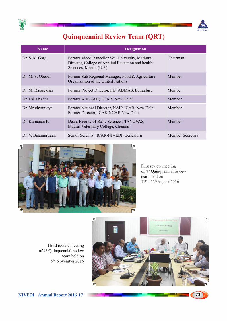
NIVEDI - Annual Report 2016-17 73
Quinquennial Review Team (QRT)
Name Designation
Dr. S. K. Garg Former Vice-Chancellor Vet. University, Mathura, Director, College of Applied Education and health Sciences, Meerat (U.P.)
Chairman
Dr. M. S. Oberoi Former Sub Regional Manager, Food & Agriculture Organization of the United Nations
Member
Dr. M. Rajasekhar Former Project Director, PD_ADMAS, Bengaluru Member
Dr. Lal Krishna Former ADG (AH), ICAR, New Delhi Member
Dr. Mruthyunjaya Former National Director, NAIP, ICAR, New DelhiFormer Director, ICAR-NCAP, New Delhi
Member
Dr. Kumanan K Dean, Faculty of Basic Sciences, TANUVAS, Madras Veterinary College, Chennai
Member
Dr. V. Balamurugan Senior Scientist, ICAR-NIVEDI, Bengaluru Member Secretary
First review meeting of 4th Quinquennial review team held on 11th - 13th August 2016
Third review meeting of 4th Quinquennial review
team held on 5th November 2016
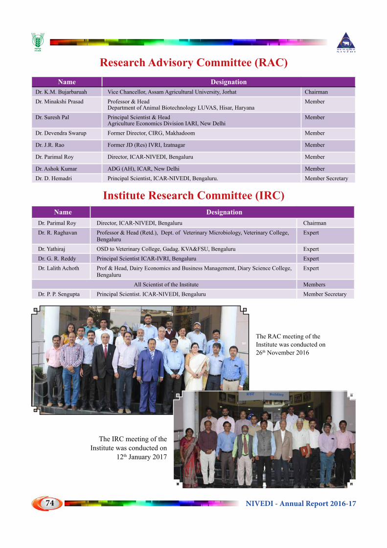
NIVEDI - Annual Report 2016-1774
Research Advisory Committee (RAC)Name Designation
Dr. K.M. Bujarbaruah Vice Chancellor, Assam Agricultural University, Jorhat ChairmanDr. Minakshi Prasad Professor & Head
Department of Animal Biotechnology LUVAS, Hisar, HaryanaMember
Dr. Suresh Pal Principal Scientist & HeadAgriculture Economics Division IARI, New Delhi
Member
Dr. Devendra Swarup Former Director, CIRG, Makhadoom Member
Dr. J.R. Rao Former JD (Res) IVRI, Izatnagar Member
Dr. Parimal Roy Director, ICAR-NIVEDI, Bengaluru Member
Dr. Ashok Kumar ADG (AH), ICAR, New Delhi MemberDr. D. Hemadri Principal Scientist, ICAR-NIVEDI, Bengaluru. Member Secretary
Institute Research Committee (IRC)Name Designation
Dr. Parimal Roy Director, ICAR-NIVEDI, Bengaluru ChairmanDr. R. Raghavan Professor & Head (Retd.), Dept. of Veterinary Microbiology, Veterinary College,
BengaluruExpert
Dr. Yathiraj OSD to Veterinary College, Gadag. KVA&FSU, Bengaluru ExpertDr. G. R. Reddy Principal Scientist ICAR-IVRI, Bengaluru ExpertDr. Lalith Achoth Prof & Head, Dairy Economics and Business Management, Diary Science College,
BengaluruExpert
All Scientist of the Institute MembersDr. P. P. Sengupta Principal Scientist. ICAR-NIVEDI, Bengaluru Member Secretary
The RAC meeting of the Institute was conducted on 26th November 2016
The IRC meeting of the Institute was conducted on
12th January 2017

NIVEDI - Annual Report 2016-17 75
Institutional Animal Ethics Committee (IAEC)Name Designation
Dr. Parimal Roy Director, ICAR-NIVEDI, Bengaluru ChairmanDr. S.P. Muthukumar CPCSEA Nominee MemberDr. B R Shome Principal Scientist, ICAR-NIVEDI, Bengaluru. MemberDr. Rajeswari Shome Principal Scientist, ICAR-NIVEDI, Bengaluru. MemberDr. Divakar Hemadri Principal Scientist, ICAR-NIVEDI, Bengaluru. MemberDr. P.P. Sengupta Principal Scientist, ICAR-NIVEDI, Bengaluru. MemberDr. P. Krishnamoorthy Scientist, ICAR-NIVEDI, Bengaluru. Member Secretary
Institute Management Committee (IMC)Name Designation
Dr. Parimal Roy Director, NIVEDI, Bengaluru ChairmanADG (AH) ICAR, New Delhi ICAR RepresentativeDr. R. Bhatta Director, NIANP, Bengaluru MemberDr. K.P. Ramesha Principal Scientist, Southern Region Station,
NDRI, BengaluruMember
Dr. B. R. Shome Principal Scientist, NIVEDI, Bengaluru MemberDr. A. N. Shylesha Principal Scientist, NBAIR, Bengaluru MemberMr. Rajeevlochana AAO, ICAR-NIVEDI, Bengaluru Member Secretary
ICAR-NIVEDI conducted 9th Institutional Animal Ethics Committee (IAEC) meeting on 30th December, 2016 under the Chairmanship of Dr. Parimal Roy, Director.
IMC Meeting held on 8th December 2016

NIVEDI - Annual Report 2016-1776
DISTINGUISHED VISITORS1. Dr. Trilochan Mohapatra, Secretary DARE & DG, ICAR
2. Dr. H. Rahman, DDG (AS), ICAR, New Delhi.
3. Dr .R.K. Singh, Director, ICAR-IVRI, Izatnagar
4. Dr. A. K. Srivatsava, Director, ICAR-NDRI, Karnal
5. Dr. K.M. Bujarbaruah, Vice-Chancellor, AAU, Guwahati
6. Dr. Madhan Mohan, Advisor, DBT, New Delhi
7. Dr. S. K. Garg, Director, CAEHS, Meerut.
8. Dr. M.S. Oberoi, Former Sub Regional Manager, FAO, United Nations
9. Dr. R.N. S. Gowda, Former Vice Chancellor, KVAFSU, Bidar
10. Dr. M. Rajasekhar, Former Project Director, PD_ADMAS, Bengaluru
11. Ashok Kumar, ADG (AH), ICAR, New Delhi
12. Dr. J R Rao, Former Professor & Head IVRI, Izatnagar
13. Dr. Minakshi Prasad, Professor, & Head, LUVAS, Hisar
14. Dr. Johanna, CDC, USA
15. Dr. Kayla Laserson, Country Director, CDC, India
16. Dr. Apoorva Chakraborthy, Director of Research, AAU, Guwahati
17. Dr. Lalkrishna, Sr. Consultant, (NER-BPMC), DBT, New Delhi
18. Prof. Alan Jenkins, Deputy Director, Water & Pollution Science Director, Centre for Ecology & Hydrology, UK
19. Prof. Mark Bailey, Director, Centre for Ecology & Hydrology, UK
20. Dr. S. Yathiraj, Dean Veterinary College, Bengaluru.
21. Dr. Raghavendra Bhatta, Director, ICAR-NIANP, Bengaluru.
22. Dr. Kumanan. K, Dean Faculty of Basic Sciences, TANUVAS, Chennai
23. Dr. B. N. Tripati, Director, ICAR-NRC on Equines, Hisar
24. Dr. Mruthyunjaya, Former National Director, NAIP
25. Dr. R. Raghavan, Retd. Professor, Dept. of Veterinary Microbiology, Veterinary College, Bengaluru
26. Dr. K. P. Ramesha, Station In-charge, ICAR-NDRI (SRS), Bengaluru
27. Dr. Lalith Achoth, Professor & Head, Dairy Science College, Bengaluru
28. Dr. G.R. Reddy, Principal Scientist, ICAR-IVRI, Bengaluru

NIVEDI - Annual Report 2016-17 77
STAFF POSITION DURING 2016-17Sl. No. NAME OF THE STAFF Designation
1 Dr. Parimal Roy Director (RMP)SCIENTIFIC STAFF
2 Dr. B.R. Shome Principal Scientist
3 Dr. (Mrs) R. Shome Principal Scientist
4 Dr. D. Hemadri Principal Scientist
5 Dr. P.P. Sengupta Principal Scientist
6 Dr. K.P. Suresh Principal Scientist
7 Dr. V. Balamurugan Senior Scientist
8 Dr. S.S. Patil Senior Scientist
9 Dr. Sathish B Shivachandra Senior Scientist
10 Dr. G. Govindaraj Senior Scientist
11 Dr. P. Krishnamoorthy Scientist
12 Dr. Md. Muddassar Chanda Scientist
13 Dr. Jagadish Hiremath Scientist
14 Dr. M. Nagalingam Scientist
15 Dr. (Mrs.). R. Sridevi Scientist
16 Dr. G. B. Manjunatha Reddy Scientist
17 Dr. (Mrs.). Siju Susan Jacob ScientistTECHNICAL STAFF
18 Dr. R. Yogisharadhya Senior Technical Officer
19 Dr. Awadesh Prajapati Senior Technical OfficerADMINISTRATIVE STAFF
20 Sh. Rajeevalochana Assistant Administrative Officer
21 Sh. Babu R. K Assistant Finance &Accounts Officer
22 Smt. Divya C. N Assistant
23 Sh. N. Narayanaswamy Assistant
24 Smt. A. Saranya Stenographer Grade-III
25 Mr. K. Vijayraj Stenographer Grade-III
26 Smt. G. C. Sridevi Lower Division Clerk
27 Sh. Gangadhareshwara Lower Division Clerk SKILLED SUPPORT STAFF
28 Sh. M. K Ramu Skilled Support Staff
29 Sh. Hanumanthuraju Skilled Support Staff
30 Mr. H. S. Umesh Skilled Support Staff
NIVEDI - Annual Report 2016-1778
Joining: Dr. Parimal Roy joined this Institute as Director ICAR - NIVEDI on 30-9-2016
Promotion: 1) Dr. Mohd. Mudassar Chanda, Scientist placed in Rs.7000/- RGP w.e.f.10-2-20142) Dr. R. Sridevi, Scientist placed in Rs.7000/- RGP w.e.f. 10-2-20133) Dr.M.Nagalingam, Scientist placed in Rs.7000/- RGP w.e.f.21-4-20144) Dr. Jagadish Hiremath, Scientist placed in Rs.7000/- RGP w.e.f. 1-3-2012
Probation clearance :1) Dr. R. Yogisharadhya , STO w.e.f. 16-12-2015
2) Dr. Awadhesh Prajapathi, STO w.e.f. 1-1-2016
Demise:
Dr. Gururao Shrinivas Desai (9th July, 1970 - 20th June 2016)
NIVEDI family is saddened to inform that our beloved colleague Dr. Gururao S. Desai, Principal Scientist has left for heavenly abode on 20th June 2016 following a brief illness. He joined at ICAR-NIVEDI, Bengaluru on 2nd May 2014 from ICAR-IVRI on transfer. Dr. Desai joined ARS in the discipline of Veterinary Microbiology at ICAR-NIHSAD (former HSADL), Bhopal on 25th November, 1999. He obtained his Ph.D., in Veterinary Virology from ICAR-IVRI, Izatnagar/Mukteswar as in-service candidate in 2007.
He served as the Biosafety Officer for the newly established state of art BSL2++ facility at ICAR- NIVEDI. His contributions in the field of conventional virology and molecular epidemiology of Classical Swine Fever, Bluetongue, PPR, Influenza, Ganjam disease and research contribution in NAIP and NICRA projects are highly recognized.
The NIVEDI family prays almighty for departed soul for eternal peace and gives enough strength to the bereaved family to bear his loss.

NIVEDI - Annual Report 2016-17 79
BUDGETRevised Estimate and Expenditure under Plan & Non Plan for 2016-17
(In lakh rupees)
Major Heads PLAN NON PLANRevised Estimate Expenditure Revised
Estimate Expenditure
Grants for Creation of Capital Assets (Capital)Works 376.15 376.15 Equipments 43.63 43.62 3.00 2.97Information Technology 1.19 1.17 Library Books & Journals 1.00 1.01 Furniture & Fixtures 2.54 2.54 1.00 1.00Grants in Aid - Salaries (Revenue) Establishment Expenses (Salaries) 405.00 405.00Grants in Aid - General (Revenue) Pension & Other Retirement BenefitsTravelling Allowances 10.00 10.00 2.00 2.00Research & Operational Expenses 101.00 101.00 62.00 62.00Administrative Expenses 25.50 25.49 161.50 161.49Miscellaneous Expenses 3.00 2.98 1.50 1.51AICRP on ADMAS 173.99 173.98 NEH 60.00 60.00 TSP 15.00 15.00 Grant Total 813.00 812.94 636.00 635.97
Revenue Receipts during 2016-17
Description Amount (Rs.)
License Fee 29760
Interest earned on loans & advances 209481
Application Fee from Candidates 1630
Interest earned on short term deposits 3024417
Receipt from Schemes 309391
Income generated on sale of kits 930366
Miscellaneous receipts 13352909
Total 17857954

NIVEDI - Annual Report 2016-1780

NIVEDI - Annual Report 2016-17 81
NIVEDI ACTIVITIES

NIVEDI - Annual Report 2016-1782

NIVEDI - Annual Report 2016-17 83
Dr. D. Hemadri, Pr. Scientist along with DBT-BBSRC FADH Project partners at University of Glasgow.
Dr. V. Balamurugan, Senior Scientist, participated in the DBT-BBSRC FADH PPR Annual Review Meeting
held at The Pirbright Institute, London, United Kingdom during 24-27th June, 2016.
Organization of International training cum workshop on to ‘Improve Diagnostic Capacity and Foster Regional Co-operation for Anthrax Control and Prevention’ from 14-16th June, 2016, at ICAR-NIVEDI. Delegates from Bangladesh, AICRP-ADMAS centers and Manipal University, participated in the programme.
Scientists of NIVEDI investigating Anthrax outbreak in Doddaballapura Taluk, Karnataka
NIVEDI Scientists providing demonstration and interaction with participants from Bangladesh about Biosafety (BSL-2+) laboratory procedures in handling and processing of clinical material as part of training cum workshop on Anthrax on 15th June, 2016, at ICAR-NIVEDI.
The staffs from ICAR-NIVEDI participated in the Krishi Unnati Mela 2017 organized at Indian Agricultural Research Institute, New Delhi during 15-17th February, 2017.

NIVEDI - Annual Report 2016-1784
ICAR Sponsored Short Course on “Advances livestock disease surveillance: Integration of molecular biology and statistical veterinary epidemiology” from 1st -10th September, 2016, ICAR-NIVEDI, Bengaluru.
Sixth Institute joint staff council of ICAR-NIVEDI meeting held on 20th January 2017 under chairmanship of Dr. Parimal Roy.
Inaugural session of “Sampling techniques and data analysis using GIS” training programme organized by ICAR-NIVEDI in collaboration with ADMaC, Core Laboratory-I, AAU, College of Veterinary Science, Guwahati from 2nd to 4th March, 2017.
Inaugural session of NSPAAD website at the Annual Review Meeting from 27th to 29th May, 2016.
NIVEDI staffs participating in the essay writing competition held during Hindi Week from 14 to 20th Sept 2016.
Quarterly meeting of Institute Hindi Implementation Committee under the chairmanship of Director NIVE-DI.

NIVEDI - Annual Report 2016-17 85
International Women’s Day celebration-2017 at ICAR-NIVEDI, Bangalore.
Brucellosis 2016 International Research Conference New Delhi, India November 17-19, 2016.
Dr. Trilochan Mohapatra, Secretary, DARE and Director General, ICAR laid the Foundation stone for the Training cum Farmers Hostel of ICAR-NIVEDI, Bengaluru on 19th May, 2016. Dr. H. Rahman, DDG (AS), Dr. A.K. Srivatsava, Director, ICAR-NDRI, Dr.Raghavendra Bhatta, Director, ICAR-NIANP and Dr. A. Sanyal, Joint Director, ICAR-IVRI, Bengaluru Campus were present.
Sensitization training program to Veterinarians of Ani-mal Husbandry Department, Puducherry under Brucel-losis Control Program on 27.04.2016 by ICAR-NIVE-DI Scientists.
NIVEDI Scientist visited selected village in Shimo-ga district of Karnataka to collect data and samples to evaluate the Vaccine Effectivenss of vaccine against FMD on 11th September 2016.
Training of BVSC internship students from KVAFSU, Bengaluru

NIVEDI - Annual Report 2016-1786
World Veterinary Day was celebrated with Guest lecture delivered by Dr. N. Balakrishnan, Joint Director, National Centre for Disease Control, Bengaluru on 30th April, 2016.
Dr. Parimal Roy, Director and Scientists interacted with officers of M/s. Merck India Pvt Ltd, Mumbai on development of seroepidemiological survey studies on IBR and Leptospirosis models under Public Private Partnership Projects on 7th November, 2016.
Organized Diagnostic Health Camp in association with RV Metropolis Diagnostic and Health care, Bengaluru for all the staff of ICAR-NIVEDI on the eve of Institute Foundation day on 1st July, 2016.
70th Independence day was celebrated at NIVEDI on 15th August, 2016.
Vigilance awareness week was celebrated during 31st October - 5th November, 2016 at ICAR-NIVEDI and various competitions and activities were organized on this occasion.
Agricultural Education day was celebrated at ICAR-NIVEDI by organizing the competitions to Gov-ernment School children from Ramagondanahalli on 2nd December, 2016.
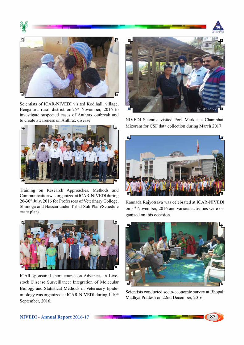
NIVEDI - Annual Report 2016-17 87
Scientists of ICAR-NIVEDI visited Kodihalli village, Bengaluru rural district on 25th November, 2016 to investigate suspected cases of Anthrax outbreak and to create awareness on Anthrax disease.
Training on Research Approaches, Methods and Communication was organized at ICAR-NIVEDI during 26-30th July, 2016 for Professors of Veterinary College, Shimoga and Hassan under Tribal Sub Plam/Schedule caste plans.
ICAR sponsored short course on Advances in Live-stock Disease Surveillance: Integration of Molecular Biology and Statistical Methods in Veterinary Epide-miology was organized at ICAR-NIVEDI during 1-10th September, 2016.
NIVEDI Scientist visited Pork Market at Champhai, Mizoram for CSF data collection during March 2017
Kannada Rajyotsava was celebrated at ICAR-NIVEDI on 3rd November, 2016 and various activities were or-ganized on this occasion.
Scientists conducted socio-economic survey at Bhopal, Madhya Pradesh on 22nd December, 2016.

NIVEDI - Annual Report 2016-1788
ICAR-NIVEDI organized Sensitization training for the implementation of Brucellosis control programme for field veterinarians of Dept. of Animal Husbandry, Govt. of Kerala on 4th November, 2016.
ICAR-NIVEDI participated in the exhibition of XIII Agricultural Science Congress 2017 organized at GKVK, UAS Bengaluru from 21 - 24th February 2017
ICAR-NIVEDI participated in the exhibition of Regional Horticulture fair organized at ICAR-IIHR Bengaluru from 15 - 19th January 2017
Vigilance awareness week was celebrated during 31st October - 5th November, 2016 at ICAR-NIVEDI and various competitions and activities were organized on this occasion.
Brucellosis 2016- International Research Conference, 17th to 19th December, 2016 at NAAS Complex, New Delhi, India.
Dr. Trilochan Mohapatra, Secretary, DARE and Director General, ICAR and Dr. J. K. Jena, DDG (Fisheries) along with ICAR Animal Science Institute Directors and Scientists in interactive meeting held on 14th January 2017 at ICAR-NIANP, Bengaluru
2nd Annual Review meet of DBT-NER-Advanced An-imal Disease Diagnosis and Management Consortium (ADMaC) held at ICAR-NIVEDI, Bengaluru on 25th November, 2016.

NIVEDI - Annual Report 2016-17 89
Infrastructure Development : Completed
Infrastructure Development : Ongoing
Solar panel (50 kw) has been installed on the roof top of the Laboratory Building
Aadhaar based biometric attendance system Swiping Machine (POS) : Cashless transaction
Director’s Residence Building 1st and 2nd Floor Utility Building
Farmers’ Hostel
Digital Franking Machine : Cashless transaction

NIVEDI - Annual Report 2016-1790
Collaborative Linkages with ICAR-NIVEDIICAR-NIVEDI has established Memorandum of Understanding (MoU) with Universities and IIT for collaborative research :
SKUAST-Kashmir FVSc & AH, Srinagar, Jammu and Kashmir (07.09.2016 to 06.08.2021)
Department of Biosciences and Bioengineering, IIT, Guwahati (26.04.2016 to 25.03.2021)
College of Veterinary Sciences & Animal Husbandry, Bhubaneswar, Odisha, OUAT (28.06.2016 to 27.05.2021)