Embed Size (px)
Citation preview
Anterior Intraparietal Sulcus is Sensitiveto Bottom–Up Attention Driven
by Stimulus Salience
Joy J. Geng and George R. Mangun
Abstract
& Frontal eye fields (FEF) and anterior intraparietal sulcus (aIPS)are involved in the control of voluntary attention in humans,but their functional differences remain poorly understood. Weexamined the activity in these brain regions as a function oftask-irrelevant changes in target and nontarget perceptual sa-lience during a sustained spatial attention task. Both aIPS and FEFwere engaged during selective attention. FEF, but not aIPS, was
sensitive to the direction of spatial attention. Conversely, aIPS, butnot FEF, was modulated by the relative perceptual salience of thetarget and nontarget stimuli. These results demonstrate separableroles for FEF and aIPS in attentional control with FEF moreinvolved in goal-directed spatial attention and aIPS relatively moresensitive to bottom–up attentional influences driven by stimulussalience. &
INTRODUCTION
The ability to filter out distracting visual information iscritical for successful performance of an individual’sgoals. For example, it is vital to pay attention to theroad when driving despite the fact that an accident hasoccurred on the side, but it is also necessary to maintaina certain level of unfocused vigilance in case an unex-pected, but behaviorally relevant, event occurs, such asa car suddenly merging into your lane. The tension be-tween maintaining voluntary attention on task-relevantobjects and automatic orienting toward perceptuallysalient, but currently task-irrelevant, events has a longhistory within the psychological literature (e.g., Proulx& Egeth, 2006b; Berger, Henik, & Rafal, 2005; Geng &Behrmann, 2005; Kristjansson, Wang, & Nakayama, 2002;Yantis & Egeth, 1999; Lavie, 1995; Theeuwes, 1995; Muller& Rabbitt, 1989; Nakayama & Mackeben, 1989; Jonides& Yantis, 1988; Gibson, 1986; Eriksen & Yeh, 1985;James, 1890/1983; Jonides & Irwin, 1981; Posner, Snyder,& Davidson, 1980). These studies report costs in task-related behavioral outcomes (e.g., lower accuracy and/or slower reaction times) when nontargets have greaterperceptual salience and/or similarity with the target.Although there is disagreement regarding whether com-pletely task-irrelevant stimuli can capture attention ornot (e.g., Leblanc, Prime, & Jolicoeur, 2008; Hickey,McDonald, & Theeuwes, 2006), there is good agree-ment that behavioral decrements in target discrimination
reflect nontarget competition for target-related atten-tional processing, which lead to attentional capture bythe nontarget.
The neural system for controlling voluntary visual at-tention in humans is well known to include the frontaleye fields (FEF) and areas in the parietal cortex, par-ticularly the intraparietal sulcus (IPS) and the superiorparietal lobule (SPL) (e.g., Kelley, Serences, Giesbrecht,& Yantis, 2007; Serences & Yantis, 2007; Slagter et al.,2007; Sylvester, Shulman, Jack, & Corbetta, 2007; Genget al., 2006; Shomstein & Behrmann, 2006; Kincade,Abrams, Astafiev, Shulman, & Corbetta, 2005; Kristjansson,Vuilleumier, Malhotra, Husain, & Driver, 2005; Behrmann,Geng, & Shomstein, 2004; Corbetta & Shulman, 2002;Yantis et al., 2002; Driver & Frackowiak, 2001; Corbetta,Kincade, Ollinger, McAvoy, & Shulman, 2000; Hopf et al.,2000; Hopfinger, Buonocore, & Mangun, 2000; Nobre,Gitelman, Dias, & Mesulam, 2000; Gitelman et al., 1999;Paus, 1996). These areas have been characterized as at-tentional ‘‘control’’ regions that bias responses in thesensory cortex (e.g., by increasing response gain) associ-ated with the attended location or feature (see alsoBestmann, Ruff, Blakemore, Driver, & Thilo, 2007; Liu,Larsson, & Carrasco, 2007; Serences & Boynton, 2007;McMains & Somers, 2004; Hopfinger et al., 2000; Kastner& Ungerleider, 2000; Brefczynski & DeYoe, 1999; Gandhi,Heeger, & Boynton, 1999; Luck, Chelazzi, Hillyard, &Desimone, 1997; Chelazzi et al., 1995); Mircrostimulationand transcranial magnetic stimulation of FEF and theparietal cortex in monkeys and humans have shown di-rect modulation of activity in the retinotopic visual cortex,which results in increased perceptual sensitivity to stimuliUniversity of California, Davis
D 2008 Massachusetts Institute of Technology Journal of Cognitive Neuroscience 21:8, pp. 1584–1601
in corresponding locations of the visual field (e.g., Ruffet al., 2006, 2008; Armstrong, Fitzgerald, & Moore, 2006;Chambers, Stokes, Janko, & Mattingley, 2006; Mevorach,Humphreys, & Shalev, 2006; Muggleton et al., 2006;Silvanto, Lavie, & Walsh, 2006; Hung, Driver, & Walsh,2005; O’Shea, Muggleton, Cowey, & Walsh, 2004; Moore& Armstrong, 2003; Grosbras & Paus, 2002). These stud-ies suggest that frontal and parietal areas contain repre-sentations of attentional priority that selectively enhanceprocessing in the sensory cortex of the related stimu-lus or feature. Such attentional enhancement of thesensory cortex leads in turn to better behavioral perfor-mance as indexed by faster detections and more accuratediscriminations.
In comparison to studies of voluntary attentional se-lection, there have been relatively few studies examiningthe representation of sensory-driven attentional pro-cesses. These studies frequently employed spatial cuesthat either incorrectly predicted the location of an up-coming target stimulus or captured attention automati-cally (e.g., Posner et al., 1980). For invalid spatial cues,effects of sensory-based attentional reorienting towardthe target have been reported in the temporal–parietaljunction (TPJ) near the intersection of the angular gy-rus and the superior temporal lobe (Corbetta, Patel, &Shulman, 2008; He et al., 2007; Indovina & Macaluso,2007; Astafiev, Shulman, & Corbetta, 2006; Vossel, Thiel,& Fink, 2006; Kincade et al., 2005; Thiel, Zilles, & Fink,2004; Mort et al., 2003; Corbetta & Shulman, 2002;Downar, Crawley, Mikulis, & Davis, 2000; Friedrich, Egly,Rafal, & Beck, 1998). Importantly, under conditions ofinvalid spatial cues, sensory-driven attentional reorient-ing was always in response to violations in task-relatedexpectancies. In the case of exogenous spatial cues thatautomatically capture attention, more mixed results havebeen reported. Some studies have also found exogenouscues to activate the inferior parietal lobe in or withinthe vicinity of TPJ (Mayer, Dorflinger, Rao, & Seidenberg,2004; Peelen, Heslenfeld, & Theeuwes, 2004; Mort et al.,2003; Corbetta et al., 2000; Rosen et al., 1999), but othershave also reported overlap with structures involved involuntary attentional tasks such as FEF and the su-perior parietal cortex (e.g., Kincade et al., 2005; Mayer,Dorflinger, et al., 2004; Mayer, Seidenberg, Dorflinger, &Rao, 2004; Peelen et al., 2004; Kim et al., 1999; Rosen et al.,1999). Among these studies however, it was not alwaysclear whether areas were driven specifically by the exog-enous cue or also by the appearance of the target. Whenthe exogenous cue was dissociated from the appearanceof the target, the overlap with areas involved in voluntaryattention was considerably less (e.g., Kincade et al., 2005).
In addition to studies with a spatial cue, sensory-driven attentional processes have also been examinedby manipulating target and nontarget similarity. Forexample, Serences and Yantis (2007) examined the ef-fects of ‘‘contingent’’ attentional capture by nontargetsthat share the target’s color (and therefore the sub-
jects’ attentional control settings). Greater activation wasfound in contralateral IPS and FEF in response to target-colored nontargets compared to non-target-colorednontargets, demonstrating that target-colored nontar-gets were more likely to capture attention and havehigher attentional priority (Serences & Yantis, 2007;Mevorach et al., 2006; Serences et al., 2005). The ideathat frontal and parietal areas operate as an attentional‘‘salience’’ or ‘‘priority’’ maps (e.g., Gottlieb, 2007; Itti& Koch, 2000) that code the behavioral relevance andperceptual salience of objects has also been suggestedby results from single-cell recording studies in mon-key FEF (e.g., Balan & Gottlieb, 2006; Bichot & Schall,2002) and the lateral intraparietal sulcus (LIP; Bisley &Goldberg, 2006; Goldberg, Bisley, Powell, & Gottlieb,2006; Ipata, Gee, Gottlieb, Bisley, & Goldberg, 2006;Kusunoki, Gottlieb, & Goldberg, 2000; Platt & Glimcher,1999; Thompson, Bichot, & Schall, 1997). Here, the de-gree to which a stimulus drove neuronal firing was de-termined by goal-directed attributes such as being thetarget but also by perceptual qualities such as luminanceor contrast. Similar to many human studies, however,the sensory-driven properties were also often relatedto the target (e.g., by being a potential target in a visualsearch display, or by predicting the location of an up-coming target).
Taken together, these previous studies suggest thatFEF and IPS may not be strictly a ‘‘voluntary’’ attentionalsystem, but rather reflect the integration of attentionalpriority for various objects in the visual field. In thisstudy, we explored the degree to which FEF and IPSeach represent sensory-driven attentional orienting. Weused a sustained spatial attention task in which the lo-cation of the target was blocked and known in advanceof each target appearance. Subjects were explicitly cuedwith 100% validity to attend to one of two ‘‘mask’’stimuli located in the lower left and right visual fieldsso that there was no ambiguity regarding the locationto which spatial attention should be directed. Percep-tual salience was manipulated by luminance and contrast(Proulx & Egeth, 2006a; Carrasco, Ling, & Read, 2004;Reynolds & Desimone, 2003; Bundesen, 1990), and wasrandomly assigned to be a feature of the attended or un-attended object, or be absent.
Importantly, perceptual salience was irrelevant to tar-get detection, discrimination, or response and, therefore,any attentional capture by a salient stimulus (and partic-ularly a salient nontarget) can be understood as beingsensory-driven and not dependent on target-related at-tentional control settings. The location of the target andthe required discrimination task was completely orthog-onal to the feature of perceptual salience and this designconstituted a conservative test of sensory-driven atten-tional processing. We additionally included a control con-dition in which the same stimuli were presented underconditions of passive viewing, which controlled for visualeffects when no attention was required.
Geng and Mangun 1585
To anticipate our results, we found that both the ante-rior IPS (aIPS) and FEF were engaged during selectiveattention, but that FEF was additionally sensitive to thedirection of goal-directed spatial attention. Conversely,aIPS integrated voluntary and sensory-driven attentionsuch that activation was greatest for salient nontargets.aIPS activation also correlated strongest with RT when thenontarget was salient. These results demonstrate separa-ble roles for FEF and aIPS in attentional control, with FEFmore involved in goal-directed spatial attention and aIPSintegrating information about the perceptual saliency ofvisual inputs during attention.
METHODS
Subjects
Fifteen volunteers (6 women, 15 right-handed), ranging inage from 18 to 34 years in age, participated. All werescreened for MRI compatibility and gave written informedconsent in accord with the local ethics clearance as ap-proved by NIH. All had normal or corrected-to-normalvision. Data from three subjects were excluded becausethey failed to perform the task to criterion (70% accuracy).
Experimental Design
The experiment had a 3 � 3 factorial design. The factorswere location of cued spatial attention (left, right, pas-sive) and location of a perceptually salient stimulus (left,right, absent). Spatial attention was mini-blocked ingroups of six trials. Within each block, salience was pseu-dorandomly selected for each trial, with the constraint
that each block contained two samples from each saliencecondition. Each block lasted 24 sec and was followed bya 12-sec period of blank fixation. Each of the three at-tention blocks occurred in a random order within a meta-block that included one sample of each spatial attentioncondition and three blank fixation periods. Four meta-blocks were included in each of five sessions.
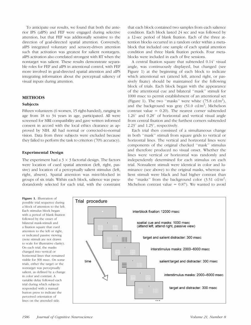
A central fixation square that subtended 0.148 visualangle, was continuously displayed, but changed (seeFigure 1) at the beginning of each block to indicatewhich attentional set (attend left, attend right, or pas-sively fixate) should be maintained for the followingblock of trials. Each block began with the appearanceof the attentional cue and bilateral ‘‘mask’’ stimuli for1000 msec to permit establishment of an attentional set(Figure 1). The two ‘‘masks’’ were white (75.8 cd/m2),and the background was gray (51.0 cd/m2; Michelsoncontrast value = 0.20). The nearest corner subtended1.268 and 0.288 of horizontal and vertical visual anglefrom central fixation and the furthest corners subtended2.238 and 1.258, respectively.
Each trial then consisted of a simultaneous changein both ‘‘mask’’ stimuli from square grids to vertical orhorizontal lines. The vertical and horizontal lines werecomponents of the original checked ‘‘mask’’ stimulusand therefore produced no visual onset. Whether thelines were vertical or horizontal was randomly andindependently determined for each stimulus on eachtrial. Nonsalient stimuli were identical in color and lu-minance (see above) to the original masks, whereas sa-lient stimuli were black and had higher contrast thanthe ‘‘masks’’ from the background color (3.51 cd/m2;Michelson contrast value = 0.87). We wanted to avoid
Figure 1. Illustration of
possible trial sequence duringa block of attention to the left.
Each stimulus block began
with a period of blank fixation
followed by the onset ofbilateral mask-stimuli and
a fixation square that cued
attention to the left or right,
or indicated passive viewing(note stimuli are not drawn
to scale for illustrative clarity).
On each trial, the masks
changed into vertical orhorizontal lines that remained
visible for 300 msec. On some
trials, either the target or thenontarget was perceptually
salient, as defined by a change
in color and contrast. A
variable delay followed eachtrial during which subjects
responded with a manual
button press to indicate the
perceived orientation oflines on the attended side.
1586 Journal of Cognitive Neuroscience Volume 21, Number 8
using visual onsets to capture attention and thereforechose a change in color and luminance to define per-ceptual ‘‘salience,’’ a method known to capture at-tention (e.g., Carrasco, 2006; Proulx & Egeth, 2006a;Carrasco et al., 2004; Reynolds & Desimone, 2003;Bundesen, 1990). The target and nontarget were visi-ble for 300 msec after which the ‘‘mask’’ gratings reap-peared (Figure 1).
The average intertrial interval varied between 2 and6 sec to allow for uniform sampling of event-relatedBOLD responses across the whole TR and to maintainbehavioral uncertainty regarding the onset of the nexttarget stimulus. Subjects were instructed to indicate viaright-handed button press using an MRI-compatible re-sponse box whether the stimulus on the attended sidewas composed of vertical or horizontal lines. Mappingof response choice to the index or middle finger wascounterbalanced across subjects. Behavioral trainingwas conducted outside the scanner until the participantunderstood the task and demonstrated the ability toperform correctly. All subjects were instructed to main-tain visual fixation throughout the experiment and theireye position was monitored throughout the experi-ment (see below). Stimuli were presented via a videoprojector and front projection screen. The screen wasviewed via a mirror system attached to the head coil.
Eye Tracking
Eye tracking was performed at 60 Hz using AppliedScience Laboratories (ASL, Bedford, MA) model 504 withlong-range remote optics. Data were acquired from 10out of 12 subjects; two subjects could not be adequatelytracked. For analysis, eye position data were filteredby removing a linear trend and values exceeding ap-proximately twice the distance of the furthest edge ofthe stimulus, which could be caused by artifacts unre-lated to the experimental manipulation such as blinks,or loss of data. The data were then smoothed with a5-point moving average to remove remaining noiseartifacts.
Image and Data Processing
MRI data were acquired from a 3-T Siemens Trio scanner(Siemens, Erlangen, Germany) equipped with an eight-channel phased array head coil. A T2*-weighted echo-planar imaging (EPI) sequence was used to acquirevolumes of 34 slices of 3 mm thickness (3 � 3 mm in-plane resolution) with a distance factor of 10%, every2000 msec. Slices were axially oriented and coveredthe whole brain. Two hundred twenty-two volumeswere collected in each session of five sessions. Imagedata were analyzed using SPM5 (Wellcome Departmentof Imaging Neuroscience, London, UK; Friston et al.,1995). Prior to statistical analysis, the first four volumes
were discarded to allow for T1 equilibrium effects. Im-ages were realigned and unwarped to correct for inter-actions between movement and field inhomogeneities(Andersson, Hutton, Ashburner, Turner, & Friston, 2001);normalized to the MNI EPI template available in SPM 5,and resampled to a resolution of 2 � 2 � 2 mm. Dataused in group image analyses were additionally smoothedwith a three-dimensional 6-mm FWHM Gaussian kernel.Region-of-interest (ROI) data extracted from each partic-ipant separately were not spatially smoothed.
High-resolution (1 � 1 � 1) T1-weighted structuralimages were acquired using an MP-RAGE sequence, co-registered with each subject’s EPI images, and normalizedto the MNI template brain. These were used for identify-ing individual anatomical landmarks for ROI selection (seebelow). An average structural image was created from thenormalized T1-weighted images for the purpose of dis-playing functional results from the group.
Experimental factors were first modeled for eachsubject by a stick function convolved with a canonicalhemodynamic response function. Linear contrasts of pa-rameter estimates were estimated for each subject andcombined for the group level in random effects generallinear model. Results from the group level were used toguide individual ROI selection (see below). Behavioraldata and image data extracted from individual ROIs wereanalyzed using R software (www.r-project.org).
Regions of Interest Identification in Individuals
Six ROIs corresponding to human FEF, aIPS, and dorsalmiddle occipital gyrus (dMOG) bilaterally were extractedfrom each subject. In contrast to data used for groupanalysis, data extracted from individual ROIs were notadditionally spatially smoothed after realignment and nor-malization. Selection of ROIs was accomplished by usingresults from group random effects analyses to guide se-lection of the functional peak in each individual nearestto anatomical landmarks. Using general task constraintscombined with anatomical criteria provided individualspecificity while still maintaining the advantages groupstatistics that permit generalization to the population(Ikkai & Curtis, 2007; Stephan, Marshall, Penny, Friston,& Fink, 2007).
For aIPS and FEF, coordinates from the group ran-dom effects analyses were based on the contrast of at-tention minus passive viewing, ( p < .0001, cluster size�10 voxels; Table 1) and those for the left and rightdMOG were based on contrasts between contralateraland ipsilateral attention to isolate regions with visual rep-resentations corresponding to the lower left and rightvisual field, ( p < .001, cluster size �10 voxels; Table 1).Data from one hemisphere could not be reliably ex-tracted for two subjects in dMOG and data from thosesubjects were not included in ROI analyses.
The ROI-defining contrasts were orthogonal to anycomparisons involving the salience conditions, which
Geng and Mangun 1587
were the subject of primary investigation. The ROI cen-ter within each individual was determined by the localmaximum ( p < .001 and p < .01, for attention minuspassive viewing and attention left vs. right, respectively)closest to peak coordinates from the correspondinggroup random effects analysis, in the appropriate ana-tomical landmark (Table 2). Anatomical definitions ofeach region were as follows: the anterior portion withinthe depth of IPS (e.g., Ikkai & Curtis, 2007; Kincade et al.,2005; Donner, Kettermann, Diesch, Villringer, & Brandt,2003; Wojciulik & Kanwisher, 1999), the junction be-tween the middle frontal gyrus and the precentral sulcusfor FEF (e.g., Connolly, Goodale, Menon, & Munoz, 2002;Paus, 1996), and dMOG (e.g., Hopfinger, Woldorff, Fletcher,& Mangun, 2001).
The ROI center was located at the peak of activationnearest to the group coordinate within the appropriateanatomical region and included voxels within a spherewith a radius of 2 mm (see Figure 2; Tables 1 and 2). In
addition to identifying specific voxels, we extracted thetime series from all voxels within each ROI using theeigenvariate tool in SPM 5. The time series was trans-formed into percent signal change by normalizing valuesby the baseline signal during blank fixation. Using coor-dinates from the group results as guides provided someuniformity between subjects and generality to the pop-ulation, but locating the ROI center based on individualfunctional contrasts and anatomy resulted in greaterprecision. All ROIs were identified in both hemispheres,and where no differences arose between hemispheres,the data were collapsed.
RESULTS
Behavioral Response and Eye Position Analyses
Subjects were instructed to only attend to the stimulusin the cued visual field and report the orientation of
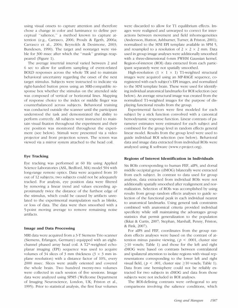
Table 1. Significant Cortical and Subcortical Regions from Whole-brain Group Analysis
Cluster Size t Score Z Score x y z mm Region Name
Attention Left + Right > Passive Viewing
465 8.94 4.73 �28 �48 44 L intraparietal sulcus
13.98 5.58 �40 �40 46
5.52 �20 �66 46 L posterior intraparietal sulcus
12 6.18 3.98 32 �50 44 R intraparietal sulcus
18 7.38 4.35 44 �36 46 R intraparietal sulcus
334 11.17 5.16 �36 �4 50 L posterior middle frontal gyrus (frontal eye fields)
39 6.15 3.97 32 �4 48 R posterior middle frontal gyrus (frontal eye fields)
392 9.86 4.92 �4 4 52 L + R medial frontal gyrus
44 8.13 4.54 �60 8 26 L inferior frontal gyrus
1250 14.25 5.62 �24 12 4 L putamen
502 11.01 5.14 22 10 4 R putamen
52 8.55 4.64 �32 �34 74 L postcentral gyrus
10 7.46 4.37 �6 2 10 L thalamus
11 6.34 4.03 18 0 14 R thalamus
Attention Left > Attention Right
145 6.91 4.21 28 �98 18 R cuneus and middle occipital gyrus
Attention Right > Attention Left
175 9.25 4.80 �22 �100 6 L cuneus and middle occipital gyrus
96 6.19 3.98 �28 �66 �12 L fusiform gyrus
40 5.87 3.87 16 �2 46 R cingulate gyrus
18 5.30 3.66 �40 �76 6 L middle occipital gyrus
15 5.03 3.55 �36 �80 �10 L inferior occipital gyrus
1588 Journal of Cognitive Neuroscience Volume 21, Number 8
lines whenever the ‘‘mask’’ was replaced with a target.On some trials, either the target in the attended locationor the nontarget in the unattended location was per-ceptually salient (see Methods). Subjects were informedthat perceptual salience was task-irrelevant and that they
should instead always attend to the cued stimulus. Theprimary question in this experiment dealt with the effectof task-irrelevant perceptual salience on behavioral andbrain responses to a voluntarily attended lateralizedstimulus.
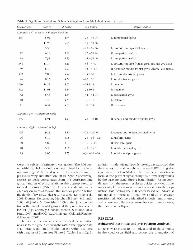
Table 2. Individual and Mean Coordinate Values for Regions of Interest
Anterior Intraparietal Sulcus Frontal Eye Fields Middle Occipital Gyrus
Subject L x y z mm R x y z mm L x y z mm R x y z mm L x y z mm R x y z mm
1 �26 �42 42 36 �42 42 �28 �6 50 30 �6 48 �30 �92 12 34 �88 18
2 �32 �48 40 40 �40 42 �38 �12 52 36 �10 48 �22 �90 8 – – –
3 �28 �46 42 38 �44 44 �38 �6 50 38 �4 64 �26 �92 6 32 �92 14
4 �30 �38 46 34 �44 48 �28 �10 60 38 �4 52 – – – 26 �90 10
5 �30 �50 44 40 �50 44 �32 �2 56 36 0 50 �26 �96 16 30 �92 24
6 �32 �52 56 32 �56 42 �26 �8 50 30 �8 54 �26 �98 16 22 �100 8
7 �42 �42 54 36 �32 40 �20 �10 58 28 0 56 �28 �98 2 22 �94 6
8 �28 �44 40 36 �44 42 �38 �8 48 32 �10 50 �24 �94 14 26 �92 2
9 �26 �46 52 34 �46 50 �28 �8 50 30 �2 48 �24 �94 6 34 �92 8
10 �30 �36 46 34 �32 42 �26 �6 48 36 �4 52 �18 �92 4 22 �90 10
11 �40 �36 38 38 �38 38 �28 �6 44 46 �2 48 �22 �92 8 24 �100 4
12 �40 �44 44 34 �42 40 �32 0 56 38 �4 48 �26 �98 6 22 �88 2
Mean �32 �44 45 36 �43 43 �30 �7 52 35 �5 52 �25 �94 9 27 �93 10
Figure 2. (A) Behavioral reaction time (RT; left) and accuracy (right) data showing a main effect of perceptual salience. Responses were
significantly slower and less accurate when the nontarget was perceptually salient than all other conditions. (B) Eye position data showing the
proportion of time spent at a distance from central fixation (in degrees of visual angle). The three cued spatial attention conditions of left, right,and passive are plotted in blue, red, and gray, respectively. The panels correspond to pretarget, target–target, and posttarget time epochs.
Geng and Mangun 1589
Behavioral data from the six conditions of main in-terest given by crossing spatial attention (left, right) andperceptual salience (attended target, unattended non-target, absent) were entered into a repeated measuresANOVA, which resulted in only a significant main effectof perceptual salience [RT: F(2, 22) = 20.3, p < .0001,Figure 2A, left; Accuracy: F(2, 22) = 5.4, p < .05, Fig-ure 2A, right]. Paired t tests demonstrated significantdifferences between all salience conditions in RT, andbetween salient attended targets and unattended non-targets in the accuracy data (Bonferroni corrected,p < .05). Salient nontargets interfered with responses,verifying that our experimental manipulation of saliencewas effective in capturing attention even when the lo-cation of the target was known in advance.
Eye position data were successfully collected from10 subjects during the fMRI experiment. Data from cor-rect trials that were also included in the fMRI analyseswere analyzed. For each subject, eye position data fromeach of the three attentional cue conditions (left, right,passive) were divided into the following time epochs: a500-msec ‘‘pretarget’’ period prior to the onset of thetarget and the nontarget, a 300-msec ‘‘target-visible’’period during which the target and the nontarget werevisible, and a 500-msec ‘‘posttarget’’ period followingthe reappearance of the mask stimuli. Histograms of
the proportion of time spent from central fixation in de-grees of visual angle during each of attention condi-tion and time epoch are plotted in Figure 2B. More than87% of the time was spent within 18 of visual angle ineach of the time epochs, indicating that subjects wereable to maintain central fixation. Importantly, when therecorded eye position was outside of 18 (<13% of total),there were no differences between any attention con-ditions (left, right, passive) in time spent either to theleft or right of fixation [i.e., eye positions were notskewed toward the attended side; all t(9) < 1.2, p > .28;see Figure 2B]. Subjects were able to perform the taskwithout differences in eye position between the differentconditions of spatial attention and, therefore, activationsobserved during imaging could not be a consequenceof systematic differences in eye movements betweenconditions.
Cued Spatial Attention
In order to select functional coordinates for aIPS andFEF ROIs based on the general attentional demands ofthe task, we contrasted trials with lateralized attentionwith passive viewing (see Methods, Figure 3A, Table 1 forresults from whole-brain analyses, and Table 2 for indi-vidual ROI coordinates). To determine if the attentional
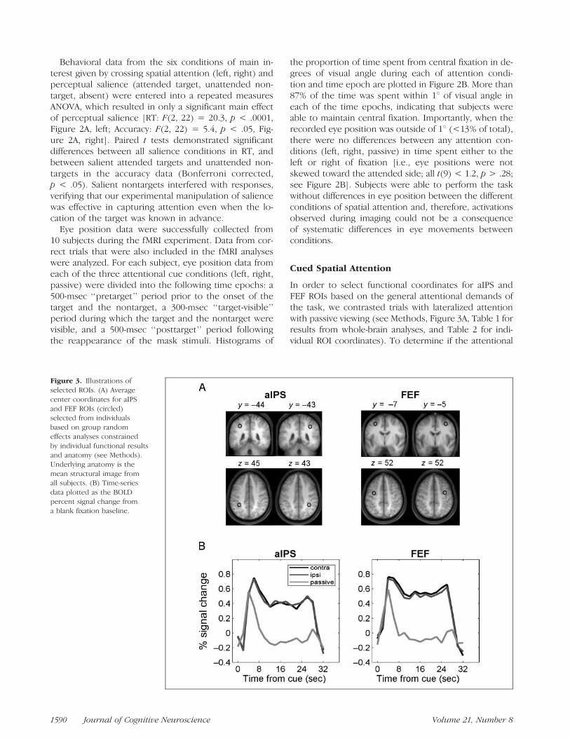
Figure 3. Illustrations of
selected ROIs. (A) Average
center coordinates for aIPS
and FEF ROIs (circled)selected from individuals
based on group random
effects analyses constrained
by individual functional resultsand anatomy (see Methods).
Underlying anatomy is the
mean structural image fromall subjects. (B) Time-series
data plotted as the BOLD
percent signal change from
a blank fixation baseline.
1590 Journal of Cognitive Neuroscience Volume 21, Number 8
modulations within aIPS and FEF depended on theduration of the attentional block, we examined the BOLDtime series beginning from the first onset of the spatialcue (and masks; see Methods) until the end ofthe attentional block 24 sec later.
The results are plotted in Figure 3 and demonstratethat responses in aIPS and FEF were greater for bothcontralateral and ipsilateral attention than for passiveviewing beginning 6 sec after the cue onset and lastinguntil 4 and 2 sec after the offset of the spatial cue atthe end of the block, respectively [all t(11) > 2.6, p <.05 with Bonferroni correction; Figure 3B]. The apparentrapid return to baseline is likely due to subjects dis-engaging their attention from the task immediately afterresponding to the sixth and final target, which was stillfollowed by an average interstimulus interval (ISI) of4 sec. The substantial and sustained difference from pas-sive viewing demonstrates that the responses in aIPSand FEF were not due to alerting or visual effects relatedto the onset of stimuli, but rather due to their involve-ment in establishing and maintaining task-related atten-tional control throughout the entire block. In additionto their similarities, FEF activation peaked slightly earlierat 4 sec after cue onset and aIPS activation peaked 6 secafter cue onset.
Effect of Perceptual Salience
Having established the presence of voluntary attentionalresponses in aIPS and FEF, we next tested for additional
sensitivity in each ROI to the manipulation of task-irrelevantperceptual salience. Recall that salient nontargets cap-tured attention away from the target, as indexed by slowerand less accurate behavioral responses to the target whenthe nontarget was perceptually salient (see Figure 2A). Abrain area that codes the attentional priority of objectsbased on both goal-directed and sensory-driven infor-mation should similarly have greatest activation in re-sponse to perceptually salient nontargets. In contrast, anarea with a specific spatial representation of the targetlocation, which would not change based on features ofthe unattended stimulus, should not be modulated byperceptual salience.
We compared for each ROI separately the conditionscorresponding to attention (contralateral, ipsilateral)crossed with perceptual salience (attended target, unat-tended nontarget, absent) using repeated measuresANOVA. The two hemispheres (left, right) were also in-cluded to test for any differences between homologousROIs from different hemispheres, but no significant dif-ferences involving hemisphere were found.
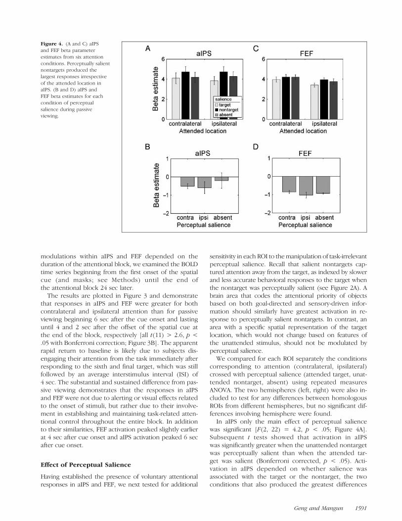
In aIPS only the main effect of perceptual saliencewas significant [F(2, 22) = 4.2, p < .05; Figure 4A].Subsequent t tests showed that activation in aIPSwas significantly greater when the unattended nontargetwas perceptually salient than when the attended tar-get was salient (Bonferroni corrected, p < .05). Acti-vation in aIPS depended on whether salience wasassociated with the target or the nontarget, the twoconditions that also produced the greatest differences
Figure 4. (A and C) aIPS
and FEF beta parameter
estimates from six attention
conditions. Perceptually salientnontargets produced the
largest responses irrespective
of the attended location inaIPS. (B and D) aIPS and
FEF beta estimates for each
condition of perceptual
salience during passiveviewing.
Geng and Mangun 1591
in behavioral response. The presence of the same per-ceptually salient stimuli produced no significant effectswhen subjects simply viewed them passively [F(2, 22) =0.8; Figure 4B]. This demonstrated that aIPS was sensi-tive to perceptual salience specifically as a function ofthe current location of spatial attention: Different re-sponses to perceptual salience only occurred when at-tention was engaged at a cued location suggesting thatthis difference carried information about the distribu-tion of attention rather than information about the per-ceptual qualities of the stimulus per se.
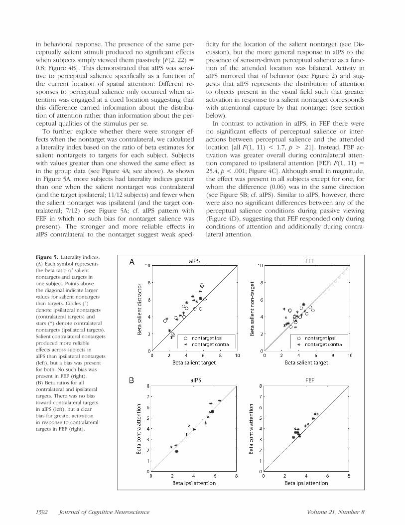
To further explore whether there were stronger ef-fects when the nontarget was contralateral, we calculateda laterality index based on the ratio of beta estimates forsalient nontargets to targets for each subject. Subjectswith values greater than one showed the same effect asin the group data (see Figure 4A; see above). As shownin Figure 5A, more subjects had laterality indices greaterthan one when the salient nontarget was contralateral(and the target ipsilateral; 11/12 subjects) and fewer whenthe salient nontarget was ipsilateral (and the target con-tralateral; 7/12) (see Figure 5A; cf. aIPS pattern withFEF in which no such bias for nontarget salience waspresent). The stronger and more reliable effects inaIPS contralateral to the nontarget suggest weak speci-
ficity for the location of the salient nontarget (see Dis-cussion), but the more general response in aIPS to thepresence of sensory-driven perceptual salience as a func-tion of the attended location was bilateral. Activity inaIPS mirrored that of behavior (see Figure 2) and sug-gests that aIPS represents the distribution of attentionto objects present in the visual field such that greateractivation in response to a salient nontarget correspondswith attentional capture by that nontarget (see sectionbelow).
In contrast to activation in aIPS, in FEF there wereno significant effects of perceptual salience or inter-actions between perceptual salience and the attendedlocation [all F(1, 11) < 1.7, p > .21]. Instead, FEF ac-tivation was greater overall during contralateral atten-tion compared to ipsilateral attention [FEF: F(1, 11) =25.4, p < .001; Figure 4C]. Although small in magnitude,the effect was present in all subjects except for one, forwhom the difference (0.06) was in the same direction(see Figure 5B; cf. aIPS). Similar to aIPS, however, therewere also no significant differences between any of theperceptual salience conditions during passive viewing(Figure 4D), suggesting that FEF responded only duringconditions of attention and additionally during contra-lateral attention.
Figure 5. Laterality indices.
(A) Each symbol representsthe beta ratio of salient
nontargets and targets in
one subject. Points above
the diagonal indicate largervalues for salient nontargets
than targets. Circles (8)denote ipsilateral nontargets
(contralateral targets) andstars (*) denote contralateral
nontargets (ipsilateral targets).
Salient contralateral nontargetsproduced more reliable
effects across subjects in
aIPS than ipsilateral nontargets
(left), but a bias was presentfor both. No such bias was
present in FEF (right).
(B) Beta ratios for all
contralateral and ipsilateraltargets. There was no bias
toward contralateral targets
in aIPS (left), but a clear
bias for greater activationin response to contralateral
targets in FEF (right).
1592 Journal of Cognitive Neuroscience Volume 21, Number 8
Although aIPS and FEF were engaged by voluntaryspatial attention bilaterally, the finding of a differencebetween them in sensitivity to task-irrelevant perceptualsalience and the location of spatial attention argues forseparable roles in attentional control. aIPS activationintegrated goal-directed and sensory-driven informationand represented the attentional cost of capture by a per-ceptually salient nontarget, suggesting that aIPS codesthe distribution of attention to objects in the visual field.In contrast, FEF activity was consistently greater whenvoluntary attention was directed toward the contralat-eral visual field, suggesting a stronger role in maintaininggoal-directed spatial attention.
Correlation with Reaction Time
We next explored the functional significance of aIPSactivation by examining the relationship between aIPSresponse amplitude and RT within individual salienceconditions. Although our manipulation of salience wassuccessful in altering patterns of behavioral performanceoverall, trial-by-trial variations in attentional focus priorto target onset should have also impacted the ability toselect the target and filter out the nontarget stimulus.Thus, we would expect RT to be related to the strengthof attentional focus on the cued target location prior totarget onset (pretarget period) as well as the amount ofattentional capture away from the target location oncethe target and nontarget appeared (target-evoked period).An area involved in pretarget attentional enhancementshould be inversely related to RT and an area involvedin representing nontarget attentional capture should in-crease monotonically with RT.
BOLD time series (in terms of % signal change fromthe fixation baseline) were extracted from FEF, aIPS, anddMOG ROIs. dMOG ROIs were defined by greater acti-vation during contralateral compared to ipsilateral at-tention (Table 2; see Methods), and were expected toreflect the strength of attentional enhancement at thecued target location (Bestmann et al., 2007; Ruff et al.,2006; Weissman, Roberts, Visscher, & Woldorff, 2006;Moore & Armstrong, 2003). dMOG ROIs were locatedcontralateral to the target stimulus. The group-averagex y z MNI coordinates were the following: left =�25 �94 9 and right = 27 �93 10 (see Table 2; forsimilar coordinates in lateralized attention tasks, see alsoHopf et al., 2006; Hopfinger et al., 2001).
For each salience condition (nontarget, target, andabsent), data were divided into four bins based on eachsubject’s RT distribution. Boarders between bins weredefined by standard deviations from the mean (<�1SD,�1SD, +1SD, >+1SD) to account for the positive skewin RT distributions (Hockely, 1984; Ratcliff & Murdock,1976; McCormack & Wright, 1964). An average of 15,71, 39, and 20 trials occurred in each of the four bins,respectively. Data from one subject were incompleteand so were excluded from all time-series analyses.
Time-series data from each salience condition and ROIare plotted in Figure 6.
Consistent with the expectation that pretarget activa-tion in contralateral target-occipital cortex should reflectthe degree of attentional enhancement at the cued tar-get location due to prior knowledge, Spearman’s rank-order correlation between the response amplitude indMOG (at 0 sec) and RT bin was significant for eachsalience condition (rs: nontarget = �.37 p < .05, tar-get = �.55 p < .0001, absent = �.42, p < .005; seeFigure 7). Target-evoked activity was only significant indMOG at 8 sec when the target was salient (rs = �.34,p < .05). Significant correlations between dMOG activa-tions and RT were not found during ipsilateral attention(rs: nontarget = �.08, target = �.22, absent = �.05, allp > .14), indicating that the negative correlation wasspecific to areas of the visual cortex representing thetarget location. The negative correlations between target-contralateral dMOG and RT was significant for all salienceconditions, but pretarget occipital activation explainedthe most variance in RT when the target was perceptuallysalient (rs
2: nontarget = 13.7%, target = 30.2%, absent =17.6%) and when sensory-driven salience matched the at-tended target location.
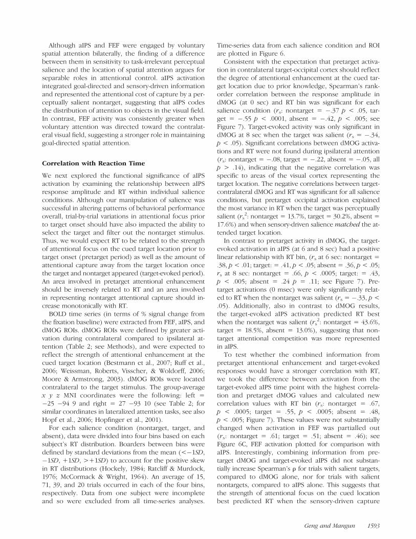
In contrast to pretarget activity in dMOG, the target-evoked activation in aIPS (at 6 and 8 sec) had a positivelinear relationship with RT bin, (rs at 6 sec: nontarget =.38, p < .01; target: = .41, p < .05; absent = .36, p < .05;rs at 8 sec: nontarget = .66, p < .0005; target: = .43,p < .005; absent = .24 p = .11; see Figure 7). Pre-target activations (0 msec) were only significantly relat-ed to RT when the nontarget was salient (rs = �.33, p <.05). Additionally, also in contrast to dMOG results,the target-evoked aIPS activation predicted RT bestwhen the nontarget was salient (rs
2: nontarget = 43.6%,target = 18.5%, absent = 13.0%), suggesting that non-target attentional competition was more representedin aIPS.
To test whether the combined information frompretarget attentional enhancement and target-evokedresponses would have a stronger correlation with RT,we took the difference between activation from thetarget-evoked aIPS time point with the highest correla-tion and pretarget dMOG values and calculated newcorrelation values with RT bin (rs: nontarget = .67,p < .0005; target = .55, p < .0005; absent = .48,p < .005; Figure 7). These values were not substantiallychanged when activation in FEF was partialled out(rs: nontarget = .61; target = .51; absent = .46); seeFigure 6C, FEF activation plotted for comparison withaIPS. Interestingly, combining information from pre-target dMOG and target-evoked aIPS did not substan-tially increase Spearman’s r for trials with salient targets,compared to dMOG alone, nor for trials with salientnontargets, compared to aIPS alone. This suggests thatthe strength of attentional focus on the cued locationbest predicted RT when the sensory-driven capture
Geng and Mangun 1593
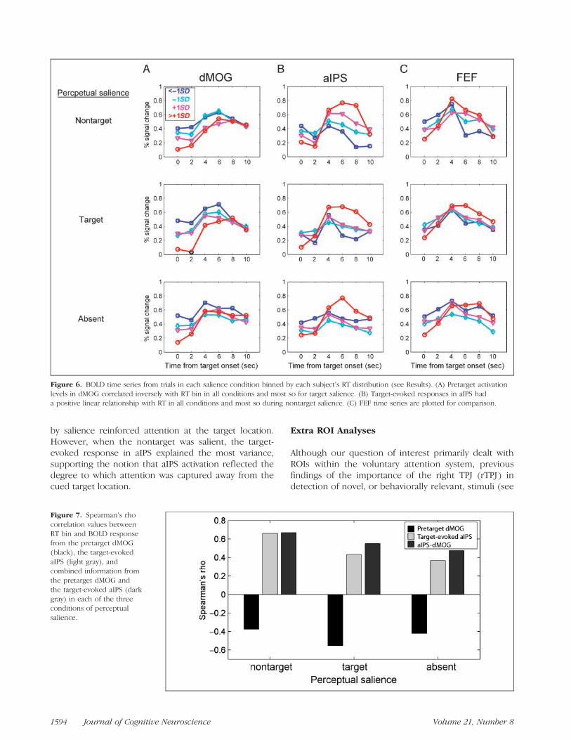
by salience reinforced attention at the target location.However, when the nontarget was salient, the target-evoked response in aIPS explained the most variance,supporting the notion that aIPS activation reflected thedegree to which attention was captured away from thecued target location.
Extra ROI Analyses
Although our question of interest primarily dealt withROIs within the voluntary attention system, previousfindings of the importance of the right TPJ (rTPJ) indetection of novel, or behaviorally relevant, stimuli (see
Figure 6. BOLD time series from trials in each salience condition binned by each subject’s RT distribution (see Results). (A) Pretarget activation
levels in dMOG correlated inversely with RT bin in all conditions and most so for target salience. (B) Target-evoked responses in aIPS hada positive linear relationship with RT in all conditions and most so during nontarget salience. (C) FEF time series are plotted for comparison.
Figure 7. Spearman’s rho
correlation values betweenRT bin and BOLD response
from the pretarget dMOG
(black), the target-evoked
aIPS (light gray), andcombined information from
the pretarget dMOG and
the target-evoked aIPS (dark
gray) in each of the threeconditions of perceptual
salience.
1594 Journal of Cognitive Neuroscience Volume 21, Number 8
Introduction), compelled us to look at the rTPJ specif-ically for effects of nontarget salience. However, we didnot see any significant pattern indicating that salientnontargets produced greater beta parameters than sa-lient targets in either whole-brain analyses, or in individ-ual ROIs based on coordinates of the rTPJ (e.g., fromDownar et al., 2000; x y z coordinates at 54 �42 13) andthe right STG from Corbetta & Shulman (2002; x y zcoordinates at 57 �45 12). Transformation of Talairachcoordinates to MNI was accomplished using a function byMatthew Brett (http://imaging.mrc-cbu.cam.ac.uk/imaging/MniTalairach). Our null finding is consistent with theidea that attentional capture in rTPJ is subject to strongmodulation by task relevance. In this task, the perceptu-ally salient feature was completely task-irrelevant andthe location of the target was known in advance. Thus,attenuating the response of a system that reorientsattention makes good sense (e.g., see Corbetta et al.,2008).
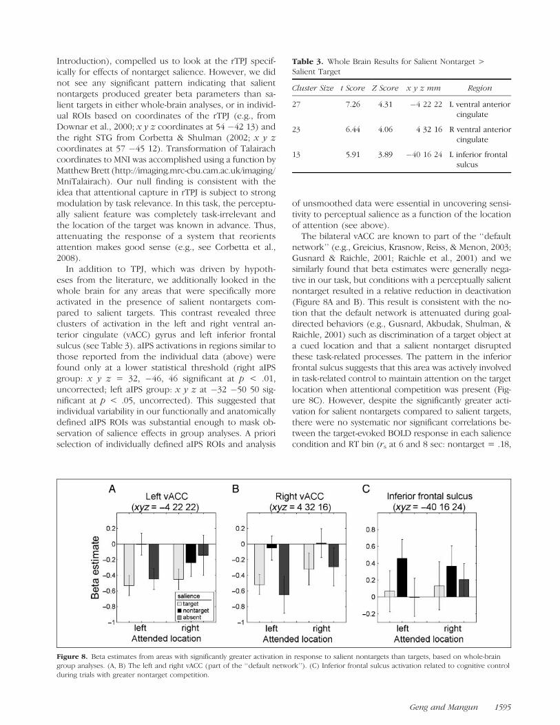
In addition to TPJ, which was driven by hypoth-eses from the literature, we additionally looked in thewhole brain for any areas that were specifically moreactivated in the presence of salient nontargets com-pared to salient targets. This contrast revealed threeclusters of activation in the left and right ventral an-terior cingulate (vACC) gyrus and left inferior frontalsulcus (see Table 3). aIPS activations in regions similar tothose reported from the individual data (above) werefound only at a lower statistical threshold (right aIPSgroup: x y z = 32, �46, 46 significant at p < .01,uncorrected; left aIPS group: x y z at �32 �50 50 sig-nificant at p < .05, uncorrected). This suggested thatindividual variability in our functionally and anatomicallydefined aIPS ROIs was substantial enough to mask ob-servation of salience effects in group analyses. A prioriselection of individually defined aIPS ROIs and analysis
of unsmoothed data were essential in uncovering sensi-tivity to perceptual salience as a function of the locationof attention (see above).
The bilateral vACC are known to part of the ‘‘defaultnetwork’’ (e.g., Greicius, Krasnow, Reiss, & Menon, 2003;Gusnard & Raichle, 2001; Raichle et al., 2001) and wesimilarly found that beta estimates were generally nega-tive in our task, but conditions with a perceptually salientnontarget resulted in a relative reduction in deactivation(Figure 8A and B). This result is consistent with the no-tion that the default network is attenuated during goal-directed behaviors (e.g., Gusnard, Akbudak, Shulman, &Raichle, 2001) such as discrimination of a target object ata cued location and that a salient nontarget disruptedthese task-related processes. The pattern in the inferiorfrontal sulcus suggests that this area was actively involvedin task-related control to maintain attention on the targetlocation when attentional competition was present (Fig-ure 8C). However, despite the significantly greater acti-vation for salient nontargets compared to salient targets,there were no systematic nor significant correlations be-tween the target-evoked BOLD response in each saliencecondition and RT bin (rs at 6 and 8 sec: nontarget = .18,
Figure 8. Beta estimates from areas with significantly greater activation in response to salient nontargets than targets, based on whole-braingroup analyses. (A, B) The left and right vACC (part of the ‘‘default network’’). (C) Inferior frontal sulcus activation related to cognitive control
during trials with greater nontarget competition.
Table 3. Whole Brain Results for Salient Nontarget >Salient Target
Cluster Size t Score Z Score x y z mm Region
27 7.26 4.31 �4 22 22 L ventral anteriorcingulate
23 6.44 4.06 4 32 16 R ventral anteriorcingulate
13 5.91 3.89 �40 16 24 L inferior frontalsulcus
Geng and Mangun 1595
�.09; target = .24 �.15; nosal = .05, �.28). This sug-gested that this region was not related specifically to at-tentional orientation, but rather to other task demandsassociated with trials that were more or less difficult.
DISCUSSION
FEF and aIPS are areas that are commonly thought to beinvolved in controlling voluntary spatial attention bybiasing processing in the visual cortex (e.g., Ruff et al.,2008; Sylvester et al., 2007; Silvanto et al., 2006; Moore& Armstrong, 2003; Corbetta et al., 2000; Hopfingeret al., 2000; Kastner, Pinsk, De Weerd, Desimone, &Ungerleider, 1999), but their respective roles in top–down versus bottom–up processes remain unclear. Inthe present study, we used a lateralized spatial attentiontask in which bilateral stimuli were presented and thelocation of the target was always known in advance. Sub-jects were required to make a perceptual judgment aboutthe attended-location stimulus. On some trials, either thetarget in the attended location or the nontarget in theunattended location was perceptually salient. Perceptualsalience was always task-irrelevant. This design enabled aconservative test of sensory-driven attentional selectionbecause there was no reason to voluntarily attend to thestimulus in the nontarget location. Nevertheless, the be-havioral data demonstrated that our manipulation of per-ceptual salience captured visual attention: Performancewas slower and less accurate when the nontarget wassalient compared to the target (see Figure 2).
Sustained BOLD activations were present in FEFand aIPS in both hemispheres over the entire dura-tion of the attentional block (e.g., Fan et al., 2007; Ikkai& Curtis, 2007; Kelley et al., 2007; Serences & Yantis,2007; Silver, Ress, & Heeger, 2005, 2007; Moore &Armstrong, 2003; Yantis et al., 2002; Vandenbergheet al., 2000), confirming that both ROIs were activelyinvolved in maintaining goal-directed spatial attention.In addition to general effects of spatial attention, how-ever, only FEF had greater activity during attention tothe contralateral visual field. The contralateral bias sug-gested that FEF was involved in maintenance of goal-directed spatial attention, which would be consistentwith literature showing that FEF activation plays a causalrole in biasing activity in the visual cortex, and enhancingperceptual sensitivity in corresponding regions of thevisual field (e.g., Ruff et al., 2006, 2008; Armstrong et al.,2006; Chambers et al., 2006; Mevorach et al., 2006;Muggleton et al., 2006; Silvanto et al., 2006; Hung et al.,2005; O’Shea et al., 2004; Moore & Armstrong, 2003;Grosbras & Paus, 2002). Greater pretarget activation inthe target-contralateral dMOG presumably leads to higherquality representations of the target stimulus when it ap-pears and, therefore, faster RTs (Weissman et al., 2006).Consistent with this, we found that pretarget activation inthe target-contralateral (but not target-ipsilateral) dMOG
was inversely related to RT in all salience conditions, butmore so when the salient stimulus was the target; pre-target occipital activations had the highest correlationwith RT when nontarget competition for attention wasthe lowest (i.e., when only the target was salient; seeFigures 6 and 7).
In contrast to FEF, aIPS showed no contralateral bias inactivation as a function of target location. Instead, aIPSactivation was greatest when the unattended (nontarget)stimulus was perceptually salient. Target-evoked activa-tions in aIPS also correlated positively with RT in allconditions, but was highest when the nontarget wassalient. Greater target-evoked aIPS activations occurredon trials with slower RTs and this was opposite to thepretarget effects found in dMOG (described above; seeFigures 6 and 7), suggesting that aIPS reflected thedegree to which attention was directed away from thetarget stimulus. This result argues against the idea thatbigger aIPS activation could represent greater attentionalsuppression of the nontarget. If greater activation meantgreater suppression, then higher amplitude responsesshould have lead to faster RTs, not slower.
Another possibility is that aIPS is involved in holdingitems in visual short-term memory (e.g., Xu, 2007) andtrials with greater activation reflected the storage ofboth the target and nontarget objects in memory. How-ever, the linear relationship between aIPS responseamplitudes and RT (even within conditions with iden-tical stimuli) argues against the categorical storage ofobject items in this study. If aIPS activation reflected thestorage of a discrete number of objects, then we wouldexpect only two response levels based on whether oneor two items were stored. Instead, it seems that activa-tion in aIPS ref lected the distribution of attention,which can be understood in different ways: one is thatit reflects a shift of attention of varying durations (orengage and disengage operations; e.g., Posner, Walker,Friedrich, & Rafal, 1984; Posner, Cohen, & Rafal, 1982)to the nontarget and then to the target on trials whenattention was captured by the salient nontarget. Alter-natively, the distribution of attention could represent anattentional priority map that codes the relative strengthof competition for attention by all stimuli in the visualfield (e.g., Gottlieb, 2007; Goldberg et al., 2006; Itti &Koch, 2000). Here, greater activation (e.g., in response totrials with a salient nontarget) would reflect the presenceof more items with higher attentional priority, even if at-tention is ultimately only shifted to the ‘‘winner’’ with thehighest relative priority (e.g., Bisley & Goldberg, 2003). Itis not possible to know from the current data whetherattention was actually shifted to the nontarget becausewe did not explicitly measure behavioral performanceat the nontarget location. However, the response in aIPSclearly contained a graded representation of the state ofcompetition for attention under all conditions of percep-tual salience and this suggests that aIPS represents thestate of attention, rather than stimulus salience per se
1596 Journal of Cognitive Neuroscience Volume 21, Number 8
(although stimulus salience obviously had a strong affecton the state of attention).
An unexpected aspect of our aIPS results was the lackof stronger hemispheric lateralization based on the at-tended visual field. Although aIPS contralateral to thenontarget did produce more robust results, it perhapsseems surprising that the results were not more lat-eralized given recent reports of topographically orga-nized attentional maps in the human IPS as well asFEF (e.g., Saygin & Sereno, 2008; Kastner et al., 2007;Swisher, Halko, Merabet, McMains, & Somers, 2007;Schluppeck, Curtis, Glimcher, & Heeger, 2006; Silveret al., 2005; Sereno, McDonald, & Allman, 1994). How-ever, it is worth noting that data from studies that usedcognitive tasks rather than specific mapping procedureshave often found strong bilateral effects in both IPS andFEF, which only sometimes also contained weaker con-tralateral sensitivity (e.g., Curtis & Connolly, 2008; Ikkai& Curtis, 2007; Indovina & Macaluso, 2007; Kelley et al.,2007; Serences & Yantis, 2007; Geng et al., 2006; Kincadeet al., 2005; Donner et al., 2002; Hopfinger et al., 2000;Wojciulik & Kanwisher, 1999). In addition, areas withretinotopic attentional maps in IPS such as IPS1 andIPS2 (Silver et al., 2005), and IPS3 and IPS4 (Swisheret al., 2007), tend to be more posterior than IPS regionsthat show bilateral attentional effects (see above), includ-ing our current findings. It is unclear whether discrep-ancies in topographic specificity arise due to meaningfulfunctional differences between subareas within IPS, ordue to experimental design factors (e.g., stimulus eccen-tricity), and requires further investigation. Nevertheless, itis clear that regions within FEF and IPS in both hemi-spheres are critically involved in the control of sustainedvoluntary spatial attention (e.g., Fan et al., 2007; Ikkai &Curtis, 2007; Serences & Yantis, 2007; Silver et al., 2005;Yantis et al., 2002; Vandenberghe et al., 2000) and thisstudy extends those findings by demonstrating differ-ences in function between these areas such that onlyaIPS was sensitive to salience of stimuli as a function ofthe location of attention.
Our results that show sensitivity in aIPS to task-irrelevant perceptual salience may initially seem at oddswith reports that salient stimuli that are completely taskunrelated are not represented in attentional controlregions, or even early visual areas (e.g., Beck & Kastner,2005; Schwartz et al., 2005). Those studies differed fromours, in that subjects were required to engage in a taskat central vision and the task-irrelevant salient stimuliappeared in the periphery. It may be that peripheral sa-lient stimuli can be effectively inhibited when attentionis concentrated centrally or when they occur during pe-riods when task stimuli are absent (Indovina & Macaluso,2007). In line with this, we also found that neither aIPSnor FEF responded significantly to the presence of thesalient stimuli during passive viewing when no atten-tional task was required (although we did find greateractivation to perceptually salient stimuli in dMOG
ROIs). This suggests that our result in aIPS of percep-tual salience was a function of the attended location:aIPS activation reflected the degree to which a per-ceptually salient nontarget captured attention away fromthe target and argues for a model of attention in whichvoluntary and stimulus-driven properties are representedwithin the same areas that control covert and overtselection.
We additionally examined areas outside of our ROIsusing whole-brain voxelwise analyses to test for otherregions that might also show sensitivity to salient non-targets as a function of attention. In the rTPJ, an areahypothesized to be involved in sensory-driven attentionalreorienting, we found no effects of nontarget salience.This result is consistent with the rTPJ being involvedwith attentional orienting specifically toward goal-relevantstimuli that may also violate expectations (e.g., Vossel,Weidner, Thiel, & Fink, 2009; Corbetta et al., 2008;Kincade et al., 2005; Downar et al., 2000), and notstimulus-driven attentional capture by irrelevant featuresof behavioral nontargets. We also found a reduction innegative activation for salient nontargets in the ACCbilaterally. The ACC is considered part of the ‘‘defaultnetwork’’ (e.g., Greicius, Supekar, Menon, & Dougherty,2008; Raichle et al., 2001), suggesting that the presenceof salient nontargets required greater task engagement.There was also greater activation for salient nontargetsin the inferior frontal sulcus, but we did not find anyparametric relationship between activation in the inferiorfrontal sulcus and RT, suggesting that the role was not inattentional control per se, but rather in executive func-tions related to staying on task.
In summary, we examined FEF and aIPS in a sustainedattention task when the perceptual salience of stimulivaried randomly. Although both regions were involvedwith selective spatial attention in general, we observeddifferences in the representation of attended spatiallocations and perceptual salience within FEF and aIPS.FEF activation was biased by the location of spatialattention, whereas aIPS was modulated by the relativeperceptual salience of the target and nontarget stimuli.The target-evoked response in aIPS scaled linearly withincreasing RT in all conditions, but especially when thenontarget was salient. aIPS was most correlated with RTwhen attention was captured by the salient nontarget,suggesting that aIPS responses are sensitive to task-irrelevant perceptual salience within the context ofgoal-directed spatial attention. Despite strong bidirec-tional anatomical and functional connections betweenFEF and IPS (e.g., Pesaran, Nelson, & Andersen, 2008;Buschman & Miller, 2007; Ferraina, Pare, & Wurtz, 2002;Blatt, Andersen, & Stoner, 1990), these results dem-onstrate separable roles for FEF and aIPS in attention-al control, with FEF more involved in goal-directedspatial attention and aIPS relatively more sensitive tobottom–up attentional inf luences driven by stimulussalience.
Geng and Mangun 1597
Acknowledgments
This work was supported by MH055714 and NSF0727115 toG. R. M. Joy J. Geng was supported by postdoctoral trainingfellowship NEI T32 EY015387 to the UCD Vision Science Re-search Group. We thank Ali Mazaheri, Cameron Carter, JohnRyan, and Michael Buonocore for valuable advice and assistance.
Reprint requests should be sent to Joy J. Geng, Center for Mindand Brain, 267 Cousteau Pl., Davis, CA 95618, or via e-mail:[email protected].
REFERENCES
Andersson, J. L., Hutton, C., Ashburner, J., Turner, R., &Friston, K. (2001). Modeling geometric deformations inEPI time series. Neuroimage, 13, 903–919.
Armstrong, K. M., Fitzgerald, J. K., & Moore, T. (2006).Changes in visual receptive fields with microstimulationof frontal cortex. Neuron, 50, 791–798.
Astafiev, S. V., Shulman, G. L., & Corbetta, M. (2006).Visuospatial reorienting signals in the human temporo-parietal junction are independent of response selection.European Journal of Neuroscience, 23, 591–596.
Balan, P. F., & Gottlieb, J. (2006). Integration of exogenousinput into a dynamic salience map revealed by perturbingattention. Journal of Neuroscience, 26, 9239–9249.
Beck, D. M., & Kastner, S. (2005). Stimulus contextmodulates competition in human extrastriate cortex.Nature Neuroscience, 8, 1110–1116.
Behrmann, M., Geng, J. J., & Shomstein, S. (2004). Parietalcortex and attention. Current Opinion in Neurobiology,14, 212–217.
Berger, A., Henik, A., & Rafal, R. (2005). Competitionbetween endogenous and exogenous orienting of visualattention. Journal of Experimental Psychology. General,134, 207–221.
Bestmann, S., Ruff, C. C., Blakemore, C., Driver, J., & Thilo,K. V. (2007). Spatial attention changes excitability ofhuman visual cortex to direct stimulation. Current Biology,17, 134–139.
Bichot, N. P., & Schall, J. D. (2002). Priming in macaquefrontal cortex during popout visual search: Feature-basedfacilitation and location-based inhibition of return.Journal of Neuroscience, 22, 4675–4685.
Bisley, J. W., & Goldberg, M. E. (2003). Neuronal activity inthe lateral intraparietal area and spatial attention. Science,299, 81–86.
Bisley, J. W., & Goldberg, M. E. (2006). Neural correlates ofattention and distractibility in the lateral intraparietal area.Journal of Neurophysiology, 95, 1696–1717.
Blatt, G. J., Andersen, R. A., & Stoner, G. R. (1990). Visualreceptive field organization and cortico-cortical connectionsof the lateral intraparietal area (area LIP) in the macaque.Journal of Comparative Neurology, 299, 421–445.
Brefczynski, J. A., & DeYoe, E. A. (1999). A physiologicalcorrelate of the ‘‘spotlight’’ of visual attention. NatureNeuroscience, 2, 370–374.
Bundesen, C. (1990). A theory of visual attention.Psychological Review, 97, 523–547.
Buschman, T. J., & Miller, E. K. (2007). Top–down versusbottom–up control of attention in the prefrontal andposterior parietal cortices. Science, 315, 1860–1862.
Carrasco, M. (2006). Covert attention increases contrastsensitivity: Psychophysical, neurophysiological andneuroimaging studies. Progress in Brain Research,154, 33–70.
Carrasco, M., Ling, S., & Read, S. (2004). Attention altersappearance. Nature Neuroscience, 7, 308–313.
Chambers, C. D., Stokes, M. G., Janko, N. E., & Mattingley,J. B. (2006). Enhancement of visual selection duringtransient disruption of parietal cortex. Brain Research,1097, 149–155.
Chelazzi, L., Biscaldi, M., Corbetta, M., Peru, A., Tassinari, G.,& Berlucchi, G. (1995). Oculomotor activity and visual spatialattention. Behavioural Brain Research, 71, 81–88.
Connolly, J. D., Goodale, M. A., Menon, R. S., & Munoz,D. P. (2002). Human fMRI evidence for the neuralcorrelates of preparatory set. Nature Neuroscience, 5,1345–1352.
Corbetta, M., Kincade, J. M., Ollinger, J. M., McAvoy, M. P., &Shulman, G. L. (2000). Voluntary orienting is dissociatedfrom target detection in human posterior parietal cortex.Nature Neuroscience, 3, 292–297.
Corbetta, M., Patel, G., & Shulman, G. L. (2008).The reorienting system of the human brain: Fromenvironment to theory of mind. Neuron, 58, 306–324.
Corbetta, M., & Shulman, G. L. (2002). Control ofgoal-directed and stimulus-driven attention in thebrain. Nature Reviews Neuroscience, 3, 201–215.
Curtis, C. E., & Connolly, J. D. (2008). Saccade preparationsignals in the human frontal and parietal cortices. Journalof Neurophysiology, 99, 133–145.
Donner, T. H., Kettermann, A., Diesch, E., Ostendorf, F.,Villringer, A., & Brandt, S. A. (2002). Visual featureand conjunction searches of equal difficulty engageonly partially overlapping frontoparietal networks.Neuroimage, 15, 16–25.
Donner, T. H., Kettermann, A., Diesch, E., Villringer, A.,& Brandt, S. A. (2003). Parietal activation during visualsearch in the absence of multiple distractors. NeuroReport,14, 2257–2261.
Downar, J., Crawley, A. P., Mikulis, D. J., & Davis, K. D.(2000). A multimodal cortical network for the detection ofchanges in the sensory environment. Nature Neuroscience,3, 277–283.
Driver, J., & Frackowiak, R. S. (2001). Neurobiologicalmeasures of human selective attention. Neuropsychologia,39, 1257–1262.
Eriksen, C. W., & Yeh, Y. Y. (1985). Allocation of attentionin the visual field. Journal of Experimental Psychology:Human Perception and Performance, 11, 583–597.
Fan, J., Byrne, J., Worden, M. S., Guise, K. G., McCandliss, B. D.,Fossella, J., et al. (2007). The relation of brain oscillationsto attentional networks. Journal of Neuroscience, 27,6197–6206.
Ferraina, S., Pare, M., & Wurtz, R. H. (2002). Comparisonof cortico-cortical and cortico-collicular signals for thegeneration of saccadic eye movements. Journal ofNeurophysiology, 87, 845–858.
Friedrich, F. J., Egly, R., Rafal, R. D., & Beck, D. (1998).Spatial attention deficits in humans: A comparison ofsuperior parietal and temporal–parietal junction lesions.Neuropsychology, 12, 193–207.
Friston, K. J., Holmes, A. P., Worsley, K. J., Poline, J. B., Frith, C.,& Frackowiak, R. S. J. (1995). Statistical parametric mapsin functional imaging: A general linear approach. HumanBrain Mapping, 2, 189–210.
Gandhi, S. P., Heeger, D. J., & Boynton, G. M. (1999).Spatial attention affects brain activity in human primaryvisual cortex. Proceedings of the National Academy ofSciences, U.S.A., 96, 3314–3319.
Geng, J. J., & Behrmann, M. (2005). Spatial probabilityas an attentional cue in visual search. Perception &Psychophysics, 67, 1252–1268.
1598 Journal of Cognitive Neuroscience Volume 21, Number 8
Geng, J. J., Eger, E., Ruff, C. C., Kristjansson, A., Rotshtein, P.,& Driver, J. (2006). On-line attentional selection fromcompeting stimuli in opposite visual fields: Effects onhuman visual cortex and control processes. Journal ofNeurophysiology, 96, 2601–2612.
Gibson, J. J. (1986). The ecological approach to visualperception. Hillsdale, NJ: Erlbaum.
Gitelman, D. R., Nobre, A. C., Parrish, T. B., LaBar, K. S.,Kim, Y. H., Meyer, J. R., et al. (1999). A large-scaledistributed network for covert spatial attention: Furtheranatomical delineation based on stringent behaviouraland cognitive controls. Brain, 122, 1093–1106.
Goldberg, M. E., Bisley, J. W., Powell, K. D., & Gottlieb, J.(2006). Saccades, salience and attention: The role of thelateral intraparietal area in visual behavior. Progress inBrain Research, 155, 157–175.
Gottlieb, J. (2007). From thought to action: The parietalcortex as a bridge between perception, action, andcognition. Neuron, 53, 9–16.
Greicius, M. D., Krasnow, B., Reiss, A. L., & Menon, V.(2003). Functional connectivity in the resting brain:A network analysis of the default mode hypothesis.Proceedings of the National Academy of Sciences,U.S.A., 100, 253–258.
Greicius, M. D., Supekar, K., Menon, V., & Dougherty, R. F.(2008). Resting-state functional connectivity reflectsstructural connectivity in the default mode network.Cerebral Cortex. Epub ahead of print.
Grosbras, M. H., & Paus, T. (2002). Transcranial magneticstimulation of the human frontal eye field: Effects onvisual perception and attention. Journal of CognitiveNeuroscience, 14, 1109–1120.
Gusnard, D. A., Akbudak, E., Shulman, G. L., & Raichle,M. E. (2001). Medial prefrontal cortex and self-referentialmental activity: Relation to a default mode of brain function.Proceedings of the National Academy of Sciences, U.S.A.,98, 4259–4264.
Gusnard, D. A., & Raichle, M. E. (2001). Searching for abaseline: Functional imaging and the resting humanbrain. Nature Reviews Neuroscience, 2, 685–694.
He, B. J., Snyder, A. Z., Vincent, J. L., Epstein, A., Shulman,G. L., & Corbetta, M. (2007). Breakdown of functionalconnectivity in frontoparietal networks underliesbehavioral deficits in spatial neglect. Neuron, 53, 905–918.
Hickey, C., McDonald, J. J., & Theeuwes, J. (2006).Electrophysiological evidence of the capture of visualattention. Journal of Cognitive Neuroscience, 18,604–613.
Hockely, W. E. (1984). Analysis of response time distributions inthe study of cognitive processes. Journal of ExperimentalPsychology: Learning, Memory, and Cognition, 10, 598–615.
Hopf, J. M., Luck, S. J., Boelmans, K., Schoenfeld, M. A.,Boehler, C. N., Rieger, J., et al. (2006). The neural siteof attention matches the spatial scale of perception.Journal of Neuroscience, 26, 3532–3540.
Hopf, J. M., Luck, S. J., Girelli, M., Hagner, T., Mangun,G. R., Scheich, H., et al. (2000). Neural sources of focusedattention in visual search. Cerebral Cortex, 10, 1233–1241.
Hopfinger, J. B., Buonocore, M. H., & Mangun, G. R. (2000).The neural mechanisms of top–down attentional control.Nature Neuroscience, 3, 284–291.
Hopfinger, J. B., Woldorff, M. G., Fletcher, E. M., & Mangun,G. R. (2001). Dissociating top–down attentional controlfrom selective perception and action. Neuropsychologia,39, 1277–1291.
Hung, J., Driver, J., & Walsh, V. (2005). Visual selection andposterior parietal cortex: Effects of repetitive transcranialmagnetic stimulation on partial report analyzed by
Bundesen’s theory of visual attention. Journal ofNeuroscience, 25, 9602–9612.
Ikkai, A., & Curtis, C. E. (2007). Cortical activity time lockedto the shift and maintenance of spatial attention. CerebralCortex, 18, 1384–1394.
Indovina, I., & Macaluso, E. (2007). Dissociation of stimulusrelevance and saliency factors during shifts of visuospatialattention. Cerebral Cortex, 17, 1701–1711.
Ipata, A. E., Gee, A. L., Gottlieb, J., Bisley, J. W., & Goldberg,M. E. (2006). LIP responses to a popout stimulus arereduced if it is overtly ignored. Nature Neuroscience, 9,1071–1076.
Itti, L., & Koch, C. (2000). A saliency-based search mechanismfor overt and covert shifts of visual attention. VisionResearch, 40, 1489–1506.
James, W. (1890/1983). The principles of psychology.Cambridge: Harvard University Press.
Jonides, J., & Irwin, D. E. (1981). Capturing attention.Cognition, 10, 145–150.
Jonides, J., & Yantis, S. (1988). Uniqueness of abrupt visualonset in capturing attention. Perception & Psychophysics,43, 346–354.
Kastner, S., DeSimone, K., Konen, C. S., Szczepanski, S. M.,Weiner, K. S., & Schneider, K. A. (2007). Topographicmaps in human frontal cortex revealed in memory-guidedsaccade and spatial working-memory tasks. Journal ofNeurophysiology, 97, 3494–3507.
Kastner, S., Pinsk, M. A., De Weerd, P., Desimone, R., &Ungerleider, L. G. (1999). Increased activity in humanvisual cortex during directed attention in the absenceof visual stimulation. Neuron, 22, 751–761.
Kastner, S., & Ungerleider, L. G. (2000). Mechanisms ofvisual attention in the human cortex. Annual Reviewof Neuroscience, 23, 315–341.
Kelley, T. A., Serences, J. T., Giesbrecht, B., & Yantis, S.(2007). Cortical mechanisms for shifting and holdingvisuospatial attention. Cerebral Cortex, 18, 114–125.
Kim, Y.-H., Gitelman, D. R., Nobre, A. C., Parrish, T. B., LaBar,K. S., & Mesulam, M. M. (1999). The large-scale neuralnetwork for spatial attention displays multifunctionaloverlap but differential asymmetry. Neuroimage, 9, 269–277.
Kincade, J. M., Abrams, R. A., Astafiev, S. V., Shulman, G. L.,& Corbetta, M. (2005). An event-related functionalmagnetic resonance imaging study of voluntary andstimulus-driven orienting of attention. Journal ofNeuroscience, 25, 4593–4604.
Kristjansson, A., Vuilleumier, P., Malhotra, P., Husain, M.,& Driver, J. (2005). Priming of color and position duringvisual search in unilateral spatial neglect. Journal ofCognitive Neuroscience, 17, 859–873.
Kristjansson, A., Wang, D., & Nakayama, K. (2002). The roleof priming in conjunctive visual search. Cognition, 85,37–52.
Kusunoki, M., Gottlieb, J., & Goldberg, M. E. (2000). The lateralintraparietal area as a salience map: The representationof abrupt onset, stimulus motion, and task relevance.Vision Research, 40, 1459–1468.
Lavie, N. (1995). Perceptual load as a necessary condition forselective attention. Journal of Experimental Psychology:Human Perception and Performance, 21, 451–468.
Leblanc, E., Prime, D. J., & Jolicoeur, P. (2008). Tracking thelocation of visuospatial attention in a contingent captureparadigm. Journal of Cognitive Neuroscience, 20, 657–671.
Liu, T., Larsson, J., & Carrasco, M. (2007). Feature-basedattention modulates orientation-selective responses inhuman visual cortex. Neuron, 55, 313–323.
Luck, S. J., Chelazzi, L., Hillyard, S. A., & Desimone, R.(1997). Neural mechanisms of spatial selective attention
Geng and Mangun 1599
in areas V1, V2, and V4 of macaque visual cortex. Journalof Neurophysiology, 77, 24–42.
Mayer, A. R., Dorflinger, J. M., Rao, S. M., & Seidenberg, M.(2004). Neural networks underlying endogenous andexogenous visual–spatial orienting. Neuroimage, 23,534–541.
Mayer, A. R., Seidenberg, M., Dorflinger, J. M., & Rao, S. M.(2004). An event-related fMRI study of exogenous orienting:Supporting evidence for the cortical basis of inhibition ofreturn? Journal of Cognitive Neuroscience, 16, 1262–1271.
McCormack, P. D., & Wright, N. M. (1964). The positiveskew observed in reaction time distributions. CanadianJournal of Psychology, 18, 43–51.
McMains, S. A., & Somers, D. C. (2004). Multiple spotlightsof attentional selection in human visual cortex. Neuron,42, 677–686.
Mevorach, C., Humphreys, G. W., & Shalev, L. (2006).Opposite biases in salience-based selection for the leftand right posterior parietal cortex. Nature Neuroscience,9, 740–742.
Moore, T., & Armstrong, K. M. (2003). Selective gating ofvisual signals by microstimulation of frontal cortex. Nature,421, 370–373.
Mort, D. J., Malhotra, P., Mannan, S. K., Rorden, C., Pambakian,A., Kennard, C., et al. (2003). The anatomy of visualneglect. Brain, 126, 1986–1997.
Muggleton, N. G., Postma, P., Moutsopoulou, K.,Nimmo-Smith, I., Marcel, A., & Walsh, V. (2006). TMSover right posterior parietal cortex induces neglect in ascene-based frame of reference. Neuropsychologia, 44,1222–1229.
Muller, H. J., & Rabbitt, P. M. (1989). Reflexive and voluntaryorienting of visual attention: Time course of activationand resistance to interruption. Journal of ExperimentalPsychology: Human Perception and Performance, 15,315–330.
Nakayama, K., & Mackeben, M. (1989). Sustained and transientcomponents of focal visual attention. Vision Research,29, 1631–1647.
Nobre, A. C., Gitelman, D. R., Dias, E. C., & Mesulam, M. M.(2000). Covert visual spatial orienting and saccades:Overlapping neural systems. Neuroimage, 11, 210–216.
O’Shea, J., Muggleton, N. G., Cowey, A., & Walsh, V. (2004).Timing of target discrimination in human frontal eyefields. Journal of Cognitive Neuroscience, 16, 1060–1067.
Paus, T. (1996). Location and function of the humanfrontal eye-field: A selective review. Neuropsychologia,34, 475–483.
Peelen, M. V., Heslenfeld, D. J., & Theeuwes, J. (2004).Endogenous and exogenous attention shifts are mediatedby the same large-scale neural network. Neuroimage,22, 822–830.
Pesaran, B., Nelson, M. J., & Andersen, R. A. (2008). Freechoice activates a decision circuit between frontal andparietal cortex. Nature, 453, 406–409.
Platt, M. L., & Glimcher, P. W. (1999). Neural correlatesof decision variables in parietal cortex. Nature, 400,233–238.
Posner, M. I., Cohen, Y., & Rafal, R. D. (1982). Neuralsystems control of spatial orienting. PhilosophicalTransactions of the Royal Society of London, Series B,Biological Sciences, 298, 187–198.
Posner, M. I., Snyder, C. R., & Davidson, B. J. (1980).Attention and the detection of signals. Journal ofExperimental Psychology, 109, 160–174.
Posner, M. I., Walker, J. A., Friedrich, F. J., & Rafal, R. D.(1984). Effects of parietal injury on covert orienting ofattention. Journal of Neuroscience, 4, 1863–1874.
Proulx, M. J., & Egeth, H. E. (2006a). Biased competitionand visual search: The role of luminance and sizecontrast. Psychological Research, 72, 106–113.
Proulx, M. J., & Egeth, H. E. (2006b). Target–nontargetsimilarity modulates stimulus-driven control in visualsearch. Psychonomic Bulletin & Review, 13, 524–529.
Raichle, M. E., MacLeod, A. M., Snyder, A. Z., Powers, W. J.,Gusnard, D. A., & Shulman, G. L. (2001). A default modeof brain function. Proceedings of the National Academyof Sciences, U.S.A., 98, 676–682.
Ratcliff, R., & Murdock, B. B. (1976). Retrieval processes inrecognition memory. Psychological Review, 83, 190–214.
Reynolds, J. H., & Desimone, R. (2003). Interacting roles ofattention and visual salience in V4. Neuron, 37, 853–863.
Rosen, A. C., Rao, S. M., Caffarra, P., Scaglioni, A., Bobholz,J. A., Woodley, S. J., et al. (1999). Neural basis of endogenousand exogenous spatial orienting. A functional MRI study.Journal of Cognitive Neuroscience, 11, 135–152.
Ruff, C. C., Bestmann, S., Blankenburg, F., Bjoertomt, O.,Josephs, O., Weiskopf, N., et al. (2008). Distinct causalinfluences of parietal versus frontal areas on humanvisual cortex: Evidence from concurrent TMS fMRI.Cerebral Cortex, 18, 817–827.
Ruff, C. C., Blankenburg, F., Bjoertomt, O., Bestmann, S.,Freeman, E., Haynes, J. D., et al. (2006). ConcurrentTMS–fMRI and psychophysics reveal frontal influenceson human retinotopic visual cortex. Current Biology,16, 1479–1488.
Saygin, A. P., & Sereno, M. I. (2008). Retinotopy andattention in human occipital, temporal, parietal, andfrontal cortex. Cerebral Cortex. Epub ahead of print.
Schluppeck, D., Curtis, C. E., Glimcher, P. W., & Heeger,D. J. (2006). Sustained activity in topographic areas ofhuman posterior parietal cortex during memory-guidedsaccades. Journal of Neuroscience, 26, 5098–5108.
Schwartz, S., Vuilleumier, P., Hutton, C., Maravita, A., Dolan,R. J., & Driver, J. (2005). Attentional load and sensorycompetition in human vision: Modulation of fMRI responsesby load at fixation during task-irrelevant stimulation inthe peripheral visual field. Cerebral Cortex, 15, 770–786.
Serences, J. T., & Boynton, G. M. (2007). Feature-basedattentional modulations in the absence of direct visualstimulation. Neuron, 55, 301–312.
Serences, J. T., Shomstein, S., Leber, A. B., Golay, X., Egeth,H. E., & Yantis, S. (2005). Coordination of voluntary andstimulus-driven attentional control in human cortex.Psychological Science, 16, 114–122.
Serences, J. T., & Yantis, S. (2007). Spatially selectiverepresentations of voluntary and stimulus-drivenattentional priority in human occipital, parietal, andfrontal cortex. Cerebral Cortex, 17, 284–293.
Sereno, M. I., McDonald, C. T., & Allman, J. M. (1994).Analysis of retinotopic maps in extrastriate cortex.Cerebral Cortex, 4, 601–620.
Shomstein, S., & Behrmann, M. (2006). Cortical systemsmediating visual attention to both objects and spatiallocations. Proceedings of the National Academy ofSciences, U.S.A., 103, 11387–11392.
Silvanto, J., Lavie, N., & Walsh, V. (2006). Stimulation ofthe human frontal eye fields modulates sensitivity ofextrastriate visual cortex. Journal of Neurophysiology,96, 941–945.
Silver, M. A., Ress, D., & Heeger, D. J. (2005). Topographicmaps of visual spatial attention in human parietal cortex.Journal of Neurophysiology, 94, 1358–1371.
Silver, M. A., Ress, D., & Heeger, D. J. (2007). Neural correlatesof sustained spatial attention in human early visual cortex.Journal of Neurophysiology, 97, 229–237.
1600 Journal of Cognitive Neuroscience Volume 21, Number 8
Slagter, H. A., Giesbrecht, B., Kok, A., Weissman, D. H.,Kenemans, J. L., Woldorff, M. G., et al. (2007). fMRIevidence for both generalized and specialized componentsof attentional control. Brain Research, 1177, 90–102.
Stephan, K. E., Marshall, J. C., Penny, W. D., Friston, K. J.,& Fink, G. R. (2007). Interhemispheric integration ofvisual processing during task-driven lateralization.Journal of Neuroscience, 27, 3512–3522.
Swisher, J. D., Halko, M. A., Merabet, L. B., McMains, S. A.,& Somers, D. C. (2007). Visual topography of humanintraparietal sulcus. Journal of Neuroscience, 27, 5326–5337.
Sylvester, C. M., Shulman, G. L., Jack, A. I., & Corbetta, M.(2007). Asymmetry of anticipatory activity in visual cortexpredicts the locus of attention and perception. Journalof Neuroscience, 27, 14424–14433.
Theeuwes, J. (1995). Abrupt luminance change pops out;abrupt color change does not. Perception & Psychophysics,57, 637–644.
Thiel, C. M., Zilles, K., & Fink, G. R. (2004). Cerebralcorrelates of alerting, orienting and reorienting ofvisuospatial attention: An event-related fMRI study.Neuroimage, 21, 318–328.
Thompson, K. G., Bichot, N. P., & Schall, J. D. (1997).Dissociation of visual discrimination from saccadeprogramming in macaque frontal eye field. Journalof Neurophysiology, 77, 1046–1050.
Vandenberghe, R., Duncan, J., Arnell, K. M., Bishop, S. J.,Herrod, N. J., Owen, A. M., et al. (2000). Maintaining
and shifting attention within left or right hemifield.Cerebral Cortex, 10, 706–713.
Vossel, S., Thiel, C. M., & Fink, G. R. (2006). Cue validitymodulates the neural correlates of covert endogenousorienting of attention in parietal and frontal cortex.Neuroimage, 32, 1257–1264.
Vossel, S., Weidner, R., Thiel, C. M., & Fink, G. R. (2009).What is ‘‘odd’’ in Posner’s location-cueing paradigm?Neural responses to unexpected location and featurechanges compared. Journal of Cognitive Neuroscience, 21,30–41.
Weissman, D. H., Roberts, K. C., Visscher, K. M., & Woldorff,M. G. (2006). The neural bases of momentary lapses inattention. Nature Neuroscience, 9, 971–978.
Wojciulik, E., & Kanwisher, N. (1999). The generality of parietalinvolvement in visual attention. Neuron, 23, 747–764.
Xu, Y. (2007). The role of the superior intraparietalsulcus in supporting visual short-term memory formultifeature objects. Journal of Neuroscience, 27,11676–11686.
Yantis, S., & Egeth, H. E. (1999). On the distinction betweenvisual salience and stimulus-driven attentional capture.Journal of Experimental Psychology: Human Perceptionand Performance, 25, 661–676.
Yantis, S., Schwarzbach, J., Serences, J. T., Carlson, R. L.,Steinmetz, M. A., Pekar, J. J., et al. (2002). Transientneural activity in human parietal cortex during spatialattention shifts. Nature Neuroscience, 5, 995–1002.
Geng and Mangun 1601