Embed Size (px)
Citation preview
Instructions for use
Title Anti-neoplastic effects of topoisomerase inhibitors in canine mammary carcinoma, melanoma, and osteosarcoma celllines
Author(s) Ong, Siew Mei; Yamamoto, Hiroki; Saeki, Kohei; Tanaka, Yuiko; Yoshitake, Ryohei; Nishimura, Ryohei; Nakagawa,Takayuki
Citation Japanese Journal of Veterinary Research, 65(1), 17-28
Issue Date 2017-02
DOI 10.14943/jjvr.65.1.17
Doc URL http://hdl.handle.net/2115/64788
Type bulletin (article)
File Information 65-1_017-028.pdf
Hokkaido University Collection of Scholarly and Academic Papers : HUSCAP
Japanese Journal of Veterinary Research 65(1): 17-28, 2017
REGULAR PAPER Experimental Research
Anti-neoplastic effects of topoisomerase inhibitors in canine mammary carcinoma, melanoma, and osteosarcoma cell lines
AbstractNumerous topoisomerase inhibitors with proven efficacy have been used extensively to treat various human neoplasms. However, among these, only doxorubicin has been used and studied extensively in veterinary oncology. The current study was performed to evaluate the responsiveness of canine osteosarcoma (cOSA), mammary gland tumour (cMGT), and malignant melanoma (cMM) cell lines to several topoisomerase inhibitors. In addition, the correlation between the sensitivity to treatment and multi-drug resistant (MDR) factors was investigated. cOSA cell lines exhibited higher sensitivity than cMGT and cMM cell lines to all the topoisomerase inhibitors tested in vitro; this was associated with the levels of multi-drug resistance protein 1 (MDR1) gene expression in the cOSA cell lines. Treatment of cOSA (HMPOS) and cMGT cell line (CHMp) xenograft mouse models with etoposide markedly delayed tumour progression in HMPOS xenografts, but failed to elicit lasting anti-tumour effects on CHMp xenograft mice. The present findings suggest that MDR1 represents a molecular signature for prediction of treatment efficacy of topoisomerase inhibitors, especially that of etoposide, which may be a clinically useful anti-tumour agent for cOSA; however, further study is necessary to refine the treatment protocol. Key Words: Topoisomerase inhibitors; canine osteosarcoma; canine mammary gland tumour; canine malignant melanoma; multi-drug resistant factors
Siew Mei Ong1,†), Hiroki Yamamoto1,†), Kohei Saeki1), Yuiko Tanaka1), Ryohei Yoshitake1), Ryohei Nishimura1) and Takayuki Nakagawa1,*)
1) Laboratory of Veterinary Surgery, Graduate School of Agricultural and Life Sciences, The University of Tokyo, 1-1-1, Yayoi, Bunkyo-ku, Tokyo 113-8657, Japan
Received for publication, November 29, 2016; accepted, December 27, 2016
†Both authors contributed equally to the paper*Corresponding author: Takayuki Nakagawa, Laboratory of Veterinary Surgery, Graduate School of Agricultural and Life Sciences, The University of Tokyo, 1-1-1, Yayoi, Bunkyo-ku, Tokyo 113-8657, JapanPhone: +81 3 58415414. Fax: +81 3 58415420. E-mail: [email protected] (T. Nakagawa)doi: 10.14943/jjvr.65.1.17
Introduction
The use of chemotherapeutic agents in veterinary medicine is frequently extrapolated from information obtained in human medicine. Direct extrapolation of chemotherapeutic protocols from human medicine is debatable due to inter-
species differences in pharmacokinetic parameters and sensitivity of tumour cells to cancer therapeutic compounds. Furthermore, there are differences between tumours in different species, and the relevance of certain canine cancers as therapeutic models for human cancer, or vice versa, is yet to be defined.29,44) For instance, unlike
Topoisomerase inhibitors in dog cancers18
in human breast cancer, no chemotherapeutic agents have been shown to be effective in canine mammary gland tumours44) (cMGT) with the exception of doxorubicin, to which a subset of patients have exhibited responsiveness. Adjuvant treatment of canine osteosarcoma (cOSA) with doxorubicin or a platinum-based drug, alone or in combination, is generally employed by veterinary oncologists; however, the protocols practiced by human oncologists for the same disease are more aggressive and complex.2,3,34,39) Therefore, it is necessary to explore anti-neoplastic drugs for canine patients with cancer via veterinary oncology studies. Topoisomerases are nuclear enzymes that are essential for relaxing supercoiled DNA during cell replication. Two major forms of topoisomerase have been established: topoisomerase I, which transforms DNA topology by introducing single-strand breaks in DNA, and topoisomerase II, which causes double-strand breaks.14,37,45) Various topoisomerase inhibitors that inhibit either topoisomerase I (irinotecan, topotecan, and camptothecin) or topoisomerase II (doxorubicin, etoposide, and daunorubicin) have been developed to treat a wide spectrum of human neoplasms.14,15,37,48) However, only doxorubicin has been extensively studied and frequently employed in veterinary chemotherapeutic protocols for certain malignancies based on its efficacy against human cancers. However, its efficacy in canine patients may not be equivalent to that reported in humans. Several clinical trials have demonstrated the efficacy of etoposide, either as single agent or in combination with other anti-neoplastic drugs, against canine lymphoma and hemangiosarcoma.18,23) However, this drug is not extensively used in veterinary oncology due to lack of clinical research on its efficacy and limited experience. In addition, basic research on the therapeutic effects of topoisomerase inhibitors, including doxorubicin and etoposide, against canine cancers is scarce. The efficacy of chemotherapy is constrained by drug resistance, which may be either intrinsic,
i.e. pre-existing resistance factors in the tumour that render tumour cells unresponsive to therapy; or acquired, where tumour cells that were primarily sensitive develop resistance during the course of treatment.24) Various resistance mechanisms have been identified: one of the factors that decrease tumour sensitivity to topoisomerase inhibitors is the development of multidrug resistance (MDR), which has been reported in both human and canine oncology. The mechanism of resistance is extensively studied, and a wide range of molecular mechanisms have been linked to resistance to topoisomerase inhibitors; in particular, mutation or decreased expression of topoisomerase I or II, and increased drug efflux by multidrug resistance protein 1 (MDR1) and multidrug resistance associated protein 1 (MRP1).9,11,17,27,40,42,49) Therefore, elucidation of the relationship between the expression of MDR factors and sensitivity to topoisomerase inhibitors may help predict responsiveness of canine tumours to these chemotherapeutic agents. The aims of this study were to assess the anti-tumour effects of topoisomerase inhibitors on cOSA, cMGT, and canine malignant melanoma (cMM) in vitro and in vivo, and evaluate the association between the expression of several MDR factors and sensitivity of canine tumours to topoisomerase inhibitors.
Materials and methods
Cell lines and reagents: Three cOSA cell lines, namely HMPOS,12) HOS,19) and OOS19), three cMGT cell lines (CHMp, CIPp, and CTBp),43) and six cMM cell lines (CMec-1, CMec-2, CMM1, CMM2, KMec, and LMec)20,32) were maintained in RPMI 1640 medium (Wako Pure Chemical, Osaka, Japan) supplemented with 10% foetal bovine serum (FBS) (Gibco BRL, NY, USA) and 5 mg/L gentamicin (Sigma-Aldrich, MO, USA), and incubated in a humidified atmosphere containing 5% CO2 at 37°C. Topoisomerase inhibitors for in
Siew Mei Ong et al. 19
vitro studies were topotecan (Tocris Bioscience, Bristol, UK), irinotecan (LKT Laboratories, MN, USA), camptothecin, doxorubicin, etoposide, and daunorubicin (Wako Pure Chemical), were reconstituted in dimethyl sulphoxide (DMSO) and stored at -20°C. For each experiment, drugs were diluted with medium supplemented with 10% FBS such that the DMSO concentration was 0.5%. Meanwhile, etoposide for in vivo experiments was acquired from Nippon Kayaku (Tokyo, Japan).
Growth inhibition assay: Tumour cells were plated at 1 × 10³ to 4.5 × 10³ cells per well into 96-well plates in quadruplicate. Cell seeding number for each cell line was optimized in preliminary experiments (data not shown). After 24 h, cells were treated with topoisomerase inhibitors at the concentrations indicated in Table 1. Cell viability was determined using the Cell Counting Kit-8 assay (Dojindo Laboratories, Kumamoto, Japan) in accordance with the manufacturer’s instructions at 48 h after treatment. Independent assays were performed three times.
Quantitative real-time PCR analysis: Total RNA was extracted from cells during exponential growth using TRI Reagent® (Molecular Research Center, Inc., Ohio, USA). cDNA was synthesized using ReverTra Ace® qPCR RT Master Mix with gDNA Remover (Toyobo, Osaka, Japan) according to the manufacturers’ protocol. Quantitative real-time PCR was performed with THUNDERBIRD®
Probe qPCR Mix (Toyobo) using a Step One Plus Real-Time PCR system (Applied Biosystems, Foster City, CA, USA). Primers used to detect the expression of topoisomerase I, IIα, and IIβ, MDR1, and MRP1 genes were designed using the NCBI Primer-BLAST Tool (http://www.ncbi.nlm.nih.gov/tools/primer-blast). The primer sequences were as follows: (a) topoisomerase I forward (5´-ATCACAGTGGCTTGGTGCAA-3´) and reverse (5´-TTTTCACAGAACCCTGCCCC-3´); (b) topoisomerase IIα forward (5´-ACCCCAGGTGGG AAGTATGT-3´) and reverse (5´-GTCTGCCACCC TTGGAGGTA-3´); (c) topoisomerase IIβ forward (5´-GTGTGTGGATACTCTTCGGGG-3´) and reverse (5´-AAAGAGTCCACAGAGCCACAC-3´); (d) MDR1 forward (5´-CAGTGGTTCAGGTGGC CCT-3´) and reverse (5´-CGAACTGTAGACAAA CGATGAGCT-3´); (e) MRP1 forward (5´-GGCTAT CAAGGGCTCAGTGG-3´) and reverse (5´-GCA CAGGCTTCAATCACAGC-3´); and (f) GAPDH forward (5´-TGACACCCACTCTTCCACCTTC-3´) and reverse (5´-CGGTTGCTGTAGCCAAATTCA-3´). PCR conditions consisted of 1 cycle of 50°C for 2 min and 95°C for 10 min, followed by 40 cycles of 95°C for 15 s and 60°C for 1 min. The expression levels were normalized to those of the housekeeping gene GAPDH. All experiments were performed in triplicate and independent assays were performed thrice.
Animal study: This study was performed with the approval of The University of Tokyo Animal Care and Use Committee. HMPOS and CHMp
Table 1. Concentrations of topoisomerase inhibitors used to determine the dose response curve
Topoisomerase inhibitor Concentration (μM)
Topoisomerase I inhibitors
Camptothecin 0.001-10
Irinotecan 0.01-100
Topotecan 0.01-100
Topoisomerase II inhibitors
Daunorubicin 0.001-25
Doxorubicin 0.001-100
Etoposide 0.01-100
Topoisomerase inhibitors in dog cancers20
cells (1 × 10⁷) suspended in PBS were inoculated subcutaneously into five-week-old female BALB/c nu/nu mice (SLC Japan, Tokyo, Japan). When the average tumour volume (TV) reached 100 mm3, the mice were randomly assigned to control (n = 6) and etoposide (50 mg/kg/day, orally, n = 6) groups. TV was assessed using a calliper and calculated according to the following formula: TV = (length × width2) /2. The relative TV was obtained by dividing the TV on the day of assessment with the TV on the day of the initiation of treatment. The mice were sacrificed after three weeks of treatment or when they developed signs of distress (body weight loss > 20%, moribund).
Statistical analysis: The 50% inhibitory concentration (IC50) value was defined as the concentration of drug that inhibits cell viability by 50%, and the results were calculated using R package drc (http://cran.r-project.org). ANOVA was used to analyse statistical differences between the average IC50 values of cOSA, cMGT, and cMM cell lines while in vivo results were compared by Student’s t test performed using SPSS (version 23, SPSS Inc.) or Excel (Microsoft Corporation). Statistical significance was set at P < 0.05.
Results
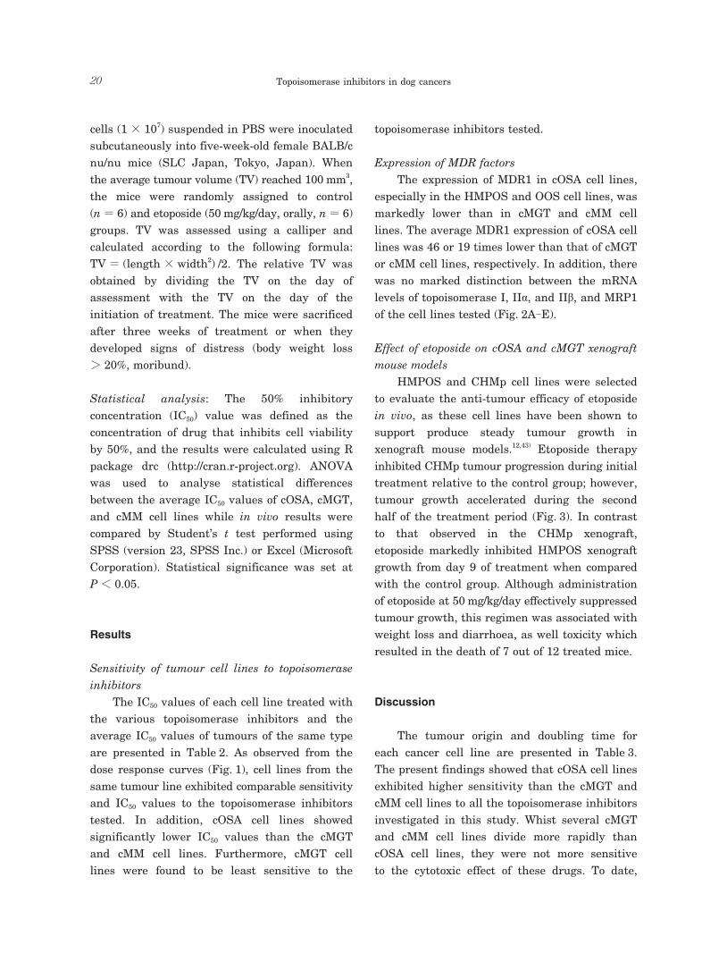
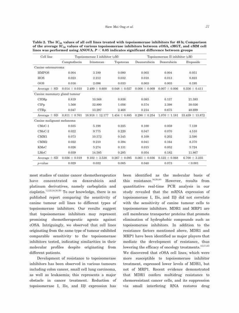
Sensitivity of tumour cell lines to topoisomerase inhibitors The IC50 values of each cell line treated with the various topoisomerase inhibitors and the average IC50 values of tumours of the same type are presented in Table 2. As observed from the dose response curves (Fig. 1), cell lines from the same tumour line exhibited comparable sensitivity and IC50 values to the topoisomerase inhibitors tested. In addition, cOSA cell lines showed significantly lower IC50 values than the cMGT and cMM cell lines. Furthermore, cMGT cell lines were found to be least sensitive to the
topoisomerase inhibitors tested.
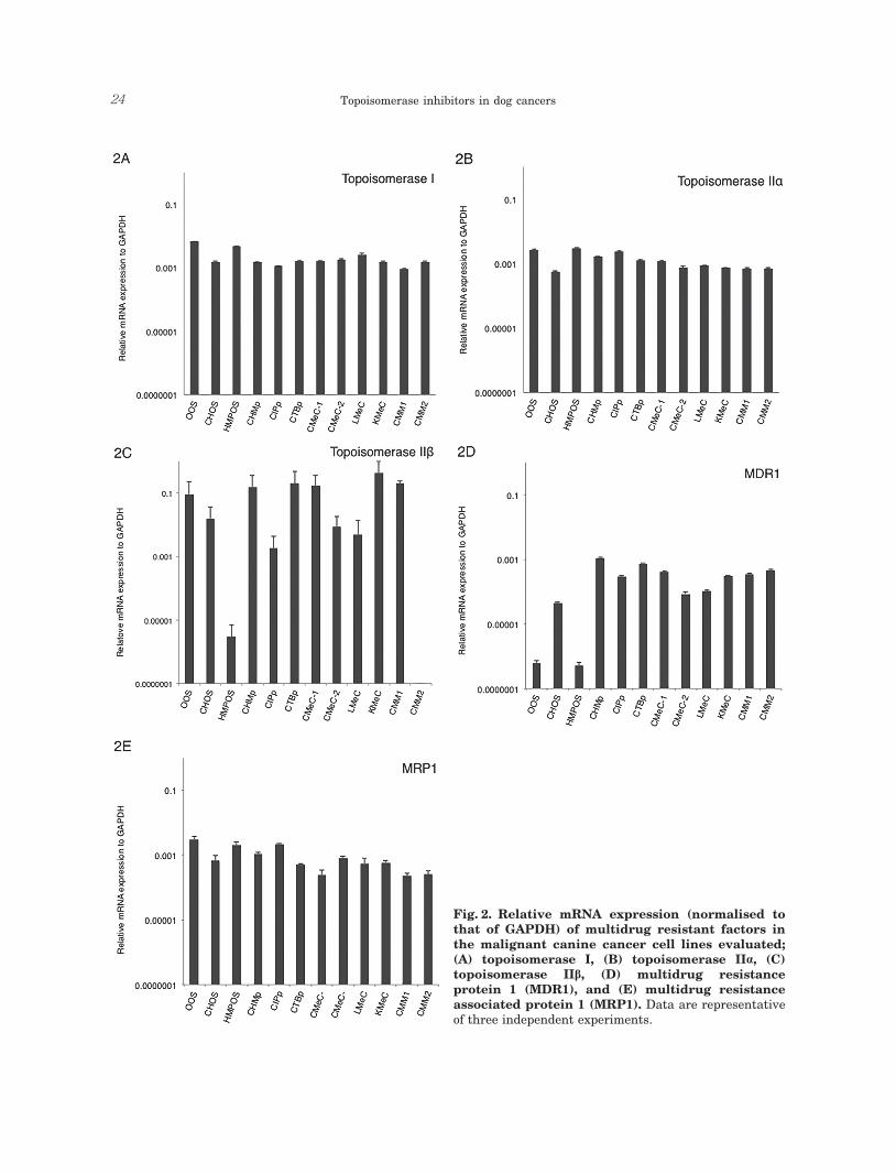
Expression of MDR factors The expression of MDR1 in cOSA cell lines, especially in the HMPOS and OOS cell lines, was markedly lower than in cMGT and cMM cell lines. The average MDR1 expression of cOSA cell lines was 46 or 19 times lower than that of cMGT or cMM cell lines, respectively. In addition, there was no marked distinction between the mRNA levels of topoisomerase I, IIα, and IIβ, and MRP1 of the cell lines tested (Fig. 2A-E).
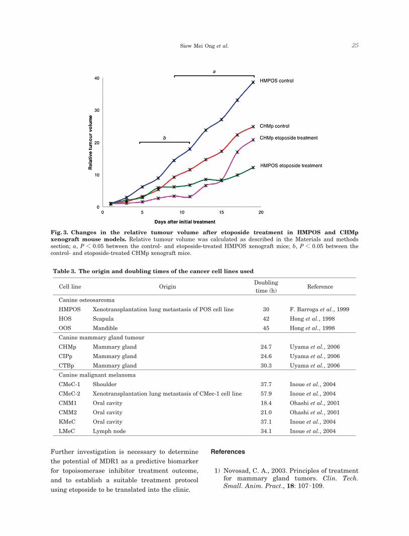
Effect of etoposide on cOSA and cMGT xenograft mouse models HMPOS and CHMp cell lines were selected to evaluate the anti-tumour efficacy of etoposide in vivo, as these cell lines have been shown to support produce steady tumour growth in xenograft mouse models.12,43) Etoposide therapy inhibited CHMp tumour progression during initial treatment relative to the control group; however, tumour growth accelerated during the second half of the treatment period (Fig. 3). In contrast to that observed in the CHMp xenograft, etoposide markedly inhibited HMPOS xenograft growth from day 9 of treatment when compared with the control group. Although administration of etoposide at 50 mg/kg/day effectively suppressed tumour growth, this regimen was associated with weight loss and diarrhoea, as well toxicity which resulted in the death of 7 out of 12 treated mice.
Discussion
The tumour origin and doubling time for each cancer cell line are presented in Table 3. The present findings showed that cOSA cell lines exhibited higher sensitivity than the cMGT and cMM cell lines to all the topoisomerase inhibitors investigated in this study. Whist several cMGT and cMM cell lines divide more rapidly than cOSA cell lines, they were not more sensitive to the cytotoxic effect of these drugs. To date,
Siew Mei Ong et al. 21
Table 2. The IC50 values of all cell lines treated with topoisomerase inhibitors for 48 h; Comparison of the average IC50 values of various topoisomerase inhibitors between cOSA, cMGT, and cMM cell lines was performed using ANOVA. P < 0.05 indicates significant difference between groups
Cell line Topoisomerase I inhibitor (μM) Topoisomerase II inhibitor (μM)
Camptothecin Irinotecan Topotecan Daunorubicin Doxorubicin Etoposide
Canine osteosarcoma
HMPOS 0.004 3.189 0.080 0.003 0.004 0.051
HOS 0.023 2.212 0.032 0.018 0.013 0.823
OOS 0.016 2.096 0.033 0.003 0.003 0.195
Average ± SD 0.014 ± 0.010 2.499 ± 0.600 0.048 ± 0.027 0.008 ± 0.009 0.007 ± 0.006 0.356 ± 0.411
Canine mammary gland tumour
CHMp 0.819 10.568 0.838 0.083 0.137 21.593
CIPp 1.568 32.890 1.056 0.574 2.398 30.026
CTBp 0.047 13.297 2.468 0.214 0.675 48.699
Average ± SD 0.811 ± 0.761 18.918 ± 12.177 1.454 ± 0.885 0.290 ± 0.254 1.070 ± 1.181 33.439 ± 13.872
Canine malignant melanoma
CMeC-1 0.035 5.198 0.225 0.100 0.059 7.139
CMeC-2 0.022 9.775 0.220 0.047 0.070 4.510
CMM1 0.073 10.572 0.345 0.109 0.202 2.590
CMM2 0.022 9.210 0.394 0.041 0.164 8.370
KMeC 0.026 5.274 0.131 0.015 0.052 5.724
LMeC 0.039 14.585 0.285 0.054 0.182 11.867
Average ± SD 0.036 ± 0.019 9.102 ± 3.538 0.267 ± 0.095 0.061 ± 0.036 0.122 ± 0.068 6.700 ± 3.235
p-value 0.029 0.032 0.005 0.040 0.073 <0.001
most studies of canine cancer chemotherapeutics have concentrated on doxorubicin and platinum derivatives, namely carboplatin and cisplatin.1,2,22,34,38,39) To our knowledge, there is no published report comparing the sensitivity of canine tumour cell lines to different types of topoisomerase inhibitors. Our results suggest that topoisomerase inhibitors may represent promising chemotherapeutic agents against cOSA. Intriguingly, we observed that cell lines originating from the same type of tumour exhibited comparable sensitivity to the topoisomerase inhibitors tested, indicating similarities in their molecular profiles despite originating from different patients. Development of resistance to topoisomerase inhibitors has been observed in various tumours including colon cancer, small cell lung carcinoma, as well as leukaemia; this represents a major obstacle in cancer treatment. Reduction of topoisomerase I, IIα, and IIβ expression has
been identified as the molecular basis of this resistance.16,38,40) However, results from quantitative real-time PCR analysis in our study revealed that the mRNA expression of topoisomerase I, IIα, and IIβ did not correlate with the sensitivity of canine tumour cells to topoisomerase inhibitors. MDR1 and MRP1 are cell membrane transporter proteins that promote elimination of hydrophobic compounds such as topoisomerase inhibitors. In addition to the resistance factors mentioned above, MDR1 and MRP1 have been identified as major players that mediate the development of resistance, thus lowering the efficacy of oncology treatments.13,17,33) We discovered that cOSA cell lines, which were more susceptible to topoisomerase inhibitor treatment, expressed lower levels of MDR1, but not of MRP1. Recent evidence demonstrated that MDR1 confers multidrug resistance to chemoresistant cancer cells, and its suppression via small interfering RNA restores drug
Topoisomerase inhibitors in dog cancers22
sensitivity.30,35,47) In addition, Gramer et al. has reported that MDR1 upregulation is associated with disease progression in canine patients with lymphoma receiving chemotherapy. Our findings are in agreement with several previous publications questioning the contribution of MRP1 to clinical drug resistance.6,10) Therefore, we suggest that MDR1 plays a more direct role in the development of resistance to topoisomerase inhibitor therapy and may serve as a predictive biomarker for treatment outcome in canine cancers. We observed that topoisomerase inhibitors are effective against cOSA cell lines. cOSA is locally invasive and requires wide marginal excision via amputation or limb salvage procedures; however, a major challenge is the presence of micrometastases at the time of
presentation or diagnosis.26,41) Although adjuvant treatment of cOSA with doxorubicin or platinum-based drugs extends overall survival of dogs with OSA from 11-21% to 35-50% at 1 year, it fails to impede the progression of the metastatic disease which is the ultimate cause of death.2,3,4,5,7,8,26,
28,31,34,39,41) Therefore, a novel chemotherapeutic protocol for cOSA is necessary. We evaluated the anti-tumour effect of etoposide in HMPOS and CHMp xenograft mouse models. Etoposide was selected for in vivo study because the use of this chemotherapeutic agent in veterinary oncology is currently limited, and its potential for clinical application is yet to be explored. Interestingly, our study demonstrated that etoposide therapy delayed tumour progression of both HMPOS and CHMp xenografts; however, tumour growth of the latter increased rapidly at the later phase.
Siew Mei Ong et al. 23
This may be attributed to the high basal expression of MDR1 in CHMp cells, which increased drug efflux or treatment-induced acquisition of drug resistance via upregulation of MDR1 expression.13,42)
The adverse effects of etoposide include myelosuppression and gastrointestinal toxicity.15,36,46) The xenograft mice that underwent etoposide therapy showed signs consistent with toxicity. As a similar treatment protocol has been employed in a previous study without severe adverse effects, the cause of the observed discrepancy with our study is unknown.25) Further studies are necessary to establish a suitable treatment protocol for translation into canine patients with cancer. Taken together, our results reveal that
topoisomerase inhibitors, specifically etoposide, exhibit effective anti-tumour effects against cOSA both in vitro and in vivo; these may be associated with lower levels of MDR1 expression in cOSA cell lines when compared with cMGT and cMM cell lines. The caveats of this study include lack of pre- and post-treatment quantification of intra-tumoural MDR1 gene expression to explore the effect of etoposide therapy on MDR1 level, which may have explained the progression of tumour growth in CHMp xenograft mice during the latter half of the treatment period. In addition, the dose of etoposide employed led to the development of toxicity; therefore, further studies are required to determine the optimal etoposide dose for maximal efficacy and minimal toxicity.
Fig. 1. Dose response curve of the malignant canine cancer cell lines to topoisomerase I inhibitors; (A) camptothecin, (B) irinotecan, (C) topotecan; and topoisomerase II inhibitors; (D) daunorubicin, (E) doxorubicin, (F) etoposide. cOSA, canine osteosarcoma; cMGT, canine mammary gland tumour; cMM, canine malignant melanoma. Data are expressed as means ± standard error of means (SEMs).
Topoisomerase inhibitors in dog cancers24
Fig. 2. Relative mRNA expression (normalised to that of GAPDH) of multidrug resistant factors in the malignant canine cancer cell lines evaluated; (A) topoisomerase I, (B) topoisomerase IIα, (C) topoisomerase IIβ, (D) multidrug resistance protein 1 (MDR1), and (E) multidrug resistance associated protein 1 (MRP1). Data are representative of three independent experiments.
Siew Mei Ong et al. 25
Further investigation is necessary to determine the potential of MDR1 as a predictive biomarker for topoisomerase inhibitor treatment outcome, and to establish a suitable treatment protocol using etoposide to be translated into the clinic.
Fig. 3. Changes in the relative tumour volume after etoposide treatment in HMPOS and CHMp xenograft mouse models. Relative tumour volume was calculated as described in the Materials and methods section; a, P < 0.05 between the control- and etoposide-treated HMPOS xenograft mice; b, P < 0.05 between the control- and etoposide-treated CHMp xenograft mice.
Table 3. The origin and doubling times of the cancer cell lines used
Cell line OriginDoubling time (h)
Reference
Canine osteosarcoma
HMPOS Xenotransplantation lung metastasis of POS cell line 30 F. Barroga et al., 1999
HOS Scapula 42 Hong et al., 1998
OOS Mandible 45 Hong et al., 1998
Canine mammary gland tumour
CHMp Mammary gland 24.7 Uyama et al., 2006
CIPp Mammary gland 24.6 Uyama et al., 2006
CTBp Mammary gland 30.3 Uyama et al., 2006
Canine malignant melanoma
CMeC-1 Shoulder 37.7 Inoue et al., 2004
CMeC-2 Xenotransplantation lung metastasis of CMec-1 cell line 57.9 Inoue et al., 2004
CMM1 Oral cavity 18.4 Ohashi et al., 2001
CMM2 Oral cavity 21.0 Ohashi et al., 2001
KMeC Oral cavity 37.1 Inoue et al., 2004
LMeC Lymph node 34.1 Inoue et al., 2004
References
1) Novosad, C. A., 2003. Principles of treatment for mammary gland tumors. Clin. Tech. Small. Anim. Pract., 18: 107-109.

Topoisomerase inhibitors in dog cancers26
metabolites in rats. J. Pharmacol. Exp. Ther., 281: 304-314.
12) Barroga, E. F., Kadosawa, T., Okumura, M. and Fujinaga, T. 1999. Establishment and characterization of the growth and pulmonary metastasis of a highly lung metastasizing cell line from canine osteosarcoma in nude mice. J. Vet. Med. Sci., 61: 361-367.
13) Gramer, I., Kessler, M. and Geyer, J. 2015. Determination of MDR1 gene expression for prediction of chemotherapy tolerance and treatment outcome in dogs with lymphoma. Vet. Comp. Oncol., 13: 363-372.
14) Hande, K. R. 1998. Clinical applications of anticancer drugs targeted to topoisomerase II. Biochim. Bhiophys. Acta., 1400: 173-184.
15) Hande, K. R. 1998. Etoposide: four decades of development of a topoisomerase II inhibitor. Eur. J. Cancer, 34: 1514-1521.
16) Harker, W. G., Slade, D. L., Drake, F. H. and Parr, R. L. 1991. Mitoxantrone resistance in HL-60 leukemia cells: reduced nuclear topoisomerase II catalytic activity and drug-induced DNA cleavage in association with reduced expression of the topoisomerase II. beta. isoform. Biochemistry, 30: 9953-9961.
17) Hasegawa, S., Abe, T., Naito, S., Kotoh, S., Kumazawa, J., Hipfner, D. R., Deeley, R. G., Cole, S. P. and Kuwano, M. 1995. Expression of multidrug resistance-associated protein (MRP), MDR1 and DNA topoisomerase II in human multidrug-resistant bladder cancer cell lines. Br. J. Cancer, 71: 907-913.
18) Hohenhaus, A. E. and Matus, R. E. 1990. Etoposide (VP-16). J. Vet. Intern. Med., 4: 239-241.
19) Hong, S. H., Kadosawa, T., Mochizuki, M., Matsunaga, S., Nishimura, R. and Sasaki, N. 1998. Establishment and characterization of two cell lines derived from canine spontaneous osteosarcoma. J. Vet. Med. Sci., 60: 757-760.
20) Inoue, K., Ohashi, E., Kadosawa, T., Hong, S. H., Matsunaga, S., Mochizuki, M., Nishimura, R. and Sasaki, N. 2004. Establishment and characterization of four canine melanoma cell lines. J. Vet. Med. Sci., 66: 1437-1440.
21) Kadosawa, T., Nozaki, K., Sasaki, N. and Takeuchi, A. 1994. Establishment and characterization of a new cell line from a canine osteosarcoma. J. Vet. Med. Sci., 56: 1167-1169.
22) Knapp, D. W., Glickman, N. W., Widmer, W. R., DeNicola, D. B., Adams, L. G., Kuczek, T., Bonney, P. L., DeGortari, A. E., Han, C. and Glickman, L. T. 2000. Cisplatin versus cisplatin combined with piroxicam in a canine
2) Bacon, N. J., Ehrhart, N. P., Dernell, W. S., Lafferty, M. and Withrow, S. J. 2008. Use of alternating administration of carboplatin and doxorubicin in dogs with microscopic metastases after amputation for appendicular osteosarcoma: 50 cases (1999-2006). J. Am. Vet. Med. Assoc., 232: 1504-1510.
3) Berg, J., Weinstein, M. J., Schelling, S. H. and Rand, W. M. 1992. Treatment of dogs with osteosarcoma by administration of cisplatin after amputation or limb-sparing surgery: 22 cases (1987-1990). J. Am. Vet. Med. Assoc., 200: 2005-2008.
4) Berg, J., Weinstein, M., Springfield, D. and Rand, W. 1995. Results of surgery and doxorubicin chemotherapy in dogs with osteosarcoma. J. Am. Vet. Med. Assoc., 206: 1555-1560.
5) Bergman, P. J., MacEwen, E. G., Kurzman, I. D., Henry, C. J., Hammer, A. S., Knapp, D. W., Hale, A., Kruth, S. A., Klein, M. K., Klausner, J. and Norris, A. M. 1996. Amputation and carboplatin for treatment of dogs with osteosarcoma: 48 cases (1991 to 1993). J. Vet. Intern. Med., 10: 76-81.
6) Borst, P. 2012. Cancer drug pan-resistance: pumps, cancer stem cells, quiescence, epithelial to mesenchymal transition, blocked cell death pathways, persisters or what? Open Biol., 2: 120066.
7) Boston, S. E., Ehrhart, N. P., Dernell, W. S., Lafferty, M. and Withrow, S. J. 2006. Evaluation of survival time in dogs with stage III osteosarcoma that undergo treatment: 90 cases (1985-2004). J. Am. Vet. Med. Assoc., 228: 1905-1908.
8) Brodey, R. S. 1979. The use of naturally occurring cancer in domestic animals for research into human cancer: general considerations and a review of canine skeletal osteosarcoma. Yale J. Biol. Med., 52: 345-361.
9) Bugg, B. Y., Danks, M. K., Beck, W. T. and Suttle, D. P. 1991. Expression of a mutant DNA topoisomerase II in CCRF-CEM human leukemic cells selected for resistance to teniposide. Proc. Natl. Acad. Sci. U.S.A., 88: 7654-7658.
10) Chen, Z. S. and Tiwari, A. K. 2011. Multidrug resistance proteins (MRPs/ABCCs) in cancer chemotherapy and genetic diseases. FEBS J., 278: 3226-3245.
11) Chu, X. Y., Kato, Y., Niinuma, K., Sudo, K. I., Hakusui, H. and Sugiyama, Y. 1997. Multispecific organic anion transporter is responsible for the biliary excretion of the camptothecin derivative irinotecan and its
Siew Mei Ong et al. 27
model of human invasive urinary bladder cancer. Cancer Chemother. Pharmacol., 46: 221-226.
23) Lana, S., U’ren, L., Plaza, S., Elmslie, R., Gustafon, D., Morley, P. and Dow, S. 2007. Continuous low-dose oral chemotherapy for adjuvant therapy of splenic hemangiosarcoma in dogs. J. Vet. Intern. Med., 21: 764-769.
24) Longley, D. B. and Johnston, P. G, 2005. Molecular mechanisms of drug resistance. J. Pathol., 205: 275-292.
25) Matsumoto, S., Mashiba, H., Okamoto, K. and Ekimoto, H. 1999. Evaluation of antitumor activity of etoposide administered orally for 21 consecutive days against human uterine cancer subcutaneous and/or orthotopic xenografts in nude mice. Gan To Kagaku Ryoho, 26: 1313-1320. (in Japanese).
26) Mauldin, G. N., Matus, R. E., Withrow, S. J. and Patnaik, A. K. 1988. Canine osteosarcoma. J. Vet. Intern. Med., 2: 177-180.
27) Meijer, C., Mulder, N. H., Timmer-Bosscha, H., Sluiter, W. J., Meersma, G. J. and Vries, E. G. 1992. Relationship of cellular glutathione to the cytotoxicity and resistance of seven platinum compounds. Cancer Res., 52: 6885- 6889.
28) Moore, A. S., Dernell, W. S., Ogilvie, G. K., Kristal, O., Elmslie, R., Kitchell, B., Susaneck, S., Rosenthal, R., Klein, M. K., Obradovoich, J. and Legendre, A. 2007. Doxorubicin and BAY 12-9566 for the treatment of osteosarcoma in dogs: a randomized, double-blind, place-controlled study. J. Vet. Intern. Med., 21: 783-790.
29) Mueller, F., Fuchs, B. and Kaser-Hotz, B. 2007. Comparative biology of human and canine osteosarcoma. Anticancer Res., 27: 155-164.
30) Nourbakhsh, M., Jaafari, M. R., Lage, H., Abnous, K., Mosaffa, F., Badiee, A. and Behravan, J. 2015. Nanolipoparticles-mediated MDR1 siRNA delivery reduces doxorubicin resistance in breast cancer cells and silences MDR1 expression in xenograft model of human breast cancer. Iran. J. Basic Med. Sci., 18: 385-392.
31) Oblak, M. L., Boston, S. E., Higginson, G., Patten, S. G., Monteith, G. J. and Woods, J. P. 2012. The impact of pamidronate and chemotherapy on survival times in dogs with appendicular primary bone tumors treated with palliative radiation therapy. Vet. Surg., 41: 430-435.
32) Ohashi, E., Hong, S. H., Takahashi, T., Nakagawa, T., Mochizuki, M., Nishimura, R.
and Sasaki, N. 2001. Effect of retinoids on growth inhibition of two canine melanoma cell lines. J. Vet. Med. Sci., 63: 83-86.
33) Pawlowski, K. M., Mucha, J., Majchrzak, K., Motyl, T. and Król, M. 2013. Expression and role of PGP, BCRP, MRP1 and MRP3 in multidrug resistance of canine mammary cancer cells. BMC Vet. Res., 9: 119.
34) Phillips, B., Powers, B. E., Dernell, W. S., Straw, R. C., Khanna, C., Hogge, G. S. and Vail, D. M. 2009. Use of single-agent carboplatin as adjuvant or neoadjuvant therapy in conjunction with amputation for appendicular osteosarcoma in dogs. J. Am. Anim. Hosp. Assoc., 45: 33-38.
35) Pop, I., Pop, L., Vitetta, E. S. and Ghetie, M. A. 2008. Generation of multidrug resistant lymphoma cell lines stably expressing P-glycoprotein. Oncol. Rep., 19: 889-896.
36) Rose, P. G., Blessing, J. A., Mayer, A. R. and Homesley, H. D. 1998. Prolonged oral etoposide as second-line therapy for platinum-resistant and platinum-sensitive ovarian carcinoma: a Gynecologic Oncology Group study. J. Clin. Oncol., 16: 405-410.
37) Rothenberg, M. L. 1997. Topoisomerase I inhibitors: review and update. Ann. Oncol., 8: 837-855.
38) Simon, D., Knebel, J. W., Baumgärtner, W., Aufderheide, M., Meyer-Lindenberg, A. and Nolte, I. 2001. In vitro efficacy of chemotherapeutics as determined by 50% inhibitory concentrations in cell cultures of mammary gland tumors obtained from dogs. Am. J. Vet. Res., 62: 1825-1830.
39) Straw, R. C., Withrow, S. J., Richter, S. L., Powers, B. E., Klein, M. K., Postorino, N. C., LaRue, S. M., Ogilvie, G. K., Vail, D. M., Morrison, W. B., McGee, M. and Dickinson, K. 1991. Amputation and cisplatin for treatment of canine osteosarcoma. J. Vet. Intern. Med., 5: 205-210.
40) Sugimoto, Y., Tsukahara, S., Oh-Hara, T., Isoe, T. and Tsuruo, T. 1990. Decreased expression of DNA topoisomerase I in camptothecin-resistant tumor cell lines as determined by a monoclonal antibody. Cancer Res., 50: 6925-6930.
41) Szewczyk, M., Lechowski, R. and Zabielska, K. 2015. What do we know about canine osteosarcoma treatment?-review. Vet. Res. Commun., 39: 61-67.
42) Thomas, H. and Coley, H. M. 2003. Overcoming multidrug resistance in cancer: an update on the clinical strategy of inhibiting p-glycoprotein. Cancer Control, 10:
Topoisomerase inhibitors in dog cancers28
159-165.43) Uyama, R., Nakagawa, T., Hong, S. H.,
Mochizuki, M., Nishimura, R. and Sasaki, N. 2006. Establishment of four pairs of canine mammary tumour cell lines derived from primary and metastatic origin and their E-cadherin expression. Vet. Comp. Oncol., 4: 104-113.
44) Vail, D. M. and MacEwen, E. G. 2000. Spontaneously occurring tumors of companion animals as models for human cancer. Cancer Invest., 18: 781-792.
45) Wang, J. C. 1996. DNA topoisomerases. Annu. Rev. Biochem., 65: 635-692.
46) Wood, L. J., Nail, L. M., Perrin, N. A., Elsea, C. R., Fischer, A. and Druker, B. J. 2006. The cancer chemotherapy drug etoposide (VP-16) induces proinflammatory cytokine production
and sickness behavior-like symptoms in a mouse model of cancer chemotherapy-related symptoms. Biol. Res. Nurs., 8: 157-169.
47) Wu, H., Hait, W. N. and Yang, J. M. 2003. Small interfering RNA-induced suppression of MDR1 (P-glycoprotein) restores sensitivity to multidrug-resistant cancer cells. Cancer Res., 63: 1515-1519.
48) Xu, Y. and Her, C. 2015. Inhibition of topoisomerase (DNA) I (TOP1): DNA damage repair and anticancer therapy. Biomolecules, 5: 1652-1670.
49) Zandvliet, M., Teske, E. and Schrickx, J. A. 2014. Multi-drug resistance in a canine lymphoid cell line due to increased P-glycoprotein expression, a potential model for drug-resistant canine lymphoma. Toxicol. In Vitro, 28: 1498-1506.

![4β-[4’-(1-(Aryl)ureido)benzamide]podophyllotoxins as DNA ... · 1 4 -[4 -(1-(Aryl)ureido)benzamide]podophyllotoxins as DNA Topoisomerase I and IIα Inhibitors and Apoptosis Inducing](https://img.pdfslide.net/doc/110x75/5e7ee9e4bc140f3b9414d72f/4-4a-1-arylureidobenzamidepodophyllotoxins-as-dna-1-4-4-1-arylureidobenzamidepodophyllotoxins.jpg)