Embed Size (px)
Citation preview

Anti-VEGF
Developments in Ophthalmology
Vol. 46
Series Editors
F. Bandello Milan
W. Behrens-Baumann Magdeburg
Anti-VEGF
Volume Editors
F. Bandello Milan
M. Battaglia Parodi Milan
Co-Editors
A.J. Augustin Karlsruhe
P. Iacono Rome
R.O. Schlingemann Amsterdam
U. Schmidt-Erfurth Vienna
F.D. Verbraak Amsterdam
12 figures, 5 in color, and 13 tables, 2010
Basel · Freiburg · Paris · London · New York · Bangalore ·
Bangkok · Shanghai · Singapore · Tokyo · Sydney
Bibliographic Indices. This publication is listed in bibliographic services, including Current Contents® and Index Medicus.
Disclaimer. The statements, opinions and data contained in this publication are solely those of the individual authors and
contributors and not of the publisher and the editor(s). The appearance of advertisements in the book is not a warranty,
endorsement, or approval of the products or services advertised or of their effectiveness, quality or safety. The publisher and the
editor(s) disclaim responsibility for any injury to persons or property resulting from any ideas, methods, instructions or products
referred to in the content or advertisements.
Drug Dosage. The authors and the publisher have exerted every effort to ensure that drug selection and dosage set forth in this
text are in accord with current recommendations and practice at the time of publication. However, in view of ongoing research,
changes in government regulations, and the constant flow of information relating to drug therapy and drug reactions, the reader
is urged to check the package insert for each drug for any change in indications and dosage and for added warnings and
precautions. This is particularly important when the recommended agent is a new and/or infrequently employed drug.
All rights reserved. No part of this publication may be translated into other languages, reproduced or utilized in any form or by
any means electronic or mechanical, including photocopying, recording, microcopying, or by any information storage and
retrieval system, without permission in writing from the publisher.
© Copyright 2010 by S. Karger AG, P.O. Box, CH–4009 Basel (Switzerland)
www.karger.com
Printed in Switzerland on acid-free and non-aging paper (ISO 9706) by Reinhardt Druck, Basel
ISSN 0250–3751
ISBN 978–3–8055–9529-2
e-ISBN 978–3–8055–9530-8
Library of Congress Cataloging-in-Publication Data
Anti-VEGF / volume editors, Francesco Bandello, M. Battaglia Parodi ;
co-editors, A. Augustin ... [et al.].
p. ; cm. -- (Developments in ophthalmology, ISSN 0250-3751 ; v. 46)
Includes bibliographical references and indexes.
ISBN 978-3-8055-9529-2 (hard cover : alk. paper) -- ISBN 978-3-8055-9530-8
(e-ISBN)
1. Eye--Blood-vessels--Diseases--Chemotherapy. 2. Vascular endothelial
growth factors--Antagonists--Therapeutic use. I. Bandello, F. (Francesco)
II. Battaglia Parodi, M. (Maurizio) III. Series: Developments in
ophthalmology ; v. 46. 0250-3751
[DNLM: 1. Eye Diseases--therapy. 2. Vascular Endothelial Growth
Factors--therapeutic use. 3. Neovascularization, Pathologic--therapy. W1
DE998NG v.46 2010 / WW 166 A6334 2010]
RE720.A58 2010
617.7�061--dc22
2010025184
Francesco BandelloDepartment of OphthalmologyUniversity Vita-Salute Scientific InstituteSan RaffaeleVia Olgettina, 60IT–20132 Milano (Italy)
Maurizio Battaglia ParodiDepartment of OphthalmologyUniversity Vita-Salute Scientific InstituteSan RaffaeleVia Olgettina, 60IT–20132 Milano (Italy)
Section Title
Contents
VII List of Contributors
IX Preface Bandello, F. (Milan)
1 Angiostatic and Angiogenic Factors de Groot, H.; Schmit-Eilenberger, V.; Kirchhof, J.; Augustin, A.J. (Karlsruhe)
4 Mechanisms of Ocular Angiogenesis and Its Molecular Mediators Siemerink, M.J. (Amsterdam); Augustin, A.J. (Karlsruhe);
Schlingemann, R.O. (Amsterdam)
21 Antivascular Endothelial Growth Factors in Age-Related Macular Degeneration
Schmidt-Erfurth, U.; Pollreisz, A.; Mitsch, C.; Bolz, M. (Vienna)
39 Antivascular Endothelial Growth Factor in Diabetic Retinopathy Iacono, P. (Rome); Battaglia Parodi, M.; Bandello, F. (Milan)
54 Retinal Vein Occlusions Buehl, W.; Sacu, S.; Schmidt-Erfurth, U. (Vienna)
73 Antivascular Endothelial Growth Factor for Choroidal Neovascularization in Pathologic Myopia
Battaglia Parodi, M. (Milan); Iacono, P. (Rome); Bandello, F. (Milan)
84 Antivascular Endothelial Growth Factors for Inflammatory Chorioretinal Disorders
Battaglia Parodi, M. (Milan); Iacono, P. (Rome); Verbraak, F.D. (Amsterdam); Bandello, F. (Milan)
96 Antivascular Endothelial Growth Factor Treatment in Pseudoxanthoma Elasticum Patients
Verbraak, F.D. (Amsterdam)
V

VI Contents
107 Antivascular Endothelial Growth Factor in Hereditary Dystrophies Battaglia Parodi, M. (Milan); Iacono, P. (Rome); Bandello, F. (Milan)
111 Antivascular Endothelial Growth Factor as an Approach for Macular Edema Buchholz, P.M.; Buchholz, A.P.; Augustin, A.J. (Karlsruhe)
123 Angiogenesis and Vascular Endothelial Growth Factors in Intraocular Tumors
Missotten, G.S. (Leuven/Amsterdam); Schlingemann, R.O. (Amsterdam); Jager, M.J. (Leiden)
133 Antivascular Endothelial Growth Factors in Anterior Segment Diseases Scholl, S.; Kirchhof, J.; Augustin, A.J. (Karlsruhe)
140 Author Index
141 Subject Index
List of Contributors
VII
A.J. Augustin, Prof.Augenklinik Moltkestrasse 90DE–76133 Karlsruhe (Germany)E-Mail [email protected]
F. Bandello, Prof.Department of OphthalmologyUniversity Vita-Salute Scientific InstituteSan RaffaeleVia Olgettina, 60IT–20132 Milano (Italy)E-Mail [email protected]
M. Battaglia Parodi, Dr.Department of OphthalmologyUniversity Vita-Salute Scientific InstituteSan RaffaeleVia Olgettina, 60IT–20132 Milano (Italy)E-Mail [email protected]
M. Bolz, Dr.Department of Ophthalmology and OptometryMedical University ViennaWaehringer Guertel 18-20AT–1090 Vienna (Austria)E-Mail [email protected]
A. Buchholz, Dr.AugenklinikMoltkestrasse 90DE–76133 Karlsruhe (Germany)E-Mail [email protected]
P. Buchholz, Dr.AugenklinikMoltkestrasse 90DE–76133 Karlsruhe (Germany)E-Mail [email protected]
W. Buehl, Dr.Department of Ophthalmology and OptometryMedical University ViennaWaehringer Guertel 18-20AT–1090 Vienna (Austria)E-Mail [email protected]
H. de Groot, Dr.Augenklinik Moltkestrasse 90DE–76133 Karlsruhe (Germany)E-Mail [email protected]
P. Iacono, Dr.Fondazione G.B. Bietti per l’OftalmologiaIRCCS (Istituto di Ricovero e Cura a Carattere Scientifico)Via Livenza 3IT–00198 Rome (Italy)E-Mail [email protected]
M.J. Jager, Prof.Department of OphthalmologyLUMCPO Box 9600NL–2300 RC Leiden (The Netherlands)E-Mail [email protected]
J. Kirchhof, Dr.AugenklinikMoltkestrasse 90DE–76133 Karlsruhe (Germany)E-Mail [email protected]
G.S. Missotten, Prof.Department OphthalmologyAcademic Medical CenterMeibergdreef 9NL–1100DD Amsterdam (The Netherlands)E-Mail [email protected]
C. Mitsch, Dr.Department of Ophthalmology and OptometryMedical University ViennaWaehringer Guertel 18-20AT–1090 Vienna (Austria)E-Mail [email protected]
A. Pollreisz, Dr.Department of Ophthalmology and OptometryMedical University ViennaWaehringer Guertel 18-20AT–1090 Vienna (Austria)E-Mail [email protected]
S. Sacu, Dr.Department of Ophthalmology and OptometryMedical University ViennaWaehringer Guertel 18-20AT–1090 Vienna (Austria)E-Mail [email protected]
R.O. Schlingemann, Prof.Medical Retina Unit and Ocular Angiogenesis GroupDepartment of OphthalmologyAcademic Medical CenterUniversity of Amsterdam, A2-122NL–1100 DD Amsterdam (The Netherlands)E-Mail [email protected]
U. Schmidt-Erfurth, Prof.Department of Ophthalmology and OptometryMedical University ViennaWaehringer Guertel 18-20AT–1090 Vienna (Austria)E-Mail [email protected]
V. Schmit-Eilenberger, Dr.AugenklinikMoltkestrasse 90DE–76133 Karlsruhe (Germany)E-Mail [email protected]
S. Scholl, Dr.AugenklinikMoltkestrasse 90DE–76133 Karlsruhe (Germany)E-Mail [email protected]
M.J. Siemerink, Dr.Medical Retina Unit and Ocular Angiogenesis GroupDepartment of OphthalmologyAcademic Medical CenterUniversity of Amsterdam, A2-122NL–1100 DD Amsterdam (The Netherlands)E-Mail [email protected]
F.D. Verbraak, Dr.Departments of Ophthalmology and Biomedical Engineering & PhysicsAcademic Medical CenterMeibergdreef 9NL–1105 AZ Amsterdam (The Netherlands)E-Mail [email protected]
VIII List of Contributors
Section Title
Preface
The development of vascular endothelial growth factor inhibitors for the treatment of
ocular neovascularization and macular edema can be regarded as the beginning of a
new era in ophthalmological therapy. Before the year 2000, the treatment of any vas-
cular abnormality in the macular region was merely restricted to conventional laser
photocoagulation. Indubitably, laser treatment represents a destructive procedure
which leads to a permanent scar and brings about a retinal sensitivity deterioration
in all cases. Since 2000, photodynamic therapy with verteporfin has been introduced
as the first attempt to couple laser energy with a light-sensitive drug in an attempt to
treat choroidal neovascularization through a relatively non-destructive form of ther-
apy. Nevertheless, photodynamic therapy can provide only a very limited visual acu-
ity improvement, especially in choroidal neovascularization secondary to age-related
macular degeneration and pathologic myopia.
In an attempt to improve the functional outcomes, many researchers have studied
the potential application of anti-angiogenic agents on ocular diseases. Previous inves-
tigations have demonstrated that vascular endothelial growth factor plays an impor-
tant role in promoting angiogenesis and vascular leakage in several ocular pathologic
conditions. The main goals of antivascular endothelial growth factor therapy are the
inhibition of growth and development of new vessels, along with the reduction of
vascular permeability.
The encouraging results of the most important randomized clinical trials regarding
the efficacy of ranibizumab and pegaptanib on subfoveal choroidal neovasculariza-
tion in relation to age-related macular degeneration have greatly influenced current
medical practice. As a result, in the last few years, many applications have been pro-
posed in an effort to treat several vascular diseases of the eye.
This aim of this book is to help update residents, general ophthalmologists, and
retina specialists on the latest applications of antivascular endothelial growth factor
Section Title
IX

therapy in ocular diseases. After an outline of the treatment principles, it covers a
large number of topics, including age-related macular degeneration, pathologic myo-
pia, angioid streaks, inflammatory diseases, hereditary dystrophies, retinal vein occlu-
sions, diabetic retinopathy, ocular tumors, and anterior segment neovascularizations.
We hope that each chapter will stimulate the interest of readers working in this field.
Francesco Bandello, Milan
X Preface
Bandello F, Battaglia Parodi M (eds): Anti-VEGF.
Dev Ophthalmol. Basel, Karger, 2010, vol 46, pp 1–3
Angiostatic and Angiogenic FactorsHeink de Groot � Vera Schmit-Eilenberger � Janna Kirchhof �
Albert J. Augustin
Augenklinik, Karlsruhe, Germany
AbstractBoth diminution of angiostatic and increment of angiogenic factors seem to contribute to neovascu-
larization in the eye under pathologic conditions. They are presented here separately. The involved
proteins can change their role during the process of neovascularization from promoters to inhibitors
and vice versa. Angiostatic factors can be divided into passive, active, unspecific and specific ones.
Some of them act during neovascularization as members of feedback loops by modifying the effects
of their angiogenic counterparts. Among the angiogenic factors VEGF is the most important.
Nevertheless other stimulating proteins exist in large numbers. Together with their static counter-
parts they form a complex network which controls neovascularization under physiologic as well as
pathologic conditions. Copyright © 2010 S. Karger AG, Basel
A short introduction into the topics of angiostatic and angiogenic factors is given. All
molecules mentioned and their interactions within the organism will be discussed in
the following article.
Angiostatic Factors in the Eye
Under healthy conditions the vascular system of the eye is thought to be stable.
Normal angiogenesis is concluded during early childhood and only reappears under
certain pathologic conditions. While one common trigger of neovascularization in
many eye diseases is ischemia, neovascularization can also occur without significant
ischemia. This is the case in wet age-related macular degeneration (AMD). However,
hypoxia and/or alterations of the perfusion are still under discussion to be an impor-
tant cofactor in the pathogenesis of this disease entity.
In ischemic neovascularization, new capillaries typically sprout from branches of
the retinal arteries. In contrast, the neovascularization in AMD originates from the
2 de Groot · Schmit-Eilenberger · Kirchhof · Augustin
choriocapillary layer. The physiological stability of the ocular vascular system is an
equilibrium between angiostatic and angiogenic factors. The vasculature is stable as
long as the angiostatic factors are ahead. Pathologic conditions such as ischemia or
inflammation shift the balance towards angiogenic factors which are released by the
damaged cells. On the other hand the unpredictable appearance of neovasculariza-
tion during dry AMD which cannot be prevented by anti-inflammatory treatment
strongly points out that also a loss of angiostatic factors alone can lead to instability of
the constructive vascular boundaries of the eye.
The strong angiostasis that is crucial for the function of the eye is maintained by
angiostatic factors in every involved tissue starting from the specialized guards of the
blood-retinal barrier down to unspecific ingredients of the blood fluid. The angio-
static effect is not only locally distributed but also stepwise during stages of angiogen-
esis. Due to the defensive nature of static concepts, not only active components such
as inhibitor proteins but also passive stabilizing members of the extracellular matrix
can be accounted to the angiostatic system.
Thus, collagens, elastins and fibrin constitute a first barrier for angiogenesis. These
molecules have to be actively degraded and the respective proteases are controlled
by protease inhibitors. Tissue inhibitors of metalloproteinases are specific metallo-
proteinase inhibitors while the serum component α2-macroglobulin unspecifically
inhibits metalloproteinases. Another protein that interferes with pericellular prote-
olysis required for migration and proliferation of endothelial cells is thrombospondin
which is present in platelet granules and is released following platelet activation. If
proteolytic degradation of capillary basement membranes occurs, a fragment of the
collagen type 18 called endostatin is released. It specifically inhibits proliferation of
endothelial cells and angiogenesis.
Other passive components of vascular stability are the VE cadherins that are
involved in intercellular tight junctions – the constituting basis of the blood-retinal
barrier. VE cadherins are members of a large family of adhesion proteins called cad-
herins that build intercellular contacts like desmosomes throughout the body. VE
cadherins have to be degraded before angiogenesis can occur. Their degradation is
triggered by vascular endothelial growth factors (VEGF) via the VEGFR-2 receptor.
More active components of vascular structural stability of the eye are proteins that
are secreted by the cells of the blood-retinal barrier. A protein that maintains stabil-
ity after maturation of newly grown capillaries is angiopoietin-1. It is produced by
pericytes. Its presence in mature capillaries improves continuity of the basal mem-
branes and the adherence of pericytes to endothelial cells. During angiogenesis it pro-
motes capillary growth. It is antagonized by angiopoietin-2 which binds to the same
endothelial cell-specific receptor Tie-2. TGF-β has among its many other effects a
similar role as it is secreted by pericytes and stabilizes the basal membrane of newly
built capillaries.
Pigment epithelium-derived factor is a cytokine that despite its name is produced
in many human cells including endothelial cells and retinal pigment epithelial cells
Angiostatic and Angiogenic Factors 3
where it was originally detected. Among other effects it is a potent inhibitor of angio-
genesis. It also has immunomodulatory features and contributes by this indirectly to
prevention of neovascularization.
The vasoinhibins act as negative feedback regulators upon the effect of VEGF.
They are upregulated in endothelial cells by VEGF and specifically inhibit migra-
tion and proliferation of these. Angiostatin also specifically inhibits proliferation of
endothelial cells. It is a fragment of plasminogen and therefore exists as a plasma fac-
tor throughout the body.
Angiogenic Factors
The growth of new blood vessels is an important natural process occurring in the
body, both in health and disease. Angiogenesis is a physiological process involving
the growth of new blood vessels from preexisting vessels whereas vasculogenesis
describes the formation of vascular structures from circulating or tissue-resident
endothelial stem cells (angioblasts) which proliferate into de novo endothelial cells.
The healthy body controls angiogenesis through a series of ‘on’ and ‘off ’ switches.
The main ‘on’ switches are known as angiogenesis-stimulation growth factors, or sim-
ply angiogenic factors. Stimulation of angiogenesis is performed by various angio-
genic proteins, including several growth factors, whereas the VEGF family has been
demonstrated to be a major contributor to angiogenesis. Additionally, a large number
of mediators exist which are involved in angiogenesis like insulin-like growth factor,
the family of fibroblast growth factor, interleukins, angiopoietins, epidermal growth
factor, transforming growth factors, platelet-derived growth factor, tumor necrosis
factor-α and vascular endothelial cadherin.
The balance between angiogenesis and inhibitors of new vessel growth is con-
trolled by a sophisticated interaction between different factors and mediators which
will be described explicitly in the following chapter.
Prof. A.J. Augustin
Augenklinik
Moltkestrasse 90
DE–76133 Karlsruhe (Germany)
Tel. +49 721 9742001, Fax +49 721 9742009, E-Mail [email protected]
Bandello F, Battaglia Parodi M (eds): Anti-VEGF.
Dev Ophthalmol. Basel, Karger, 2010, vol 46, pp 4–20
Mechanisms of Ocular Angiogenesis and Its Molecular MediatorsMartin J. Siemerinka � Albert J. Augustinb � Reinier O. Schlingemanna,c
aOcular Angiogenesis Group, Department of Ophthalmology, Academic Medical Center, Amsterdam, The
Netherlands; bAugenklinik, Karlsruhe, Germany, and cNetherlands Institute for Neuroscience, Royal Netherlands
Academy of Arts and Sciences, Amsterdam, The Netherlands
AbstractAngiogenesis is defined as the formation of new blood vessels from the existing vasculature. It is a
highly coordinated process occurring during development of the retinal vasculature, ocular wound
healing, and in pathological conditions. Complex interactions are involved between non-vascular
and microvascular cells, such as endothelial cells and pericytes, via several angiogenic growth fac-
tors and inhibitors. Of these growth factors, vascular endothelial growth factor (VEGF) has emerged
as the single most important causal agent of angiogenesis in health and disease in the eye. During
the angiogenic process, endothelial cells shift from a homogeneous quiescent population into a
population of heterogeneous phenotypes, each with a distinct cellular fate. So far, three angiogenic
specialized phenotypes have been identified: (1) ‘tip cells’, which pick up guidance signals and
migrate through the extracellular matrix; (2) ‘stalk cells’, which proliferate, form junctions, produce
extracellular matrix, and form a lumen, and (3) ‘phalanx cells’, which do not proliferate, but align and
form a smooth monolayer. Eventually, a robust mature new blood vessel is formed which is capable
of supplying blood and oxygen to tissues. Pathological angiogenesis is a key component of several
irreversible causes of blindness. In most of these conditions, angiogenesis is part of a wound healing
response culminating, via an angiofibrotic switch, in fibrosis and scar formation which leads to blind-
ness. Currently, VEGF-A antagonists are standard care in the treatment of exudative age-related
macular degeneration, and have been found to be a valuable additional treatment strategy in sev-
eral other vascular retinal diseases. Copyright © 2010 S. Karger AG, Basel
Blood vessels form an intricate hollow network of arteries, capillaries, and veins for
the transport of liquids, solutes, gases, macromolecules, and cells throughout the ver-
tebrate body. The vascular network is formed during early stages of development, and
its correct and early function is absolutely critical for survival of the embryo. New
blood vessels originate from endothelial precursor cells (angioblasts) by a process
called vasculogenesis or from preexisting blood vessels by angiogenesis [1, 2]. Once a
functional adult vascular system has been formed completely, blood vessels become

Mechanisms of Ocular Angiogenesis and Its Molecular Mediators 5
quiescent. The growth potential of smaller blood vessels, however, is retained and is
employed during wound healing and tissue regeneration.
Beyond its physiological roles, angiogenesis is also a hallmark of many pathologi-
cal conditions, including neovascular diseases in the eye [3–5]. Excessive angiogenesis
occurs when diseased cells produce abnormal amounts of angiogenic factors, over-
whelming the effects of natural angiogenesis inhibitors. As the newly formed vessels
mainly serve a role in a wound healing response, they usually do not restore the tis-
sue integrity, but rather cause visual impairment when they are located in normally
avascular, transparent tissues such as the cornea and vitreous. Strategies for inhibition
of angiogenesis include approaches that can block the angiogenesis cascade at several
steps [4, 6].
Angiogenesis: Mechanisms and Molecular Mediators
Endothelial Cell Differentiation
All blood vessels are lined by endothelial cells (ECs), which form the interface between
circulating blood in the lumen and the rest of the vessel wall. Under normal condi-
tions, ECs are a remarkably quiescent cell type, undergoing division approximately
once every 1,000 days, but when activated, cell division can occur every 1–2 days [7].
Sprouting angiogenesis requires selection of ECs from an existing blood vessel which
will be activated to form the new vessel, while at the same time, surrounding ECs
remain quiescent in their current position. From recent studies a model has emerged
in which ECs differentiate into three specialized cell types with distinct phenotypes
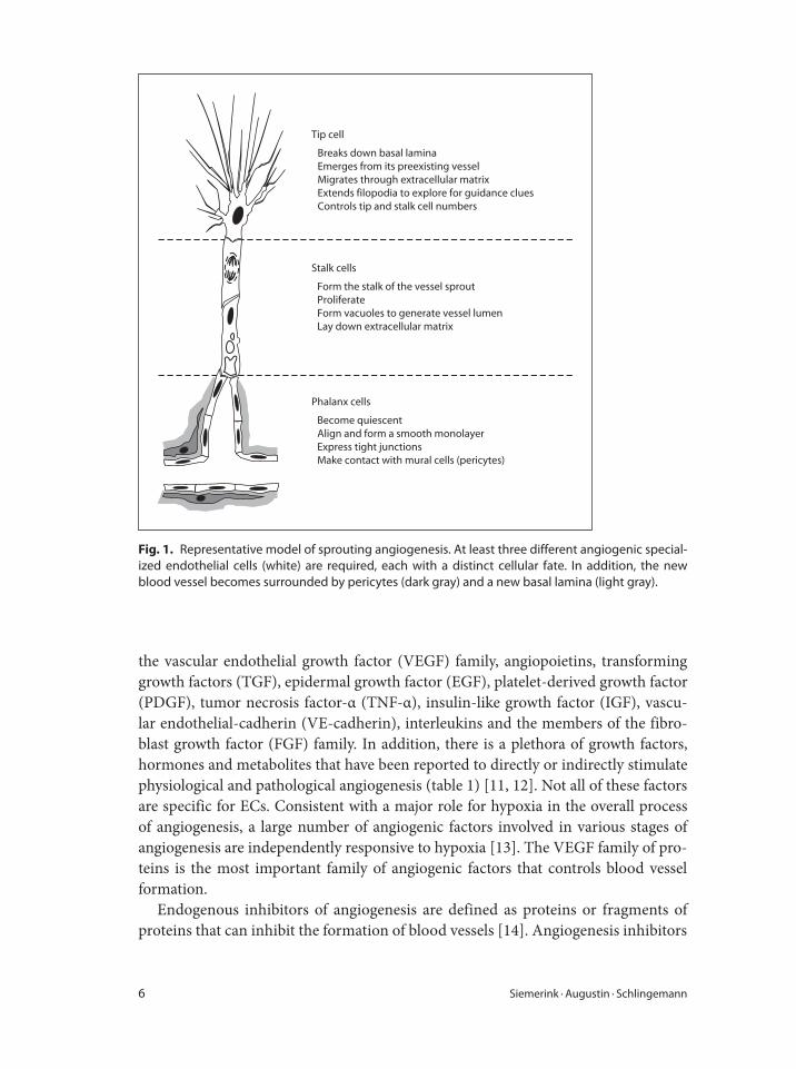
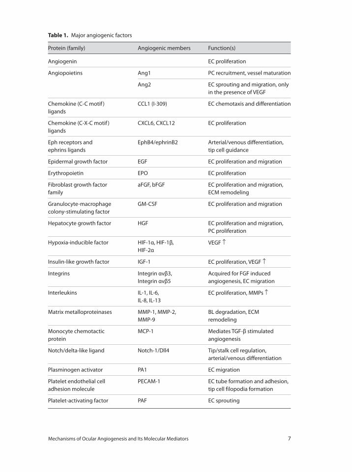
during angiogenesis (fig. 1) [8–10]. First, a single ‘tip cell’ develops. This EC breaks
down the basal lamina, emerges from its parent blood vessel and becomes the lead-
ing cell of the sprouting vessel. The tip cell migrates into the extracellular matrix and
senses microenvironmental attractive and repulsive signals for guidance. Secondly,
following directly behind the migrating tip cell, other ECs differentiate under the
influence of the adjacent tip cell into ‘stalk cells’ that proliferate and bridge the gap
between the tip cell and the parent vasculature. Stalk cells generate the blood vessel
lumen through the formation of intracellular vacuoles, a process called ‘lumenogen-
esis’. Thirdly, ECs behind the stalk cells differentiate into ‘phalanx cells’, and align in a
smooth cobblestone monolayer, becoming the most inner cell layer in the new blood
vessel. Phalanx cells no longer proliferate, express tight junctions and make contact
with mural cells.
Angiogenesis Inducers and Inhibitors
Angiogenesis is tightly controlled by closely interacting angiogenic and angiostatic
factors, and their balance ultimately determines if, where and when the ‘angiogenic
switch’ is turned on with angiogenesis as the result [2, 9]. Over the past decades,
numerous inducers of angiogenesis have been identified, including the members of
6 Siemerink · Augustin · Schlingemann
the vascular endothelial growth factor (VEGF) family, angiopoietins, transforming
growth factors (TGF), epidermal growth factor (EGF), platelet-derived growth factor
(PDGF), tumor necrosis factor-α (TNF-α), insulin-like growth factor (IGF), vascu-
lar endothelial-cadherin (VE-cadherin), interleukins and the members of the fibro-
blast growth factor (FGF) family. In addition, there is a plethora of growth factors,
hormones and metabolites that have been reported to directly or indirectly stimulate
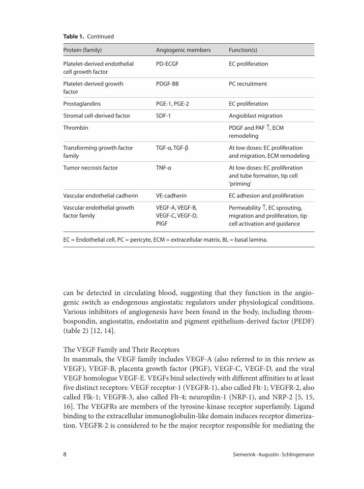
physiological and pathological angiogenesis (table 1) [11, 12]. Not all of these factors
are specific for ECs. Consistent with a major role for hypoxia in the overall process
of angiogenesis, a large number of angiogenic factors involved in various stages of
angiogenesis are independently responsive to hypoxia [13]. The VEGF family of pro-
teins is the most important family of angiogenic factors that controls blood vessel
formation.
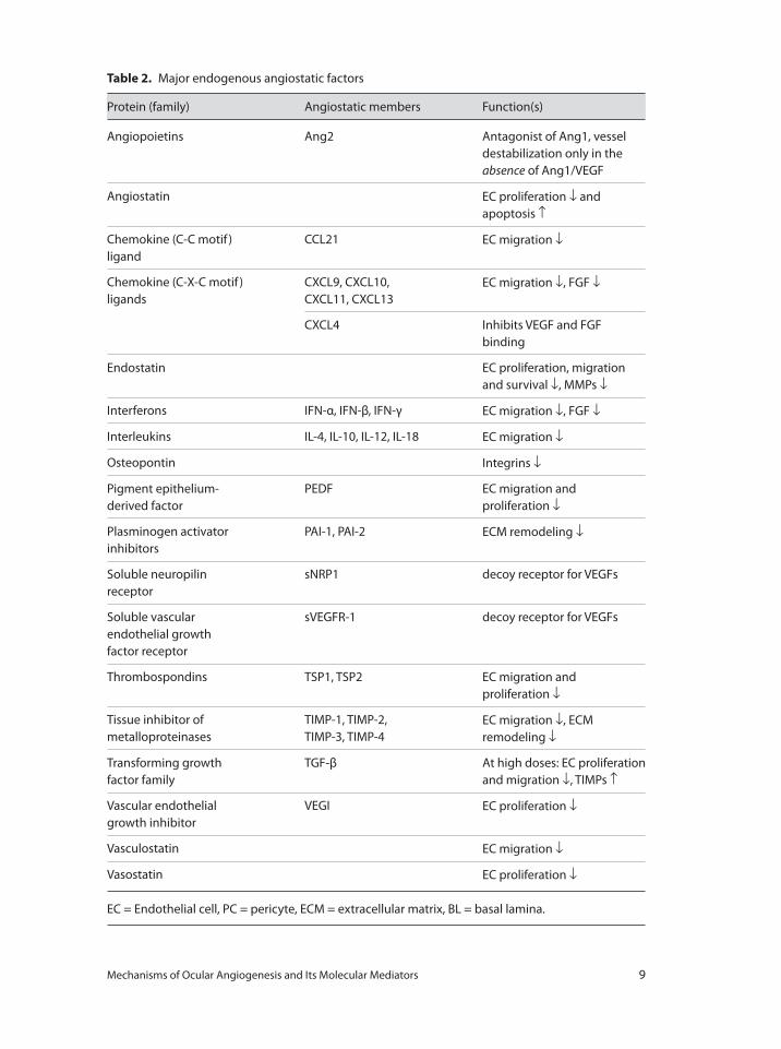
Endogenous inhibitors of angiogenesis are defined as proteins or fragments of
proteins that can inhibit the formation of blood vessels [14]. Angiogenesis inhibitors
Tip cell
Breaks down basal lamina Emerges from its preexisting vesselMigrates through extracellular matrixExtends filopodia to explore for guidance cluesControls tip and stalk cell numbers
Stalk cells
Form the stalk of the vessel sproutProliferateForm vacuoles to generate vessel lumenLay down extracellular matrix
Phalanx cells
Become quiescentAlign and form a smooth monolayerExpress tight junctionsMake contact with mural cells (pericytes)
Fig. 1. Representative model of sprouting angiogenesis. At least three different angiogenic special-
ized endothelial cells (white) are required, each with a distinct cellular fate. In addition, the new
blood vessel becomes surrounded by pericytes (dark gray) and a new basal lamina (light gray).
Mechanisms of Ocular Angiogenesis and Its Molecular Mediators 7
Table 1. Major angiogenic factors
Protein (family) Angiogenic members Function(s)
Angiogenin EC proliferation
Angiopoietins Ang1 PC recruitment, vessel maturation
Ang2 EC sprouting and migration, only
in the presence of VEGF
Chemokine (C-C motif )
ligands
CCL1 (I-309) EC chemotaxis and differentiation
Chemokine (C-X-C motif )
ligands
CXCL6, CXCL12 EC proliferation
Eph receptors and
ephrins ligands
EphB4/ephrinB2 Arterial/venous differentiation,
tip cell guidance
Epidermal growth factor EGF EC proliferation and migration
Erythropoietin EPO EC proliferation
Fibroblast growth factor
family
aFGF, bFGF EC proliferation and migration,
ECM remodeling
Granulocyte-macrophage
colony-stimulating factor
GM-CSF EC proliferation and migration
Hepatocyte growth factor HGF EC proliferation and migration,
PC proliferation
Hypoxia-inducible factor HIF-1α, HIF-1β,
HIF-2α
VEGF ↑
Insulin-like growth factor IGF-1 EC proliferation, VEGF ↑
Integrins Integrin αvβ3,
Integrin αvβ5
Acquired for FGF induced
angiogenesis, EC migration
Interleukins IL-1, IL-6,
IL-8, IL-13
EC proliferation, MMPs ↑
Matrix metalloproteinases MMP-1, MMP-2,
MMP-9
BL degradation, ECM
remodeling
Monocyte chemotactic
protein
MCP-1 Mediates TGF-β stimulated
angiogenesis
Notch/delta-like ligand Notch-1/Dll4 Tip/stalk cell regulation,
arterial/venous differentiation
Plasminogen activator PA1 EC migration
Platelet endothelial cell
adhesion molecule
PECAM-1 EC tube formation and adhesion,
tip cell filopodia formation
Platelet-activating factor PAF EC sprouting
8 Siemerink · Augustin · Schlingemann
can be detected in circulating blood, suggesting that they function in the angio-
genic switch as endogenous angiostatic regulators under physiological conditions.
Various inhibitors of angiogenesis have been found in the body, including throm-
bospondin, angiostatin, endostatin and pigment epithelium-derived factor (PEDF)
(table 2) [12, 14].
The VEGF Family and Their Receptors
In mammals, the VEGF family includes VEGF-A (also referred to in this review as
VEGF), VEGF-B, placenta growth factor (PlGF), VEGF-C, VEGF-D, and the viral
VEGF homologue VEGF-E. VEGFs bind selectively with different affinities to at least
five distinct receptors: VEGF receptor-1 (VEGFR-1), also called Flt-1; VEGFR-2, also
called Flk-1; VEGFR-3, also called Flt-4; neuropilin-1 (NRP-1), and NRP-2 [5, 15,
16]. The VEGFRs are members of the tyrosine-kinase receptor superfamily. Ligand
binding to the extracellular immunoglobulin-like domain induces receptor dimeriza-
tion. VEGFR-2 is considered to be the major receptor responsible for mediating the
Table 1. Continued
Protein (family) Angiogenic members Function(s)
Platelet-derived endothelial
cell growth factor
PD-ECGF EC proliferation
Platelet-derived growth
factor
PDGF-BB PC recruitment
Prostaglandins PGE-1, PGE-2 EC proliferation
Stromal cell-derived factor SDF-1 Angioblast migration
Thrombin PDGF and PAF ↑, ECM
remodeling
Transforming growth factor
family
TGF-α, TGF-β At low doses: EC proliferation
and migration, ECM remodeling
Tumor necrosis factor TNF-α At low doses: EC proliferation
and tube formation, tip cell
‘priming’
Vascular endothelial cadherin VE-cadherin EC adhesion and proliferation
Vascular endothelial growth
factor family
VEGF-A, VEGF-B,
VEGF-C, VEGF-D,
PlGF
Permeability ↑, EC sprouting,
migration and proliferation, tip
cell activation and guidance
EC = Endothelial cell, PC = pericyte, ECM = extracellular matrix, BL = basal lamina.
Mechanisms of Ocular Angiogenesis and Its Molecular Mediators 9
Table 2. Major endogenous angiostatic factors
Protein (family) Angiostatic members Function(s)
Angiopoietins Ang2 Antagonist of Ang1, vessel
destabilization only in the
absence of Ang1/VEGF
Angiostatin EC proliferation ↓ and
apoptosis ↑
Chemokine (C-C motif )
ligand
CCL21 EC migration ↓
Chemokine (C-X-C motif )
ligands
CXCL9, CXCL10,
CXCL11, CXCL13
EC migration ↓, FGF ↓
CXCL4 Inhibits VEGF and FGF
binding
Endostatin EC proliferation, migration
and survival ↓, MMPs ↓
Interferons IFN-α, IFN-β, IFN-γ EC migration ↓, FGF ↓
Interleukins IL-4, IL-10, IL-12, IL-18 EC migration ↓
Osteopontin Integrins ↓
Pigment epithelium-
derived factor
PEDF EC migration and
proliferation ↓
Plasminogen activator
inhibitors
PAI-1, PAI-2 ECM remodeling ↓
Soluble neuropilin
receptor
sNRP1 decoy receptor for VEGFs
Soluble vascular
endothelial growth
factor receptor
sVEGFR-1 decoy receptor for VEGFs
Thrombospondins TSP1, TSP2 EC migration and
proliferation ↓
Tissue inhibitor of
metalloproteinases
TIMP-1, TIMP-2,
TIMP-3, TIMP-4
EC migration ↓, ECM
remodeling ↓
Transforming growth
factor family
TGF-β At high doses: EC proliferation
and migration ↓, TIMPs ↑
Vascular endothelial
growth inhibitor
VEGI EC proliferation ↓
Vasculostatin EC migration ↓
Vasostatin EC proliferation ↓
EC = Endothelial cell, PC = pericyte, ECM = extracellular matrix, BL = basal lamina.
10 Siemerink · Augustin · Schlingemann
angiogenic effects of VEGF-A. The role of VEGFR-1 in angiogenesis remains contro-
versial as its activation has been shown to both stimulate and suppress angiogenesis.
However, soluble VEGFR-1 (sVEGFR-1) inhibits retinal angiogenesis in vivo [17].
VEGFR-3 is highly expressed in angiogenic sprouts in vivo and, like VEGFR-2, its
signaling mediates angiogenesis [18]. NRPs are VEGF-A165-, PlGF-, and VEGF-B-
specific receptors, and form receptor complexes with VEGFRs: NRP-1 partners with
VEGFR-2, whereas NRP-2 can form a complex with VEGFR-2 and VEGFR-3 [16].
VEGF-A, the best characterized and most studied of the VEGF family members,
was originally described as a permeability factor, as it increases permeability of the
endothelium through the formation of intercellular gaps and fenestrations. At least
six human VEGF-A mRNA species, encoding VEGF-A isoforms of 121, 145, 165,
183, 189 and 206 amino acids, are produced by alternative splicing of the VEGF-A
mRNA [15, 16]. In mouse, the VEGF-A isoforms are one amino acid shorter, i.e.
VEGF-A120, etc. It is widely accepted that VEGF-A is crucial for both vasculogenesis
and angiogenesis: loss of only a single allele in mice or zebrafish is lethal, resulting
in severe vascular defects and cardiovascular abnormalities [19]. VEGF-A exerts its
biologic effect through interaction with VEGFR-1 and VEGFR-2, and the neuropilin
receptors NRP-1 and NRP-2 [15].
VEGF-B yields two isoforms, VEGF-B167 and VEGF-B186 by alternative splicing,
which signal through VEGFR-1 and NRP-1 [16]. VEGF-B is widely expressed in vari-
ous tissues, including retina, but it is particularly abundant in the heart and skeletal
muscle [15]. VEGF-B is able to directly stimulate EC growth and migration in vitro
and in vivo [15]. However, the precise role of VEGF-B is not known, and genetic
studies have revealed that VEGF-B-deficient mice are healthy and fertile, and do not
display vascular defects, which indicate that VEGF-B is not involved or redundant in
angiogenesis [15, 16].
PlGF is predominantly expressed in the placenta, heart and lungs, and binds
VEGFR-1 and NRP-1 [16]. The binding of PlGF to VEGFR-1 leads to the forma-
tion of a complex between VEGFR-1 and -2, which enhances VEGF-A signaling and
stimulates angiogenesis [15]. PlGF upregulates the expression of VEGF-A, FGF-2,
PDGF-B, matrix metalloproteinases (MMPs) and other angiogenic factors, suggest-
ing that ECs are able to enhance their own responsiveness to VEGF-A by producing
PlGF. Furthermore, PlGF can promote blood vessel maturation via the recruitment of
mural cells [15].
VEGF-C and VEGF-D both bind VEGFR-2, but with a lower affinity than they
bind to VEGFR-3. Like VEGF-A, both VEGF-C and VEGF-D are able to stimulate
the migration and proliferation of ECs in vitro and in vivo [15]. VEGFR-3 expression
is more abundant on tip cells than on stalk cells [18], whereas VEGFR-3 expression
is absent on phalanx cells. It has been suggested that VEGF-C may cooperate with
VEGF-A to activate ECs for angiogenic sprouting via VEGFR-2/VEGFR-3 receptor
complex.
The viral VEGF homologue VEGF-E is a potent angiogenic factor as well [16].
Mechanisms of Ocular Angiogenesis and Its Molecular Mediators 11
Matrix Degradation
Before ECs can grow out from preexisting vessels, the EC basal lamina must be
degraded and the extracellular matrix needs to be remodeled [8, 9, 12]. This is achieved
by a complex interplay of angiogenic growth factors, mural cells, and ECs. Acidic and
basic FGFs (aFGF and bFGF, respectively) and VEGF stimulate the production of
collagenase and MMPs, and upregulate urokinase-type plasminogen activator in ECs
[20]. Collagenases are enzymes that break the peptide bonds in collagens; urokinase-
type plasminogen activator converts plasminogen into plasmin, leading to fibrinoly-
sis; and MMPs are capable of degrading all kinds of extracellular matrix proteins.
Furthermore, low-dose stimulation by TGF-β upregulates proteases in ECs [21]. At
the same time, FGFs and VEGF downregulate endogenous inhibitors of proteolytic
enzymes such as tissue inhibitors of metalloproteinases (TIMPs) [20].
Tip and Stalk Cell Regulation
The selection of an endothelial tip cell from a population of quiescent ECs has to be
tightly regulated since excessive tip cell formation would result in a poorly patterned,
hyperdense vessel network that may not be functional. Clearly both tip and stalk
cells are stimulated by the same growth factor, VEGF, and both respond through
VEGFR-2 signaling [9, 10, 22]. However, their behavior is very different and in vivo
studies show that tip and stalk cells carry a differential transcriptional signature [9,
10, 22]. In tip cells, VEGFR-2 signaling induces the expression of the Notch ligand
delta-like 4 (DLL4), which is transported to the cell membrane and binds to Notch
receptors on adjacent ECs [22–26]. After ligand binding, Notch is cleaved in these
future stalk cells, generating the Notch intracellular domain that acts as a transcrip-
tional regulator. In these stalk cells, notch activation downregulates the expression
of VEGFR-2, VEGFR-3 and NRP-1, while inducing the transcription of VEGFR-1
and its soluble splice variant sVEGFR-1 [24–26]. Experimental inhibition of Dll4-
Notch1 signaling raised the number of tip cells during early postembryonic angio-
genesis, leading to increased sprout densities and change in vascular patterning [22].
Overactivation of Notch signaling, on the other hand, reduced the migratory behav-
ior of ECs [22]. Other Notch ligands expressed by sprouting vessels are Jagged1 and
Dll1, and loss of each of these also results in vascular defects. However, Dll4 is the
only ligand expressed in tip cells, whereas Jagged1 and Dll1 are present in stalk cells
[27]. These data indicate that the graded distribution of VEGF together with Dll-
Notch signaling regulates angiogenic behavior of ECs by limiting the number of cells
that become tip cell.
Endothelial Proliferation
The stimulatory effects of VEGFs on EC proliferation have been well reported in
vitro and in vivo [5, 16]. Interestingly, during angiogenesis, adjacent ECs exhibit dis-
tinct cellular behavior patterns, even when exposed to a similar degree of VEGF-A,
indicating that several other key molecules are involved in EC differentiation into tip
12 Siemerink · Augustin · Schlingemann
cells, stalk cells or phalanx cells [22]. Co-expression of NRPs with VEGFR-2 is typical
for endothelial tip cells, where it enhances VEGF-A binding to VEGFR-2, VEGFR-2
phosphorylation and VEGF-induced signaling, all of which are required for migra-
tion. In stalk cells, where NRP expression is absent, VEGF-A signaling via VEGFR-2
promotes proliferation but not migration [8, 10, 22].
At low doses, TGF-β contributes to the angiogenic switch by upregulating angio-
genic factors in ECs, but it has inhibitory effects at higher concentrations [21]. TGF-β
family ligands stimulate type II receptors that phosphorylate type I receptors (such
as activin receptor-like kinase (ALK)) and activate the downstream signaling Smads.
Endoglin is a type III receptor, which facilitates ALK1/TGF-β signaling in ECs, and
ALK1/Endoglin/TGF-β signaling also promotes EC proliferation and migration.
Addition of a neutralizing antibody against TGF-β strongly inhibited angiogenesis in
vitro and in vivo [21]. The angiogenic effects of TNF-α are similar to those of TGF-β,
as it promotes EC proliferation and tube formation in lower doses, but inhibits angio-
genesis in higher doses [28].
Angiopoietin-2 (Ang-2) can act as an angiogenic factor depending on the presence
of co-stimulatory molecules. For example, in the presence of VEGF, Ang-2 induces
migration and proliferation of ECs by binding to the Tie2 receptor and thereby block-
ing Tie2 signaling of angiopoietin-1 (Ang-1). In the absence of VEGF, however, Ang-2
causes apoptosis of ECs and regression of blood vessels. Ang-1 has an antagonizing
effect on Tie2 and inhibits EC proliferation. Ang-1 secreted by pericytes binds to Tie2
on ECs, and is important for maintenance of vessel integrity and quiescence.
Several other molecules have been reported to stimulate EC proliferation, includ-
ing FGFs, EGF, CXC chemokines and insulin-like growth factor-1 (IGF-1) [12, 29].
Endothelial Cell-Cell Interaction
EC junctions are composed of a complex network of adhesion proteins that are linked
to the intracellular cytoskeletal network and signaling molecules. VE-cadherin is spe-
cifically localized to the inter-EC junction, and is known to be required for main-
taining a restrictive endothelial barrier. VE-cadherin is critical for proper vascular
development: VE-cadherin-null mice die in early embryonic stages because of vas-
cular defects [30]. The functions of cadherins are modulated by catenins, which bind
with the intracellular tail of the cadherins. After activation of VEGFR-2 by VEGF,
catenins become highly phosphorylated, leading to loss of cell-cell junctions, allowing
EC to differentiate and move from their current position. Later on during angiogen-
esis, the phosphorylation of catenins decreases, allowing restabilization of EC cell-cell
junctions and the differentiation into quiescent phalanx cells.
Platelet EC adhesion molecule-1 (PECAM-1) is expressed on ECs, and like
VE-cadherin, it is enriched in intercellular junctions. PECAM-1 mediated cell-cell
junctions are necessary for the organization of ECs in to tubular networks in vitro,
and PECAM-1 has been shown to stimulate the formation of tip cell filopodia in
vivo [31].
Mechanisms of Ocular Angiogenesis and Its Molecular Mediators 13
Blood Vessel Guidance
Endothelial tip cells pick up attractive or repulsive signals from the tissue environ-
ment and translate them into a dynamic process of adhesion and de-adhesion, leading
to migration. In this process the tip cell forms lamellipodia (short cytoskeletal projec-
tion) and filopodia (long finger-like plasma membrane extensions) [8]. Lamellipodia
are located on the mobile edge of the cell. They adhere and connect the intracel-
lular cytoskeleton to the extracellular matrix, allowing stress fibers of actin/myosin
filaments to pull the cell forward. Filopodia protrude from the lamellipodial actin
network and function as antennae with which tip cells probe their environment. The
main regulators of filopodia and lamellipodia formation are members of the Rho
small GTPases, which are induced by VEGF [32].
An extracellular VEGF-A gradient appears to be a strong attractant for migrating
ECs via binding to VEGFR-2 and NRPs, which are prominent on tip cell filopodia.
An important biological property of the different VEGF-A isoforms is their heparin
and heparan-sulfate-binding ability. The larger VEGF-A isoforms bind very tightly
to heparin and remain sequestered in the extracellular matrix, whereas the shorter
VEGF-A isoforms are freely diffusible. It is well established that VEGF-A189 and
VEGF-A165 function as a chemoattractive signal that promote the polarized extension
of tip cell filopodia, whereas VEGF-A121 can support EC proliferation but not tip cell
guidance [22, 33].
Furthermore, the function of endothelial tip cells bears remarkable similarity to
that of axonal growth cones. Blood vessels and nerve fibers course throughout the
body alongside one another and it has been reported that during embryogenesis,
their patterning is guided in large part by similar attractive and repulsive guidance
cues. Thus far, four major families of receptors have been shown to regulate guid-
ance events during axonal and vascular morphogenesis: Plexin/NRP complexes with
their ligands class 3 semaphorins; ‘uncoordinated-5’ (UNC5) family and ‘deleted in
colorectal cancer’ (DCC) with their ligands netrins; ‘Roundabout’ (Robo) with their
ligands Slits, and Eph and their ligands ephrins [9, 33, 34].
Lumen Formation
While migrating, the leading tip cell creates a tunnel throughout the extracellu-
lar matrix space. Behind the tip cell, stalk cells flatten onto the wall of this tube-
like space in the extracellular matrix, resulting in an apical and basal face of the
endothelium. Stalk cells form large intracellular vacuoles by fusion of intracellu-
lar vesicles, mediated by integrins, which fuse together to form a lumen [9, 35].
Multiple integrins as well as the transcription factor myocyte enhancer binding
factor 2C (MEF2C) are able to participate in vesicle formation and fusion in vitro
[35]. EC interactions with the extracellular matrix establish signaling cascades
downstream of integrin ligation leading to activation of the Rho family of GTPases.
Inhibition of Rho GTPases results in complete blockade of EC vacuole and lumen
formation in vitro [35].
14 Siemerink · Augustin · Schlingemann
Recruitment of Mural Cells and Maturation
After the initial vessel formation through angiogenesis, determination of artery or
vein identity is regulated by a variety of molecular factors which specify EC fate.
Distinct arterial and venous molecular markers are evident even before the initia-
tion of circulatory flow, suggesting that molecular determinants play a critical role
in arterial/venous differentiation. Several relevant genes have now been identified in
vivo, including the Hedgehog family of secreted morphogens, notch signaling, NRPs,
EphB4, ephrinB2, and VEGF [36].
The two major classes of mural cells are the vascular smooth muscle cells, which
coat veins and arteries, and the pericytes, which are present in variable amounts
around capillaries. The mural cells are indispensable to provide survival and antipro-
liferative factors that stabilize the newly-formed vessel. However, the hypothesis that
pericyte loss initializes the first steps of angiogenesis whereas pericyte recruitment
only occurs at the completion of angiogenesis is controversial, since many pericytes
are found to be present in endothelial sprouts in vivo [37].
The development and the recruitment of vascular mural cells require the function
of PDGF signaling, Ang-1 and its receptor Tie-2, and Ephrin-Eph interaction. PDGFs
exist as heterodimers (PDGF-AB) or homodimers composed of chains A and B.
Endothelial tip cells from growing vascular sprouts generate a PDGF-B concentration
gradient that promotes the recruitment of pericytes expressing the PDGF-B receptor
[37, 38]. This in turn activates TGF-β in pericytes, which introduces the production
of basal lamina components that are required for final blood vessel maturation and
stabilization [21].
Ang-1, expressed by perivascular cells, binds to and activates the Tie2 receptor,
thereby stimulating mural cell attachment. In agreement, a poor association between
ECs and surrounding mural cells was seen in Ang-1 and Tie2 knockouts. Ang-2 was
shown to have an antagonizing effect on Tie2 inducing pericyte loss and capillary
degeneration in the retina. However, endothelial expression of Tie2 has been observed
on newly formed vessels that are still immature [37].
Retinal Circulation
Retinal Vascular System
The retina has a dual vascular supply: the outer one-third of the retina is supplied
from the choroidal circulation and the inner two-thirds by the central retinal artery.
The choroidal arteries pierce the sclera around the optic nerve and fan out to form
the choriocapillaris, providing nutrients and oxygen to the retinal pigment epithe-
lium and the photoreceptors in the outer part of retina. The corresponding venous
lobules drain into the venules and veins that run anterior towards the equator of the
eyeball to enter the vortex veins. The vortex veins penetrate the sclera and ultimately
merge into the ophthalmic vein. The central retinal artery pierces the optic nerve
Mechanisms of Ocular Angiogenesis and Its Molecular Mediators 15
close to the eyeball, emerges at the optic disk, and sends 4 main branches over the
human retina, lying close to the inner limiting membrane. Each of the 4 branches
of the central retinal artery supplies one quadrant of the inner retina. The venous
equivalent of the central retinal artery is the retinal vein. The anatomy of the veins of
the orbit of the eye varies between individuals. In some the central retinal vein drains
into the superior ophthalmic vein whereas in others it drains directly into the cavern-
ous sinus [39,40].
Unique Characteristics of Retinal Circulation
Regulation of the microenvironment of the retina, e.g. the controlled fluid and molec-
ular movement between the ocular vascular bed and the retinal tissues, is fundamental
for appropriate retinal function and vision. Therefore, the retina has a unique blood-
retinal barrier that separates the retina from the circulating blood [41]. The blood-
retinal barrier is formed by complex tight junctions of retinal capillary ECs (the inner
barrier) and retinal pigment epithelial cells (the outer barrier), corresponding to the
two main circulations. The choroidal capillaries themselves are fenestrated, like most
of the highly permeable capillaries throughout the human body. Retinal capillary ECs
express a variety of unique transporters which play a key role in the influx trans-
port of essential molecules and the efflux transport of neurotransmitter metabolites,
toxins, and drugs [41]. Therefore, systemic drug administration is not suitable for
the treatment of retinal diseases because of poor drug permeability across the blood-
retinal barrier.
Retinal circulation is characterized by a low blood flow and a high level of oxy-
gen extraction; arteriovenous difference in pO2 is about 40% [42]. Autonomic nerve
endings extend to the uvea and the extraocular part of the retinal blood vessels, but
not to the intraocular segments of the retinal blood vessels. Therefore, retinal arterial
tone is largely regulated by local factors such as local variations in perfusion pressure
and in pO2, pCO2 and pH [42]. The presence of mechanisms that autoregulate retinal
circulation may well reflect important survival strategies for the retina which are not
yet fully understood.
Development of the Retinal Vasculature
During embryogenesis, the vascular network that supplies the retina undergoes dra-
matic changes and reorganization [39]. The choroidal vasculature develops in an
early stage and is preceded by a peak of VEGF-A production by the retinal pigment
epithelium, suggesting that VEGF-A is involved in the development of the choroidal
vasculature [43]. Initially, the inner part of the eye is metabolically supported by the
hyaloidal vasculature, an arterial network in the vitreous. Blood enters through the
central hyaloid artery in the optic nerve, runs through hyaloid vessels in the vitre-
ous and then exits through an annular collection vessel at the front of the eye. The
hyaloid vessel system is a dense, but transient intraocular circulatory system that
undergoes progressive and nearly complete regression during the latest stage of
16 Siemerink · Augustin · Schlingemann
ocular development as the lens, the vitreous and the retina mature. Due to the natural
regression of the hyaloidal vasculature, as well as increasing metabolic demands of
maturing neurons, the retina becomes hypoxic, and therefore the formation of the
retinal vasculature is induced.
Retinal Angioblasts
Recent observations have suggested that the initial human retinal vasculature develops
by differentiation, and organization of vascular precursor cells that are CD39+ (angio-
blast marker) and CD34–/CD31– (EC markers) at around 14 weeks of gestation [44].
These cells seem to emerge from a pool of precursor cells that are CXCR4+/c-Kit+
(angioblast receptors), and were found in the neuroblastic layer of human embryonic
retina at 7 weeks of gestation. CD39+/CXCR4+/c-Kit+ cells start to migrate ante-
riorly into the retinal nerve fiber layer where stroma-derived factor-1 (SDF-1, the
ligand for CXCR4) and stem cell factor (SCF, the ligand for c-Kit) levels are at their
highest. With apparent migration of these vascular precursors, the expression of c-Kit
declines and differentiation into angioblasts and alignment with nerve fibers occurs.
A gradient of SDF-1 towards the ora serrata suggests that the angioblasts migrate
towards the higher concentration. CXCR4 expression is regulated by SCF, FGF-2 and
VEGF, and angioblasts continue to express CXCR4 until they become ECs that are
CD34+/CD31+. These results suggest, at least in part, that vasculogenesis might con-
tribute to growth of the primordial vessels in the central retina [44, 45]. However, the
identification and lineage of angioblasts within the developing retina is still contro-
versial [46].
Retinal Angiogenesis
After formation of primordial vessels, new blood vessels sprout into the retina by
means of angiogenesis, forming the vasculature of the inner retina. Retinal angiogen-
esis begins in the most superficial retinal layer at the optic nerve head, and radiates
outwards from this central point [3, 39, 45]. The superficial plexus develops in a cen-
trifugal fashion across the inner surface of the retina, with the exception of the primate
fovea from which blood vessels are excluded. Retinal angiogenesis is closely regulated
by supply and demand of oxygen. High oxygen tension suppresses hypoxia-induced
VEGF production, and less VEGF results in less blood vessel growth [3, 39]. Additional
capillary networks in deeper retinal layers then arise by sprouting from the superficial
arteries to form the deeper vascular plexus. Vascular pruning in the developing retina
results from EC migration from retracting vessels into the surrounding newly develop-
ing vessels. The process of natural pruning can be accelerated by experimental expo-
sure to hyperoxia [3]. The process of retinal vascular development is completed shortly
before birth in humans, and a few weeks after birth in several other mammalian spe-
cies including rodents. With development of the capillary plexuses and the resulting
increase in oxygen tension, a capillary-free zone develops around the major blood ves-
sels, followed by vessel retraction in the superficial plexus [40].
Mechanisms of Ocular Angiogenesis and Its Molecular Mediators 17
Vascular Patterning
The process of sprouting angiogenesis during development of the retinal vasculature
is preceded by an invasion of migrating astrocytes in a centrifugal fashion across the
inner surface of the retina [3, 22, 40, 46, 47]. Ganglion cells secrete PDGF-A to stim-
ulate proliferation of astrocytes [48, 49]. The retinal vascular plexus initially forms
superimposed on the astrocyte network. Astrocytes at the leading edge and immedi-
ately ahead of the vascular plexus secrete high levels of VEGF-A compared to more
distally located astrocytes that already have established contact with ECs. During this
burst of angiogenesis, all endothelial tip cells are closely attached to astrocytes and
their filopodia orientate along the astrocyte cell bodies and processes. Experimental
overexpression of PDGF-A in ganglion cells resulted in a large increase in the num-
ber of retinal astrocytes and subsequent overgrowth of the retinal vasculature in vivo
[49]. However, blocking PDGF-A receptor reduced astrocyte network formation but
showed only small changes in blood vessel formation [49].
Pathological Ocular Angiogenesis
What Is Unique in Ocular Angiogenesis?
Several ocular diseases are hallmarked by angiogenesis, including diabetic retinopa-
thy, age-related macular degeneration, and retinopathy of prematurity [3, 5, 46, 50].
In all these conditions, angiogenesis is probably stimulated by local tissue hypoxia
resulting from neuronal metabolism, with varying contributions from inflamma-
tory signals and oxidative stress. In retinal neovascularization, VEGF plays a central
role [3, 5, 51]. At least five retinal cell types have the capacity to produce and secrete
VEGF. These include the retinal pigmented epithelium, astrocytes, Müller cells, ECs
and ganglion cells. However, they differ widely in their responses to hypoxia; in vitro
studies show that Müller cells and astrocytes generally produce the greatest amounts
of VEGF under hypoxic conditions [1, 22, 43]. The two most important forms of ocu-
lar angiogenesis are preretinal angiogenesis, originating from the retinal vasculature,
and subretinal (or choroidal) neovascularization.
Preretinal Angiogenesis
Preretinal angiogenesis occurs as a final common pathway in several diseases associ-
ated with capillary non-perfusion and local retinal ischemia, including diabetic retin-
opathy. Angiogenesis is induced by the ischemic retinal areas, and ultimately results in
the formation of large contractile fibrovascular membranes within the vitreous cavity.
These membranes and the associated hemorrhages cause blindness by obscuration
of the visual axis and retinal detachment. When the retinal ischemia is widespread,
angiogenesis and scarring can also occur on the iris and cause an untreatable form
of glaucoma. Destruction of the ischemic retinal areas with laser can be effective in
inducing regression and fibrosis of the newly formed vessels.
18 Siemerink · Augustin · Schlingemann
1 Patan S: Vasculogenesis and angiogenesis as mecha-
nisms of vascular network formation, growth and
remodeling. J Neurooncol 2000;50:1–15.
2 Risau W: Mechanisms of angiogenesis. Nature 1997;
386:671–674.
3 Gariano RF, Gardner TW: Retinal angiogenesis in
development and disease. Nature 2005;438:960–
966.
4 Schlingemann RO, Witmer AN: Treatment of reti-
nal diseases with VEGF antagonists. Prog Brain Res
2009;175:253–267.
5 Witmer AN, Vrensen GFJM, van Noorden CJF, Sch-
lingemann RO: Vascular endothelial growth factors
and angiogenesis in eye disease. Prog Retin Eye Res
2003;22:1–29.
6 Andreoli CM, Miller JW: Antivascular endothelial
growth factor therapy for ocular neovascular dis-
ease. Curr Opin Ophthalmol 2007;18:502–508.
7 Cines DB, Pollak ES, Buck CA, Loscalzo J, Zim-
merman GA, McEver RP, et al: Endothelial cells in
physiology and in the pathophysiology of vascular
disorders. Blood 1998;91:3527–3561.
Subretinal Angiogenesis
Subretinal, or choroidal neovascularization, results from a series of pathological
events affecting the retinal pigment epithelium, Bruch’s membrane, and the choroid.
Typically, subretinal angiogenesis is a wound healing response that occurs only when
an anatomical discontinuation of Bruch’s membrane is present, in combination with
a driving force such as inflammation, hypoxia, and oxidative stress. For most condi-
tions it is unknown to what extent these three mechanisms contribute to the initia-
tion of subretinal angiogenesis. Subretinal angiogenesis is a hallmark of age-related
macular degeneration, occurring either between the retinal pigment epithelium and
Bruch’s membrane (occult choroidal neovascularization), or between the retinal pig-
ment epithelium and the neuroretina (classic choroidal neovascularization).
New vessels formed by subretinal angiogenesis can later regress, leaving an atro-
phic retinal area, or the wound healing can progress with formation of a fibrotic scar.
In both cases, the overlying neuroretina will slowly degenerate, leading to loss of
sharp sight, contrast sensitivity, and color vision.
Ocular Angiogenesis and Wound Healing Responses
In most instances, pathological ocular angiogenesis is a wound healing-like response
in which the formation of blood vessels is accompanied by influx of inflammatory
cells, followed by myofibroblast formation [52]. Therefore, during disease progres-
sion, the angiogenic phase can be followed by a fibrotic phase. It has been shown that
VEGF-driven angiogenesis upregulates profibrotic factors such as TGF-β1 and con-
nective tissue growth factor (CTGF) [53]. CTGF levels strongly correlate with degree
of fibrosis in vitreoretinal conditions [52]. When the balance between the angio-
genic (VEGF) and fibrotic (CTGF) factors shifts to a certain threshold ratio in favor
of fibrosis, the ‘angiofibrotic switch’ occurs and fibrosis and scarring develop [52].
Administration of anti-VEGF drugs to patients as a therapy to regress neovascular-
ization could therefore lead to a temporary increase in fibrosis, a phenomenon that is
indeed observed in the clinic.
References
Mechanisms of Ocular Angiogenesis and Its Molecular Mediators 19
8 De Smet F, Segura I, De Bock K, Hohensinner PJ,
Carmeliet P: Mechanisms of vessel branching filo-
podia on endothelial tip cells lead the way. Arterio-
scler Thromb Vasc Biol 2009;29:639–649.
9 Adams RH, Alitalo K: Molecular regulation of
angiogenesis and lymphangiogenesis. Nat Rev Mol
Cell Biol 2007;8:464–478.
10 Le Noble F, Klein C, Tintu A, Pries A, Buschmann I:
Neural guidance molecules, tip cells, and mechani-
cal factors in vascular development. Cardiovasc Res
2008;78:232–241.
11 Otrock ZK, Mahfouz RAR, Makarm JA, Shamsed-
dine AI: Understanding the biology of angiogenesis:
review of the most important molecular mecha-
nisms. Blood Cells Mol Dis 2007;39:212–220.
12 Distler JHW, Hirth A, Kurowska-Stolarska M, Gay
RE, Gay S, Distler O: Angiogenic and angiostatic
factors in the molecular control of angiogenesis. Q J
Nucl Med 2003;47:149–161.
13 Fong GH: Regulation of angiogenesis by oxygen
sensing mechanisms. J Mol Med 2009;87:549–560.
14 Tabruyn SP, Griffioen AW: Molecular pathways of
angiogenesis inhibition. Biochem Biophys Res
Commun 2007;355:1–5.
15 Tammela T, Enholm B, Alitalo K, Paavonen K: The
biology of vascular endothelial growth factors.
Cardiovasc Res 2005;65:550–563.
16 Holmes DIR, Zachary I: The vascular endothelial
growth factor family: angiogenic factors in health
and disease. Genome Biol 2005;6.
17 Aiello LP, Pierce EA, Foley ED, Takagi H, Chen H,
Riddle L, et al: Suppression of retinal neovascular-
ization in vivo by inhibition of vascular endothelial
growth-factor (VEGF) using soluble VEGF-receptor
chimeric proteins. Proc Natl Acad Sci USA 1995;92:
10457–10461.
18 Witmer AN, van Blijswijk BC, Dai J, Hofman P,
Partanen TA, Vrensen GFJM, et al: VEGFR-3 in
adult angiogenesis. J Pathol 2001;195:490–497.
19 Carmeliet P, Ferreira V, Breier G, Pollefeyt S,
Kieckens L, Gertsenstein M, et al: Abnormal blood
vessel development and lethality in embryos lacking
a single VEGF allele. Nature 1996;380:435–439.
20 Presta M, Dell’Era P, Mitola S, Moroni E, Ronca R,
Rusnati M: Fibroblast growth factor/fibroblast
growth factor receptor system in angiogenesis.
Cytokine Growth Factor Rev 2005;16:159–178.
21 Goumans MJ, Liu Z, ten Dijke P: TGF-β signaling in
vascular biology and dysfunction. Cell Res 2009;19:
116–127.
22 Gerhardt H, Golding M, Fruttiger M, Ruhrberg C,
Lundkvist A, Abramsson A, et al: VEGF guides
angiogenic sprouting utilizing endothelial tip cell
filopodia. J Cell Biol 2003;161:1163–1177.
23 Hellstrom M, Phng LK, Hofmann JJ, Wallgard E,
Coultas L, Lindblom P, et al: Dll4 signalling through
Notch1 regulates formation of tip cells during
angiogenesis. Nature 2007;445:776–780.
24 Leslie JD, Ariza-McNaughton L, Bermange AL,
McAdow R, Johnson SL, Lewis J: Endothelial sig-
nalling by the Notch ligand delta-like 4 restricts
angiogenesis. Development 2007;134:839–844.
25 Harrington LS, Sainson RCA, Williams CK, Taylor
JM, Shi W, Li JL, et al: Regulation of multiple angio-
genic pathways by D114 and Notch in human
umbilical vein endothelial cells. Microvasc Res
2008;75:144–154.
26 Hainaud P, Contreres JO, Villemain A, Liu LX,
Plouet J, Tobelem G, et al: The role of the vascular
endothelial growth factor-delta-like 4 ligand/
Notch4-ephrin B2 cascade in tumor vessel remodel-
ing and endothelial cell functions. Cancer Res 2006;
66:8501–8510.
27 Roca C, Adams RH: Regulation of vascular mor-
phogenesis by Notch signaling. Genes Dev 2007;21:
2511–2524.
28 Sainson RCA, Johnston DA, Chu HC, Holderfield
MT, Nakatsu MN, Crampton SP, et al: TNF primes
endothelial cells for angiogenic sprouting by induc-
ing a tip cell phenotype. Blood 2008;111:4997–
5007.
29 Otrock ZK, Mahfouz RAR, Makarm JA, Shamsed-
dine AI: Understanding the biology of angiogenesis:
review of the most important molecular mecha-
nisms. Blood Cells Mol Dis 2007;39:212–220.
30 Wallez Y, Vilgrain I, Huber P: Angiogenesis: the
VE-cadherin switch. Trends Cardiovasc Med 2006;
16:55–59.
31 Cao GY, Fehrenbach ML, Williams JT, Finklestein
JM, Zhu JX, DeLisser HM: Angiogenesis in platelet
endothelial cell adhesion molecule-1-null mice. Am
J Pathol 2009;175:903–915.
32 Mattila PK, Lappalainen P: Filopodia: molecular
architecture and cellular functions. Nat Rev Mol
Cell Biol 2008;9:446–454.
33 Carmeliet P: Blood vessels and nerves: common sig-
nals, pathways and diseases. Nat Rev Genet 2003;4:
710–720.
34 Larrivee B, Freitas C, Suchting S, Brunet I, Eichmann
A: Guidance of vascular development lessons from
the nervous system. Circ Res 2009;104:428–441.
35 Iruela-Arispe ML, Davis GE: Cellular and molecu-
lar mechanisms of vascular lumen formation. Dev
Cell 2009;16:222–231.
36 Swift MR, Weinstein BM: Arterial-venous specifica-
tion during development. Circ Res 2009;104:576–
588.
20 Siemerink · Augustin · Schlingemann
37 Witmer AN, van Blijswijk BC, van Noorden CJF,
Vrensen GFJM, Schlingemann RO: In vivo angio-
genic phenotype of endothelial cells and pericytes
induced by vascular endothelial growth factor-A. J
Histochem Cytochem 2004;52:39–52.
38 Penfold PL, Provis JM, Madigan MC, Vandriel D,
Billson FA: Angiogenesis in normal human retinal
development – the involvement of astrocytes and
macrophages. Graefes Arch Clin Exp Ophthalmol
1990;228:255–263.
39 Saint-Geniez M, D’Amore PA: Development and
pathology of the hyaloid, choroidal and retinal vas-
culature. Int J Dev Biol 2004;48:1045–1058.
40 Provis JM: Development of the primate retinal vas-
culature. Prog Retin Eye Res 2001;20:799–821.
41 Kaur C, Foulds WS, Ling EA: Blood-retinal barrier
in hypoxic ischaemic conditions: basic concepts,
clinical features and management. Prog Retin Eye
Res 2008;27:622–647.
42 Hardy P, Beauchamp M, Sennlaub F, Gobeil F,
Tremblay L, Mwaikambo B, et al: New insights into
the retinal circulation: inflammatory lipid media-
tors in ischemic retinopathy. Prostaglandins Leukot
Essent Fatty Acids 2005;72:301–325.
43 Gogat K, Le Gat L, Van den Berghe L, Marchant D,
Kobetz A, Gadin S, et al: VEGF and KDR gene
expression during human embryonic and fetal eye
development. Invest Ophthalmol Vis Sci 2004;45:7–
14.
44 Hasegawa T, McLeod DS, Prow T, Merges C, Grebe
R, Lutty GA: Vascular precursors in developing
human retina. Invest Ophthalmol Vis Sci 2008;49:
2178–2192.
45 Hughes S, Yang HJ, Chan-Ling T: Vascularization of
the human fetal retina: roles of vasculogenesis and
angiogenesis. Invest Ophthalmol Vis Sci 2000;41:
1217–1228.
46 Gariano RF: Cellular mechanisms in retinal vascu-
lar development. Prog Retin Eye Res 2003;22:295–
306.
47 Chan-Ling T, McLeod DS, Hughes S, Baxter L, Chu
Y, Hasegawa T, et al: Astrocyte-endothelial cell
relationships during human retinal vascular devel-
opment. Invest Ophthalmol Vis Sci 2004;45:2020–
2032.
48 Sandercoe TM, Madigan MC, Billson FA, Penfold
PL, Provis JM: Astrocyte proliferation during devel-
opment of the human retinal vasculature. Exp Eye
Res 1999;69:511–523.
49 Fruttiger M, Calver AR, Kruger WH, Mudhar HS,
Michalovich D, Takakura N, et al: PDGF mediates a
neuron-astrocyte interaction in the developing ret-
ina. Neuron 1996;17:1117–1131.
50 Campochiaro PA, Hackett SF: Ocular neovascular-
ization: a valuable model system. Oncogene 2003;22:
6537–6548.
51 Blaauwgeers HGT, Hotkamp BW, Rutten H, Witmer
AN, Koolwijk P, Partanen TA, et al: Polarized vascu-
lar endothelial growth factor secretion by human
retinal pigment epithelium and localization of vas-
cular endothelial growth factor receptors on the
inner choriocapillaris – evidence for a trophic para-
crine relation. Am J Pathol 1999;155:421–428.
52 Kuiper EJ, Van Nieuwenhoven FA, de Smet MD, van
Meurs JC, Tanck MW, Oliver N, et al: The angiofi-
brotic switch of VEGF and CTGF in proliferative
diabetic retinopathy. PLoS One 2008;3:e2675.
53 Kuiper EJ, Hughes JM, Van Geest RJ, Vogels IMC,
Goldschmeding R, van Noorden CJF, et al: Effect of
VEGF-A on expression of profibrotic growth factor
and extracellular matrix genes in the retina. Invest
Ophthalmol Vis Sci 2007;48:4267–4276.
Prof. Dr. R.O. Schlingemann
Medical Retina Unit and Ocular Angiogenesis Group, Department of Ophthalmology
Academic Medical Center, University of Amsterdam, A2-122
PO Box 22660, NL–1100 DD Amsterdam (The Netherlands)
Tel. +31 205 663 682, Fax +31 205 669 048, E-Mail [email protected]
Bandello F, Battaglia Parodi M (eds): Anti-VEGF.
Dev Ophthalmol. Basel, Karger, 2010, vol 46, pp 21–38
Antivascular Endothelial Growth Factors in Age-Related Macular DegenerationUrsula Schmidt-Erfurth � Andreas Pollreisz � Christoph Mitsch �
Matthias Bolz
Department of Ophthalmology and Optometry, Medical University Vienna,
Vienna, Austria
AbstractAge-related macular degeneration (AMD) is the leading cause of irreversible vision loss in adults
aged over 50 years in developed countries. Until recently, argon laser photocoagulation and photo-
dynamic therapy (PDT) were the only treatments available for the neovascular form of AMD. The
introduction of new intravitreally injectable inhibitors of vascular endothelial growth factor (VEGF)
revolutionized the management of the wet form. Pegaptanib was the first anti-VEGF agent to be
approved by the US Food and Drug Administration (FDA) for use in neovascular AMD. The VISION
study showed that patients receiving pegaptanib lost vision in a smaller rate than those treated with
conventional care. Bevacizumab is a full-length recombinant humanized monoclonal antibody
which binds to all isoforms of VEGF-A. It is licensed for the intravenous administration for the treat-
ment of malignant solid tumors and is available for off-label use in the treatment of AMD-related
CNV. Numerous retrospective studies have shown beneficial effects of bevacizumab in patients with
neovascular AMD. Ranibizumab is a recombinant, humanized antibody antigen-binding fragment
(Fab) that binds to all known active forms of VEGF-A. The US FDA approved ranibizumab for treat-
ment of all subtypes of choroidal neovascularization secondary to AMD. VEGF trap is a pharmaco-
logically engineered protein that binds VEGF with higher affinity than pegaptanib or ranibizumab
and thus prevents VEGF binding to its cellular receptor offering a theoretically longer interval
between necessary treatments. A number of studies have shown that OCT imaging allows identifica-
tion of functionally relevant factors like subretinal fluid or retinal thickness, which are important for
the establishment of optimized individual dosing regimen during anti-angiogenesis therapies.
Copyright © 2010 S. Karger AG, Basel
Age-related macular degeneration (AMD) is the leading cause of irreversible vision
loss in adults aged over 50 years in developed countries. Large population studies
report prevalence rates of early AMD between 5.8 and 15.1% [1, 2]. Giving demo-
graphic development in the aging of the population, its prevalence will increase
22 Schmidt-Erfurth · Pollreisz · Mitsch · Bolz
dramatically in the following years [3, 4]. AMD should therefore not only be regarded
as a medical problem but also as a major socioeconomic burden.
AMD is a term used to summarize different pathological age-related changes of
the macula, namely drusen maculopathy, geographic atrophy (dry form) and chor-
oidal neovascularization (CNV) (wet or neovascular form). AMD is characterized
by a progressive loss of central vision resulting from degenerative and neovascular
changes in the macula [5].
Although during the last decade numerous studies were addressed to understand
the pathophysiological mechanisms, the underlying processes and modulating factors
still need to be elucidated. A growing body of evidence indicates that AMD patho-
genesis involves chronic inflammation, complement activation and autoimmunity
[6–8].
All subtypes of AMD are complex, multifactorial diseases with only limited
means for prevention or cure. Until recently, argon laser photocoagulation and pho-
todynamic therapy (PDT) were the only treatments available for the neovascular
form of AMD. The introduction of new intravitreally injectable inhibitors of vas-
cular endothelial growth factor (VEGF) revolutionized the management of the wet
form [9, 10].
VEGF is a homodimeric glycoprotein with a molecular weight of 45 kDa which
exists in four major isoforms (VEGF-121, -165, -189, -206) [11]. The different iso-
forms are characterized by their molecular weight, ability to bind to heparin and
acidity. VEGF is a key regulator of physiological angiogenesis during embryogene-
sis but has also been implicated in pathological angiogenesis such as tumor growth
or intraocular neovascular diseases [12]. In vivo mouse models demonstrated the
impact of VEGF on ocular neovascularization and identified it as a central molecu-
lar mediator of CNV, which is the major cause of visual loss in patients with exuda-
tive AMD [13]. The binding of VEGF to its receptors on retinal vascular endothelial
cells initiates several intracellular signaling pathways resulting in proliferation, dif-
ferentiation and migration of endothelial cells [14, 15]. In addition, VEGF acts as
a potent vascular permeability factor resulting in increased fluid leakage across the
blood vessel walls. CNV membranes obtained from patients with AMD contain
VEGF as shown in immunohistochemical studies [16, 17]. Analysis of different
retinal cell types in post-mortem eyes with AMD revealed higher VEGF levels in
the retinal pigment epithelial (RPE) cell layer and the outer nuclear layer than in
healthy control eyes [18]. The ability of RPE cells to secrete VEGF has also been
shown in in vitro experiments when cells are cultured under hypoxic conditions
[19]. Furthermore, significantly increased levels of VEGF have been found in the
aqueous humor of human eyes with neovascular AMD as compared to healthy
controls [20].
Finally, evidence from clinical trials with anti-VEGF agents in humans, as described
below, highlight the pathognomonic role of VEGF in the pathogenesis of neovascular
AMD.
Anti-VEGF in Age-Related Macular Degeneration 23
Pegaptanib
Pegaptanib, an oligonucleotide aptamer, was the first anti-VEGF agent to be
approved by the US Food and Drug Administration (FDA) for use in neovascular
AMD. Aptamers are nucleic acid ligands, which bind different targets such as pro-
teins with high specificity and affinity. They are complex three-dimensional struc-
tures isolated from oligonucleotides by a selection process in vitro. By binding to
molecules, aptamers may change the shape of the molecule or inhibit the molecule’s
biological function [21–23]. When the anti-VEGF aptamer binds to VEGF it blocks
the binding of VEGF to its receptor, preventing the initiation of the intracellular cas-
cade [24]. Pegaptanib consists of a 28-base RNA oligonucleotide with two branched
20-kDa polyethylene glycol moieties, which binds selectively to the VEGF-165 iso-
form [25].
In August 1998, a phase 1A trial was initiated as a multicenter, open-label, dose-
escalation study by the Eyetech Study Group. A total of 15 patients aged between 64
and 92 years with subfoveal CNV secondary to AMD were recruited and received
a single injection of pegaptanib at doses between 0.25 and 3.00 mg per eye. Three
months after injection, 80% of eyes had an improved or stable vision and 26.7% of
eyes showed a significantly improved vision defined as a three-line or greater increase
of vision on the ETDRS chart. There were no unexpected visual safety events reported
and evaluation of color photographs and fluorescein angiograms showed no signs of
choroidal or retinal toxicity [26].
Following the phase 1A trial, a phase 2 study was started to determine the safety
profile of a multiple injection therapy. The trial was performed as a multicenter,
open-label, repeat-dose study of 3 mg per eye of pegaptanib with and without PDT.
21 patients with subfoveal CNV secondary to AMD were followed up at 11 sites in
the USA. Pegaptanib was administered as an intravitreal injection on three occa-
sions at 28-day intervals. PDT with verteporfin was given 5–10 days before admin-
istration of the anti-VEGF aptamer in eyes with predominantly classic CNV. There
were no serious drug-related adverse events reported 3 months after treatment.
87.5% of patients receiving pegaptanib alone had stabilized or improved vision
after 3 months of treatment and 25% of eyes demonstrated a three-line or greater
improvement on the ETDRS charts. When combined with PDT, 60% of eyes showed
a gain of three lines on the ETDRS charts compared to 2.2% for those eyes treated
with PDT alone. 40% of pegaptanib-treated cases were re-treated with PDT at 3
months while this rate increased to 93% in those eyes treated with PDT alone. The
phase 2 trial showed that multiple intravitreal injections of pegaptanib were well
tolerated and had a positive effect on vision improvement especially in combination
with PDT [27].
By 2001, two concurrent, prospective, randomized, double-blind, multicenter,
dose-ranging, controlled clinical trials, named VISION (VEGF Inhibition Study in
Ocular Neovascularization) were started at 117 sites throughout the world (USA
24 Schmidt-Erfurth · Pollreisz · Mitsch · Bolz
and Canada – Study 1004; other centers worldwide – Study 1003). In total, 1,208
patients 50 years of age or older with subfoveal CNV due to AMD were included.
VISION compared three dosages of intravitreal pegaptanib (0.3, 1.0 or 3.0 mg) with
sham injections. Injections were given into one eye every 6 weeks over a period of
48 weeks. The primary endpoint was the proportion of patients who had lost fewer
than 15 letters of visual acuity (VA) between baseline and week 54. At week 54, 70%
of patients randomized to 0.3 mg pegaptanib (p < 0.001), 71% of patients assigned
to 1.0 mg pegaptanib (p < 0.001) and 65% of patients randomized to 3.0 mg pegap-
tanib (p = 0.03) showed a loss of fewer than 15 letters of VA as compared to 55%
of patients randomized to sham injections. In addition, fewer patients of the 0.3-mg
pegaptanib group required adjunctive PDT – applied at the investigator’s discretion
– than patients randomized to the sham group, which was an indirect sign of the
beneficial effect of the drug. The results for both concurrent trials were similar with
both reaching statistical significance between all doses of pegaptanib and the sham
injection for the primary endpoint between baseline and week 54 [28]. There was
no dose-response relationship between the three different doses of pegaptanib but
a higher percentage of patients receiving the 0.3- or 1.0-mg doses lost fewer than 15
letters compared to the 3.0-mg group.
After 54 weeks, patients who had received pegaptanib were re-randomized to
either continue therapy or discontinue treatment. Patients who were re-randomized
to continue pegaptanib for another year were statistically significantly more likely to
maintain stable VA, defined as loss of less than 15 letters, than those who discontin-
ued the drug. However, when the study was analyzed separately (Study 1004 vs. Study
1003), Study 1004 revealed efficacy at 2 years, whereas this was not the case for Study
1003 for any of the active doses at the second year [29].
In summary, the VISION Study showed that patients receiving pegaptanib lost
vision in a smaller rate than those treated with conventional care.
Bevacizumab
Bevacizumab is a full-length recombinant humanized monoclonal antibody with a
molecular weight of 149 kDa [30, 31]. It binds to and inhibits all isoforms of vascular
endothelial growth factor A (VEGF-A) [30]. Unlike ranibizumab it is glycosylated
and has an Fc fragment. Bevacizumab is licensed for the intravenous administration
for the treatment of malignant solid tumors [32–34] and is available for off-label use
in the treatment of AMD-related CNV [34].
Preliminary preclinical studies in primates showed a lack of retinal penetration of
an intravitreally injected full-length antibody directed against epithelial growth factor
receptor, which had structural similarities with bevacizumab [35]. However, when
the commercially available formulation of bevacizumab was used in retinal penetra-
tion studies it was clearly shown that it can penetrate the retina and is transported
Anti-VEGF in Age-Related Macular Degeneration 25
into the photoreceptor outer segments, the retinal pigment epithelium and the chor-
oid [36, 37].
In two small experimental uncontrolled clinical trials performed in the years 2004
and 2005 by the same study group, bevacizumab was used intravenously in patients
with neovascular AMD [38, 39]. Improvements in VA and a decrease in macular
thickness as assessed by optical coherence tomography (OCT) have been reported
from these trials. The drug was well tolerated by the patients and no serious ocu-
lar adverse events occurred except that the treatment was associated with elevated
blood pressure, which required antihypertensive medication. Geitzenauer et al. [40]
compared safety, VA and anatomic outcomes of 2.5 and 5 mg/kg intravenous bevaci-
zumab in patients with neovascular AMD and reported similar improvements of these
parameters in both treatment groups. In patients with pigment epithelial detachment
secondary to neovascular AMD, systemic bevacizumab therapy proved beneficial in
decreasing lesion height and diameter [41].
In order to reduce the risk of systemic side effects the efficacy of intravitreally
injected bevacizumab on a patient with neovascular AMD who responded poorly to
intravitreal pegaptanib was investigated [42]. Within 1 week, OCT revealed a resolu-
tion of subretinal fluid and an improvement in VA in this patient, which was main-
tained for the following 4 weeks. This encouraging result led to increased interest in
the intravitreal application of bevacizumab for the treatment of wet AMD resulting in
the initiation of a growing number of clinical trials [43, 44].
A prospective, non-randomized open-label phase 1 dose-escalation study was
performed to investigate the safety and tolerability of a single intravitreal injection
of bevacizumab at different doses (1.0, 1.5 or 2.0 mg) [45]. Of the 45 patients with
AMD and subfoveal CNV, 43 patients completed the study. Twelve weeks after the
injection, vision was stabilized or improved and no unfavorable neovascular lesion-
macular changes could be observed. A clinical study evaluating the effect of intra-
vitreal bevacizumab on anterior chamber inflammatory activity showed that there
was no inflammatory response detectable clinically or by laser flare meter measure-
ment [46].
Growing evidence for the beneficial use of bevacizumab in patients with wet AMD
came from a large number of clinical studies describing improvements of VA and a
decrease of macular thickness compared to treatment with PDT and/or intravitreal
triamcinolone [47, 48]. Numerous retrospective studies have shown beneficial effects
of bevacizumab in patients with neovascular AMD. However, results from these trials
need to be interpreted with caution, as there are several limitations like retrospective
study design, limited number of patients, non-standardized VA testing and a limited-
follow-up.
Spaide et al. [49] conducted a retrospective study of 266 eyes of 266 patients with
CNV due to AMD over a period of 12 weeks. They reported a VA improvement in
38.8% of patients at the 3-month follow-up visit. Mean central macular thickness
decreased by 127 μm from 340 to 213 μm, which was statistically significant [49].
26 Schmidt-Erfurth · Pollreisz · Mitsch · Bolz
Similar results from a retrospective study were reported by Rich et al. [50]. Three
months after the first intravitreal injection of bevacizumab, 44% of patients had at
least a three-line improvement of VA together with a decrease in the 1-mm central
retinal thickness, which proved to be statistically significant.
An interventional, consecutive, retrospective case series by Avery et al. [51]
included 81 eyes of 79 patients receiving intravitreal bevacizumab on a monthly basis
until resolution of macular edema, subretinal fluid or pigment epithelium detach-
ment and revealed a median gain of 20 letters at 8 weeks. Mean retinal thickness of
the central 1 mm was also statistically significantly decreased.
Although intravitreal bevacizumab seems to be well tolerated in general accord-
ing to the published data [52], there are several reports about serious ocular inflam-
mation after intravitreal injection [53–57]. A possible explanation for an increased
risk of endophthalmitis might be the Fc domain, which is part of bevacizumab but
not ranibizumab. This portion of the antibody is known to facilitate immunologi-
cal processes like phagocyte activation or complement fixation rendering it more
susceptible to pro-inflammatory responses. Furthermore, bevacizumab is produced
through a cellular pathway by cultures of mammalian ovarian cancer cells, a process
associated with glycosylation of proteins with higher immunogenic potential than
the non-glycosylated pure proteins obtained by a bacterial pathway as is the case with
ranibizumab. Moreover, bevacizumab is produced for intravenous use and therefore
undergoes a less strict purification process than drugs primarily intended for intra-
ocular use.
Ranibizumab
Ranibizumab is a recombinant, humanized antibody antigen-binding fragment (Fab)
that binds and neutralizes all known active forms of VEGF-A, a protein that is believed
to play a critical role in the formation of new blood vessels (fig. 1).
The US FDA approved ranibizumab for treatment of all fluorescein-angiographic
subtypes of CNV secondary to AMD. This approval was based on the results of two
phase III studies: the MARINA (Minimally Classic/Occult Trial of the Anti-VEGF
Antibody Ranibizumab in the Treatment of Neovascular AMD) Study, which com-
pared ranibizumab against sham treatment in patients with minimally classic or
occult with no classic CNV secondary to AMD [58], and the ANCHOR (Anti-VEGF
Antibody for the Treatment of Predominantly Classic Choroidal Neovascularization
in Age-Related Macular Degeneration) Study, which compared ranibizumab against
verteporfin PDT in patients with predominantly classic CNV [59]. Verteporfin PDT
was selected as the compared treatment in the latter study because it has been shown
to slow the progression of vision loss in patients with this typically more aggressive
type of lesion [60, 61], and was the standard treatment when this study was initi-
ated. These two studies (described in detail below) were pivotal evaluations of the
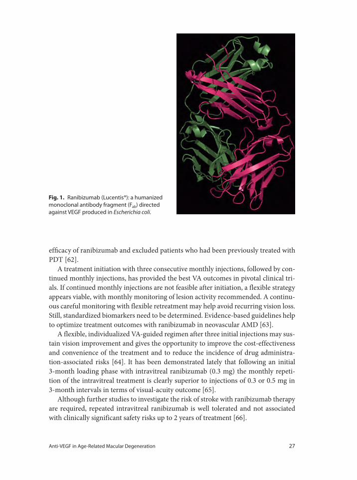
Anti-VEGF in Age-Related Macular Degeneration 27
efficacy of ranibizumab and excluded patients who had been previously treated with
PDT [62].
A treatment initiation with three consecutive monthly injections, followed by con-
tinued monthly injections, has provided the best VA outcomes in pivotal clinical tri-
als. If continued monthly injections are not feasible after initiation, a flexible strategy
appears viable, with monthly monitoring of lesion activity recommended. A continu-
ous careful monitoring with flexible retreatment may help avoid recurring vision loss.
Still, standardized biomarkers need to be determined. Evidence-based guidelines help
to optimize treatment outcomes with ranibizumab in neovascular AMD [63].
A flexible, individualized VA-guided regimen after three initial injections may sus-
tain vision improvement and gives the opportunity to improve the cost-effectiveness
and convenience of the treatment and to reduce the incidence of drug administra-
tion-associated risks [64]. It has been demonstrated lately that following an initial
3-month loading phase with intravitreal ranibizumab (0.3 mg) the monthly repeti-
tion of the intravitreal treatment is clearly superior to injections of 0.3 or 0.5 mg in
3-month intervals in terms of visual-acuity outcome [65].
Although further studies to investigate the risk of stroke with ranibizumab therapy
are required, repeated intravitreal ranibizumab is well tolerated and not associated
with clinically significant safety risks up to 2 years of treatment [66].
Fig. 1. Ranibizumab (Lucentis®): a humanized
monoclonal antibody fragment (Fab) directed
against VEGF produced in Escherichia coli.
28 Schmidt-Erfurth · Pollreisz · Mitsch · Bolz
PIER Study
The PIER Study evaluated the efficacy and safety of ranibizumab administered
monthly for 3 months and then quarterly in patients with CNV secondary to AMD.
Year 1 results were published in October 2007.
This study was a phase IIIb, multicenter, randomized, double-masked, sham injec-
tion-controlled trial in patients with predominantly or minimally classic or occult with
no classic CNV lesions. Patients were randomized 1:1:1 to 0.3 mg ranibizumab (60
patients), 0.5 mg ranibizumab (61 patients), or sham (63 patients) treatment groups.
The primary efficacy endpoint was the mean change from baseline VA at year 1.
Those mean changes from baseline VA were –16.3, –1.6, and –0.2 letters for the
sham, 0.3-mg, and 0.5-mg groups, respectively (p ≤ 0.0001, each ranibizumab dose
vs. sham). Ranibizumab stopped growth of the CNV lesions and reduced leakage.
However, in the ranibizumab groups the treatment effect declined during quarterly
dosing (for example, at 3 months the mean changes from baseline VA had been gains
of 2.9 and 4.3 letters for the 0.3- and 0.5-mg doses, respectively). Results of subgroup
analyses of mean change from baseline VA at 12 months by baseline age, VA, and
lesion characteristics were consistent with the overall results. Few serious ocular or
non-ocular adverse events occurred in any group.
The conclusion was that ranibizumab administered monthly for 3 months and
then quarterly provided a significant benefit in VA to patients with AMD-related sub-
foveal CNV and was well tolerated. The incidence of serious ocular or non-ocular
adverse events was low [67].
FOCUS Study
The FOCUS Study assessed the efficacy and adverse-events profile of a combined
treatment with ranibizumab and verteporfin PDT in patients with predominantly
classic CNV secondary to AMD during a 2-year, multicenter, randomized, single-
masked, controlled study. The 2-year results were published in December 2007.
106 patients received monthly intravitreal injections of ranibizumab 0.5 mg and 56
patients received sham injections. All patients received PDT on day 0, and then every
3 months as needed. The efficacy assessment included VA changes and changes of the
morphology of the lesion and the PDT frequency. Adverse events were summarized
by incidence and severity. At the end of year 2, 88% of the patients receiving com-
bined ranibizumab and PDT treatment had lost fewer than 15 letters from baseline
VA, whereas 75% of the patients receiving PDT alone had lost fewer than 15 letters.
25% of the patients in the combined treatment group had gained more than 15 let-
ters (7% for the PDT-alone group). The two treatment arms differed by 12.4 letters in
mean VA change. The VA benefit of adding ranibizumab to PDT in year 1 persisted
through year 2.
Anti-VEGF in Age-Related Macular Degeneration 29
On average, patients in the combined treatment group developed less lesion growth
and showed greater reduction of CNV leakage and subretinal fluid accumulation, and
required fewer PDT retreatments than patients receiving PDT alone (mean 0.4 vs. 3.0
PDT retreatments). Endophthalmitis and serious intraocular inflammation occurred
in 2.9% and 12.4% of ranibizumab + PDT patients and 0% of PDT-alone patients.
Incidences of serious non-ocular adverse events were similar in the two treatment
groups [62].
MARINA Study
In the MARINA (Minimally Classic/Occult Trial of the Anti-VEGF Antibody
Ranibizumab in the Treatment of Neovascular AMD) Study, 716 patients with either
minimally classic or occult (with no classic lesions) CNV secondary to AMD were
randomly assigned to receive 24 monthly intravitreal injections of ranibizumab
(either 0.3 or 0.5 mg) or sham injections. The primary endpoint was the proportion
of patients losing fewer than 15 letters from baseline VA at 12 months.
At 12 months, 94.5% of the group given 0.3 mg of ranibizumab and 94.6% of those
given 0.5 mg had lost fewer than 15 letters, whereas 62.2% of the patients receiving
sham injections did so. VA improved by 15 or more letters in 24.8% of the 0.3-mg
group and 33.8% of the 0.5-mg group, but only 5.0% of the sham-injection group
gained 15 or more letters in VA. Mean increases were 6.5 letters in the 0.3-mg group
and 7.2 letters in the 0.5-mg group; the main decrease was 10.4 letters in the sham-
injection group. The benefit in VA was maintained at 24 months. During 24 months,
presumed endophthalmitis was identified in 5 patients (1.0%) and serious uveitis in 6
patients (1.3%) given ranibizumab.
The study showed that the intravitreal administration of ranibizumab during 2
years did prevent vision loss and improved mean VA and had low rates of serious
adverse events, in patients with minimally classic or occult CNV secondary to AMD
[58].
ANCHOR Study
The ANCHOR Study (published 2009) demonstrated that ranibizumab was superior
to PDT with respect to VA and morphologic efficacy outcomes and that intravitreal
treatment of predominantly classic CNV in AMD with ranibizumab had a low rate of
serious ocular adverse events.
Of 423 patients (143 PDT, 140 each in the two ranibizumab groups), the majority
(77% in each group) completed the 2-year study. At month 24, the VA benefit from
ranibizumab was statistically significant (p 0.0001 vs. PDT) and clinically meaning-
ful: 89.9–90.0% of ranibizumab-treated patients had lost fewer than 15 letters from
30 Schmidt-Erfurth · Pollreisz · Mitsch · Bolz
baseline (vs. 65.7% of PDT patients); 34–41.0% had gained 15 or more letters (vs.
6.3% of PDT group). On average, VA had improved from baseline by 8.1–10.7 letters
(vs. a mean decline of 9.8 letters in PDT group). Changes in morphological character-
istics on FA also favored ranibizumab.
There was no difference among groups in rates of serious ocular and non-ocular
adverse events. Three out of 277 patients (1.1%) in the ranibizumab groups developed
endophthalmitis in the study eye. Ranibizumab provided greater clinical benefit than
verteporfin PDT in patients with AMD with new-onset, predominantly classic CNV.
PrONTO Study
The long-term efficacy of a variable-dosing regimen with ranibizumab was
assessed in the ‘Prospective Optical Coherence Tomography Imaging of Patients
with Neovascular Age-Related Macular Degeneration Treated with Intraocular
Ranibizumab’ (PrONTO) Study, in which patients were followed for 2 years. The
2-year results were published in January 2009.
The study design was a 2-year prospective, uncontrolled, variable-dosing regimen
with intravitreal ranibizumab treatment based on OCT findings. In this open-label,
prospective, single-center, uncontrolled clinical study, patients with AMD involving
the central fovea and a central retinal thickness of 300 μm or more as measured by
OCT received 3 consecutive monthly intravitreal injections of ranibizumab (0.5 mg).
During the first year, a retreatment with ranibizumab was performed at each monthly
visit if any criterion was fulfilled such as an increase in central retinal thickness of
at least 100 μm as assessed by OCT or a loss of 5 letters or more. During the second
year, the retreatment criteria were amended to include retreatment if any qualitative
increase in the amount of fluid was detected using OCT.
40 patients were enrolled and 37 completed the 2-year study. At month 24, mean
VA improved by 11.1 letters (p < 0.001) and the OCT-CRT decreased by 212 μm (p
< 0.001). VA improved by 15 letters or more in 43% of patients. These VA and OCT
outcomes were achieved with an average of 9.9 injections over 24 months.
The PrONTO Study using an OCT-guided variable-dosing regimen with intravit-
real ranibizumab resulted in VA outcomes comparable with the outcomes from the
phase III clinical studies, but fewer intravitreal injections were required [68].
New Anti-VEGF Treatments for Neovascular Age-Related Macular Degeneration
Vascular Endothelial Growth Factor Trap
The VEGF trap is a pharmacologically engineered protein that binds VEGF and thus
prevents VEGF binding to its cellular receptor. The VEGF trap is composed of two
Anti-VEGF in Age-Related Macular Degeneration 31
different binding domains from VEGF receptor 1 and 2, and is designed to bind the
VEGF-A isoform with higher affinity than pegaptanib or ranibizumab to offer a theo-
retically longer interval between necessary treatments [69].
The intravitreal VEGF trap has been evaluated in a phase 1/2 study, the Clinical
Evaluation of Anti-Angiogenesis in the Retina (CLEAR)-IT 1 and 2. 21 participants
with neovascular AMD were treated with a single intravitreal dose (0.05, 0.15, 0.5, 1,
2, or 4 mg) of the VEGF trap. In the two highest dose groups (2 and 4 mg), the mean
increase in best corrected VA was 13.5 letters. In addition, the mean time for retreat-
ment was 150 days (range 45–420). No serious ocular or systemic adverse events were
noted [70].
Combined Therapy Regiments
The effect of combined treatment of CNV secondary to AMD is subject to actual
research. The pro-angiogenic and pro-inflammatory effects of verteporfin could be
counteracted by combined treatment with anti-VEGF agents [71]. During a single-
center, prospective clinical study conducted at the Department of Ophthalmology
of the Medical University of Vienna, the combined treatment with verteporfin/
ranibizumab was associated with CNV occlusion, reduced edema, improved
visual function and retinal sensitivity [72]. Same-day verteporfin and ranibizumab
appears to be safe and is not associated with severe vision loss or severe ocular
inflammation [73].
Imaging Anti-VEGF Effects on Retinal Morphology in Age-Related Macular
Degeneration
During recent years, OCT has emerged as an important tool in the diagnosis of
AMD and during the follow-up period following anti-VEGF treatment [74]. OCT
allows a detailed evaluation of retinal morphology, similar to in vivo histology,
providing insights into characteristic in vivo changes that occur during disease
progression as well as secondary to treatment. The fourth-generation OCT, spec-
tral-domain OCT (SD-OCT), uses a fast spectral domain technique and performs
scans in a raster pattern throughout the entire macular area at a very high resolu-
tion [75, 76] (fig. 2).
The fifth-generation OCT, the polarization sensitive OCT (PS-OCT), enables to
detect retinal tissue due to its different qualities of polarization and allows specifi-
cally the detection of RPE cells [77, 78]. The combination of PS-SD-OCT provides
a precise identification of retinal pigment epithelium in AMD, allowing the recog-
nition of disease-specific patterns in order to identify the status and progression of
AMD [79].
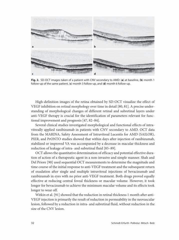
32 Schmidt-Erfurth · Pollreisz · Mitsch · Bolz
High-definition images of the retina obtained by SD-OCT visualize the effect of
VEGF inhibition on retinal morphology over time in detail [80, 81]. A precise under-
standing of morphological changes of different retinal and subretinal layers under
anti-VEGF therapy is crucial for the identification of parameters relevant for func-
tional improvement and prognosis [47, 82–84].
Several clinical studies investigated morphological and functional effects of intra-
vitreally applied ranibizumab in patients with CNV secondary to AMD. OCT data
from the MARINA, Safety Assessment of Intravitreal Lucentis for AMD (SAILOR),
PIER, and PrONTO studies showed that within days after injection of ranibizumab,
stabilized or improved VA was accompanied by a decrease in macular thickness and
reduction of leakage of intra- and subretinal fluid [85–89].
OCT allows the quantitative determination of efficacy and potential effective dura-
tion of action of a therapeutic agent in a non-invasive and simple manner. Shah and
Del Priore [90] used sequential OCT measurements to determine the magnitude and
time course of the initial response to anti-VEGF treatment and the subsequent return
of exudation after single and multiple intravitreal injections of bevacizumab and
ranibizumab in eyes with no prior anti-VEGF treatment. Both drugs proved equally
effective at reducing central foveal thickness or macular volume. However, it took
longer for bevacizumab to achieve the minimum macular volume and its effects took
longer to wear off.
Witkin et al. [91] showed that the reduction in retinal thickness 1 month after anti-
VEGF injection is primarily the result of reduction in permeability in the neovascular
lesion, followed by a reduction in intra- and subretinal fluid, without reduction in the
size of the CNV lesion.
a b
dc
Fig. 2. SD-OCT images taken of a patient with CNV secondary to AMD: (a) at baseline, (b) month 1
follow-up of the same patient, (c) month 3 follow-up, and (d) month 6 follow-up.
Anti-VEGF in Age-Related Macular Degeneration 33
Size and location of the CNV lesion in relation to the RPE level were qualitatively
and quantitatively determined two-dimensionally by Fremme et al. [92], in order to
get an impression of classic and occult CNV components. The authors reported that
under ranibizumab monotherapy there was no clinically significant regression of
CNV.
In a prospective study, Bolz et al. [93] identified and quantified the effects of intra-
vitreal ranibizumab therapy given as a loading regimen with three injections dur-
ing the first 3 months with Stratus OCT. Significant morphological and functional
therapeutic effects were observed as early as 1 week following the first treatment with
ranibizumab. During the loading-dose period, central retinal thickness, intraretinal
cysts and subretinal fluid decreased fast and significantly, whereas pigment epithe-
lial detachment resolved less rapidly. The initial administration of intravitreal ranibi-
zumab induced a significant effect on intra- and subretinal fluid and visual function;
subsequent injections had a less pronounced effect. The change in morphology and
function was only significant between baseline and week 1, while there were no sig-
nificant additional morphological or functional benefits following the second and
third injection.
Similar results were reported from Ahlers et al. [94] in a prospective case series
analyzing the effect of 3-monthly doses of ranibizumab on retinal morphology
and visual outcome in patients with AMD. The study showed that the central mil-
limeter thickness of the retina as measured by Stratus OCT significantly decreased
within the first week after initial ranibizumab treatment. Most of the maximal
therapeutic effect was obtained within this time period. A further decrease in
retinal thickness could be observed continuously up to month 3. Furthermore,
OCT revealed that subretinal fluid was reduced 1 week after the first injection
and was almost eliminated 1 month later. Over the period of the next 2 months,
no additional major changes could be observed. Pigment epithelial detachment
was also significantly reduced within the first week after the initial treatment with
a continuous resolution over the next 3 months. Furthermore, the study showed
that a reduction of retinal thickness correlated with an improvement in BCVA.
The same could be demonstrated for the decrease in subretinal fluid, which cor-
related significantly with an improvement in BCVA. In contrast, there was no sig-
nificant correlation between pigment epithelial detachment and VA. Overall, the
best therapeutic effect of VA improvement was observed 1 month after the first
injection of ranibizumab suggesting that morphological recovery occurred at an
earlier timepoint than visual improvement. In a similar study design, Kiss et al.
[95] evaluated the effect of three injections of ranibizumab on retinal function and
morphology in 23 patients affected by neovascular AMD. The results of the study
suggest that the RPE lesion area may be more relevant for visual function than
retinal thickness.
Apart from a morphological benefit, there are case reports describing RPE tears
following anti-VEGF therapy. RPE tears may occur after intravitreal injection
34 Schmidt-Erfurth · Pollreisz · Mitsch · Bolz
1 VanNewkirk MR, Nanjan MB, Wang JJ, et al: The
prevalence of age-related maculopathy: the visual
impairment project. Ophthalmology 2000;107:1593–
1600.
2 Mitchell P, Smith W, Attebo K, Wang JJ: Prevalence
of age-related maculopathy in Australia. The Blue
Mountains Eye Study. Ophthalmology 1995;102:
1450–1460.
3 Buch H, Nielsen NV, Vinding T, et al: Fourteen-year
incidence, progression, and visual morbidity of age-
related maculopathy: the Copenhagen City Eye
Study. Ophthalmology 2005;112:787–798.
4 Klein R, Klein BE, Knudtson MD, et al: Fifteen-year
cumulative incidence of age-related macular degen-
eration: the Beaver Dam Eye Study. Ophthalmology
2007;114:253–262.
5 Witmer AN, Vrensen GF, Van Noorden CJ, Schlin-
gemann RO: Vascular endothelial growth factors
and angiogenesis in eye disease. Prog Retin Eye Res
2003;22:1–29.
6 Reynolds R, Hartnett ME, Atkinson JP, et al: Plasma
complement components and activation fragments:
associations with age-related macular degeneration
genotypes and phenotypes. Invest Ophthalmol Vis
Sci 2009;50:5818–5827.
7 Morohoshi K, Goodwin AM, Ohbayashi M, Ono SJ:
Autoimmunity in retinal degeneration: autoim-
mune retinopathy and age-related macular degen-
eration. J Autoimmun 2009;33:247–254.
8 Okemefuna AI, Nan R, Miller A, et al: Complement
factor H binds at two independent sites to C-reactive
protein in acute phase concentrations. J Biol Chem
2010;285:1053–1065.
9 Schmidt-Erfurth U, Pruente C: Management of
neovascular age-related macular degeneration. Prog
Retin Eye Res 2007;26:437–451.
10 Schmidt-Erfurth U, Richard G, Augustin A, et al:
Guidance for the treatment of neovascular age-
related macular degeneration. Acta Ophthalmol
Scand 2007;85:486–494.
11 Ferrara N, Gerber HP, LeCouter J: The biology of
VEGF and its receptors. Nat Med 2003;9:669–676.
12 Carmeliet P: Mechanisms of angiogenesis and arte-
riogenesis. Nat Med 2000;6:389–395.
13 Ishibashi T, Hata Y, Yoshikawa H, et al: Expression
of vascular endothelial growth factor in experimen-
tal choroidal neovascularization. Graefes Arch Clin
Exp Ophthalmol 1997;235:159–167.
14 Kvanta A: Ocular angiogenesis: the role of growth
factors. Acta Ophthalmol Scand 2006;84:282–288.
15 Petrova TV, Makinen T, Alitalo K: Signaling via vas-
cular endothelial growth factor receptors. Exp Cell
Res 1999;253:117–130.
16 Frank RN, Amin RH, Eliott D, et al: Basic fibroblast
growth factor and vascular endothelial growth fac-
tor are present in epiretinal and choroidal neovas-
cular membranes. Am J Ophthalmol 1996;122:
393–403.
of ranibizumab in patients with neovascular AMD, probably because of the rapid
regression of the fibrovascular membrane [96, 97]. A small ratio of CNV size to pig-
ment epithelial detachment is more common in eyes with RPE tears. In addition,
a larger size of pigment epithelial detachment is considered as a predictor for RPE
tears. However, vision may be preserved despite RPE tears [98]. Although a pigment
epithelial tear in neovascular AMD can represent natural history, it is suggested that
clinicians should be aware of and monitor patients for the possibility of this com-
plication after intravitreal injections of ranibizumab [99]. However, it remains to be
determined if a pigment epithelial tear should be considered as a possible therapeuti-
cally induced complication or if it rather represents natural history in neovascular
AMD.
In summary, a number of studies have shown that OCT imaging allows identifica-
tion of functionally relevant factors like subretinal fluid or retinal thickness, which
are important for the establishment of an optimized individual dosing regimen dur-
ing antiangiogenesis therapies.
References
Anti-VEGF in Age-Related Macular Degeneration 35
17 Kvanta A, Algvere PV, Berglin L, Seregard S:
Subfoveal fibrovascular membranes in age-related
macular degeneration express vascular endothelial
growth factor. Invest Ophthalmol Vis Sci 1996;37:
1929–1934.
18 Kliffen M, Sharma HS, Mooy CM, et al: Increased
expression of angiogenic growth factors in age-
related maculopathy. Br J Ophthalmol 1997;81:154–
162.
19 Shima DT, Adamis AP, Ferrara N, et al: Hypoxic
induction of endothelial cell growth factors in reti-
nal cells: identification and characterization of vas-
cular endothelial growth factor as the mitogen. Mol
Med 1995;1:182–193.
20 Funk M, Karl D, Georgopoulos M, et al: Neovascular
age-related macular degeneration: intraocular
cytokines and growth factors and the influence of
therapy with ranibizumab. Ophthalmology 2009;
116:2393–2399.
21 Waheed NK, Miller JW: Aptamers, intramers, and
vascular endothelial growth factor. Int Ophthalmol
Clin 2004;44:11–22.
22 Famulok M, Mayer G, Blind M: Nucleic acid aptam-
ers – from selection in vitro to applications in vivo.
Acc Chem Res 2000;33:591–599.
23 Ellington AD, Szostak JW: In vitro selection of RNA
molecules that bind specific ligands. Nature
1990;346:818–822.
24 Schachat AP: New treatments for age-related macular
degeneration. Ophthalmology 2005;112:531–532.
25 Ruckman J, Green LS, Beeson J, et al: 2�-Fluoro-
pyrimidine RNA-based aptamers to the 165-amino-
acid form of vascular endothelial growth factor
(VEGF-165). Inhibition of receptor binding and
VEGF-induced vascular permeability through
interactions requiring the exon 7 encoded domain. J
Biol Chem 1998;273:20556–20567.
26 Eyetech Study Group: Preclinical and phase 1A clin-
ical evaluation of an anti-VEGF pegylated aptamer
(EYE001) for the treatment of exudative age-related
macular degeneration. Retina 2002;22:143–152.
27 Eyetech Study Group: Anti-vascular endothelial
growth factor therapy for subfoveal choroidal neo-
vascularization secondary to age-related macular
degeneration: phase II study results. Ophthalmology
2003;110:979–986.
28 Gragoudas ES, Adamis AP, Cunningham ET Jr, et al:
Pegaptanib for neovascular age-related macular
degeneration. N Engl J Med 2004;351:2805–2816.
29 Spaide R: New treatments for AMD. Ophthalmology
2006;113:160–161.
30 Ferrara N, Hillan KJ, Gerber HP, Novotny W:
Discovery and development of bevacizumab, an
anti-VEGF antibody for treating cancer. Nat Rev
Drug Discov 2004;3:391–400.
31 Presta LG, Chen H, O’Connor SJ, et al: Humanization
of an anti-vascular endothelial growth factor mono-
clonal antibody for the therapy of solid tumors and
other disorders. Cancer Res 1997;57:4593–4599.
32 Adding a humanized antibody to vascular endothe-
lial growth factor (bevacizumab, Avastin) to chemo-
therapy improves survival in metastatic colorectal
cancer. Clin Colorectal Cancer 2003;3:85–88.
33 Kabbinavar F, Hurwitz HI, Fehrenbacher L, et al:
Phase II, randomized trial comparing bevacizumab
plus fluorouracil (FU)/leucovorin (LV) with FU/LV
alone in patients with metastatic colorectal cancer. J
Clin Oncol 2003;21:60–65.
34 Hurwitz H, Fehrenbacher L, Novotny W, et al: Beva-
cizumab plus irinotecan, fluorouracil, and leuco-
vorin for metastatic colorectal cancer. N Engl J Med
2004;350:2335–2342.
35 Mordenti J, Cuthbertson RA, Ferrara N, et al:
Comparisons of the intraocular tissue distribution,
pharmacokinetics, and safety of 125I-labeled full-
length and Fab antibodies in rhesus monkeys follow-
ing intravitreal administration. Toxicol Pathol 1999;
27:536–544.
36 Heiduschka P, Fietz H, Hofmeister S, et al:
Penetration of bevacizumab through the retina after
intravitreal injection in the monkey. Invest Oph-
thalmol Vis Sci 2007;48:2814–2823.
37 Shahar J, Avery RL, Heilweil G, et al: Electro-
physiologic and retinal penetration studies follow-
ing intravitreal injection of bevacizumab (Avastin).
Retina 2006;26:262–269.
38 Michels S, Rosenfeld PJ, Puliafito CA, et al: Systemic
bevacizumab (Avastin) therapy for neovascular age-
related macular degeneration 12-week results of an
uncontrolled open-label clinical study. Ophthal-
mology 2005;112:1035–1047.
39 Moshfeghi AA, Rosenfeld PJ, Puliafito CA, et al:
Systemic bevacizumab (Avastin) therapy for neo-
vascular age-related macular degeneration: 24-week
results of an uncontrolled open-label clinical study.
Ophthalmology 2006;113:2002.e1–12.
40 Geitzenauer W, Michels S, Prager F, et al: Com-
parison of 2.5 and 5 mg/kg systemic bevacizumab in
neovascular age-related macular degeneration:
24-week results of an uncontrolled, prospective
cohort study. Retina 2008;28:1375–1386.
41 Bolz M, Michels S, Geitzenauer W, et al: Effect of
systemic bevacizumab therapy on retinal pigment
epithelial detachment. Br J Ophthalmol 2007;91:
785–789.
42 Rosenfeld PJ, Fung AE, Puliafito CA: Optical coher-
ence tomography findings after an intravitreal injec-
tion of bevacizumab (Avastin) for macular edema
from central retinal vein occlusion. Ophthalmic
Surg Lasers Imaging 2005;36:336–339.
36 Schmidt-Erfurth · Pollreisz · Mitsch · Bolz
43 Weigert G, Michels S, Sacu S, et al: Intravitreal beva-
cizumab (Avastin) therapy versus photodynamic
therapy plus intravitreal triamcinolone for neovas-
cular age-related macular degeneration: 6-month
results of a prospective, randomised, controlled
clinical study. Br J Ophthalmol 2008;92:356–360.
44 Hahn R, Sacu S, Michels S, et al: Intravitreal bevaci-
zumab versus verteporfin and intravitreal triamci-
nolone acetonide in patients with neovascular
age-related macula degeneration (in German).
Ophthalmologe 2007;104:588–593.
45 Costa RA, Jorge R, Calucci D, et al: Intravitreal bev-
acizumab for choroidal neovascularization caused
by AMD (IBeNA Study): results of a phase 1 dose-
escalation study. Invest Ophthalmol Vis Sci 2006;
47:4569–4578.
46 Kiss C, Michels S, Prager F, et al: Evaluation of ante-
rior chamber inflammatory activity in eyes treated
with intravitreal bevacizumab. Retina 2006;26:877–
881.
47 Leydolt C, Michels S, Prager F, et al: Effect of intrav-
itreal bevacizumab (Avastin®) in neovascular age-
related macular degeneration using a treatment
regimen based on optical coherence tomography: 6-
and 12-month results. Acta Ophthalmol 2009:
[Epub ahead of print].
48 Sacu S, Michels S, Prager F, et al: Randomised clini-
cal trial of intravitreal Avastin vs. photodynamic
therapy and intravitreal triamcinolone: long-term
results. Eye (Lond) 2009;23:2223–2227.
49 Spaide RF, Laud K, Fine HF, et al: Intravitreal beva-
cizumab treatment of choroidal neovascularization
secondary to age-related macular degeneration.
Retina 2006;26:383–390.
50 Rich RM, Rosenfeld PJ, Puliafito CA, et al: Short-
term safety and efficacy of intravitreal bevacizumab
(Avastin) for neovascular age-related macular
degeneration. Retina 2006;26:495–511.
51 Avery RL, Pieramici DJ, Rabena MD, et al: Intra-
vitreal bevacizumab (Avastin) for neovascular age-
related macular degeneration. Ophthalmology
2006;113:363–372.e5.
52 Fung AE, Rosenfeld PJ, Reichel E: The International
Intravitreal Bevacizumab Safety Survey: using the
internet to assess drug safety worldwide. Br J Oph-
thalmol 2006;90:1344–1349.
53 Artunay O, Yuzbasioglu E, Rasier R, et al: Incidence
and management of acute endophthalmitis after
intravitreal bevacizumab (Avastin) injection. Eye
(Lond) 2009;23:2187–2193.
54 Sato T, Emi K, Ikeda T, et al: Severe intraocular
inflammation after intravitreal injection of bevaci-
zumab. Ophthalmology 2010;117:512–516.e1–2.
55 Yenerel NM, Dinc UA, Gorgun E: A case of sterile
endophthalmitis after repeated intravitreal bevaci-
zumab injection. J Ocul Pharmacol Ther 2008;24:
362–363.
56 Yamashiro K, Tsujikawa A, Miyamoto K, et al:
Sterile endophthalmitis after intravitreal injection
of bevacizumab obtained from a single batch. Retina
2010;30:485–490.
57 Georgopoulos M, Polak K, Prager F, et al: Charact-
eristics of severe intraocular inflammation follow-
ing intravitreal injection of bevacizumab (Avastin).
Br J Ophthalmol 2009;93:457–462.
58 Rosenfeld PJ, Brown DM, Heier JS, et al: Ranibi-
zumab for neovascular age-related macular degen-
eration. N Engl J Med 2006;355:1419–1431.
59 Brown DM, Kaiser PK, Michels M, et al: Rani-
bizumab versus verteporfin for neovascular age-
related macular degeneration. N Engl J Med 2006;
355:1432–1444.
60 TAP Report 1: Photodynamic therapy of subfoveal
choroidal neovascularization in age-related macular
degeneration with verteporfin: one-year results of
two randomized clinical trials. Arch Ophthalmol
1999;117:1329–1345.
61 TAP Report 2: Photodynamic therapy of subfoveal
choroidal neovascularization in age-related macular
degeneration with verteporfin: two-year results of
two randomized clinical trials. Arch Ophthalmol
2001;119:198–207.
62 Andrew NA, Lisa T, Carol YC, Angele S: Ranibi-
zumab combined with verteporfin photodynamic
therapy in neovascular age-related macular degen-
eration (FOCUS): year 2 results. Am J Ophthalmol
2008;145:862–874.e3.
63 Mitchell P, Korobelnik JF, Lanzetta P, et al: Rani-
bizumab (Lucentis) in neovascular age-related mac-
ular degeneration: evidence from clinical trials. Br J
Ophthalmol 2009;20:20.
64 Holz FG, Korobelnik JF, Lanzetta P, et al: The effects
of a flexible visual acuity-driven ranibizumab treat-
ment regimen in age-related macular degeneration:
outcomes of a drug and disease model. Invest
Ophthalmol Vis Sci 2009;51:405–412.
65 Schmidt-Erfurth U, Eldem B, Guymer R, et al.; on
behalf of the EXCITE Study Group: Efficacy and
safety of monthly versus quarterly ranibizumab
treatment in neovascular age-related macular
degeneration. Ophthalmology 2010 (in press).
66 Schmidt-Erfurth U: Clinical safety of ranibizumab
in age-related macular degeneration. Expert Opin
Drug Saf 2010;9:149–165.
67 Carl DR, David MB, Prema A, et al: Randomized,
double-masked, sham-controlled trial of ranibi-
zumab for neovascular age-related macular degen-
eration: PIER Study year 1. Am J Ophthalmol
2008;145:239–248.e5.
Anti-VEGF in Age-Related Macular Degeneration 37
68 Lalwani GA, Rosenfeld PJ, Fung AE, et al: A vari-
able-dosing regimen with intravitreal ranibizumab
for neovascular age-related macular degeneration:
year 2 of the PrONTO Study. Am J Ophthalmol
2009;148:43–58.e1.
69 Quan Dong N, Syed Mahmood S, Gulnar H, et al: A
phase I trial of an IV-administered vascular endo-
thelial growth factor trap for treatment in patients
with choroidal neovascularization due to age-related
macular degeneration. Ophthalmology 2006;113:
1522.e1–14.
70 Do DV: Antiangiogenic approaches to age-related
macular degeneration in the future. Ophthalmology
2009;116(suppl 1):S24–S6.
71 Schmidt-Erfurth U, Schlotzer-Schrehard U, Cur-
siefen C, et al: Influence of photodynamic therapy
on expression of vascular endothelial growth factor
(VEGF), VEGF receptor 3, and pigment epithelium-
derived factor. Invest Ophthalmol Vis Sci 2003;
44:4473–4480.
72 Kiss CG, Simader C, Michels S, Schmidt-Erfurth U:
Combination of verteporfin photodynamic therapy
and ranibizumab: effects on retinal anatomy, chor-
oidal perfusion and visual function in the protect
study. Br J Ophthalmol 2008;92:1620–1627.
73 Schmidt-Erfurth U, Wolf S: Same-day administra-
tion of verteporfin and ranibizumab 0.5 mg in
patients with choroidal neovascularisation due to
age-related macular degeneration. Br J Ophthalmol
2008;92:1628–1635.
74 Srinivasan VJ, Wojtkowski M, Witkin AJ, et al:
High-definition and three-dimensional imaging of
macular pathologies with high-speed ultrahigh-res-
olution optical coherence tomography. Ophthal-
mology 2006;113:2054.e1–14.
75 Golbaz I, Ahlers C, Goesseringer N, et al: Automatic
and manual segmentation of healthy retinas using
high-definition optical coherence tomography. Acta
Ophthalmol 2009: [Epub ahead of print].
76 Mylonas G, Ahlers C, Malamos P, et al: Comparison
of retinal thickness measurements and segmenta-
tion performance of four different spectral and time
domain OCT devices in neovascular age-related
macular degeneration. Br J Ophthalmol 2009;93:
1453–1460.
77 Gotzinger E, Pircher M, Baumann B, et al: Three-
dimensional polarization sensitive OCT imaging
and interactive display of the human retina. Opt
Express 2009;17:4151–4165.
78 Ahlers C, Schmidt-Erfurth U: Three-dimensional
high resolution OCT imaging of macular pathology.
Opt Express 2009;17:4037–4045.
79 Ahlers C, Gotzinger E, Pircher M, et al: Imaging of
the retinal pigment epithelium in age-related macu-
lar degeneration using polarization-sensitive optical
coherence tomography. Invest Ophthalmol Vis Sci
2010;51:2149–2157.
80 Puliafito CA, Hee MR, Lin CP, et al: Imaging of
macular diseases with optical coherence tomogra-
phy. Ophthalmology 1995;102:217–229.
81 Pieroni CG, Witkin AJ, Ko TH, et al: Ultrahigh res-
olution optical coherence tomography in non-exu-
dative age-related macular degeneration. Br J
Ophthalmol 2006;90:191–197.
82 Malamos P, Sacu S, Georgopoulos M, et al:
Correlation of high-definition optical coherence
tomography and fluorescein angiography imaging
in neovascular macular degeneration. Invest Oph-
thalmol Vis Sci 2009;50:4926–4933.
83 Golbaz I, Ahlers C, Schutze C, et al: Influence of
antiangiogenetic therapy on retinal thickness values
in age-related macular degeneration (in German).
Ophthalmologe 2009;106:1103–1110.
84 Einwallner E, Ahlers C, Golbaz I, et al: Neovascular
age-related macular degeneration under anti-angio-
genic therapy: subretinal fluid is a relevant prognos-
tic parameter (in German). Ophthalmologe 2010;
107:158–164.
85 Andreoli CM, Miller JW: Antivascular endothelial
growth factor therapy for ocular neovascular dis-
ease. Curr Opin Ophthalmol 2007;18:502–508.
86 Brown DM, Regillo CD: Anti-VEGF agents in the
treatment of neovascular age-related macular
degeneration: applying clinical trial results to the
treatment of everyday patients. Am J Ophthalmol
2007;144:627–637.
87 Brown DM, Kaiser PK, Michels M, et al: Rani-
bizumab versus verteporfin for neovascular age-
related macular degeneration. N Engl J Med 2006;
355:1432–1444.
88 Kaiser PK, Blodi BA, Shapiro H, Acharya NR:
Angiographic and optical coherence tomographic
results of the MARINA Study of ranibizumab in
neovascular age-related macular degeneration.
Ophthalmology 2007;114:1868–1875.
89 Regillo CD, Brown DM, Abraham P, et al: Ran-
domized, double-masked, sham-controlled trial of
ranibizumab for neovascular age-related macular
degeneration: PIER Study year 1. Am J Ophthalmol
2008;145:239–248.
90 Shah AR, Del Priore LV: Duration of action of intra-
vitreal ranibizumab and bevacizumab in exudative
AMD eyes based on macular volume measure-
ments. Br J Ophthalmol 2009;93:1027–1032.
38 Schmidt-Erfurth · Pollreisz · Mitsch · Bolz
91 Witkin AJ, Vuong LN, Srinivasan VJ, et al: High-
speed ultrahigh resolution optical coherence tomo-
graphy before and after ranibizumab for age-related
macular degeneration. Ophthalmology 2009;116:
956–963.
92 Framme C, Panagakis G, Birngruber R: Effects on
choroidal neovascularization after anti-VEGF
upload using intravitreal ranibizumab, as determined
by spectral domain-optical coherence tomography.
Invest Ophthalmol Vis Sci 2010;51:1671–1676.
93 Bolz M, Simader C, Ritter M, et al: Morphological
and functional analysis of the loading regimen with
intravitreal ranibizumab in neovascular age-related
macular degeneration. Br J Ophthalmol 2010;94:
185–189.
94 Ahlers C, Golbaz I, Stock G, et al: Time course of
morphologic effects on different retinal com-
partments after ranibizumab therapy in age-related
macular degeneration. Ophthalmology 2008;115:
e39–e46.
95 Kiss CG, Geitzenauer W, Simader C, et al: Evaluation
of ranibizumab-induced changes in high-resolution
optical coherence tomographic retinal morphology
and their impact on visual function. Invest Oph-
thalmol Vis Sci 2009;50:2376–2383.
96 Bakri SJ, Kitzmann AS: Retinal pigment epithelial
tear after intravitreal ranibizumab. Am J Ophthalmol
2007;143:505–507.
97 Kiss C, Michels S, Prager F, et al: Retinal pigment
epithelium tears following intravitreal ranibizumab
therapy. Acta Ophthalmol Scand 2007;85:902–903.
98 Chan CKM, Meyer CHM, et al: Retinal pigment
epithelial tears after intravitreal bevacizumab injec-
tion for neovascular age-related macular degenera-
tion. Retina 2007;27:541–551.
99 Apte RS: Retinal pigment epithelial tear after intrav-
itreal ranibizumab for subfoveal CNV secondary to
AMD. Int Ophthalmol 2007;27:59–61.
Prof. Ursula Schmidt-Erfurth
Department of Ophthalmology and Optometry
Medical University Vienna
Waehringer Guertel 18-20, AT–1090 Vienna (Austria)
E-Mail [email protected]
Bandello F, Battaglia Parodi M (eds): Anti-VEGF.
Dev Ophthalmol. Basel, Karger, 2010, vol 46, pp 39–53
Antivascular Endothelial Growth Factor in Diabetic RetinopathyPierluigi Iaconoa � Maurizio Battaglia Parodib � Francesco Bandellob
aFondazione G.B. Bietti per l’Oftalmologia, IRCCS (Istituto di Ricovero e Cura a Carattere Scientifico),
Rome, and bDepartment of Ophthalmology, University Vita-Salute, Scientific Institute San Raffaele,
Milan, Italy
AbstractDiabetic macular edema (DME) and proliferative diabetic retinopathy (PDR) represent the most com-
mon causes of vision loss in patients affected by diabetes mellitus. Diabetic retinopathy (DR) needs
special attention because of its high public health impact and impact on quality of life of patients.
Actually, laser retinal photocoagulation is the standard of care for the treatment of DR. However,
laser treatment reduces the risk of moderate visual loss by approximately 50%, without a remark-
able vision recovery. Thus, new approaches in the treatment of DR have been taken into account
and, more specifically, the therapy employing antivascular endothelial growth factor (anti-VEGF)
drugs could play a meaningful role. VEGF is a pluripotent growth factor that functions as an endothe-
lial cell-specific mitogen and vasopermeability factor. Through these mechanisms VEGF plays a criti-
cal role in promoting angiogenesis and vascular leakage. A high level of VEGF has been detected in
eyes presenting DME and PDR, and thereby VEGF is an attractive candidate as therapeutic target of
pharmacological treatment in the management of DR. In the current chapter, the concepts and
results of anti-VEGF therapy in the treatment of the DME and PDR are presented.
Copyright © 2010 S. Karger AG, Basel
Diabetic retinopathy (DR) is considered the most frequent vascular disorder being
detectable in about 40% of diabetic patients 40 years and older [1]. Today, DR
is the leading cause of acquired blindness among young adults throughout the
developed countries [2]. Population-based epidemiological studies have estimated
that after 20 years, DR is recognized to a certain extent, and that after 30 years
a proliferative DR is present in the 70% of patients with diabetes mellitus type 1
[3]. The World Health Organization estimates that about 171 million persons are
affected by diabetes with an expected doubling of prevalence expected in the next
20 years [4].
40 Iacono · Battaglia Parodi · Bandello
Role of the Vascular Endothelial Growth Factor in Diabetic Retinopathy
Hyperglycemia is the main factor involved in the pathogenesis of DR. It results in the
production of glycation end products, activation of the polyol pathway, and altered
transduction of cellular signals [5–7]. The following damage to endothelial cells and
pericytes, through activation of oxidative and inflammatory mechanisms, produces
diabetic microangiopathy affecting small-caliber retinal vessel [8]. These altera-
tions result in the deregulation of the mechanism of flow control with subsequently
hypoxia and accumulation of fluid in the retinal tissue. Hypoxia represents the likely
major inducer of vascular endothelial growth factor (VEGF) gene transcription, but
the overexpression of VEGF is also upregulated in response to high glucose, pro-
tein kinase C activation, and glycation end products, all elements characterizing the
impairment of glycometabolic control [5–7, 9].
VEGF is a pluripotent growth factor that functions as an endothelial cell-specific
mitogen and vasopermeability factor and through these mechanisms the VEGF plays
a critical role in promoting angiogenesis and vascular leakage [10–13]. In DR, the
impairment of the blood retinal barrier and the increased permeability are respon-
sible for the diabetic macular edema (DME) and several investigations underline the
active role of VEGF. By disrupting the intercellular tight junctions between the retinal
endothelial cells, VEGF increases the extracellular accumulation of fluid from the
intravascular compartment [8]. Moreover, VEGF shows a role in mediating active
intraocular neovascularization.
Elevations of VEGF levels in ocular fluids from human patients with tissue hypoxia
and active neovascularization secondary to DR have been well documented [14]. The
increased levels of VEGF decline when treatment with panretinal photocoagulation
induces regression of neovascularization. Thus, these studies demonstrated a tempo-
ral correlation between VEGF elevations and active proliferative retinopathy evidenc-
ing the role of VEGF as a key mediator of intraocular neovascularization secondary
to DR. In essence, VEGF is an attractive candidate as therapeutic target of pharmaco-
logical treatment in the management of DR.
Anti-VEGF Therapy in the Treatment of Diabetic Macular Edema and Proliferative
Diabetic Retinopathy
The VEGF molecular family includes five members: placental growth factor, VEGF-A,
VEGF-B, VEGF-C and VEGF-D [15]. Each of the different factors may link one or
more of three VEGF receptors. Moreover, between the different factors, VEGF-A plays
a major role in angiogenesis and vascular permeability. Alternative splicing of VEGF
gene produces nine VEGF-A isoforms (VEGF121, VEGF145, VEGF148, VEGF162, VEGF165, VEGF165b, VEGF183, VEGF189, VEGF206) and among them VEGF165 is the most abun-
dantly expressed isoform and is detected as being mainly responsible for DR.
Anti-VEGF in Diabetic Retinopathy 41
The pathway enclosed between VEGF gene transcription and the activation of the
VEGF receptor is the object of a new therapeutic approach based on the use of the
VEGF antagonist. Pegaptanib, ranibizumab, bevacizumab and VEGF Trap are mol-
ecules that are able to directly bind the VEGF protein. A new and interesting thera-
peutic approach is the employment of bevasiranib. This molecule, interfering with
messenger RNA, interrupts the synthesis of the VEGF protein. Last of all, rapamycin,
employed commonly as an immunosuppressive, anti-inflammatory or antimycotic
drug, reduces the activity of VEGF molecules interfering with the promoting signal,
the active synthesis of VEGF, and reduces the response of endothelial cells to VEGF.
Ranibizumab
Ranibizumab is an antigen-binding fragment (Fab) derived from a humanized anti-
VEGF antibody and this Fab inhibits all biologically active isoforms and active prote-
olytic fragments of VEGF-A. Currently, ranibizumab is approved by the Food and Drug
Administration for the treatment of neovascular age-related macular degeneration.
Chun et al. [16] reported the first pilot study exploring the effects of two dos-
ing regimens of ranibizumab in eyes affected by clinically significant DME. Of 10
patients enrolled, 5 received 0.3 mg ranibizumab and 5 received 0.5 mg ranibizumab
at baseline and at 1 and 2 months. At month 3, 40% of patients gained more than 15
letters, 50% gained more than 10 letters, and 80% obtained an improvement of at
least 1 letter in best corrected visual acuity (BCVA). At month 3, the mean decrease
in central retinal thickness was 45.3 and 197.8 μm in the low- and high-dose groups,
respectively. Intravitreal injections of ranibizumab were generally well tolerated and
no systemic adverse events were reported.
Nguyen et al. [17] investigated the role of ranibizumab in DME in the open-label
study READ-1 (Ranibizumab for Edema of the Macula in Diabetes: Phase 1). Ten
patients with chronic DME received intraocular injections of 0.5 mg ranibizumab
at baseline and at 1, 2, 4, and 6 months. The main outcome measures were changes
in BCVA, central retinal thickness as assessed by optical coherence tomography
(OCT) measurement at the 7-month examination. Mean and median values of BCVA
improved at 7 months by 12.3 and 11 letters respectively. Compared to the baseline,
mean foveal thickness showed a meaningful reduction decreasing from 503 to 257
μm with a 85% reduction of the excess foveal thickness present at baseline. The injec-
tions were well tolerated with no ocular or systemic adverse events.
More recently, the results of the READ-2 study were reported. READ-2 was a pro-
spective, randomized, interventional, multicenter clinical trial designed to compare
ranibizumab with focal/grid laser, alone or in combination, in DME [18]. 126 patients
were randomized to receive 0.5 mg ranibizumab, focal/grid laser photocoagulation
or a combination of 0.5 mg ranibizumab and focal/grid laser. Group 1, 42 patients,
received 0.5 mg ranibizumab at baseline and months 1, 3, and 5. Group 2, 42 patients,
42 Iacono · Battaglia Parodi · Bandello
received focal/grid laser photocoagulation at baseline and month 3 if needed (center
subfield thickness was >250 μm). Group 3, 42 patients, received a combination of 0.5
mg ranibizumab and focal/grid laser at baseline and month 3. The primary outcome
was the change in BCVA at month 6 in comparison with baseline. At month 6, the
group receiving ranibizumab alone showed a significant improvement in mean BCVA
with respect to the patients receiving focal/grid laser. The group receiving combined
therapy was not statistically different from groups 1 or 2. Resolutions of 50, 33, and
45% of excess foveal thickening were assessed in groups 1, 2, and 3, respectively.
The RESOLVE study was specifically designed to evaluate the efficacy and safety of
ranibizumab 0.5 mg in patients with visual impairment due to DME. The RESOLVE
trial (a randomized, double-masked, multicenter, phase 2 study assessing the safety
and efficacy of two concentrations of ranibizumab compared with non-treatment
control for the treatment of DME with center involvement) evaluated the effect of
ranibizumab on retinal edema and visual acuity (VA) in 151 patients with clinically
significant DME. Patients with a central macular thickness of ≥300 μm were random-
ized to receive three monthly injections with either 0.3 or 0.5 mg ranibizumab or pla-
cebo. After the three monthly intravitreal injections, treatment was administrated on
pro re nata basis for 9 months. The primary endpoint in the 1-year study was visual
function at 6 months. The study design allowed the investigator to double the dose
of ranibizumab if after 1 month the resolution of macular edema was incomplete.
Moreover, retinal photocoagulation could be administered if needed.
The preliminary results were partially presented at the 2009 ARVO Meeting [19].
During the 12-month follow-up period, mean BCVA increased and mean CRT
decreased continuously over time. The mean change in BCVA from baseline to the
12-month examination was –1.4 letters in the sham group. The groups receiving 0.3
and 0.5 mg gained, respectively, 11.8 and 8.8 letters. In order to provide further clarity
on the effectiveness of treatments based on administration of steroidal or anti-VEGF
drugs in comparison to conventional laser treatment, the DRCR net has designed
a randomized, multicenter clinical trial which addressed the effects on visual acu-
ity and on central retinal thickness in four groups receiving, respectively, intravitreal
ranibizumab alone or associated with laser photocoagulation or triamcinolone asso-
ciated with the laser treatment or laser treatment alone.
The study recruited 691 patients and examined a total of 854 eyes in a follow-up
period of 2 years. Two hundred ninety-three eyes were randomized to received laser
alone, 187 eyes were assigned to the group receiving 0.5 mg ranibizumab + prompt laser,
188 eyes received 0.5 mg ranibizumab + deferred laser (at least 24 weeks), and 186 eye
were included in the group receiving 4 mg intravitreal triamcinolone + prompt laser.
At 1-year examination, the mean change in the visual acuity letter score respect
to the baseline value showed a statistically significant improvement in the ranibi-
zumab + prompt laser group (+9±11 letters) and ranibizumab + deferred laser group
(+9±12) but not in the triamcinolone + prompt laser group (+4±13) compared with
the laser group (+3±13).
Anti-VEGF in Diabetic Retinopathy 43
Over the 2 years of follow-up a different correlation between visual acuity change
and retinal thickness was observed in each group. A progressive reduction in mean
central subfield thickness was noted in the laser group during the 24 months of fol-
low-up; however, the mean change in visual acuity did not continue to increase from
the 1- to 2-year visit as noted instead during the first year of follow-up.
In the triamcinolone + laser group, during the first year of follow-up an improve-
ment of visual function was associated with a significant reduction in CST whereas
from the 1- to 2-year examination the mean CST increased in parallel with a visual
acuity reduction.
Ranibizumab groups showed a parallel visual acuity improvement associated
with a CST reduction from baseline to 12-month visit and following the OCT results
remained relatively stable up to 24-month examination and paralleled the visual acu-
ity outcomes during this time.
Intraocular hypertension and cataract surgery were more frequently noted in
the triamcinolone + prompt laser group in comparison to groups receiving ranibi-
zumab + laser or laser alone.
The current large prospective randomized clinical trial confirms the preliminary
promising results in the treatment of DME and suggests as a combined therapy might
offer a more efficacious approach in this disorder where the multi-factorial pathogen-
esis involves several processes [20].
Actually, several multicenter international clinical trials (e.g. RESTORE, RIDE and
RISE) are ongoing in order to evaluate the efficacy and safety of ranibizumab 0.5 mg
as monotherapy or in combination with laser photocoagulation in eyes affected by
DME.
With regard to the effects of ranibizumab on proliferative diabetic retinopathy
(PDR), no studies are available in the literature evaluating this topic. However, DRCR.
net has designed a prospective, randomized, comparative clinical trial to evaluate the
role of ranibizumab or triamcinolone intravitreal injection as adjunctive treatment to
panretinal photocoagulation for PDR.
Pegaptanib
Pegaptanib is a pegylated 28-nucleotide RNA aptamer that binds to the VEGF164/165
isoform at high affinity. VEGF165 levels are present in human eyes affected by DR
with increased concentration and play an active role in promoting angiogenesis and
in enhancing vascular permeability. Initially, pegaptanib was employed only in the
treatment of neovascular age macular degeneration, where it obtained the approval of
the Food and Drug Administration. Considering the role of VEGF165 in DR and the
safety and tolerability profile of intravitreally administered pegaptanib, a phase II trial
was specifically designed to investigate the effects of pegaptanib in the management
of DME.
44 Iacono · Battaglia Parodi · Bandello
The Macugen Diabetic Retinopathy Study was a randomized, sham-controlled, dou-
ble-masked, dose-finding phase II trial designed to evaluate the effect of three doses of
intravitreal pegaptanib vs. sham injection in patients affected by clinically significant
DME [21]. The patients were randomized to receive 0.3, 1.0 or 3.0 mg of pegaptanib or
sham injection at baseline, week 6 or week 12. If needed, further injections were admin-
istrated every 6 weeks up to a maximum of three additional injections. Retinal laser
photocoagulation could be delivered if the investigators judged it to be necessary. The
main outcome measures were changes in BCVA, central retinal thickness as assessed
by OCT measurement, and additional therapy with photocoagulation between weeks
12 and 36. At the final visit at week 36, the group of patients receiving pegaptanib 0.3
mg was significantly superior to sham injection, as measured by mean change in VA
(+4.7 vs. –0.4 letters; p = 0.04), proportions of patients gaining >10 letters of VA (34 vs.
10%; p = 0.003), change in mean central retinal thickness (68 μm reduction vs. 3.7 μm
increase; p = 0.02). Moreover, only 25% of patients receiving pegaptanib required reti-
nal photocoagulation in comparison with 40% of patients receiving a sham injection (p
= 0.04). It is noteworthy that patients receiving 1.0 or 3.0 mg did not show a significant
improvement compared to 0.3 mg with regard to BCVA or CRT changes. In general,
pegaptanib was well tolerated at various concentrations; endophthalmitis occurred in 1
of 652 injections and was successfully treated without severe visual loss.
The Macugen Diabetic Retinopathy Study also provided new information on the
ability of pegaptanib sodium to lead to regression of retinal neovascularization in
proliferative DR [22]. Of 16 patients having retinal neovascularization in the study
eye at baseline, 13 were assigned to receive pegaptanib treatment and 3 were assigned
to sham injections. At 36 weeks, 8 of 13 (62%) in the pegaptanib treatment group
showed regression of neovascularization, as assessed by fundus photography or fluo-
rescein angiography, whereas no such regression occurred in 3 sham-treated eyes.
In 3 of 8 with regression, neovascularization progressed at week 52 after cessation
of pegaptanib at week 30, suggesting the necessity of repeated injections to control
retinal neovascularization.
More recently, Gonzalez et al. [23] reported the results of a prospective, random-
ized, controlled, open-label, exploratory study designed to compare the efficacy of
intravitreal pegaptanib vs. panretinal laser photocoagulation (PRP) in the treatment
of active PDR. 20 subjects with active PDR were assigned at a 1:1 ratio to receive
pegaptanib treatment in 1 eye every 6 weeks for 30 weeks or with PRP. In 90% of eyes
randomized to pegaptanib, retinal neovascularization showed a complete regression
by week 3. By week 12, in all eyes receiving pegaptanib a complete regression of retinal
proliferation was obtained and preserved through week 36. In the PRP-treated group,
at the 9-month examination, 25% demonstrated complete regression, 25% showed
partial regression, and 50% showed persistent active PDR.
With regard to BCVA and although the difference between the groups was not
statistically significant (p = 0.22), pegaptanib-treated eyes showed an increase of 5.8
letters in BCVA at 36 weeks, whereas the PRP-treated eyes lost 6.0 letters.
Anti-VEGF in Diabetic Retinopathy 45
Bevacizumab
Bevacizumab is a full-length recombinant humanized antibody active against all iso-
forms of VEGF and currently it is approved for the treatment of metastatic colon
cancer. However, anti-VEGF treatment approved for wet AMD, ranibizumab and
pegaptanib is restricted in many countries; this situation has induced many retinal
specialists to off-label use of bevacizumab and contemporarily it has allowed to under-
line the role of this VEGF antagonist in many retinal disorders including neovascular
AMD, macular edema in non-ischemic central retinal vein occlusion, pseudophakic
cystoid macular edema and DR.
Short-term effects of bevacizumab for DME in a large randomized phase II clinical
trial were initially reported by the Diabetic Retinopathy Clinical Research Network
[24]. 109 subjects with DME and Snellen acuity equivalent ranging from 20/32 to
20/320 were prospectively enrolled and randomized to 1 of 5 groups: (a) focal photo-
coagulation at baseline, (b) intravitreal injection of 1.25 mg bevacizumab at baseline
and 6 weeks, (c) intravitreal injection of 2.5 mg bevacizumab at baseline and 6 weeks,
(d) intravitreal injection of 1.25 mg bevacizumab at baseline and sham injection at 6
weeks, or (e) intravitreal injection of 1.25 mg bevacizumab at baseline and 6 weeks
with photocoagulation at 3 weeks.
BCVA in groups receiving bevacizumab alone showed a median one-line improve-
ment at the 3-week visit, which was preserved up to 12 weeks and was greater than
the change in the group receiving only focal photocoagulation at baseline. A similar
trend was observed with regard to central retinal thickness; comparing focal photo-
coagulation vs. bevacizumab alone, a greater reduction in central retina thickness was
observed in the bevacizumab groups at 3 weeks. Subsequently, only a trend towards a
greater reduction at 6, 9, and 12 weeks was detected.
No significant differences among groups receiving bevacizumab 1.25 vs. 2.5 mg for
changes in BCVA or central retinal thickness were observed. Moreover, no meaningful
differences were found comparing bevacizumab groups with groups receiving com-
bined treatment in reduction in central subfield thickening or improvement in VA.
Also, Lam et al. [25] evaluated the efficacy of two dosing regimens of bevacizumab at
the 6-month follow-up. 48 patients were randomized to receive three monthly intra-
vitreal injections of 1.25 or 2.5 mg bevacizumab. At each monthly scheduled visit
a significant mean central foveal thickness reduction was observed in both groups.
Similarly, mean logMAR BCVA showed a statistically significant improvement from
baseline to the final visit at 6 months (from 0.63 to 0.52 in the 1.25-mg group and from
0.60 to 0.47 in the 2.5-mg group). No significant difference in BCVA was observed
between the two groups. Moreover, the study confirmed the effects of bevacizumab
when the intravitreal injection reached a plateau of action at 3 weeks with a subse-
quent decline, and repeated injections were necessary to maintain the initial effect.
More recently, Arevalo et al. [26] reported the results of a retrospective, multicenter,
interventional, comparative case series with a long-term follow-up that extended to
46 Iacono · Battaglia Parodi · Bandello
24 months. The study evaluated 139 eyes that underwent an intravitreal injection of
1.25 or 2.5 mg bevacizumab. The main outcome measures were central foveal thick-
ness as measured by OCT and changes in BCVA. Additional injections were adminis-
tered if recurrence of macular edema was detected on OCT associated with VA loss.
At 1 month, both groups showed a statistically significant improvement in BCVA
and subsequently the gain was preserved up to the 24-month examination. The 1.25-
mg group improved from 20/150 to 20/107 at 1 month, and to 20/75 at 24 months. In
the 2.5-mg group, BCVA improved from 20/168 to 20/118 at 1 month, and to 20/114
at the final visit. OCT examination evidenced a good anatomical response in both
groups. At 1 month, the mean central macular thickness measurements decreased
significantly from 446 to 333 μm; during the subsequent period a similar trend was
observed up to 24 months with a final mean value of 279.7 μm. Similar results were
observed in the 2.5-mg group. Over the 24-month follow-up period, 807 injections
were performed and the mean number of injections per eye was 5.8 (range 1–15) at a
mean interval of 12.2 ± 10.4 weeks.
Long-term efficacy of repeated injections of intravitreal 1.25 mg bevacizumab for
the treatment of chronic diffuse DME was also reported by Kook et al. [27]. The study
included 126 patients affected by chronic, diffuse, clinically significant DME in part
not responsive to previous treatments including laser photocoagulation (62% focal
laser treatment, 38% panretinal laser treatment), triamcinolone intravitreal injection
(41%) or vitrectomy (11%). 67 and 59 patients completed the scheduled visits to 6 and
12 months, respectively. At the 6-month examination, logMAR BCVA ranged from
a baseline value of 0.82 to 0.74 considering all patients. The mean BCVA of patients
who completed the 12-month follow-up improved similarly from 0.82 to 0.74. Mean
central retinal thickness decreased from 463 to 374 μm after 6 months, and to 357 μm
after 12 months with a statistically significant difference. The authors concluded that
even in cases with chronic diffuse ischemic DME not responding to other therapies,
a successful treatment with repeated intravitreal injections of bevacizumab can be
observed in a long-term follow-up.
Other studies compared intravitreal bevacizumab treatment with intravitreal
triamcinolone or focal retinal photocoagulation in refractory DME or as primary
treatment. Paccola et al. [28] designed a randomized, prospective study in order to
evaluate the anatomical response and VA outcomes after a single intravitreal injec-
tion of 4 mg triamcinolone acetonide or 1.25 mg bevacizumab in the refractory dif-
fuse DME. The study enrolled 26 patients; at baseline, logMAR BCVA was 0.936
and 0.937 in the triamcinolone and bevacizumab groups, respectively. At 6 months,
BCVA improved to 0.91 and 0.92 without achieving a significant difference; how-
ever, interim analysis at the 1-, 2- and 3-month examinations evidenced a signifi-
cant improvement in the triamcinolone group in comparison with the bevacizumab
group. Central macular thickness was significantly reduced in the intravitreal triam-
cinolone group compared with the bevacizumab group at weeks 4, 8, 12 and 24. The
analysis of changes in CMT over the follow-up showed a significant from baseline
Anti-VEGF in Diabetic Retinopathy 47
at weeks 4, 8 and 12 in the triamcinolone group and at weeks 4 and 8 in the bevaci-
zumab group.
A similar prospective and comparative case series was reported by Shimura et
al. [29]. The study recruited 14 patients with bilateral long-standing DME; in each
patient, 1 eye was selected to receive a single intravitreal injection of triamcinolone
(4 mg) and the other to receive a single intravitreal bevacizumab injection (1.25 mg).
logMAR BCVA in the triamcinolone group improved significantly from 0.64 to
0.33 at 1 week, and the gain was subsequently preserved up to 12 weeks. At a final
observation period of 24 weeks, BCVA decreased to 0.47 but was still significantly dif-
ferent from the baseline value. Similarly, BCVA in the bevacizumab group improved
from 0.61 to 0.39 at 1 week and maintained the initial gain up to 4 weeks. At 12 weeks,
BCVA returned to the initial level. No further decrease or improvement was observed
in the following 3 months. Between the two groups, a statistically significant differ-
ence of BCVA was observed in favor of the triamcinolone group at 3 and 6 months.
Morphological analysis revealed a significant decrease of foveal thickness from
522 to 342.6 μm in the triamcinolone group at 1 week. At 12 weeks the foveal thick-
ness maintained the improvement, but at 6 months it increased slightly to 410.4 μm.
The bevacizumab group showed a significant foveal thickness reduction at 1 week
from 527 to 397 μm and preserved the improvement also at 4 weeks. In the following
weeks, the foveal thickness showed a progressive worsening and reached the initial
level at 12 weeks; no further worsening or improvement was observed in the follow-
ing 12 weeks. Between the two groups, a statistically significant difference of foveal
thickness was observed in favor of the triamcinolone group at 1, 3 and 6 months.
A more recently, randomized, three-arm clinical trial comparing intravitreal beva-
cizumab injection (1.25 mg) alone or in combination with intravitreal triamcinolone
acetonide (2 mg) versus macular laser photocoagulation as a primary treatment of DME
was published by Soheilian et al. [30]. Each arm enrolled 50 patients receiving the treat-
ment at baseline and at 12 weeks as needed. The main outcome measure was change in
BCVA at the 4-month examination, but the study also provided the results at 36 weeks.
The bevacizumab group showed a significant BCVA improvement from 0.71 to 0.54 at 6
weeks; the initial gain was maintained at each following visit at 12, 24 and 36 weeks. The
patients who underwent to combined treatment showed a significant BCVA improve-
ment from 0.73 to 0.60 at 6 weeks. This group maintained BCVA also at 12 weeks but
lost the statistically significant improvement at 6 and 9 months. In the macular photoco-
agulation group, BCVA showed a stabilization at 6 weeks in comparison to the baseline
value (0.60 vs. 0.55) and similar values were observed at all follow-up visits; however, we
should consider that the three groups differed with regard to the baseline VA values.
The mean values of central macular thickness decreased significantly in all groups
only at 6 weeks in comparison to the baseline values and although the reduction was
greater in the bevacizumab group with respect to the other two groups, no statisti-
cally significant differences were registered at any of the follow-ups among the three
groups. Retreatment was administered to 27 eyes up to 6 months and specifically 14
48 Iacono · Battaglia Parodi · Bandello
eyes received an additional bevacizumab injection, 10 eyes received combined treat-
ment and macular photocoagulation was performed in 3 eyes.
Another interesting element of this study is represented by regression of retinal
neovascularization that presented initially in 9 patients. After the first bevacizumab
treatment, the neovascularization resolved in all subjects. Treatment of retinal neo-
vascularization associated with DR has been the object of study in several investiga-
tions exploring the employment of bevacizumab as an antiangiogenic drug.
In a retrospective study, Avery et al. [31] investigated the biologic effects of intra-
vitreal bevacizumab in patients with retinal and iris neovascularization secondary to
diabetes mellitus in 45 eyes. The patients received intravitreal bevacizumab in a dose-
escalating regimen (6.2 g to 1.25 mg). Changes in fluorescein angiographic leakage
and in BCVA were the primary and secondary outcome, respectively. At the 1-week
examination, fluorescein angiography disclosed a complete or at least partial reduction
in leakage of the neovascularization. Recurrence was registered in variable time: in 1
case recurrence of retinal neovascularization was detected after 2 weeks whereas in
the other cases no recurrent leakage was registered at the last follow-up of 11 weeks.
In a prospective, non-randomized open-label study, Jorge et al. [32] evaluated the
effects of bevacizumab in patients affected by active proliferative DR refractory to
laser treatment and with a BCVA value inferior to 20/40. Each patient received a sin-
gle intravitreal bevacizumab injection of 1.5 mg. 15 patients completed the scheduled
visits up to 12 weeks of follow-up. Mean logMAR BCVA improved significantly from
0.90 at baseline to 0.76 at week 1; subsequently the improvement was preserved up to
the 12-week examination. At baseline the mean neovascularization leakage area was
27.79 mm2. At the 1- and 12-week examinations, the mean neovascularization leak-
age area decreased significantly to 5.43 and 5.50, respectively.
Additional data on the use of bevacizumab in patients affected by proliferative
DR complicated by vitreous hemorrhages has been provided by Moradian et al. [33].
38 patients were enrolled and prospectively followed up to 20 weeks. Mean logMAR
BCVA increased from 1.13 to 0.86 1 week after the injection; a further improvement
was observed at 6, 12 and 20 weeks, with a final mean value of 0.53. Vitreous hemor-
rhages resolved significantly at the 1- and 12-week examinations and about 50% of
patients showed a complete resolution. At 20 weeks, only the 23% of the eyes pre-
sented a slight degree of vitreous hemorrhages.
A more recent investigation corroborates these results. Huang et al. [34] evaluated
the efficacy of intravitreal bevacizumab combined with panretinal photocoagulation
in the treatment of PDR complicated by vitreous hemorrhage. In their prospective
study, 40 patients underwent an intravitreal bevacizumab injection (1.25 mg) fol-
lowed by PRP. An additional injection was administered if no signs of decreased vit-
reous hemorrhages were noted. In the event of persistent vitreous hemorrhage over
12 weeks, vitrectomy was performed. The vitreous clear-up time and rate of vitrec-
tomy were registered and compared with the control group treated with conventional
methods. The vitreous clear-up time in the bevacizumab group was significantly
Anti-VEGF in Diabetic Retinopathy 49
lower than in the control group (11.9 vs. 18.1 weeks). Similarly, the bevacizumab
group required vitrectomy in 10% of the patients compared to 45% of the control
group. 31 and 9 patients respectively received 1 and 2 injections.
VEGF Trap
The VEGF Trap-Eye (Regeneron Inc.) is a 115-kDa recombinant fusion protein of
portions of VEGF receptors 1 and 2, and the Fc region of human IgG which binds all
VEGF-A isoforms with higher affinity in comparison to other anti-VEGFs, including
bevacizumab and ranibizumab [35]. Moreover, VEGF Trap-Eye has a longer half-life
in the eye after intraocular injection and it binds other members of the VEGF family
including placental growth factors 1 and 2, which have been shown to contribute to
excessive vascular permeability. This higher affinity will probably allow to employ
lower doses and to maintain a longer duration of action [36, 37].
A study phase I exploring the safety and bioactivity of a single injection of 4.0 mg
VEGF Trap-Eye in subjects with DME has been reported by Do et al. [38]. At 4 weeks,
5 of the 5 subjects showed a good anatomical response with a meaningful reduction in
excess retinal foveal thickness. All but 1 subject maintained the reduction at 6 weeks
after the injection. The median improvement in BCVA was 9 letters on the ETDRS
chart at 1 month and 3 letters at 6 weeks. In general, a single intravitreal injection of
4.0 mg VEGF Trap-Eye was well tolerated and no serious ocular adverse events were
reported. An extended phase II study on a larger sample of patients and with a longer
follow-up is waited with great interest.
Bevasiranib
Small interfering RNA (siRNA) molecules are able to inactive the RNA messenger
and suppress RNA translation. Bevasiranib is a specific siRNA designed to decrease
the level and activity of VEGF mRNA. In this regard, bevasiranib may have a role in
the treatment of DR [39–41].
The RACE trial was a double-masked, randomized, phase 2 study, designed to
investigate the safety and efficacy of bevasiranib in patients with DME [42]. At the
end of the follow-up, 48 patients had completed the scheduled visit. Each patient was
assigned to one of three arms receiving respectively bevasiranib 0.2, 1.5 or 3.0 mg.
Bevasiranib was administered monthly for 3 months. Primary and secondary out-
come measures were changes on macular edema as measured by OCT and on BCVA.
In this pilot study, there was a trend showing a decrease in macular thickness between
weeks 8 and 12, where the higher doses result in a larger reduction in thickness than
the lowest dose. BCVA showed a stabilization in 91% of patients. No severe side effect
was observed, however 4 patients reported mild uveitis.
50 Iacono · Battaglia Parodi · Bandello
Rapamycin
Rapamycin is a macrocyclic antibiotic produced by the bacterium Streptomyces
hygroscopicus and found in the soil of Easter Island and it was initially classified as a
macrolide fungicide; nevertheless, it shows a wide range of actions including immu-
nosuppressive, antitumor and antiangiogenic properties. Rapamycin binds specifically
the FKBP12; the active complex inhibits the mammalian target of rapamycin (mTOR),
a kinase, which integrates growth factor-activated signals including signals promoting
angiogenesis mediated by VEGF. Moreover, mTOR is an activator of hypoxia-inducible
factor 1a, which upregulates the transcription of VEGF. In hypoxic cells, rapamycin
can interfere with HIF-1a activation by increasing the rate of its degradation [43–45].
In essence, the antiangiogenic properties of rapamycin are associated with a decrease
in VEGF production and a reduction in the response of vascular endothelial cells to
stimulation by VEGF. Rapamycin may therefore have a meaningful role as therapy for
retinal disorders characterized by pathological vascular permeability and proliferation.
Preliminary results of application of rapamycin for DME were presented at the 2008
ARVO Meeting by Blumenkranz et al. [46]. A multicenter, open-label phase 1 dose-
escalation study was designed with the aim of evaluating the safety and pharmacologi-
cal activity of rapamycin. 50 patients were randomly assigned to receive either a single
intravitreal (44, 110, 176, 264, or 352 μg) or a single subconjunctival (220, 440, 880,
1,320, or 1,760 μg) rapamycin injection. The primary outcome was changes in BCVA
evaluated on the ETDRS chart and changes on central retinal thickness evaluated by
OCT at 14, 45, 90 and 180 days. With regard to BCVA, a significant improvement was
observed at 14, 45 and 90 days in both groups receiving a 440-μg subconjunctival injec-
tion and the group receiving a 352-μg intravitreal injection. Specifically, the intravitreal
injection group showed a mean gain of 11.6, 6.4, and 7.8 letters at 14, 45 and 90 days.
The subconjunctival injection group gained 8.8, 11.4, and 7.4 letters. A significant cen-
tral macular thickness reduction was observed in both groups with mean OCT reduc-
tions of 33, 78, and 54 μm in the patients receiving a subconjunctival injection and
72, 42, and 61 μm in the patients receiving an intravitreal injection. No dose-limiting
toxicities were observed and no serious ocular adverse events were registered.
Conclusion
Anti-VEGF therapy has opened a new perspective in the treatment of DME and of
proliferative retinopathy. However, the pathogenesis and the course of the DR will
demand a more complex approach. Currently, retinal laser photocoagulation is the
only recommended treatment in the clinically significant macular edema and in PDR.
These statements are derived from large clinical randomized, controlled trials which
are not available for the pharmacological classes analyzed above [47]. For each mol-
ecule, anti-VEGF must be defined as the more effective scheme of administration in
Anti-VEGF in Diabetic Retinopathy 51
1 Kempen JH, O’Colmain BJ, Leske MC, et al: Eye
Diseases Prevalence Research Group: The preva-
lence of diabetic retinopathy among adults in the
United States. Arch Ophthalmol 2004;122:552–563.
2 Klein R: Retinopathy in a population-based study.
Trans Am Ophthalmol Soc 1992;90:561–594.
3 Orchard TJ, Dorman JS, Maser RE, et al: Prevalence
of complications in IDDM by sex and duration.
Pittsburgh Epidemiology of Diabetes Complications
Study II. Diabetes 1990;39:1116–1124.
4 Wild S, Roglic G, Green A, Sicree R, et al: Global
prevalence of diabetes: estimates for the year 2000
and projections for 2030. Diabetes Care 2004;27:
1047–1053.
5 Caldwell RB, Bartoli M, Behzadian MA, et al:
Vascular endothelial growth factor and diabetic
retinopathy: pathophysiological mechanisms and
treatment perspectives. Diabetes Metab Res Rev
2003;19:442–455.
6 Lu M, Kuroki M, Amano S, et al: Advanced glyca-
tion end products increase retinal vascular endothe-
lial growth factor expression. J Clin Invest 1998;101:
1219–1224.
7 Gabbay KH: The sorbitol pathway and the complica-
tions of diabetes. N Engl J Med 1973;288:831–836.
8 Gardner TW, Antonetti DA, Barber AJ, et al:
Diabetic retinopathy: more than meets the eye. Surv
Ophthalmol 2002;47(suppl 2):253–262.
9 Aiello LP, Northrup JM, Keyt BA, et al: Hypoxic
regulation of vascular endothelial growth factor in
retinal cells. Arch Ophthalmol 1995;113:1538–1544.
10 Neufeld G, Cohen T, Gengrinovitch S, et al: Vascular
endothelial growth factor and its receptors. FASEB J
1999,13:9–22.
11 Grant MB, Afzal A, Spoerri P, et al: The role of growth
factors in the pathogenesis of diabetic retinopathy.
Expert Opin Investig Drugs 2004;13:1275–1293.
12 Plate KH, Breier G, Weich HA, et al: Vascular
endothelial growth factor is a potential tumour
angiogenesis factor in human gliomas in vivo.
Nature 1992 29;359:845–848.
13 Shweiki D, Itin A, Soffer D, et al: Vascular endothe-
lial growth factor induced by hypoxia may mediate
hypoxia-initiated angiogenesis. Nature 1992;359:
843–845.
14 Aiello LP, Avery RL, Arrigg PG, et al: Vascular
endothelial growth factor in ocular fluid of patients
with diabetic retinopathy and other retinal disor-
ders. N Engl J Med 1994;331:1480–1487.
15 Ferrara N: Vascular endothelial growth factor: basic
science and clinical progress. Endocr Rev 2004;25:
581–611.
16 Chun DW, Heier JS, Topping TM, et al: A pilot study
of multiple intravitreal injections of ranibizumab in
patients with center-involving clinically significant
diabetic macular edema. Ophthalmology 2006;113:
1706–1712.
17 Nguyen QD, Tatlipinar S, Shah SM, et al: Vascular
endothelial growth factor is a critical stimulus for
diabetic macular edema. Am J Ophthalmol 2006;
142:961–969.
18 Nguyen QD, Shah SM, Heier JS, et al: READ-2
Study Group: Primary endpoint (6 months) results
of the ranibizumab for edema of the macula in dia-
betes (READ-2) study. Ophthalmology 2009;116:
2175–21781.e1.
reference to the characteristics of the single molecules, the dosing, and for some also
the routes of administration; moreover, the use of selective vs. non-selective mole-
cules has to be better defined. A better definition of anti-VEGF treatment in reference
to the characteristics and stadiation of DR is necessary; thus, more specific inclu-
sion and exclusion criteria are required including the presence of early or advanced
DR, duration of symptoms and signs, visual function, presence of concomitant reti-
nal edema and proliferation, and previous treatments. A head-to-head comparison
between anti-VEGF and conventional laser therapy is necessary, and, considering the
multifactor pathogenesis, verification of the effectiveness of combined treatments,
including laser treatment and corticosteroid therapy, is needed.
Finally, the surgical approach could receive additional support in the management
of complications of advanced DR, neovascular glaucoma and in the progression of
macular edema associated with cataract surgery.
References
52 Iacono · Battaglia Parodi · Bandello
19 Wolf A, Massin P, Bandello F, et al: RESOLVE Study
Group: Safety and efficacy of ranibizumab treat-
ment in patients with diabetic macular edema:
12-month results of the RESOLVE study. ARVO
Meeting, Fort Lauderdale, 2009.
20 The Diabetic Retinopathy Clinical Research Net-
work, Elman MJ, Aiello LP, Beck RW, Bressler NM,
Bressler SB, Edwards AR, Ferris FL 3rd, Friedman
SM, Glassman AR, Miller KM, Scott IU, Stockdale
CR, Sun JK: Randomized Trial Evaluating Ranibizu-
mab Plus Prompt or Deferred Laser or Triamcinol-
one Plus Prompt Laser for Diabetic Macular Edema.
Ophthalmology 2010;117:1064 –1077.
21 Cunningham ET Jr, Adamis AP, Altaweel M, et al:
Macugen Diabetic Retinopathy Study Group: A
phase II randomized double-masked trial of pegap-
tanib, an antivascular endothelial growth factor
aptamer, for diabetic macular edema. Ophthal-
mology 2005;112:1747–1757.
22 Adamis AP, Altaweel M, Bressler NM, et al: Macugen
Diabetic Retinopathy Study Group: Changes in reti-
nal neovascularization after pegaptanib (Macugen)
therapy in diabetic individuals. Ophthalmology
2006;113:23–28.
23 Gonzalez VH, Giuliari GP, Banda RM, et al:
Intravitreal injection of pegaptanib sodium for pro-
liferative diabetic retinopathy. Br J Ophthalmol
2009;93:1474–1478.
24 Diabetic Retinopathy Clinical Research Network: A
phase II randomized clinical trial of intravitreal
bevacizumab for diabetic macular edema. Ophthal-
mology 2007;114:1860–1867.
25 Lam DS, Lai TY, Lee VY, et al: Efficacy of 1.25 vs.
2.5 mg intravitreal bevacizumab for diabetic macu-
lar edema: six-month results of a randomized con-
trolled trial. Retina 2009;29:292–299.
26 Arevalo JF, Sanchez JG, Wu L, et al: Pan-American
Collaborative Retina Study Group: Primary intrav-
itreal bevacizumab for diffuse diabetic macular
edema: the Pan-American Collaborative Retina
Study Group at 24 months. Ophthalmology 2009;
116:1488–1497.
27 Kook D, Wolf A, Kreutzer T, et al: Long-term effect
of intravitreal bevacizumab (Avastin) in patients
with chronic diffuse diabetic macular edema. Retina
2008;28:1053–1060.
28 Paccola L, Costa RA, Folgosa MS, et al: Intravitreal
triamcinolone versus bevacizumab for treatment of
refractory diabetic macular oedema (IBEME study).
Br J Ophthalmol 2008;92:76–80.
29 Shimura M, Nakazawa T, Yasuda K, et al: Com-
parative therapy evaluation of intravitreal bevaci-
zumab and triamcinolone acetonide on persistent
diffuse diabetic macular edema. Am J Ophthalmol
2008;145:854–861.
30 Soheilian M, Ramezani A, Obudi A, et al: Random-
ized trial of intravitreal bevacizumab alone or
combined with triamcinolone versus macular pho-
tocoagulation in diabetic macular edema. Oph-
thalmology 2009;116:1142–1150.
31 Avery RL, Pearlman J, Pieramici DJ, et al: Intravitreal
bevacizumab (Avastin) in the treatment of prolifer-
ative diabetic retinopathy. Ophthalmology 2006;113:
1695.e1–15.
32 Jorge R, Costa RA, Calucci D, et al: Intravitreal bev-
acizumab (Avastin) for persistent new vessels in
diabetic retinopathy (IBEPE study). Retina 2006;26:
1006–1013.
33 Moradian S, Ahmadieh H, Malihi M, et al: Intra-
vitreal bevacizumab in active progressive prolifera-
tive diabetic retinopathy. Graefes Arch Clin Exp
Ophthalmol 2008;246:1699–1705.
34 Huang YH, Yeh PT, Chen MS, et al: Intravitreal
bevacizumab and panretinal photocoagulation for
proliferative diabetic retinopathy associated with
vitreous haemorrhage. Retina 2009;29:1134–1140.
35 Economides AN, Carpenter LR, Rudge JS, et al:
Cytokine traps: multi-component, high-affinity
blockers of cytokine action. Nat Med 2003;9:47–52.
36 Holash J, Davis S, Papadopoulos N, et al: VEGF
Trap: a VEGF blocker with potent antitumor effects.
Proc Natl Acad Sci USA 2002;99:11393–11398.
37 Stewart MW, Rosenfeld PJ: Predicted biological
activity of intravitreal VEGF Trap. Br J Ophthalmol
2008;92:667–668.
38 Do DV, Nguyen QD, Shah SM, et al: An exploratory
study of the safety, tolerability and bioactivity of a
single intravitreal injection of vascular endothelial
growth factor Trap-eye in patients with diabetic
macular oedema. Br J Ophthalmol 2009;93:144–
149.
39 Reich SJ, Fosnot J, Kuroki A, et al: Small interfering
RNA targeting VEGF effectively inhibits ocular
neovascularization in a mouse model. Mol Vis 2003;
9:210–216.
40 Shen J, Samul R, Silva RL, et al: Suppression of ocu-
lar neovascularization with siRNA targeting VEGF
receptor 1. Gene Ther 2006;13:225–234.
41 Singerman LJ: Combination therapy using the small
interfering RNA bevasiranib. Retina 2009;29(suppl):
49–50.
42 Singerman LJ: Intravitreal bevasiranib in exudative
age-related macular degeneration or diabetic macu-
lar edema. 25th Annual Meeting of the American
Society of Retina Specialists, Indian Wells, Decem-
ber 1–5, 2007.
43 Sausville EA, Elsayed Y, Monga M, et al: Signal
transduction-directed cancer treatments. Annu Rev
Pharmacol Toxicol 2003;43:199–231.
Anti-VEGF in Diabetic Retinopathy 53
44 Guba M, von Breitenbuch P, Steinbauer M, et al:
Rapamycin inhibits primary and metastatic tumor
growth by antiangiogenesis: involvement of vascu-
lar endothelial growth factor. Nat Med 2002;8:128–
135.
45 Sehgal SN: Rapamune (RAPA, rapamycin, siroli-
mus): mechanism of action immunosuppressive
effect results from blockade of signal transduction
and inhibition of cell cycle progression. Clin Bio-
chem 1998;31:335–340.
46 Blumenkranz MS, Dugel PU, Solley WA, et al: A
randomized dose-escalation trial of locally-admin-
istered sirolimus to treat diabetic macular edema.
2008 Annual Meeting of the Association of Research
in Vision and Ophthalmology, Fort Lauderdale
2008, v49.
47 Early Treatment Diabetic Retinopathy Study
(ETDRS) Research Group: Treatment techniques
and clinical guidelines for photocoagulation of dia-
betic macular edema. ETDRS Report No. 2. Oph-
thalmology 1987;94:761–774.
Francesco Bandello, MD, FEBO
Professor and Chairman, Department of Ophthalmology
University Vita-Salute Scientific Institute, San Raffaele
Via Olgettina, 60, IT–20132 Milan (Italy)
Tel. +39 02 2643 2648, Fax +39 02 2648 3643, E-Mail [email protected]
Bandello F, Battaglia Parodi M (eds): Anti-VEGF.
Dev Ophthalmol. Basel, Karger, 2010, vol 46, pp 54–72
Retinal Vein OcclusionsWolf Buehl � Stefan Sacu � Ursula Schmidt-Erfurth
Department of Ophthalmology and Optometry, Medical University Vienna,
Vienna, Austria
AbstractRetinal vein occlusion (RVO) is a common cause of vision loss in elderly people. The complex patho-
genesis of central RVO (CRVO), hemi-RVO (HRVO) and branch RVO (BRVO) makes it an interdisciplin-
ary task. Treatment of RVO should aim at eliminating the complications and vision-disturbing effects
of RVO but also include prophylactic measures in order to avoid recurrence of the disease. Problems
are mainly caused by the ischemic form of RVO, leading to neovascularization. Several treatment
methods have been investigated over the past decades, including drug therapy and surgical meth-
ods. Until recently, sufficient evidence-based studies were only available for the effect of grid and
scatter laser therapy on RVO. New studies have shown a positive effect of intravitreal therapy with
vascular endothelial growth factor inhibitors (anti-VEGF therapy) on the progression of the disease.
Ongoing studies are now focusing on different combination therapies. Larger randomized studies
will hopefully lead to a commonly accepted regimen for treatment of CRVO and BRVO in the near
future. Copyright © 2010 S. Karger AG, Basel
Background
Retinal vein occlusion (RVO) is the second most common vascular retinal disease,
following diabetic retinopathy [1, 2]. There are three types of RVO: central RVO
(CRVO), hemi-RVO (HRVO), and branch RVO (BRVO). The prevalence of CRVO
ranges from 0.1 to 0.4 in the population aged 40 and older. BRVO occurs three times
more often than CRVO and is a common reason for (unilateral) decreased visual acu-
ity in elderly people [3, 4]. The average age at the first occurrence lies around 70 years.
Patients suffer from reduced visual acuity and visual field defects. The decrease in
visual acuity is caused by macular edema, macular ischemia, vitreous hemorrhage,
retinal hemorrhages, development of epiretinal membranes, or retinal degeneration
[1]. Complications of RVO include vitreous hemorrhage, rubeosis iridis and neovas-
cularization glaucoma.
RVO leads to reduced or missing outflow of venous blood, followed by retinal
vascular leakage and macular edema due to an increased intracapillary pressure.
Retinal Vein Occlusions 55
Exudation of blood components causes segmental intraretinal hemorrhages. Two
occlusion types (ischemic and non-ischemic) can be distinguished [3–6]. The preva-
lence of BRVO lies between 0.6 and 1.6%, the incidence around 2.14 per 1,000 people
aged 40 or older [3, 7–9]. The highest risk exists at the age of 60–70 years. There are
no gender-specific differences [10].
The precise etiology of RVO is not entirely clear. There are five pathogenetic
mechanisms which may lead to less perfusion of the retinal vein: neurovascular
compression, alteration of the vascular wall, increased blood viscosity, hematologic
abnormalities, and inflammation.
BRVO typically occurs at arteriovenous crossings, often being located in the super-
otemporal quadrant [11–13]. The main risk factors for the development of BRVO are
arteriosclerotic, hypertensive and diabetic changes in retinal arterioles [6, 14]. In con-
trast to CRVO, local factors such as glaucoma or ocular hypertension do not seem to
play an essential role in the pathogenesis of BRVO.
Although several methods of treatment have been described for RVO, most of
these options lack a sufficient amount of prospective and randomized studies assess-
ing their effectiveness and several studies do not differentiate between CRVO, HRVO
and BRVO. However, there are substantial differences between CRVO and BRVO
concerning visual prognosis, risk of complications and efficiency of different thera-
pies. Laser photocoagulation has been the only evidence-based treatment for patients
with macular edema secondary to BRVO and CRVO since 1984 [1, 2]. Recently, sev-
eral studies focusing on the effect of intravitreal antivascular endothelial growth fac-
tor (anti-VEGF) treatment have been published.
Pathogenesis and Risk Factors
Although RVO is a common retinal vascular disorder, its precise pathogenesis is
still not entirely understood. Undoubtedly, acute RVO is associated with throm-
botic changes in the vascular system, leading to intraluminal narrowing and venous
congestion with increased venous pressure [15]. This intravascular alteration con-
secutively causes a breakdown of the blood retina barrier with extravasation and typi-
cal, flame-shaped intraretinal bleedings. The barrier defect results from a primarily
unobstructed arterial blood flow and simultaneously reduced venous outflow. In case
of CRVO or BRVO with affection of the central retina, stasis with hypertension also
causes the typical extracellular macular edema: local ischemia with expression of
VEGF and related cytokines and the particular anatomic structure of the macular
retina play an important role in its genesis. Depending on the severity of the vascular
obstruction with concomitant capillary dropout and arteriolar alteration, retinal isch-
emia may develop and can lead to neovascularization.
Similar to other vascular diseases, there are three main mechanisms in the devel-
opment of RVO: (degenerative) changes of the vascular wall, pathologic hematologic
56 Buehl · Sacu · Schmidt-Erfurth
factors, and reduced vascular flow rate. CRVO may also be caused by neurovascular
compression (increased intraocular pressure) and by inflammatory processes.
Degenerative Changes of the Vessel Wall
Histological observations revealed typical alterations of the vascular endothelium
and intima media in the vicinity of arteriovenous crossings. These changes, perhaps
resulting from the compression of the vein by the overlying artery, and subsequent
formation of a thrombus, seem to be important steps in the genesis of RVO. Sclerosis
of the retinal artery, which is often associated with systemic diseases such as hyper-
tension, atherosclerosis, diabetes, and enhanced by smoking, may lead to further
compression of the vein, resulting in turbulent blood flow causing further damage of
the venous vessel wall and finally leading to formation of a thrombus. Consequently,
there is a positive feedback loop between the pathogenesis of reduced vascular flow
rate and that of degenerative changes of the vessel wall.
Hematologic Factors
Severe disorders of the blood components such as an increased cell count or unbal-
anced plasma protein levels (leukemia, polycythemia, etc.) as well as minor changes in
blood viscosity – especially hyperviscosity due to increased hematocrit values (hyper-
viscosity syndrome) – may contribute to the pathogenesis of RVO. On the other hand,
the influence of blood coagulation factors (including resistance to activated protein C
and deficiency of protein C, protein S, antithrombin, and others) on the development
of RVO has been discussed controversially [16–19]. Studies in elderly patients (>45
years) with blood coagulation disorders did not find a higher risk for RVO compared
to a control group. However, particularly young patients (<45 years) with RVO with-
out arteriosclerotic risk factors should be referred to a hematologist. In particular,
these patients should be screened for homocysteinemia (relative risk: 8.9) and for
antiphospholipid antibodies (mainly anticardiolipin antibodies) and lupus anticoagu-
lants (relative risk: 3.9). However, in the vast majority of patients with RVO, blood
and plasma parameters are unremarkable.
Reduced Vascular Flow Rate
The retinal vascular flow rate is influenced by the anatomic characteristics of the
vascular architecture of the retina and at the optic nerve head. Artery and vein are
located in close vicinity, sharing a common adventitia, and enter the globe through
the lamina cribrosa. Therefore, sclerotic changes in the arterial wall may impede the
blood flow of the adjacent vein, separating the pathogenesis of RVOs from that of
peripheral vein occlusions such as deep venous thrombosis. Furthermore, it is also
generally accepted that the mechanical narrowing of the venous lumen at arterio-
venous crossings plays an important role in the pathogenesis of BRVO. At the major-
ity of arteriovenous crossings, the retinal vein is located between the more rigid artery
and the cellular retina.
Retinal Vein Occlusions 57
Neurovascular Compression
Open angle glaucoma, ischemic optic neuropathy, pseudotumor cerebri, tilted optic
nerve heads, and optic nerve head drusen are risk factors for the development of
CRVO. Contrary to CRVO, these local risk factors do not seem to play a major role in
the pathogenesis of BRVO.
Inflammation
Inflammatory/autoimmune vasculitis (systemic lupus erythematodes) as well as
infectious vasculitis (sarcoidosis, HIV, syphilis, herpes zoster) may play a role in the
pathogenesis of RVO. There are also a number of purely retinal causes of BRVO,
namely Von Hippel’s disease, Coats’ disease, Eales’ disease, Behçet’s syndrome and
toxoplasmosis. Further risk factors, which may be associated with a higher incidence
of RVO, include retrobulbar block, dehydration, pregnancy, and different medica-
tions, e.g. oral contraceptives, diuretics, hepatitis B vaccine.
There are also some factors which seem to decrease the risk for RVO, including
frequent physical activity, moderate alcohol consumption, higher serum levels of
high-density lipoprotein cholesterol, and exogenous estrogens (in women). Table 1
summarizes the major commonly accepted risk factors for RVO. Although there is no
evidence for a causal association with specific systemic diseases [20], patients should
always be referred to a specialist for internal medicine to detect and treat any underly-
ing disease and to assess the patient’s risk profile with special regard to arterial hyper-
tension and blood coagulation disorders.
Symptoms and Natural Course
Central Retinal Vein Occlusion (CRVO)
There are two types of CRVO:
(1) Non-ischemic (perfused) CRVO (NICRVO) or venous stasis retinopathy.
Compared with eyes that have the ischemic type, eyes with NICRVO typically have
a visual acuity of 20/200 or better, relatively few cotton-wool spots, and less marked
retinal hemorrhage. Approximately 20% of NICRVO will progress to ischemic
CRVOs. The ophthalmoscopic features are similar to ischemic CRVO, but are much
less extensive. Engorgement of the venous tree (including the capillaries) is promi-
nent; increased tortuosity and dilation of the veins and a darker appearance of the
blood column are common findings; retinal hemorrhages vary markedly. Cotton-
wool spots are rare. Vision may be decreased because of macular edema or macular
hemorrhage.
(2) Ischemic (non-perfused) CRVO (ICRVO) or hemorrhagic retinopathy. ICRVO is
defined by more than 9 disc areas in diameter of retinal capillary non-perfusion on
angiography. 35% of ischemic eyes develop iris neovascularization or angular neo-
vascularization. Confluent hemorrhages are concentrated in the posterior pole, many
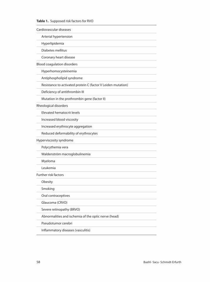
58 Buehl · Sacu · Schmidt-Erfurth
Table 1. Supposed risk factors for RVO
Cardiovascular diseases
Arterial hypertension
Hyperlipidemia
Diabetes mellitus
Coronary heart disease
Blood coagulation disorders
Hyperhomocysteinemia
Antiphospholipid syndrome
Resistance to activated protein C (factor V Leiden mutation)
Deficiency of antithrombin III
Mutation in the prothrombin gene (factor II)
Rheological disorders
Elevated hematocrit levels
Increased blood viscosity
Increased erythrocyte aggregation
Reduced deformability of erythrocytes
Hyperviscosity syndrome
Polycythemia vera
Waldenström macroglobulinemia
Myeloma
Leukemia
Further risk factors
Obesity
Smoking
Oral contraceptives
Glaucoma (CRVO)
Severe retinopathy (BRVO)
Abnormalities and ischemia of the optic nerve (head)
Pseudotumor cerebri
Inflammatory diseases (vasculitis)
Retinal Vein Occlusions 59
hemorrhages are flame-shaped, and dot and punctate hemorrhages are interspersed
and indicate involvement of the deeper retinal layers. Bleedings may be extensive,
erupting through the internal limiting membrane to form a preretinal hemorrhage
or extending into the vitreous. Typically, the entire venous tree is tortuous, engorged,
dilated, and dark. The retina appears edematous, particularly in the posterior pole,
and cotton-wool patches (soft exudates) are often present.
Hemicentral Vein Occlusion (HRVO)
In approximately 20% of eyes, the branch retinal veins draining the superior and infe-
rior halves of the retina enter the lamina cribrosa separately before joining to form a
single central retinal vein. HRVO is an occlusion of one of these dual trunks of the
central retinal vein within the nerve. HRVO therefore involves the venous drainage
from approximately half of the retina, either the superior or the inferior retina. Three
factors that were significantly associated with this type of occlusion compared to con-
trol were systemic hypertension, diabetes mellitus, and glaucoma.
Branch Retinal Vein Occlusion (BRVO)
BRVO typically occurs at the intersection of a branch retinal artery with a vein. It is
generally less visually disabling than CRVO or HRVO. Most BRVOs involve veins
located temporal to the optic disc; there are significantly more vein-posterior than
vein-anterior crossings in the superotemporal than the inferotemporal quadrant,
and vein-posterior crossings are more likely to be obstructed than vein-anterior
crossings.
Based on the amount of capillary non-perfusion (ischemic index) present on the
fluorescein angiogram, Magargal and co-workers classified BRVO, in a manner simi-
lar to that for CRVO, into three types: (1) hyperpermeable (non-ischemic), (2) inde-
terminate, and (3) ischemic. The ischemic index is the percentage of non-perfused
retina based on the amount of retina that is obstructed, rather than the entire retina.
Hayreh and co-workers categorized BRVO as mild, moderate, and marked, based on
the degree of capillary non-perfusion seen angiographically. As in CRVO, there is a
spectrum of capillary non-perfusion in BRVO, ranging from little, if any, non-perfu-
sion to extensive or almost complete non-perfusion. It is probably most clinically use-
ful to classify eyes as non-ischemic and ischemic because neovascularization generally
occurs only in the ischemic cases. The key to the diagnosis lies in the unilateral and
segmental distribution of the ophthalmoscopic findings. This distribution of findings
distinguishes BRVO from other disorders involving hemorrhage, cotton-wool spots,
and retinal edema. Visual field defects range from relative to absolute scotomata and
peripheral depression in the involved corresponding segment. Eyes with branch reti-
nal vein obstruction and retinal capillary non-perfusion can develop neovasculariza-
tion of the retina and/or disc.
Due to the compromised function of the central retina, CRVO, HRVO and major
BRVO are usually characterized by a sudden onset of clinical symptoms. Patients
60 Buehl · Sacu · Schmidt-Erfurth
complain about decreased visual acuity and visual field defects. Typically, these com-
plaints are more severe in the morning, caused by the increased venous pressure
(lower head position) during sleep and symptoms may disappear entirely during the
day. Floaters and metamorphopsia are further typical complaints. Weeks following
the initial event, a second occlusion may lead to a further decline in visual acuity.
Other than CRVO and major BRVO, peripheral BRVO may remain unnoticed until
complications occur.
Typical early complications of RVO with macular involvement include macular
edema, macular non-perfusion and vitreous hemorrhage. Ophthalmoscopically, cot-
ton-wool spots and lipid deposits in the retinal nerve fiber layer as well as tortuositas
and dilatation of retinal veins in the affected area are common findings [4, 21].
The natural history of eyes with CRVO, particularly in older patients presenting
with reduced visual acuity, is usually poor. In the Central Retinal Vein Occlusion
Study, 16% of eyes developed iris and/or angle neovascularization, a total of 34% con-
verted to the ischemic type of the disease. After 3 years of follow-up, 58% of eyes –
especially in patients who initially present with poor visual acuity – will have vision
worse than 20/200 [2].
The prognosis regarding visual acuity after BRVO depends mostly on the localiza-
tion of the occlusion [14]. A study by the branch vein occlusion group found a spon-
taneous increase in visual acuity after BRVO by two or more lines (logMAR scale) in
37%, but only in 17% a decrease of 2 or more lines [1]. Subsequently, a mean increase
in visual acuity of 2.3 lines was documented after 3 years. 34% of patients attained
a final visual acuity of 20/40 or better, only 23% a final visual acuity below 20/200.
Reasons for a recovery of visual acuity are resolution of intraretinal bleedings and
cotton-wool spots, the development of collateral vessels and capillary compensation,
leading to restored blood flow and resorption of retinal edema [14, 16]. The severity
of the occlusion and extent of ischemia are important prognostic factors for the natu-
ral course of visual acuity after RVO [14].
Differentiation between ischemic and non-ischemic RVO is not always easy and
usually cannot be made by ophthalmoscopy alone. In addition, slit-lamp examination
and fluorescein angiography are required [3]. The only definite proof of ischemic RVO
is the occurrence of neovascular complications, including vascular proliferation, vit-
reous hemorrhage, rubeosis iridis and secondary (neovascular) glaucoma (‘100-day
glaucoma’). Typically, venous filling is delayed (>20 s) by fluorescein angiography.
Retinal neovascularization only occurs in the ischemic type of RVO in an estimated
two thirds of cases. It has been shown to occur in 36% of eyes with a non-perfusion
area larger than 5 optic disc diameters [1, 3].
Long-standing RVO are characterized by less or no residual intraretinal blood
and resolution of cotton-wool spots. While there is less tortuositas of vessels in the
affected segment, shunt vessels (collaterals) can frequently be seen. Macular edema
can persist or may leave neurosensory atrophy with irregularities of the pigment epi-
thelium after resorption.
Retinal Vein Occlusions 61
Treatment of RVO
During the past decades, several treatment methods have been described for RVO.
Most therapies focus on eliminating late complications and vision-disturbing effects
of RVO. Overall, there is still only little evidence for the efficacy of specific treatment
options due to lack of large randomized, prospective studies. In general, therapeu-
tic strategies can be divided into acute interventions (within the first 3 months after
RVO) and prophylactic measures aiming at reducing the risk of late complications.
Besides, any underlying non-ocular disease should be diagnosed and treated, if pos-
sible. Because of the complex pathogenesis, treatment and prophylaxis of RVO is an
interdisciplinary task and all patients with acute RVO should be referred to a special-
ist for internal medicine.
Acute Interventions
The aim of early therapeutic measures should be to improve the perfusion of the
affected vessel and to reduce macular edema. Rheological therapy has long been con-
sidered to be the first-line treatment in acute RVO.
Rheological Therapy
Hemodilution can be performed as hyper- or isovolemic hemodilution. The latter
can be controlled more easily and allows for significantly lower hematocrit values in
the capillary bed. Isovolemic hemodilution is performed by simultaneous venesec-
tion and replacement of the lost volume by a plasma substitute, usually hydroxyethyl
starch or dextran, leading to reduced blood viscosity and improving retinal blood
flow [25]. Several authors described significantly better visual outcomes in treated
patients [22–24]. However, these results have been discussed controversially because
of the small study populations and because combined therapies were applied in some
of the studies. Nevertheless, isovolemic hemodilution is accepted by several special-
ists as a first-line treatment within the first 8 weeks after BRVO, as it increases retinal
perfusion and may prevent capillary closure and progressive retinal ischemia. The
therapy is usually well tolerated; side effects include headache, dyspnea, deep vein
thrombosis and hypotension [16]. Nevertheless, patients have to be carefully selected
and should not have any severe cardiorespiratory or renal disease. Early treatment
seems to be important in order to reduce the risk of subsequent ischemic compli-
cations. Late treatment does not seem to influence ischemia or prevent secondary
glaucoma, therefore eyes showing established neovascularization will need additional
laser coagulation [25].
Other rheologically active substances which have been tested for treatment of
RVO include troxerutin and pentoxifylline. The first is thought to improve micro-
circulation in capillaries and venules by inhibiting erythrocyte and platelet aggrega-
tion improving erythrocyte deformability [26]. Pentoxifylline leads to vessel dilation
and improves retinal flow [27]. Both substances have been used in the treatment of
62 Buehl · Sacu · Schmidt-Erfurth
peripheral vein occlusions of the extremities. The efficacy of these drugs in patients
with RVO has not sufficiently been proven in prospective studies yet.
Thrombolytic and Coagulation-Inhibiting Therapy
Currently there is only one randomized study comparing the effect of dalteparin and
acetylsalicylate in patients with BRVO. No difference was found between the two anti-
coagulants. Apart from this study, no randomized clinical studies have been published
concerning systemic treatment of BRVO with acetylsalicylate, heparin or intravasal
thrombolysis with tissue plasminogen activator (rTPA). None of these treatments has
been proven to be effective in the treatment of CRVO, and some of them have severe
side effects when being used systemically [28]. One recently published study found
a positive effect of systemic treatment of CRVO and BRVO with low-dose rTPA but
no preventive effect concerning neovascularization [29]. Another case series found
increased visual acuity after systemic treatment of RVOs with enoxaparin, a low-
molecular-weight heparin [30].
Local thrombolysis with rTPA – either by intravitreal injection or, during vitreo-
retinal surgery, by direct injection into the occluded vessel – has been shown to be
effective in patients with CRVO [31, 32]; currently there are no studies on its effec-
tiveness in BRVO. However, treatment with rTPA has serious risks and was associated
with complications, including vitreous hemorrhage and increase in macular edema
[25, 28]. Its application is therefore very limited.
One study found a significant effect of the platelet aggregation inhibitor ticlopi-
dine on visual acuity when administered within the first 3 weeks after vein occlusion
and for a period of 6 months [33]. In general, anticoagulants should not be used with-
out underlying coagulopathy and are contraindicated at the early stage because of the
increased risk of subretinal bleedings.
Laser Treatment
Macular grid laser photocoagulation has been shown to be effective and is a com-
monly used method in patients with RVO. Many studies have been published, inves-
tigating different methods of laser coagulation, mainly macular grid-pattern laser
for treatment of macular edema and peripheral scatter laser photocoagulation for
prophylaxis and treatment of neovascularization. The clinical results vary largely
between the different studies. The largest randomized, prospective trial (Branch Vein
Occlusion Study Group) showed a positive effect of central grid laser on visual acuity
in patients with BRVO [1]. However, other studies found no significant benefit for
patients after central grid laser [34, 35]. In CRVO, grid laser photocoagulation was
shown to decrease macular edema angiographically but not to improve visual acu-
ity in patients with persistent macular edema [2]. It has therefore been largely aban-
doned for macular edema secondary to CRVO. However, grid laser treatment may
be used in BRVO to treat a macular edema, which persists longer than 3–6 months
or with serous retinal detachment [1, 36]. If fluorescein angiography shows macular
Retinal Vein Occlusions 63
non-perfusion, the effect of grid laser therapy is not guaranteed [1]. Grid laser treat-
ment has been shown to be more effective in combination with intravitreal injections
of TA in one study [56]. An argon laser was used by most authors. Grid laser treat-
ment usually is not effective in RVO older than 1 year and in eyes with a VA of 20/200
or worse [37].
Peripheral scatter laser photocoagulation in ischemic (non-perfusion) areas sig-
nificantly reduces the risk of retinal neovascularization and vitreous hemorrhage [2,
38]. The effectiveness of peripheral laser coagulation was demonstrated for eyes with
non-perfusion areas, but also for eyes already showing neovascularization. However,
the effect was greater when treatment was performed early, before neovasculariza-
tion had developed. On the other hand, there are important side effects of scatter
laser coagulation, primarily visual field defects. For that reason it has been proposed
to wait with laser treatment until the first signs of neovascularization are clinically
visible [38].
Another method, which has been suggested by several authors, is to create an
anastomosis between a retinal vein and the choroid. This can be achieved by a neo-
dymium yttrium-aluminum garnet (Nd:YAG) laser spot following intense argon laser
treatment of a retinal vein segment [39]. Alternatively, it can be performed intraop-
eratively during vitrectomy, using an erbium:YAG laser [40]. Success rates of up to
54% have been reported, however a complication rate of 20% has also been described
for laser-induced chorioretinal anastomosis.
Surgical Methods
The aim (and theoretical advantage) of surgical treatment of RVO is to remove the
occlusion itself and to restore normal retinal blood flow. One method, which has
been employed by several authors, is arteriovenous dissection (sheathotomy). Its pur-
pose is to dissect the retinal artery from the adjacent vein in the area surrounding the
vascular occlusion [41]. The procedure requires vitrectomy, followed by an incision
of the adventitial sheath adjacent to the arteriovenous crossing and separation of the
adhesions. Several studies have shown a positive effect of sheathotomy on visual acu-
ity [41–49]. Different surgical techniques were used and surgery was combined with
additional application of intravitreal triamcinolone, tissue plasminogen activator
or other treatment methods in some of these (non-randomized) studies. Therefore,
the effect of sheathotomy alone remains unclear. One study found no advantage of
sheathotomy over treatment of BRVO with intravitreal TA [43].
The vitreous and the intact vitreoretinal surface are supposed to play a role in
the development of neovascularization [25]. It has been postulated that vitrectomy
alone with surgical detachment of the posterior hyaloid may be more relevant for the
positive effect on visual acuity than arteriovenous dissection itself [45, 46, 50]. One
study found no advantage of sheathotomy over sole vitrectomy [45]. Another study
found no advantage of combined vitrectomy and intravitreal TA over vitrectomy
alone [51].
64 Buehl · Sacu · Schmidt-Erfurth
An alternative surgical method which has been shown to improve visual acuity
after RVO with persisting macular edema, is combined vitrectomy and peeling of the
internal limiting membrane [52–54]. However, because of the lack of randomized
prospective studies, the rationale why vitreoretinal surgery reduces the symptoms of
BRVO remains unclear.
Corticosteroids
Corticosteroids lead to reduced permeability of the affected retinal vasculature and
offer an anti-inflammatory and angiostatic effect, therefore reducing macular edema
and the associated chronic damage to photoreceptors [55]. Several studies have shown
a positive effect of intravitreal injections of triamcinolone acetonide (TA) [56–68].
In vitro, corticosteroids also inhibit VEGF expression and may thus prevent neovas-
cularization and reduce the VEGF-mediated increase in retinal capillary permeabil-
ity [25]. Unfortunately, most of the clinical studies were not randomized or did not
distinguish between different types (ischemic/non-ischemic) of RVO. Some studies
found a superior effect of combined intravitreal TA and macular grid laser photoco-
agulation therapy on visual acuity [56, 69, 70]. However, the positive anti-edematous
effect of TA seems to be temporary, and several retreatments are usually necessary
to avoid reoccurrence of macular edema and associated loss in visual acuity. The
repetitive application in turn raises the risk for side effects of the treatment, mainly
increased intraocular pressure, cataract progression and endophthalmitis [71–73].
Early treatment seems to be important, because chronic macular edema with per-
sistence beyond 1 year often does not respond well to the treatment with intravitreal
steroids or does not lead to an increase in visual acuity [25].
For a longer lasting therapeutic effect, the implantation of an intravitreous dex-
amethasone delivery system (Posurdex) has also been shown to be effective in the
treatment of BRVO [74]. The side effects, mainly intraocular pressure rise, are similar
to repeated intravitreal injections of TA. Another intraocular slow-release device is
already approved in the USA and approval by the European authorities is pending.
Some effects on visual acuity have also been shown for retrobulbar injections of
TA [67, 75–78], however these were less reliable than after intravitreal application of
TA. Systemic application of steroids has also been reported to reduce macular edema
and improve visual acuity, but should – because of the possible side effects – only be
considered for younger patients with concurrent papillary edema or with an inflam-
matory component, especially in patients with systemic vasculitic disorder. However,
currently there are only reports on the effectiveness of systemic steroid therapy in
patients with CRVO [28], but no data regarding its potential usefulness in the treat-
ment of BRVO.
Anti-VEGF Therapy
RVO leads to increased expression of VEGFs by retina and retinal pigment epithe-
lium and an increased release into the vitreous, causing neovascularization, vascular
Retinal Vein Occlusions 65
hyperpermeability with subsequent breakdown of the blood retina barrier and thus
macular edema. All three currently available VEGF inhibitors (ranibizumab, beva-
cizumab, pegaptanib) have been applied successfully in RVO. However, many pro-
spective, randomized studies evaluating these agents against each other or a control
group are still ongoing and the results have not been published yet. Therefore any use
of anti-VEGF agents in this indication has to be considered as off-label. Most of the
published studies used different doses (1.25–2.5 mg) of bevacizumab (Avastin®). In
all of these series visual acuity increased and macular thickness was reduced for 3–9
weeks after the treatment [79]. However, most trials did not differentiate between
different types of RVO. Recently, a number of studies focusing on the effect of beva-
cizumab on BRVO have been published, also demonstrating a significant increase
in visual acuity and regression of the macular edema [80–90]. Encouraging results
have also been published for anti-VEGF treatment of CRVO [79, 91–94]. It became
obvious that not only central visual acuity, but also the overall retinal sensitivity
improved significantly after anti-VEGF therapy [86]. One group demonstrated that
patients who respond well to the initial injection of bevacizumab are most likely to
achieve a long-term benefit from the treatment [85]. Two studies found a correlation
between the intraocular VEGF concentrations and the severity of macular edema as
well as a significant decrease of these factors in eyes treated with bevacizumab [95,
96]. Since central retinal thickness measurements correlated significantly with VEGF
concentrations, optical coherence tomography (OCT) appears to offer an excellent
biomarker for disease activity [95]. In many cases of recurrent macular edema after
the first treatment, retreatment with bevacizumab again led to a reduction of the
macular thickness. Altogether, there was a mean increase in visual acuity of more
than three lines [16]. Similar results have been found for the treatment of BRVO with
ranibizumab in the first published study [97]. In general, intravitreal treatment with
VEGF inhibitors seems to have only a low risk of complications. The most common
side effects include hyposphagma and conjunctival hyperemia. The disadvantage of
anti-VEGF therapy is – similar to the treatment with corticosteroids – the short dura-
tion of the drug effect, usually requiring several retreatments to maintain the positive
effects on macular edema and visual acuity. The effect of a single injection seems to
last 6–8 weeks (fig. 1). Most authors used 2–3 injections over the first 5–6 months
[80]. Also patients who do not respond well to laser therapy seem to benefit from
intravitreal anti-VEGF therapy [80]. Two randomized, multicenter phase III studies
of ranibizumab (Lucentis) (BRAVO study, CRUISE study) show early and sustained
improvement in vision in patients with BTVO and CRVO [Retina Congress 2009;
publication pending]. However, the condition of central macular edema does not
necessarily reflect the overall ischemic situation of the entire retina and despite the
absence of clinically significant macular edema, neovascularization of the disc may
occur and lead to vitreous hemorrhage. These neovascular complications appear to
be retarded under anti-VEGF therapy, but may still occur with a prolonged pathoge-
netic development.
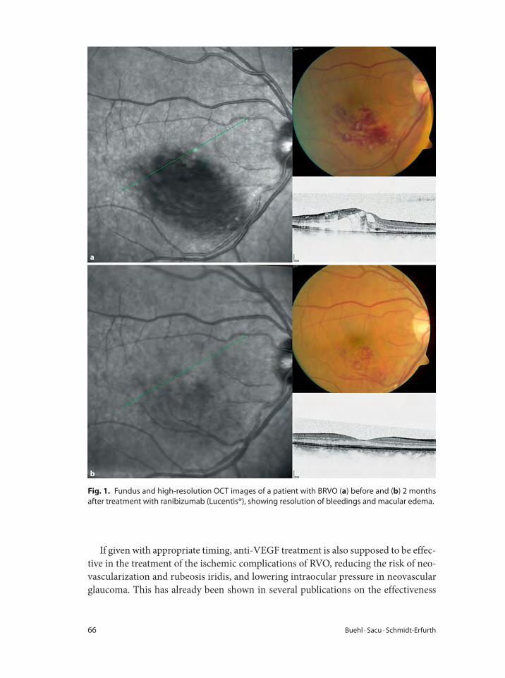
66 Buehl · Sacu · Schmidt-Erfurth
If given with appropriate timing, anti-VEGF treatment is also supposed to be effec-
tive in the treatment of the ischemic complications of RVO, reducing the risk of neo-
vascularization and rubeosis iridis, and lowering intraocular pressure in neovascular
glaucoma. This has already been shown in several publications on the effectiveness
a
b
Fig. 1. Fundus and high-resolution OCT images of a patient with BRVO (a) before and (b) 2 months
after treatment with ranibizumab (Lucentis®), showing resolution of bleedings and macular edema.
Retinal Vein Occlusions 67
of anti-VEGF therapy in late complications of other retinal vascular diseases, mainly
in diabetic retinopathy [98, 99]. Bevacizumab has also been applied intracamerally to
successfully suppress neovascular glaucoma and rubeosis iridis [100].
Prophylactic Measures
In the late stadium of RVO the main focus should be directed to the prevention of
secondary glaucoma. Ischemic areas larger than 5 optic disc diameters are associated
with a distinctly increased risk of neovascular complications and should therefore be
treated [1, 38]. Randomized studies have shown that peripheral scatter laser coagula-
tion of non-perfusion areas can lead to a significant reduction of neovascular prolif-
erations in those cases [38]. Consequently, follow-up examinations at short intervals
are mandatory in all cases of RVO to detect non-perfusion areas in time. Future stud-
ies will have to show whether the use of anti-VEGF agents may perhaps replace the
preventive effect of peripheral laser coagulation.
As mentioned before, prophylactic measures also include investigation and treat-
ment of all associated risk factors, mainly arterial hypertension, diabetes mellitus,
blood lipid and hematological disorders, in order to reduce the risk of a second event
in the same or the contralateral eye.
Conclusion
Although several treatment methods for RVO have been shown to be effective, there
is still a lack of large prospective, randomized studies for most therapies. The com-
plex genesis of the disease with numerous risk factors and the variable progression,
ranging from spontaneous recovery to exacerbation with severe ischemic complica-
tions, makes it difficult to find a common treatment scheme for BRVO. Laser coagu-
lation is effective in the treatment of macular edema and neovascularization, but not
in all cases, and it has significant functional side effects. Favorable results were also
found for treatment with intravitreal triamcinolone; however, the effect is temporary
and associated with severe ocular side effects. The approval of slow-release implants
may improve the prospective of this strategy. The results for surgical treatment of
RVO lack sufficient evidence, are largely variable and not appropriated per se to be
applied in larger populations. The recently published data on intravitreal treatment
with anti-VEGF substances is encouraging and gives hope for a further step ahead
in the treatment of this frequent sight-threatening disease. Further studies will have
to show whether the positive short-term effects of anti-VEGF therapy on macular
edema and visual performance are only transient or may finally lead to a long-lasting
stabilization and persistent improvement in visual acuity. The main problem is the
short therapeutic effect of a single intravitreal injection. However, in contrast to other
substances such as intravitreal TA, treatment with VEGF inhibitors has been shown
to have remarkably few side effects even after many repeated injections. Perhaps
68 Buehl · Sacu · Schmidt-Erfurth
1 Branch Vein Occlusion Study Group: Argon laser
photocoagulation for macular edema in branch vein
occlusion. Am J Ophthalmol 1984;98:271–282.
2 Central Vein Occlusion Study Group: Natural his-
tory and clinical management of central retinal vein
occlusion. Arch Ophthalmol 1997;115:486–491.
3 Parodi MB, Bandello F: Branch retinal vein occlu-
sion: classification and treatment. Ophthalmologica
2009;223:298–305.
4 Klein R, Klein BE, Moss SE, Meuer SM: The epide-
miology of retinal vein occlusion: the Beaver Dam
Eye Study. Trans Am Ophthalmol Soc 2000;98:133–
141.
5 Noma H, Funatsu H, Yamasaki M, et al: Pathogenesis
of macular edema with branch retinal vein occlu-
sion and intraocular levels of vascular endothelial
growth factor and interleukin-6. Am J Ophthalmol
2005;140:256–261.
6 Kumar B, Yu DY, Morgan WH, Barry CJ, Constable
IJ, McAllister IL: The distribution of angioarchitec-
tural changes within the vicinity of the arterio-
venous crossing in branch retinal vein occlusion.
Ophthalmology 1998;105:424–427.
7 Mitchell P, Smith W, Chang A: Prevalence and asso-
ciations of retinal vein occlusion in Australia. The
Blue Mountains Eye Study. Arch Ophthalmol 1996;
114:1243–1247.
8 Cugati S, Wang JJ, Rochtchina E, Mitchell P: Ten-
year incidence of retinal vein occlusion in an older
population. The Blue Mountains Eye Study. Arch
Ophthalmol 2006;124:726–732.
9 David R, Zangwill L, Badarna M, Yassur Y: Epidemi-
ology of retinal vein occlusion and its association
with glaucoma and increased intraocular pressure.
Ophthalmologica 1998;197:69–74.
10 Bearelly S, Fekrat S: Controversy in the manage-
ment of retinal venous occlusive disease. Int Oph-
thalmol Clin 2004;44:85–102.
11 Weinberg D, Dodwell DG, Fern SA: Anatomy of
arteriovenous crossings in branch retinal vein occlu-
sion. Am J Ophthalmol 1990;109:298–302.
12 Scott IU: Vitreoretinal surgery for complications of
branch retinal vein occlusion. Curr Opin Ophthal-
mol 2002;13:161–166.
13 Feist RM, Ticho BH, Shapiro MJ, Farber M: Branch
retinal vein occlusion and quadratic variation in
arteriovenous crossings. Am J Ophthalmol 1992;
113:664–668.
14 Christoffersen NL, Larsen M: Pathophysiology and
hemodynamics of branch retinal vein occlusion.
Ophthalmology 1999;106:2054–2062.
15 Janssen MC, den Heijer M, Cruysberg JR, Woller-
sheim H, Bredie SJ: Retinal vein occlusion: a form of
venous thrombosis or a complication of atheroscle-
rosis? A meta-analysis of thrombophilic factors.
Thromb Haemost 2005;93:1021–1026.
16 Rehak J, Rehak M: Branch retinal vein occlusion:
pathogenesis, visual prognosis, and treatment
modalities. Curr Eye Res 2008;33:111–131.
17 Ingerslev J: Thrombophilia: a feature of importance
in retinal vein thrombosis? Acta Ophthalmol Scand
1999;77:619–621.
18 Tekeli O, Gürsel E, Buyurgan H: Protein C, protein
S and antithrombin III deficiencies in retinal vein
occlusion. Acta Ophthalmol Scand 1999;77:628–
630.
19 Tost F: Venous branch occlusion due to APC resis-
tance. Ophthalmologe 1999;96:777–780.
20 Hayreh SS: Prevalent misconceptions about acute
retinal vascular occlusive disorders. Prog Retin Eye
Res 2005;24:493–519.
21 Barbazetto IA, Schmidt-Erfurth UM: Evaluation of
functional defects in branch retinal vein occlusion
before and after laser treatment with scanning laser
perimetry. Ophthalmology 2000;107:1089–1098.
22 Chen HC, Wiek J, Gupta A, et al: Effect of isovolae-
mic haemodilution on visual outcome in branch
retinal vein occlusion. Br J Ophthalmol 1998;82:162–
167.
23 Hansen LL, Wiek J, Arntz R: Randomized study of
the effect of isovolemic hemodilution in retinal
branch vein occlusion (in German). Fortschr Oph-
thalmol 1988;85:514–516.
combination of anti-VEGF therapy with other forms of treatment may reduce the
number of required intravitreal injections. Prospective randomized trials evaluating
different compounds and different regimen are ongoing. With respect to the mul-
tifactorial pathophysiology, including vascular and hematologic events as well as
angiogenic and inflammatory stimuli, and the chronic nature of the disease, com-
bined approaches may be required for an optimized management.
References
Retinal Vein Occlusions 69
24 Poupard P, Eledjam JJ, Dupeyron G, et al: Role of
acute normovolemic hemodilution in treating reti-
nal venous occlusions (in French). Ann Fr Anesth
Reanim 1986;5:229–233.
25 Shahid H, Hossain P, Amoaku WM: The manage-
ment of retinal vein occlusion: is interventional
ophthalmology the way forward? Br J Ophthalmol
2006;90:627–639.
26 Glacet-Bernard A, Coscas G, Chabanel A, et al: A
randomized, double-masked study on the treatment
of retinal vein occlusion with troxerutin. Am J
Ophthalmol 1994;118:421–429.
27 De Sanctis MT, Cesarone MR, Belcaro G, et al:
Treatment of retinal vein thrombosis with pentoxi-
fylline: a controlled, randomized trial. Angiology
2002;53(suppl 1):35–38.
28 Sharma A, D’Amico DJ: Medical and surgical man-
agement of central retinal vein occlusion. Int Oph-
thalmol Clin 2004;44:1–16.
29 Hattenbach LO, Friedrich Arndt C, Lerche R, et al:
Retinal vein occlusion and low-dose fibrinolytic
therapy: a prospective, randomized, controlled mul-
ticenter study of low-dose recombinant tissue plas-
minogen activator versus hemodilution in retinal
vein occlusion. Retina 2009;29:932–940.
30 Steigerwalt RD Jr, Cesarone MR, Belcaro G, et al:
Retinal and orbital venous occlusions treated with
enoxaparin. J Ocul Pharmacol Ther 2008;24:421–
426.
31 Lahey JM, Fong DS, Kearney J: Intravitreal tissue
plasminogen activator for acute central retinal vein
occlusion. Ophthalmic Surg Lasers 1999;30;427–
434.
32 Weiss JN, Bynoe LA: Injection of tissue plasmino-
gen activator into a branch retinal vein in eyes with
central retinal vein occlusion. Ophthalmology 2001;
108:2249–2257.
33 Houtsmuller AJ, Vermeulen JA, Klompe M, et al:
The influence of ticlopidine on the natural course of
retinal vein occlusion. Agents Actions Suppl 1984;
15:219–229.
34 Parodi MB, Saviano S, Bergamini L, Ravalico G:
Grid laser treatment of macular edema in macular
branch retinal vein occlusion. Doc Ophthalmol
1999;97:427–431.
35 Parodi MB, Saviano S, Ravalico G: Grid laser treat-
ment in macular branch retinal vein occlusion.
Graefes Arch Clin Exp Ophthalmol 1999;237:1024–
1027.
36 Parodi MB, DI Stefano G, Ravalico G: Grid laser
treatment for exudative retinal detachment second-
ary to ischemic branch retinal vein occlusion. Retina
2008;28:97–102.
37 McIntosh RL, Mohamed Q, Saw SM, Wong TY:
Interventions for branch retinal vein occlusion: an
evidence-based systematic review. Ophthalmology
2007;114:835–846.
38 Branch Vein Occlusion Study Group: Argon laser
scatter photocoagulation for prevention of neovas-
cularization and hemorrhage in branch vein occlu-
sion. Arch Ophthalmol 1986;104:34–41.
39 McAllister IL, Douglas JP, Constable IJ, et al: Laser-
induced chorioretinal venous anastomosis for non-
ischemic central retinal vein occlusion: evaluation
of the complications and their risk factors. Am J
Ophthalmol 1998;126:219–229.
40 Quiroz-Mercado H, Sanchez-Buenfil E, Guerrero-
Naranjo JL, et al: Successful erbium: YAG laser-
induced chorioretinal venous anastomosis for the
management of ischemic central retinal vein occlu-
sion. A report of two cases. Graefes Arch Clin Exp
Ophthalmol 2001;239:872–875.
41 Osterloh MD, Charles S: Surgical decompression of
branch retinal vein occlusions. Arch Ophthalmol
1988,106:1469–1471.
42 Chung EJ, Freeman WR, Koh HJ: Visual acuity and
multifocal electroretinographic changes after arte-
riovenous crossing sheathotomy for macular edema
associated with branch retinal vein occlusion. Retina
2008,28:220–225.
43 Chung EJ, Lee H, Koh HJ: Arteriovenous crossing
sheathotomy versus intravitreal triamcinolone ace-
tonide injection for treatment of macular edema
associated with branch retinal vein occlusion.
Graefes Arch Clin Exp Ophthalmol 2008;246:967–
974.
44 Garcia-Arumi J, Martinez-Castillo V, Boixadera A,
Blasco H, Corcostegui B: Management of macular
oedema in branch retinal vein occlusion with she-
athotomy and recombinant tissue plasminogen acti-
vator. Retina 2004;24:530–540.
45 Yamamoto S, Saito W, Yagi F, Takeuchi S, Sato E,
Mizunoya S: Vitrectomy with or without arterio-
venous adventitial sheathotomy for macular edema
associated with branch retinal vein occlusion. Am J
Ophthalmol 2004;138:907–914.
46 Kumagai K, Furukawa M, Ogino N, Uemura A,
Larson E: Long-term outcomes of vitrectomy with
or without arteriovenous sheathotomy in branch
retinal vein occlusion. Retina 2007;27:49–54.
47 Avci R, Inan U, Kaderli B: Evaluation of arterio-
venous crossing sheathotomy for decompression of
branch retinal vein occlusion. Eye 2008;22:120–
127.
48 Horio N, Horiguchi M: Effect of arteriovenous
sheathotomy on retinal blood flow and macular
edema in patients with branch retinal vein occlu-
sion. Am J Ophthalmol 2005;139:739–740.
70 Buehl · Sacu · Schmidt-Erfurth
49 Mason J, Feist R, White M Jr, Swanner J, McGwin G
Jr, Emond T: Sheathotomy to decompress branch
retinal vein occlusion: a matched control study.
Ophthalmology 2004;111:540–545.
50 Han DP, Bennett SR, Williams DF, Dev S: Arterio-
venous crossing dissection without separation of
the retina vessels for treatment of branch retinal
vein occlusion. Retina 2003;23:145–151.
51 Uemura A, Yamamoto S, Sato E, Sugawara T, Mita-
mura Y, Mizunoya S: Vitrectomy alone versus vit-
rectomy with simultaneous intravitreal injection of
triamcinolone for macular edema associated with
branch retinal vein occlusion. Ophthalmic Surg
Lasers Imaging 2009;40:6–12.
52 Ma J, Yao K, Zhang Z, Tang X: 25-Gauge vitrectomy
and triamcinolone acetonide-assisted internal limit-
ing membrane peeling for chronic cystoid macular
edema associated with branch retinal vein occlu-
sion. Retina 2008;28:947–956.
53 Mandelcorn MS, Nrusimhadevara RK: Internal lim-
iting membrane peeling for decompression of mac-
ular edema in retinal vein occlusion: a report of 14
cases. Retina 2004;24:348–355.
54 Mandelcorn MS, Mandelcorn E, Guan K, Adatia
FA: Surgical macular decompression for macular
edema in retinal vein occlusion. Can J Ophthalmol
2007;42:116–122.
55 McAllister IL, Vijayasekaran S, Chen SD, Yu DY:
Effect of triamcinolone acetonide on vascular endo-
thelial growth factor and occludin levels in branch
retinal vein occlusion. Am J Ophthalmol 2009;147:
838–846.
56 SCORE Study Research Group: A randomized trial
comparing the efficacy and safety of intravitreal tri-
amcinolone with standard care to treat vision loss
associated with macular edema secondary to branch
retinal vein occlusion: The Standard Care vs.
Corticosteroid for Retinal Vein Occlusion (SCORE)
Study Report 6. Arch Ophthalmol 2009;127:1115–
1128.
57 Avitabile T, Longo A, Reibaldi A: Intravitreal triam-
cinolone compared with macular laser grid photo-
coagulation for the treatment of cystoid macular
edema. Am J Ophthalmol 2005;140:695–702.
58 Ozkiris A, Evereklioglu C, Erkilic K, Dogan H:
Intravitreal triamcinolone acetonide for treatment
of persistent macular edema in branch retinal vein
occlusion. Eye 2007;20:13–17.
59 Jonas JB, Akkoyun I, Kamppeter B, Kreissig I,
Degenring RF: Branch retinal vein occlusion treated
by intravitreal triamcinolone acetonide. Eye 2005;
19:65–71.
60 Cekic O, Chang S, Tseng JJ, et al: Intravitreal triam-
cinolone injection for treatment of macular edema
secondary to branch retinal vein occlusion. Retina
2005;25:851–855.
61 Lee H, Shah GK: Intravitreal triamcinolone as pri-
mary treatment of cystoid macular edema second-
ary to branch retinal vein occlusion. Retina 2005;
25:551–555.
62 Yepremyan M, Wertz FD, Tivnan T, Eversman L,
Marx JL: Early treatment of cystoid macular edema
secondary to branch retinal vein occlusion with
intravitreal triamcinolone acetonide. Ophthalmic
Surg Lasers Imaging 2005;36:30–36.
63 Chen SD, Sundaram V, Lochhead J, Patel CK: Intra-
vitreal triamcinolone for the treatment of ischemic
macular edema associated with branch retinal vein
occlusion. Am J Ophthalmol 2006;141:876–883.
64 Karacorlu M, Ozdemir H, Karacorlu SA: Resolution
of serous macular detachment after intravitreal tri-
amcinolone acetonide treatment of patients with
branch retinal vein occlusion. Retina 2005;25:856–
860.
65 Roth DB, Cukras C, Radhakrishnan R, et al: Intra-
vitreal triamcinolone acetonide injections in the
treatment of retinal vein occlusions. Ophthalmic
Surg Lasers Imaging 2008;39:446–454.
66 Cakir M, Dogan M, Bayraktar Z, et al: Efficacy of
intravitreal triamcinolone for the treatment of
macular edema secondary to branch retinal vein
occlusion in eyes with or without grid laser photo-
coagulation. Retina 2008;28:465–472.
67 Patel PJ, Zaheer I, Karia N: Intravitreal triamcino-
lone acetonide for macular oedema owing to retinal
vein occlusion. Eye 2008;22:60–64.
68 Ozdek S, Deren YT, Gurelik G, Hasanreisoglu B:
Posterior subtenon triamcinolone, intravitreal tri-
amcinolone and grid laser photocoagulation for the
treatment of macular edema in branch retinal vein
occlusion. Ophthalmic Res 2008;40:26–31.
69 Riese J, Loukopoulos V, Meier C, Timmermann M,
Gerding H: Combined intravitreal triamcinolone
injection and laser photocoagulation in eyes with
persistent macular edema after branch retinal vein
occlusion. Graefes Arch Clin Exp Ophthalmol 2008;
246:1671–1676.
70 Parodi MB, Iacono P, Ravalico G: Intravitreal triam-
cinolone acetonide combined with subthreshold
grid laser treatment for macular oedema in branch
retinal vein occlusion: a pilot study. Br J Ophthalmol
2008;92:1046–1050.
71 Jonas JB, Degenring RF, Kreissig I, Akkoyun I,
Kamppeter BA: Intraocular pressure elevation after
intravitreal triamcinolone acetonide injection. Oph-
thalmology 2005;112:593–598.
72 Jonas JB, Degenring RF, Kreissig I, Akkoyun I:
Safety of intravitreal high-dose reinjections of tri-
amcinolone acetonide. Am J Ophthalmol 2004;138:
1054–1055.
Retinal Vein Occlusions 71
73 Scott IU, Flynn HW Jr: Reducing the risk of endo-
phthalmitis following intravitreal injections. Retina
2007;27:10–12.
74 Kuppermann BD, Blumenkranz MS, Haller JA, et al:
Dexamethasone DDS Phase II Study Group: Ran-
domized controlled study of an intravitreous dex-
amethasone drug delivery system in patients with
persistent macular edema. Arch Ophthalmol 2007;
125:309–317.
75 Kawaji T, Takano A, Inomata Y, et al: Trans-Tenon’s
retrobulbar triamcinolone acetonide injection for
macular oedema related to branch retinal vein
occlusion. Br J Ophthalmol 2008;92:81–83.
76 Kawaji T, Hirata A, Awai N, et al: Trans-Tenon ret-
robulbar triamcinolone injection for macular edema
associated with branch retinal vein occlusion
remaining after vitrectomy. Am J Ophthalmol 2005;
140:540–542.
77 Hayashi K, Hayashi H: Intravitreal versus retrobul-
bar injections of triamcinolone for macular edema
associated with branch retinal vein occlusion. Am J
Ophthalmol 2005;139:972–982.
78 Wakabayashi T, Okada AA, Morimura Y, et al:
Trans-Tenon retrobulbar triamcinolone infusion for
chronic macular edema in central and branch reti-
nal vein occlusion. Retina 2004;24:964–967.
79 Prager F, Michels S, Kriechbaum K, et al: Intravitreal
bevacizumab (Avastin) for macular oedema sec-
ondary to retinal vein occlusion: 12-month results
of a prospective clinical trial. Br J Ophthalmol 2009;
93:452–456.
80 Badalà F: The treatment of branch retinal vein
occlusion with bevacizumab. Curr Opin Ophthalmol
2008;19:234–238.
81 Gregori NZ, Rattan GH, Rosenfeld PJ, et al: Safety
and efficacy of intravitreal bevacizumab (Avastin)
for the management of branch and hemiretinal vein
occlusion. Retina 2009;29:913–925.
82 Ahmadi AA, Chuo JY, Banashkevich A, Ma PE,
Maberley DA: The effects of intravitreal bevaci-
zumab on patients with macular edema secondary
to branch retinal vein occlusion. Can J Ophthalmol
2009;44:154–159.
83 Jaissle GB, Leitritz M, Gelisken F, Ziemssen F, Bartz-
Schmidt KU, Szurman P: One-year results after
intravitreal bevacizumab therapy for macular edema
secondary to branch retinal vein occlusion. Graefes
Arch Clin Exp Ophthalmol 2009;247:27–33.
84 Abegg M, Tappeiner C, Wolf-Schnurrbusch U,
Barthelmes D, Wolf S, Fleischhauer J: Treatment of
branch retinal vein occlusion induced macular
edema with bevacizumab. BMC Ophthalmol 2008;
29:8–18.
85 Chung EJ, Hong YT, Lee SC, Kwon OW, Koh HJ:
Prognostic factors for visual outcome after intravit-
real bevacizumab for macular edema due to branch
retinal vein occlusion. Graefes Arch Clin Exp Oph-
thalmol 2008;246:1241–1247.
86 Kriechbaum K, Michels S, Prager F, et al: Intravitreal
Avastin for macular oedema secondary to retinal
vein occlusion: a prospective study. Br J Ophthalmol
2008;92:518–522.
87 Kreutzer TC, Alge CS, Wolf AH, et al: Intravitreal
bevacizumab for the treatment of macular oedema
secondary to branch retinal vein occlusion. Br J
Ophthalmol 2008;92:351–355.
88 Wu L, Arevalo JF, Roca JA, et al: Pan-American
Collaborative Retina Study Group (PACORES):
Comparison of two doses of intravitreal bevaci-
zumab (Avastin) for treatment of macular edema
secondary to branch retinal vein occlusion: results
from the Pan-American Collaborative Retina Study
Group at 6 months of follow-up. Retina 2008;28:212–
219.
89 Rabena MD, Pieramici DJ, Castellarin AA, Nasir
MA, Avery RL: Intravitreal bevacizumab (Avastin)
in the treatment of macular edema secondary to
branch retinal vein occlusion. Retina 2007;27:419–
425.
90 Spandau U, Wickenhauser A, Rensch F, Jonas J:
Intravitreal bevacizumab for branch retinal vein
occlusion. Acta Ophthalmol Scand 2007;85:118–
119.
91 Stahl A, Struebin I, Hansen LL, Agostini HT, Feltgen
N: Bevacizumab in central retinal vein occlusion: a
retrospective analysis after 2 years of treatment. Eur
J Ophthalmol 2010;20:180–185.
92 Guthoff R, Meigen T, Hennemann K, Schrader W:
Comparison of bevacizumab and triamcinolone for
treatment of macular edema secondary to central
retinal vein occlusion – a matched-pairs analysis.
Ophthalmologica 2010;224:126–132.
93 Gregori NZ, Gaitan J, Rosenfeld PJ, Puliafito CA,
Feuer W, Flynn HW Jr, Berrocal AM, Al-Attar L,
Dubovy S, Smiddy WE, Schwartz SG, Lee WH,
Murray TG: Long-term safety and efficacy of intra-
vitreal bevacizumab (Avastin) for the management
of central retinal vein occlusion. Retina 2008;28:
1325–1337.
94 Wu WC, Cheng KC, Wu HJ: Intravitreal triamcino-
lone acetonide vs. bevacizumab for treatment of
macular oedema due to central retinal vein occlu-
sion. Eye 2009;23:2215–2222.
95 Funk M, Kriechbaum K, Prager F, et al: Intraocular
concentrations of growth factors and cytokines in
retinal vein occlusion and the effect of therapy with
bevacizumab. Invest Ophthalmol Vis Sci 2009;50:
1025–1032.
72 Buehl · Sacu · Schmidt-Erfurth
96 Park SP, Ahn JK: Changes of aqueous vascular
endothelial growth factor and pigment epithelium-
derived factor following intravitreal bevacizumab
for macular oedema secondary to branch retinal
vein occlusion. Clin Exp Ophthalmol 2009;37:490–
495.
97 Campochiaro PA, Hafiz G, Shah SM, et al: Rani-
bizumab for macular edema due to retinal vein
occlusions: implication of VEGF as a critical stimu-
lator. Mol Ther 2008;16:791–799.
98 Iliev ME, Domig D, Wolf-Schnurrbursch U, Wolf S,
Sarra GM: Intravitreal bevacizumab (Avastin) in
the treatment of neovascular glaucoma. Am J
Ophthalmol 2006;142:1054–1056.
99 Yazdani S, Hendi K, Pakravan M: Intravitreal beva-
cizumab (Avastin) injection for neovascular glau-
coma. J Glaucoma 2007;16:437–439.
100 Grisanti S, Biester S, Peters S, Tatar O, Ziemssen F,
Bartz-Schmidt KU: Tübingen Bevacizumab Study
Group: Intracameral bevacizumab for iris rubeosis.
Am J Ophthalmol 2006;142:158–160.
Prof. Ursula Schmidt-Erfurth
Department of Ophthalmology and Optometry
Medical University Vienna
Waehringer Guertel 18-20, AT–1090 Vienna (Austria)
E-Mail [email protected]
Bandello F, Battaglia Parodi M (eds): Anti-VEGF.
Dev Ophthalmol. Basel, Karger, 2010, vol 46, pp 73–83
Antivascular Endothelial Growth Factor for Choroidal Neovascularization in Pathologic MyopiaMaurizio Battaglia Parodia � Pierluigi Iaconob � Francesco Bandelloa
aDepartment of Ophthalmology, University Vita-Salute, Scientific Institute San Raffaele, Milan, and bFondazione
G.B. Bietti per l’Oftalmologia, Istituto di Ricovero e Cura a Carattere Scientifico, Rome, Italy
AbstractChoroidal neovascularization (CNV) is the most common vision-threatening macular complication in
pathologic myopia (PM) being detectable in 4–11% of the affected eyes. Treatment of PM-related
CNV is still controversial. Intravitreal injections of antivascular endothelial growth factor molecules
are able to inhibit the vascular endothelial growth factor isoforms and have shown promise in the
treatment of myopic CNV. The present review describes the beneficial effects of this approach both
for subfoveal and juxtafoveal CNV. However, considering the lack of randomized clinical trials and
the relatively short follow-up in most of the studies, a multicentric clinical trial should be necessary
to validate the positive results in the long term. Copyright © 2010 S. Karger AG, Basel
Pathologic myopia (PM) represents one of the major causes of legal blindness in many
developed countries [1], presenting a prevalence of about 2% in the general popula-
tion [2]. PM is defined as an eye having a minimum refractive error of –6 dpt, an
axial length >26 mm, associated with degenerative changes of the retina, choroid and
sclera at the posterior segment [3]. Common findings are posterior staphyloma, cho-
rioretinal atrophy, and lacquer cracks in the Bruch’s membrane [4]. Choroidal neo-
vascularization (CNV) is the most common vision-threatening macular complication
in PM [5], being detectable in 4–11% of the eyes affected by PM [2, 4, 5].
Myopic CNV has typical characteristics, such as small dimensions (usually less than
1 disc diameter), more often subfoveal or juxtafoveal location, complete absence or lim-
ited presence of subretinal fluid, hemorrhage, or hard exudates [4]. Moreover, myo-
pic CNV has specific histopathological features being a type 2 CNV, which is situated
between the sensory retina and retinal pigment epithelium (RPE), and is entirely coated
by RPE. More specifically, PM-related CNV is characterized by a RPE layer on its inner
surface and a RPE inverted layer on its outer surface, creating a twofold RPE layer [6].
74 Battaglia Parodi · Iacono · Bandello
Natural History
The long-term visual prognosis of subfoveal myopic CNV without treatment is poor,
with a visual acuity of 20/200 or less after 5–10 years of follow-up [7]. In particular,
the CNV tends to a progressive enlargement with fibrotic evolution, whereas cho-
rioretinal atrophic changes develop around the myopic CNV especially in older
patients [8–10]. It is noteworthy that patients under the age of 40 have significantly
better visual outcome than patients over the age of 40 [9].
Less data are available regarding the natural history of juxtafoveal myopic CNV. A
retrospective, observational study on 9 patients affected by juxtafoveal myopic CNV
with a mean follow-up of 5.8 years has revealed that final visual acuity was 20/40 in
77% of the cases, but unfortunately the authors do not report the baseline VA values,
making the interpretation of these data difficult [11]. The results have been confirmed
by another investigation involving 11 eyes with juxtafoveal myopic CNV, which, after a
follow-up of 5 years, showed a final VA <5/10 in 45% of the examined cases [12–15].
Treatment Options
Treatment of PM-related CNV is still controversial. During the past decades, laser
photocoagulation has been the only effective means of treating non-subfoveal CNV
associated with PM, but the functional outcome has turned out to be unsatisfactory
in the long term. In the largest published study, the comparison of laser photocoagu-
lation with the natural history showed laser-treated eyes to have a statistically better
visual acuity at 2 years, but this difference was not statistically significant after 5 years
of follow-up [16]. Such late failure is generally due to the typical expansion of the
laser scar, which is seen in 90–100% of the treated eyes [16]. Two limited case series
have reported positive results about photodynamic therapy (PDT) for juxtafoveal
myopic CNV [17, 18].
Despite the relative positive effects of PDT in subfoveal myopic CNV demon-
strated at 12 months by the VIP (Verteporfin in Photodynamic Therapy) study [19],
the 2-year outcomes failed to reveal a statistically significant treatment benefit [20].
Alternative therapies like radiotherapy [21], surgical removal of the CNV [22] and
macular translocation [23] have not been shown to be worth the collateral damage
incurred by them.
In PM, mechanical tissue strain caused by axial length elongation may lead to the
development of choroidal ischemia followed by atrophy of the RPE and overlying
retina and subsequent vascular endothelial growth factor (VEGF) release [4]. The
risk of developing CNV was, thus, found to be greatest in eyes with patchy atrophy
or lacquer cracks [4]. Intravitreal injections of anti-VEGF molecules, such as beva-
cizumab and ranibizumab, are able to inhibit all the VEGF isoforms [24] and have
shown promise in the treatment of myopic CNV.
Anti-VEGF for CNV in Pathologic Myopia 75
Anti-VEGF for Myopic CNV
Several investigators have tried to assess the effects of intravitreal anti-VEGF admin-
istration for the treatment of myopic CNV reporting positive results [25–37]. Both
naive CNV [25–30] and PDT-treated CNV [29–32] have been taken into consider-
ation by a number of surveys. Subfoveal CNV has been analyzed by most of the stud-
ies, but some case series examined also cases presenting juxtafoveal CNV [25, 28,
32]. Intravitreal bevacizumab has been employed by the majority of the investigations
[25–34], whereas only a few studies have made use of ranibizumab [35, 36].
Dosages used varied from 1 to 2.5 mg, and in general the protocol required a
monthly injection for the first 3 months, followed by an as per needed injection for
the subsequent period [25–36]. Overall, the anti-VEGF approach for PM-related
CNV has been demonstrated to offer improved functional and anatomic advantages
with respect to the previous treatment options, but all the studies available in the
current literature are burdened by several limitations, including a limited number of
patients, a short follow-up (3–12 months), the lack of a control group, the variability
in the treatment protocol, and often the retrospective design of the analysis.
Subfoveal Myopic CNV
The results of VIP studies indicated that PDT-treated eyes were more likely to lose
fewer than 8 letters (72%) than placebo-treated eyes (44%) after 1 year, 32% of treated
eyes gaining at least 5 letters with respect to 15% of the placebo-treated eyes [19].
Unfortunately, the results were no longer statistically significant at the second year
of follow-up [20]. Starting from these unsatisfactory results, several authors have
reported interesting data about the use of anti-VEGF drugs in the treatment of
PM-related subfoveal CNV.
All the studies have shown a visual acuity improvement over their follow-ups [25–
37]. Nevertheless, a significant reduction of mean central macular thickness (CMT)
was registered only by a few analyses [29, 30, 32, 37]. This optical coherence tomo-
graphic (OCT) feature seems to be a characteristic finding of myopic CNV, which
generally show a mild degree of exudation [4]. All the studies have described reduc-
tion or stoppage of dye leakage on fluorescein angiography (FA) or CNV size reduc-
tion as an indication of response to therapy [25–37].
Interestingly, a single study has found that patients aged less and greater than 50
achieved a similar visual acuity improvement of about 8 letters [37], showing that
intravitreal bevacizumab is effective also in older patients on the contrary to PDT,
where 50% of older patients compared to 20% of younger patients lose at least 15 let-
ters [38].
Our experience is related to a prospective, single-center, interventional case series
with a planned follow-up of 2 years. 30 eyes of 30 patients were enrolled in the study.
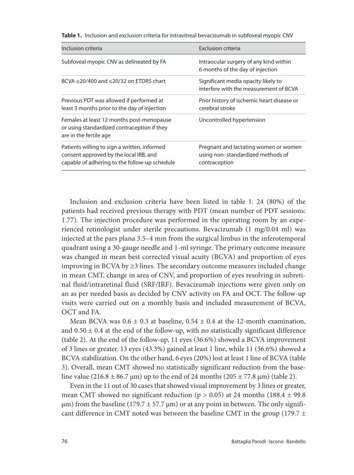
76 Battaglia Parodi · Iacono · Bandello
Inclusion and exclusion criteria have been listed in table 1. 24 (80%) of the
patients had received previous therapy with PDT (mean number of PDT sessions:
1.77). The injection procedure was performed in the operating room by an expe-
rienced retinologist under sterile precautions. Bevacizumab (1 mg/0.04 ml) was
injected at the pars plana 3.5–4 mm from the surgical limbus in the inferotemporal
quadrant using a 30-gauge needle and 1-ml syringe. The primary outcome measure
was changed in mean best corrected visual acuity (BCVA) and proportion of eyes
improving in BCVA by ≥3 lines. The secondary outcome measures included change
in mean CMT, change in area of CNV, and proportion of eyes resolving in subreti-
nal fluid/intraretinal fluid (SRF/IRF). Bevacizumab injections were given only on
an as per needed basis as decided by CNV activity on FA and OCT. The follow-up
visits were carried out on a monthly basis and included measurement of BCVA,
OCT and FA.
Mean BCVA was 0.6 ± 0.3 at baseline, 0.54 ± 0.4 at the 12-month examination,
and 0.50 ± 0.4 at the end of the follow-up, with no statistically significant difference
(table 2). At the end of the follow-up, 11 eyes (36.6%) showed a BCVA improvement
of 3 lines or greater. 13 eyes (43.3%) gained at least 1 line, while 11 (36.6%) showed a
BCVA stabilization. On the other hand, 6 eyes (20%) lost at least 1 line of BCVA (table
3). Overall, mean CMT showed no statistically significant reduction from the base-
line value (216.8 ± 86.7 μm) up to the end of 24 months (205 ± 77.8 μm) (table 2).
Even in the 11 out of 30 cases that showed visual improvement by 3 lines or greater,
mean CMT showed no significant reduction (p > 0.05) at 24 months (188.4 ± 99.8
μm) from the baseline (179.7 ± 57.7 μm) or at any point in between. The only signifi-
cant difference in CMT noted was between the baseline CMT in the group (179.7 ±
Table 1. Inclusion and exclusion criteria for intravitreal bevacizumab in subfoveal myopic CNV
Inclusion criteria Exclusion criteria
Subfoveal myopic CNV as delineated by FA Intraocular surgery of any kind within
6 months of the day of injection
BCVA ≥20/400 and ≤20/32 on ETDRS chart Significant media opacity likely to
interfere with the measurement of BCVA
Previous PDT was allowed if performed at
least 3 months prior to the day of injection
Prior history of ischemic heart disease or
cerebral stroke
Females at least 12 months post-menopause
or using standardized contraception if they
are in the fertile age
Uncontrolled hypertension
Patients willing to sign a written, informed
consent approved by the local IRB, and
capable of adhering to the follow-up schedule
Pregnant and lactating women or women
using non- standardized methods of
contraception
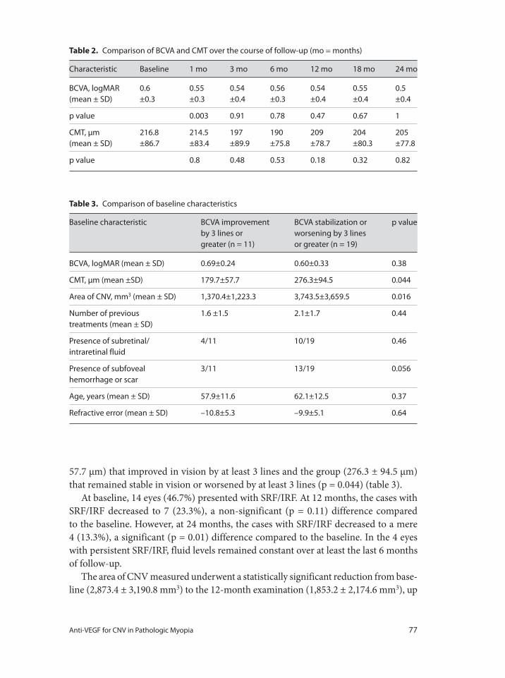
Anti-VEGF for CNV in Pathologic Myopia 77
57.7 μm) that improved in vision by at least 3 lines and the group (276.3 ± 94.5 μm)
that remained stable in vision or worsened by at least 3 lines (p = 0.044) (table 3).
At baseline, 14 eyes (46.7%) presented with SRF/IRF. At 12 months, the cases with
SRF/IRF decreased to 7 (23.3%), a non-significant (p = 0.11) difference compared
to the baseline. However, at 24 months, the cases with SRF/IRF decreased to a mere
4 (13.3%), a significant (p = 0.01) difference compared to the baseline. In the 4 eyes
with persistent SRF/IRF, fluid levels remained constant over at least the last 6 months
of follow-up.
The area of CNV measured underwent a statistically significant reduction from base-
line (2,873.4 ± 3,190.8 mm3) to the 12-month examination (1,853.2 ± 2,174.6 mm3), up
Table 2. Comparison of BCVA and CMT over the course of follow-up (mo = months)
Characteristic Baseline 1 mo 3 mo 6 mo 12 mo 18 mo 24 mo
BCVA, logMAR
(mean ± SD)
0.6
±0.3
0.55
±0.3
0.54
±0.4
0.56
±0.3
0.54
±0.4
0.55
±0.4
0.5
±0.4
p value 0.003 0.91 0.78 0.47 0.67 1
CMT, μm
(mean ± SD)
216.8
±86.7
214.5
±83.4
197
±89.9
190
±75.8
209
±78.7
204
±80.3
205
±77.8
p value 0.8 0.48 0.53 0.18 0.32 0.82
Table 3. Comparison of baseline characteristics
Baseline characteristic BCVA improvement
by 3 lines or
greater (n = 11)
BCVA stabilization or
worsening by 3 lines
or greater (n = 19)
p value
BCVA, logMAR (mean ± SD) 0.69±0.24 0.60±0.33 0.38
CMT, μm (mean ±SD) 179.7±57.7 276.3±94.5 0.044
Area of CNV, mm3 (mean ± SD) 1,370.4±1,223.3 3,743.5±3,659.5 0.016
Number of previous
treatments (mean ± SD)
1.6 ±1.5 2.1±1.7 0.44
Presence of subretinal/
intraretinal fluid
4/11 10/19 0.46
Presence of subfoveal
hemorrhage or scar
3/11 13/19 0.056
Age, years (mean ± SD) 57.9±11.6 62.1±12.5 0.37
Refractive error (mean ± SD) –10.8±5.3 –9.9±5.1 0.64
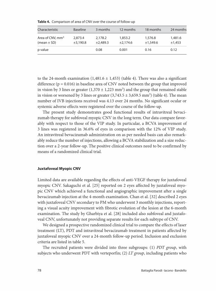
78 Battaglia Parodi · Iacono · Bandello
to the 24-month examination (1,481.6 ± 1,453) (table 4). There was also a significant
difference (p = 0.016) in baseline area of CNV noted between the group that improved
in vision by 3 lines or greater (1,370 ± 1,223 mm3) and the group that remained stable
in vision or worsened by 3 lines or greater (3,743.5 ± 3,659.5 mm3) (table 4). The mean
number of IVB injections received was 4.13 over 24 months. No significant ocular or
systemic adverse effects were registered over the course of the follow-up.
The present study demonstrates good functional results of intravitreal bevaci-
zumab therapy for subfoveal myopic CNV in the long term. Our data compare favor-
ably with respect to those of the VIP study. In particular, a BCVA improvement of
3 lines was registered in 36.6% of eyes in comparison with the 12% of VIP study.
An intravitreal bevacizumab administration on as per needed basis can also remark-
ably reduce the number of injections, allowing a BCVA stabilization and a size reduc-
tion over a 2-year follow-up. The positive clinical outcomes need to be confirmed by
means of a randomized clinical trial.
Juxtafoveal Myopic CNV
Limited data are available regarding the effects of anti-VEGF therapy for juxtafoveal
myopic CNV. Sakaguchi et al. [25] reported on 2 eyes affected by juxtafoveal myo-
pic CNV which achieved a functional and angiographic improvement after a single
bevacizumab injection at the 4-month examination. Chan et al. [32] described 2 eyes
with juxtafoveal CNV secondary to PM who underwent 3 monthly injections, report-
ing a visual acuity improvement with fibrotic evolution of the lesion at the 6-month
examination. The study by Gharbiya et al. [28] included also subfoveal and juxtafo-
veal CNV, unfortunately not providing separate results for each subtype of CNV.
We designed a prospective randomized clinical trial to compare the effects of laser
treatment (LT), PDT and intravitreal bevacizumab treatment in patients affected by
juxtafoveal myopic CNV over a 24-month follow-up period. Inclusion and exclusion
criteria are listed in table 5.
The recruited patients were divided into three subgroups: (1) PDT group, with
subjects who underwent PDT with verteporfin; (2) LT group, including patients who
Table 4. Comparison of area of CNV over the course of follow-up
Characteristic Baseline 3 months 12 months 18 months 24 months
Area of CNV, mm3
(mean ± SD)
2,873.4
±3,190.8
2,178.2
±2,489.3
1,853.2
±2,174.6
1,576.8
±1,549.6
1,481.6
±1,453
p value 0.08 0.001 0.16 0.12
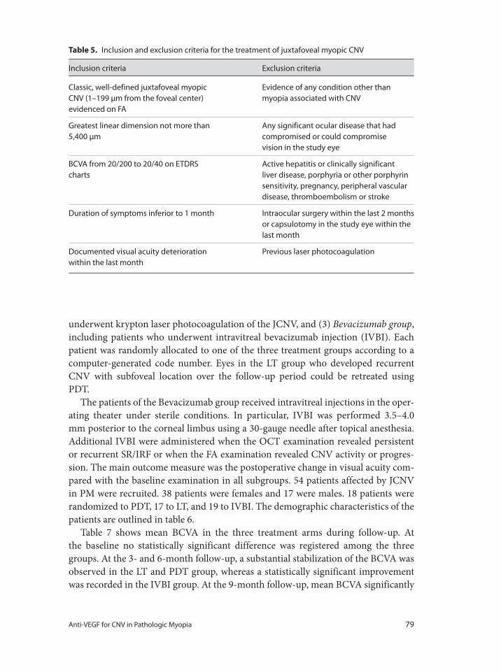
Anti-VEGF for CNV in Pathologic Myopia 79
underwent krypton laser photocoagulation of the JCNV, and (3) Bevacizumab group,
including patients who underwent intravitreal bevacizumab injection (IVBI). Each
patient was randomly allocated to one of the three treatment groups according to a
computer-generated code number. Eyes in the LT group who developed recurrent
CNV with subfoveal location over the follow-up period could be retreated using
PDT.
The patients of the Bevacizumab group received intravitreal injections in the oper-
ating theater under sterile conditions. In particular, IVBI was performed 3.5–4.0
mm posterior to the corneal limbus using a 30-gauge needle after topical anesthesia.
Additional IVBI were administered when the OCT examination revealed persistent
or recurrent SR/IRF or when the FA examination revealed CNV activity or progres-
sion. The main outcome measure was the postoperative change in visual acuity com-
pared with the baseline examination in all subgroups. 54 patients affected by JCNV
in PM were recruited. 38 patients were females and 17 were males. 18 patients were
randomized to PDT, 17 to LT, and 19 to IVBI. The demographic characteristics of the
patients are outlined in table 6.
Table 7 shows mean BCVA in the three treatment arms during follow-up. At
the baseline no statistically significant difference was registered among the three
groups. At the 3- and 6-month follow-up, a substantial stabilization of the BCVA was
observed in the LT and PDT group, whereas a statistically significant improvement
was recorded in the IVBI group. At the 9-month follow-up, mean BCVA significantly
Table 5. Inclusion and exclusion criteria for the treatment of juxtafoveal myopic CNV
Inclusion criteria Exclusion criteria
Classic, well-defined juxtafoveal myopic
CNV (1–199 μm from the foveal center)
evidenced on FA
Evidence of any condition other than
myopia associated with CNV
Greatest linear dimension not more than
5,400 μm
Any significant ocular disease that had
compromised or could compromise
vision in the study eye
BCVA from 20/200 to 20/40 on ETDRS
charts
Active hepatitis or clinically significant
liver disease, porphyria or other porphyrin
sensitivity, pregnancy, peripheral vascular
disease, thromboembolism or stroke
Duration of symptoms inferior to 1 month Intraocular surgery within the last 2 months
or capsulotomy in the study eye within the
last month
Documented visual acuity deterioration
within the last month
Previous laser photocoagulation
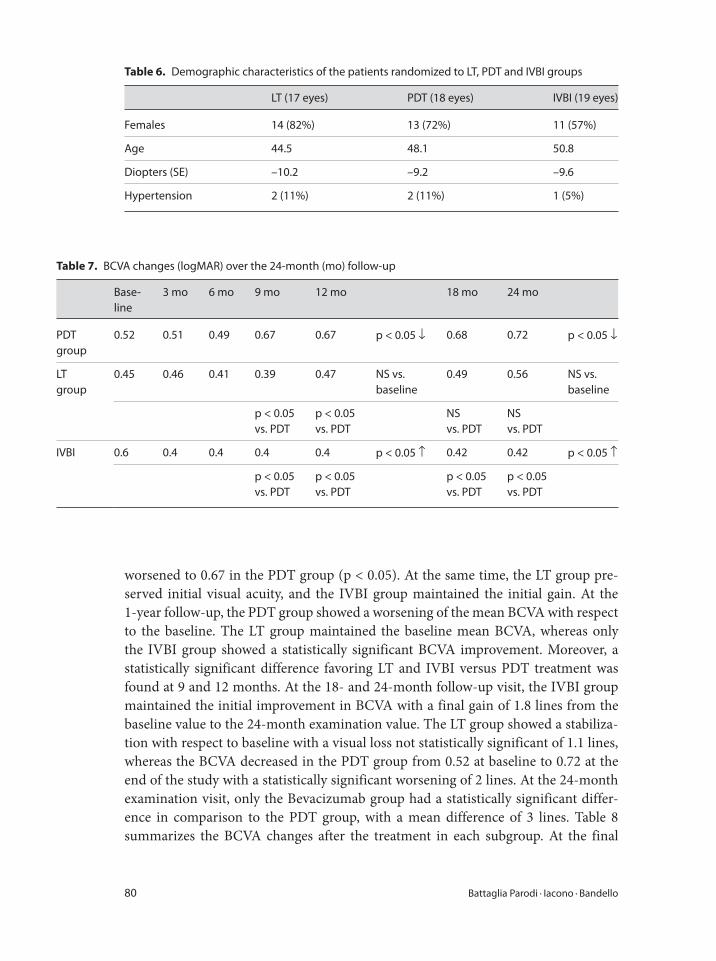
80 Battaglia Parodi · Iacono · Bandello
worsened to 0.67 in the PDT group (p < 0.05). At the same time, the LT group pre-
served initial visual acuity, and the IVBI group maintained the initial gain. At the
1-year follow-up, the PDT group showed a worsening of the mean BCVA with respect
to the baseline. The LT group maintained the baseline mean BCVA, whereas only
the IVBI group showed a statistically significant BCVA improvement. Moreover, a
statistically significant difference favoring LT and IVBI versus PDT treatment was
found at 9 and 12 months. At the 18- and 24-month follow-up visit, the IVBI group
maintained the initial improvement in BCVA with a final gain of 1.8 lines from the
baseline value to the 24-month examination value. The LT group showed a stabiliza-
tion with respect to baseline with a visual loss not statistically significant of 1.1 lines,
whereas the BCVA decreased in the PDT group from 0.52 at baseline to 0.72 at the
end of the study with a statistically significant worsening of 2 lines. At the 24-month
examination visit, only the Bevacizumab group had a statistically significant differ-
ence in comparison to the PDT group, with a mean difference of 3 lines. Table 8
summarizes the BCVA changes after the treatment in each subgroup. At the final
Table 6. Demographic characteristics of the patients randomized to LT, PDT and IVBI groups
LT (17 eyes) PDT (18 eyes) IVBI (19 eyes)
Females 14 (82%) 13 (72%) 11 (57%)
Age 44.5 48.1 50.8
Diopters (SE) –10.2 –9.2 –9.6
Hypertension 2 (11%) 2 (11%) 1 (5%)
Table 7. BCVA changes (logMAR) over the 24-month (mo) follow-up
Base-
line
3 mo 6 mo 9 mo 12 mo 18 mo 24 mo
PDT
group
0.52 0.51 0.49 0.67 0.67 p < 0.05 ↓ 0.68 0.72 p < 0.05 ↓
LT
group
0.45 0.46 0.41 0.39 0.47 NS vs.
baseline
0.49 0.56 NS vs.
baseline
p < 0.05
vs. PDT
p < 0.05
vs. PDT
NS
vs. PDT
NS
vs. PDT
IVBI 0.6 0.4 0.4 0.4 0.4 p < 0.05 ↑ 0.42 0.42 p < 0.05 ↑
p < 0.05
vs. PDT
p < 0.05
vs. PDT
p < 0.05
vs. PDT
p < 0.05
vs. PDT
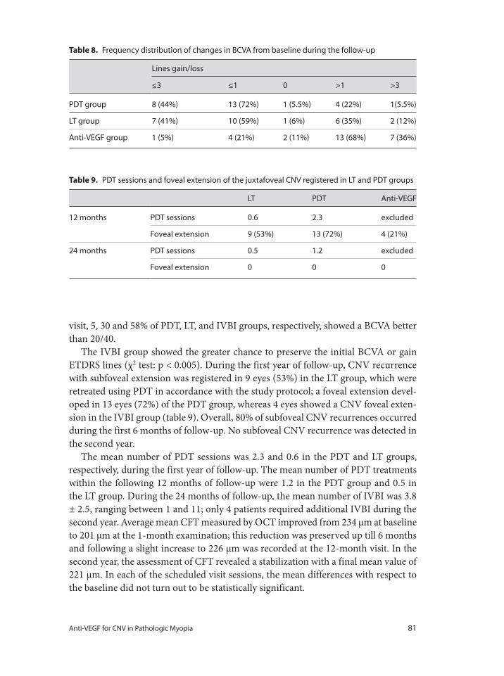
Anti-VEGF for CNV in Pathologic Myopia 81
visit, 5, 30 and 58% of PDT, LT, and IVBI groups, respectively, showed a BCVA better
than 20/40.
The IVBI group showed the greater chance to preserve the initial BCVA or gain
ETDRS lines (χ2 test: p < 0.005). During the first year of follow-up, CNV recurrence
with subfoveal extension was registered in 9 eyes (53%) in the LT group, which were
retreated using PDT in accordance with the study protocol; a foveal extension devel-
oped in 13 eyes (72%) of the PDT group, whereas 4 eyes showed a CNV foveal exten-
sion in the IVBI group (table 9). Overall, 80% of subfoveal CNV recurrences occurred
during the first 6 months of follow-up. No subfoveal CNV recurrence was detected in
the second year.
The mean number of PDT sessions was 2.3 and 0.6 in the PDT and LT groups,
respectively, during the first year of follow-up. The mean number of PDT treatments
within the following 12 months of follow-up were 1.2 in the PDT group and 0.5 in
the LT group. During the 24 months of follow-up, the mean number of IVBI was 3.8
± 2.5, ranging between 1 and 11; only 4 patients required additional IVBI during the
second year. Average mean CFT measured by OCT improved from 234 μm at baseline
to 201 μm at the 1-month examination; this reduction was preserved up till 6 months
and following a slight increase to 226 μm was recorded at the 12-month visit. In the
second year, the assessment of CFT revealed a stabilization with a final mean value of
221 μm. In each of the scheduled visit sessions, the mean differences with respect to
the baseline did not turn out to be statistically significant.
Table 8. Frequency distribution of changes in BCVA from baseline during the follow-up
Lines gain/loss
≤3 ≤1 0 >1 >3
PDT group 8 (44%) 13 (72%) 1 (5.5%) 4 (22%) 1(5.5%)
LT group 7 (41%) 10 (59%) 1 (6%) 6 (35%) 2 (12%)
Anti-VEGF group 1 (5%) 4 (21%) 2 (11%) 13 (68%) 7 (36%)
Table 9. PDT sessions and foveal extension of the juxtafoveal CNV registered in LT and PDT groups
LT PDT Anti-VEGF
12 months PDT sessions 0.6 2.3 excluded
Foveal extension 9 (53%) 13 (72%) 4 (21%)
24 months PDT sessions 0.5 1.2 excluded
Foveal extension 0 0 0
82 Battaglia Parodi · Iacono · Bandello
1 Hotchkiss ML, Fine SL: Pathologic myopia and
choroidal neovascularization. Am J Ophthalmol
1981;91:177–183.
2 Curtin BJ: Basic science and clinical management;
in Curtin BJ (ed): The Myopias. Philadelphia,
Harper & Row, 1985, pp 237–245.
3 Tokoro T: Criteria for diagnosis of pathologic myo-
pia; in Tokoro T (ed): Atlas of Posterior Fundus
Changes in Pathologic Myopia. New York, Springer,
1998, pp 1–2.
4 Grossniklaus HE, Green WR: Pathologic findings in
pathologic myopia. Retina 1992;12:127–133.
5 Avila MP, Weiter JJ, Jalkh AE, et al: Natural history
of choroidal neovascularization in degenerative
myopia. Ophthalmology 1984;91:1573–1581.
6 Grossniklaus HE, Gass JDM: Clinicopathologic cor-
relations of surgically excised type 1 and type 2 sub-
macular choroidal neovascular membranes. Am J
Ophthalmol 1998;126:59–69.
7 Takeshi Y, Kyoko OM, Kenjiro Y, et al: Myopic chor-
oidal neovascularization: a 10-year follow-up. Oph-
thalmology 2003;110:1297–1305.
8 Vongphanit J, Mitchell P, Wang JJ: Prevalence and
progression of myopic retinopathy in an older pop-
ulation. Ophthalmology 2002;109:704–711.
9 Yoshida T, Ohno-Matsui K, Ohtake Y, et al: Long-
term visual prognosis of choroidal neovasculariza-
tion in high myopia. Ophthalmology 2002;109:
712–719.
10 Yoshida T, Ohno-Matsui K, Yasazumi K, et al:
Myopic choroidal neovascularization: a 10-year fol-
low-up. Ophthalmology 2003;110:1297–1305.
11 Bottoni F, Tilanus M: The natural history of juxtafo-
veal and subfoveal choroidal neovascularization in
high myopia. Int Ophthalmol 2001;24:249–255.
12 Hayashi K, Ohno-Matsui K, Yoshida T, Kobayashi
K, Kojima A, Shimada N, Yasuzumi K, Futagami S,
Tokoro T, Mochizuki M: Characteristics of patients
with a favorable natural course of myopic choroidal
neovascularization. Graefes Arch Clin Exp Ophthal-
mol 2005;243:13–19.
13 Brancato R, Menchini U, Pece A, et al: Dye laser
photocoagulation of macular subretinal neovascu-
larization in pathological myopia. A randomized
study of three different wavelengths. Int Ophthalmol
1988;11:235–238.
14 Fardeau C, Soubrane G, Coscas G: Photocoagulation
de néovaisseaux sous-rétinien compliquant la dégé-
nérescence myopique. Bull Soc Ophtalmol Fr 1992;
3:239–242.
15 Soubrane G, Pisan J, Bornert P, Perrenoud F, Coscas
G: Néovaisseaux sous-retiniens de la myopie dégé-
nérative: résultats de la photocoagulation. Bull Soc
Ophtalmol Fr 1986;86:269–272.
16 Secretan M, Kuhn D, Soubrane G, et al: Long-term
visual outcome of choroidal neovascularization in
pathologic myopia: natural history and laser treat-
ment. Eur J Ophthalmol 1997;7:307–316.
17 Cohen SY, Bulik A, Dubois L, Quentel G: Photo-
dynamic therapy for juxtafoveal choroidal neovas-
cularization in myopic eyes. Am J Ophthalmol 2003;
136:371–374.
18 Lam DS, Liu DT, Fan DS, et al: Photodynamic ther-
apy with verteporfin for juxtafoveal choroidal neo-
vascularization secondary to pathologic myopia:
one-year results of a prospective series. Eye 2005;
19:834–840.
19 Verteporfin in Photodynamic Therapy (VIP) Study
Group: Photodynamic therapy of subfoveal choroi-
dal neovascularization in pathologic myopia with
verteporfin: one-year results of a randomized clini-
cal trial - VIP report No. 1. Ophthalmology 2001;
108:841–852.
20 Blinder KJ, Blumenkranz MS, Bressler NM, et al:
Verteporfin therapy of subfoveal choroidal neovas-
cularization in pathologic myopia: two-year results
of a randomized clinical trial – VIP report No. 3.
Ophthalmology 2003;110:667–673.
21 Kobayashi H, Kobayashi K: Radiotherapy for subfo-
veal neovascularization associated with pathologi-
cal myopia: a pilot study. Br J Ophthalmol 2000;84:
761–766.
Our study demonstrates that at 24 months of follow-up, the IVBI offers the greatest
chance to restore the initial VA loss in comparison to PDT or LT in patients affected
by juxtafoveal myopic CNV, with documented VA deterioration. However, consider-
ing the small sample of this study and the relative low frequency of JCNV secondary
to PM, a multicentric clinical trial with a longer follow-up is necessary to validate our
results.
References
Anti-VEGF for CNV in Pathologic Myopia 83
22 Ruiz-Moreno JM, de la Vega C: Surgical removal of
subfoveal choroidal neovascularization in highly
myopic patients. Br J Ophthalmol 2001;85:1041–
1043.
23 Hamelin N, Glacet-Bernard A, Brindeau C, et al:
Surgical treatment of subfoveal neovascularization
in myopia: macular translocation versus surgical
removal. Am J Ophthalmol 2002;133:530–536.
24 Ferrara N, Gerber HP, LeCouter J: The biology of
VEGF and its receptors. Nat Med 2003;9:669–676.
25 Sakaguchi H, Ikuno Y, Kamei M, et al: Intravitreal
injection of bevacizumab for choroidal neovascu-
larisation associated with pathological myopia. Br J
Ophthalmol 2007;91:161–165.
26 Mandal S, Venkatesh P, Sampangi R, et al: Intra-
vitreal bevacizumab (avastin) as primary treatment
for myopic choroidal neovascularization. Eur J
Ophthalmol 2007;17:620–626.
27 Rensch F, Spandau UH, Schlichtenbrede F, et al:
Intravitreal bevacizumab for myopic choroidal neo-
vascularization. Ophthalmic Surg Lasers Imaging
2008;39:182–185.
28 Gharbiya M, Allievi F, Mazzeo L, Gabrieli CB: Intra-
vitreal bevacizumab treatment for choroidal neo-
vascularization in pathologic myopia: 12-month
results. Am J Ophthalmol 2009;147:84–93.
29 Wu PC, Chen YJ: Intravitreal injection of bevaci-
zumab for myopic choroidal neovascularization:
one-year follow-up. Eye 2009;23:356–361.
30 Ikuno Y, Sayanagi K, Soga K, et al: Intravitreal beva-
cizumab for choroidal neovascularization attribut-
able to pathological myopia: one-year results. Am J
Ophthalmol 2009;147:94–100.
31 Ruiz-Moreno JM, Montero JA, Gomez-Ulla F, Ares
S: Intravitreal bevacizumab to treat subfoveal chor-
oidal neovascularisation in highly myopic eyes: one-
year outcome. Br J Ophthalmol 2009;93:448–451.
32 Chan WM, Lai TY, Chan KP, et al: Intravitreal beva-
cizumab (avastin) for myopic choroidal neovascu-
larisation: one-year results of a prospective pilot
study. Br J Ophthalmol 2009;93:150.
33 Hayashi K, Ohno-Matsui K, Shimada N, et al:
Intravitreal bevacizumab on myopic choroidal neo-
vascularization that was refractory to or had
recurred after photodynamic therapy. Graefes Arch
Clin Exp Ophthalmol 2009;247:609–618.
34 Ruiz-Moreno JM, Gomez-Ulla F, Montero JA, et al:
Intravitreous bevacizumab to treat subfoveal chor-
oidal neovascularization in highly myopic eyes:
short-term results. Eye 2009;23:334–348.
35 Lai TY, Chan WM, Liu DT, Lam DS: Intravitreal
ranibizumab for the primary treatment of choroidal
neovascularization secondary to pathologic myopia.
Retina 2009;29:750–756.
36 Silva RM, Ruiz-Moreno JM, Nascimento J, et al:
Short-term efficacy and safety of intravitreal ranibi-
zumab for myopic choroidal neovascularization.
Retina 2008;28:1117–1123.
37 Arias L, Planas L, Prades S, et al: Intravitreal bevaci-
zumab (avastin) for choroidal neovascularisation
secondary to pathologic myopia: six-month results.
Br J Ophthalmol 2008;92:1035–1039.
38 Axer-Siegel L, Ehrlich R, Weimberger D, et al:
Photodynamic therapy of subfoveal choroidal neo-
vascularization in high myopia in a clinical setting:
visual outcome in relation to age at treatment. Am J
Ophthalmol 2004;138:602–607.
Francesco Bandello, MD, FEBO
Professor and Chairman, Department of Ophthalmology
University Vita-Salute Scientific Institute, San Raffaele
Via Olgettina, 60, IT–20132 Milan (Italy)
Tel. +39 02 26432648, Fax +39 02 26483643, E-Mail [email protected]
Bandello F, Battaglia Parodi M (eds): Anti-VEGF.
Dev Ophthalmol. Basel, Karger, 2010, vol 46, pp 84–95
Antivascular Endothelial Growth Factors for Inflammatory Chorioretinal DisordersMaurizio Battaglia Parodia � Pierluigi Iaconob � Frank D. Verbraakc �
Francesco Bandelloa
aDepartment of Ophthalmology, University Vita-Salute, Scientific Institute San Raffaele, Milan; bFondazione G.B.
Bietti per l’Oftalmologia, Istituto di Ricovero e Cura a Carattere Scientifico, Rome, Italy, and cAcademic Medical
Center, Department of Ophthalmology, Amsterdam, The Netherlands
AbstractMacular edema (ME) and choroidal neovascularization (CNV) can complicate the course of several
inflammatory chorioretinal diseases, leading to a severe visual function impairment. The most fre-
quently involved clinical entities include for example multifocal choroiditis, presumed ocular histo-
plasmosis syndrome, Beçhet’s disease, multiple evanescent white dot syndrome, birdshot
chorioretinopathy, acute multifocal posterior placoid pigment epitheliopathy, serpiginous choroidi-
tis, and persistent placoid maculopathy. Results that have reported on antivascular endothelial
growth factor (anti-VEGF) treatment in uveitic patients with CNV or ME have demonstrated positive
results in many cases. However, bearing in mind that it has been proven impossible to perform ran-
domized clinical trials with anti-VEGF in uveitic patients with CNV or ME, further studies with longer
follow-ups are necessary to assess the value of this therapeutic approach.
Copyright © 2010 S. Karger AG, Basel
Macular edema (ME) and choroidal neovascularization (CNV) can complicate the
course of several inflammatory chorioretinal diseases, leading to a severe visual func-
tion impairment. ME can be ascribed to the dysfunction of or the damage to either
the inner or the outer blood-retinal barrier. CNV development can be related to a
number of features, comprising retinal pigment epithelium (RPE) damage and inter-
ruption of Bruch’s membrane. Many cytokines and other molecules involved in the
inflammatory process are also implicated in the pathogenetic mechanisms leading to
a profound alteration of vascular permeability and angiogenesis.
Systemic or topical treatments aiming at reducing the inflammatory reaction often
do not fully control ME and CNV development, and do not allow a recovery of the
visual function. The recent advent of intravitreal antivascular endothelial growth fac-
tor (anti-VEGF) therapy has opened new perspectives for the treatment of ME and
Anti-VEGF for Inflammatory Chorioretinal Disorders 85
CNV of inflammatory origin. Although the evidence supporting the use of anti-VEGF
therapy in inflammatory chorioretinal diseases is not based on controlled trials, the
positive results of a multitude of case series have demonstrated its value. These find-
ings all support the beneficial effects of anti-VEGF molecules in the treatment of exu-
dative complications of inflammatory chorioretinal disorders.
Inflammatory chorioretinal diseases comprise a heterogenous group of disorders.
Some of these diseases are especially prone to the development of CNV, like multifo-
cal choroiditis, presumed ocular histoplasmosis syndrome, Beçhet’s disease, multiple
evanescent white dot syndrome, birdshot chorioretinopathy, acute multifocal poste-
rior placoid pigment epitheliopathy, serpiginous choroiditis, and persistent placoid
maculopathy. Anti-VEGF therapy has been advocated for these diseases to success-
fully control CNV.
Most inflammatory chorioretinal diseases can lead to persistent uveitic cystoid
macular edema (CME). Especially intermediate uveitis is known for this complica-
tion. Anti-VEGF therapy has been used to treat uveitic CME, and has shown promis-
ing results.
Multifocal Choroiditis and Punctate Inner Choriodopathy
Initially described by Nozik and Dorsch [1], multifocal choroiditis (MC) is a chronic
inflammatory disease occurring most frequently in young, myopic, female subjects.
Involvement of the anterior segment is detectable in about 50% of cases, whereas the
posterior segment is invariably affected [1–10]. Typical features of the disorder are
multiple punched-out chorioretinal lesions at the posterior pole and mid-periphery
associated with vitritis. The visual prognosis in patients affected by MC is generally
good, except for cases presenting macular involvement. A marked reduction in visual
acuity (VA) is related to a number of complications, including CME, foveal scarring,
epireretinal membrane, or CNV [1–10]. More specifically CNV has been reported
as the most common cause of visual loss in MC, occurring in 27–32% of the cases
[1–10].
Currently, there are no evidence-based guidelines for therapy of CNV second-
ary to MC. Some studies have described the beneficial effects of corticosteroid and
immunosuppressant treatment, conventional laser treatment of extrafoveal CNV, and
photodynamic therapy (PDT), and surgical excision for the management of sub- and
juxtafoveal CNV [11–18].
Corticosteroid and immunosuppressant therapy have been reported to be benefi-
cial in controlling the inflammation related to MC, but the efficacy in stabilizing the
visual function in eyes with subfoveal CNV is still controversial [5, 8, 11–14]. Surgical
CNV excision seems to offer good results, even though results are burdened with
complications related to surgery, and a high recurrence rate [15, 16]. Small uncon-
trolled case series have reported that PDT can stabilize the visual function in eyes
86 Battaglia Parodi · Iacono · Verbraak · Bandello
with sub- or juxtafoveal CNV, however without achieving a significant VA improve-
ment [17–19].
Promising results have been reported with the use of intravitreal bevacizumab [20–
22]. In particular, a retrospective study of the effect of bevacizumab administered for
CNV not related to age-related macular degeneration also included 12 eyes affected
by MC. The results were encouraging, even though a specific analysis is not allowed
because only the cumulative results have been published [21]. More recently, Fine et
al. [22] have reported on bevacizumab and ranibizumab intravitreal injections in 6
eyes affected by CNV secondary to MC. At the end of the available follow-up (mean
follow-up 42 weeks, range 25–69 weeks), 5 eyes showed an improvement of the visual
function with a final VA values better than 20/30 and reduced activity of CNV lesion.
Only 1 patient experienced a reduction of visual function to 20/400 due to a subfoveal
rip of the RPE. Anti-VEGF treatment will also inhibit the proinflammatory effects of
VEGF present in these eyes.
The role of VEGF in the pathogenesis of CNV secondary to MC is supported by a
recent study by Shimada et al. [23] who demonstrated that VEGF overexpression is
detectable in samples of active CNV obtained after surgical excision. The pathology
of excised neovascularizations did not differ between cases of MC or punctuate inner
choroidopathy. An early neovascular ‘bridging’ between originally separated inflam-
matory and/or neovascular foci may bring about a severe VA loss after intravitreal
bevacizumab therapy, however this is a process that can occur independent of any
treatment, and the relation with the injection remains unclear [24].
Our experience refers to a prospective interventional case series including 10 eyes
of 10 patients with MC-related subfoveal CNV, who underwent intravitreal bevaci-
zumab injection, starting with three monthly injections, followed by retreatment as
needed, with decisions to retreat based on loss of VA of more than 5 letters, and/or
signs of active leakage on optical coherence tomography (OCT), over a 12-month
follow-up. Mean best corrected VA (BCVA) and mean foveal thickness (FT) at the
baseline were 0.58 logMAR (±0.48 SD) and 290 μm (±92 μm SD), respectively. At the
3-month examination, mean BCVA improved to 0.42 ± 0.48 and mean FT decreased
to 268 ± 172 μm SD, whereas at the 12-month examination, mean BCVA was 0.40 ±
0.49 and mean FT was 255 ± 163 μm. Moreover, at the 12-month examination, 83%
of patients showed a final VA better than 20/50, and overall, 9 out of 12 treated eyes
(75%) showed at least one line of BCVA improvement, whereas 3 eyes (25%) revealed
one line of worsening. The mean number of administered injections was 3.8, ranging
from 3 to 7.
All the studies regarding the treatment of MC-related CNV have important limita-
tions, especially the small number of patients and the absence of a control group. MC
is a rare condition which makes the planning of a randomized clinical trial difficult.
However, the reported improvement in VA in patients treated with anti-VEGF injec-
tions seems promising enough to consider this treatment in patients with multifocal
choroiditis and a juxta- or subfoveal CNV.
Anti-VEGF for Inflammatory Chorioretinal Disorders 87
Ocular Histoplasmosis Syndrome
Ocular histoplasmosis syndrome (OHS) is determined by the systemic infection of
Histoplasma capsulatum and occurs in areas where H. capsulatum is endemic. Outside
these endemic areas a similar ocular syndrome is recognized without any evidence
of H. capsulatum and has been called ocular histoplasmosis. OHS is characterized
by small, round, mid-peripheral chorioretinal lesions and peripapillary atrophy [25].
CNV is a well-known complication of OHS, which may lead to metamorphopsia and
severe VA loss.
Fluorescein angiography evaluation of OHS reveals the presence of initially hypo-
fluorescent spots which show staining in the late phases. Indocyanine green angiog-
raphy unveils both hyper- and hypofluorescent spots, which often are not detectable
on clinical examination. CNV secondary to OHS typically occurs during the second
to fifth decade of life and shows a rapid course [25, 26]. Up to 75% of patients with
CNV secondary to OHS experience a decline in vision to 20/200 or worse over the
course of a 2- to 3-year period [27, 28]. Possible treatment options for CNV associ-
ated with OHS include laser photocoagulation, extrafoveal CNV, systemic and intra-
vitreal corticosteroids, PDT, and surgical removal or macular displacement surgery,
but all these treatment modalities have turned out to provide limited results regarding
preservation of VA, sometimes with severe complications, and have shown a high
recurrence rate [29–36].
In the absence of a beneficial treatment for OHS-related CNV, some authors have
administered intravitreal anti-VEGF drugs. Positive functional and anatomical out-
comes have been described in a single case report, with VA improvement from 20/200 to
20/60 over a 1-year follow-up and a single intravitreal injection of bevacizumab [37].
A retrospective interventional case series of 28 patients presenting either juxta- or
subfoveal CNV related to OHS has demonstrated that intravitreal bevacizumab leads to
a VA improvement in 71% eyes and a stabilization in 14% of cases over an average fol-
low-up of 22 weeks [38]. Interestingly, all the patients presenting treatment-naive CNV
stabilized or improved, in contrast to eyes previously treated with PDT or that received
a combined treatment. This finding is consistent with published data about treatment
results of CNV in AMD patients, where PDT-naive patients responded better [39].
Again a randomized controlled trial with a large number of patients would be
needed to prove the effectiveness, safety, and long-term results of this therapy, but the
results of the reported case series seems promising enough to consider this treatment
in patients with OHS and a juxta- or subfoveal CNV.
Behçet’s Disease
Behçet’s disease is a complex systemic inflammatory disease with a severe inflammatory
ocular involvement as one of the major manifestations of the disease. More specifically,
88 Battaglia Parodi · Iacono · Verbraak · Bandello
the macula becomes involved with the development of CME, and, very seldom, a sub-
foveal CNV. A recent article has shown that intravitreal bevacizumab injection can be
beneficial in the treatment of CME secondary to ocular Behçet’s disease. In this investi-
gation, 12 eyes of 11 patients underwent an intravitreal injection of bevacizumab. The
results were encouraging showing a BCVA improvement in 7 eyes (58%), although the
FT and macular volume, measured with OCT, did not reveal a statistically significant
difference with respect to the baseline values [40]. More studies are needed to assess the
value of the anti-VEGF approach in the therapy of ME secondary to Behçet’s disease.
Multiple Evanescent White Dot Syndrome
Multiple evanescent white dot syndrome (MEWDS), originally described by Jampol et
al. [41], is an acute, usually unilateral chorioretinopathy that occurs mostly in young
subjects. Affected patients report blurred vision, photopsia, or visual field defects.
MEWDS is clinically characterized by multiple yellow or white spots throughout the
posterior pole and the peripheral retina, and punctated lesions surrounding the fovea.
Fluorescein angiography shows hyperfluorescence of the yellow spots, with dif-
fuse or patchy staining at the RPE level and possible optic disc staining during the
late phases. Early indocyanine green angiography demonstrates a pattern of hypo-
fluorescent spots which are more apparent and more numerous with respect to the
simple biomicroscopic examination. Such lesions varied in size from 50 to 500 μm
in diameter [42]. Full-field and multifocal electroretinograms disclose dysfunction
at the photoreceptors level [43, 44]. A typical OCT finding is the disruption of the
photoreceptor inner/outer segment junction line which can recover along with the
resolution of the disease [45]. The prognosis is generally good with complete sponta-
neous resolution of the inflammation within 6 months [45]. Nevertheless, subsequent
recurrence, bilateral involvement or CNV development have been reported [46].
Intravitreal anti-VEGF has been employed to treat CNV which may complicate
the course of the disease. In particular, a single case presenting peripapillary CNV
and central subretinal fluid was treated by means of a single intravitreal injection
of ranibizumab, achieving a positive functional and anatomical outcome. BCVA
improved from 20/40 to 20/20, the CNV completely regressed, and the MEWDS find-
ings disappeared over a follow-up of 6 months [47].
Birdshot Chorioretinopathy
The clinical manifestation of birdshot chorioretinopathy (BCR) is characterized by a
posterior segment involvement with vitritis, ‘birdshot’ appearance of scattered, sub-
retinal spots located anywhere in the fundus, but more frequently nasal to the optic
disc and in the inferior retina. The lesions are oval, non-pigmented, creamy yellow,
Anti-VEGF for Inflammatory Chorioretinal Disorders 89
and flat with indistinct margins [48–51]. Macular and optic disc edema, CNV, and
vasculitis may complicate the clinical picture [48–51]. HLA-A29 testing is very useful
to confirm the diagnosis [52].
Electroretinography may reveal a decreased b-wave amplitude and delayed
implicit time, especially in scotopic condition [51]. Fluorescein angiography shows
early hypofluorescence and late hyperfluorescence of the spots. The optic disc usu-
ally reveals a dye leakage or staining, indicating the inflammatory involvement. On
indocyanine green angiography the spots are hypofluorescent throughout the course
of the examination. Many more spots are visible on indocyanine green angiography
than with either biomicroscopy or fluorescein angiography. Very rarely, BCR may be
complicated by the occurrence of a foveal CNV.
A case of BCR complicated by subfoveal CNV has been reported in an interven-
tional case series including CNV forms other than AMD. The patient had previously
received an intravitreous injection of triamcinolone acetonide without beneficial
response and was retreated with intravitreal bevacizumab achieving a regression of
the CNV [21]. An additional case with positive response to intravitreal bevacizumab
was included in another case series [53].
BRC is more frequently complicated by chronic diffuse CME that can extend over
the whole posterior pole. Interestingly, in a case of BRC presenting with CME, an
improvement of BCVA in association with a reduction of the retinal thickness was
reported after a single intravitreal injection of bevacizumab [54]. Experience with
anti-VEGF in this disease is limited and needs further investigation.
Acute Multifocal Posterior Placoid Pigment Epitheliopathy
Acute multifocal posterior placoid pigment epitheliopathy (AMPPPE) is an acute
self-limiting chorioretinal inflammatory disorder characterized by the development
of multifocal, yellow-white, flat, placoid lesions at the level of the RPE at the poste-
rior pole and at the mid-peripheral fundus [55]. The disease usually presents with
an acute loss of vision. During the first weeks the expansion of the placoid lesions
can occur, while new lesions can appear in formerly unaffected areas. This process
continues for a few months with the development of new lesions and the healing of
the older ones. Eventually all the lesions resolve, leading to the typical clinical aspect
characterized by mottling, atrophy, and hyperpigmentation of the RPE, generally
associated with a good functional recovery. The fluorescein angiograms during the
acute phases show early hypofluorescence followed by staining of the lesions in the
later phases. The indocyanine green angiograms reveal a choroidal hypofluorescence
during the whole examination. Generally, AMPPPE has a good long-term prognosis
for VA, although most patients have residual symptoms and paracentral scotomas
[56]. Seldom, AMPPPE can lead to a severe loss of vision due to atrophic macular
changes, subretinal fibrosis and CNV [57–60]. More specifically, CNV is a very rare
90 Battaglia Parodi · Iacono · Verbraak · Bandello
complication of AMPPPE [61, 62]. As a consequence, no proven treatment is cur-
rently available for this complication. A single case report has recently described a
14-year-old girl who developed CNV as a complication of AMPPPE. The patient was
effectively treated with a single intravitreal ranibizumab injection. BCVA improved
from 20/40 to 20/20 through a follow-up of 12 months along with stabilization of the
CNV and progressive reduction of subretinal fluid on OCT [63].
Serpiginous Choroiditis
Serpiginous choroiditis (SC) is a rare, progressive, chronically recurring inflamma-
tory disorder. SC is usually bilateral, with onset between the ages of 30 and 70 years,
and no race or sex predilection [64–67]. SC is characterized by the occurrence of
grayish-yellow lesions at the RPE level, most frequently located close to the optic disc
with peripapillary lesions and fingerlike projections extending outward. In rare cases
the disease takes place in the macula, or in other retinal areas, without peripapil-
lary involvement [67]. The active lesions last from weeks to months and then there
is a spontaneous resolution with pigment loss or clumping and retinochoroidal atro-
phy [68]. Patients affected by SC have typically recurrences at intervals varying from
weeks to years, with extension to the periphery. About one third of patients reveal an
inflammatory reaction in the posterior and the anterior segment [66, 69].
Occasionally, findings of SC may be associated with other manifestations, includ-
ing retinal vasculitis, papillitis, vitritis, branch retinal vein occlusion, serous neuro-
sensory detachment, RPE detachment, and optic disc neovascularization [70–72].
CNV occurs in up to 35% of cases of SC and may be seen at the time of active chor-
oiditis or in between episodes [72–75]. The treatment of the CNV secondary to SC is
still debated, but a recent case report has described a favorable outcome after treat-
ment with intravitreal injection of ranibizumab [76]. Other studies are necessary to
validate the clinical relevance of this therapy.
Persistent Placoid Maculopathy
Persistent placoid maculopathy (PPM) is a new clinical entity characterized by bilat-
eral symmetric macular involvement characterized by a whitish plaque-like lesion at
the level of the RPE centered around the fovea, which is often complicated by CNV
development with disciform evolution [77, 78]. Owing to the rarity of this compli-
cation, currently there is no proven treatment able to manage PPM-related CNV.
Corticosteroids have been tried in a previous report, without halting the progression
toward CNV development and disciform scarring [77, 78].
We have diagnosed and treated a 60-year-old man affected by PPM and bilat-
eral CNV [79]. At presentation, BCVA was 20/800 in the right eye, due to an old
Anti-VEGF for Inflammatory Chorioretinal Disorders 91
fibrovascular scar as a result of the natural evolution of a subfoveal CNV, and 20/32
in the left eye, which hosted a juxtafoveal CNV. The patient was treated with intrav-
itreal injection of 1.25 mg bevacizumab in the left eye. One week later, BCVA in the
treated eye was 20/25 and a partial resolution of the subretinal fluid on OCT could be
noticed. At the 30-day examination, BCVA was still 20/25, with persistence of mini-
mal intraretinal fluid, and some dye leakage on fluorescein angiography. A second
treatment was provided and 1 week later, BCVA improved to 20/20 with complete
resolution of fluid on OCT. Fluorescein angiography revealed the absence of dye leak-
age. The patient has been followed up monthly for 18 months, without registering any
further change on OCT and angiography.
Uveitic Cystoid Macular Edema
CME is the most common cause of significant visual loss in patients affected by
intraocular inflammation [80]. The actual incidence of uveitic CME differs according
to the underlying etiology and inflammation site. In particular, CME has been regis-
tered in about 30% of HLA-B27-positive patients with anterior uveitis, as compared
with 8% of HLA-B27-negative patients [81, 82]. CME is associated more commonly
with more posterior locations of uveitis (intermediate, posterior, and panuveitis) [83].
The pathogenesis of uveitic CME is not completely understood and may result from
dysfunction of and/or damage to either the inner or the outer blood-retinal barrier.
Many inflammatory cytokines and other molecules such as arachidonic acid have
been implicated in the etiology of this common complication, and it is probable that
a combination of different factors concur to the development of this condition [84].
Untreated uveitic CME tends to cause progressive visual loss because of a progres-
sive photoreceptor damage. Medical treatment includes several approaches, such as
topical and systemic non-steroidal anti-inflammatory drugs, corticosteroids (topical,
systemic, transseptal, or intravitreal), systemic carbonic anhydrase inhibitors, and
octreotide [85]. The difficulties in obtaining a favorable functional outcome together
with the resistance to the treatment modalities have led several authors to search for
new therapies. A recent investigation has taken into consideration 13 patients with
CME secondary to different uveitic forms, causing significant visual impairment,
who were resistant to medical therapy including not only initial use of topical and/or
systemic anti-inflammatory drugs, but also to intravitreal injections of triamcinolone
acetonide in the majority of cases [54]. The use of intravitreal bevacizumab achieved
encouraging results, with significant improvement in vision and central retinal thick-
ness in patients with this type of recalcitrant CME. Survival analysis showed that the
probability of any improvement in VA increased progressively starting at 6 weeks and
reached 81% at 14 weeks. These preliminary data suggest that intravitreal bevaci-
zumab may be a beneficial choice for the treatment of recalcitrant uveitic CME, and
more definitive studies into this matter are warranted.
92 Battaglia Parodi · Iacono · Verbraak · Bandello
1 Nozik RA, Dorsch W: A new chorioretinopathy
associated with anterior uveitis. Am J Ophthalmol
1973;76:758–762.
2 Tatar O, Yoeruek E, Szurman P, et al: Effect of beva-
cizumab on inflammation and proliferation in
human choroidal neovascularization. Arch Ophthal-
mol 2008;126:782–790.
3 Dryer RF, Gass JDM: Multifocal choroiditis and
panuveitis. A syndrome that mimics ocular histo-
plasmosis. Arch Ophthalmol 1984;102:1776–1784.
4 Deutsch TA, Tessler HH: Inflammatory pseudohis-
toplasmosis. Ann Ophthalmol 1985;17:451–465.
5 Cantrill HL, Folk JC: Multifocal choroiditis associ-
ated with progressive subretinal fibrosis. Am J Oph-
thalmol 1986;101:170–180.
6 Morgan CM, Schatz H: Recurrent multifocal chor-
oiditis. Ophthalmology 1986;93:1138–1147.
7 Lardenoye CW, Van der Lelij A, de Loos WS, et al:
Peripheral multifocal chorioretinitis: a distinct clin-
ical entity? Ophthalmology 1997;104:1820–1826.
8 Jampol LM, Wiredu A: MEWDS, MFC, PIC, AMN,
AIBSE, and AZOOR: one disease or many? Retina
1995;15:373–378.
Conclusions
Anti-VEGF treatment in uveitic patients has been provided for two reasons: the devel-
opment of CNV and persistent, treatment-resistant ME. Choroidal neovasculariza-
tions in uveitic patients are frequently seen in some diseases entities like MC, but are
a more rare complication in others. In the development of CNV in uveitic patients,
more so than in AMD patients, inflammations play a pivotal role. Anti-inflammatory
drugs, locally or systemically given, can be effective in the treatment of CNV in many
cases, but the response may be too slow, leaving the eye permanently damaged, with
loss of VA, which can be profound in patients with involvement of the fovea. Anti-
VEGF could be a solution for this problem and could be especially helpful in pre-
venting this permanent loss of vision in the acute phase of diseases. This approach is
interesting because it directly acts on the pathogenetic mechanisms of the CNV devel-
opment, eliminating the inflammatory rebound typical of other treatment modalities,
such as laser photocoagulation or PDT.
In some uveitic diseases, like Beçhet’s disease, BCR, SC, adequate immune suppres-
sion, most of the time requiring systemic corticosteroids in combination with other
immune suppressors like methotrexate, azathioprine or mycophenolate, cyclosporine,
is mandatory in addition to intraocular anti-VEGF. While in others, with less inflam-
mation, like OHS or multifocal choroiditis, anti-VEGF could probably be sufficient
to treat the CNV adequately. The role of treatment with anti-VEGF in CME is more
controversial and should probably be reserved for those cases that are unresponsive
to other anti-inflammatory treatments, although here again the anti-inflammatory
effect of anti-VEGF will be beneficial, albeit probably only for a relatively short time.
Results reported on the anti-VEGF treatment in uveitic patients with CNV or ME
have demonstrated positive results. However, as always, it has been proven impossible
to perform randomized clinical trials with anti-VEGF in uveitic patients with CNV
or ME, and for the time being we will have to base our treatment decisions not on
evidence-based protocols, but on case series, the experience of others, and by one’s
own expertise and common sense.
References
Anti-VEGF for Inflammatory Chorioretinal Disorders 93
9 Brown J, Folk JC, Reddy CV, Kimura AE: Visual
prognosis of multifocal choroiditis, punctate inner
choroidopathy, and the diffuse subretinal fibrosis
syndrome. Ophthalmology 1996;103:1100–1105.
10 Slakter JS, Giovannini A, Yannuzzi LA, et al: Indo-
cyanine green angiography of multifocal choroidi-
tis. Ophthalmology 1997;104:1813–1819.
11 Michel SS, Ekong A, Baltatzis S, Foster S: Multifocal
choroiditis and panuveitis: immunomodulatory
therapy. Ophthalmology 2002;109:378–383.
12 Flaxel CJ, Owens SL, Mulholland B, et al: The use of
corticosteroids for choroidal neovascularisation in
young patients. Eye 1998;12:266–272.
13 Nussenblatt RB, Coleman H, Jirawuthiworavong G,
et al: The treatment of multifocal choroiditis associ-
ated choroidal neovascularization with sirolimus
(rapamycin). Acta Ophthalmol Scand 2007;85:230–
231.
14 Michel SS, Ekong A, Baltatzis S, Foster CS: Multifocal
choroiditis and panuveitis: immunomodulatory
therapy. Ophthalmology 2002;109:378–383.
15 Olsen TW, Capone A, Sternberg P, et al: Subfoveal
choroidal neovascularization in punctate inner chor-
oidopathy. Surgical management and pathologic
findings. Ophthalmology 1996;103:2061–2069.
16 Thomas MA, Dickinson JD, Melberg NS, et al:
Visual results after surgical removal of subfoveal
choroidal neovascular membranes. Ophthalmology
1994;101:1384–1396.
17 Spaide RF, Freund KB, Slakter J, et al: Treatment of
subfoveal choroidal neovascularization associated
with multifocal choroiditis and panuveitis with
photodynamic therapy. Retina 2002;22:545–549.
18 Parodi MB, Di Crecchio L, Lanzetta P, et al:
Photodynamic therapy with verteporfin for subfo-
veal choroidal neovascularization associated with
multifocal choroiditis. Am J Ophthalmol 2004;138:
263–269.
19 Parodi MB, Iacono P, Spasse S, Ravalico G:
Photodynamic therapy for juxtafoveal choroidal
neovascularization associated with multifocal chor-
oiditis. Am J Ophthalmol 2006;141:23–26.
20 Chan WM, Lai TY, Liu DT, Lam DS: Intravitreal
bevacizumab (Avastin) for choroidal neovascular-
ization secondary to central serous chorioretinopa-
thy, secondary to punctate inner choroidopathy, or
of idiopathic origin. Am J Ophthalmol 2007;143:
977–983.
21 Chang LK, Spaide RF, Brue C, et al: Bevacizumab
treatment for subfoveal choroidal neovasculariza-
tion from causes other than age-related macular
degeneration. Arch Ophthalmol 2008;126:941–945.
22 Fine HF, Zhitomirsky IB, Freund KB, et al:
Bevacizumab (Avastin) and ranibizumab (Lucentis)
for choroidal neovascularization in multifocal chor-
oiditis. Retina 2009;29:8–12.
23 Shimada H, Yuzawa M, Hirose T, et al: Pathological
findings of multifocal choroiditis with panuveitis
and punctate inner choroidopathy. Jpn J Ophthalmol
2008;52:282–288.
24 Reche-Frutos J, Calvo-Gonzalez C, Donate-Lopez J,
Garcia-Feijoo J: Early neovascular bridging of chor-
oidal neovascularization after ranibizumab treat-
ment. Graefes Arch Clin Exp Ophthalmol 2009;247:
1427–1430.
25 Prasad AG, Van Gelder RN: Presumed ocular histo-
plasmosis syndrome. Curr Opin Ophthalmol 2005;
16:364–368.
26 Brown DM, Weingeist TA, Smith RE: Histoplasmosis;
in Pepose JS, Holland GN, Wilhelmus KR (eds):
Ocular Infection and Immunity. St Louis, Mosby,
1996, pp 1252–1261.
27 Kleiner RC, Ratner CM, Enger C, Fine SL: Subfoveal
neovascularization in the ocular histoplasmosis
syndrome: a natural history study. Retina 1988;8:
225–229.
28 Ciulla TA, Piper HC, Xiao M, Wheat LJ: Presumed
ocular histoplasmosis syndrome: update on epide-
miology, pathogenesis, and photodynamic, antian-
giogenic, and surgical therapies. Curr Opin
Opthalmol 2001;12:442–449.
29 Olk JR, Burgess DB, McCormick PA: Subfoveal and
juxtafoveal subretinal neovascularization in the pre-
sumed ocular histoplasmosis syndrome: visual
prognosis. Ophthalmology 1984;91:1592–1602.
30 Macular Photocoagulation Study Group: Krypton
laser photocoagulation for neovascular lesions of
ocular histoplasmosis: results of a randomized clini-
cal trial. Arch Ophthalmol 1987;105:1499–1507.
31 Martidis A, Miller DG, Ciulla TA, Danis RP,
Moorthy RS: Corticosteroids as an antiangiogenic
agent for histoplasmosis-related subfoveal choroidal
neovascularization. J Ocul Pharmacol Ther 1999;15:
425–428.
32 Rechtman E, Allen VD, Danis RP, et al, Intravitreal
triamcinolone for choroidal neovascularization in
ocular histoplasmosis syndrome. Am J Ophthalmol
2003;136:739–741.
33 Breger AS, Conway M, del Priore LV, et al: Sub-
macular surgery for subfoveal choroidal neovascular
membranes in patients with presumed ocular histo-
plasmosis. Arch Ophthalmol 1997;115:991–996.
34 Submacular Surgery Trials Research Group: Surgical
removal vs. observation for subfoveal choroidal
neovascularization, either associated with the ocu-
lar histoplasmosis syndrome or idiopathic. I.
Ophthalmic findings from a randomized clinical
trial: Submacular Surgery Trials (SST) Group H
Trial: SST Report No 9. Arch Ophthalmol 2004;122:
1597–1611.
94 Battaglia Parodi · Iacono · Verbraak · Bandello
35 Verteporfin in Ocular Histoplasmosis (VOH) Study
Group: Photodynamic therapy of subfoveal choroi-
dal neovascularization with verteporfin in the ocu-
lar histoplasmosis syndrome: one-year results of an
uncontrolled, prospective case series. Ophthal-
mology 2002;109:1499–1505.
36 Verteporfin in Ocular Histoplasmosis Study Group:
Photodynamic therapy with verteporfin in ocular
histoplasmosis: uncontrolled, open-label two-year
study. Ophthalmology 2004;111:1725–1733.
37 Adán A, Navarro M, Casaroli-Marano RP, et al:
Intravitreal bevacizumab as initial treatment for
choroidal neovascularization associated with pre-
sumed ocular histoplasmosis syndrome. Graefes
Arch Clin Exp Ophthalmol 2007;245:1873–1875.
38 Schadlu R, Blinder KJ, Shah GK, et al: Intravitreal
bevacizumab for choroidal neovascularization in
ocular histoplasmosis. Am J Ophthalmol 2008;145:
875–878.
39 Yoganathan P, Deramo VA, Lai JC, et al: Visual
improvement following intravitreal bevacizumab
(Avastin) in exudative age-related macular degen-
eration. Retina 2006;26:994–998.
40 Mirshahi A, Namavari S, Djalilian A, et al: Intra-
vitreal bevacizumab (Avastin) for the treatment of
cystoid macular edema in Beçhet’s disease. Ocular
Immunol Inflamm 2009;17:59–64.
41 Jampol LM, Sieving PA, Pugh D, et al: Multiple eva-
nescent white dot syndrome. I. Clinical findings.
Arch Ophthalmol 1984;102:671–674.
42 Gross NE, Yannuzzi LA, Freund KB, et al: Multiple
evanescent white dot syndrome. Arch Ophthalmol
2006;124:493–500.
43 Sieving PA, Fishman GA, Jampol LM, Pugh D:
Multiple evanescent white dot syndrome. II.
Electrophysiology of the photoreceptors during ret-
inal pigment epithelium disease. Arch Ophthalmol
1984;102:675–679.
44 Feigl B, Haas A, El-Shabrawi Y: Multifocal ERG in
multiple evanescent white dot syndrome. Graefes
Arch Clin Exp Ophthalmol 2002;240:615–621.
45 Li D, Kishi S: Restored photoreceptor outer segment
damage in multiple evanescent white dot syndrome.
Ophthalmology 2009:116:762–770.
46 Wyhinny GJ, Jackson JL, Jampol JM: Subretinal neo-
vascularization following multiple evanescent white
dot syndrome. Arch Ophthalmol 1990;108:1384.
47 Rouvas AA, Ladas ID, Papakostas TD, et al: Intra-
vitreal ranibizumab in a patient with choroidal neo-
vascularization secondary to multiple evanescent
white dot syndrome. Eur J Ophthalmol 2007;17:
996–999.
48 Ryan SJ, Maumenee AE: Birdshot retinochoroidop-
athy. Am J Ophthalmol 1980;89:31–45.
49 Soubrane G, Bokobza R, Coscas G: Late developing
lesions in birdshot retinochoroidopathy. Am J
Ophthalmol 1990;109:204–210.
50 Brucker AJ, Deglin EA, Bene C, Hoffman ME:
Subretinal choroidal neovascularization in birdshot
retinochoroidopathy. Am J Ophthalmol 1985;99:40–
44.
51 Fuerst DJ, Tessler HH, Fishman GA, et al: Birdshot
retinochoroidopathy. Arch Ophthalmol 1984;102:
214–219.
52 Nussenblatt RB, Mittal KK, Ryan SJ, et al: Birdshot
retinochoroidopathy associated with HLA-A20
antigen and immune responsiveness to retinal
S-antigen. Am J Ophthalmol 1982;94:147–158.
53 Nguyen QD, Shah SM, Hafiz G, et al: intravitreal
bevacizumab causes regression of choroidal neovas-
cularization secondary to diseases other than age-
related macular degeneration. Am J Ophthalmol
2008;145:257–266.
54 Cordero Coma M, Sobrin L, Onal S, et al: Intra-
vitreal bevacizumab for treatment of uveitic macu-
lar edema. Ophthalmology 2007;114:1574–1579.
55 Gass JDM: Acute posterior multifocal placoid pig-
ment epitheliopathy. Arch Ophthalmol 1968;80:177–
185.
56 Wolf MD, Alward WL, Folk JC: Long-term visual
function in acute posterior multifocal placoid pig-
ment epitheliopathy. Arch Ophthalmol 1991;109:
800–803.
57 Isashiki M, Koide H, Yamashita T, Ohba N: Acute
posterior multifocal placoid pigment epitheliopathy
associated with diffuse retinal vasculitis and late
haemorrhagic macular detachment. Br J Ophthalmol
1986;70:255–259.
58 Williams DF, Mieler WF: Long-term follow-up of
acute multifocal posterior placoid pigment epithe-
liopathy. Br J Ophthalmol 1989;73:985–990.
59 Daniele S, Daniele C, Orcidi F, Tavano A:
Progression of choroidal atrophy in acute posterior
multifocal placoid pigment epitheliopathy. Ophthal-
mologica 1998;212:66–72.
60 Pagliarini S, Piguet B, Ffytche TJ, Bird AC: Foveal
involvement and lack of visual recovery in APMPPE
associated with uncommon features. Eye 1995;9:42–
47.
61 Bowie EM, Sletten KR, Kayser DL, Folk JC: Acute
posterior multifocal placoid pigment epitheliopathy
and choroidal neovascularization. Retina 2005;25:
362–364.
62 Espinasse-Berrod MA, Gotte D, Parent de Curzon
H, Bayen MC, Campinchi R: A case of multiple epi-
theliopathy associated with subretinal neovascular-
izations. J Fr Ophtalmol 1988;11:191–194.
Anti-VEGF for Inflammatory Chorioretinal Disorders 95
63 Mavrakanas N, Mendrinos E, Tabatabay C, Pour-
naras CJ: Intravitreal ranibizumab for choroidal
neovascularization secondary to acute multifocal
posterior placoid pigment epitheliopathy. Acta
Ophthalmol 2010;88:e54–e55.
64 Schatz H, Maumenee AE, Patz A: Geographic heli-
coid peripapillary choroidopathy: clinical presenta-
tion and fluorescein angiographic findings. Trans
Am Acad Ophthalmol Otolaryngol 1974;78:747–
751.
65 Blumenkranz MS, Gass JDM, Clarkson JG: Atypical
serpiginous choroiditis. Arch Ophthalmol 1982;100:
1773–1778.
66 Laatikainen L, Erkkila H: Serpiginous choroiditis.
Br J Ophthalmol 1974;58:777–783.
67 Mansour AM, Jampol LK, Packo HK, Hrisalamos
NF: Macular serpiginous choroiditis. Retina 1988;
8:125–129.
68 Chisholm IH, Gass JDM, Hutton WL: The late
stages of serpiginous (geographic) choroiditis. Am J
Ophthalmol 1976;82:343–348.
69 Masi RJ, O’Connor R, Kimura SJ: Anterior uveitis in
geographic or serpiginous choroiditis. Am J Oph-
thalmol 1978;86:228–231.
70 Sahel JA, Weber M, Conlon MR, Albert DM:
Pathology of the uveal tract; in Albert DM, Jakobiec
FA (eds): Principles and Practices of Ophthalmology:
Clinical Practice. Philadelphia. Saunders, 1994, vol
182, pp 2145–2179.
71 Wojno T, Meredith TA: Unusual findings in serpigi-
nous choroiditis. Am J Ophthalmol 1982;94:650–
653.
72 Laatikainen L, Erkkila H: Subretinal and disc neo-
vascularization in serpiginous choroiditis. Br J Oph-
thalmol 1982;66:326–328.
73 Jampol JK, Orth D, Daily MJ, Rabb MF: Subretinal
neovascularization with geographic (serpiginous)
choroiditis. Am J Ophthalmol 1979;88:683–686.
74 Gass JDM: Serpiginous Choroiditis. Stereoscopic
Atlas of Macular Diseases: Diagnosis and Treatment,
ed 4. St Louis, Mosby, 1997, pp 158–165.
75 Christmas NH, Ok KT, Oh DM, Folk JC: Long-term
follow-up of patients with serpiginous choroiditis.
Retina 2002;22:550–556.
76 Song MH, Roh YJ: Intravitreal ranibizumab for
choroidal neovascularisation in serpiginous chor-
oiditis. Eye (Lond) 2009;23:1873–1875.
77 Golchet PR, Jampol LM, Wilson D, Yannuzzi LA,
Ober M, Stroh E: Persistent placoid maculopathy. A
new clinical entity. Trans Am Ophthalmol Soc 2006;
104:108–120.
78 Golchet PR, Jampol LM, Wilson D, Yannuzzi LA,
Ober M, Stroh E: Persistent placoid maculopathy. A
new clinical entity. Ophthalmology 2007;114:1530–
1540.
79 Parodi MB, Iacono P, Bandello F: Juxtafoveal chor-
oidal neovascularization secondary to persistent
placoid maculopathy treated with intravitreal beva-
cizumab. In press.
80 Rothova A, Suttorp-van Schulten MS, Frits Treffers
W, Kijlstra A: Causes and frequency of blindness in
patients with intraocular inflammatory disease. Br J
Ophthalmol 1996;80:332–336.
81 Rosenbaum JT: Characterization of uveitis associ-
ated with spondyloarthritis. J Rheumatol 1989;16:
792–796.
82 Bayen H, Bayen MC, De Curzon HP, et al:
Involvement of the posterior eye segment in HLA-
B27+ iridocyclitis: incidence; value of surgical treat-
ment. J Fr Ophtalmol 1988;11:561–566.
83 Markomichelakis NN, Halkiadakis I, Pantelia E, et
al: Patterns of macular edema in patients with
uveitis: qualitative and quantitative assessment
using optical coherence tomography. Ophthalmology
2004;111:946–953.
84 Guex-Crosier Y: The pathogenesis and clinical pre-
sentation of macular edema in inflammatory dis-
eases. Doc Ophthalmol 1999;97:297–309.
85 Papadaki T, Zacharopoulos I, Iaccheri B, et al:
Somatostatin for uveitic cystoid macular edema.
Ocul Immunol Inflamm 2005;13:469–470.
Francesco Bandello, MD, FEBO
Professor and Chairman, Department of Ophthalmology
University Vita-Salute Scientific Institute, San Raffaele
Via Olgettina, 60, IT–20132 Milan (Italy)
Tel. +39 02 2643 2648, Fax +39 02 2648 3643, E-Mail [email protected]
Bandello F, Battaglia Parodi M (eds): Anti-VEGF.
Dev Ophthalmol. Basel, Karger, 2010, vol 46, pp 96–106
Antivascular Endothelial Growth Factor Treatment in Pseudoxanthoma Elasticum PatientsFrank D. Verbraak
Departments of Ophthalmology and Biomedical Engineering & Physics, Academic Medical Center, Amsterdam,
The Netherlands
AbstractThe eye in patients with pseudoxanthoma elasticum, an autosomal recessive disease, shows several
lesions, like peau d’orange, angioid streaks, comet lesions, and paired hyperpigmented smudges. The
most devastating ocular complication is the development of choroidal neovascularizations (CNV). This
exudative disease of the central retina leads to loss of visual acuity, and results of treatments in the
past have been disappointing. From the present evidence it may be concluded that intravitreal anti-
vascular endothelial growth factor (anti-VEGF) therapy with ranibizumab or bevacizumaab is benefi-
cial for the treatment of CNV secondary to angioid streaks associated with PXE. Especially in the early
stages of the disease, visual acuity can be maintained or even improved over a prolonged period of
time, even with a low number of injections. Later in the course of the disease, when more widespread
atrophic changes have occurred, the perspective is more bleak. Although there seem to be arguments
to treat selected patients with a maintenance treatment of intravitreal injections once every 2 months,
an as-needed regimen is the most used strategy. Patients need to be aware of the off-label nature of
the treatment with anti-VEGF and also need to be informed about the possible increased risk of cardio-
vascular and/or thromboembolic events, although at present no definite proof has been documented
of this higher risk in patients with or without PXE treated with intravitreal anti-VEGF. Overall, based on
the evidence available, intravitreal treatment with anti-VEGF seems to be the best choice at present to
treat patients with CNV secondary to angioid streaks and PXE. Copyright © 2010 S. Karger AG, Basel
History and General Disease
Pseudoxanthoma elasticum (PXE) was already described at the end of the 19th century
by dermatologists. The characteristic ocular angioid streaks were described slightly
later, but it was only as late as 1929 that the association between the two abnormali-
ties was realized by the Swedish ophthalmologist Groenblad and the dermatologist
Strandberg. The PXE syndrome has since their description also been known as the
Groenblad-Strandberg syndrome [1–3].

Anti-VEGF Treatment in PXE Patients 97
The skin lesions precede the other manifestations and are most often the reason a
patient is diagnosed as being affected by PXE. Small, 1–5 mm, soft papules with a yel-
lowish color present themselves in a reticular pattern, already at the age of 13, but are
most often diagnosed the first time at the age of 22 years. They occur typically on the
neck, axillae, groin, and the back of the knee. Plaques or redundant skin have also been
described. Although the skin lesions are very characteristic, they are not pathogno-
monic for PXE, nor does the absence of skin changes exclude a diagnosis of PXE [3–6].
Cardiovascular complications due to generalized calcification of tissues and vessels
are often seen. Hypertension (22.5%), atherosclerosis (25%), intermittent claudicatio
(18%), gastrointestinal hemorrhage (13%), angina pectoris (19%), and early myocar-
dial infarction all belong to the spectrum of these complications, and to a large extent
determine the course of the disease [2–6].
The eye shows several lesions, like peau d’orange, angioid streaks, comet lesions,
and paired hyperpigmented smudges. The most devastating ocular complication is
the development of choroidal neovascularizations (CNV). This exudative disease of
the central retina leads to loss of visual acuity (VA), and results of treatment have been
disappointing. However, as in exudative age-related macular degeneration (AMD),
the use of antivascular endothelial growth factor (anti-VEGF) intraocular injections
with ranibizumab or bevacizumab have been proven to maintain or even improve VA
in many of these patients.
Genetics of PXE
Initially, PXE was considered to be sporadic, but it is now known to be an autosomal
recessive disease. Cases with pseudodominance with diseased family members in two
generations have been described, but never families with three generations. The prev-
alence in the general population is between 1 in 25,000 to 1 in 100,000 live births [1].
For years the disease was assumed to be caused by abnormal elastic fibers, and muta-
tions were expected in the genes coding for the synthesis of elastic fibers, like elastin,
fibrillin, or microfibrillar-associated proteins and/or enzymes, but were never found.
Positional cloning of a linked 16d13.1 region, found through linkage analysis to be
involved in PXE, identified a small region of 500 kb, which included 5 genes. Direct
sequencing detected that mutations in one of these genes, the ABCC6 gene, was the
cause of PXE. Up till now, more than 200 mutations in the ABCC6 gene have been
identified to be the cause of PXE [1–3, 6–9].
The ABCC6 gene encodes for an ATP-binding cassette transporter (ABC), which
actively transports from the intra- to the extracellular space or into cell compart-
ments. The protein encoded by the gene regulates the calcium concentration, and
reduces the calcium phosphate precipitation outside the cells. Loss of function leads
to dystrophic mineralization and fragmentation of elastic fibers, abnormalities of col-
lagen fibers, and defects in the extracellular matrix [10].
98 Verbraak
Interestingly, certain vascular endothelial growth factor gene polymorphisms seem
to be a prognostic factor for the manifestation of CNV in patients with PXE [11]. The
most significant single nucleotide polymorphism associated with the development of
CNV was c.-460C>T, with an odds ratio of 3.83 (95% CI 2.01–7.31, p = 0.0003). These
findings suggest an involvement of VEGF in the development of PXE-associated ocu-
lar manifestations. This VEGF gene polymorphism could also be used as a prognostic
marker, identifying patients at high risk for the development of CNV.
Ocular Signs and Symptoms of PXE
Fine yellow drusen-like pigment abnormalities temporal of the fovea, peau d’orange,
are the first visible lesions in the eye of patients with PXE. These abnormalities are
located at the level of the retinal pigment epithelium (RPE), and do not influence
retinal function. No histological correlates have been identified for these changes. It is
one of the typical lesions seen in PXE, but can be highly variable in expression. It can
be seen in over 90% of PXE patients with the typical skin lesions [12–14].
Later in life the angioid streaks become visible, usually not before the age of 10
years [2, 12, 13]. They originate from the optic disc, sometimes even encircle the disk,
and radiate towards the periphery in an irregular pattern, as brownish lines with a
varying width. The prevalence of angioid streaks is as high as 99% 20 years follow-
ing the diagnosis of PXE in patients [2, 12, 13]. Peau d’orange and angioid streaks are
usually bilateral. The angioid streaks most often are easily recognized, but sometimes
small angioid streaks are difficult to identify. On angiography, especially indocyanine
green angiography, the angioid streaks are much more pronounced, and can more
easily be identified [15, 16]. Once formed, the angioid streaks seem to remain station-
ary, although long-term studies have shown a slow increase in width and length over
the years. Late in the course of the disease, angioid streaks become more atrophic, an
atrophy that extends to the adjacent RPE and choriocapillaris, and can lead to large
areas of atrophy in the center of the macula [12, 17].
In an autofluorescence study of patients with PXE, abnormalities were detected
in the RPE-photoreceptor complex that were more widespread than expected from
conventional fundus imaging [18]. Lobular or multilobular and diffuse areas of RPE
atrophy could be found in 72% of the examined eyes. The extensive alteration of the
RPE suggested an important role of pathologic RPE changes in the evolution of visual
loss in PXE patients, also in the absence of neovascularizations. The RPE changes
correlate with changes in Bruch’s membrane that shows thickening and calcification
over large areas. In addition, a pattern dystrophy-like alteration is also part of the
spectrum of macular changes, seen in 10% of cases, and could also be associated with
progressive vision loss.
In the mid-periphery, comet-like lesions have been found in PXE patients [2, 14, 19,
20]. These chorioretinal atrophic lesions present in 60% of patients show a localized
Anti-VEGF Treatment in PXE Patients 99
RPE and underlying choroid atrophy with a slightly depigmented tail pointing from
the lesion towards the posterior pole, like a comet’s tail. They seem to be pathogno-
monic for PXE. The lesions are generally small (around 125 μm) and located outside
the fovea, and do not influence vision. Another asymptomatic ocular sign present
in 50% of PXE patients is the presence of paired hyperpigmented smudges, like the
wings of a predator bird, aligning an angioid streak [12]. A non-specific ocular find-
ing, associated with angioid streaks, is the presence of optic disc drusen that can be
present in up to 20% of patients [21].
Secondary hemorrhagic and exudative changes in the macula can be found in
73–86% of the cases [2, 13, 17, 22–24]. In contrast to the other manifestations of PXE,
these lesions do lead to a profound loss of VA. Small traumata can cause severe bleed-
ing and cause extensive subretinal hemorrhages, especially around angiod streaks.
Patients should be made aware of this risk and avoid unnecessary traumata [24].
CNV’s develop, which grow through the breaks in Bruch’s membrane at the site of
angioid streaks. In contrast to AMD a CNV in PXE develops at a much younger age.
In many cases the first presentation is juxtafoveal, and not subfoveal as in AMD, how-
ever later on in the course of the disease, the recurrences will include the fovea. Many
cases become bilateral within 1–2 years [2, 13, 22, 23, 25–28].
Treatment of CNV
The treatment of CNV associated with the angioid streaks seen in PXE before the
use of anti-VEGF injections had only a very limited efficacy. Laser photocoagulation,
transpupillary thermotherapy, selective indocyanine green-mediated photothrombo-
sis of ingrowth site vessels, and rare cases of macular translocation have all been used
in the past, and with these treatments stability of the lesions was the best result in
some series, but most patients continued to lose their VA [29–34]. The best results
were reported with photodynamic therapy (PDT), with or without additional intraoc-
ular injection with triamcinolone, but unfortunately, also PDT was not able to stop
the progression of the development of CNV, and perhaps also due to the unwanted
collateral damage inherent to this treatment, vision loss could not be stopped [35].
The success of anti-VEGF treatment of CNV in other diseases encouraged many
ophthalmologists to treat patients with CNV due to angioid streaks off-label with
either bevacizumab or ranibizumab. The rarity of the disease prevented large series
to be studied, and most case series did have a relatively short follow-up [28, 36–43].
However, the results were very encouraging.
Two larger case series were reported and one of these also reported on the long-
term outcomes, after a follow-up of more than 24 months [39, 42] (table 1). In these
two series a total of 11 women and 14 men were treated with a mean age of 53 years
(range 24–72). Mean follow-up was 8.6 months in the study by Finger et al. [39],
patients 1–15, and 28.5 months in the study by Myung et al. [42], patients 16–24.
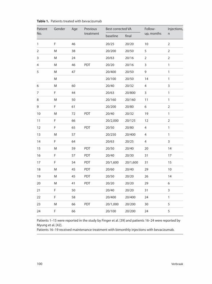
100 Verbraak
Table 1. Patients treated with bevacizumab
Patient
No.
Gender Age Previous
treatment
Best corrected VA Follow-
up, months
Injections,
nbaseline final
1 F 46 20/25 20/20 10 2
2 M 38 20/200 20/50 5 2
3 M 24 20/63 20/16 2 2
4 M 46 PDT 20/20 20/16 3 1
5 M 47 20/400 20/50 9 1
M 20/100 20/50 14 1
6 M 60 20/40 20/32 4 3
7 F 44 20/63 20/800 3 1
8 M 50 20/160 20/160 11 1
9 F 61 20/200 20/80 6 2
10 M 72 PDT 20/40 20/32 19 1
11 F 66 20/2,000 20/125 12 2
12 F 65 PDT 20/50 20/80 4 1
13 M 57 20/250 20/400 4 1
14 F 64 20/63 20/25 4 3
15 M 59 PDT 20/50 20/40 20 14
16 F 57 PDT 20/40 20/30 31 17
17 F 54 PDT 20/1,600 20/1,600 31 15
18 M 45 PDT 20/60 20/40 29 10
19 M 45 PDT 20/50 20/20 26 14
20 M 41 PDT 20/20 20/20 29 6
21 F 50 20/40 20/20 31 3
22 F 58 20/400 20/400 24 1
23 M 66 PDT 20/1,000 20/200 30 5
24 F 66 20/100 20/200 24 5
Patients 1–15 were reported in the study by Finger et al. [39] and patients 16–24 were reported by
Myung et al. [42].
Patients 16–19 received maintenance treatment with bimonthly injections with bevacizumab.
Anti-VEGF Treatment in PXE Patients 101
Patients 16–19 received a regular maintenance therapy, with injections once every
2 months. These patients had already lost their vision in the other eye, and seemed to
be at a greater risk of losing vision in the treated eye. The other patients received an
on-demand treatment in case of recurrent or persistent activity of the CNV, either on
optical coherence tomography (OCT) examination or on FA. The number of retreat-
ments in these patients was relatively low, starting with only 1 injection, and with
a mean of 2–3 injections per year during follow-up. Four patients received only 1
injection and remained stable during 1 year. In certain patients with choroidal CNV
associated with angioid streaks, intravitreal bevacizumab can restore VA and normal-
ize the macular morphology for an extended period of time. However, most patients
needed more than one treatment, and regular follow-up once every month seems to
be mandatory in all patients.
Only 4 patients experienced a loss of VA, and in another 4 patients VA did not
change. VA in all other patients improved. Seven patients had a VA of 20/40 or more,
and none of these patients lost vision. Following treatment, 12 patients had a VA of
20/40 or more. The sooner one starts treatment at a stage of the disease with minimal
abnormalities in the posterior pole, the better one is able to stop the progression of
the CNV, and preserve vision. Five patients had a VA <20/200, of which 3 showed a
modest improvement and the others remained the same.
Patients who received previous PDT treatment, in general, showed more extensive
alterations in the posterior pole, especially larger areas of RPE atrophy, and most started
with a lower VA at baseline and did not improve much (fig. 1). However, the first pre-
sentation of a CNV in patients with PXE is in most cases a classical type of CNV, with
limited dimensions, and these types of CNV in general respond favorably to treatment
(fig. 2). Some patients have been reported with a prolonged recurrence-free period fol-
lowing intravitreal treatment with bevacizumab. One patient has been reported with
an improvement in VA from 20/70 to 20/20 for a period of 32 months, following three
monthly injections with bevacizumab [44]. The majority of patients will experience a
recurrence of the CNV, and need additional treatments. In some of these patients the
recurrences are frequent and these patients need careful follow-up to prevent loss of
VA. As stated, some authors think the risk of recurrence and subsequent vision loss is
so great that a maintenance therapy with injections once every 2 or 3 months is justi-
fied [36, 42]. Indeed the rate of recurrence seemed to be lower in patients treated with
a maintenance treatment schedule compared to those treated on demand, but both
regimens were equally capable of improving or stabilizing VA and there are insufficient
data at present to draw definite conclusions on the optimal dosing schedule.
Later in the course of the disease, at a time when secondary atrophic changes have
occurred in the posterior pole, the CNV changes to a more occult type, with larger
dimensions and a more widespread leakage. In this type of lesion the patient is gener-
ally older, the VA is already lower, and the response to treatment, even though ana-
tomically successful, will not lead to a great improvement in VA but most of the time
will stabilize vision.
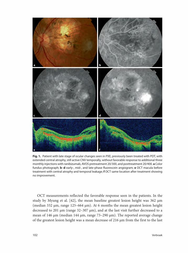
102 Verbraak
OCT measurements reflected the favorable response seen in the patients. In the
study by Myung et al. [42], the mean baseline greatest lesion height was 362 μm
(median 332 μm, range 125–664 μm). At 6 months the mean greatest lesion height
decreased to 201 μm (range 32–307 μm), and at the last visit further decreased to a
mean of 146 μm (median 144 μm, range 73–290 μm). The reported average change
of the greatest lesion height was a mean decrease of 216 μm from the first to the last
a b
dc
e f
Fig. 1. Patient with late stage of ocular changes seen in PXE, previously been treated with PDT, with
extended central atrophy, still active CNV temporally, without favorable response to additional three
monthly injections with ranibizumab, AVOS pretreatment 20/300, and posttreatment 20/400. a Color
fundus photograph; b–d early-, mid-, and late-phase fluorescein angiogram; e OCT macula before
treatment with central atrophy and temporal leakage; f OCT same location after treatment showing
no improvement.
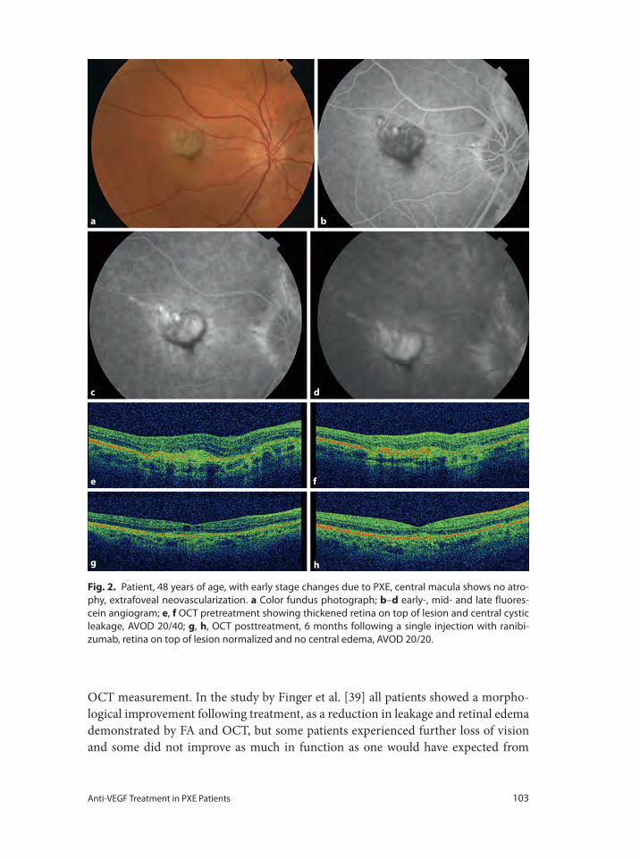
Anti-VEGF Treatment in PXE Patients 103
OCT measurement. In the study by Finger et al. [39] all patients showed a morpho-
logical improvement following treatment, as a reduction in leakage and retinal edema
demonstrated by FA and OCT, but some patients experienced further loss of vision
and some did not improve as much in function as one would have expected from
a
c
e
g h
f
d
b
Fig. 2. Patient, 48 years of age, with early stage changes due to PXE, central macula shows no atro-
phy, extrafoveal neovascularization. a Color fundus photograph; b–d early-, mid- and late fluores-
cein angiogram; e, f OCT pretreatment showing thickened retina on top of lesion and central cystic
leakage, AVOD 20/40; g, h, OCT posttreatment, 6 months following a single injection with ranibi-
zumab, retina on top of lesion normalized and no central edema, AVOD 20/20.
104 Verbraak
the morphological improvement. Treatment with bevacizumab was most efficient in
patients with fewer changes at the posterior pole, at a stage where the disease process
was not far progressed.
PXE patients are prone to cardiovascular events. Widespread atherosclerosis,
intermittent claudicatio, angina pectoris, and early myocardial infarction belong to
the spectrum of cardiovascular abnormalities seen in PXE. One of the possible side
effects of intravitreal anti-VEGF therapy could be an increased risk to just these car-
diovascular events, although to date no definite proof of this increased risk has been
documented. In the PXE patients reported in literature, treated with anti-VEGF, no
patient suffered from heart attack or stroke. One case has been described of a non-
arteritic anterior optic neuropathy, occurring 2 weeks following an injection with
bevacizumab in a patient with PXE, and a CNV secondary to angioid streaks [45].
However, a direct causal relationship in the presented case could neither be proven
nor rejected.
Conclusions
From the present evidence it may be concluded that intravitreal anti-VEGF therapy
with ranibizumab or bevacizumaab is beneficial for the treatment of CNV secondary
to angioid streaks associated with PXE. The positive outcome described in the earliest
studies, mostly in case series, including a limited number of patients and a relative
short follow-up, were confirmed by two larger case series with follow-up of up to 2
years [39, 42].
No other treatment modality was able to reach the same positive results. Even with
the most promising therapy, PDT, results were poor regarding the preservation of
VA, probably to the collateral damage inherent to this treatment 30, 35]. Although
there seem to be arguments to treat selected patients with a maintenance treatment
of intravitreal injections once every 2 months, the majority of patients will be treated
with an as-needed regimen. Unfortunately the low number of patients prevents larger
controlled trials necessary to draw conclusions based on sound research. In addi-
tion, it seems to be unethical to withhold treatment in patients with CNV. A larger
prospective uncontrolled clinical trial about the efficacy and safety of ranibizumab in
treatment of CNV in PXE patients has been started to recruit in 2010 (PXE-CNV 06
htpp://www.clinicaltrilas.gov).
Patients need to be aware of the off-label nature of the treatment with anti-VEGF.
Patients also need to be informed about the possible increased risk of cardiovascu-
lar and/or thromboembolic events, although at present no definite proof has been
documented of this higher risk in patients with or without PXE treated with intravit-
real anti-VEGF. Overall, based on available evidence, intravitreal treatment with anti-
VEGF seems to be the best choice at present to treat patients with CNV secondary to
angioid streaks and PXE.
Anti-VEGF Treatment in PXE Patients 105
1 Chassaing N, Martin L, Calvas P, Le BM, Hovnanian
A: Pseudoxanthoma elasticum: a clinical, pathophys-
iological and genetic update including 11 novel
ABCC6 mutations. J Med Genet 2005;42:881–892.
2 Finger RP, Charbel IP, Ladewig MS, Gotting C,
Szliska C, Scholl HP, et al: Pseudoxanthoma elasti-
cum: genetics, clinical manifestations and therapeu-
tic approaches. Surv Ophthalmol 2009;54:272–285.
3 Plomp AS, Toonstra J, Bergen AA, van Dijk MR, de
Jong PT: Proposal for updating the pseudoxan-
thoma elasticum classification system and a review
of the clinical findings. Am J Med Genet A 2010;
152A:1049–1058.
4 Fabbri E, Forni GL, Guerrini G, Borgna-Pignatti C:
Pseudoxanthoma-elasticum-like syndrome and tha-
lassemia: an update. Dermatol Online J 2009;15:7.
5 Laube S, Moss C: Pseudoxanthoma elasticum. Arch
Dis Child 2005;90:754–756.
6 Li Q, Jiang Q, Pfendner E, Varadi A, Uitto J: Pseu-
doxanthoma elasticum: clinical phenotypes, molec-
ular genetics and putative pathomechanisms. Exp
Dermatol 2009;18:1–11.
7 Bergen AA, Plomp AS, Hu X, de Jong PT, Gorgels
TG: ABCC6 and pseudoxanthoma elasticum. Pflu-
gers Arch 2007;453:685–691.
8 Pfendner EG, Vanakker OM, Terry SF, Vourthis S,
McAndrew PE, McClain MR, et al: Mutation detec-
tion in the ABCC6 gene and genotype-phenotype
analysis in a large international case series affected
by pseudoxanthoma elasticum. J Med Genet 2007;
44:621–628.
9 Vanakker OM, Leroy BP, Coucke P, Bercovitch LG,
Uitto J, Viljoen D, et al: Novel clinico-molecular
insights in pseudoxanthoma elasticum provide an
efficient molecular screening method and a com-
prehensive diagnostic flowchart. Hum Mutat 2008;
29:205.
10 Gorgels TG, Hu X, Scheffer GL, van der Wal AC,
Toonstra J, de Jong PT, et al: Disruption of Abcc6 in
the mouse: novel insight in the pathogenesis of
pseudoxanthoma elasticum. Hum Mol Genet 2005;
14:1763–1773.
11 Zarbock R, Hendig D, Szliska C, Kleesiek K, Gotting
C: Vascular endothelial growth factor gene poly-
morphisms as prognostic markers for ocular mani-
festations in pseudoxanthoma elasticum. Hum Mol
Genet 2009;18:3344–3351.
12 Neldner KH: Pseudoxanthoma elasticum. Clin Der-
matol 1988;6:1–159.
13 Neldner KH: Pseudoxanthoma elasticum. Int J Der-
matol 1988;27:98–100.
14 Lebwohl M, Neldner K, Pope FM, De Paepe A,
Christiano AM, Boyd CD, et al: Classification of
pseudoxanthoma elasticum: report of a consensus
conference. J Am Acad Dermatol 1994;30:103–107.
15 Lafaut BA, Priem H, De Laey JJ: Indocyanine green
angiography in angioid streaks. Bull Soc Belge Oph-
talmol 1997;265:21–24.
16 Lafaut BA, Leys AM, Scassellati-Sforzolini B, Priem
H, De Laey JJ: Comparison of fluorescein and indo-
cyanine green angiography in angioid streaks.
Graefes Arch Clin Exp Ophthalmol 1998;236:346–
353.
17 Mansour AM, Ansari NH, Shields JA, Annesley
WH Jr, Cronin CM, Stock EL: Evolution of angioid
streaks. Ophthalmologica 1993;207:57–61.
18 Finger RP, Charbel IP, Ladewig M, Gotting C, Holz
FG, Scholl HP: Fundus autofluorescence in pseu-
doxanthoma elasticum. Retina 2009;29:1496–1505.
19 Hu X, Plomp AS, van SS, Wijnholds J, de Jong PT,
Bergen AA: Pseudoxanthoma elasticum: a clinical,
histopathological, and molecular update. Surv Oph-
thalmol 2003;48:424–438.
20 Mansour AM, Annesley WH: Comet-tailed drusen
of the retinal pigment epithelium in angioid streaks.
Eye (Lond) 1998;12:943–944.
21 Pierro L, Brancato R, Minicucci M, Pece A: Echo-
graphic diagnosis of drusen of the optic nerve head
in patients with angioid streaks. Ophthalmologica
1994;208:239–242.
22 Popescu MP: Presence of choroidal neovascular
membranes in pseudoxanthoma elasticum. Rev
Chir Oncol Radiol ORL Oftalmol Stomatol Ser
Oftalmol 1987;31:219–220.
23 Puig J, Garcia-Arumi J, Salvador F, Sararols L,
Calatayud M, Alforja S: Subretinal neovasculariza-
tion and hemorrhages in angioid streaks (in
Spanish). Arch Soc Esp Oftalmol 2001;76:309–314.
24 Soutome N, Sugahara M, Okada AA, Hida T:
Subretinal hemorrhages after blunt trauma in pseu-
doxanthoma elasticum. Retina 2007;27:807–808.
25 Atmaca LS, Ozmert E: Macular involvement in
angioid streaks. Gronblad-Strandberg syndrome (in
French). J Fr Ophtalmol 1992;15:249–253.
26 Donaldson EJ: Angioid streaks. Aust J Ophthalmol
1983;11:55–58.
27 Sato K, Ikeda T: Fluorescein angiographic features
of neovascular maculopathy in angioid streaks. Jpn
J Ophthalmol 1994;38:417–422.
28 Schiano LD, Parravano MC, Chiaravalloti A, Varano
M: Choroidal neovascularization in angioid streaks
and pseudoxanthoma elasticum: 1 year follow-up.
Eur J Ophthalmol 2009;19:151–153.
References
106 Verbraak
29 Browning AC, Chung AK, Ghanchi F, Harding SP,
Musadiq M, Talks SJ, et al: Verteporfin photody-
namic therapy of choroidal neovascularization in
angioid streaks: one-year results of a prospective
case series. Ophthalmology 2005;112:1227–1231.
30 Chung AK, Gauba V, Ghanchi FD: Photodynamic
therapy using verteporfin for juxtafoveal choroidal
neovascularisation in angioid streaks associated
with pseudoxanthoma elasticum: 40 months’ results.
Eye (Lond) 2006;20:629–631.
31 Hedaya J, Freeman WR: The effect of treatments
given to patients before initiation of antivascular
endothelial growth factor treatment. Retina 2008;
28:911–912.
32 Roth DB, Estafanous M, Lewis H: Macular translo-
cation for subfoveal choroidal neovascularization in
angioid streaks: Am J Ophthalmol 2001;131:390–
392.
33 Singerman LJ, Hatem G: Laser treatment of choro-
idal neovascular membranes in angioid streaks.
Retina 1981;1:75–83.
34 Tassignon MJ, Merckaert I, De CA, Cornelis MT:
Treatment of choroidal neovascular membranes in a
case of pseudoxanthoma elasticum (in French). Bull
Soc Belge Ophtalmol 1985;214:107–112.
35 Menchini U, Virgili G, Introini U, Bandello F,
Ambesi-Impiombato M, Pece A, et al: Outcome of
choroidal neovascularization in angioid streaks
after photodynamic therapy. Retina 2004;24:763–
771.
36 Bhatnagar P, Freund KB, Spaide RF, Klancnik JM Jr,
Cooney MJ, Ho I, et al: Intravitreal bevacizumab for
the management of choroidal neovascularization in
pseudoxanthoma elasticum. Retina 2007;27:897–
902.
37 Derriman L, Marshall J, Moorman C, Downes SM:
The use of intravitreal bevacizumab to treat choroi-
dal neovascular membranes. Retina 2008;28:910–
911.
38 Donati MC, Virgili G, Bini A, Giansanti F, Rapizzi
E, Giacomelli G, et al: Intravitreal bevacizumab
(Avastin) for choroidal neovascularization in angi-
oid streaks: a case series. Ophthalmologica 2009;223:
24–27.
39 Finger RP, Charbel IP, Ladewig M, Holz FG, Scholl
HP: Intravitreal bevacizumab for choroidal neovas-
cularisation associated with pseudoxanthoma elas-
ticum. Br J Ophthalmol 2008;92:483–487.
40 Japiassu RM, Japiassu MA, Pecego MG: Intravitreal
bevacizumab in choroidal neovascularization sec-
ondary to Gronblad-Strandberg syndrome: case
report. Arq Bras Oftalmol 2008;71:427–429.
41 Lommatzsch A: Management of choroidal vascu-
larisation. Br J Ophthalmol 2008;92:445–446.
42 Myung JS, Bhatnagar P, Spaide RF, Klancnik JM Jr,
Cooney MJ, Yannuzzi LA, et al: Long-term out-
comes of intravitreal antivascular endothelial
growth factor therapy for the management of chor-
oidal neovascularisation in pseudoxanthoma elasti-
cum. Retina 2009.
43 Rinaldi M, Dell’Omo R, Romano MR, Chiosi F,
Cipollone U, Costagliola C: Intravitreal bevaci-
zumab for choroidal neovascularization secondary
to angioid streaks. Arch Ophthalmol 2007;125:1422–
1423.
44 Kovach JL, Schwartz SG, Hickey M, Puliafito CA:
Thirty-two month follow-up of successful treatment
of choroidal neovascularization from angioid
streaks with intravitreal bevacizumab. Ophthalmic
Surg Lasers Imaging 2009;40:77–79.
45 Ganssauge M, Wilhelm H, Bartz-Schmidt KU,
Aisenbrey S: Non-arteritic anterior ischemic optic
neuropathy after intravitreal injection of bevaci-
zumab (Avastin) for treatment of angioid streaks in
pseudoxanthoma elasticum. Graefes Arch Clin Exp
Ophthalmol 2009;247:1707–1710.
Frank D. Verbraak, MD, PhD
Departments of Ophthalmology and Biomedical Engineering & Physics
Academic Medical Center, Meibergdreef 9
NL–1105 AZ Amsterdam (The Netherlands)
Tel. +31 20 566 4351, E-Mail [email protected]
Bandello F, Battaglia Parodi M (eds): Anti-VEGF.
Dev Ophthalmol. Basel, Karger, 2010, vol 46, pp 107–110
Antivascular Endothelial Growth Factor in Hereditary DystrophiesMaurizio Battaglia Parodia � Pierluigi Iaconob � Francesco Bandelloa
aDepartment of Ophthalmology, University Vita-Salute, Scientific Institute San Raffaele, Milan, and bFondazione
G.B. Bietti per l’Oftalmologia, Istituto di Ricovero e Cura a Carattere Scientifico, Rome, Italy
AbstractChoroidal neovascularization can rarely complicate the course of a number of chorioretinal heredi-
tary dystrophies leading to an even further impaired vision function. In recent years, several case
reports and case series have shown that intravitreal injections of antivascular endothelial growth
factor drugs can be effective in treating subfoveal choroidal neovascularization secondary to chori-
oretinal dystrophies either improving vision, or at least halting its progressive loss. Additional stud-
ies are warranted to confirm the initial positive response and to assess the best therapeutic
regimen. Copyright © 2010 S. Karger AG, Basel
Choroidal neovascularization (CNV) can rarely complicate the course of several cho-
rioretinal dystrophies. Little is known regarding the natural history of this form of
CNV. Visual acuity (VA) can be stable for a long time with a late evolution toward
fibrosis and atrophy [1]. Nevertheless, the occurrence of a CNV, especially with sub-
foveal location, is often associated with a visual function deterioration. Some authors
have employed laser photocoagulation and photodynamic therapy to reverse or halt
the consequent visual impairment, achieving limited beneficial effects [2, 3]. The
advent of therapy based on intravitreal injection of antivascular endothelial growth
factor drugs has brought new hope in the treatment of this complication of chori-
oretinal dystrophies. Additional studies are warranted to confirm the initial positive
response and to assess the best therapeutic regimen.
Best’s Dystrophy
Two reports have described a positive effect of intravitreal injections of both bevaci-
zumab and ranibizumab for the treatment of a subfoveal CNV related to vitelliform
108 Battaglia Parodi · Iacono · Bandello
Best’s disease [4, 5]. In both cases, optical coherence tomography (OCT) examination
revealed the complete resolution of the sensory retinal detachment, over a short-term
follow-up. Bearing in mind the young age of patients affected by Best vitelliform dystro-
phy, this approach could be extremely beneficial for visual prognosis and quality of life.
Pattern Dystrophy
A single report describes a case of adult-onset foveomacular vitelliform dystrophy
associated and complicated by occult CNV who underwent intravitreal bevacizumab
with progressive decrease of subretinal fluid, but with no VA improvement [6].
Our experience includes 8 patients affected by subfoveal CNV associated with pat-
tern dystrophy of the retinal pigment epithelium, who were treated with intravitreal
bevacizumab and prospectively followed up for 24 months. The mean best corrected
VA (BCVA) and the mean foveal thickness (FT) at baseline were 0.73 ± 0.37 (logMAR
± SD) and 281 ± 104 μm (CMT ± SD), respectively. At the 3-month examination,
mean BCVA improved significantly to 0.52 ± 0.29 and mean FT decreased to 222 ±
78 μm. Subsequently, at the 6- and 12-month examinations, a substantial stabilization
of the mean BCVA was observed, whereas the mean FT changed to 217 ± 52 and 208
± 58 μm, respectively. At the final visit, mean BCVA showed a statistically significant
improvement of 2.4 lines in comparison to the baseline value. The mean FT finally
decreased to 197 ± 36 μm.
Overall, a mean number of 4.4 intravitreal bevacizumab injections was admin-
istered during the 24 months of follow-up. No side effect or complication was reg-
istered during the follow-up period. On the basis of our practice, we believe that
intravitreal injection of bevacizumab can be regarded as a beneficial treatment for
pattern dystrophy-related subfoveal CNV. Further studies are necessary to assess the
best therapeutic regimen and the most appropriate monitoring procedures.
Intravitreal injection of bevacizumab was performed in a case affected by adult-
onset vitelliform dystrophy with no evidence of CNV [7]. During 4 months of follow-
up the spectral domain OCT showed a progressive decrease of yellowish deposits and
complete resolution of subretinal fluid which accompanied the lipofuscin-like mate-
rial. Unfortunately, a mild decrease in VA and persistence of metamorphopsia was
registered.
Retinitis Pigmentosa
Some authors have recently reported on the use of intravitreal bevacizumab to treat
cystoid macular edema complicating retinitis pigmentosa. This complication occurs
in about 20% of cases [8] and oral carbonic anhydrase inhibitors have been reported
as the most effective treatment to manage it.
Anti-VEGF in Hereditary Dystrophies 109
1 Parodi MB: Eredodistrofie Corioretiniche. Rome,
Verduci Editore, 2002.
2 Parodi MB: Choroidal neovascularization in fundus
pulverulentus. Acta Ophthalmol Scand 2002;80:559–
560.
3 Parodi MB, Liberali T, Pedio M, Francis PJ, Piccolino
FC, Fiotti N, Romano M, Ravalico G: Photodynamic
therapy of subfoveal choroidal neovascularization
secondary to reticular pattern dystrophy: three-year
results of an uncontrolled, prospective case series.
Am J Ophthalmol 2006;141:1152–1154.
4 Querques G, Bocco MCA, Soubrane G, Souied EH:
Intravitreal ranibizumab (Lucentis) for choroidal
neovascularisation associated with vitelliform mac-
ular dystrophy. Acta Ophthalmol 2008;86:694–695.
5 Leu J, Schrage NF, Degenring RF: Choroidal neo-
vascularisation secondary to Best’s disease in a
13-year-old boy treated by intravitreal Bevacizumab.
Graefes Arch Clin Exp Ophthalmol 2007;245:1723–
1725.
6 Montero JA, Ruiz-Moreno JM, De La Vega C:
Intravitreal bevacizumab for adult-onset vitelliform
dystrophy: a case report. Eur J Ophthalmol 2007;17:
983–986.
A former interventional case series of 2 eyes treated with intravitreal bevacizumab
injections demonstrated the absence of a positive effect [9], whereas another inter-
ventional case series including 13 eyes of 7 patients revealed a progressive reduction
of the central macular thickness in association with visual function improvement
[10]. Positive results have also been reported in a single case of CME secondary to a
poor response to oral acetazolamide and following treatment with pegaptanib intra-
vitreal injection [11].
Further studies with a larger population and longer follow-up period are warranted
to assess the efficacy of this approach.
Fundus Flavimaculatus
A single case of fundus flavimaculatus associated with CNV was submitted to intrav-
itreal ranibizumab injections at monthly intervals. Six months after three injections,
VA improved from 20/64 to 20/32, with resolution of serous retinal detachment on
OCT [12]. This case report indicates that intravitreal ranibizumab may be considered
a possible valuable therapeutic option for this rare association.
Sorsby Fundus Dystrophy
A case of CNV secondary to genetically demonstrated Sorsby fundus dystrophy was
treated with three initial intravitreal injections of bevacizumab at a dose of 5 mg/kg
at 2-week intervals, followed by an additional injection because of CNV recurrence
at the 7-month follow-up [13]. After 16 months of follow-up, VA had improved from
20/50 at baseline to 20/25, whereas OCT and fluorescein angiography showed no evi-
dence of CNV activity.
References
110 Battaglia Parodi · Iacono · Bandello
7 Lee JY, Lim J, Chung H, Kim JG, Yoon YH: Spectral
domain optical coherence tomography in a patient
with adult-onset vitelliform dystrophy treated with
intravitreal bevacizumab. Ophthalmic Surg Lasers
Imaging 2009;40:319–321.
8 Fishman JA, Maggiano JM, Fishman M: Foveal
lesions seen in retinitis pigmentosa. Arch Oph-
thalmol 1977;95:1993–1996.
9 Melo GB, Farah ME, Aggio FB: Intravitreal injec-
tion of bevacizumab for cystoid macular edema in
retinitis pigmentosa. Acta Ophthalmol 2007;85:461–
463.
10 Yuzbasioglu E, Artunay O, Rasier R, Sengul A,
Bahcecioglu H: Intravitreal bevacizumab (Avastin)
injection in retinitis pigmentosa. Curr Eye Res 2009;
34:231–237.
11 Querques G, Prascina F, Iaculli C, et al: Intravitreal
pegaptanib sodium (Macugen) for refractory cys-
toid macular edema in pericentral retinitis pigmen-
tosa. Int Ophthalmol 2009;29:103–107.
12 Quijano C, Querques G, Massamba N, Soubrane G,
Souied EH: Type 3 choroidal neovascularization
associated with fundus flavimaculatus. Ophthalmic
Res 2009;42:152–154.
13 Prager F, Michels S, Geitzenauer W, Schmidt-
Erfurth U: Choroidal neovascularization secondary
to Sorsby fundus dystrophy treated with systemic
bevacizumab (Avastin). Acta Ophthalmol Scand
2007;85:904–906.
Francesco Bandello, MD, FEBO
Professor and Chairman, Department of Ophthalmology
University Vita-Salute Scientific Institute, San Raffaele
Via Olgettina, 60, IT–20132 Milan (Italy)
Tel. +39 02 2643 2648, Fax +39 02 2648 3643, E-Mail [email protected]
Bandello F, Battaglia Parodi M (eds): Anti-VEGF.
Dev Ophthalmol. Basel, Karger, 2010, vol 46, pp 111–122
Antivascular Endothelial Growth Factor as an Approach for Macular EdemaP.M. Buchholz � A.P. Buchholz � A.J. Augustin
Augenklinik, Karlsruhe, Germany
AbstractMacular edema is an abnormal thickening of the macula associated with the accumulation of excess
fluid in the extracellular space of the neurosensory retina. The following chapter looks at the basic
pathomechanisms of macular edema as well as major pathologic conditions leading to it: special
focus is on diabetic retinopathy, retinal venous occlusions and a number of inflammatory disorders.
Currently available data on up-to-date pharmacologic treatment options such as steroids and anti-
VEGF compounds is presented and discussed. Copyright © 2010 S. Karger AG, Basel
Broadly defined, macular edema (ME) is an abnormal thickening of the macula asso-
ciated with the accumulation of excess fluid in the extracellular space of the neu-
rosensory retina. Intracellular edema involving Müller cells has also been observed
histopathologically in some cases. The term cystoid macular edema (CME) applies
when there is evidence by biomicroscopy, fluorescein angiography (FA) and/or opti-
cal coherence tomography (OCT) of fluid accumulation into multiple cyst-like spaces
within the macula.
Several basic pathophysiologic processes may contribute to the development of
ME, which occurs in association with a wide variety of pathologic conditions. ME
presents the final common pathway in many prevalent retinal disorders and can be
considered the leading cause of central vision loss in the developed world. It is thus of
markedly medical and socioeconomic importance.
Since the pathogenesis of ME depends on the underlying etiology and because
it may be multifactorial, an effective management is based upon recognizing and
addressing each factor that is expressed in a given clinical setting. The treatment of
ME has evolved dramatically over the past two decades. Research has led to a better
understanding of its causes, but also to the development of new therapeutic options
[1].
112 Buchholz · Buchholz · Augustin
Basic Pathophysiologic Mechanisms
Normally, volume and composition of the extracellular compartment of the neuro-
sensory retina and subretinal space is regulated by retinal capillary endothelial cell
and retinal pigment epithelium (RPE) tight junctions as well as the pumping func-
tions of RPE cells. However, if there is a loss of function in such fluid barriers or
effective RPE pump mechanisms, intraretinal fluid will accumulate. OCT imaging
reveals that ME is located mostly in the outer retinal layers [2]. Most commonly, ME
is a result of pathologic hyperpermeability of retinal blood vessels. Increased vascular
permeability leads to extravasation of fluid, proteins and other molecules into the ret-
inal interstitium. Depending on the underlying disease, the latter may include pros-
taglandins, leukotrienes, protein kinase C, nitric oxide and various cytokines such as
vascular endothelial growth factor (VEGF), TNF-α and interleukins.
Clinically leakage from retinal blood vessels is best detected with FA, while OCT
imaging is a sensitive method for detecting and quantifying macular thickening
regardless of its cause. Leakage of fluid is further enhanced by factors that increase
retinal blood flow such as vasodilation, rising intraluminal pressure and increasing
blood volume. Additionally there is evidence that alterations in the outer blood-
retinal (RPE) barrier may contribute directly to ME in conditions such as diabetic
retinopathy and postoperative states [1]. Furthermore, tractional stress by perifoveal
vitreous detachment or epiretinal membrane as well as various abnormalities causing
fluid leakage from the optic nerve head can lead to intraretinal fluid accumulation
and ME. Regardless of the pathogenic mechanisms causing ME, the resulting loss
in visual acuity (VA) essentially depends on macular thickening and various other
factors such as duration of edema, perfusion of macular capillaries, photoreceptor
impairment, dysfunction and media opacities [2].
Major Pathologic Conditions Leading to ME: Retinal Vascular Disease
Diabetic Retinopathy
DME is the leading cause of vision loss in patients with diabetes. It occurs in diabetes
types 1 and 2 and increases in incidence with the severity of diabetic retinopathy.
Chronic hyperglycemia is a major initiator of microvascular complications in dia-
betes. It sets off a series of metabolic events, stimulating the expressions of multiple
cytokines such as VEGF and produces vascular dysfunction and damage that includes
loss of endothelial cells, increased permeability and leukocyte adhesion and altera-
tions in blood flow leading to diabetic retinopathy and potentially diabetic macular
edema (DME).
The prevalence of diabetic retinopathy is available from major European countries
and the USA. According to a survey published in Germany in 2000, the prevalence of
diabetic retinopathy was estimated at 22% in diabetic patients [3]. Data from a study
Anti-VEGF as an Approach for Macular Edema 113
carried out in 2002 by the University of Valladolid in Spain gave a similar prevalence
(20.9%) with regard to retinopathy in diabetic patients [4]. In the UK, up to 10%
of people with diabetes will have retinopathy requiring ophthalmologic follow-up or
treatment [5]. It has been estimated that if untreated, 6–9% of patients with prolifera-
tive retinopathy or severe non-proliferative retinopathy will become blind each year
[6]. Within the Wisconsin Epidemiologic Study of Diabetic Retinopathy (WESDR),
a population-based study from a well-defined area, researchers found a prevalence
of 71% for diabetic retinopathy in a group of 996 patients who were below the age of
30 at onset of diabetes [7]. Data on incidence is much more limited. In Germany for
example, 2.13% of diabetics are estimated to develop diabetic retinopathy each year
[5]. The WESDR reported an incidence of 59% over a 4-year interval [8].
The prevalence of DME has also been recorded. In Spain, out of 20.9% of diabetic
patients with retinopathy, ME was found in 5.7% [4]. In the UK, there are an esti-
mated 380,000 blind people and 579,000 partially sighted people. According to the
Royal College of Ophthalmologists of London, ME is responsible for 70% of visual
loss in diabetic patients [5]. The prevalence of ME in the WESDR, severe enough to
be a likely cause of visual disturbance, ranged between 4 and 11%. Based on these
data it was estimated (as of 1993) that of approximately 7,800,000 people in North
America with diabetes, 84,000 would develop proliferative retinopathy and about
95,000 would develop ME over a 10-year period [9].
The pathophysiology of ME in diabetic eyes is complex. Increases in retinal blood
flow may partly explain the extravasation of fluid into the extracellular compartment,
the most important mechanism however is the breakdown of the blood-retinal bar-
riers. VEGF is a highly potent vasopermeability factor and has demonstrated to play
a major role in this process. In the past, focal laser photocoagulation has been the
only proven treatment as well as standard of care in patients with clinically significant
DME. Pharmacologic therapies are usually considered in eyes with perfused, non-
tractional, diffuse DME which is refractory and unlikely to respond to laser treatment
[1].
Retinal Venous Occlusions
An immediate consequence of retinal venous thrombosis is elevation of the intravas-
cular pressure in retinal veins distal to the occlusion site. The breakdown of the blood-
retinal barrier in hypoxic retina is mediated by upregulated cytokines such as VEGF,
which causes leakage of fluids and proteins. By these mechanisms both central retinal
vein occlusions (CRVO) and branch retinal vein occlusions (BRVO) commonly result
in ME which tends to be chronic, difficult to treat and visually disabling [1]. A recent
study has also highlighted that a reduction in perifoveal capillary blood-flow velocity
may be involved in the development of ME in patients with BRVO [10].
A recent survey found that few epidemiology data are available for BRVO as well
as CRVO. BRVO is the second most common retinal vascular disorder after diabetic
retinopathy [11]. There were no European population-based studies identified in
114 Buchholz · Buchholz · Augustin
literature. Such studies however are needed to better understand the epidemiology
of RVO in Europe and yield reliable, valid, generalizable incidence and prevalence
estimates of BRVO and CRVO. It was concluded that from available material the
Beaver Dam Eye Study currently provides the best estimate [12]. The Beaver Dam
Eye Study found a prevalence and incidence for BRVO of 0.6 an 0.12% respectively.
Furthermore, the study showed a respective prevalence and incidence for CRVO of
0.1 and 0.04% [13].
Based on the findings from the Beaver Dam Eye Study, the following extrapola-
tions suggest that prevalence of BRVO ranges from 130,000 (Spain) to 270,000 cases
(Germany), and incidence from 26,000 to 54,000 cases. For CRVO, prevalence ranges
from 22,000 and 45,000 cases and incidence from 9,000 to 18,000 cases [12]. Following
a CRVO or BRVO incident, vision loss is often exacerbated by macular hemorrhage,
macular ischemia or submacular fluid with secondary RPE damage. In randomized
clinical trials, grid laser treatment has shown to produce at least modest visual benefit
in eyes with BRVO, however not in cases of CRVO. There are thus very limited treat-
ment options in CRVO. In recent times, pharmacologic therapies have been intro-
duced as alternative approaches for treatment of vein occlusions [1].
Inflammatory Disorders
Several different underlying inflammatory disorders also result in ME as a common
phenotype.
CME following cataract surgery (Irvine-Gass syndrome) can be detected by FA
and remains the most common cause of visual loss in this setting. While it occurs in
approximately 20% of uncomplicated cases, it will only cause significant decrease in
VA in 1–2% of operated eyes. Risk of developing postoperative CME is substantially
greater in patients with preexisting conditions known to produce vasoactive stimuli
such as diabetic retinopathy and uveitis [1].
A recent pilot study has demonstrated the positive correlation of VEGF concentra-
tion in aqueous samples with clinically meaningful changes in central subfield thick-
ness measured by OCT in a subpopulation of diabetic patients undergoing cataract
surgery. To further elucidate the relationship, more research is warranted [14].
The development of ME is also a known complication of laser procedures such
as panretinal photocoagulation for retinal vascular disease (e.g. diabetic retinopathy,
vein occlusion). Although the mechanism is not clearly understood, laser-induced
inflammatory mediators such as VEGF and transudation from increased macular
blood flow may play a significant role. In addition, the usage of other types of laser
procedures, such as YAG laser procedures, may also enhance occurrence of CME.
CME is the most frequent complication in uveitis. It typically develops in patients
with intermediate and posterior uveitis components. It may occur in a wide variety of
uveitis syndromes, whether they are caused by underlying autoimmune diseases (pars
planitis) or infectious (toxoplasmosis), toxic (rifabutin-associated) or idiopathic (sar-
coidosis) etiologies. In all these conditions the most important pathogenic mechanism
Anti-VEGF as an Approach for Macular Edema 115
is loss of inner blood-retinal barrier integrity caused by inflammatory mediators like
VEGF, which are generated by the underlying uveitic process [1].
Pharmacologic Treatment Options in ME with a Focus on Anti-VEGF Therapies
Diabetic Macular Edema
Options to treat DME effectively have not been too numerous. Apart from different
types of laser treatments – which present the standard of care in treatment of DME
– surgical management in the form of vitrectomy has also played a role in cases unre-
sponsive to laser treatment.
In the category of pharmacological treatment approaches, mainly corticosteroids
have been applied in the past. Corticosteroids are well-known potent anti-inflam-
matory compounds. Additionally they possess strong antiangiogenesis effects, also
acting by suppressing VEGF gene activity and its metabolic pathway. Especially
intravitreally applied triamcinolone (IVTA) has been employed with a fair number
of randomized controlled clinical trials demonstrating significant improvement in
DME and VA [15]. However many of the trials had small numbers of participants as
well as short follow-up time periods. Also, since no IVTA compound has obtained
marketing approval for the treatment of ME, concentrations and dosing schedules
of triamcinolone varied across trials (e.g. 4 up to 25 mg), making comparisons in
terms of efficacy and safety a challenge. The most common choice however seems to
be the 4-mg concentration. Substantial adverse events were recorded in most trials,
among them infections, glaucoma and cataract formation [16]. A study performed
by the Diabetic Retinopathy Clinical Research (DRCR) Network investigators com-
pared the efficacy and safety of focal/grid laser photocoagulation compared to IVTA
doses of 1 or 4 mg respectively over a time period of 2 years. While at 4 months the
VA in steroid-treated subjects was better than in the laser group, the difference was
no more apparent in the long term. From 16 months to 2 years, laser-treated patients
had better vision. These findings show that longer-term data need to be generated for
the treatment of DME to determine the most effective treatment in specific patient
populations [17].
More recently, intravitreal implants have been developed. These allow a more
precise titration as well as extended duration of drug delivery. Examples of licensed
compounds are a surgically implanted fluocinolone acetonide device as well as an
injectable, biodegradable dexamethasone-loaded matrix. Data available report on
improvements of VA and macular thickness, but also on steroid-typical adverse effects
as previously seen with IVTA [18].
Although many agents in development inhibit the production of VEGF, research
has also focused on agents that can antagonize circulating ocular VEGF. The major-
ity of VEGF antagonists are in active development for retinal neovascularization
disorders such as age-related macular degeneration. However, by virtue of the
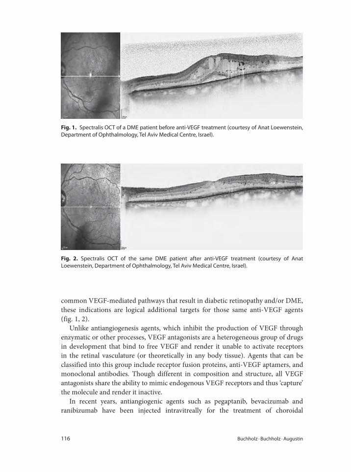
116 Buchholz · Buchholz · Augustin
common VEGF-mediated pathways that result in diabetic retinopathy and/or DME,
these indications are logical additional targets for those same anti-VEGF agents
(fig. 1, 2).
Unlike antiangiogenesis agents, which inhibit the production of VEGF through
enzymatic or other processes, VEGF antagonists are a heterogeneous group of drugs
in development that bind to free VEGF and render it unable to activate receptors
in the retinal vasculature (or theoretically in any body tissue). Agents that can be
classified into this group include receptor fusion proteins, anti-VEGF aptamers, and
monoclonal antibodies. Though different in composition and structure, all VEGF
antagonists share the ability to mimic endogenous VEGF receptors and thus ‘capture’
the molecule and render it inactive.
In recent years, antiangiogenic agents such as pegaptanib, bevacizumab and
ranibizumab have been injected intravitreally for the treatment of choroidal
Fig. 1. Spectralis OCT of a DME patient before anti-VEGF treatment (courtesy of Anat Loewenstein,
Department of Ophthalmology, Tel Aviv Medical Centre, Israel).
Fig. 2. Spectralis OCT of the same DME patient after anti-VEGF treatment (courtesy of Anat
Loewenstein, Department of Ophthalmology, Tel Aviv Medical Centre, Israel).
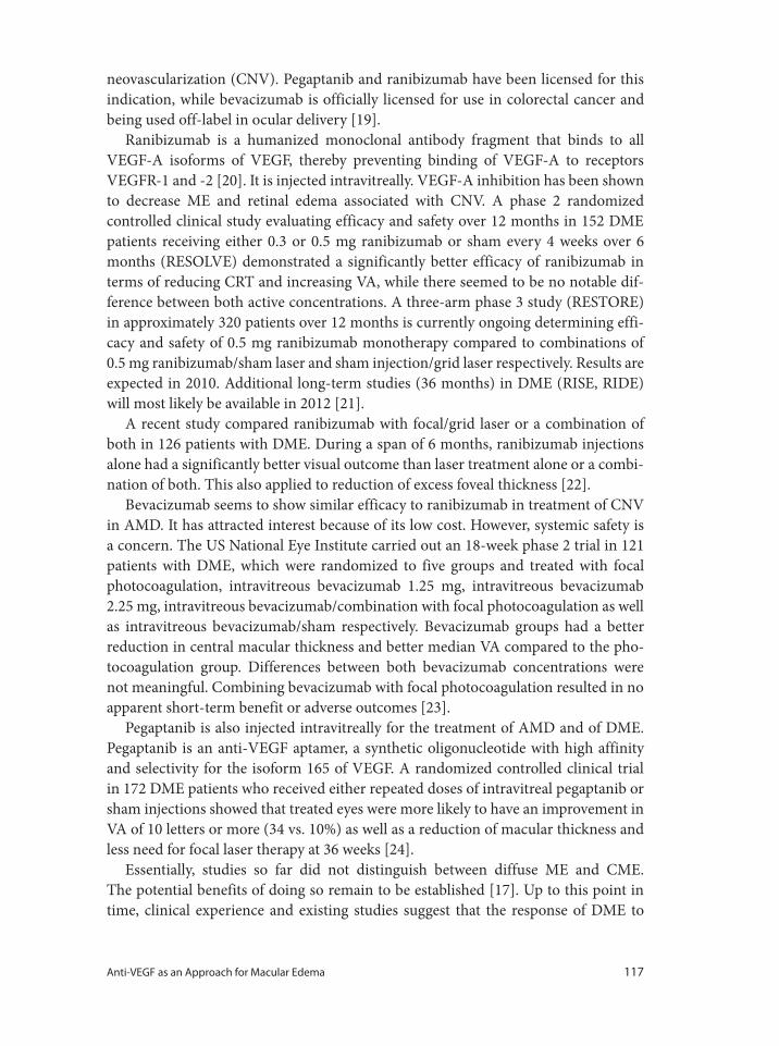
Anti-VEGF as an Approach for Macular Edema 117
neovascularization (CNV). Pegaptanib and ranibizumab have been licensed for this
indication, while bevacizumab is officially licensed for use in colorectal cancer and
being used off-label in ocular delivery [19].
Ranibizumab is a humanized monoclonal antibody fragment that binds to all
VEGF-A isoforms of VEGF, thereby preventing binding of VEGF-A to receptors
VEGFR-1 and -2 [20]. It is injected intravitreally. VEGF-A inhibition has been shown
to decrease ME and retinal edema associated with CNV. A phase 2 randomized
controlled clinical study evaluating efficacy and safety over 12 months in 152 DME
patients receiving either 0.3 or 0.5 mg ranibizumab or sham every 4 weeks over 6
months (RESOLVE) demonstrated a significantly better efficacy of ranibizumab in
terms of reducing CRT and increasing VA, while there seemed to be no notable dif-
ference between both active concentrations. A three-arm phase 3 study (RESTORE)
in approximately 320 patients over 12 months is currently ongoing determining effi-
cacy and safety of 0.5 mg ranibizumab monotherapy compared to combinations of
0.5 mg ranibizumab/sham laser and sham injection/grid laser respectively. Results are
expected in 2010. Additional long-term studies (36 months) in DME (RISE, RIDE)
will most likely be available in 2012 [21].
A recent study compared ranibizumab with focal/grid laser or a combination of
both in 126 patients with DME. During a span of 6 months, ranibizumab injections
alone had a significantly better visual outcome than laser treatment alone or a combi-
nation of both. This also applied to reduction of excess foveal thickness [22].
Bevacizumab seems to show similar efficacy to ranibizumab in treatment of CNV
in AMD. It has attracted interest because of its low cost. However, systemic safety is
a concern. The US National Eye Institute carried out an 18-week phase 2 trial in 121
patients with DME, which were randomized to five groups and treated with focal
photocoagulation, intravitreous bevacizumab 1.25 mg, intravitreous bevacizumab
2.25 mg, intravitreous bevacizumab/combination with focal photocoagulation as well
as intravitreous bevacizumab/sham respectively. Bevacizumab groups had a better
reduction in central macular thickness and better median VA compared to the pho-
tocoagulation group. Differences between both bevacizumab concentrations were
not meaningful. Combining bevacizumab with focal photocoagulation resulted in no
apparent short-term benefit or adverse outcomes [23].
Pegaptanib is also injected intravitreally for the treatment of AMD and of DME.
Pegaptanib is an anti-VEGF aptamer, a synthetic oligonucleotide with high affinity
and selectivity for the isoform 165 of VEGF. A randomized controlled clinical trial
in 172 DME patients who received either repeated doses of intravitreal pegaptanib or
sham injections showed that treated eyes were more likely to have an improvement in
VA of 10 letters or more (34 vs. 10%) as well as a reduction of macular thickness and
less need for focal laser therapy at 36 weeks [24].
Essentially, studies so far did not distinguish between diffuse ME and CME.
The potential benefits of doing so remain to be established [17]. Up to this point in
time, clinical experience and existing studies suggest that the response of DME to
118 Buchholz · Buchholz · Augustin
intravitreal injection of anti-VEGF drugs is transient, variable and of modest magni-
tude in most patients. Proof of long-term efficacy and safety will require large phase
III clinical trials. Such studies should also address whether this relative short-term
efficacy could be extended over a longer time by combination with subsequent laser
photocoagulation [1].
ME Secondary to CRVO and BRVO
Similar to the situation in diabetic retinopathy and ME, available therapeutic options
have been scarce, especially in the management of CRVO. Hemodilution measures as
part of a hospitalization approach have displayed large variations and employed mul-
tiple agents, making a generalized recommendation difficult [25]. The only random-
ized study investigating the effect of hemodilution in an outpatient setting in CRVO
showed no significant benefit [26].
Effectiveness of laser treatment in ME was investigated as part of the Central
Retinal Vein Occlusion Study. Data demonstrated that grid photocoagulation was not
effective in improving VA in eyes with ME secondary to perfused CRVO, although
there was a trend in patients younger than 60 years [27]. While the short-term results
of intravitreal triamcinolone treatment of ME secondary to CRVO appear to be prom-
ising, anatomical and visual improvements seem to be often transient; safety and effi-
cacy have so far only been shown for a specific concentration in one controlled clinical
study. The SCORE (Standard Care versus Corticosteroid for Retinal Vein Occlusion)
Study is a multicenter phase 3 study in 630 patients with ME in CRVO randomized to
intravitreal triamcinolone 4- or 1-mg injections respectively or standard care (obser-
vation). Intravitreal triamcinolone was shown to be superior to observation for treat-
ing vision loss associated with ME. The 1-mg dose had a safety profile superior to that
of the 4-mg dose. The authors suggest that intravitreal triamcinolone in a 1-mg dose,
following the retreatment criteria applied in the SCORE Study, should be considered
for up to 1 year, and possibly 2 years, for patients with characteristics similar to those
in the SCORE-CRVO trial [28]. A trial in which patients received a dexamethasone
implant containing either 350 or 700 μg has also been finalized. The results have not
yet been published [29].
Several case series show that intravitreal anti-VEGF therapy may cause decrease
in macular thickness and improvement in VA [19], however the reported follow-up
series are short and no recommendations can be made at present [18]. Results of a
currently ongoing phase 3 trial (CRUISE) are awaited in 2010. The study compares
the efficacy and safety of monthly administered 0.5 or 0.3 mg ranibizumab or sham
injections for a period of 6 months followed by another 6 months of observation in
390 patients. Another ongoing study (ROCC) is investigating the efficacy and safety
of ranibizumab 10-mg intravitreal injections every 3 months compared to sham
[19].
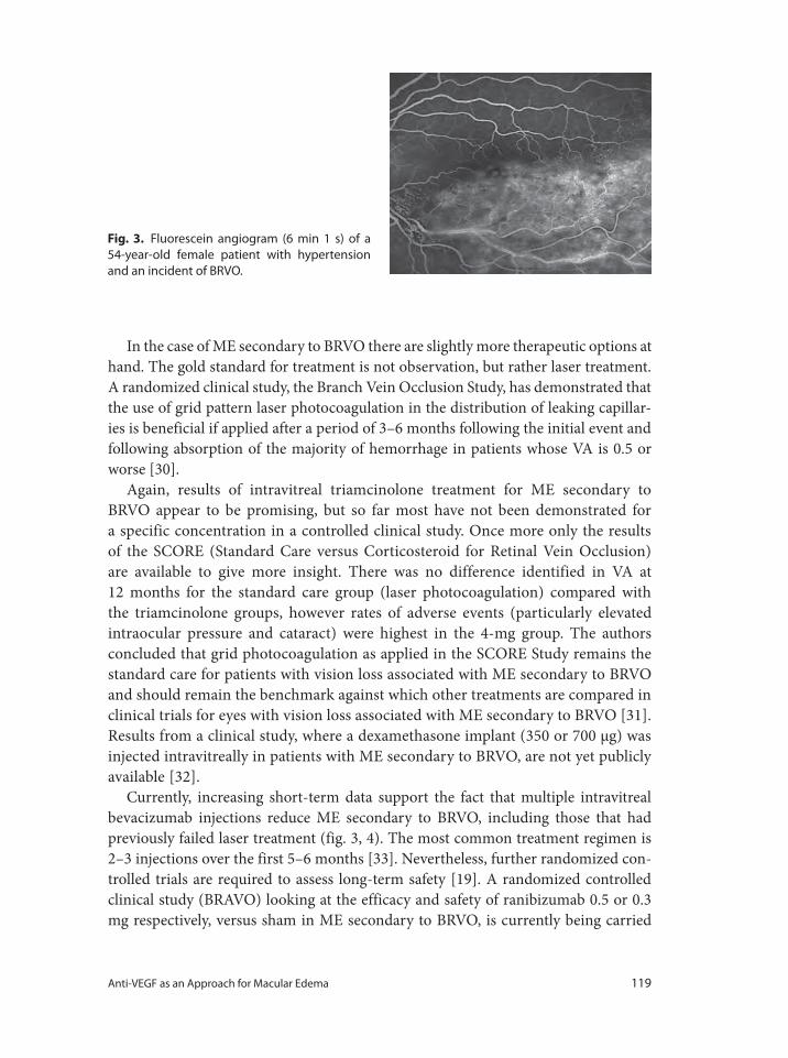
Anti-VEGF as an Approach for Macular Edema 119
In the case of ME secondary to BRVO there are slightly more therapeutic options at
hand. The gold standard for treatment is not observation, but rather laser treatment.
A randomized clinical study, the Branch Vein Occlusion Study, has demonstrated that
the use of grid pattern laser photocoagulation in the distribution of leaking capillar-
ies is beneficial if applied after a period of 3–6 months following the initial event and
following absorption of the majority of hemorrhage in patients whose VA is 0.5 or
worse [30].
Again, results of intravitreal triamcinolone treatment for ME secondary to
BRVO appear to be promising, but so far most have not been demonstrated for
a specific concentration in a controlled clinical study. Once more only the results
of the SCORE (Standard Care versus Corticosteroid for Retinal Vein Occlusion)
are available to give more insight. There was no difference identified in VA at
12 months for the standard care group (laser photocoagulation) compared with
the triamcinolone groups, however rates of adverse events (particularly elevated
intraocular pressure and cataract) were highest in the 4-mg group. The authors
concluded that grid photocoagulation as applied in the SCORE Study remains the
standard care for patients with vision loss associated with ME secondary to BRVO
and should remain the benchmark against which other treatments are compared in
clinical trials for eyes with vision loss associated with ME secondary to BRVO [31].
Results from a clinical study, where a dexamethasone implant (350 or 700 μg) was
injected intravitreally in patients with ME secondary to BRVO, are not yet publicly
available [32].
Currently, increasing short-term data support the fact that multiple intravitreal
bevacizumab injections reduce ME secondary to BRVO, including those that had
previously failed laser treatment (fig. 3, 4). The most common treatment regimen is
2–3 injections over the first 5–6 months [33]. Nevertheless, further randomized con-
trolled trials are required to assess long-term safety [19]. A randomized controlled
clinical study (BRAVO) looking at the efficacy and safety of ranibizumab 0.5 or 0.3
mg respectively, versus sham in ME secondary to BRVO, is currently being carried
Fig. 3. Fluorescein angiogram (6 min 1 s) of a
54-year-old female patient with hypertension
and an incident of BRVO.
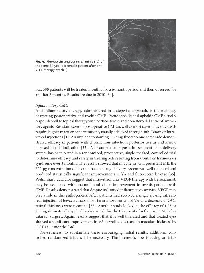
120 Buchholz · Buchholz · Augustin
out. 390 patients will be treated monthly for a 6-month period and then observed for
another 6 months. Results are due in 2010 [34].
Inflammatory CME
Anti-inflammatory therapy, administered in a stepwise approach, is the mainstay
of treating postoperative and uveitic CME. Pseudophakic and aphakic CME usually
responds well to topical therapy with corticosteroid and non-steroidal anti-inflamma-
tory agents. Resistant cases of postoperative CME as well as most cases of uveitic CME
require higher macular concentrations, usually achieved through sub-Tenon or intra-
vitreal injections [1]. An implant containing 0.59 mg fluocinolone acetonide demon-
strated efficacy in patients with chronic non-infectious posterior uveitis and is now
licensed in this indication [35]. A dexamethasone posterior-segment drug delivery
system has been tested in a randomized, prospective, single-masked, controlled trial
to determine efficacy and safety in treating ME resulting from uveitis or Irvine-Gass
syndrome over 3 months. The results showed that in patients with persistent ME, the
700-μg concentration of dexamethasone drug delivery system was well tolerated and
produced statistically significant improvements in VA and fluorescein leakage [36].
Preliminary data also suggest that intravitreal anti-VEGF therapy with bevacizumab
may be associated with anatomic and visual improvement in uveitis patients with
CME. Results demonstrated that despite its limited inflammatory activity, VEGF may
play a role in this pathogenesis. After patients had received a single 2.5-mg intravit-
real injection of bevacizumab, short-term improvement of VA and decrease of OCT
retinal thickness were recorded [37]. Another study looked at the efficacy of 1.25 or
2.5 mg intravitreally applied bevacizumab for the treatment of refractory CME after
cataract surgery. Again, results suggest that it is well tolerated and that treated eyes
showed a significant improvement in VA as well as decrease in macular thickness by
OCT at 12 months [38].
Nevertheless, to substantiate these encouraging initial results, additional con-
trolled randomized trials will be necessary. The interest is now focusing on trials
Fig. 4. Fluorescein angiogram (7 min 38 s) of
the same 54-year-old female patient after anti-
VEGF therapy (week 6).
Anti-VEGF as an Approach for Macular Edema 121
1 Johnson M: Perspective – etiology and treatment of
macular edema. Am J Ophthalmol 2009;147:11–16.
2 Catier A, Tadayoni R: Characterization of macular
edema from various etiologies by optical coherence
tomography. Am J Ophthalmol 2005;140:200–206.
3 Zietz B, Kasparbauer A, Ottman S, Spiegel D,
Palitzsch KD: Diabetic retinopathy and associated
risk factors in type 1 and type 2 diabetics in the upper
palatinate (in German). Dtsch Med Wochenschr
2000;125:783–788.
4 López IM, Diez A, Velilla S, Rueda A, Alvarez A,
Pastor CJ: Prevalence of diabetic retinopathy and
eye care in a rural area of Spain. Institute of Applied
Ophthalmobiology (IOBA), University of Valladolid,
Valladolid, Spain. Ophthalmic Epidemiol 2002;205–
214.
5 Hayward LM, Burden ML, Burden AC, Blackledge
H, Raymond NT, Botha JL, Karwakowski WSS,
Duke T, Chang YF: What is the prevalence of visual
impairment in the general and diabetic population:
Are there ethnic and gender differences? Diabet
Med 2002;19:27–34.
6 Philipps RP, Clearkin L: Screening for diabetic
retinopathy: an overview; in Somdutt Prasad, MS,
FRCS, Fellow in Diabetic Eye Disease: Arrowe Park
Hospital. Upton, Wirral. The Problem of Diabetic
Retinopathy: NHS NSC Diabetic Retinopathy
Screening (http://www.diabetic-retinopathy.screen-
ing.nhs.uk/diabetic-retinopathy.html).
7 Klein R, Klein B: The Wisconsin Epidemiologic
Study of Diabetic Retinopathy. II. Prevalence and
risk of diabetic retinopathy when age at diagnosis is
less than 30 years. Arch Ophthalmol 1984;102:520–
526.
8 Klein R, Klein B: The Wisconsin Epidemiologic
Study of Diabetic Retinopathy. XIV. Ten-year inci-
dence and progression of diabetic retinopathy. Arch
Ophthalmol 1994;112:1217–1228.
9 Klein B, Klein R: Diabetic retinopathy; in Johnson
GJ, Minassian DC (eds): The Epidemiology of Eye
Disease. New York, Oxford University Press, 2003,
pp 342–345.
10 Noma H, Funatsu H: Macular microcirculation and
macular edema in branch retinal vein occlusion. Br
J Ophthalmol 2009;93:630–633.
11 Rehak J, Rehak M: Branch retinal vein occlusions:
pathogenesis, visual prognosis, and treatment
modalities. Curr Eye Res 2008;33:111–131.
12 Soubrane G, Buchholz P, Mahabhashyam S: The
epidemiology of retinal vein occlusions in Europe: a
literature review of existing data. Euretina Annual
Meeting, Nice, France 2009.
13 Klein R: The epidemiology of retinal vein occlusion:
the Beaver Dam Eye Study. Trans Am Ophthalmol
Soc 2000;98:133–141.
14 Hartnett E, Tinkham N: Aqueous vascular endothe-
lial growth factor as a predictor of macular thicken-
ing following cataract surgery in patients with
diabetes mellitus. Am J Ophthalmol 2009;148:895–
901.
15 Jonas JB, Kreissig I, Söfker A, Degenering RF:
Intravitreal injection of triamcinolone for diffuse
diabetic macular edema. Arch Ophthalmol 2003;
121:57–61.
16 O’Doherty M, Dooley I: Interventions for diabetic
macular edema: a systematic review of the litera-
ture. Br J Ophthalmol 2008;92:1585–1587.
17 Diabetic Retinopathy Clinical Research Network: A
randomized trial comparing intravitreal triamcino-
lone acetonide and focal/grid photocoagulation for
diabetic macular edema. Ophthalmology 2008;115:
1447–1459.
18 Royal College of Ophthalmologists: Retinal vein
occlusion (RVO) interim guidelines. February 2009,
pp 15–16.
19 NHS UK – National Horizon Scanning Centre:
Ranibizumab for macular oedema secondary to
central or branch retinal vein occlusion. April 2009,
p 2.
20 Anonymous: Wirksamkeit und Sicherheit von
Ranibizumab unter Praxisbedingungen. Addendum.
Ophthalmologe 2009;106:3.
21 Nguyen QD, Shah SM: Primary endpoint (6 months)
results of ranibizumab for edema of the macula in
diabetes (READ-2) study. Ophthalmology 2009;116:
2175–2181.
22 Scott IU, Edwards AR: A phase II randomized clini-
cal trial of intravitreal bevacizumab for diabetic
macular edema Ophthalmology 2007;114:1860–
1867.
that evaluate combination therapies and/or direct comparisons between treatments.
Finally, clinical experience and the actual oxygen supply will lead the physician to an
individualized treatment in this complicated disease entity.
References
122 Buchholz · Buchholz · Augustin
23 Cunningham ET, Adamis AP: Macugen Diabetic
Retinopathy Study Group: A phase II randomized
double masked trial of pegaptanib, an anti VEGF
aptamer for diabetic macular edema. Ophthalmol-
ogy 2005;112:1747–1757.
24 Quresh M, McIntosh R: Interventions for central
retinal vein occlusions. An evidence-based system-
atic review. Ophthalmology 2007;114:507–519.
25 Luckie AP, Wroblewski JJ: A randomized prospec-
tive study of outpatient hemodilution for central
retinal vein obstruction. Aust NZ J Ophthalmol
1996;24:223–232.
26 The Central Retinal Vein Occlusion Study Group
Report: Evaluation of grid pattern photocoagula-
tion for macular edema in central retinal vein occlu-
sion: Ophthalmology 1995;102:1434–1444.
27 Ip MS, Scott IU: A randomized trial comparing the
efficacy and safety of intravitreal triamcinolone
with observation to treat vision loss associated with
macular edema secondary to central retinal vein
occlusion: the Standard Care versus Corticosteroid
for Retinal Vein Occlusion (SCORE) study report 5.
Arch Ophthalmol 2009;127:1101–1114.
28 h t t p : / / w w w . c l i n t r i a l s . g o v / c t 2 / s h o w /
NCT00168324/00168298.
29 Pieramici DJ, Rabena M: Ranibizumab for the treat-
ment of macular edema associated with perfused
central retinal vein occlusions. Ophthalmology
2008;115:e47–e54.
30 The Branch Vein Occlusion Study Group (BVOS):
Argon laser photocoagulation for macular edema in
branch vein occlusion. Am J Ophthalmol 1984;98:
271–282.
31 Scott IU, Ip MS: A randomized trial comparing the
efficacy and safety of intravitreal triamcinolone with
standard care to treat vision loss associated with
macular edema secondary to branch retinal vein
occlusion: the Standard Care versus Corticosteroid
for Retinal Vein Occlusion (SCORE) study report 6.
Arch Ophthalmol 2009;127:1115–1128.
32 h t t p : / / w w w . c l i n t r i a l s . g o v / c t 2 / s h o w /
NCT00168324/00168298.
33 Prager F, Michels S: Intravitreal bevacizumab
(Avastin) for macular edema secondary to retinal
vein occlusion – twelve month results of a prospec-
tive clinical trial. Br J Ophthalmol 2009;93:452–456.
34 NHS UK – National Horizon Scanning Centre:
Ranibizumab for macular oedema secondary to cen-
tral or branch retinal vein occlusion. April 2009, p 3
( h t t p : / / w w w. c l i n t r i a l s . g o v / c t 2 / s h o w /
NCT00486018).
35 Jaffe GJ, Martin D: Fluocinolone acetonide implant
(Retisert) for non-infectious posterior uveitis: 34
week results of a multicentre randomized clinical
study. Ophthalmology 2006;113:1020–1027.
36 Williams GA, Haller JA: Dexamethasone posterior-
segment drug delivery system in the treatment of
macular edema resulting from uveitis or Irvine-
Gass syndrome. Am J Ophthalmol 2009;147:1048–
1054.
37 Cordero Coma M, Sobrin L: Intravitreal bevaci-
zumab for treatment if uveitic macular edema.
Ophthalmology 2007;114:1574–1579.
38 Arevalo F, Maia M: Intravitreal bevacizumab for
refractory pseudophakic cystoids macular edema:
The Pan-American Collaborative Retina Study
Group. Ophthalmology 2009;116:1481–1487.
Prof. A.J. Augustin
Augenklinik
Moltkestrasse 90
DE–76133 Karlsruhe (Germany)
Tel. +49 721 9742001, Fax +49 721 9742009, E-Mail [email protected]
Bandello F, Battaglia Parodi M (eds): Anti-VEGF.
Dev Ophthalmol. Basel, Karger, 2010, vol 46, pp 123–132
Angiogenesis and Vascular Endothelial Growth Factors in Intraocular TumorsGuy S. Missottena,b � Reinier O. Schlingemannb � Martine J. Jagerc
aDepartment of Ophthalmology, Catholic University Leuven, Belgium; bDepartment of Ophthalmology,
Academic Medical Center, Amsterdam, and cDepartment of Ophthalmology, Leiden University, Leiden,
The Netherlands
AbstractThe role of angiogenesis in tumors appears obvious: without vessels, tumors cannot grow. However,
the long-held belief that all human solid tumors are angiogenesis-dependent has been challenged
by the universally disappointing results of anti-angiogenesis therapy in cancer. This may be explained
by the fact that cooption of preexisting vasculature as a primary or secondary mechanism of tumor
vascularization is more important than previously thought. Nevertheless, anti-angiogenesis therapy
may play an important (adjuvant) role in the prevention of metastases of intraocular tumors (uveal
melanoma and retinoblastoma). Antivascular endothelial growth factor therapy already plays an
important role in the management of irradiation complications in tumor eyes.
Copyright © 2010 S. Karger AG, Basel
The most frequent intraocular tumors are retinoblastoma in children (incidence
6–12 per million children per year) and uveal melanoma in adults (incidence 6–8 per
million per year) [1]. A special characteristic of intraocular tumors is their pathway
of metastasis: with the exception of massive extraocular growth or optic nerve out-
growth in retinoblastoma, metastatic dissemination is mainly hematogenous due to
the absence of lymphatic vessels in the eye.
The role of angiogenesis in tumors appears obvious: without vessels, tumors can-
not grow. In the 1970s, Folkman and co-workers started to work on angiogenesis,
particularly in ophthalmology, which opened a large field of investigation. Folkman’s
hypothesis that solid tumors are angiogenesis-dependent, initiated studies of angio-
genesis in tumor biology [2]. Vascular endothelial growth factor (VEGF) and many
other angiogenic proteins were found to play a major role in ocular and general
angiogenesis.
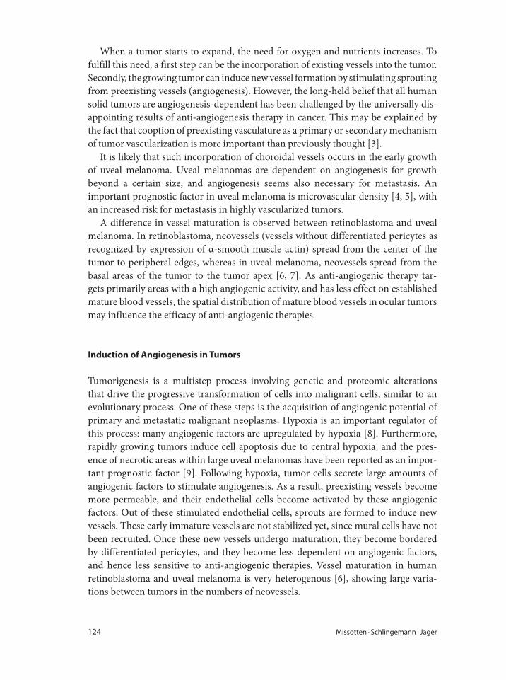
124 Missotten · Schlingemann · Jager
When a tumor starts to expand, the need for oxygen and nutrients increases. To
fulfill this need, a first step can be the incorporation of existing vessels into the tumor.
Secondly, the growing tumor can induce new vessel formation by stimulating sprouting
from preexisting vessels (angiogenesis). However, the long-held belief that all human
solid tumors are angiogenesis-dependent has been challenged by the universally dis-
appointing results of anti-angiogenesis therapy in cancer. This may be explained by
the fact that cooption of preexisting vasculature as a primary or secondary mechanism
of tumor vascularization is more important than previously thought [3].
It is likely that such incorporation of choroidal vessels occurs in the early growth
of uveal melanoma. Uveal melanomas are dependent on angiogenesis for growth
beyond a certain size, and angiogenesis seems also necessary for metastasis. An
important prognostic factor in uveal melanoma is microvascular density [4, 5], with
an increased risk for metastasis in highly vascularized tumors.
A difference in vessel maturation is observed between retinoblastoma and uveal
melanoma. In retinoblastoma, neovessels (vessels without differentiated pericytes as
recognized by expression of α-smooth muscle actin) spread from the center of the
tumor to peripheral edges, whereas in uveal melanoma, neovessels spread from the
basal areas of the tumor to the tumor apex [6, 7]. As anti-angiogenic therapy tar-
gets primarily areas with a high angiogenic activity, and has less effect on established
mature blood vessels, the spatial distribution of mature blood vessels in ocular tumors
may influence the efficacy of anti-angiogenic therapies.
Induction of Angiogenesis in Tumors
Tumorigenesis is a multistep process involving genetic and proteomic alterations
that drive the progressive transformation of cells into malignant cells, similar to an
evolutionary process. One of these steps is the acquisition of angiogenic potential of
primary and metastatic malignant neoplasms. Hypoxia is an important regulator of
this process: many angiogenic factors are upregulated by hypoxia [8]. Furthermore,
rapidly growing tumors induce cell apoptosis due to central hypoxia, and the pres-
ence of necrotic areas within large uveal melanomas have been reported as an impor-
tant prognostic factor [9]. Following hypoxia, tumor cells secrete large amounts of
angiogenic factors to stimulate angiogenesis. As a result, preexisting vessels become
more permeable, and their endothelial cells become activated by these angiogenic
factors. Out of these stimulated endothelial cells, sprouts are formed to induce new
vessels. These early immature vessels are not stabilized yet, since mural cells have not
been recruited. Once these new vessels undergo maturation, they become bordered
by differentiated pericytes, and they become less dependent on angiogenic factors,
and hence less sensitive to anti-angiogenic therapies. Vessel maturation in human
retinoblastoma and uveal melanoma is very heterogenous [6], showing large varia-
tions between tumors in the numbers of neovessels.
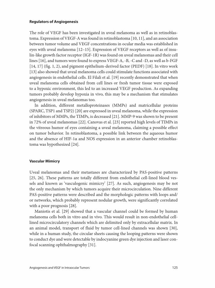
Angiogenesis and VEGF in Intraocular Tumors 125
Regulators of Angiogenesis
The role of VEGF has been investigated in uveal melanoma as well as in retinoblas-
toma. Expression of VEGF-A was found in retinoblastoma [10, 11], and an association
between tumor volume and VEGF concentrations in ocular media was established in
eyes with uveal melanoma [12–15]. Expression of VEGF receptors as well as of insu-
lin-like growth factor receptor (IGF-1R) was found on uveal melanomas and their cell
lines [16], and tumors were found to express VEGF-A, -B, -C and -D, as well as b-FGF
[14, 17] (fig. 1, 2), and pigment epithelium-derived factor (PEDF) [18]. In vitro work
[13] also showed that uveal melanoma cells could stimulate functions associated with
angiogenesis in endothelial cells. El Filali et al. [19] recently demonstrated that when
uveal melanoma cells obtained from cell lines or fresh tumor tissue were exposed
to a hypoxic environment, this led to an increased VEGF production. As expanding
tumors probably develop hypoxia in vivo, this may be a mechanism that stimulates
angiogenesis in uveal melanomas too.
In addition, different metalloproteinases (MMPs) and matricellular proteins
(SPARC, TSP1 and TSP2) [20] are expressed in uveal melanoma, while the expression
of inhibitors of MMPs, the TIMPs, is decreased [21]. MMP-9 was shown to be present
in 72% of uveal melanomas [22]. Canovas et al. [23] reported high levels of TIMPs in
the vitreous humor of eyes containing a uveal melanoma, claiming a possible effect
on tumor behavior. In retinoblastoma, a possible link between the aqueous humor
and the absence of HIF-1α and NOS expression in an anterior chamber retinoblas-
toma was hypothesized [24].
Vascular Mimicry
Uveal melanomas and their metastases are characterized by PAS-positive patterns
[25, 26]. These patterns are totally different from endothelial cell-lined blood ves-
sels and known as ‘vasculogenic mimicry’ [27]. As such, angiogenesis may be not
the only mechanism by which tumors acquire their microcirculation. Nine different
PAS-positive patterns were described and the morphologic patterns with loops and/
or networks, which probably represent nodular growth, were significantly correlated
with a poor prognosis [28].
Maniotis et al. [29] showed that a vascular channel could be formed by human
melanoma cells both in vitro and in vivo. This would result in non-endothelial cell-
lined microcirculatory channels which are delimited only by extracellular matrix. In
an animal model, transport of fluid by tumor cell-lined channels was shown [30],
while in a human study, the circular sheets causing the looping patterns were shown
to conduct dye and were detectable by indocyanine green dye injection and laser con-
focal scanning ophthalmography [31].
126 Missotten · Schlingemann · Jager
1 2 3 4 50
Primary tumor cell cultures
200
400
600
800
1,000
VEGF-A
(pg
/ml)
NormoxiaHypoxia
Fig. 1. VEGF-A protein expres-
sion in primary uveal mela-
noma cultures. The amount of
VEGF-A protein expression
measured with ELISA in super-
natant of primary uveal mela-
noma cell cultures (cultures
1–5) under normoxic (black)
and hypoxic (gray) exposure
after 24 h. Expression is dem-
onstrated in amount protein
(pg/ml). 130 × 86 mm (300 ×
300 dpi) [from 19].
Fig. 2. Expression of VEGF-A
in human uveal melanoma tis-
sue. In situ hybridization of
VEGF-A with uveal melanoma
demonstrated diffuse VEGF-A
expression throughout the
tumors, especially around
blood vessels [from 15].
Angiogenesis and VEGF in Intraocular Tumors 127
Inflammation and Angiogenesis
In cancer, chronic inflammation has been demonstrated within tumor foci and a
number of studies suggest that tumor-associated macrophages promote the growth,
proliferation, and metastasis of neoplastic cells and can induce angiogenesis leading
to tumor progression. In uveal melanoma, PAS-positive patterns were associated with
endothelial cells expressing endothelial monocyte-activating polypeptide (EMAP)-II,
a cytokine that attracts macrophages [32]. The presence of high numbers of mac-
rophages carries an adverse prognosis in uveal melanoma [33]. As tumors with high
numbers of infiltrating macrophages also show an increased expression of HLA class
I and II and a lymphocytic infiltrate, we coined the term inflammatory phenotype for
this category of uveal melanomas [34].
Radiation Retinopathy
Eyes that had to be removed because of complications after irradiation of uveal mela-
noma showed an increase in the production of VEGF in the eye, probably as a result
of the tissue hypoxia caused by enhanced destruction of vascular endothelial cells in
the tumor [15]. This would suggest that complications of radiation retinopathy might
be susceptible to treatment with VEGF inhibitors.
Radiation retinopathy is an occlusive microangiopathy, with focal capillary clo-
sure, relative early sparing of pericytes and irregular dilation of the neighboring
microvasculature [35, 36]. Eventually, capillaries as well as pericytes are lost, with
resulting inner retinal non-perfusion and inner and outer retinal atrophy. Macular
edema is one of the earliest signs of radiation maculopathy. Shields et al. [37] defined
clinical radiation maculopathy as retinal capillary bed changes (dilation, microaneu-
rysm, retinal hemorrhage), retinal edema, retinal exudation, nerve fiber layer infarc-
tion, or vascular sheathing affecting the area within 3 mm of the foveola. Radiation
retinopathy generally occurs more than 1 year after brachytherapy. In an analysis of
1,300 patients treated with plaque radiotherapy for posterior uveal melanoma, clini-
cal evidence of radiation maculopathy was found in 43% of patients at 5-year follow-
up [38]. In a study on 125 patients, 17% had macular edema at 6 months, 40% at 12
months, 57% at 18 months and 61% at 24 months, despite treatment (transpupillary
thermotherapy and laser coagulation) [39].
The size of the tumor, the radiation dose to the tumor base, and the radiation dose
to the optic disk are all significant factors in the development of macular edema [39].
Depending on the size of the tumor, radiation retinopathy can be up to 25% and neo-
vascular glaucoma may occur in 15% [40], especially after proton beam therapy for
extremely large tumors [41].
128 Missotten · Schlingemann · Jager
Local Treatment of Radiation Retinopathy
One of the treatments of radiation retinopathy is intravitreal injection of triamci-
nolone, a corticosteroid. This drug had no effect on the growth of uveal melanoma
cells in vitro [42]. It is currently investigated whether VEGF inhibitors have any effect
on tumor cells in vitro, as different receptors and mRNA of VEGF-A isoforms are
expressed in uveal melanoma cells [17, 43]. The effectiveness of the use of triamci-
nolone and anti-angiogenic treatments is being investigated in tumor eyes suffering
from radiation retinopathy. A prospective study including 163 patients treated with
three injections of periocular triamcinolone during the first year after brachytherapy
showed [37] a benefit of treatment in the first 18 months after plaque radiotherapy and
a significant reduction of the number of patients with poor visual acuity. Following
injection of bevacizumab, 1 case of extensive cystoid macular edema showed an
increase of visual acuity and decrease of central retinal optical coherence tomography
thickness in the first months after the injection [44].
Treatment of Radiation-Induced Neovascular Glaucoma
Neovascular glaucoma can occur following radiation therapy of large ocular tumors.
An intravitreal injection of bevacizumab (1.25 mg) can cause regression of iris and
angle neovascularization within 1 week [45–47], both in iris and posterior segment
melanoma, and resolution of a serous detachment within 1 month (intravitreal injec-
tion 14 months following iodine brachytherapy). Rapid regression was also seen with
intravitreal injection with ranibizumab, with an improvement of rubeosis iridis 2 days
after administration, and this effect lasted 6 months [48].
Metastasis and Angiogenesis
The primary site for metastases of uveal melanoma is the liver, but metastases may
also occur in the lung, skin and brain. Micrometastasis of approximately 100 μm are
assumed to remain dormant in the liver for many years, and are thought to become
only active after angiogenesis is induced. In a murine model, metastatic uveal mela-
noma cells are shown to express higher VEGF levels than the primary tumor cells
[49]. Nevertheless, no correlation between VEGF expression on the primary tumor
and metastasis formation was found [12]. Patients with clinical metastases have higher
VEGF levels in their serum compared to patients without metastases [19].
For metastasis treatment, combining various agents with different activities, includ-
ing anti-angiogenic functions, may be a reasonable approach. A study combining low-
dose thalidomide and interferon-α2b in 6 metastatic uveal melanoma patients achieved
a stabilization of metastases in the liver and lung that lasted 12 months [50].
Angiogenesis and VEGF in Intraocular Tumors 129
In an animal model, low-dose angiostatin could reduce the number of hepatic
micrometastases in a murine ocular melanoma model [51]. It was assumed that an
alteration of VEGF expression occurred in the melanoma cells and that angiostatin
inhibited migration of the melanoma cells [49]. Inhibition of VEGF in a mouse model
of retinoblastoma with intravitreal bevacizumab showed a decrease in the growth of
retinoblastoma in vitro and vivo [52]. Similar results were found in a murine model of
uveal melanoma [53]. PEDF is an angiogenesis inhibitor that balances angiogenesis in
the eye and blocks tumor progression. Although PEDF has no effect on proliferation
of retinoblastoma cells in vitro, treatment with PEDF injections inhibited the growth
of retinoblastoma xenografts in vivo by downregulating HIF-1α and VEGF [53]. Also,
PEDF overexpression in uveal melanoma cells inhibited intraocular growth and the
development of liver metastases in a murine model [53]. The use of adjuvant anecor-
tave acetate also reduced the tumor load significantly in a retinoblastoma mouse
model [54]. However, this treatment induced the development of hypoxic areas in
the tumor [55]. The addition of carboplatin and also of glycolytic inhibitors had an
additional treatment effect [56].
In vitro, reduction of VEGF and tumor growth was reached with RNAi targeting
VEGF [57]. Nevertheless, the heterogeneity and spatial distribution of vessels, both in
retinoblastoma as well as in uveal melanoma, limits anti-angiogenic treatments that
only target immature vasculature. Anti-angiogenesis treatment of uveal melanoma
metastases in humans has not yet been reported.
Picropodophyllin, an inhibitor of IGF-1R, was shown to block uveal melanoma
growth [16]. As this drug can be given orally, clinical studies are anxiously awaited.
Inhibition of ocular tumor growth was also achieved in an in vivo model by gene
transduction which led to an increase in interleukin-12, a strong anti-angiogenic
cytokine [58].
Treatment
The final proof of principle that cancer patients can be effectively treated with angio-
genesis inhibitors is still awaited [3, 59]. Various preclinical in vitro and in vivo exper-
iments have proven that most tumors need new vessel formation in order to grow and
to form metastases, and this is also true for intraocular tumors. First of all, tumors
placed in the avascular cornea do not grow until new blood vessels reach the implant.
Secondly, the introduction of only one angiogenic gene can cause a switch from tumor
dormancy to progressive tumor growth. Thirdly, tumor growth can be inhibited and
sometimes tumor regression can be obtained just by attacking the vascular compart-
ment with specific angiogenesis inhibitors [3].
In these model systems, angiogenesis is necessary for tumors >2 mm, and there are
no indications that the same should not be true for metastases. This would say that
(multiple?) metastases can form without any need for angiogenesis, especially the
130 Missotten · Schlingemann · Jager
1 Egan KM, Seddon JM, Glynn RJ, Gragoudas ES,
Albert DM: Epidemiologic aspects of uveal mela-
noma. Surv Ophthalmol 1988;32:239–251.
2 Folkman J: Tumour angiogenesis: therapeutic impli-
cations. N Engl J Med 1971;285:1182–1186.
3 Verheul HM, Voest EE, Schlingemann RO: Are
tumours angiogenesis-dependent? J Pathol 2004;
202:5–13.
4 Foss AJ, Alexander RA, Jefferies LW, Hungerford JL,
Harris AL, Lightman S: Microvessel count predicts
survival in uveal melanoma. Cancer Res 1996;56:
2900–2903.
5 Makitie T, Summanen P, Tarkkanen A, Kivela T:
Microvascular density in predicting survival of
patients with choroidal and ciliary body melanoma.
Invest Ophthalmol Vis Sci 1999;40:2471–2480.
6 Pina Y, Boutrid H, Schefler A, Dubovy S, Feuer W,
Jockovich ME, Murray TG: Blood vessel maturation
in retinoblastoma tumors: spatial distribution of
neovessels and mature vessels and its impact on
ocular treatment. Invest Ophthalmol Vis Sci 2009;
50:1020–1024.
7 Pina Y, Cebulla CM, Murray TG, Alegret A, Dubovy
SR, Boutrid H, Feuer W, Mutapcic L, Jockovich ME:
Blood vessel maturation in human uveal melanoma:
spatial distribution of neovessels and mature vascu-
lature. Ophthalmic Res 2009;41:160–169.
8 Witmer AN, Vrensen GF, van Noorden CJ, Schlin-
gemann RO: Vascular endothelial growth factors
and angiogenesis in eye disease. Prog Retin Eye Res
2003;22:1–29.
9 Missotten GS: Doctoral Thesis. Angiogenesis and
Screening in Uveal Melanoma. Leiden, The Nether-
lands, 2005.
10 Kvanta A, Steen B, Seregard S: Expression of VEGF
in retinoblastoma but not in posterior uveal mela-
noma. Exp Eye Res 1996;63:511–518.
11 Stitt AW, Simpson DA, Boocock C, Gardiner TA,
Murphy GM, Archer DB: Expression of VEGF and
its receptors is regulated in eyes with intraocular
tumours. J Pathol 1998;186:306–312.
12 Sheidow TG, Hooper PL, Crukley C, Young J,
Heathcote JG: Expression of VEGF in uveal mela-
noma and its correlation with metastasis. Br J
Ophthalmol 2000;84:750–756.
13 Boyd SR, Tan D, Bruce C, Gittos A, Neale MH,
Hungerford JL, Charnock-Jones S, Cree IA: Vascular
endothelial growth factor is elevated in ocular fluids
of eyes harbouring uveal melanoma: identification
of a potential therapeutic window. Br J Ophthalmol
2002;86:448–452.
14 Boyd SR, Tan DS, de Souza L, Neale MH, Myatt NE,
Alexander RA, Robb M, Hungerford JL, Cree IA:
Uveal melanomas express vascular endothelial
growth factor and basic fibroblast growth factor and
support endothelial cell growth. Br J Ophthalmol
2002;86:440–447.
15 Missotten GS, Notting IC, Schlingemann RO, Zijl-
mans HJ, Lau C, Eilers PH, Keunen JE, Jager MJ:
Vascular endothelial growth factor-A in eyes with
uveal melanoma. Arch Ophthalmol 2006;124:1428–
1434.
micrometastasis that are assumed to exist in uveal melanoma. Anti-angiogenesis may
play an important role as adjuvant therapy, but a complete remediation of metastatic
tumor disease based only on this mechanism seems unlikely.
After a few years of clinical experience with other malignancies, a number of con-
siderations and questions have emerged. First, the idea of starving tumors has been
called into question, because VEGF-R treatment not only starves tumors but also
normalizes the highly abnormal tumor vasculature, thereby yielding the advantage
of improving the delivery of cytotoxic drugs. Secondly, VEGF-R inhibitory treatment
is limited by resistance/escape, and provides no definitive cure, but survival prolon-
gation in the range of months rather than years. Also, VEGF-R inhibitors induce
adverse effects because of the importance of VEGF signaling for the maintenance of
quiescent endothelium in healthy organs, and probably of the neuroretina in the eye
[8, 60]. Thus, anti-VEGF therapy as monotherapy of intraocular tumors or its metas-
tases will not be an option, and novel strategies focusing on combination treatments
are needed.
References
Angiogenesis and VEGF in Intraocular Tumors 131
16 Economou MA, Andersson S, Vasilcanu D, All-
Ericsson C, Menu E, Girnita A, Girnita L, Axelson
M, Seregard S, Larsson O: Oral picropodophyllin is
well tolerated in vivo and inhibits IGF-1R expres-
sion and growth of uveal melanoma. Invest Oph-
thalmol Vis Sci 2008;49:2337–2342.
17 Notting IC, Missotten GS, Sijmons B, Boonman ZF,
Keunen JE, van der Pluijm G: Angiogenesic profile
of uveal melanoma. Curr Eye Res 2006;31:775–785.
18 Yang H, Cheng R, Liu G, Zhong Q, Li C, Cai W,
Yang Z, Ma J, Yang X, Gao G: PEDF inhibits growth
of retinoblastoma by anti-angiogenic activity.
Cancer Sci 2009;100:2419–2425.
19 El Filali M, Missotten GS, Maat W, Ly LV, Luyten
GPM, van der Velden PA, Jager MJ: Invest Ophthal-
mol Vis Sci 2010;51:2329–2337.
20 Ordonez JL, Paraoan L, Hiscott P, Gray D, Garcia-
Finana M, Grierson I, Damato B: Differential exp-
ression of angioregulatory matricellular proteins in
posterior uveal melanoma. Melanoma Res 2005;15:
495–502.
21 Van der Velden PA, Zuidervaart W, Hurks MH,
Pavey S, Ksander BR, Krijgsman E, Frants RR,
Tensen CP, Willemze R, Jager MJ, Gruis NA: Exp-
ression profiling reveals that methylation of TIMP-3
is involved in uveal melanoma development. Int J
Cancer 2003;106:472–479.
22 Sahin A, Kiratli H, Tezel GG, Soylemezoglu F, Bilgic
S: Expression of vascular endothelial growth factor
A, matrix metalloproteinase-9 and extravascular
matrix patterns in iris and ciliary body melanomas.
Ophthalmic Res 2007;39:40–44.
23 Canovas D, Rennie IG, Nichols CE, Sisley K: Local
environmental influences on uveal melanoma.
Vitreous humor promotes uveal melanoma inva-
sion, whereas the aqueous can be inhibitory. Cancer
2008;112:1787–1794.
24 Crosby MB, Hubbard GB, Gallie BL, Grossniklaus
HE: Anterior diffuse retinoblastoma: mutational
analysis and immunofluorescence staining. Arch
Pathol Lab Med 2009;133:1215–1218.
25 Rummelt V, Mehaffey MG, Campbell RJ, Pe’er J,
Bentler SE, Woolson RF, Naumann GO, Folberg R:
Microcirculation architecture of metastases from
primary ciliary body and choroidal melanomas. Am
J Ophthalmol 1998;126:303–305.
26 Folberg R, Rummelt V, Parys-van Ginderdeuren R,
Hwang T, Woolson RF, Pe’er J, Gruman LM: The
prognostic value of tumour blood vessel morphol-
ogy in primary uveal melanoma. Ophthalmology
1993;100:1389–1398.
27 Bhat P, Jakobiec FA, Folberg R: Comparison of
tumor-associated vasculatures in uveal and cutane-
ous melanomas. Semin Ophthalmol 2009;24:166–
171.
28 Makitie T, Summanen P, Tarkkanen A, Kivela T:
Microvascular loops and networks as prognostic
indicators in choroidal and ciliary body melanomas.
J Natl Cancer Inst 1999;91:359–367.
29 Maniotis AJ, Folberg R, Hess A, Seftor EA, Gardner
LMG, Pe’er J, Trent JM, Meltzer PS, Hendrix MJC:
Vascular channel formation by human melanoma
cells in vivo and in vitro; vascular mimicry. Am J
Pathol 1999;155:739–752.
30 Clarijs R, Otte-Holler I, Ruiter DJ, de Waal RMW:
Presence of a fluid-conducting meshwork in xeno-
grafted cutaneous and primary human uveal mela-
noma. Invest Ophthalmol Vis Sci 2002;43:912–918.
31 Shields CL, Shields JA, De Potter P: Patterns of
indocyanine green videoangiography of choroidal
tumors. Br J Ophthalmol 1995;79:237–245.
32 Clarijs R, Schalkwijk L, Ruiter DJ, de Waal RM:
EMAP-II expression is associated with macrophage
accumulation in primary uveal melanoma. Invest
Ophthalmol Vis Sci 2003;44:1801–1806.
33 Makitie T, Summanen P, Tarkkanen A, Kivela T:
Tumor-infiltrating macrophages (CD68+ cells) and
prognosis in malignant uveal melanoma. Invest
Ophthalmol Vis Sci 2001;42:1414–1421.
34 Maat W, Ly LV, Jordanova ES, de Wolff-Rouendaal
D, Schalij-Delfos NE, Jager MJ: Monosomy of chro-
mosome 3 and an inflammatory phenotype occur
together in uveal melanoma. Invest Ophthalmol Vis
Sci 2008;49:505–510.
35 Archer DB: Doyne Lecture. Responses of retinal
and choroidal vessels to ionizing radiation. Eye
1993;7:1–13.
36 Irvine AR, Woor IS: Radiation retinopathy as an
experimental model for ischemic proliferative retin-
opathy and rubeosis iridis. Am J Ophthalmol 1987;
103:790–797.
37 Horgan N, Shields CL, Mashayekhi A, Salazar PF,
Materin MA, O’Reagan M, Shields JA: Periocular
triamcinolone for prevention of macular edema
after plaque radiotherapy of uveal melanoma: a ran-
domized controlled trial. Arch Ophthalmol 2009;
116:1383–1390.
38 Gunduz K, Shields CL, Shileds JA, Cater J, Freire JE,
Brady LW: Radiation retinopathy following plaque
radiotherapy for posterior uveal melanoma. Arch
Ophthalmol 1999;117:609–614.
39 Horgan N, Shields CL, Mashayekhi A, Teixeira LF,
Materin MA, Shields JA: Early macular morpho-
logical changes following plaque radiotherapy for
uveal melanoma. Retina 2008;28:263–273.
40 Krema H, Simpson ER, Pavlin CJ, Payne D, Vasquez
LM, McGowan H: Management of ciliary body mel-
anoma with iodine-125 plaque therapy. Can J
Ophthalmol 2009;44:395–400.
132 Missotten · Schlingemann · Jager
41 Boyd SR, Gittos A, Richter M, Hungerford JL,
Errington RD, Cree IA: Proton beam therapy and
iris neovascularisation in uveal melanoma. Eye
2006;20:832–836.
42 El Filali M, Homminga I, Maat W, Van der Velden
PA, Jager MJ: Triamcinolone acetonide and anecor-
tave acetate do not stimulate uveal melanoma cell
growth. Mol Vis 2008;14:1752–1759.
43 Ijland SA, Jager MJ, Heijdra BM, Westphal JR, Peek
R: Expression of angiogenic and immunosuppres-
sive factors by uveal melanoma cell Lines. Melanoma
Res 1999;9:445–450.
44 Ziemssen F, Voelker M, Altpeter E, Bartz-Schmidt
KU, Gelisken F: Intravitreal bevacizumab treatment
of radiation maculopathy due to brachytherapy in
choroidal melanoma. Acta Ophthalmol 2007;85:
579–580.
45 Jaissle GB, Ulmer A, Henke-Fahle S, Fierlbeck G,
Bartz-Schmidt KU, Szurman P: Suppression of mel-
anoma-associated neoangiogenesis by bevacizumab.
Arch Dermatol 2008;144:525–527.
46 Bianciotto C, Shields CL, Kang B, Shields JA: Treat-
ment of iris melanoma and secondary neovascular
glaucoma using bevacizumab and plaque radiother-
apy. Arch Ophthalmol 2008;126:578–579.
47 Vásquez LM, Somani S, Altomare F, Simpson ER:
Intracameral bevacizumab in the treatment of neo-
vascular glaucoma and exudative retinal detach-
ment after brachytherapy in choroidal melanoma.
Can J Ophthalmol 2009;44:106–107.
48 Dunavoelgyi R, Zehetmayer M, Simader C,
Schmidt-Erfurth U: Rapid improvement of radia-
tion-induced neovascular glaucoma and exudative
retinal detachment after a single intravitreal ranibi-
zumab injection. Clin Exp Ophthalmol 2007;35:878–
880.
49 Yang H, Xu Z, Luvone PM, Grossniklaus HE: Angio-
statin decreases cell migration and VEGF to PEDF
RNA ratio in vitro and in a murine ocular mela-
noma model. Mol Vis 2006;12:511–517.
50 Solti M, Berd D, Mastrangelo MJ, Sato T: A pilot
study of low-dose thalidomide and interferon-α2b in
patients with metastatic melanoma who failed prior
treatment. Melanoma Res 2007;17:225–231.
51 Yang H, Akor C, Dithmar S, Grossniklaus HE: Low-
dose adjuvant angiostatin decreases hepatic micro-
metastasis in murine ocular melanoma model. Mol
Vis 2004;10:987–995.
52 Lee SY, Kim DK, Cho JH, Koh JY, Yoon YH:
Inhibitory effect of bevacizumab on the angiogene-
sis and growth of retinoblastoma. Arch Ophthalmol
2008;126:953–958.
53 Yang H, Grossniklaus HE: Constitutive overexpres-
sion of pigment epithelium-derived factor inhibits
ocular melanoma growth and metastasis. Invest
Ophthalmol Vis Sci 2010;51:28–34.
54 Jockovich M, Murray TG, Escalona-Benz E, Her-
nandez E, Feuer W: Anecortave acetate as single and
adjuvant therapy in the treatment of retinal tumors
of LHBETATAG mice. Invest Ophthalmol Vis Sci
2006;47:1264–1268.
55 Boutrid H, Pina Y, Cebulla CM, Feuer WJ, Lampidis
TJ, Jockovich ME, Murray TG: Invest Ophthalmol
Vis Sci 2009;50:5537–5543.
56 Boutrid H, Jockovich ME, Murray TG, Pina Y, Feuer
WJ, Lampidis TJ, Cebulla CM: Targeting hypoxia, a
novel treatment for advanced retinoblastoma. Invest
Ophthalmol Vis Sci 2008;49:2799–2805.
57 Jia RB, Zhang YX, Zhou YX, Song X, Liu HY, Wang
LZ, Luo M, Lu J, Ge SF, Fan XQ: VEGF-targeted
RNA interference suppresses angiogenesis and
tumor growth of retinoblastoma. Ophthalmic Res
2007;32:108–115.
58 Albini A, Fassini G, Nicolo M, Dell’Eva R, Vene R,
Cammarota R, Barberis M, Noonan DM: Inhibition
of a vascular ocular tumor growth by IL-12 gene
transfer. Clin Exp Metastasis 2007;24:485–493.
59 Jager MJ: Angiogenesis in uveal melanoma. Oph-
thalmic Res 2006;38:248–250.
60 Saint-Geniez M, Maharaj AS, Walshe TE, Tucker
BA, Sekiyama E, Kurihara T, Darland DC, Young
MJ, D’Amore PA: Endogenous VEGF is required for
visual function: evidence for a survival role on
Müller cells and photoreceptors. PLoS One 2008;3:
e3554.
Guy S. Missotten
Department Ophthalmology, Academic Medical Center
Meibergdreef 9, Post Box 22660
NL–1100DD Amsterdam (The Netherlands)
Tel. +31 20 566 5907, Fax +31 20 566 9078, E-Mail [email protected]
Bandello F, Battaglia Parodi M (eds): Anti-VEGF.
Dev Ophthalmol. Basel, Karger, 2010, vol 46, pp 133–139
Antivascular Endothelial Growth Factors in Anterior Segment DiseasesStefan Scholl � Janna Kirchhof � Albert J. Augustin
Augenklinik, Karlsruhe, Germany
AbstractProangiogenic growth factors, mainly VEGF (vascular endothelial growth factor) play a significant
role in anterior segment diseases, characterized by neovascularization. Newly grown vessels in the
cornea can lead to an impairment of transparency and visual acuity. Neovascularization of the iris
(rubeosis iridis) and the anterior chamber angle are caused by ischemic retinopathies, usually lead-
ing to neovascular glaucoma with serious loss of vision. A pterygium is characterized, amongst oth-
ers, by fibrovascular proliferation and may have vision threatening consequences if left untreated.
Several antiangiogenic drugs have evolved in the last decade, mainly used for the treatment of chor-
oidal neovascularization in age-related macular degeneration. Bevacizumab though, is also widely
used off-label, in topic form or as an intracameral injection, to treat anterior segment neovasculariza-
tion with encouraging results. Copyright © 2010 S. Karger AG, Basel
Antivascular Endothelial Growth Factor in Corneal Neovascularization
Under physiologic conditions the cornea has the unique feature of being avascular,
actively maintained by expression of antiangiogenic and antilymphangiogenic fac-
tors. Under pathologic conditions, vessels invade the cornea form the limbal vascular
plexus. Corneal neovascularization (NV) is a final pathway common to numerous
ocular insults and disorders such as infection, inflammation, ischemia, degeneration,
loss of the limbal stem cell barrier and trauma. The result is an impairment in cor-
neal transparency and visual acuity. Occasionally, the newly grown vessels can serve a
beneficial role in clearing infections, wound healing and arresting stromal melts. The
disadvantages overweigh though. Corneal NV often causes tissue scarring, corneal
edema, lipid deposition and persistent inflammation. Corneal NV has been reported
in about 4% of patients presenting for general ophthalmologic care in the USA, rep-
resenting an estimated 1.4 million individuals [1]. About 12% of these cases are asso-
ciated with an impairment in visual acuity [1]. The most common causes of corneal
134 Scholl · Kirchhof · Augustin
infectious blindnesses in the Western world (herpetic keratitis) and the developing
countries (trachoma) are accompanied by corneal NV.
An extended wearing of soft contact lenses is another major cause of corneal NV.
It is estimated that about 125,000–470,000 people in the USA wearing soft contact
lenses show a certain degree of corneal NV [1].
An impaired visual acuity is not the only negative result of newly grown cor-
neal vessels. Corneal NV leads to the loss of the immune privilege of the cornea.
The result is a worsening of the prognosis of penetrating keratoplasty being a major
cause for corneal graft rejection. On the other hand, various risk factors have been
shown to be associated with an increased likelihood of corneal NV after penetrating
keratoplasty [2].
Currently, topical steroids remain the first-choice therapy, because corneal NV is
assumed to be secondary to some degree of inflammation. Well-known side effects
include cataract, glaucoma and the increased risk for infections. When inflammation
is not the cause, such as in diseases associated with deficiency of limbal cells or cor-
neal hypoxia, anti-inflammatory corticosteroids have little or no effect on the growth
of the vessels. Other therapeutic options include topical non-steroidal anti-inflam-
matory agents, laser photocoagulation, fine-needle diathermy, photodynamic therapy
and restoration of the ocular surface with the use of conjunctival, limbal or amniotic
membrane transplantation. The treatment is often ineffective or vessel recanalization
occurs, requiring multiple treatment sessions.
The rapid progress in angiogenesis research in the last few years has led to the
development of several novel, specific antiangiogenic drugs for use in both oncol-
ogy and ophthalmology. A major focus of the research into antiangiogenic therapy
is vascular endothelial growth factor (VEGF), which is known to promote several
steps in angiogenesis, including proteolytic activities, endothelial cell proliferation,
endothelial cell migration and capillary tube formation [3]. Its essential role in nor-
mal embryogenic vasculogenesis and angiogenesis was supported by findings that
inactivation of a single VEGF allele in mice resulted in death of the embryo. VEGF
is both necessary and sufficient for the occurrence of pathologic ocular NV in mul-
tiple ocular tissues. It is thought to be a key mediator in the development of corneal
NV. VEGF is upregulated in inflamed and vascularized corneas. Corneal epithe-
lial and endothelial cells, vascular endothelial cells of limbal vessels, fibroblasts and
macrophages in scar tissue have all been found to excrete VEGF. The concentration
of VEGF molecules and receptors is considerably higher in diseased corneas than
in normal or avascular abnormal corneas. VEGF exerts its activity by binding to
several high-affinity transmembrane endothelial cell receptors, especially VEGFR-1
(Flt-1) and VEGFR-2 (KDR/Flk-1). This leads to intracellular receptor phosphory-
lation, which in turn triggers the relevant intracellular downstream receptor path-
ways [4].
VEGF inhibitors, such as pegaptanib (Macugen®), ranibizumab (Lucentis®) and
bevacizumab (Avastin®) are currently used for the treatment of neovascular age-
Anti-VEGF in Anterior Segment Diseases 135
related macular degeneration. The first two drugs have been approved by the US
Food and Drug Administration for use in wet AMD.
Bevacizumab has been approved for use in oncology, but is also widely used on an
off-label basis to treat macular edema resulting from retinal vein occlusion (RVO),
proliferative diabetic retinopathy (PDR) and to treat corneal NV. Bevacizumab is a
full-length recombinant humanized monoclonal antibody that binds to and inhibits
the biological activity of all five human VEGF-A isoforms (VEGF-115, -121, -165,
-189, -206). It prevents VEGF-A from ligating to its endothelial receptors, but does
not influence other members of the VEGF gene family. Following reports that the
systemic application of bevacizumab in animal models inhibited inflammatory
corneal NV [5, 6], bevacizumab could have been shown to be safe and efficient in
reducing corneal NV without local or systemic side effects [7, 8], administered topi-
cally as eyedrops [9–11] and subconjunctivally [12–14]. It has also been suggested
that it might be used as pretransplantation treatment in penetrating keratoplasty
[15]. Many aspects of topically administered bevacizumab though, including the
optimal dosing for modulating the neovascular process, the long-term safety and
long-term stability of treatment results, are not well known yet with only a few stud-
ies existing.
Anti-VEGF in the Treatment of Rubeosis Iridis
Neovascular glaucoma (NVG) is a potentially devastating glaucoma, where delayed
diagnosis or poor management can result in complete loss of vision or, quite pos-
sibly, loss of the globe itself. Early diagnosis of the disease, followed by immedi-
ate treatment, is imperative. Retinal ischemia is the most common and important
mechanism in most, if not all, cases that result in the anterior segment changes
causing NVG. Ischemic retinopathies such as PDR and RVO can cause new vessel
growth on the iris (rubeosis iridis, iris rubeosis, iris NV) and in the anterior cham-
ber angle that can lead to NVG with serious consequences for the patients. RVO is
the most common retinal vascular disease after diabetic retinopathy with a cumula-
tive 10-year incidence of 1.6% [16]. Central retinal artery occlusion can also cause
NV of the iris and NVG as a result of ocular ischemia, with an incidence of 1–20%
[17–19].
The disease management usually attempts to control the ocular ischemia, aiming
for a regression of the iris rubeosis. Panretinal photocoagulation (PRP) is the only
routine treatment of choice. However, PRP often takes several weeks to induce neo-
vascular regression. During this period, progressive angle closure and optic nerve
damage may occur as a result of the elevated intra-ocular pressure [20]. VEGF lev-
els are indirectly reduced after PRP in patients with ischemic retinal disorders [21].
However, PRP alone is not successful in halting iris NV in every patient, especially
those with severe and rapid neovascular progression [22].
136 Scholl · Kirchhof · Augustin
VEGF is an important regulator of pathological NV of the iris (rubeosis iridis,
iris rubeosis, iris NV) in patients with NVG secondary to proliferative vasculopa-
thies. The concentration of VEGF is elevated in the aqueous humor of these patients
and is up to 40- to 113-fold higher in patients with NVG compared to patients with
open angle glaucoma or cataract. In addition to VEGF, basic fibroblast growth fac-
tor, platelet-derived growth factor and insulin like growth factor have a role in the
development of iris NV (rubeosis). Nevertheless, direct targeting of VEGF might be
a possible therapeutic strategy to treat NV. The reduced neovascular activity may
lead to a decreased release of inflammatory cytokines from the iris.
Intracameral injection of bevacizumab is currently tested in clinical trials. It has
been shown that intracameral injection of bevacizumab reduces the aqueous humor
level of VEGF and the iris NV itself. A marked regression of anterior segment NV and
relief of symptoms could be observed as early as 48 h after the intracameral injection
of the drug. A complete remission of the iris NV could be observed within 3 weeks
after the injection [23]. In some cases, recurrent leakage was seen as early as 4 weeks
necessitating repeat injection [23].
The aqueous humor concentration of VEGF has been shown to be reduced by
about 10- to 30-fold 1 week after the treatment [24, 25]. Intracameral bevacizumab
has no harmful effects on corneal endothelium. No significant progression in poly-
megatism and pleomorphism of corneal endothelial cells could be observed [25].
Several case reports and case series exist proving the short-term efficacy of intrac-
ameral bevacizumab in iris NV. In 2007, Raghuram et al. [26] have shown that intra-
cameral injection of bevacizumab helped in the successful regression of an anterior
chamber neovascular membrane in a painful blind eye with the effect being persistent
even after 6 months of follow-up.
With the positive effect of intracameral bevacizumab as monotherapy proven,
the effects of a combination therapy have been studied. Patients with rubeotic
glaucoma secondary to ischemic central RVO were treated with intracameral beva-
cizumab in order to affect the outcome of their disease. In addition, the patients
underwent episodes of cycloablative and panretinal laser treatment. They were fol-
lowed for more than 6 months and achieved some stability with a combination of
these modalities [27]. Despite these encouraging results, larger studies are needed
to prove the long-term efficacy and safety of intracameral bevacizumab for iris
rubeosis.
Anti-VEGF in the Treatment of Pterygium
A pterygium (Greek word ‘ptery’ meaning ‘wing’) is a very common degenerative
condition of the conjunctiva, usually appearing in the form of a triangular growth of
conjunctival fibrovascular tissue on the corneal surface. Although the exact cause of
this lesion is not completely understood, it is associated with numerous risk factors
Anti-VEGF in Anterior Segment Diseases 137
such as infrared and ultraviolet radiation, trauma and topical irritations. A pterygium
can have vision threatening consequences if left untreated.
Histopathologically a pterygium is characterized by elastotic degeneration of
collagen and fibrovascular proliferation. It is suggested that an initial disruption
of the limbal corneal-conjunctival barrier is followed by a progressive active ‘con-
junctivalization’ of the cornea in which cellular proliferation, inflammation and
angiogenesis are implicated. Recently the role of this inflammation and fibrovas-
cular proliferation has been particularly highlighted as important factors in the
pathogenesis of pterygia. Many growth factors, amongst them VEGF, chemically
stimulate angiogenesis and have been observed in fibroblastic and inflammatory
pterygium cells. It has been suggested that not only an overexpression of VEGF,
but also the absence of angiogenesis inhibitors play a decisive role in the pathogen-
esis of pterygia [28].
The treatment options include medical and surgical approaches. The administra-
tion of artificial tears as well as steroids or non-steroidal anti-inflammatory agents
can reduce the inflammatory response and relieve the symptoms. Despite a wide vari-
ety of surgical techniques and adjunct drugs (5-fluorouracil, mitomycin C) the recur-
rence rates show high variations from 50–80% for the simple excision to 5–15% for
the more advanced techniques [29, 30].
The overexpression of VEGF in pterygium tissue led to the hypothesis that the
application (subconjunctival or topical) of an anti-VEGF agent could induce regres-
sion when used at early stages or prevent recurrence when used as an adjunct to
pterygium surgery or/and at the early stages of recurrent pterygium. Bahar et al. [30]
have shown that a single subconjunctival injection of bevacizumab at the limbus had
no effect on new vessel formation in recurrent pterygium.
A case series [31] proved that a single subconjunctival application of ranibizumab
in 1 case and bevacizumab in 2 cases was effective in causing regression of conjunc-
tival microvessels in inflamed or residual pterygia. Although hopes could be raised,
a randomized prospective clinical study [32] conducted in 30 patients suggested that
the single use of 1.25 mg bevacizumab did not affect the recurrence rate or early post-
operative conjunctival erythema or healing of the cornea following pterygium exci-
sion. Another study [33] has shown that bevacizumab was not effective in preventing
a recurrence after pterygium excision. Bevacizumab has been proven to completely
prevent the recurrence in only a few cases [34]. In most cases the anti-VEGF drug
could only delay the recurrence of the pterygium [35].
Currently the data in the administration of subconjunctival or topical anti-VEGF
drugs for the treatment of pterygia are not conclusive. More controlled prospective
randomized clinical studies incorporating a large number of patients and long-term
follow-up would be necessary to better understand and to investigate the different
treatment strategies, the dosage needed as well as the route of application.
138 Scholl · Kirchhof · Augustin
1 Lee P, Wang CC, Adamis AP: Ocular neovascular-
ization: an epidemiologic review. Surv Ophthalmol
1998;43:245–269.
2 Dana MR, Strellein JW: Loss and restoration of
immune privilege in eyes with corneal neovasculariza-
tion. Invest Ophthalmol Vis Sci 1996;37:2485–2494.
3 Chang JH, Gabison EE, Kato T, et al: Corneal neo-
vascularization. Curr Opin Ophthalmol 2001;12:
242–249.
4 Penn JS, Madan A, Caldwell RB, et al: Vascular
endothelial growth factor in eye disease. Prog Retin
Eye Res 2008;27:331–371.
5 Manzano RP, Peyman GA, Khan P, et al: Inhibition
of experimental corneal neovascularization by bev-
acizumab (Avastin). Br J Ophthalmol 2007;91:804–
807.
6 Bock F, Onderka J, Rummelt C, et al: Safety profile
of topical VEGF neutralization at the cornea. Invest
Ophthalmol Vis Sci 2009;50:2095–2102.
7 Dastjerdi MH, Al-Arfaj KM, Nallasamy N, et al:
Topical bevacizumab in the treatment of corneal
neovascularization: results of a prospective, open
label, noncomparative study. Arch Ophthalmol
2009;127:381–389.
8 Jacobs DS, Lim M, Carrasquillo KG, et al: Beva-
cizumab for corneal neovascularization. Ophthal-
mology 2009;116:592–593.
9 De Stafeno JJ, Kim T: Topical bevacizumab therapy
for corneal neovascularization. Arch Ophthalmol
2007;125:834–836.
10 Kim SW, Ha BJ, Kim EK, et al: The effect of topical
bevacizumab on corneal neovascularization. Oph-
thalmology 2008;115:e33–e38.
11 Uy HS, Chan PS, Ang RE: Topical bevacizumab and
ocular surface neovascularization in patients with
Stevens-Johnson syndrome. Cornea 2008;27:70–73.
12 Erdurmus M, Totan Y: Subconjunctival bevaci-
zumab for corneal neovascularization. Graefes Arch
Clin Exp Ophthalmol 2007;245:1577–1579.
13 Doctor PP, Bhat PV, Foster CS: Subconjunctival bev-
acizumab for corneal neovascularization. Cornea
2008;27:992–995.
14 Bahar I, Kaiserman I, McAllum P, et al: Subcon-
junctival bevacizumab injection for corneal neovas-
cularization in recurrent pterygium. Curr Eye Res
2008;33:23–28.
15 Mackenzie SE, Tucker WR, Poole TR: Bevacizumab
(Avastin) for corneal neovascularization-corneal
light shield soaked application. Cornea 2009;28:246–
247.
16 Wand M, Dueker DK, Aiello LM, et al: Effects of
panretinal photocoagulation on rubeosis iridis,
angle neovascularization and neovascular glau-
coma. Am J Ophthalmol 1978;86:332–339.
17 Gartner S, Henkind P: Neovascularization of the iris
(rubeosis iridis). Surv Ophthalmol 1978;22:291–
312.
18 Duker JS, Brown GC: Neovascularization of the
optic nerve associated with obstruction of the cen-
tral retinal artery. Ophthalmology 1989;96:87–91.
19 Hayreh SS, Podhajsky P: Ocular neovascularization
with retinal vascular occlusion. II. Occurrence in
central and branch retinal artery occlusion. Arch
Ophthalmol 1982;100:1585–1596.
20 Ehlers JP, Spirn MJ, Lam A, et al: Comparison intra-
vitreal bevacizumab/panretinal photocoagulation
versus panretinal photocoagulation alone in the
treatment of neovascular glaucoma. Retina 2008;28:
696–702.
21 Tripathi RC, Li J, Tripathi BJ, et al: Increased level of
vascular endothelial growth factor in aqueous
humor of patients with neovascular glaucoma. Oph-
thalmology 1998;105:232–237.
22 Sivak-Callcott JA, O’Day DM, Gass JD, Tsai JC:
Evidence-based recommendations for the diagnosis
and treatment of neovascular glaucoma. Ophthal-
mology 2001;108:1767–1776.
23 Chalam KV, Guphta SK, Grover S, et al: Intracameral
Avastin dramatically resolves iris neovasculariza-
tion and reverses neovascular glaucoma. Eur J Oph-
thalmol 2008;18:255–262.
24 Sawada O, Kawamura H, Kakinoki M, et al: Vascular
endothelial growth factor in aqueous humor before
and after intravitreal injection of bevacizumab in
eyes with diabetic retinopathy. Arch Ophthalmol
2007;125:1363–1366.
25 Lim TH, Bae SH, Cho YJ, et al: Concentration of
vascular endothelial growth factor after intracam-
eral bevacizumab injection in eyes with neovascular
glaucoma. Korean J Ophthalmol 2009;23:188–192.
26 Raghuram A, Saravanan VR, Narendran V: Intra-
cameral injection of bevacizumab (Avastin) to treat
anterior chamber neovascular membrane in a pain-
ful blind eye. Indian J Ophthalmol 2007;55:460–
462.
27 Qureshi K, Kashani S, Kelly SP: Intracameral beva-
cizumab for rubeotic glaucoma secondary to retinal
vein occlusion. Int Ophthalmol 2009;29:537–539.
28 Mauro J, Foster CS: Pterygia: pathogenesis and the
role of subconjunctival bevacizumab in treatment.
Semin Ophthalmol 2009;24:130–134.
29 Fisher JP: Pterygium: follow-up. Updated January
12, 2009. http://emedicine.nedscape.com/article/
1192527-followup.
References
Anti-VEGF in Anterior Segment Diseases 139
30 Bahar I, Kaiserman I, Weisbrod M, et al: Extensive
versus limited pterygium excision with conjunctival
autograft: outcomes and recurrence rates. Curr Eye
Res 2008;33:435–440.
31 Mansour AM: Treatment of inflamed pterygia or
residual pterygial bed. Br J Ophthalmol 2009;93:864–
865.
32 Razeghinejad MR, Hosseini H, Ahmadi F, et al:
Preliminary results of subconjunctival bevacizumab
in primary pterygium excision. Ophthalmic Res
2009;43:134–138.
33 Leippi S, Grehn F, Geerling G: Antiangiogenic ther-
apy for pterigium recurrence (in German). Ophthal-
mologe 2009;106:413–419.
34 Wu PC, Kuo HK, Tai MH, et al: Topical bevaci-
zumab eyedrops for limbal-conjunctival neovascu-
larization in impending recurrent pterygium.
Cornea 2009;28:103–104.
35 Fallah MR, Khosravi K, Hashemian MN, et al: Effi-
cacy of topical bevacizumab for inhibiting growth
of impending recurrent pterygium. Curr Eye Res
2010;35:17–22.
Prof. A.J. Augustin
Augenklinik
Moltkestrasse 90
DE–76133 Karlsruhe (Germany)
Tel. +49 721 9742001, Fax +49 721 9742009, E-Mail [email protected]

Augustin, A.J. 1, 4, 111, 133
Bandello, F. IX, 39, 73, 84, 107
Battaglia Parodi, M. 39, 73, 84, 107
Bolz, M. 21
Buchholz, A.P. 111
Buchholz, P.M. 111
Buehl, W. 54
de Groot, H. 1
Iacono, P. 39, 73, 84, 107
Jager, M.J. 123
Kirchhof, J. 1, 133
Missotten, G.S. 123
Mitsch, C. 21
Pollreisz, A. 21
Sacu, S. 54
Schlingemann, R.O. 4, 123
Schmidt-Erfurth, U. 21, 54
Schmit-Eilenberger, V. 1
Scholl, S. 133
Siemerink, M.J. 4
Verbraak, F.D. 84, 96
Author Index
140
Acute multifocal posterior placoid pigment
epitheliopathy (AMPPPE), features and
management 89, 90
Age-related macular degeneration (AMD)
epidemiology 21, 22
neovascularization features 1, 2
vascular endothelial growth factor
therapeutic targeting
ANCHOR Study 29, 30
bevacizumab 24–26
FOCUS Study 28, 29
MARINA Study 29
pegaptanib 23, 24
PIER Study 28
PrONTO study 30
ranibizumab 26–30
rationale 22
AMD, see Age-related macular degeneration
AMPPPE, see Acute multifocal posterior
placoid pigment epitheliopathy
ANCHOR Study 29, 30
Angiopoietins
angiogenic activity of angiopoietin-2 12
angiostasis 2
mural cell recruitment and maturation 14
Angiostatin, uveal melanoma metastasis
studies 128
BCR, see Birdshot chorioretinopathy
Behçet’s disease, ocular features and
management 87, 88
Best’s dystrophy, features and
management 107, 108
Bevacizumab
age-related macular degeneration
studies 24–26
Behçet’s disease management 88
Best’s dystrophy management 107, 108
birdshot chorioretinopathy management
89
choroidal neovascularization
management 75–79
cystoid macular edema management 91
diabetic retinopathy management 117
diabetic retinopathy studies 45–49
multifocal choroiditis management 86
neovascular glaucoma management 128
ocular histoplasmosis syndrome
management 87
pattern dystrophy management 108
persistent placoid maculopathy
management 91
pseudoxanthoma elasticum
management 99–101
pterygium management 137
radiation retinopathy management 128
retinal vein occlusion management 65, 67,
119
retinitis pigmentosa management 109
rubeosis iridis management 136
Sorsby fundus dystrophy management 109
Bevasiranib, diabetic retinopathy studies 49
Birdshot chorioretinopathy (BCR), features
and management 88, 89
Cadherins, VE cadherins and angiostasis 2, 12
Choroidal neovascularization (CNV), see also
specific diseases
inflammatory chorioretinal disease
association 84
natural history 74
pathologic myopia 73
treatment
juxtafoveal myopic disease 78–82
Subject Index
141
142 Subject Index
Choroidal neovascularization (CNV)
subfoveal myopic disease 75–78
vascular endothelial growth factor
inhibitors 75–79
CNV, see Choroidal neovascularization
Collagen, angiostasis 2
Corneal neovascularization, features and
management 133–135
Corticosteroids
corneal neovascularization
management 134
multifocal choroiditis management 85, 86
retinal vein occlusion management 64,
119
Diabetic retinopathy (DR)
epidemiology 39
macular edema association and vascular
endothelial growth factor therapeutic
targeting 112, 113, 115–118
vascular endothelial growth factor
therapeutic targeting
bevacizumab 45–49
bevasiranib 49
pegaptanib 43, 44
ranibizumab 41–43
rapamycin 50
rationale 40
VEGF Trap Eye 49
DR, see Diabetic retinopathy
EC, see Endothelial cell
Elastin, angiostasis 2
Endothelial cell (EC)
cell-cell interactions 12
differentiation and angiogenesis 5
proliferation 11, 12
tip and stalk cell regulation 11
FGF, see Fibroblast growth factor
Fibrin, angiostasis 2
Fibroblast growth factor (FGF), angiogenesis
role 11
FOCUS Study 28, 29
Fundus flavimaculatus, features and
management 109
Hemodilution, retinal vein occlusion
management 61
Irvine-Gass syndrome 114
Lumen, formation in angiogenesis 13
Macular edema, see also specific diseases
cystoid macular edema features and
management 91, 111
diabetic retinopathy association 112, 113,
115–118
inflammatory chorioretinal disease
association 84, 85, 114, 115, 120
pathogenesis 111, 112
retinal vein occlusion association 113, 114,
118–120
vascular endothelial growth factor
therapeutic targeting 115–120
MARINA Study 29
Matrix metalloproteinases (MMPs),
angiogenesis role 11
MC, see Multifocal choroiditis
Melanoma, see Uveal melanoma
MEWDS, see Multiple evanescent white dot
syndrome
MMPs, see Matrix metalloproteinases
Multifocal choroiditis (MC), features and
management 85, 86
Multiple evanescent white dot syndrome
(MEWDS), features and management 88
Mural cell, recruitment and maturation 14
Neovascular glaucoma (NVG)
radiation induction and management 128
rubeosis iridis 135, 136
NVG, see Neovascular glaucoma
OCT, see Optical coherence tomography
Ocular histoplasmosis syndrome (OHS),
features and management 87
OHS, see Ocular histoplasmosis syndrome
Optical coherence tomography (OCT),
vascular endothelial growth factor
therapeutic targeting findings
age-related macular degeneration 31–34
pseudoxanthoma elasticum 102, 103
Pattern dystrophy, features and
management 108
PDGF, see Platelet-derived growth factor
PDT, see Photodynamic therapy
PEDF, see Pigment epithelium-derived factor
Pegaptanib
age-related macular degeneration
studies 23, 24
Subject Index 143
diabetic retinopathy studies 43, 44, 117
Pentoxifylline, retinal vein occlusion
management 61, 62
Persistent placoid maculopathy (PPM),
features and management 90, 91
Photocoagulation
choroidal neovascularization
management 78–82
retinal vein occlusion management 62, 63
rubeosis iridis management 135
Photodynamic therapy (PDT), choroidal
neovascularization management 78–82
Picropodophyllin, uveal melanoma metastasis
management 128
PIER Study 28
PIGF, see Placenta growth factor
Pigment epithelium-derived factor (PEDF)
angiostasis 2, 3, 8
uveal melanoma metastasis studies 128
Placenta growth factor (PlGF), angiogenesis
role 10
Platelet-derived growth factor (PDGF), mural
cell recruitment and maturation 14
PPM, see Persistent placoid maculopathy
Preretinal angiogenesis, pathology 17
PrONTO study 30
Pseudoxanthoma elasticum (PXE)
clinical features 97
genetics 97, 98
history of study 96
ocular signs and symptoms 98, 99
treatment 99–104
Pterygium, features and management 136, 137
PXE, see Pseudoxanthoma elasticum
Radiation retinopathy
local treatment 128
overview 127
Ranibizumab
acute multifocal posterior placoid pigment
epitheliopathy management 90
age-related macular degeneration
studies 26–30
Best’s dystrophy management 107, 108
choroidal neovascularization
management 75
diabetic retinopathy management 117
diabetic retinopathy studies 41–43
fundus flavimaculatus management 109
multiple evanescent white dot syndrome
management 88
neovascular glaucoma management 128
pseudoxanthoma elasticum
management 99–101
pterygium management 137
retinal vein occlusion management 65,
66
Rapamycin, diabetic retinopathy studies 50
Retina
angioblasts 16
angiogenesis 16
development of vasculature 15, 16
patterning 17
unique features of circulation 15
vasculature 14, 15
Retinal vein occlusion (RVO)
macular edema association and vascular
endothelial growth factor therapeutic
targeting 113, 114, 118–120
pathogenesis 55–57
prophylaxis 67
risk factors 55–58
symptoms and course
branch retinal vein occlusion 59, 60
central retinal vein occlusion 57, 59
hemicentral retinal vein occlusion
59
treatment
acute interventions 61
corticosteroids 64
photocoagulation 62, 63
rheological therapy 61, 62
surgery 63, 64
thrombolysis 62
vascular endothelial growth factor
inhibitors 64–67
types 54
Retinitis pigmentosa, features and
management 108, 109
Retinoblastoma
angiogenesis
induction 124
inflammation 127
overview 123, 124
regulation 125, 126
epidemiology 123
treatment prospects 129, 130
Rubeosis iridis, features and management 135,
136
RVO, see Retinal vein occlusion
SC, see Serpiginous choroiditis
144 Subject Index
Serpiginous choroiditis (SC), features and
management 90
Sorsby fundus dystrophy, features and
management 109
Subretinal angiogenesis, pathology 18
TGF-β, see Transforming growth factor-β
Thalidomide, uveal melanoma metastasis
management 128
Thrombospondin, angiostasis 2
Tissue plasminogen activator (tPA), retinal
vein occlusion management 62
tPA, see Tissue plasminogen activator
Transforming growth factor-β (TGF-β),
angiogenesis role 12
Triamcinolone
diabetic retinopathy management 115
radiation retinopathy management 128
Troxerutin, retinal vein occlusion
management 61, 62
Uveal melanoma
angiogenesis
induction 124
inflammation 127
overview 123, 124
regulation 125, 126
metastasis and angiogenesis 128, 129
radiation retinopathy
local treatment 128
overview 127
treatment prospects 129, 130
vascular mimicry 125
Vascular endothelial growth factor (VEGF)
angiogenesis role 3, 6
blood vessel guidance 13
endothelial cell proliferation induction 11,
12
intravitreal trap 30, 31, 49
therapeutic targeting, see Bevacizumab,
Bevasiranib, Pegaptanib, Ranibizumab,
Rapamycin
tip and stalk cell regulation 11
types and receptors 8, 10
VEGF, see Vascular endothelial growth factor
Wound healing, homology with pathological
ocular angiogenesis 18