Embed Size (px)
Citation preview

C L I N I C A L P R A C T I C E I N V I T E D A R T I C L EEllie J. C. Goldstein, Section Editor
Antibiotic Hypersensitivity Reactions andApproaches to Desensitization
Davey P. Legendre,1 Christina A. Muzny,2 Gailen D. Marshall,3 and Edwin Swiatlo4
1Pharmacy Division, Health Management Associates, Woodstock, Georgia; 2Division of Infectious Diseases, University of Alabama at Birmingham;3Department of Medicine, Division of Clinical Immunology and Allergy, and 4Division of Infectious Diseases, University of Mississippi MedicalCenter, Jackson
Before initiating antibiotic therapy, drug hypersensitivity is an important consideration, and a common strat-egy is to avoid giving patients medications when a high likelihood of severe reactions exists. With an increase inantibiotic resistance and a decrease in novel antibiotics, there is greater pressure to consider antibiotics in pa-tients with a history of adverse reactions. The major concerns include IgE-mediated, or type I, reactions, ana-phylaxis, Stevens-Johnson syndrome, and toxic epidermal necrolysis. Some antibiotics with similarcharacteristics, such as cephalosporins and penicillins, may be given safely to patients with a certain allergyprofile. There is still greater concern when considering antibiotics for patients with reported allergy. Desen-sitization is a strategy to safely induce drug tolerance to a specific drug to limit the possibility of a type Ireaction.
Keywords. drug allergy; hypersensitivity; desensitization; β-lactam; vancomycin.
Drug hypersensitivity reactions are immunologicresponses to medications. The World Allergy Organiza-tion recommends categorizing hypersensitivity reac-tions on the basis of the timing of the appearance ofsymptoms as immediate (ie, develops within 1 hour ofdrug exposure) or delayed-type (ie, onset after 1 hourof drug exposure) reactions [1]. Immediate-type (im-munoglobulin E [IgE]–mediated) hypersensitivity reac-tions pose the greatest clinical concern because of therisk of life-threatening anaphylaxis; delayed-type reac-tions most commonly present as rashes or skin lesions.
Patient reports of reactions to antibiotics (often de-scribed as “allergies”) are commonplace. A recentstudy of self-reported antibiotic allergy prevalenceamong 411 543 outpatients in San Diego County, Cali-fornia, found that 9.0% of patients had a penicillin
allergy documented in their medical record [2]. In ad-dition, antibiotic-associated adverse events have beenimplicated in 19.3% of all emergency department visitsfor drug-related adverse events in the United States,with the majority of adverse events due to immune me-diated reactions [3]. It is thus necessary for providers tohave an accurate understanding of antibiotic hypersen-sitivity reactions to assist in their decision-making pro-cess regarding the necessity of alternative antibioticusage vs desensitization. Desensitization is becomingmore commonly used in the current era of increasingantibiotic resistance and limited antimicrobial drugdevelopment [4]. This review focuses on the pathogene-sis, clinical manifestations, diagnosis, and treatment ofimmediate and delayed-type hypersensitivity reactionsto antimicrobial medications in addition to providinga review of standardized desensitization protocols andpublished case reports and case series that are availablefor clinical use.
IMMUNE-MEDIATED HYPERSENSITIVITYREACTIONS
Hypersensitivity reactions to drugs are mediated by im-mune responses to antigenic determinants within either
Received 6 September 2013; accepted 14 December 2013; electronically pub-lished 23 December 2013.
Correspondence: Davey P. Legendre, PharmD, BCPS-AQID, Health ManagementAssociates, Pharmacy Division, 1126 Arborhill Dr, Woodstock, GA 30189 ([email protected]).
Clinical Infectious Diseases 2014;58(8):1140–8© The Author 2013. Published by Oxford University Press on behalf of the InfectiousDiseases Society of America. All rights reserved. For Permissions, please e-mail:[email protected]: 10.1093/cid/cit949
1140 • CID 2014:58 (15 April) • CLINICAL PRACTICE
by guest on June 3, 2015http://cid.oxfordjournals.org/
Dow
nloaded from

the drug molecules themselves or epitopes formed by theassociation of drug with host proteins or other macromolecules.A classic and still useful scheme to classify hypersensitivity re-actions was proposed by Gell and Coombs [5] (Table 1). Thissystem describes 4 broad mechanistic pathways that result intissue injury associated with clinical manifestations ofhypersensitivity.
PHARMACOLOGY
Antibiotics generally do not directly stimulate the immune sys-tem, because of their small molecular size. These small chemi-cals may bind with larger molecules to create a hapten-carriercomplex. Penicillins have been extensively studied for their pro-pensity to induce various types of immune-mediated hypersen-sitivity reactions. Once the β-lactam ring opens, it can bind withlysine to create the major determinant for allergic sensitivity,the penicilloyl-protein complex (Figure 1). As the β-lactammolecule undergoes isomerization to penicillanic acid, it maybind with other molecules that also stimulate the immune sys-tem. This isomer then becomes the minor determinant of aller-gy, which is a less dominant mechanism [6].
Cephalosporins, carbapenems, and monobactams may allcause allergic reactions through mechanisms similar to penicil-lins, but the cross-reactivity of penicillin allergy to these otherclasses is quite controversial. Early studies of crossover allergyrates of cephalosporins likely used reagents contaminated withtrace amounts of penicillins, leading to high rates of crossoverallergy [7]. Later studies show the crossover rate of allergy tobe much lower, but still remaining clinically significant. Thecross-reactivity rate appears to be strongly related to the charac-teristics of the side chains in addition to the conformation of theβ-lactam ring. Carbapenems replace a carbon atom for sulfur,creating a β-lactam ring very similar to penicillins (Figure 2).The resulting crossover allergy rate ranges up to 10%, althoughsome investigators have reported the rate to be much lower [8, 9].Cephalosporins add a carboxyl moiety to create a 6-memberβ-lactam ring. The crossover allergy rate is more difficult to
Table 1. Classification of Immune-Mediated HypersensitivityReactions
Classification Common Name Pathogenesis
I Immediate-typehypersensitivity
Antigen binding to membrane-bound IgE on mast cells,resulting in release of biogenicamines, arachidonic acidmetabolites, and othervasoactive molecules.
II Antibody-antigenbinding
IgG or IgM antibodies bind tocell-surface antigens orextracellular matrixcomponents.
III Soluble antigen-antibody complexes
Deposition of antigen-antibodycomplexes formed in solutionon solid substrates such ascells or tissues
IVa Delayed-typehypersensitivity
Antigen-specific T-lymphocyteactivation
Abbreviations: IgE, immunoglobulin E; IgG, immunoglobulin G; IgM,immunoglobulin M.a Type IV reactions are often subdivided into types a-d, depending on thecytokine-expression profile of the activated T lymphocytes.
Source: Adapted from Gell and Coombs [5].
Figure 1. Chemical structures of penicillins (A), penicilloyl-protein complex (B), sulfonamides (C), and N4-sulfonamidol (D).
CLINICAL PRACTICE • CID 2014:58 (15 April) • 1141
by guest on June 3, 2015http://cid.oxfordjournals.org/
Dow
nloaded from

pinpoint due to the sheer number of available medications andgenerations, but it is likely that early-generation cephalosporins,such as cephalexin and cefazolin, are more likely to have cross-over allergy than later generations, such as ceftriaxone and cefe-pime [7, 10]. Monobactams lack a second ring; crossover allergyis very rare and limited to case reports. The clinical relevance ofany cross-reactivity rate depends primarily on the nature of theprevious hypersensitivity reaction (ie, immediate vs delayed) andthe general health of the patient, which would predict the degreeof morbidity from an unexpected systemic reaction.
Sulfonamides also form hapten-carrier complexes, but unlikeβ-lactams, sulfonamides are stable and require acetylation oroxidation to form N4-sulfonamidol, which can then bond tolarger molecules and stimulate the immune system (Figure 1).In addition, sulfonamides may bind directly to T-cell receptorsand activate the immune system with no metabolism or hapten-carrier complex necessary [6]. Vancomycin is also known tocause skin reactions such as erythema and pruritus, but it is im-portant to differentiate between red man syndrome and a trueallergic reaction. Red man syndrome is a pseudoallergic reactionthat does not involve antibodies and results from direct stimu-lation of mast cells with severe reactions including hypotensionand muscle spasm. The incidence of red man syndrome is relat-ed to the rate of infusion. Whereas 1 g of vancomycin over 30minutes can often precipitate an episode, infusions of 10 mg/minute rarely cause reactions. IgE-mediated reactions or ana-phylaxis are possible with vancomycin and carry the potentialfor Stevens-Johnson syndrome (SJS) [11, 12].Drug-induced lin-ear immunoglobulin A–mediated bullous dermatosis may bedue to vancomycin with a severe case reported to mimic toxicepidermal necrolysis (TEN) [13].
Hypersensitivity reactions are possible with other antibioticclasses such as lincosamides, macrolides, and quinolones.
Patient-specific factors can change the incidence of drug allergy.For example, a patient allergic to several classes of medicationsmay be predisposed to additional allergies with other classes ofdrugs. Total daily dose and cumulative dose are disease-specificfactors that may also influence the incidence of drug allergy [6].
CLINICAL MANIFESTATIONS OF ANTIBIOTICHYPERSENSITIVITY REACTIONS
Type I Immediate Hypersensitivity ReactionsPenicillins and cephalosporins are the most commonly pre-scribed β-lactam antibiotics that can induce severe, life-threat-ening type I hypersensitivity reactions [14]. The onset of type Ireactions occurs rapidly after administration of the inciting an-tibiotic, usually within 1 hour of ingestion, and requires thepresence of drug-specific IgE [15]. IgE-mediated reactions aredose dependent, although this may not be clinically apparentas small doses of drug can cause a severe reaction.
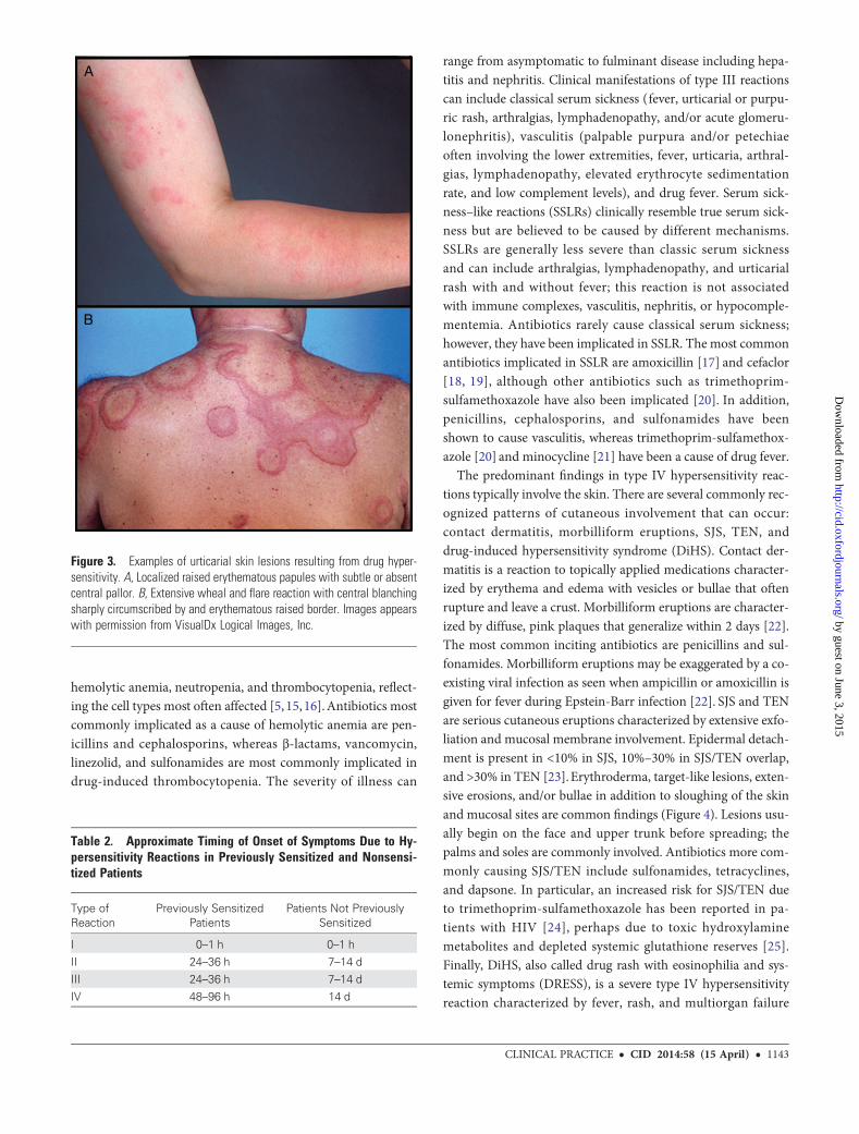
The most common signs and symptoms are an urticarial rash(with a classic wheel and flare appearance), pruritus, flushing,angioedema, wheezing, gastrointestinal symptoms, hypoten-sion, altered mental status, and anxiety [5, 15]. (Figure 3). Nei-ther fever nor elevations in C-reactive protein are seen in a typeI reaction, which can help to distinguish it from other types ofdrug reactions. In addition, type I reactions should not occurseveral days into a course of therapy, if exposure to an incitingdrug is continuous.
Delayed-Type Reactions: Types II, III, and IVDelayed-type hypersensitivity reactions (types II, III, and IV)are those in which the onset is 1 hour or more after drug expo-sure. These reactions are not mediated by IgE, and timing ofsymptoms may differ (Table 2). Type II reactions present as
Figure 2. Ring structures for penicillins (A), cephalosporins (B), carbapenems (C), and monobactams (D).
1142 • CID 2014:58 (15 April) • CLINICAL PRACTICE
by guest on June 3, 2015http://cid.oxfordjournals.org/
Dow
nloaded from
hemolytic anemia, neutropenia, and thrombocytopenia, reflect-ing the cell types most often affected [5, 15, 16].Antibiotics mostcommonly implicated as a cause of hemolytic anemia are pen-icillins and cephalosporins, whereas β-lactams, vancomycin,linezolid, and sulfonamides are most commonly implicated indrug-induced thrombocytopenia. The severity of illness can
range from asymptomatic to fulminant disease including hepa-titis and nephritis. Clinical manifestations of type III reactionscan include classical serum sickness (fever, urticarial or purpu-ric rash, arthralgias, lymphadenopathy, and/or acute glomeru-lonephritis), vasculitis (palpable purpura and/or petechiaeoften involving the lower extremities, fever, urticaria, arthral-gias, lymphadenopathy, elevated erythrocyte sedimentationrate, and low complement levels), and drug fever. Serum sick-ness–like reactions (SSLRs) clinically resemble true serum sick-ness but are believed to be caused by different mechanisms.SSLRs are generally less severe than classic serum sicknessand can include arthralgias, lymphadenopathy, and urticarialrash with and without fever; this reaction is not associatedwith immune complexes, vasculitis, nephritis, or hypocomple-mentemia. Antibiotics rarely cause classical serum sickness;however, they have been implicated in SSLR. The most commonantibiotics implicated in SSLR are amoxicillin [17] and cefaclor[18, 19], although other antibiotics such as trimethoprim-sulfamethoxazole have also been implicated [20]. In addition,penicillins, cephalosporins, and sulfonamides have beenshown to cause vasculitis, whereas trimethoprim-sulfamethox-azole [20] and minocycline [21] have been a cause of drug fever.
The predominant findings in type IV hypersensitivity reac-tions typically involve the skin. There are several commonly rec-ognized patterns of cutaneous involvement that can occur:contact dermatitis, morbilliform eruptions, SJS, TEN, anddrug-induced hypersensitivity syndrome (DiHS). Contact der-matitis is a reaction to topically applied medications character-ized by erythema and edema with vesicles or bullae that oftenrupture and leave a crust. Morbilliform eruptions are character-ized by diffuse, pink plaques that generalize within 2 days [22].The most common inciting antibiotics are penicillins and sul-fonamides. Morbilliform eruptions may be exaggerated by a co-existing viral infection as seen when ampicillin or amoxicillin isgiven for fever during Epstein-Barr infection [22]. SJS and TENare serious cutaneous eruptions characterized by extensive exfo-liation and mucosal membrane involvement. Epidermal detach-ment is present in <10% in SJS, 10%–30% in SJS/TEN overlap,and >30% in TEN [23].Erythroderma, target-like lesions, exten-sive erosions, and/or bullae in addition to sloughing of the skinand mucosal sites are common findings (Figure 4). Lesions usu-ally begin on the face and upper trunk before spreading; thepalms and soles are commonly involved. Antibiotics more com-monly causing SJS/TEN include sulfonamides, tetracyclines,and dapsone. In particular, an increased risk for SJS/TEN dueto trimethoprim-sulfamethoxazole has been reported in pa-tients with HIV [24], perhaps due to toxic hydroxylaminemetabolites and depleted systemic glutathione reserves [25].Finally, DiHS, also called drug rash with eosinophilia and sys-temic symptoms (DRESS), is a severe type IV hypersensitivityreaction characterized by fever, rash, and multiorgan failure
Figure 3. Examples of urticarial skin lesions resulting from drug hyper-sensitivity. A, Localized raised erythematous papules with subtle or absentcentral pallor. B, Extensive wheal and flare reaction with central blanchingsharply circumscribed by and erythematous raised border. Images appearswith permission from VisualDx Logical Images, Inc.
Table 2. Approximate Timing of Onset of Symptoms Due to Hy-persensitivity Reactions in Previously Sensitized and Nonsensi-tized Patients
Type ofReaction
Previously SensitizedPatients
Patients Not PreviouslySensitized
I 0–1 h 0–1 h
II 24–36 h 7–14 dIII 24–36 h 7–14 d
IV 48–96 h 14 d
CLINICAL PRACTICE • CID 2014:58 (15 April) • 1143
by guest on June 3, 2015http://cid.oxfordjournals.org/
Dow
nloaded from

with the liver, kidneys, heart, and/or lungs most commonly af-fected. Additionally, drug fever may be the sole manifestation ofa type IV hypersensitivity reaction, although hepatic or renaldysfunction, pulmonary involvement, and/or mucosal ulcera-tion may be present. The timing of the onset of fever in thiscase is not a reliable diagnostic clue [26]; in most cases fevercan occur several days to 3 weeks after the offending medicationhas been started but may take up to several year(s) in some pa-tients. Withdrawal of the offending medication usually resultsin defervescence in 72–96 hours.
DIAGNOSIS AND TREATMENT OF ANTIBIOTICALLERGY
Evaluation of the patient reporting a hypersensitivity reaction toan antimicrobial medication should begin with a detailed histo-ry and assessment of the type of clinical reaction experienced[27]. Important information to obtain includes
• source of the reported allergy history (patient, familymember, healthcare professional, etc);
• indication;
• dose/route of medication;
• signs/symptoms experienced;
• the timing of onset of the reaction in relationship to the ini-tiation of the medication;
• whether or not the reaction necessitated hospitalization;
• treatment(s) given for the reaction and response;
• whether or not the patient has taken the medication againsince the prior reaction;
• whether or not any recurrent signs or symptoms occurredwith subsequent drug exposure; and
• concurrent medications at the time that the reaction oc-curred and if any of these were newly started.
There are other classes of medications in addition to antibi-otics that can cause hypersensitivity reactions, such as antiepi-leptics, antihypertensives, antiretrovirals, muscle relaxants,nonsteroidal anti-inflammatories, allopurinol, therapeutic for-eign proteins, platinum-based chemotherapy, and opiates[16]. The patient’s medical record should be reviewed to obtainany further details regarding the reported allergy, including anylaboratory abnormalities present during the time of the report-ed event (ie, peripheral or urine eosinophilia, hematuria, etc). Insome circumstances, patients reporting an allergy to an antibi-otic medication may have actually experienced a nonallergic ad-verse effect. Examples may include gastrointestinal side effectscaused by macrolides and tetracyclines or photosensitivitycaused by tetracyclines.
Nevertheless, history alone is not always sufficient for estab-lishing antibiotic hypersensitivity. Skin testing is a next step inthe diagnostic process. However, it is important to note thatthere are comparatively few validated antibiotic skin test proce-dures available. Of these, penicillin testing has the longest his-tory and is the most frequently used methodology. Penicillinskin testing can provide additional useful information regardingan individual’s risk for a type I hypersensitivity reaction if ex-posed to the antimicrobial medication in question. Importantly,an evidence-based analysis including original studies describingthe precision of skin testing in the diagnosis of penicillin allergyfound that only 10%–20% of patients reporting this allergy weretruly allergic. Patients with positive skin test results should un-dergo desensitization [28]; virtually all patients with negativepenicillin skin tests results can take penicillin without serioussequelae [27].
If a patient with a reported allergy is deemed not allergic or ifthe allergy is simply an expected side effect, the medical recordshould be updated to reflect this change. Failure to do so maydeprive the patient of receiving essential antibiotics when no al-lergy exists. Documentation of tolerance to a similar class orproduct (ie, penicillin-allergic patient able to tolerate cephalo-sporins) is also important. This documentation should stay withthe patient across hospital admissions and outpatient records.This task may be best accomplished during medication recon-ciliation, but evaluation of allergy is an ongoing process.
The majority of deaths from anaphylaxis result from respira-tory failure followed by cardiovascular compromise [29]. Main-tenance of the airway and cardiovascular system comprise thecritical foundation of anaphylaxis management. Epinephrine
Figure 4. Mucosal membrane involvement with skin desquamation in ahuman immunodeficiency virus–infected patient with toxic epidermal nec-rolysis caused by a sulfonamide allergy.
1144 • CID 2014:58 (15 April) • CLINICAL PRACTICE
by guest on June 3, 2015http://cid.oxfordjournals.org/
Dow
nloaded from

should be administered immediately. Outside the healthcaresetting, intramuscular epinephrine given in the anterior lateralthigh is the preferred route. If intravenous access is available, abolus of epinephrine (0.2 μg/kg) should be given and followedby a low-dose infusion of a vasopressor such as norepinephrinetitrated to a systolic blood pressure ≥90 mm Hg. Fluid admin-istration with large volumes of crystalloids should occur con-currently with vasopressor infusion when the response toepinephrine is not immediate and sustained.
Secondary therapeutic modalities such as antihistamines andcorticosteroids do not immediately support blood pressure orreduce inflammation, but are commonly included in anaphy-laxis protocols. Antihistamines are useful for preventing orblunting angioedema or urticaria associated with IgE-mediateddrug reactions. Simultaneous treatment with both an H1 and anH2 antagonist is recommended over a single agent for anaphy-laxis [30]. Corticosteroids have little value in the acute phase ofanaphylaxis, but they have well-known anti-inflammatoryproperties and are frequently included in anaphylaxis treatmentalgorithms because of their utility in preventing delayed ana-phylactic reactions.
PRINCIPLES OF DESENSITIZATION
Classical desensitization protocols are designed to treat type 1(IgE–mediated) mast cell reactions [31]. The typical requestfor drug desensitization may better be described as inductionof drug tolerance without an adverse reaction [32]. This termmore accurately reflects the diverse mechanisms that may be re-sponsible for a specific drug reaction including IgE-mediated,non-IgE-mediated, and non-immune-mediated processes [16].
If an IgE-mediated sensitivity is established and the need forthe drug confirmed, a standard desensitization protocol can beinitiated. The goal of this procedure is described by some ascontrolled anaphylaxis—that is, the drug is administered at aconcentration and rate that will cause drug-specific IgE-armed mast cells to degranulate at low rates that do not precip-itate a systemic reaction. Serial doses of medication are gradu-ally increased (usually doubled) for each administration (oftenat 15- to 20-minute intervals), and the number of IgE receptorson the mast cells are suppressed, which deceases the sensitivityof the mast cell to the point where a full dose of drug can ulti-mately be safely given. This defines a clinically tolerant state tothe continued administration of the drug with little risk of a sig-nificant mast cell–mediated reaction during the course of ther-apy. It is critical to note that this procedure does not eliminatethe IgE-mediated drug sensitivity; rather, it desensitizes the in-dividual to allow him/her to receive the therapeutic course safe-ly. Once desensitized, the patient usually does not react toadministration of the drug for the duration of therapy. Oncetherapy is completed, the desensitized state will only last for
up to 4 half-lives (T½) of the drug. After that, sensitivity isassumed to have returned, and future therapeutic courses willrequire repeated desensitization protocols.
In cases where IgE-mediated sensitivity cannot be confirmedbut the patient history strongly suggests that an immediate hy-persensitivity state exists, drug allergy is assumed and the pa-tient is subjected to a standard desensitization protocol [16].In contrast, for cases where the history suggests that IgE-mediatedhypersensitivity is not responsible for a previous reaction, agraded challenge protocol can be instituted [32]. A graded chal-lenge is not intended to induce drug tolerance, and is designedprimarily to demonstrate that administration of a specific drugwill not result in an immediate reaction. A patient who toleratesa graded challenge without reaction can then be considerednonallergic, with a risk of future reaction no greater than thepopulation at large.
There is further consideration to interpreting the results of agraded challenge. If the mechanism responsible for the reactionis a non-IgE-mediated or non–immune mediated, althoughthere may be no initial reaction after the graded challenge, a de-layed reaction (such as rash or other organ dysfunction) maystill occur. This is why, in the initial assessment, establishingthe temporal relationship between initial drug exposure and
Table 3. Sample Desensitization Protocola for a 1-g Final Dose
DoseStrength,
mgVolume,
mL Preparation Instructions
1 1 30 Add 29.75 mL D5W and 0.25 mL stocksolution Ab to empty 50-mL bag
2 2 30 Add 29.5 mL D5W and 0.5 mL stocksolution A to empty 50-mL bag
3 4 30 Add 29 mL D5W and 1 mL stock solutionA to empty 50-mL bag
4 8 30 Add 28 mL D5W and 2 mL stock solutionA to empty 50-mL bag
5 16 30 Add 26 mL D5W and 4 mL stock solutionA to empty 50-mL bag
6 32 30 Add 22 mL D5W and 8 mL stock solutionA to empty 50-mL bag
7 64 30 Add 14 mL D5W and 16 mL stocksolution A to empty 50-mL bag
8 128 50 Add 18 mL D5W and 32 mL stocksolution A to empty 50-mL bag
9 250 50 Remove 2.5 mL from 50-mL D5W bagand add 2.5 mL of stock solution Bc
10 500 50 Remove 5 mL from 50-mL D5W bag andadd 5 mL of stock solution B
11 1000 50 Remove 10 mL from 50-mL D5W bagand add 10 mL of stock solution B
Administer first 10 doses over 15 minutes and last dose over 30 minutes.a Supplies needed: eight 50-mL bags, three 50-mL D5W bags, stock solution A,stock solution B.b Stock solution A: 4 mg/mL.c Stock solution B: 100 mg/mL.
CLINICAL PRACTICE • CID 2014:58 (15 April) • 1145
by guest on June 3, 2015http://cid.oxfordjournals.org/
Dow
nloaded from

first appearance of adverse clinical event is so important. Asnewer and more accurate techniques are developed to identifyspecific mechanism of antibiotic sensitivity, more specific andeffective protocols will be developed to induce drug tolerancein susceptible patients.
Before beginning a desensitization procedure, several consid-erations must be reviewed to limit any major complications.The best clinical setting should be determined (office, medicalward, intensive care unit). The desensitization protocol shouldbe reviewed with the pharmacist and nurse to ensure optimal
creation of formulas and strict adherence to the schedule. Thepharmacist should be aware that a dose may have to be remixedin the event of a dose failure. Adequate personnel should beavailable during the desensitization with the expectation thatthe process may take several hours or longer. Vital signs andadverse reactions should be monitored before and after each in-cremental dose. Medications for anaphylaxis should be imme-diately available, and some protocols advocate schedulingdiphenhydramine throughout the desensitization with epineph-rine at the bedside, whereas others recommend an intravenous
Table 4. Medication Desensitization Protocols
MedicationConcentration,
mg/mL Infusion TimeInterval Between
DosesTime toComplete
Dose Range,mg
Level ofEvidencea Final Dose
Ampicillin [33] IV Not reported Not reported 20 min 6 h 0.05–2000 IV 2000 mgCefepime [34] IV 0.04
220
5 min 15 min 4 h 0.032–2000 III 2000 mg
Ceftazidime [35] IV 0.112
15 min 15 min 2 d 0.025–2.56–3070.5–586
I Various
Ciprofloxacin [36] IV 0.112
10 min20 min lastdose
15 min 0.1–0.80.16–0.640.6–120
II 400 mg
Clarithromycin [37] oral 0.050.5550
NA 15 min 5 h 0.005–0.20.4–3.26–2450–500
III 500 mg
Clindamycin [38] oral NA NA 8 h 7 d 20–600 II 600 mgDaptomycin [39] IV Not reported 15 min 30 min 3 h 0.00035–350 II 350 mg
Imipenem [40] IV 0.00010.0010.010.11
30 min 30 min 4 h 0.00030.01–0.030.1–0.31–310–21
II 1000 mg/d
Linezolid [41] oral 0.018–1.5 NA 30 0.0366–400 II 600 mgMeropenem [42] IV 0.00008–20 20 min 20 min 5 h 0.004–1000 III 1000 mg
Penicillin [43] IV 0.11101001000
Unknown 15 min30 min after lastdose
9 h 0.01–0.080.16–0.641.2–4.810–80160–640
I 1000 mg IV
Penicillin [43] oral 0.5550
NA 15 min30 min after lastdose
4 h 0.05–3.26–2450–400
I 1000 mg IV
TMP/SMX [43] oral 40 TMP/200SMX per 5mL
NA 1 h 5 h 0.04/0.02–160/800
I 160 mg/800 mg
Tobramycin [44] IV 0.0005–0.8 20 min 30 min 8 h 0.001–16 II 80 mg
Vancomycin [12] IV rapid 0.0002–2 Various Continuous 4 h 0.02–500 I Usual doseover 2 h
Vancomycin [12] IV slow 0.001–4 5 h 5 h 3 d 0.5–1000 I 1000 mg
Abbreviations: IV, intravenous; NA, not applicable; TMP/SMX, trimethoprim/sulfamethoxazole.a I, desensitization successful in >1 patient with confirmed allergy to that medication; II, desensitization successful in 1 patient with confirmed allergy to thatmedication; III, desensitization successful in 1 patient with confirmed allergy to a medication in class; IV, desensitization successful in 1 patient withoutconfirmed allergy.
1146 • CID 2014:58 (15 April) • CLINICAL PRACTICE
by guest on June 3, 2015http://cid.oxfordjournals.org/
Dow
nloaded from
line, electrocardiography monitor, and spirometer. An adversereaction does not necessarily require stopping the desensitiza-tion protocol and may proceed by repeating the last tolerabledose and rechallenging. Patients missing a dose may have tobe desensitized again. A sample adaptable desensitization pro-tocol is listed in Table 3, and medication-specific desensitizationprotocols are listed in Table 4 [12, 33–44].
CONCLUSIONS
Antibiotic allergy remains an important barrier in providingideal care, and with fewer new antibiotics available on themarket along with increasing antibiotic resistance, the chanceof an allergy–treatment mismatch is increasing. Many patientswith declared allergy may be given that medication after differ-entiating between allergy and intolerance. When a true drug al-lergy is highly likely based on history and skin testing (whenavailable), desensitization protocols can be used to give the pa-tient an antibiotic in the safest and most responsible mannerpossible. Fully understanding the mechanisms of allergy andengaging specialists in treatment further reduces risk.
Notes
Author contributions. Conception and design: D. P. L., C. A. M., E. S.;drafting of manuscript: D. P. L., C. A. M., E. S., G. D. M.; content oversight:E. S., G. D. M.Potential conflicts of interest. All authors: No reported conflicts.All authors have submitted the ICMJE Form for Disclosure of Potential
Conflicts of Interest. Conflicts that the editors consider relevant to the con-tent of the manuscript have been disclosed.
References
1. Johansson SG, Bieber T, Dahl R, et al. Revised nomenclature for allergyfor global use: report of the Nomenclature Review Committee of theWorld Allergy Organization, October 2003. J Allergy Clin Immunol2004; 113:832–6.
2. Macy E, Poon KYT. Self-reported antibiotic allergy incidence and prev-alence: age and sex effects. Am J Med 2009; 122:778 e1–7.
3. Shehab N, Patel PR, Srinivasan A, et al. Emergency department visitsfor antibiotic-associated adverse events. Clin Infect Dis 2008;47:735–43.
4. Boucher HW, Talbot GH, Bradley JS, et al. Bad bugs, no drugs: no ES-KAPE! An update from the Infectious Diseases Society of America. ClinInfect Dis 2009; 48:1–12.
5. Coombs P, Gell PG. Classification of allergic reactions responsible forclinical hypersensitivity and disease. In: G RR, P.G.H Gell, eds. Clinicalaspects of immunology. Oxford, UK: Oxford University Press, 1968:575–96.
6. Celik W, Pochler WJ, Adkinson NF. Drug allergy. In: C CJ, ed. Middle-ton’s allergy: principles and practice. Philadelphia, PA: Elsevier Saun-ders, 2010:1205–26.
7. Pichichero ME. Cephalosporins can be prescribed safely for penicillin-allergic patients. J Fam Pract 2006; 55:106–12.
8. Sodhi M, et al. Is it safe to use carbapenems in patients with a history ofallergy to penicillin? J Antimicrob Chemother 2004; 54:1155–7.
9. Frumin J, Gallagher JC. Allergic cross-sensitivity between penicillin,carbapenem, and monobactam antibiotics: what are the chances? AnnPharmacother 2009; 43:304–15.
10. Arroliga ME, Pien L. Penicillin allergy: consider trying penicillin again.Cleve Clin J Med 2003; 70:313–4, 317–8, 320–1 passim.
11. Mandell GL, Bennett JE, Dolin R. Mandell, Douglas, and Bennett’sprinciples and practice of infectious diseases. 7th ed. Philadelphia,PA: Churchill Livingstone/Elsevier, 2010.
12. Wazny LD, Daghigh B. Desensitization protocols for vancomycin hy-persensitivity. Ann Pharmacother 2001; 35:1458–64.
13. Dellavalle RP, et al. Vancomycin-associated linear IgA bullous derma-tosis mimicking toxic epidermal necrolysis. J Am Acad Dermatol 2003;48(5 suppl):S56–7.
14. Baldo BA. Diagnosis of allergy to pencillins and cephalosporins. ACI Int2000; 12:206–12.
15. Levine BB. Immunologic mechanisms of penicillin allergy. A haptenicmodel system for the study of allergic diseases of man. N Engl J Med1966; 275:1115–25.
16. Greenberger PA. Chapter 30: drug allergy. Allergy Asthma Proc 2012;33(suppl 1):S103–7.
17. Lin B, Strehlow M. Images in emergency medicine. Serum sickness-likereaction to amoxicillin. Ann Emerg Med 2007; 50:350, 359.
18. Vial T, Pont J, Pham E, et al. Cefaclor-associated serum sickness-likedisease: eight cases and review of the literature. Ann Pharmacother1992; 26:910–4.
19. Stricker BH, Tijssen JG. Serum sickness-like reactions to cefaclor. J ClinEpidemiol 1992; 45:1177–84.
20. Slatore CG, Tilles SA. Sulfonamide hypersensitivity. Immunol AllergyClin North Am 2004; 24:477–90, vii.
21. Grim SA, Romanelli F, Jennings PR, et al. Late-onset drug fever associ-ated with minocycline: case report and review of the literature. Pharma-cotherapy 2003; 23:1659–62.
22. James WD, Berger TG, Elston DM. Contact dermatitis and drugeruptions, in Andrews’ diseases of the skin. China: Elsevier, 2011:88–137.
23. Diaz L, Ciurea AM. Cutaneous and systemic adverse reactions to anti-biotics. Dermatol Ther 2012; 25:12–22.
24. Lin D, Tucker MJ, Rieder MJ. Increased adverse drug reactions to anti-microbials and anticonvulsants in patients with HIV infection. AnnPharmacother 2006; 40:1594–601.
25. Gruchalla RS. 10. Drug allergy. J Allergy Clin Immunol 2003; 111(2suppl):S548–59.
26. Mackowiak PA. Drug fever: mechanisms, maxims and misconceptions.Am J Med Sci 1987; 294:275–86.
27. Salkind AR, Cuddy PG, Foxworth JW. The rational clinical examina-tion. Is this patient allergic to penicillin? An evidence-based analysisof the likelihood of penicillin allergy. JAMA 2001; 285:2498–505.
28. Weiss ME, Adkinson NF. Immediate hypersensitivity reactions to pen-icillin and related antibiotics. Clin Allergy 1988; 18:515–40.
29. Simons FE. Anaphylaxis pathogenesis and treatment. Allergy 2011; 66(suppl 95):31–4.
30. Simons FE. Advances in H1-antihistamines. N Engl J Med 2004;351:2203–17.
31. McLean-Tooke A, Aldridge C, Stroud C, et al. Practical management ofantibiotic allergy in adults. J Clin Pathol 2011; 64:192–9.
32. Joint Task Force on Practice Parameters; American Academy of Allergy,Asthma and Immunology; American College of Allergy, Asthma andImmunology; Joint Council of Allergy, Asthma and Immunology.Drug allergy: an updated practice parameter. Ann Allergy Asthma Im-munol 2010; 105:259–73.
33. Candela L. Caring for a patient with Listeria endocarditis: use of anti-biotic desensitization. Crit Care Nurse 2002; 22:38–43.
34. Damin D, Marney S, DiPersio D, Hargrove F. Cefepime desensitiza-tion in a patient with cystic fibrosis. J Allergy Clin Immunol 2004;113:S312.
35. Ghosal S, Taylor CJ. Intravenous desensitization to ceftazidime in cysticfibrosis patients. J Antimicrob Chemother 1997; 39:556–7.
36. Gea-Banacloche JC, Metcalfe DD. Ciprofloxacin desensitization. J Aller-gy Clin Immunol 1996; 97:1426–7.
CLINICAL PRACTICE • CID 2014:58 (15 April) • 1147
by guest on June 3, 2015http://cid.oxfordjournals.org/
Dow
nloaded from

37. Holmes NE, Hodgkinson NE, Dendle C, et al. Report of oral clarithro-mycin desensitization. Br J Clin Pharmacol 2008; 66:323–4.
38. Marcos C, Sopeña B, Luna I, et al. Clindamycin desensitization in anAIDS patient. AIDS 1995; 9:1201–2.
39. Metz GM, Thyagarajan A. A successful protocol for daptomycin desen-sitization. Ann Allergy Asthma Immunol 2008; 100:87.
40. Gorman SK, Zed PJ, Dhingra VK, et al. Rapid imipenem/cilastatin de-sensitization for multidrug-resistant Acinetobacter pneumonia. AnnPharmacother 2003; 37:513–6.
41. Cawley MJ, Lipka O. Intravenous linezolid administered orally: a noveldesensitization strategy. Pharmacotherapy 2006; 26:563–8.
42. Wilson DL, Owens RC Jr, Zuckerman JB. Successful meropenem desensiti-zation in a patient with cystic fibrosis. Ann Pharmacother 2003; 37:1424–8.
43. Gilbert DN. The Sanford guide to antimicrobial therapy 2011. Sperry-ville, VA: Antimicrobial Therapy, Inc, 2011.
44. Earl HS, Sullivan TJ. Acute desensitization of a patient with cystic fibro-sis allergic to both beta-lactam and aminoglycoside antibiotics. J AllergyClin Immunol 1987; 79:477–83.
1148 • CID 2014:58 (15 April) • CLINICAL PRACTICE
by guest on June 3, 2015http://cid.oxfordjournals.org/
Dow
nloaded from