Embed Size (px)
Citation preview
1
Antibody Structure and Function
Amit Lugade PhD
Center for Immunotherapy
Hematopoiesis
2
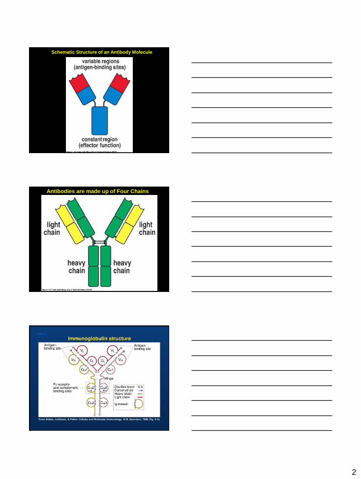
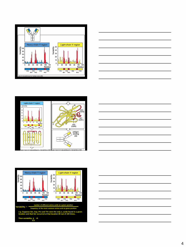
Schematic Structure of an Antibody Molecule
Antibodies are made up of Four Chains
3
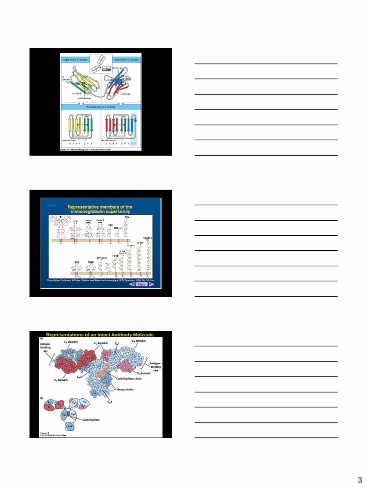
Figure 3-5
Representations of an Intact Antibody Molecule
4
Variability = number of different amino acids at a given position
frequency of the most common amino acid at given position
e.g. Suppose Ser, Asp, Pro and Thr were the only a. acids found in a given
location and that Ser occurred at that location 80 out of 100 times.
Then variability: 4 =5
0.8
5
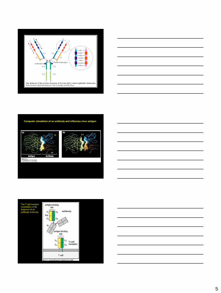
Computer simulation of an antibody and influenza virus antigen
VH
VL 8 A
The T cell receptor
resembles a Fab
fragment of an
antibody molecule
6
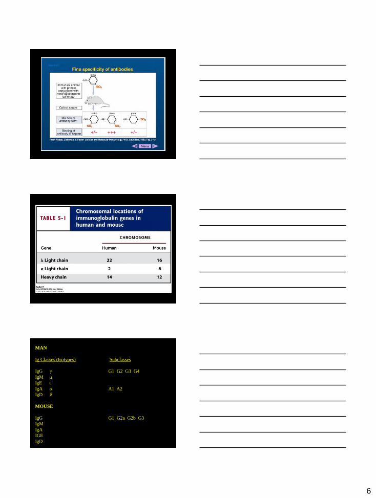
MAN
Ig Classes (Isotypes) Subclasses
IgG g G1 G2 G3 G4
IgM m
IgE e
IgA a A1 A2
IgD d
MOUSE
IgG G1 G2a G2b G3
IgM
IgA
IGE
IgD
7
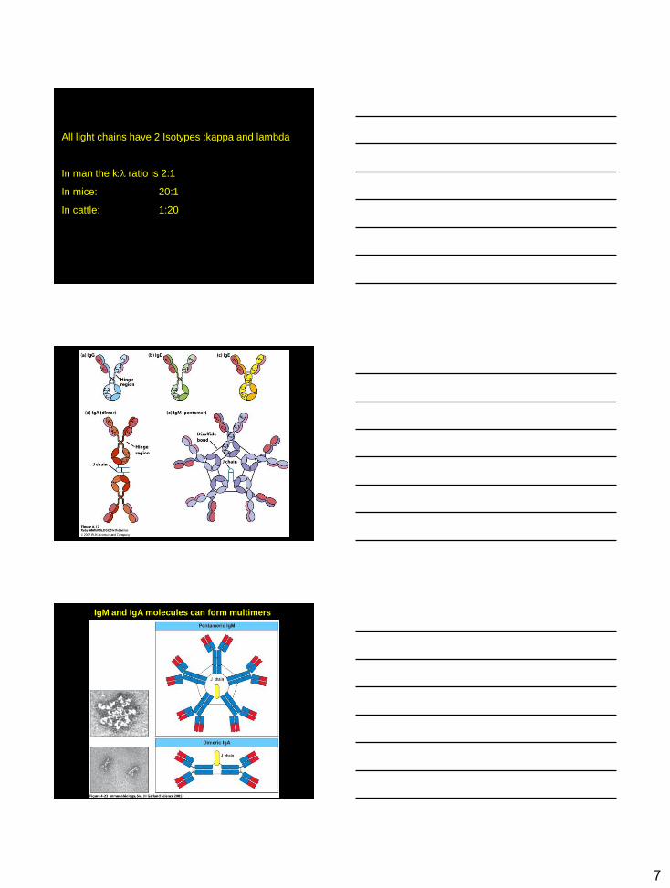
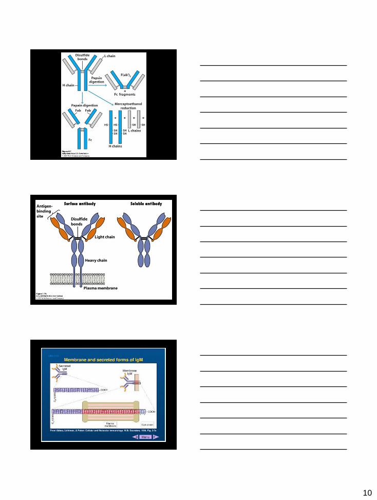
All light chains have 2 Isotypes :kappa and lambda
In man the k:l ratio is 2:1
In mice: 20:1
In cattle: 1:20
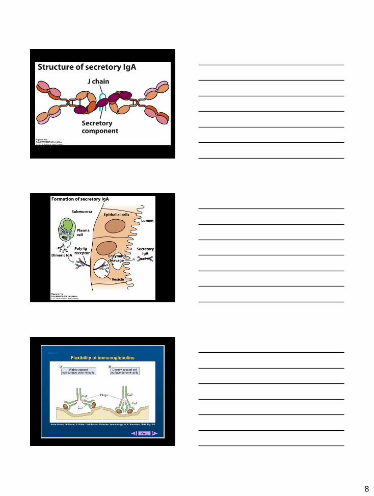
IgM and IgA molecules can form multimers
8
9
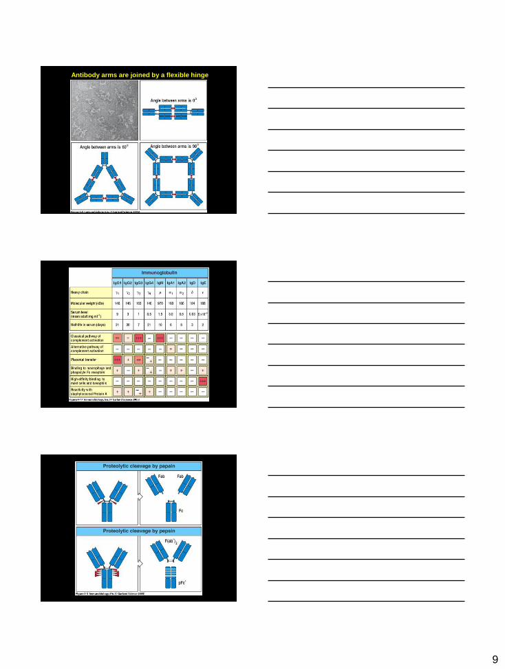
Antibody arms are joined by a flexible hinge
10
11
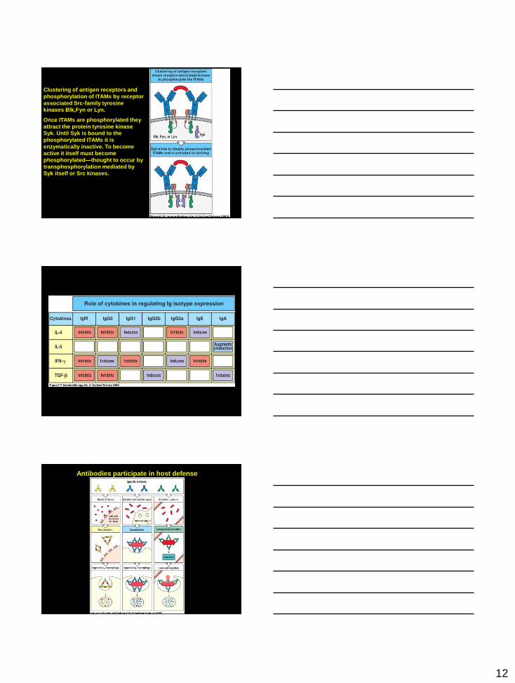
B Cells Originate from a Lymphoid Progenitor in the Bone Marrow
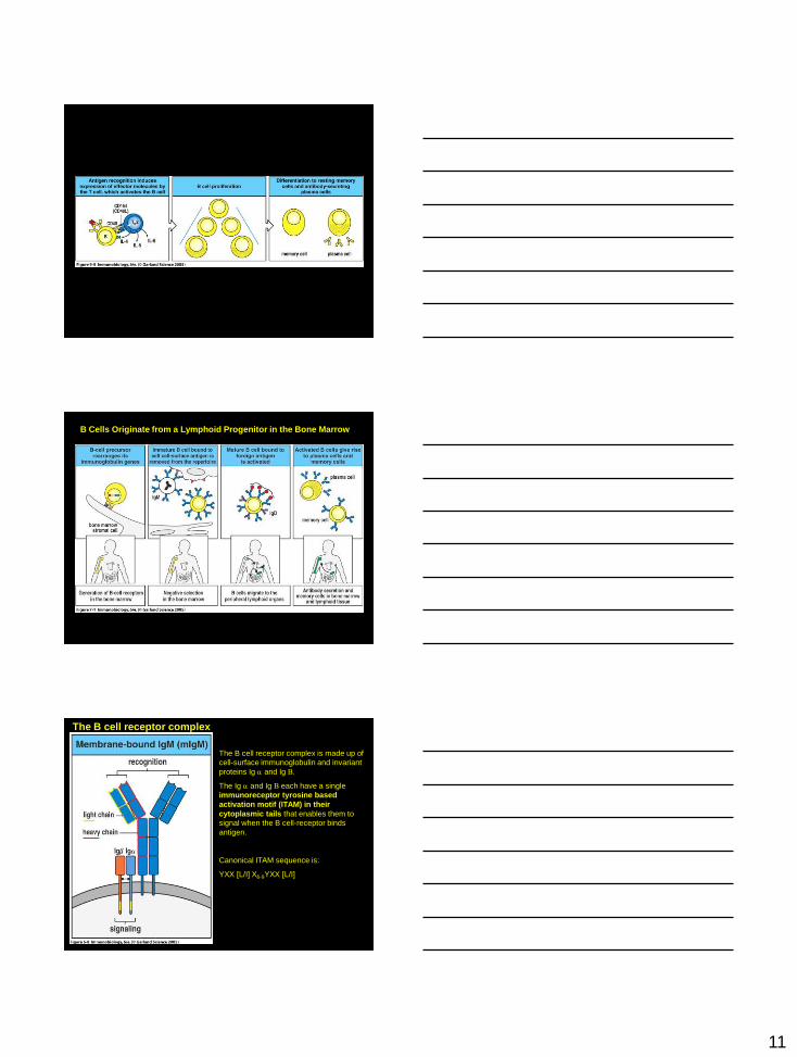
The B cell receptor complex
The B cell receptor complex is made up of
cell-surface immunoglobulin and invariant
proteins Ig a and Ig B.
The Ig a and Ig B each have a single
immunoreceptor tyrosine based
activation motif (ITAM) in their
cytoplasmic tails that enables them to
signal when the B cell-receptor binds
antigen.
Canonical ITAM sequence is:
YXX [L/I] X6-9YXX [L/I]
12
Clustering of antigen receptors and
phosphorylation of ITAMs by receptor
associated Src-family tyrosine
kinases Blk,Fyn or Lyn.
Once ITAMs are phosphorylated they
attract the protein tyrosine kinase
Syk. Until Syk is bound to the
phosphorylated ITAMs it is
enzymatically inactive. To become
active it itself must become
phosphorylated—thought to occur by
transphosphorylation mediated by
Syk itself or Src kinases.
Antibodies participate in host defense
13
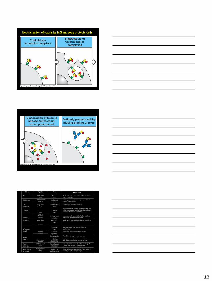
Neutralization of toxins by IgG antibody protects cells
Disease
Organism
Toxin
Effects in vivo
Tetanus
Clostridium
tetani
Tetanus
toxin
Blocks inhibitory neuron action leading to chronic
muscle contraction
Diphtheria
Corynebacterium
diptheriae
Diphtheria
toxin
Inhibits protein synthesis leading to epithelial-cell
damage, and myocarditis
Gas
Gangrene
Clostridium
perfringens
Clostridial-α
toxin
Phospholipase leading to cell death
Cholera
Vibrio
cholerae
Cholera
toxin
Activates adenylate cyclase, elevates cAMP in cells,
leading to changes in intestinal epithelial cells that
cause loss of water and electrolytes
Anthrax
Bacillus
anthracis
Anthrax toxic
complex
Increases vascular permeability leading to edema,
hemorrhage and circulatory collapse
Botulism
Clostridium
botulinum
Botulinus
toxin
Blocks release of acetylcholine leading to paralysis
Whooping
cough
Bordetella
pertussis
Pertussis
toxin
ADP-ribosylation of G proteins leading to
lymphocytosis
Tracheal
cytotoxin
Inhibits cilia and causes epithelial-cell loss
Scarlet
fever
Streptococcus
pyogenes
Erythrogenic
toxin
Vasodilation leading to scarlet-fever rash
Leukocidin
streptolysins
Kills phagocytes, allowing bacterial survival
Food
poisoning
Staphylococcus
aureus
Staphylococcal
enterotoxin
Acts on intestinal neurons to induce vomiting. Also
a potent T-cell mitogen (SE superantigen)
Toxic shock
syndrome
Staphylococcus
aureus
Toxic-shock
syndrome toxin
Causes hypotension and skin loss. Also a potent T-
cell mitogen (TSST 1 superantigen)
14
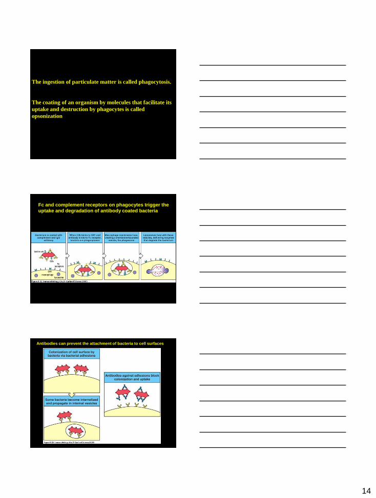
The ingestion of particulate matter is called phagocytosis.
The coating of an organism by molecules that facilitate its
uptake and destruction by phagocytes is called
opsonization
Fc and complement receptors on phagocytes trigger the
uptake and degradation of antibody coated bacteria
Antibodies can prevent the attachment of bacteria to cell surfaces
15
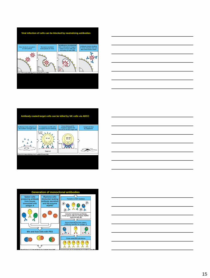
Viral infection of cells can be blocked by neutralizing antibodies
Antibody coated target cells can be killed by NK cells via ADCC
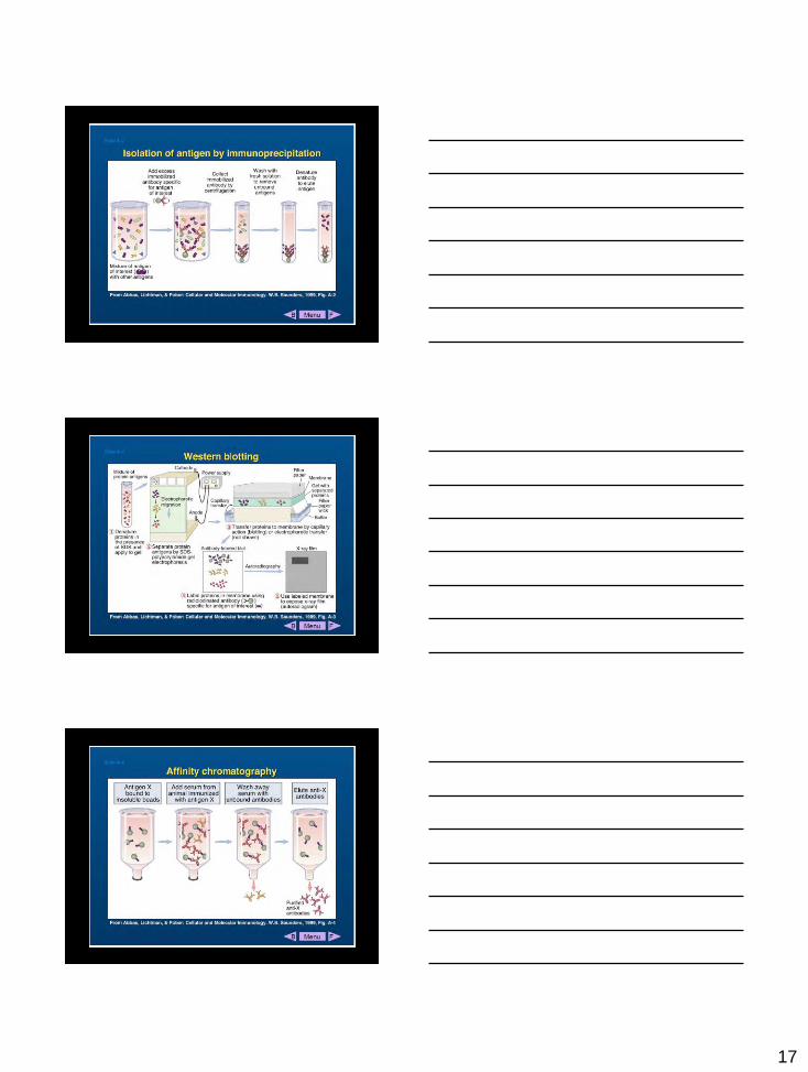
Generation of monoclonal antibodies
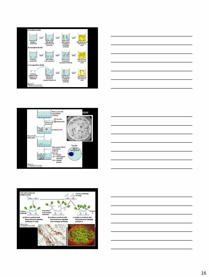
16
ELISPOT ASSAY
17
18
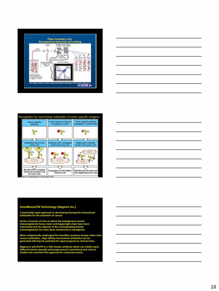
Recognition by monoclonal antibodies of tumor-specific antigens
XenoMouseTM Technology (Abgenix Inc.)
A potentially rapid approach to developing therapeutic monoclonal
antibodies for the treatment of cancer.
Series of strains of mice in which the endogenous murine
immunoglobulin heavy chain and kappa light chain have been
inactivated and the majority of the corresponding human
immunoglobulin loci have been introduced as transgenes.
When antigenically challenged the XenoMice produce human rather than
mouse antibodies. High affinity monoclonal antibodies can be
generated offering the potential for rapid progress to clinical trials.
Abgenix’s anti-EGFR is a fully human antibody which can inhibit many
different tumors (growth and progression) in preclinical and clinical
studies has received FDA approval for colorectal cancer.
19
Panitumumab represents the first fully human antibody developed from XenoMouse
technology to be approved by a regulatory agency. This has been an important
milestone in validating XenoMouse strains as well as other human immunoglobulin-
producing mouse technologies as sources for therapeutic antibodies.
The path from initiation of XenoMouse technology development to regulatory
approval took ~15 years, including 6 years for mouse strains derivation and mAb
development and 6.5 years of clinical development.
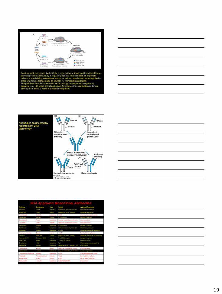
Antibodies engineered by
recombinant DNA
technology
Antibody Brand name Type Target Approved Treatments
Abciximab ReoPro chimeric inhibition of glycoprotein IIb/IIIa cardiovascular disease
Adalimumab Humira human inhibition of TNF-a signalling inflammatory diseases
Alemtuzumab Campath humanized CD52 chronic lymphocytic leukemia
Basiliximab Simulect chimeric IL-2 receptor a transplant rejection
Bevacizumab Avastin humanized vascular endothelial growth factor colorectal cancer
Cetuximab Erbitux chimeric epidermal growth factor receptor colorectal cancer
Daclizumab Zenapax humanized IL-2 receptor a transplant rejection
Eculizumab Soliris humanized complement system protein C5 inflammatory diseases
Efalizumab Raptiva humanized CD11a inflammatory diseases (psoriasis)
Ibritumomab-tiuxetan Zevalin murine CD20 Non-Hodgkin lymphoma
Infliximab Remicade chimeric inhibition of TNF-a signaling inflammatory diseases (auto-immune)
Muromonab-CD3 Orthoclone OKT3 murine T cell CD3 receptor transplant rejection
Natalizumab Tysabri humanized T cell VLA4 receptor multiple sclerosis
Omalizumab Xolair humanized IgE inflammatory diseases (asthma)
Payilizumab Synagis humanized an epitope of the F protein of RSV RSV infection
Panitumumab Vectibix human epidermal growth factor receptor colorectal cancer
Ranibizumab Lucentis humanized vascular endothelial growth factor macular degeneration
Gemtuzumab-ozogamicin Mylotarg humanized CD33 acute myelogenous leukemia
Rituximab Rituxan, Mabthera chimeric CD20 Non-Hodgkin lymphoma
Tositumomab Bexxar murine CD20 Non-Hodgkin lymphoma
Trastuzumab Herceptin humanized ErbB2/HER2/EGFR breast cancer
FDA Approved Monoclonal Antibodies
20
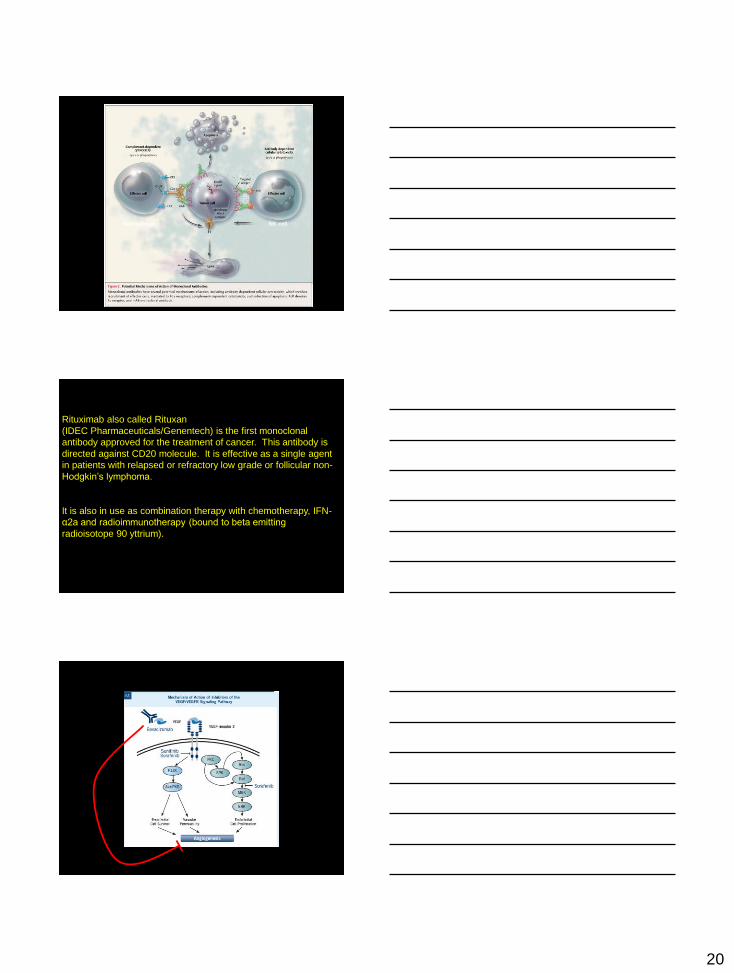
Macrophage NK cell
Rituximab also called Rituxan
(IDEC Pharmaceuticals/Genentech) is the first monoclonal
antibody approved for the treatment of cancer. This antibody is
directed against CD20 molecule. It is effective as a single agent
in patients with relapsed or refractory low grade or follicular non-
Hodgkin’s lymphoma.
It is also in use as combination therapy with chemotherapy, IFN-
α2a and radioimmunotherapy (bound to beta emitting
radioisotope 90 yttrium).
21
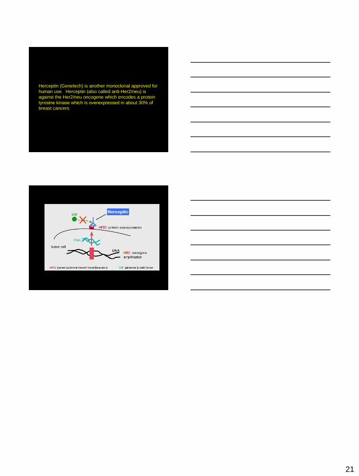
Herceptin (Genetech) is another monoclonal approved for
human use. Herceptin (also called anti-Her2/neu) is
against the Her2/neu oncogene which encodes a protein
tyrosine kinase which is overexpressed in about 30% of
breast cancers.