Embed Size (px)
Citation preview

“ t r a c e & c a t c h ”
innovation for health & wellness
Instruction for Use
Antibody to cetuximab (Erbitux®) ELISA
SHIKARI®S-ATCEnzyme immunoassay for the qualitative determination (screening) of antibodies to cetuximab (Erbitux®) in serum and plasma
i 2-8 CREF TR-ATCv1 ∑ 12 x 8
Revision # 1.2 August 2017
Matriks Biotek® Laboratories www.matriksbiotek.com

2 • SHIKARI® S-ATC
Contents Page
Intend to use....................................................................................................................... 3
Summary and Explanation…................................................................................................ 3
Test Principle ...................................................................................................................... 5
Warnings and Precautions ……............................................................................................ 6
Storage and Stability...….....................…..............................................................................7
Specimen Collection and Storage....................................................................................... 7
Materials Supplied............................................................................................................. 8
Materials Required but not Supplied ...……........................................................................ 9
Procedure Notes................................................................................................................ 9
Preparation of Component ….………................................................................................. 10
Test Procedure................................................................................................................. 11
Interpretation of Results.................................................................................................. 12
References ....................................................................................................................... 13
SHIKARI S-ATC
Cetuximab (Erbitux® ) antibodies qualitative analyse
Required Volume (µl) 10
Total Time (min) 140
Sample Serum, plazma
Sample Number 96
Detection Limit (ng/mL) +/-
Spike Recovery (%) -
Shelf Life (year) 1

SHIKARI® S-ATC • 3
Intended Use
The Matriks Biotek® Antibody to cetuximab (Erbitux®) Enzyme-Linked-ImmunoSorbent-Assay (ELISA) Kit is intended for the qualitative determination of antibodies to cetuximab (Erbitux®) in serum and plasma. It is for professional use only.
Summary and Explanation
Epidermal growth factor receptor (EGFR)
Epidermal growth factor receptor (EGFR; HER1; ErbB1) is
a transmembrane tyrosine kinase encoded by c-erb-B proto-oncogene, expressed in
normal and malignant cells and stimulated by epidermal growth factor (EGF) or
transforming growth factor-alpha (TGF-alpha) binding extracellular domain of the
receptor, leading receptor to dimerize and activating intracellular kinase domain on each
receptor, bringing about phosphorylation of tyrosine residues on each member of the
receptor pair. Then, signaling complexes form in cytoplasm to activate gene transcription
responding for such as cell proliferation. Termination of signaling occurs through
internalization of receptor-ligand complex. Activation of EGFR results in perturbation
of mitogen-activated protein kinase (MAPK), phosphatidylinositol 3-kinase, and AKT
pathways triggering tumorigenic processes, such as increased proliferation, angiogenesis
and metastasis, and prevents apoptosis. Breast, lung, colon, prostate, kidney, bladder,
head and neck, and ovary cancers have been associated to EGFR overexpression which
causes early disease progression, poor survival, and resistance to chemotherapy in many
epithelial malignancies. Epidermal growth factor receptor/human epidermal growth
factor receptor 1 (EGFR/HER1) and its ligand, transforming growth factor-alpha (TGF-
alpha) were showed to involve in hepatocarcinogenesis. EGFR
is overexpressed in hepatocellular carcinoma (HCC). To overcome the uncontrollable
effect of EGFR triggering cancer development, monoclonal antibodies have been shown
to be used as blockers in vitro and in vivo.
Cetuximab
Cetuximab (IMC-C225, Erbitux) is a chimeric monoclonal antibody of the immunoglobulin
G1 (IgG1) and FDA-approved epidermal growth factor receptor (EGFR) inhibitor. It
is a 152 kDa protein composed of four polypeptide chain. There are 32 cysteine residues
forming accordingly 16 potential disulfide bonds. Preclinical studies have shown
that cetuximab enhances the antitumour effects of chemotherapy (e.g. that
of irinotecan in colorectal cancer) as well as radiotherapy (e.g. in squamous cell
carcinoma of the head and neck) by inhibiting cell proliferation, angiogenesis and
metastasis and by promoting apoptosis is used for the treatment of metastatic colorectal
cancer, metastatic non-small cell lung cancer and head and neck cancer. Cetuximab also
blocks growth factor-induced activation of the downstream mitogen-activated

4 • SHIKARI® S-ATC
protein kinase, inhibiting cell proliferation. It has been also illustrated
that cetuximab increases the receptor internalization which is another mechanism to
silence the receptor. Cetuximab arrests cell cycle at G1 gap phase by upregulating anti-
proliferative p27kip1, which functions via complex formation with Cdk2,
and downregulating proliferating cell nuclear antigen (PCNA). It also
decreases angiogenic factors, inhibits tumor-cell invasion and
metastasis via downregulation of matrix metalloproteinases (MMPs) and VEGF, and
promotes apoptosis by upregulating apoptotic protein, Bax, with the help of other
chemotherapeutic agents. Cetuximab has been widely shown to display synergistic effect
with other agents and/or radiotherapy.
Binding of antigen-binding fragment (Fab) of Cetuximab, which displays higher affinity
comparing to ligands of EGFR, takes place via domain III of extracellular EGFR, preventing
the receptor from conformational change to be dimerized and blocking EGFR
signaling through inhibition of EGF and TGF-alpha-stimulated phosphorylation of the
receptor.
Pharmacokinetics and Pharmacodynamics
In a study conducted by Fracasso et al., patients with colorectal, breast, and head and
neck carcinomas were administered with one of different dosages of cetuximab (50, 100,
250, 400 and 500 mg/m2). For each concentration, cetuximab serum concentration was
showed to reach maximum at 3 h, and decrease slowly. Serum concentration decreased
to baseline at 96 h and 168 h for dosages 50 and 100 mg/m2, respectively. Mean
maximum observed concentrations (Cmax) increased in a dose dependent manner (from
22.8 ug/ml to 245.6 ug/ml).
It was indistinguishable for 400 mg/m2 (Cmax=228.9 ug/ml) and 500
mg/m2 (Cmax=245.6 ug/ml). The mean total body clearance based on body surface area
for cetuximab was similar following doses of >100 mg/m2 (range, 34.4-19.3 L/h/m2) but
greater in the 50 mg/m2 dose group (65.9 L/h/m2). Biopsy results showed that
maximal cytoplasmic EGFR downregulation after treatment was seen in 8 h with 400
mg/m2 dosage.
After 250 mg/m2 weekly cetuximab administration, the average trough level of patients
with both partial responses (PRs) and stable disease (SD) was 60,742 ng/ml (~400 nmol/l)
compared with those patients with progressive disease (PD; 33,208 ng/ml). In another
study, cetuximab was infused as loading dose of 400 mg/m2 followed by weekly infusions
of 250 mg/m2 in colorectal cancer patients. Median residual concentrations were 41 and
54 mg/L on days 14 and 28, respectively. It was determined that initial serum albumin
concentration was significantly related to first-order elimination clearance
of cetuximab. Central volume of distribution was 2.96 L (4%), peripheral volume of
distribution was 4.65 L (6%), elimination clearance was 0.479 L/d (4%) and distribution
clearance was 0.836 L/d (8%).

SHIKARI® S-ATC • 5
Test Principle
The Matriks Biotek® Antibody to cetuximab (Erbitux®) ELISA is a sandwich assay for
the determination of antibodies against cetuximab in serum and plasma samples.
During the first incubation period, antibodies to cetuximab (ATC) in patient serum/
plasma samples are captured by the drug cetuximab (Erbitux®) coated on the wall of the
microtiter wells. After washing away the unbound components from samples, a
peroxidase-labelled specific conjugate is added to each well and then incubated.
After a second washing step, the bound enzymatic activity is detected by addition of
tetramethylbenzidine (TMB) chromogen-substrate. Finally, the reaction is terminated
with an acidic stop solution. The intensity of the reaction color is directly proportional to
the concentration of ATC in sample.

6 • SHIKARI® S-ATC
Warnings and Precautions
1. For professional use only.
2. Before starting the assay, read the instructions completely and carefully. Use the
valid version of the package insert provided with the kit. Be sure that everything is
understood. For further information (clinical background, test performance,
automation protocols, alternative applications, literature, etc.) please refer to the
local distributor.
3. In case of severe damage of the kit package please contact Matriksbiotek or your
supplier in written form, latest one week after receiving the kit. Do not use
damaged components in test runs, but keep safe for complaint related issues.
4. Obey lot number and expiry date. Do not mix reagents of different lots. Do not
use expired reagents.
5. Follow good laboratory practice and safety guidelines. Wear lab coats, disposable
latex gloves and protective glasses where necessary.
6. Reagents of this kit containing hazardous material may cause eye and skin
irritations. See MATERIALS SUPPLIED and labels for details.
7. Chemicals and prepared or used reagents have to be treated as hazardous waste
according the national biohazard safety guidelines or regulations.
8. Avoid contact with Stop solution. It may cause skin irritations and burns.
9. Some reagents contain sodium azide (NaN3 ) as preservatives. In case of contact
with eyes or skin, flush immediately with water. NaN3 may react with lead and
copper plumbing to form explosive metal azides. When disposing reagents, flush
with large volume of water to avoid azide build-up.
10. All reagents of this test kit containing human serum or plasma have been tested
and were found negative for HIV I/II, HBsAg and HCV by FDA approved procedures.
However, a presence of these or other infectious agents cannot be excluded
absolutely and therefore reagents should be treated as potential biohazards in use
and for disposal.

SHIKARI® S-ATC• 7
Storage and Stability
The kit is shipped at ambient temperature and should be stored at 2-8°C. Keep away from heat or direct sun light. The storage and stability of specimen and prepared reagents is stated in the corresponding chapters. The strips of microtiter plate is stable up to the expiry date of the kit in the broken, but tightly closed bag when stored at 2–8°C.
Specimen Collection and Storage
Serum, Plasma (EDTA, Heparin)*
The usual precautions for venipuncture should be observed. It is important to preserve the chemical integrity of a blood specimen from the moment it is collected until it is assayed. Do not use grossly hemolytic, icteric or grossly lipemic specimens. Samples
appearing turbid should be centrifuged before testing to remove any particulate material.
Storage: 2-8°C -20°C Keep away from heat or direct sunlight
Avoid repeated freeze-thaw cyclesStability: 7 d 6 mon
*. Cetuximab (Erbitux®) infusion camouflages/masks the presence of antibody to cetuximab in serum/plasma samples. Therefore, blood sampling time is critical for detection of cetuximab. Matriks Biotek® Laboratories propose to obtain blood sample just before the infusion of cetuximab (Erbitux®) or at least 2 weeks after the infusion of cetuximab (Erbitux®).
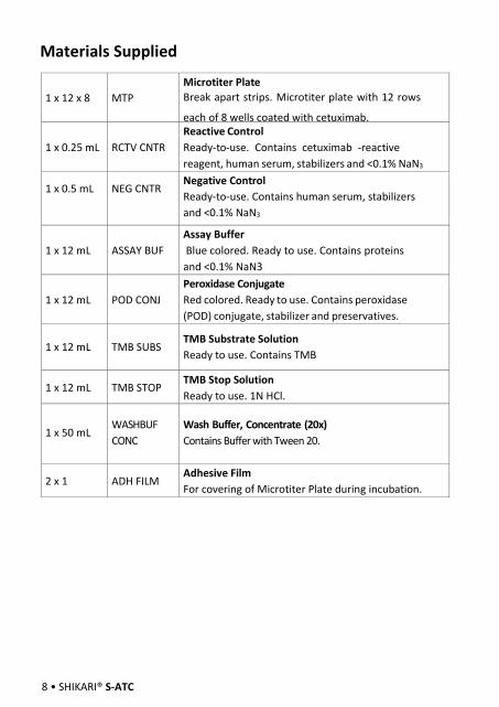
Materials Supplied
8 • SHIKARI® S-ATC
1 x 12 x 8 MTP
Microtiter Plate
Break apart strips. Microtiter plate with 12 rows
each of 8 wells coated with cetuximab.
1 x 0.25 mL RCTV CNTR
Reactive Control
Ready-to-use. Contains cetuximab -reactive
reagent, human serum, stabilizers and <0.1% NaN3
1 x 0.5 mL NEG CNTR Negative Control
Ready-to-use. Contains human serum, stabilizers
and <0.1% NaN3
1 x 12 mL ASSAY BUF
Assay Buffer
Blue colored. Ready to use. Contains proteins
and <0.1% NaN3
1 x 12 mL POD CONJ
Peroxidase Conjugate
Red colored. Ready to use. Contains peroxidase
(POD) conjugate, stabilizer and preservatives.
1 x 12 mL TMB SUBS TMB Substrate Solution
Ready to use. Contains TMB
1 x 12 mL TMB STOP TMB Stop Solution
Ready to use. 1N HCl.
1 x 50 mLWASHBUF
CONC
Wash Buffer, Concentrate (20x)
Contains Buffer with Tween 20.
2 x 1 ADH FILMAdhesive Film
For covering of Microtiter Plate during incubation.

Materials Required but not Supplied
SHIKARI® S-ATC• 9
Procedure Notes
1. Micropipettes (< 3% CV) and tips to deliver 5-1000 µL.
2. Calibrated measures.
3. Tubes (1 mL) for sample dilution.
4. Wash bottle, automated or semi-automated microtiter plate washing system.
5. Microtiter plate reader capable of reading absorbance at 450/650 nm.
6. Bidistilled or deionised water, paper towels, pipette tips and timer.
1. Any improper handling of samples or modification of the test proceduremay influence the results. The indicated pipetting volumes, incubationtimes, temperatures and pretreatment steps have to be performedstrictly according to the instructions. Use calibrated pipettes and devicesonly.
2. Once the test has been started, all steps should be completed withoutinterruption. Make sure that required reagents, materials and devices areprepared ready at the appropriate time. Allow all reagents and specimensto reach room temperature (18-25 °C) and gently swirl each vial of liquidreagent and sample before use. Mix reagents without foaming.
3. Avoid contamination of reagents, pipettes and wells/tubes. Use newdisposable plastic pipette tips for each reagent, standard or specimen. Donot interchange caps. Always cap not used vials. Do not reuse wells/tubesor reagents.
4. Use a pipetting scheme to verify an appropriate plate layout.
5. Incubation time affects results. All wells should be handled in the sameorder and time sequences. It is recommended to use an 8-channel Micropipettor for pipetting of solutions in all wells.
6. Microplate washing is important. Improperly washed wells will giveerroneous results. It is recommended to use a multichannel pipette or anautomatic microplate washing system. Do not allow the wells to drybetween incubations. Do not scratch coated wells during rinsing andaspiration. Rinse and fill all reagents with care. While rinsing, check thatall wells are filled precisely with Wash Buffer, and that there are noresidues in the wells.
7. Humidity affects the coated wells/tubes. Do not open the pouch until itreaches room temperature. Unused wells/tubes should be returnedimmediately to the resealed pouch including the desiccant.

10 • SHIKARI® S-ATC
Preparation of Component
Dilute/ disolve
Component with Diluent Relation Remarks Storage Stability
10 mL Wash Buffer* Up to
200 mL
Bidist.
water
1:20 Warm up
at 37°C to
dissolve
crystals.
Mix
vigorously.
karıştırın.
2-8 °C 2 w
*. Prepare Wash Buffer before starting assay procedure.

SHIKARI® S-ATC • 11
Test Procedure
1 Pipette 100μl of Assay Buffer non-exceptionally into each of the wells to be used.
2 QUALITATIVE ELISA TEST FORMAT
Pipette 10 µL of ready-to use Negative Control, Reactive Control, and Samples
into the respective wells of microtiter plate.
Wells
A1: Negative Control
B1: Negative Control
C1: Reactive Control
D1 and on:. Sample (Serum/Plasma)
3 Cover the plate with adhesive film. Briefly mix contents by gently shaking the
plate. Incubate 60 min at room temperature (18-25°C).
4 Remove adhesive film. Discard incubation solution. Wash plate 3 times each with 300 µL of diluted Wash Buffer. Remove excess solution by tapping the inverted plate on a paper towel.
5 Pipette 100 µL of ready-to use Peroxidase Conjugate into each well.
6 Cover the plate with adhesive film. Incubate 60 min at room temperature (18- 25°C).
7 Remove adhesive film. Discard incubation solution. Wash plate 3 times each with 300 µL of diluted Wash Buffer. Remove excess solution by tapping the inverted plate on a paper towel.
8 Pipette 100 µL of TMB Substrate Solution into each well.
9 Incubate 20 min (without adhesive foil.) at room temperature (18-25°C) in the
dark.
10 Stop the substrate reaction by adding 100 µL of Stop Solution into each well.
Briefly mix contents by gently shaking the plate. Color changes from blue to
yellow
11 Measure optical density with a photometer at 450/650 nm within 30 min after
pipetting of the Stop Solution.

12 • SHIKARI® S-ATC
Interpretation Of Results
For the run to be valid, the OD450/650 nm of Positive Control should be ≥ 1.00 and the OD450/650 nm of each Negative Control should be <0.200 0, if not, improper technique or reagent deterioration may be suspected and the run should be repeated.
The results are evaluated by a cut-off value which is estimated by multiplying
the mean OD450/650 nm of the negative controls by 3.
E.g.;
If “SampIe OD450/650 the mean OD450/650 of Negative Controls” is ≥3, the
sample is POSITIVE If “SampIe OD450/650 the mean OD450/650 of Negative
Controls” is <3, the sample is NEGATİVE
SHIKARI® S-ATC • 13
REFERENCES
1. Baselga 2001. European Journal of Cancer 37 S16-S22. 2. Fracasso et al. 2007. A Phase 1 Escalating Single-Dose and Weekly Fixed-Dose
Study of Cetuximab: Pharmacokinetic and Pharmacodynamic Rationale for Dosing. Clin. Cancer Res. 13; 3.
3. Goldstein et al. 1995. Biological efficacy of a chimeric antibody to the epidermal growth factor receptor in a human tumor xenograft model. Clinical Cancer Research 1; 1311-1318.
4. Huang SM, Harari PM. Epidermal growth factor receptor inhibition in cancer therapy: biology, rationale and preliminary clinical results. Invest New Drugs 1999, 17, 259–269.
5. Humblet 2004. Cetuximab: an IgG1 monoclonal antibody for the treatment of epidermal growth factor receptor-expressingtumors. Expert Opin Pharmacother. 5; 7 1621-1633.
6. Klapper LN, Kirschbaum MH, Sela M, Yarden Y. Biochemical and clinical implications of the ErbB/HER signaling network of growth factor receptors. Adv Cancer Res 2000, 77, 25–79. 2.
7. Li et al. 2005. Structural basis for inhibition of the epidermal growth factor receptor by cetuximab. Cancer Cell Vol 7.
8. Sato JD,Kawamoto T, LeAD,Mendelsohn J, Polikoff J, Sato H. Biological effects in vitro of monoclonal antibodies to Human epidermal growth factor receptors.Mol BiolMed 1983, 1, 511–529.
9. Zhu et al. 2007. Phase 2 Study of Cetuximab in Patients With Advanced Hepatocellular Carcinoma. Cancer 2007 1 10:581-9.


![Target audience enter shikari [autosaved]](https://img.pdfslide.net/doc/110x75/5585c792d8b42ab2048b4972/target-audience-enter-shikari-autosaved.jpg)