Embed Size (px)
Citation preview
RESEARCH ARTICLE
Anticancer activity of a novel small molecule
tubulin inhibitor STK899704
Krisada Sakchaisri1,2☯, Sun-Ok Kim1☯, Joonsung Hwang1☯, Nak Kyun Soung1☯, Kyung
Ho Lee1, Tae Woong Choi1, Yongjun Lee1, Chan-Mi Park1, Naraganahalli
R. Thimmegowda1, Phil Young Lee1, Bettaswamigowda Shwetha1,
Ganipisetti Srinivasrao1, Thi Thu Huong Pham1,3, Jae-Hyuk Jang1, Hye-Won Yum4,
Young-Joon Surh4, Kyung S. Lee5, Hwangseo Park6, Seung Jun Kim7, Yong Tae Kwon8*,
Jong Seog Ahn1*, Bo Yeon Kim1*
1 Anticancer Agents Research Center, Korea Research Institute of Bioscience and Biotechnology (KRIBB),
Ochang, Cheongwon, Korea, 2 Department of Pharmacology, Faculty of Pharmacy, Mahidol University,
Bangkok, Thailand, 3 The Key Laboratory of Enzyme & Protein Technology (KLEPT), VNU University of
Science, Vietnam National University, Hanoi, Vietnam, 4 College of Pharmacy, Seoul National University,
Seoul, Korea, 5 Laboratory of Metabolism, National Cancer Institute, National Institutes of Health, Bethesda,
Maryland, United States of America, 6 Department of Bioscience and Biotechnology, Sejong University,
Seoul, Korea, 7 Disease Target Structure Research Center, Korea Research Institute of Bioscience and
Biotechnology, Daejeon, Korea, 8 Department of Biomedical Sciences and Protein Metabolism Medical
Research Center, College of Medicine, Seoul National University, Seoul, Korea
☯ These authors contributed equally to this work.
* [email protected] (BYK); [email protected] (JSA); [email protected] (YTK)
Abstract
We have identified the small molecule STK899704 as a structurally novel tubulin inhibitor.
STK899704 suppressed the proliferation of cancer cell lines from various origins with IC50
values ranging from 0.2 to 1.0 μM. STK899704 prevented the polymerization of purified
tubulin in vitro and also depolymerized microtubule in cultured cells leading to mitotic arrest,
associated with increased Cdc25C phosphorylation and the accumulation of both cyclin B1
and polo-like kinase 1 (Plk1), and apoptosis. Unlike many anticancer drugs such as Taxol
and doxorubicin, STK899704 effectively displayed antiproliferative activity against multi-
drug-resistant cancer cell lines. The proposed binding mode of STK899704 is at the inter-
face between αβ-tubulin heterodimer overlapping with the colchicine-binding site. Our in
vivo carcinogenesis model further showed that STK 899704 is potent in both the prevention
and regression of tumors, remarkably reducing the number and volume of skin tumor by
STK899704 treatment. Moreover, it was significant to note that the efficacy of STK899704
was surprisingly comparable to 5-fluorouracil, a widely used anticancer therapeutic. Thus,
our results demonstrate the potential of STK899704 to be developed as an anticancer che-
motherapeutic and an alternative candidate for existing therapies.
Introduction
Microtubules, a major component of the cytoskeleton, are polymers of α- and β-tubulin het-
erodimers which play important roles in a variety of cellular processes including cellular
PLOS ONE | https://doi.org/10.1371/journal.pone.0173311 March 15, 2017 1 / 18
a1111111111
a1111111111
a1111111111
a1111111111
a1111111111
OPENACCESS
Citation: Sakchaisri K, Kim S-O, Hwang J, Soung
NK, Lee KH, Choi TW, et al. (2017) Anticancer
activity of a novel small molecule tubulin inhibitor
STK899704. PLoS ONE 12(3): e0173311. https://
doi.org/10.1371/journal.pone.0173311
Editor: Irina V Lebedeva, Columbia University,
UNITED STATES
Received: November 11, 2016
Accepted: February 17, 2017
Published: March 15, 2017
Copyright: This is an open access article, free of all
copyright, and may be freely reproduced,
distributed, transmitted, modified, built upon, or
otherwise used by anyone for any lawful purpose.
The work is made available under the Creative
Commons CC0 public domain dedication.
Data Availability Statement: All relevant data are
within the paper and its Supporting Information
files.
Funding: This work was supported by the Bio and
Medical Technology Development Program (NRF-
2014M3A9B5073938), the National Research
Council of Science & Technology grant (CAP-16-
03-KRIBB), Global R&D Center (NRF-2010-00719)
program and the World Class Institute Program
(WCI2009-002) of the Ministry of Science, ICT and
Future Planning of Korea and KRIBB Research
Initiative Program (KGS531162). The funders had

trafficking, maintaining cell polarity, cell signaling, cell migration, and cell proliferation [1].
During mitosis, microtubules form highly dynamic mitotic spindles, which are critical for
the proper orientation and segregation of chromosomes [2]. The impairment of mitotic
spindles leads to mitotic arrest and consequently apoptosis [3, 4]. The critical role of microtu-
bules in cell division and other cellular functions makes them an attractive target for cancer
chemotherapy.
Microtubule-targeting agents are usually classified into two main groups, stabilizers and
destabilizers, based on their mechanisms of action [5–7]. Microtubule-stabilizing agents,
including paclitaxel (Taxol) and docetaxel, inhibit depolymerization and enhance microtubule
polymerization. Most microtubule-stabilizing agents bind to the taxane-binding site or an
overlapping site on β-tubulin. Microtubule-depolymerizing agents such as colchicine and
vinca alkaloids, inhibit microtubule polymerization and usually bind to either the colchicine-
or vinca-binding site. Both stabilizers and destabilizers affect microtubule dynamics at lower
concentrations than those that affect microtubule-polymer mass [5], and arrest cells at mitosis.
Although microtubule-targeting agents especially paclitaxel and vinca alkaloids are widely
used and in clinical success, both intrinsic and acquired drug resistances in cancer cells are sig-
nificant limitations to clinical efficacy [8–10]. Resistance to microtubule-targeting agents is
often related to the expression of multidrug resistance proteins such as the drug efflux pump
P-glycoprotein (P-gp), resulting in the exportation of the agents from cancer cells preventing
the intracellular accumulation of the active drug. Resistance can also arise from mutations in
and/or alteration of tubulin isotype levels [11, 12]. In addition to drug resistance, neurotoxicity
is a common side effect, which leads to a dose limitation of microtubule targeting drugs in
clinical use [13, 14]. Therefore, in recent years, there has been great interest in the identifica-
tion of novel tubulin-targeting drugs with lowered neurotoxicity and insensitivity to chemore-
sistance providing significant clinical benefits to cancer patients.
In our screening for antiproliferative agents from a small-molecule library, we identified
STK899704 as a structurally novel antimitotic agent. STK899704 binds tubulin and inhibits its
polymerization, leading to cell cycle arrest at mitosis and cell death. Molecular docking studies
demonstrated that the binding site of STK899704 on tubulin overlaps with the colchicine-
binding site. In addition, STK899704 exhibited antiproliferative activity against a broad range
of cancer cell types regardless of multidrug-resistance phenotypes. The preclinical evaluation
of novel compounds STK899704 revealed the effect on skin carcinogenesis model in vivo, dem-
onstrating its chemopreventive and antitumor activities. As far as we know, this is the first
study indicating that STK899704 has a potent therapeutic efficacy. Thus, our data suggest that
STK899704 is a novel tubulin-depolymerizing agent with the potential to be further developed
as an anticancer agent.
Materials and methods
Reagents, antibodies, cell lines, and animals
Dulbecco’s modified Eagle’s medium (DMEM) and RPMI 1640 were purchased from
HyClone. Fetal bovine serum (FBS), McCoy’s 5a medium, and Alexa Fluor 488 conjugated α-
tubulin antibody were supplied by Invitrogen. Doxorubicin, paclitaxel, nocodazole, Hoechst
33342, DMBA (7,12-dimethylbenz[α]anthracene), TPA (12-O-tetradecanoylphorbol-13-ace-
tate), 5-fluorouracil (5-FU), and antibodies against β-actin and γ-actin were obtained from
Sigma-Aldrich. Vinblastine and colchicine were purchased from Calbiochem/EMD Chemi-
cals. All compounds were dissolved in DMSO at a 0.1% final concentration for in vitro analy-
ses. Antibodies against phospho-Histone H3 (S10), Histone H3, Cyclin B1, caspase-8, and
caspase-9 were supplied by Cell Signaling Technology. The Caspase-3 antibody was obtained
Anticancer activity of a small molecule STK899704
PLOS ONE | https://doi.org/10.1371/journal.pone.0173311 March 15, 2017 2 / 18
no role in study design, data collection and
analysis, decision to publish, or preparation of the
manuscript.
Competing interests: The authors have declared
that no competing interests exist.
from IMGENEX. Antibodies against Cdc25C, Plk1, caspase-7, PARP, and GAPDH were pur-
chased from Santa Cruz Biotechnology. Z-VAD-FMK was obtained from R&D Systems.
STK899704 was synthesized at Korea Research Institute of Bioscience and Biotechnology
(KRIBB), and both detailed synthetic procedure and characterization data are shown in S1 Fig.
All the intermediates and final compound were characterized by NMR and ESIMS analyses.
Analytical data of STK899704 as follows.1H NMR (400 MHz, DMSO-d6) δ ppm:11.838 (1H, s, CH3-NH-CO-CH3), 8.862 (1H, s,
CH3-N = CH-Ar), 8.438–8.418 (1H, d, J = 8, Ar-H), 8.355–8.307 (2H, dd, J = 8.8, 3.6, Ar-H),
8.114–8.094 (1H, d, J = 8.0, Ar-H), 8.041–8.018 (1H, d, J = 8.0, Ar-H), 7.891–7.869 (1H, d,
J = 8.8, Ar-H), 7.731–7.694 (1H, t, J = 7.2, Ar-H), 7.622–7.585 (1H, t, J = 8.0, Ar-H), 7.483–
7.461 (1H, t, J = 8.8, Ar-H), 7.217–7.198 (2H, m, Ar-H), 5.192 (2H, s, -N-CH2), 4.211–4.158
(2H, q, J = 7.2, -O-CH2), 2.505 (3H, s, -CH3), 1.249–1.214 (3H, t, J = 7.2, -CH3); 13C NMR
(100 MHz, DMSO-d6): δ ppm:168.602, 153.88, 152.217, 148.131, 145.138, 141.430, 137.031,
130.101, 128.833, 128.178, 127.482, 127.271, 125.388, 124.674, 123.720, 122.755, 122.242,
121.368, 120.985, 112.485, 109.656, 109.539, 108.235, 61.201, 44.431, 14.026, 9.961.
ESIMS found: m/z 452.6 [M-H]-, 454.6 [M+H]+, 476.5 [M+Na]+; Rf = 0.50 (hexane: ethyl
acetate = 1:2).
Human epithelioid cervical carcinoma HeLa cells, human breast adenocarcinoma MCF7
and MDA-MB-231 cells, human hepatocellular carcinoma HepG2 and Hep3B cells, human
colon adenocarcinoma HCT-116 and HT-29, human epidermoid carcinoma A431 cells,
human glioblastoma A-172, SnB-75, and U-373MG cells, human prostate carcinoma PC-3
cells, human leukemia K562 and HL-60 cells, human lung carcinoma A549 and NCI-H460
cells, and human osteosarcoma U-2OS cells were purchased from ATCC. Human gastric carci-
noma SNU-484 and SNU-601 cells were obtained from Korean Cell Line Bank. The detail
characteristics of parental MCF7 and K562 cell lines and the multidrug-resistant, P-glycopro-
tein overexpressing MCF7/ADR and K562/ADR cell lines were obtained from Bio-Evaluation
Center at KRIBB [15, 16].
The male FVB/N mice were purchased from The Jackson Laboratory. All animal care and
experimental protocols used in this study were approved by the Institutional Animal Care and
Use Committee of KRIBB (permit number: KRIBB-AEC-16081). According to KRIBB guide-
lines for the care and use of laboratory animals, the mice were housed individually in the stan-
dard cages, and maintained at 22 ± 2˚C in a room with a 12-hour light/dark cycle. Fresh food
and water were provided at all times. All procedures were performed under anesthesia by inha-
lation of Isoflurane, and all efforts were made to minimize suffering.
Chemical screening methods
All compounds from a small-molecule library obtained from Korea Chemicals Bank were eval-
uated for their antiproliferative activity on various cancer cell lines. The MTT assay was used
to determine the cytotoxic effect of compounds and each IC50 value was assessed by log-dose-
response curves. Also, the EZ-CyTox cell viability assay (Daeil Lab. Service, Korea) was per-
formed according to the manufacturer’s instructions and the absorbance was measured at 450
nm using VersaMax™ (Molecular Devices LLC, USA). Each IC50 value was calculated using a
nonlinear regression analysis using GraphPad Prism 6.0 program.
Considering the functions of microtubule in the maintenance of cellular morphology, fur-
ther analyses that involved the disruption of cellular morphology was performed by micro-
scopic examination and immunocytochemistry. The detailed methods including flow
cytometric analysis are described in the following sections.
Anticancer activity of a small molecule STK899704
PLOS ONE | https://doi.org/10.1371/journal.pone.0173311 March 15, 2017 3 / 18
Flow cytometric analysis
Following compound treatment, cells were harvested and stained with propidium iodide (PI)
according to the instruction of Cycletest Plus DNA Reagent Kit (BD Biosciences) or with anti-
Annexin V-FITC (BD Biosciences) for 30 min to determine the percentage of cells with phos-
phatidylserine externalization. Flow cytometric analysis was performed using a FACSCalibur
instrument (BD Biosciences).
Immunoblot and immunofluorescence staining
Cells were lysed with cold RIPA buffer and whole cell lysates were subjected to SDS-PAGE as
previously reported [17]. To perform immunofluorescence staining, the cells were fixed and
incubated with Alexa 488-conjugated α-Tubulin antibody. DNA was stained with Hoechst
33342 in PBS. Images were analyzed on a fluorescence microscope (Nikon Instruments Inc.).
Tubulin polymerization assay
The assay was performed according to the manufacturer’s instructions (Cytoskeleton, Inc.,
USA). In brief, tubulin proteins (>97% pure) were suspended in G-PEM buffer (80 mM
PIPES (pH 6.9), 2 mM MgCl2, 0.5 mM EDTA, and 1.0 mM GTP) to a final concentration of
4.0 mg/mL. The tubulin solution was then incubated with G-PEM buffer alone (control), and
STK899704 (10 μM, final concentration) at 37˚C. Paclitaxel and vinblastine at final concentra-
tion of 5 μM were also used as positive enhancer and inhibitor controls, respectively. The poly-
merization of tubulin was measured by continuous monitoring of the turbidity change at 340
nm (VersaMax™).
Computer modeling study
To examine the binding mode of STK899704 with respect to impairing the activity of tubulin,
we conducted docking simulations in the active site. Three dimensional atomic coordinates
were extracted from the X-ray crystal structure of tubulin (PDB code: 1SA0) as the receptor
model [18]. Gasteiger-Marsili atomic charges were determined for all the protein and ligand
atoms to calculate the electrostatic interactions between tubulin and STK899704 [19]. Docking
simulations to address the binding mode of STK899704 were then carried out with the modi-
fied version of AutoDock program whose outperformance had been well appreciated for vari-
ous target proteins [20–23]. Of the twenty binding conformations of STK899704 generated
with docking simulations, those differing by less than 1.5 Å in positional root-mean-square
deviation were clustered together. The lowest-energy binding configuration in the top-ranked
cluster was finally selected for further analysis.
Tubulin competitive binding SPA assay
The competitive binding SPA assay was performed as the manufacturer’s instructions (Cyto-
skeleton, Inc.) using biotin-labeled tubulin, streptavidin-coated PVT SPA beads (Perkin
Elmer), and Colchicine [ring C, methoxy-3H] (1 mCi/mL, specific activity 85 Ci/mmol)
(American Radiolabeled Chemicals, Inc.). Briefly, lyophilized biotin-labeled tubulin was incu-
bated with streptavidin-coated PVT SPA beads for 30 min at 4˚C, and the premix beads were
then incubated with [3H]colchicine (30 nM) and various concentrations of unlabeled colchi-
cine, STK899704, or vinblastine (0.1, 0.3, 1.0, 3.0, 10.0, 30.0, and 100 μM final concentration)
for additional 45 min at room temperature. The scintillations were then measured using 1450
MicroBeta TriLux (Perkin Elmer).
Anticancer activity of a small molecule STK899704
PLOS ONE | https://doi.org/10.1371/journal.pone.0173311 March 15, 2017 4 / 18
Skin carcinogenesis in vivo
A two-stages carcinogenesis was performed as previously described [24, 25]. The dorsal skin
area of the 6–7 week old mice was shaved 2 days before start of the experiment. Tumorigenesis
was initiated by a single topical treatment with 100 μg of DMBA in 0.2 ml of acetone over a
period of 1 week. Tumor promotion was then induced by treatment with 5 μg of TPA in 0.2
ml of acetone twice weekly. In order to measure the number and volume of skin tumors, mice
were weighed and photographed every week starting from when first measurable tumors (1
mm3) appeared. Tumor volume was calculated using the following formula: tumor volume 4π/
3(l/2)(w/2)(h/2), where l is the length, w is the width, and h is the height [19]. At the end of the
experiment, the mice were euthanized with CO2, and both tumors and skin were collected for
histological and biochemical studies.
Within 30 minutes following the above dose of TPA application, STK899704 or 5-FU at 500
nM in 0.2 ml of acetone was treated twice a week over 15 weeks. The mice were divided into
four groups and each group consisted of more than 20 mice. Two groups of mice were treated
with acetone (vehicle) or TPA only, which served as negative or positive controls, respectively.
Test for adverse effects of STK899704 treatment
To test adverse effects of STK899704 treatment, the compound was applied onto the shaved
area of dorsal skin in healthy group of mice twice weekly, 20 times in total. The effects were
compared to control group treated with acetone only, and each group was consisted of more
than 10 mice.
Collected samples of skin were fixed at 4˚C in 4% paraformaldehyde/PBS or 10% formalin
solution and then sectioned for histological analyses [15]. The sections (10 μm thickness)
were stained with hematoxylin/eosin and observed using research system microscope BX51
(Olympus).
Results
Antiproliferative effect of STK899704 on various cancer cells and
multidrug-resistant cell lines
In the course of screening for antiproliferative agents from a small-molecule library, we found
that ethyl(2-methyl-3-((E)-((naphtha(2,1-b)furan-2-ylcarbonyl)hydrazono)methyl)-1H-
indole-1-yl)acetate or STK899704 (Fig 1A) displayed a potent antiproliferative effect against
HeLa cervical cancer cells as shown in Fig 1B. STK899704 suppressed the growth of HeLa cells
in a dose-dependent manner with an IC50 of 350 nM (Fig 1C). Furthermore, STK899704
inhibited the growth of a variety of human cancer cell lines including skin, bone, breast, colon,
prostate, lung, stomach, brain, and liver cancers and leukemia with IC50 values ranging from
0.35 to 1.54 μM (Fig 1C and S1 Table).
One major mechanism of acquired resistance to anticancer drug is mediated by overexpres-
sion of drug-efflux protein P-glycoprotein [8]. The antiproliferative activity of STK899704 was
compared with doxorubicin and Taxol in K562, MCF7, and their respective P-glycoprotein
overexpressing multidrug-resistance (MDR) cell lines, K562/ADR and MCF7/ADR [15, 16].
The MDR cells were resistant to doxorubicin, Taxol, vinblastine, and colchicine with resistance
factors (ratio of IC50 of resistance cell line relative to its parental cell line) ranging from 7.6 to
582 fold (Table 1), whereas STK899704 exhibited a potent cytotoxic effect against these MDR
cell lines as judged by resistance factors of 0.12 and 0.16 for K562/ADR and MCF7/ADR,
respectively.
Anticancer activity of a small molecule STK899704
PLOS ONE | https://doi.org/10.1371/journal.pone.0173311 March 15, 2017 5 / 18

STK899704 induced mitotic arrest
The treatment of HeLa cells with STK899704 resulted in a dose-dependent accumulation of
G2/M phase cells with 4N DNA content and, concomitant decrease in G1 and S phase cells
Fig 1. STK899704 suppressed the growth of a variety of human cancer cell lines. (A) Chemical structure of STK899704. (B)
Antiproliferative effect of STK899704 on HeLa cells. Cells were seeded at 2 x 10³ cells in 96-well plate and treated with various
concentrations of STK899704 for 4 days. Cell growth was determined by MTT assay. (C) Inhibitory effects of STK899704 on the growth of
various cancer cell lines. Data were fitted with dose-response curve by using Graphpad Prism software.
https://doi.org/10.1371/journal.pone.0173311.g001
Table 1. Effect of STK899704, doxorubicin, Taxol, vinblastine, and colchicine on multidrug-resistant cell lines.
Cell line IC50 (μM)
STK899704 Doxorubicin Taxol Vinblastine Colchicine
K562 0.360 0.021 0.002 0.026 0.010
K562/ADR 0.044 (0.12) 2.925 (139.3) 1.164 (582) 0.442 (17) 0.451 (45.1)
MCF7 1.610 0.063 0.005 0.024 0.028
MCF7/ADR 0.262 (0.16) 3.767 (59.8) 1.089 (217.8) 0.183 (7.6) 0.454 (16.2)
K562, MCF7 and their MDR cell lines were cultured in microtiter plates (1~2 × 10³ cells/well) and incubated with different concentrations of STK899704 for 4
days. The MTT assay was performed to determine the cytotoxic effect of STK899704 and IC50 was assessed by log-dose-response curve. The resistant
factor (in parenthesis) was calculated by the ratio of the IC50 of the resistant to that of the parental cells. All values represent mean ± SDs from triplicate
experiments.
https://doi.org/10.1371/journal.pone.0173311.t001
Anticancer activity of a small molecule STK899704
PLOS ONE | https://doi.org/10.1371/journal.pone.0173311 March 15, 2017 6 / 18
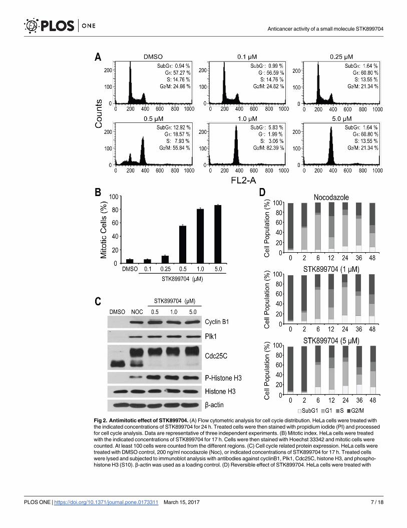
Fig 2. Antimitotic effect of STK899704. (A) Flow cytometric analysis for cell cycle distribution. HeLa cells were treated with
the indicated concentrations of STK899704 for 24 h. Treated cells were then stained with propidium iodide (PI) and processed
for cell cycle analysis. Data are representative of three independent experiments. (B) Mitotic index. HeLa cells were treated
with the indicated concentrations of STK899704 for 17 h. Cells were then stained with Hoechst 33342 and mitotic cells were
counted. At least 100 cells were counted from the different regions. (C) Cell cycle related protein expression. HeLa cells were
treated with DMSO control, 200 ng/ml nocodazole (Noc), or indicated concentrations of STK899704 for 17 h. Treated cells
were lysed and subjected to immunoblot analysis with antibodies against cyclinB1, Plk1, Cdc25C, histone H3, and phospho-
histone H3 (S10). β-actin was used as a loading control. (D) Reversible effect of STK899704. HeLa cells were treated with
Anticancer activity of a small molecule STK899704
PLOS ONE | https://doi.org/10.1371/journal.pone.0173311 March 15, 2017 7 / 18

(Fig 2A). The increased G2/M phase cells were accompanied by a marked increase in rounded,
mitotic-like cell morphology, suggesting that STK899704 might induce mitotic arrest. To
address this, we assessed the percentage of mitotic cells by quantification of the number of
cells with highly condensed chromosomes, characteristic of particular mitotic chromosomes
[26] and found that STK899704 treatment resulted in a dose-dependent increase in mitotic
indices (Fig 2B). Hence, these results indicate that STK899704 causes cell cycle arrest at M
phase.
In addition, we evaluated the effect of STK899704 on cell cycle related proteins, including
Cdc25C, histone H3, cyclin B1, and polo-like kinase (Plk1), in comparison with nocodazole, a
tubulin depolymerizing agent and well-known mitotic blocker [6]. During mitosis, Cdc25C
activity is hyperphosphorylated causing its slower migration on SDS-PAGE [27, 28], whereas
the phosphorylation of histone H3 at S10 is required for proper chromosome condensation
and segregation [29, 30]. Both Cyclin B1 and Plk1 expression oscillate throughout the cell
cycle. Their levels are minimal in G1 phase, begin to accumulate in S phase, and reach the max-
imum at G2/M boundary [31–34]. As expected, the treatment of HeLa cells with nocodazole,
as well as STK899704, resulted in elevated Cdc25C and histone H3 phosphorylation levels and
accumulated cyclin B1 and Plk1 levels (Fig 2C), confirming that STK899704 induces mitotic
arrest.
To examine whether the antimitotic activity of STK899704 is reversible, HeLa cells were
treated with STK899704 (1 and 5 μM) for 18 h. Nocodazole, a reversible inhibitor of microtu-
bule assembly [35], was included as control. As shown in Fig 2D, HeLa cells were efficiently
arrested at G2/M phase (>80%) upon treatment with nocodazole and STK899704 overnight.
After the removal of nocodazole and STK899704, the G2/M-arrested HeLa cells were able to
re-enter the cell cycle with a minimal delayed in cells treated with a high concentration of
STK899704 (5.0 μM) (Fig 2D). These results indicated that STK899704, like nocodazole, acts
in a reversible manner.
STK899704 interfered with tubulin polymerization and mitotic spindle
organization
Given the fact that microtubules are the major structural component of a mitotic spindle,
whose function is critical for chromosome segregation during mitosis [2], and that a large
number of antimitotic agents interact with tubulin and thereby alter its polymerization and
dynamics [5], we examined the effect of STK899704 on tubulin polymerization in vitro in com-
parison with known microtubule binding agents by tracking the change in turbidity over time.
In the absence of treatment, tubulin heterodimers self-assembled to form linear tubulin poly-
mers in a time-dependent manner as shown in Fig 3A. Treatment with the microtubule-stabi-
lizing agent paclitaxel resulted in enhanced tubulin polymerization, whereas the microtubule-
depolymerizing agent vinblastine, as well as STK899704, interfered with tubulin polymeriza-
tion (Fig 3A).
To further determine whether STK899704 could affect mitotic spindle organization in cells,
HeLa cells were treated with DMSO control or test compounds, followed by formaldehyde fix-
ation and subsequent staining to visualize α–tubulin, γ-tubulin, and chromosomes. As shown
in Fig 3B, Taxol, a microtubule stabilizing agent, significantly induced multipolar spindles
with highly condensed chromosomes resulting from enhanced tubulin polymerization.
nocodazole (200 ng/ml) or STK899704 (1 or 5 μM) for 17 h. Cells were washed twice and released into fresh DMEM without
nocodazole or STK899704. Cells were then stained with PI at the indicated time and processed for cell cycle analysis. Each
Bar indicates mean ± SD from three independent experiments.
https://doi.org/10.1371/journal.pone.0173311.g002
Anticancer activity of a small molecule STK899704
PLOS ONE | https://doi.org/10.1371/journal.pone.0173311 March 15, 2017 8 / 18

Fig 3. STK899704 inhibited tubulin polymerization and mitotic spindle organization. (A) Tubulin polymerization assay.
The effect of STK899704 (5 μM) on polymerization of purified tubulin in vitro was examined in a GTP-containing buffer. DMSO
was used as a negative control. Tubulin-targeting agents Taxol (5 μM) and vinblastine (5 μM) were also used as controls for
tubulin-stabilizing and tubulin-destabilizing agents, respectively. Assembly of tubulin into microtubules was determined by the
degree of turbidity at 340 nm. (B) Immunofluorescence staining of microtubules in HeLa cells. Cells were treated with DMSO,
Taxol (100 nM), nocodazole (200 ng/ml), or indicated concentrations of STK899704 for 17 h. Cells were then fixed and stained
with Alexa Fluor 488-conjugated anti-tubulin antibody and Hoechst 33342 to visualize α-tubulin and DNA, respectively. Scale
bar, 10 μm. (C) Fraction of mitotic cells. At least 100 cells from (B) were counted from the different regions. Percentages of
normal metaphase, misaligned, multipolar, and tubulin aggregate phenotypes were shown. (D) Proposed binding model of
Anticancer activity of a small molecule STK899704
PLOS ONE | https://doi.org/10.1371/journal.pone.0173311 March 15, 2017 9 / 18

Abnormal spindle morphology was also clearly observed in cells treated with lower concentra-
tions of nocodazole (0.1 μM) and STK899704 (0.25–1.0 μM) (Fig 3B and 3C). In some
instances, spindle appeared normal but chromosomes were not aligned at the metaphase plate.
In others, multipolar spindles were observed. This abnormal spindle morphology suggests that
nocodazole and STK899704 at low concentration interfere with spindle microtubule dynamics.
However, cells treated with higher concentrations of nocodazole (0.3 μM) or STK899704
(5.0 μM) exhibited condensed chromosome with aggregated tubulin and disrupted microtu-
bules. Taken together, these results suggest that STK899704, like nocodazole, is a microtubule-
depolymerizing agent.
We next search for potential binding site of STK899704 on tubulin. Using computer model-
ing and X-ray structure PDB code 1SA0 [18], we determined the proposed binding site of
STK899704 on tubulin (Fig 3D, red stick) is located between the α- and β-subunit of tubulin at
the colchicine binding site (Fig 3D, green stick). Comparison of their proposed binding modes
showed that colchicine binds to the β-tubulin subunit whereas the STK899704 ligand spans
across the intra dimer interface suggesting that the binding site of STK899704 somehow over-
laps with colchicine. We confirmed that STK899704 interacts directly with this binding site
using a tubulin competitive binding SPA assay. In addition, we showed that STK899704 com-
petitively inhibited [3H]colchicine binding to biotinylated tubulin, similar to unlabeled colchi-
cine, whereas vinblastine did not significantly influence the binding of [3H]colchicine (Fig
3E). Thus, these data indicate that STK899704 is a new class of tubulin inhibitor; and its anti-
proliferative activity is due to its binding to tubulin at the colchicine-binding site.
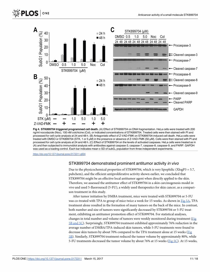
STK899704 induced cell death following prolonged mitotic arrest
We next examined whether STK899704 would eventually induce cell death after prolonged
mitotic arrest by determining the subG1 populations of HeLa cells. Similar to the effect of
colchicine and nocodazole, treatment with STK899704 resulted in a marked increase in the
subG1 population at 48 h (Fig 4A). Since caspases are the key mediators of apoptosis [36],
Z-VAD-FMK, a cell-permeable and irreversible pancaspase inhibitor, was used to examine
whether caspases are involved in the increased subG1 population after STK899704 treatment.
As shown in Fig 4B, co-treatment with Z-VAD-FMK prevented the accumulation of the subG1
population compared with STK899704 treatment alone. These results suggest that STK899704
treatment induces apoptosis in a time-dependent manner, associated with the increase in the
subG1 population.
In addition, the effects of STK899704, colchicine, and nocodazole on the cleavage of initia-
tor caspases were analyzed. Caspase-3 and caspase-7 are the effector caspases that are activated
by initiator caspases such as caspase-8 and caspase-9. As shown in Fig 4C, the cleaved caspases
were clearly detectable at 48 h in a concentration-dependent manner. The level of PARP cleav-
age was also examined since activation of effector caspases such as caspase-3 leads to down-
stream cleavage of various substrates including poly (ADP-ribose) polymerase (PARP) during
apoptosis [36, 37]. Consistent with active caspase levels, PARP cleavage was also prominent at
48 h after treatment with STK899704, colchicine, and nocodazole (Fig 4C). Taken together,
these results indicate that STK899704 induces prolonged mitotic arrest and consequently leads
to apoptosis.
STK899704 on tubulin. The αβ-tubulin heterodimer from PDB entry 1SA0 is shown as ribbon (gray, α-tubulin; cyan, β-tubulin).
STK899704 is presented in red stick while colchicine is shown in green. (E) Effect of STK899704 on tubulin binding. Tubulin
binding was tested with a SPA-based competition assay. Error bars represent mean ± SDs from three independent
experiments.
https://doi.org/10.1371/journal.pone.0173311.g003
Anticancer activity of a small molecule STK899704
PLOS ONE | https://doi.org/10.1371/journal.pone.0173311 March 15, 2017 10 / 18
STK899704 demonstrated prominent antitumor activity in vivo
Due to the physicochemical properties of STK899704, which is very lipophilic (XlogP3 = 5.7,
pubchem), and the efficient antiproliferative activity shown earlier, we concluded that
STK899704 might be an effective local antitumor agent when directly applied to the skin.
Therefore, we assessed the antitumor effect of STK899704 in a skin carcinogenesis model invivo and used 5-fluorouracil (5-FU), a widely used therapeutics for skin cancer, as a compari-
son treatment in this study.
After tumor initiation by DMBA treatment, mice were treated with STK899704 or 5-FU
was co-treated with TPA to group of mice twice a week for 15 weeks. As shown in Fig 5A, TPA
treatment alone resulted in the formation of many tumors on the back of the mice. In contrast,
both number and size of tumors were significantly decreased by STK899704 or 5-FU treat-
ment, exhibiting an antitumor promotion effect of STK899704. For statistical analyses,
changes in total number and volume of tumors were weekly monitored during treatment (Fig
5B and 5C). Surprisingly, STK899704 treatment exhibited approximately 76% reduction in the
average number of DMBA/TPA-induced skin tumors, while 5-FU treatments were found to
decrease skin tumors by about 70% compared to the TPA treatment alone at 15 weeks (Fig
5B). Similarly, STK899704 treatment reduced the tumor volume by approximately 80%, while
5-FU treatments decreased the tumor volume by about 76% at 15 weeks (Fig 5C). At 15 weeks,
Fig 4. STK899704 triggered programmed cell death. (A) Effect of STK899704 on DNA fragmentation. HeLa cells were treated with 200
ng/ml nocodazole (Noc), 100 nM colchicine (Col), or indicated concentrations of STK899704. Treated cells were then stained with PI and
processed for cell cycle analysis at 24 and 48 h. (B) Antagonistic effect of Z-VAD-FMK on STK899704-induced cell death. HeLa cells were
treated with DMSO or STK899704 (STK, 1 or 5 μM) in the presence or absence of Z-VAD-FMK (50 μM). Cells were then stained with PI and
processed for cell cycle analysis at 24 and 48 h. (C) Effect of STK899704 on the levels of activated caspases. HeLa cells were treated as in
(A) and then subjected to immunoblot analysis with antibodies against caspase-3, caspase-7, caspase-8, caspase-9, and PARP. GAPDH
was used as a loading control. Each bar indicates mean ± SD of subG1 population from three independent experiments.
https://doi.org/10.1371/journal.pone.0173311.g004
Anticancer activity of a small molecule STK899704
PLOS ONE | https://doi.org/10.1371/journal.pone.0173311 March 15, 2017 11 / 18
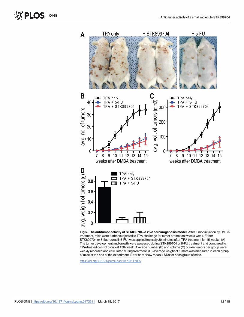
Fig 5. The antitumor activity of STK899704 in vivo carcinogenesis model. After tumor initiation by DMBA
treatment, mice were further subjected to TPA challenge for tumor promotion twice a week. Either
STK899704 or 5-fluorouracil (5-FU) was applied topically 30 minutes after TPA treatment for 15 weeks. (A)
The tumor development and growth were assessed during STK899704 or 5-FU treatment and compared to
TPA-treated control group at 15th week. Average number (B) and volume (C) of skin tumors per group were
weekly recorded and calculated during treatment. (D) Average weight of tumors was measured in each group
of mice at the end of the experiment. Error bars show mean ± SDs for each group of mice.
https://doi.org/10.1371/journal.pone.0173311.g005
Anticancer activity of a small molecule STK899704
PLOS ONE | https://doi.org/10.1371/journal.pone.0173311 March 15, 2017 12 / 18

skin tumors from each group of mice were isolated, and their weight was measured as shown
in the Fig 5D. The mean weight of the skin tumors was obviously decreased 9.4-fold in the
STK899704-treated group. These results indicate that STK899704 exhibits potent both chemo-
preventive and antitumor effects, and the efficacy of STK899704 showed beyond that of 5-FU.
The effects of STK899704 treatment on normal skin were also evaluated in healthy group of
mice. Visual inspection and histological examination of the skin sections revealed that there
were no abnormal symptoms such as skin inflammation and hyperplasia (S2 Fig).
Discussion
In our effort to discover novel antiproliferative agents, we identified STK899704 as a novel
tubulin inhibitor. STK899704 has an acylhydrazone moiety known as a useful scaffold for drug
development, especially for anticancer therapy due to its antimitotic activity [38–42]. Further
analyses showed that STK899704 strongly induces the accumulation of mitotic cells in a con-
centration-dependent and reversible manner (Fig 2A, 2B and 2D). These results suggest
that STK899704 induces cell cycle arrest in mitosis. In addition, the antimitotic effect of
STK899704 was confirmed by increased phosphorylation of Cdc25C and accumulation of
cyclin B1 and Plk1 (Fig 2C), which are associated with mitosis [18, 27, 28, 31–37]. Moreover,
prolonged mitotic arrest leads to a time-dependent increase in subG1 population (Fig 4A) and
activation of caspases (Fig 4C). The effector caspases are implicated in the late, irreversible
phase of the apoptotic pathway and carry out the proteolytic degradation of a broad range of
cellular targets, such as PARP which plays an important role in diverse cellular processes,
including DNA damage response [36, 37]. Therefore, increased DNA fragmentation and
altered levels of active caspases suggest that prolonged mitotic arrest induced by STK899704
eventually trigger programmed cell death.
We also found that the antimitotic activity of STK899704 is ascribed to its ability to prevent
tubulin polymerization in vitro and disrupt microtubules in cells (Fig 3A and 3B), indicating
that STK899704 is a microtubule-destabilizing agent. Computer modeling studies reveal that
STK899704 possibly binds to the colchicine-binding site (Fig 3D). The colchicine ligand lies in
β-tubulin subunit whereas STK899704 ligand traverses across the interface between αβ-tubulin
heterodimer. Additionally, the results from Figs 2D and 3D suggest that STK899704 does not
covalently bind to tubulin, allowing for the full reversibility of its intracellular activity.
One of the major limitations of anticancer drugs in clinical use is drug resistance, acquired
or intrinsic, especially resistance due to the expression of the drug-efflux protein P-gp [8].
Unlike Taxol and the Topoisomeras II inhibitor doxorubicin, STK899704 exhibits equal or
better potency in both parental and multidrug-resistant cells overexpressing P-glycoprotein.
Therefore, STK899704 may overcome the resistance mediated by the multidrug resistance pro-
tein P-gp.
In this study, the efficacy of STK899704 was investigated using a DMBA/TPA-induced skin
carcinogenesis model (Fig 5). Skin cancer is a generic term for malignant tumor occurring on
the skin and its incidence rate has been generally on the rise [43, 44]. The overall prevalence of
skin cancer has been reported to be higher in Caucasians and becomes higher with age, in part
due to the accumulative exposure to UV light [45–47]. Thus, skin cancer is emerging as a
global issue in terms of disease prevention ranging from Europe and the United States whose
major races are Caucasians, to Asia where the population age is steadily rising. Therefore,
development of a therapy with convenient applicability and low side effect would be in high
demand. Here we reported that topically applied STK899704 exhibited efficient antitumor
effect. In response to STK899704 treatment, approximately 80% of the total number of skin
tumors dramatically decreased (Fig 5B). Compared to the TPA-treated control group, tumors
Anticancer activity of a small molecule STK899704
PLOS ONE | https://doi.org/10.1371/journal.pone.0173311 March 15, 2017 13 / 18
size was also reduced to approximately 80% by STK899704 treatments (Fig 5C). Thus, the
prominent antitumor activity of STK899704 has been proved in vivo animal model.
Except surgical removal of the lesion, 5-FU and Imiquimod are the most widely used topical
therapeutics for skin cancer [48, 49]. However, 5-FU is occasionally accompanied by many
side effects including local pain, itchiness, burning, stinging, and dermatitis. Imiquimod acts
as immune response modifier that can stimulate the immune system to primarily produce
interferon-α (INF-α) leading to nonspecific inflammation or dermatitis. Since the antitumor
effect of STK899704 was surprisingly comparable to 5-FU (Fig 5) and did not induce abnormal
symptoms or alteration in skin conditions (S2 Fig), STK899704 is a promising alternative to
5-FU or Imiquimod for treatment of skin cancer.
Taken together, our data indicate that the antiproliferative effect of STK899704 is ascribed
to its microtubule destabilizing activity, resulting in mitotic arrest and apoptosis. In addition,
the antiproliferative activity of STK899704 seems to be effective regardless of any multidrug-
resistance phenotype. Our study suggests that STK899704 is a promising candidate for further
development as a chemotherapeutic agent, particularly for the treatment of skin cancer as well
as MDR cancer.
Supporting information
S1 Fig. Reaction scheme for the synthesis of STK899704. Synthesis of (E)-ethyl 2-(2-
methyl-3-((2-(naphtho[2,1-b]furan-2-carbonyl)hydrazono)methyl)-1H-indol-1-yl)acetate
(STK899704) is outlined in Scheme 1. The preparation of the title compound STK899704 was
started from 2-Hydroxy-1-naphthaldehyde (1) and ethyl bromoacetate to obtaine Naphtho
[2,1-b]furan-2-carboxylic acid (2a). This reaction required anhydrous K2CO3 as base and
dimethylformamide (DMF) as solvent, both condensation as well as cyclization occurred in
single step in 90% yield. The structure of the product 2a was determined by 1H NMR and
ESIMS analyses. The 7 aromatic protons characteristic signals appeared between δ8.269–
7.460 ppm in 1H NMR and 13 carbon signals appeared between δ 161.293–105.238 ppm in 13C
NMR spectra, also its mass spectrum revealed a molecular ion peak at m/z 211.4[M-H]-corre-
sponding to the molecular formula C13H8O3 confirms the structure of 2a. In the second step
of reacton, Naphtho[2,1-b]furan-2-carboxylic acid (2a) converted to the corresponding ester
ethyl naphtho[2,1-b]furan-2-carboxylate (2b) in 60% yield, by esterification reaction using eth-
anol and SOCl2. The carboxylate 2b was confirmed by the presence of new peaks quartet—
CH2 at δ 4.424–4.370 ppm (J = 7.2 Hz) and triplet—CH3 at δ 1.388–1.353 ppm (J = 7.2 Hz)
along with 7 aromatic protons in 1H NMR spectrum and also by molecular ion peak at m/z241.4 [M+H]+, 263.4 [M+Na]+ appeared in ESIMS spectrum corresponding to the molecular
formula C15H12O3.Thus compound 2b was reacted with hydrazine hydrate in third step of
reaction to obtain an intermediate compound naphtho[2,1-b]furan-2-carbohydrazide 3 in
72% yield. 1H NMR spectrum of compound 3 exhibited no peak corresponds to ester instead
it shows signals at δ 10.049 ppm for amide O = C-NH and δ 4.597 ppm for -NH2 (D2O
exchangeable) of hydrazide respectively. The structure was further confirmed by recording
its mass spectra, by its molecular ion peak at m/z 225.4 [M-H]-, 227.4 [M+H]+, 249.4
[M+Na]+corresponds to molecular formula C13H10N2O2. Vilsmeier-Haack formylation of
2-methyl indole (4) gave 2-methyl-1H-indole-3-carbaldehyde (5) in 87% yield in fourth step.
Reaction of phosphorous oxychloride with DMF at low temperature led to the formation of an
electrophile and then was slowly added 2-methyl indole. The structure of the product 5 was
confirmed by the appearance of new peak at δ 10.058 ppm for—CHO instead of one aromatic
proton in 1H NMR spectrum. ESIMS analysis data indicates the molecular ion peak at m/z158.4 [M-H]-, 160.4 [M+H]+, 182.3 [M+Na]+ which is corresponding to molecular formula
Anticancer activity of a small molecule STK899704
PLOS ONE | https://doi.org/10.1371/journal.pone.0173311 March 15, 2017 14 / 18

C10H9NO. The intermediate compound ethyl 2-(3-formyl-2-methyl-1H-indol-1-yl)acetate (6)
was obtained in 86% yield in step 5, by N-substitution of 2-methyl-1H-indole-3-carbaldehyde
(5) by ethyl bromoacetate using sodium hydride as a base in THF solvent and it was confirmed
by the absence of indole—NH signal at δ 11.962 ppm instead appeared new signals at δ 4.208–
4.155 ppm, quartet for—CH2 and δ 1.241–1.205 ppm triplet for—CH3 in 1H NMR spectrum
and also by molecular ion peak at m/z 244.4 [M-H]-, 246.4 [M+H]+, 268.4 [M+Na]+ appeared
in ESIMS spectrum corresponding to the molecular formula C14H15NO3. Finally the title com-
pound (E)-ethyl 2-(2-methyl-3-((2-(naphtho[2,1-b]furan-2-carbonyl)hydrazono)methyl)-1H-
indol-1-yl)acetate (STK899704) was obtained in 75% yield, by condensation of hydrazide 3
with aldehyde 6 in presence of catalytic amount of acetic acid in absolute ethanol. The struc-
ture of newly synthesized molecule STK899704 was confirmed by 1H NMR, 13C NMR and
mass analyses. 1H NMR of compound STK899704 exhibits singlet at δ 11.838 ppm for amide
proton O = C-NH-, singlet at δ 8.862 ppm for azomethine proton—CH = N-, 11 aromatic pro-
tons appeared between δ 8.438–7.198 ppm, δ 5.192 ppm for -N-CH2, δ 4.211–4.158 ppm for—
O-CH2-,δ 1.249–1.214 ppm for—CH3, and δ 2.505 ppm singlet appeared for—CH3 attached to
aromatic ring.13C NMR spectrum of compound STK899704 exhibits total 27 carbon signals.
The structure was further confirmed by recording its mass spectrum, by its molecular ion peak
at m/z m/z 452.6 [M-H]-, 454.6 [M+H]+, 476.5 [M+Na]+ corresponds to molecular formula
C27H23N3O4.
(TIF)
S2 Fig. The effects of STK899704 treatment on normal skin. To examine additional effects
of STK899704 treatment on normal skin, the compound or acetone was applied onto the dor-
sal skin of healthy mice twice weekly for 10 weeks. (A) The picture taken after the last treat-
ment. (B) Hematoxylin/eosin stained sections from skin samples exhibit no skin abnormalities
caused by STK899704 treatment.
(TIF)
S1 Table. Antiproliferative activity of STK899704 on various cancer cell lines. The various
cancer cell lines were cultured in microtiter plates (1–2 x 10³ cells/well) and incubated with dif-
ferent concentration of STK899704 for 4 days. The MTT assay was used to determine the cyto-
toxic effect of STK899704 and IC50s were assessed by log-dose-response curves. Data are the
average of triplicate assays.
(PDF)
Acknowledgments
This work was supported by the R&D Convergence Program (CAP-16-03-KRIBB) and the
International Cooperation Research Program of National Research Council of Science & Tech-
nology (NST) of Republic of Korea. This work was also supported the Bio and Medical Tech-
nology Development Program (NRF-2014M3A9B5073938), the Basic Science Research
Program (NRF-2016R1A2B3011389) of the Ministry of Science, ICT and Future Planning
(MSIP) of Korea and KRIBB Research Initiative Program.
Author Contributions
Conceptualization: KS SK JH NKS BYK.
Data curation: KS SK JH BYK.
Formal analysis: KS SK TWC YL CMP NRT PYL BS.
Anticancer activity of a small molecule STK899704
PLOS ONE | https://doi.org/10.1371/journal.pone.0173311 March 15, 2017 15 / 18

Funding acquisition: YTK JSA BYK.
Investigation: KS JH NKS KHL.
Methodology: GS TTHP HP.
Resources: JHJ HWY YJS.
Software: HP SJK.
Supervision: KSL SJK YTK JSA BYK.
Validation: KS SK JH NKS.
Visualization: TWC YL CMP NRT.
Writing – original draft: NS SK JH NKS.
Writing – review & editing: YTK JSA BYK.
References1. Honore S, Pasquier E, Braguer D. Understanding microtubule dynamics for improved cancer therapy.
Cell Mol Life Sci. 2005; 62(24):3039–56. https://doi.org/10.1007/s00018-005-5330-x PMID: 16314924
2. Kline-Smith SL, Walczak CE. Mitotic Spindle Assembly and Chromosome Segregation: Refocusing on
Microtubule Dynamics. Molecular Cell. 2004; 15(3):317–27. http://dx.doi.org/10.1016/j.molcel.2004.07.
012. PMID: 15304213
3. Castedo M, Perfettini J-L, Roumier T, Andreau K, Medema R, Kroemer G. Cell death by mitotic catas-
trophe: a molecular definition. Oncogene. 2004; 23(16):2825–37. https://doi.org/10.1038/sj.onc.
1207528 PMID: 15077146
4. Dalton WB, Yang VW. Role of prolonged mitotic checkpoint activation in the formation and treatment of
cancer. Future Oncology. 2009; 5(9):1363–70. https://doi.org/10.2217/fon.09.118 PMID: 19903065
5. Dumontet C, Jordan MA. Microtubule-binding agents: a dynamic field of cancer therapeutics. Nat Rev
Drug Discov. 2010; 9(10):790–803. http://www.nature.com/nrd/journal/v9/n10/suppinfo/nrd3253_S1.
html. PMID: 20885410
6. Jordan MA, Thrower D, Wilson L. Effects of vinblastine, podophyllotoxin and nocodazole on mitotic
spindles. Implications for the role of microtubule dynamics in mitosis. Journal of Cell Science. 1992; 102
(3):401–16.
7. Schiff PB, Horwitz SB. Taxol stabilizes microtubules in mouse fibroblast cells. Proceedings of the
National Academy of Sciences of the United States of America. 1980; 77(3):1561–5. PMID: 6103535
8. Fojo T, Menefee M. Mechanisms of multidrug resistance: the potential role of microtubule-stabilizing
agents. Annals of Oncology. 2007; 18(suppl 5):v3–v8.
9. Johnstone RW, Ruefli AA, Smyth MJ. Multiple physiological functions for multidrug transporter P-glyco-
protein? Trends in Biochemical Sciences. 2000; 25(1):1–6. http://dx.doi.org/10.1016/S0968-0004(99)
01493-0. PMID: 10637601
10. Orr GA, Verdier-Pinard P, McDaid H, Horwitz SB. Mechanisms of Taxol resistance related to microtu-
bules. Oncogene. 2003; 22(47):7280–95. https://doi.org/10.1038/sj.onc.1206934 PMID: 14576838
11. Huzil JT, Chen K, Kurgan L, Tuszynski JA. The roles of beta-tubulin mutations and isotype expression
in acquired drug resistance. Cancer Inform. 2007; 3:159–81. PMID: 19455242
12. Kavallaris M. Microtubules and resistance to tubulin-binding agents. Nat Rev Cancer. 2010; 10(3):194–
204. https://doi.org/10.1038/nrc2803 PMID: 20147901
13. Windebank AJ. Chemotherapeutic neuropathy. Curr Opin Neurol. 1999; 12(5):565–71. PMID:
10590893
14. Quasthoff S, Hartung HP. Chemotherapy-induced peripheral neuropathy. J Neurol. 2002; 249(1):9–17.
PMID: 11954874
15. Kim HM, Oh GT, Hong DH, Kim MS, Kang JS, Park SM, et al. MDR-1 gene expression is a minor factor
in determining the multidrug resistance phenotype of MCF7/ADR and KB-V1 cells. FEBS Lett. 1997;
412(1):201–6. PMID: 9257720
Anticancer activity of a small molecule STK899704
PLOS ONE | https://doi.org/10.1371/journal.pone.0173311 March 15, 2017 16 / 18

16. Kang MR, Lee K, Kang JS, Lee CW, Lee KH, Kim JH, et al. KBH-A42, a histone deacetylase inhibitor,
inhibits the growth of doxorubicin-resistant leukemia cells expressing P-glycoprotein. Oncol Rep. 2010;
23(3):801–9. PMID: 20127023
17. Hwang J, Kita R, Kwon HS, Choi EH, Lee SH, Udey MC, et al. Epidermal ablation of Dlx3 is linked to IL-
17-associated skin inflammation. Proceedings of the National Academy of Sciences of the United
States of America. 2011; 108(28):11566–71. https://doi.org/10.1073/pnas.1019658108 PMID:
21709238
18. Ravelli RB, Gigant B, Curmi PA, Jourdain I, Lachkar S, Sobel A, et al. Insight into tubulin regulation from
a complex with colchicine and a stathmin-like domain. Nature. 2004; 428(6979):198–202. https://doi.
org/10.1038/nature02393 PMID: 15014504
19. Gasteiger J, Marsili M. Iterative partial equalization of orbital electronegativity—a rapid access to atomic
charges. Tetrahedron. 1980; 36(22):3219–28. http://dx.doi.org/10.1016/0040-4020(80)80168-2.
20. Park H, Shin Y, Choe H, Hong S. Computational design and discovery of nanomolar inhibitors of Ikap-
paB kinase beta. J Am Chem Soc. 2015; 137(1):337–48. https://doi.org/10.1021/ja510636t PMID:
25513719
21. Park H, Lee S, Hong S. Discovery of Dual Inhibitors for Wild Type and D816V Mutant of c-KIT Kinase
through Virtual and Biochemical Screening of Natural Products. J Nat Prod. 2016; 79(2):293–9. https://
doi.org/10.1021/acs.jnatprod.5b00851 PMID: 26807861
22. Shin S, Mah S, Hong S, Park H. Discovery of Low Micromolar Dual Inhibitors for Wild Type and L1196M
Mutant of Anaplastic Lymphoma Kinase through Structure-Based Virtual Screening. J Chem Inf Model.
2016; 56(4):802–10. https://doi.org/10.1021/acs.jcim.6b00026 PMID: 27014917
23. Park H, Shin Y, Kim J, Hong S. Application of Fragment-Based de Novo Design to the Discovery of
Selective Picomolar Inhibitors of Glycogen Synthase Kinase-3 Beta. J Med Chem. 2016; 59(19):9018–
34. https://doi.org/10.1021/acs.jmedchem.6b00944 PMID: 27676184
24. Hennings H, Glick AB, Lowry DT, Krsmanovic LS, Sly LM, Yuspa SH. FVB/N mice: an inbred strain sen-
sitive to the chemical induction of squamous cell carcinomas in the skin. Carcinogenesis. 1993; 14
(11):2353–8. PMID: 8242866
25. Liu H, Hwang J, Li W, Choi TW, Liu K, Huang Z, et al. A derivative of chrysin suppresses two-stage skin
carcinogenesis by inhibiting mitogen- and stress-activated kinase 1. Cancer prevention research. 2014;
7(1):74–85. https://doi.org/10.1158/1940-6207.CAPR-13-0133 PMID: 24169959
26. Koshland D, Strunnikov A. Mitotic chromosome condensation. Annu Rev Cell Dev Biol. 1996; 12:305–
33. https://doi.org/10.1146/annurev.cellbio.12.1.305 PMID: 8970729
27. Izumi T, Walker DH, Maller JL. Periodic changes in phosphorylation of the Xenopus cdc25 phosphatase
regulate its activity. Mol Biol Cell. 1992; 3(8):927–39. PMID: 1392080
28. Bonnet J, Mayonove P, Morris MC. Differential phosphorylation of Cdc25C phosphatase in mitosis. Bio-
chemical and biophysical research communications. 2008; 370(3):483–8. https://doi.org/10.1016/j.
bbrc.2008.03.117 PMID: 18384749
29. Wei Y, Yu L, Bowen J, Gorovsky MA, Allis CD. Phosphorylation of histone H3 is required for proper
chromosome condensation and segregation. Cell. 1999; 97(1):99–109. PMID: 10199406
30. Hans F, Dimitrov S. Histone H3 phosphorylation and cell division. Oncogene. 2001; 20(24):3021–7.
https://doi.org/10.1038/sj.onc.1204326 PMID: 11420717
31. Pines J, Hunter T. Isolation of a human cyclin cDNA: evidence for cyclin mRNA and protein regulation in
the cell cycle and for interaction with p34cdc2. Cell. 1989; 58(5):833–46. PMID: 2570636
32. Hwang A, Maity A, McKenna WG, Muschel RJ. Cell cycle-dependent regulation of the cyclin B1 pro-
moter. J Biol Chem. 1995; 270(47):28419–24. PMID: 7499347
33. Lee KS, Yuan YL, Kuriyama R, Erikson RL. Plk is an M-phase-specific protein kinase and interacts with
a kinesin-like protein, CHO1/MKLP-1. Mol Cell Biol. 1995; 15(12):7143–51. PMID: 8524282
34. Winkles JA, Alberts GF. Differential regulation of polo-like kinase 1, 2, 3, and 4 gene expression in mam-
malian cells and tissues. Oncogene. 2005; 24(2):260–6. https://doi.org/10.1038/sj.onc.1208219 PMID:
15640841
35. De Brabander MJ, Van de Veire RM, Aerts FE, Borgers M, Janssen PA. The effects of methyl (5-(2-thie-
nylcarbonyl)-1H-benzimidazol-2-yl) carbamate, (R 17934; NSC 238159), a new synthetic antitumoral
drug interfering with microtubules, on mammalian cells cultured in vitro. Cancer research. 1976; 36
(3):905–16. PMID: 766963
36. Strasser A, O’Connor L, Dixit VM. Apoptosis signaling. Annu Rev Biochem. 2000; 69:217–45. https://
doi.org/10.1146/annurev.biochem.69.1.217 PMID: 10966458
37. Soldani C, Scovassi AI. Poly(ADP-ribose) polymerase-1 cleavage during apoptosis: an update. Apopto-
sis. 2002; 7(4):321–8. PMID: 12101391
Anticancer activity of a small molecule STK899704
PLOS ONE | https://doi.org/10.1371/journal.pone.0173311 March 15, 2017 17 / 18
38. Maia Rdo C, Tesch R, Fraga CA. Acylhydrazone derivatives: a patent review. Expert Opin Ther Pat.
2014; 24(11):1161–70. https://doi.org/10.1517/13543776.2014.959491 PMID: 25213630
39. Duan YT, Man RJ, Tang DJ, Yao YF, Tao XX, Yu C, et al. Design, Synthesis and Antitumor Activity of
Novel link-bridge and B-Ring Modified Combretastatin A-4 (CA-4) Analogues as Potent Antitubulin
Agents. Sci Rep. 2016; 6:25387. https://doi.org/10.1038/srep25387 PMID: 27138035
40. Rodrigues DA, Ferreira-Silva GA, Ferreira AC, Fernandes RA, Kwee JK, Sant’Anna CM, et al. Design,
Synthesis, and Pharmacological Evaluation of Novel N-Acylhydrazone Derivatives as Potent Histone
Deacetylase 6/8 Dual Inhibitors. J Med Chem. 2016; 59(2):655–70. https://doi.org/10.1021/acs.
jmedchem.5b01525 PMID: 26705137
41. Salum LB, Mascarello A, Canevarolo RR, Altei WF, Laranjeira AB, Neuenfeldt PD, et al. N-(1’-
naphthyl)-3,4,5-trimethoxybenzohydrazide as microtubule destabilizer: Synthesis, cytotoxicity, inhibi-
tion of cell migration and in vivo activity against acute lymphoblastic leukemia. Eur J Med Chem. 2015;
96:504–18. https://doi.org/10.1016/j.ejmech.2015.02.041 PMID: 25951294
42. do Amaral DN, Cavalcanti BC, Bezerra DP, Ferreira PM, Castro Rde P, Sabino JR, et al. Docking, syn-
thesis and antiproliferative activity of N-acylhydrazone derivatives designed as combretastatin A4 ana-
logues. PLoS One. 2014; 9(3):e85380. https://doi.org/10.1371/journal.pone.0085380 PMID: 24614859
43. Rogers HW, Weinstock MA, Harris AR, Hinckley MR, Feldman SR, Fleischer AB, et al. Incidence esti-
mate of nonmelanoma skin cancer in the United States, 2006. Arch Dermatol. 2010; 146(3):283–7.
https://doi.org/10.1001/archdermatol.2010.19 PMID: 20231499
44. Society AC. Cancer Facts & Figures: American Cancer Society 2014; 2014.
45. Gloster HM Jr., Neal K. Skin cancer in skin of color. J Am Acad Dermatol. 2006; 55(5):741–60; quiz 61–
4. https://doi.org/10.1016/j.jaad.2005.08.063 PMID: 17052479
46. Gilchrest BA. Photoaging. J Invest Dermatol. 2013; 133(E1):E2–6. https://doi.org/10.1038/skinbio.
2013.176 PMID: 23820721
47. El Ghissassi F, Baan R, Straif K, Grosse Y, Secretan B, Bouvard V, et al. A review of human carcino-
gens—part D: radiation. The Lancet Oncology. 2009; 10(8):751–2. PMID: 19655431
48. Bahner JD, Bordeaux JS. Non-melanoma skin cancers: photodynamic therapy, cryotherapy, 5-fluoro-
uracil, imiquimod, diclofenac, or what? Facts and controversies. Clin Dermatol. 2013; 31(6):792–8.
https://doi.org/10.1016/j.clindermatol.2013.08.020 PMID: 24160289
49. Nashan D, Meiss F, Muller M. Therapeutic strategies for actinic keratoses—a systematic review. Euro-
pean journal of dermatology: EJD. 2013; 23(1):14–32. https://doi.org/10.1684/ejd.2013.1923 PMID:
23477760
Anticancer activity of a small molecule STK899704
PLOS ONE | https://doi.org/10.1371/journal.pone.0173311 March 15, 2017 18 / 18