Embed Size (px)
Citation preview
ANTICARCINOGENIC EFFECTS OF THE ETHANOLIC EXTRACT FROM SALIX
AEGYPTIACA L. IN COLON CANCER, INVOLVEMENT OF AKT PATHWAY
A THESIS SUBMITTED TO THE GRADUATE SCHOOL OF NATURAL AND APPLIED SCIENCES
OF MIDDLE EAST TECHNICAL UNIVERSITY
BY
SHABNAM ENAYAT
IN PARTIAL FULFILLMENT OF THE REQUIREMENTS FOR THE DEGREE OF DOCTOR OF PHILOSOPHY
IN
BIOLOGY
SEPTEMBER 2013

Approval of the thesis:
ANTICARCINOGENIC EFFECTS OF THE ETHANOLIC EXTRACT FROM
SALIX AEGYPTIACA L. IN COLON CANCER, INVOLVEMENT OF AKT PATHWAY
Submitted by SHABNAM ENAYAT in partial fulfillment of the requirements for the degree of Doctor of Philosophy in Biology Department, Middle East Technical University by, Prof. Dr. Canan Özgen _____________________ Dean, Graduate School of Natural and Applied Sciences Prof. Dr. Gülay Özcengiz _____________________ Head of the Department, Biology Assoc. Prof. Dr. Sreeparna Banerjee _____________________ Supervisor, Biology Dept., METU Examining Committee Members: Assoc. Prof. Dr. Mesut Muyan _____________________ Biology Dept., METU Assoc. Prof. Dr. Sreeparna Banerjee _____________________ Biology Dept., METU Prof. Dr.Arif Ahmet Başaran _____________________ Pharmacognosy Dept, Faculty of Pharmacy., Hacettepe University, Turkey Assoc. Prof. Dr Mayda Gürsel _____________________ Biology Dept., METU Assoc. Prof. Dr. Ayşen Tezcaner _____________________ Engineering Sciences., METU Date: _____________________

iv
I hereby declare that all information in this document has been obtained and presented in accordance with academic rules and ethical conduct. I also declare that, as required by these rules and conduct, I have fully cited and referenced all material and results that are not original to this work.
Name, Last Name: Shanam Enayat
Signature :
v
ABSTRACT
ANTICARCINOGENIC EFFECTS OF THE ETHANOLIC EXTRACT FROM SALIX AEGYPTIACA L. IN COLON CANCER, INVOLVEMENT OF AKT
PATHWAY
Enayat, Shabnam
Ph.D., Department of Biology Supervisor: Assoc. Prof. Dr. Sreeparna Banerjee
September 2013, 118 pages
Salix aegyptiaca has been used for centuries as a medicinal plant for various health disorders. Our previous study demonstrated high contents of phenolics and antioxidants in its bark many of those are famed for their anti-inflammatory properties.
Since inflammation frequently accompanies the progress of colorectal cancer, in the current study we showed that the ethanolic extract of bark (EEB) from S.aegyptiaca exerted strong anti-proliferative effects on several cancer cells but not on non-cancerous CCD-18Co cells. Ethyl acetate and water fractions, obtained by solvent-solvent fractionation of EEB showed strong antiproliferative effects comparable to the effect from EEB. Both fractions were applied to tandom mass spectrometry. Catechin, salicin and catechol in Ethyl acetate and salicin in aqueous fractions were the most abundant compounds. Additionally, treatment with the extract induced apoptosis and cell cycle arrest at G1/S, not by DNA damage, but by decreasing phosphorylation and activity of Akt/PKB, stabilizing p53 and upregulating p21. The extract strongly reduced the superoxide anion production in cancer cells but not normal cells.
In addition, EEB strongly inhibited anchorage independent growth of colon cancer cell lines HCT-116 and HT-29. Moreover, EEB could inhibit the motility, determined by scratch wound healing and migration through Transwells and adhesion of cancer cells to fibronectin. These in vitro functional changes were accompanied by the downregulation of the EMT markers SNAI1, SNAI2, Twist1, MMP9 and MMP2 and a restoration of E-cadherin expression.
We propose that EEB from S.aegyptiaca, with its myriad cancer chemopreventive effects, can be consumed for its health promoting effects. Key words: Salix aegyptiaca, Ethanolic extract, Colon cancer, Apoptosis, Metastasis
vi
ÖZ
SALİX AEGYPTİACA L.NİN ETANOLİK ÖZÜTÜNÜN AKT SİNYAL YOLAĞI ARACILIĞIYLA KOLON KANSERİ ÜZERİNDE ANTİ-KARSİNOJENİK
ETKİLERİ
Shabnam Enayat
Doktora, Biyoloji Bölümü Tez Yöneticisi: Doç. Dr. Sreeparna Banerjee
Eylül 2013, 118 sayfa
Salix aegyptiaca yüzyıllar boyunca çeşitli sağlık bozuklukları için şifalı bir bitki olarak kullanılmıştır. Önceki çalışmalarımız, bu bitkinin kabuklarında anti-inflamatuvar özellikleriyle ünlü olan fenolikler ve antioksidanların yüksek oranda bulunduğunu göstermiştir. İnflamasyon çoğunlukla kolorektal kanser gelişimine eşlik ettiğinden, bu çalışmada S.aegyptiaca’dan elde edilen kabukların etanol özütlerinin (EEB) çeşitli kanser hücreleri üzerinde çoğalmayı engelleyici etkisi olduğunu gösterdik. Aynı etki kanser olmayan CCD-18Co hücrelerinde görülmemiştir. EEB’nin çekme yöntemiyle ayrıştırılmasıyla elde edilen etil asetat ve su fazları, EEB etkisi ile karşılaştırılabilir düzeyde çoğalmayı önlemiştir. Her iki faz da ardışık kütle spektrometresine tabi tutulmuş ve etil asetat fazında katekin, salisin ve katekol, su fazında ise salisin bileşiklerinin ağırlıkta olduğu gözlenmiştir. Ayrıca, hücrelerin özüt ile muamelesi, DNA hasarı yoluyla değil, Akt/PKB fosforlanmasını ve etkinliğini azaltarak, p53’ü dengeleyerek ve p21’i tetikleyerek apoptoz ve G1/S fazında hücre dönüsünün durmasına yol açmıştır. Özüt aynı zamanda kanser hücrelerinde süperoksit anyon üretimini artırmış fakat aynı etki normal hücrelerde görülmemiştir. Bunlara ek olarak EEB, kolon kanseri hücre hatları HCT-116 ve HT-29’da tutunmadan bağımsız büyüme özelliğini azaltmıştır. Ayrıca EEB, yara iyileşme yöntemiyle belirlenen hareketlilik ve Transwell içinden göç etme ve fibronektine tutunma yöntemleriyle belirlenen göç edebilme özelliklerini gözle görünür şekilde azaltmıştır. Bu in vitro işlevsel değişimlere SNAI1, SNAI2, Twist1, MMP9 ve MMP2 gibi EMT belirteçlerinde azalma ve E-cadherin ifadesinin geri kazanımı eşlik etmiştir. S.aegyptiaca’dan elde edilen EEB’nin, sayısız kanser önleyici etkileri sebebiyle, sağlığı destekleyici bir etken olarak tüketilebileceğini önermekteyiz. Anahtar kelimeler: Salix aegyptiaca, etanol özütü, kolon kanseri, apoptoz, metastaz

vii
DEDICATED
TO
MY FAMILY

viii
ACKNOWLEDGMENTS I would like to express my gratitude to my supervisor Assoc. Prof. Dr. Sreeparna Banerjee for her support, patience, and encouragement throughout my graduate studies. I feel so lucky to have an advisor who always finds the time for listening to even little problems unavoidably faced in the course of performing research.
I would like to thank Dr. Arif Ahmet Başaran at the Department of Pharmacognosy, Hacettepe University, Ankara for valuable advises and helps during the research, Dr. Mayda Gürsel at the Department of Biology, METU, Ankara for her great advises and contribution in flow cytometric analysis and Dr Ihsan Gürsel at the Department of Molecular Biology and Genetics, Bilkent University, Ankara for the use of the flow cytometry facility. I thank Dr. Ali Sonboli for his contribution in plant identification and Dr. Tamay Şeker (METU Molecular Biology and Biotechnology Central Laboratory) for her contribution to the Mass spectrometry experiments. Dr Özlen Konu and Dr Uygar Tazebay at the Department of Molecular Biology and Genetics, Bilkent University, Ankara, Dr Betul Özdemir Soyler at the Food Engineering Department and Dr Elif Erson-Bensan at the Biological Sciences Department, METU, Ankara and Dr Ali Güre, at the Department of Molecular Biology and Genetics are thanked for sharing resources.
I am indebted to all my family for their dedication and the many years of support which gave me the power to face all difficulties and made this dissertation possible. I would like to express my deepest gratitude and love to my dearest mother for her endless love, efforts and patience and to my dearest father for extreme encouragements and trust to me and to my dear brothers whose presence in my life give me inexpressible feeling of confidence and courage to move forward.
My special thanks go to all my dearest friends who are treasures in my life.
The work was funded by METU Scientific Research Fund (Project no. BAP-2008-07-02-04 and BAP-07-02-2012-007) and the Turkish Academy of Sciences Young Scientist award (TÜBA-GEBİP) to SB. I was supported by a TÜBİTAK bursary for international students (TÜBİTAK-BİDEB 2215).
ix
TABLE OF CONTENTS
ABSTRACT ............................................................................................................................. v
ÖZ............................................................................................................................................ vi
ACKNOWLEDGMENTS ..................................................................................................... viii
LIST OF FIGURES ............................................................................................................... xiii
LIST OF ABBREVIATIONS ............................................................................................. xviii
1. INTRODUCTION ................................................................................................................ 1
1.1 An overview of the history of cancer; ancient and modern treatment methods ............ 1
1.2 Carcinogens and therapeutic methods ........................................................................... 2
1.3 Major signaling pathways involved in cancer: potent targets for cancer therapy ......... 2
Oxidative stress mediated mechanisms: Involvement of Reactive Oxygen Species (ROS) .............................................................................................................................. 2
Nuclear factor-kappaB (NF-κB) signaling pathway: a bridge between inflammation and cancer .............................................................................................................................. 3
Mitogen Activated Protein Kinase/Extracellular Related Kinase (MAPKs/ERK) signaling pathway ............................................................................................................ 3
PI3K/Akt/mTOR Pathway .............................................................................................. 4
Downstream targets of PI3K/Akt signaling .................................................................... 5
Cross Talk between these pathways ................................................................................ 5
1.4 Major hallmarks of cancer ............................................................................................. 8
Sustained cell cycle progression and proliferative signaling ........................................... 8
DNA damage-mediated mechanisms .............................................................................. 8
DNA damage response and DNA repair mechanisms..................................................... 8
Resistance to programmed cell death (apoptosis) ........................................................... 9
1.5 Metastasis of epithelial tumor ..................................................................................... 10
Regulation of metastasis ................................................................................................ 10
Epithelial–mesenchymal transition and the invasive potential of tumors ..................... 11
E-cadherin a major cell adhesion molecule involved in the progression of EMT ......... 11
Hallmarks of cancer as therapeutic target for cancer therapy ....................................... 12
1.6 Chemopreventive compounds from natural sources ................................................... 12
New approaches to the value of natural products .......................................................... 13
1.7 Aims of the research .................................................................................................... 15
2. MATERIAL AND METHODS ........................................................................................ 17
x
2.1 Chemicals and Reagents ............................................................................................. 17
2.1 Plant selection ............................................................................................................. 17
2.2 Extract preparation ...................................................................................................... 18
2.3 Solvent-solvent fractionation ...................................................................................... 18
2.4 Polyphenol identification ............................................................................................ 19
2.5 Cell Culture ................................................................................................................. 20
2.6 Transfections ............................................................................................................... 20
2.6.1 Tranformation of E.Coli with rCD2p110 plasmid and plasmid purification ....... 20
2.6.2 Preparation of LB medium ................................................................................... 21
2.7 Cell viability assay ...................................................................................................... 21
2.7.1 MTT Assay .............................................................................................................. 21
2.8 BrdU incorporation assay ........................................................................................... 21
2.9 Annexin V assay ......................................................................................................... 22
2.10 RNA extraction and RT-PCR ................................................................................... 22
2.11 Immunoblotting ......................................................................................................... 23
2.12 Cell cycle analysis ..................................................................................................... 23
2.13 Measurement of superoxide anion using a modified colorimetric NBT assay ......... 23
2.14 Cellular Antioxidant Activity (CAA) Assay ............................................................. 24
2.15 Colony formation in soft agar ................................................................................... 24
2.16 Cell adhesion assay ................................................................................................... 24
2.17 In vitro scratch wound healing assay ........................................................................ 25
2.18 Cell migration assay .................................................................................................. 25
2.19 Statistical analysis. .................................................................................................... 25
3. RESULTS ........................................................................................................................... 27
3.1 Fractionation of EEB and the effect of the fractions on cellular proliferation ............ 27
A: MTT cell proliferation assay .................................................................................... 27
B: BrdU incorporation assay ......................................................................................... 34
3.2 Identification of phenolic compounds present in ethyl acetate and aqueous fractions 36
3.3 Determination of cytotoxicity of EEB on a non-transformed colon fibroblast cell lines. .......................................................................................................................................... 37
3.4 Effect of salicin on cellular proliferation .................................................................... 38
3.4 Determination of EEB effect on cellular oxidative stress, a major inducer of DNA damage and DNA damage-induced cell cycle arrest ........................................................ 39
A: Nitroblue Tetrazolium (NBT) colorimetric assay .................................................... 39
xi
B: Cellular antioxidant activity (CAA) assay: An assessment of cellular oxidative stress ....................................................................................................................................... 41
3.5 Determining the effect of ethyl acetate and aqueous fraction on superoxide anion production in HCT-116 and HT-29 cell lines using NBT colorimetric assay ................... 44
3.6 EEB induces cell cycle arrest at G1/S in HCT-116 and HT-29 cell lines. .................. 45
3.7 Determination of the effect of EEB on DNA damage induction and DNA damage related proteins. ................................................................................................................. 50
3.8 EEB inhibits Akt/PkB and the MAPK pathways to exert its antitumor effects .......... 50
3.9 EEB induces apoptosis in HCT-116 and HT-29 cells. ................................................ 53
A: Annexin V staining assay ......................................................................................... 53
B: Assessment of pro and anti-apoptotic protein expressions ....................................... 59
SECTION TWO.....................................................................................................................61
3.10 The effect of EEB on motility of cancer cells as an indicator of metastatic potential ........................................................................................................................................... 61
3.10.1 Effect of EEB on anchorage independent growth .................................................. 61
3.10.2 Effect of EEB on cellular motility .......................................................................... 64
A: Migration on transwell ............................................................................................. 64
B: Scratch wound healing assay .................................................................................... 66
3.10.3 Effect of EEB on cell adhesion .............................................................................. 69
3.10.4 Treatment with EEB results in an inhibition of EMT ............................................ 69
4. DISCUSION...................................................................................................................... 73
4.1 EEB selectively induced growth inhibitory effects in cancer cells but not in non-transformed cells ............................................................................................................... 74
4.2 The ethyl acetate and aqueous fractions were the active fractions of EEB with strong growth inhibitory effect on cancer cells ............................................................................ 74
4.3 Identification of phenolic compounds present in ethyl acetate and aqueous fractions 74
4.4 EEB and its active fractions inserted antioxidant effect on cancer cells ..................... 75
4.5 The effect of EEB on cell cycle in HCT-116 and HT-29 cell lines ............................. 76
4.6 EEB induced cell cycle arrest through downregulation of Akt/PKB pathway ............ 76
4.7 The dual targeting of Akt/PKB and MAPKs pathways by EEB ................................. 77
4.8 EEB induced apoptosis in both colon cancer cell lines via a p53 dependent pathway 77
4.9 Salicin did not effectively contribute in the cell cycle arrest or proapoptotic function of EEB ............................................................................................................................... 78
4.10 EEB induced strong antimetastatic effects on colon cancer cell lines....................... 79
4.11 EEB strongly reduced the anchorage independent growth of cancer cells ................ 79
4.12 EEB effectively reduced the motility and migration of cancer cells ......................... 79
xii
4.13 EEB strongly reduced the integrin-mediated adhesion to ECM proteins ................. 80
4.14 EEB inhibits the epithelial to mesenchymal transition of colon cancer cell lines. ... 80
5.CONCLUSION .................................................................................................................... 83
5.1 Conclusive schematic figures...................................................................................... 84
REFERENCES ....................................................................................................................... 87
APPENDICES ...................................................................................................................... 113
Appendix A ..................................................................................................................... 113
Appendix B. .................................................................................................................... 116
Appendix C. Mammelian expression vector ................................................................... 117
CURRICULUM VITAE ....................................................................................................... 118
xiii
LIST OF FIGURES Figure 1.1: The six biological alterations considered as the main hallmarks of cancer .......... 1
Figure 1. 2: Represent the effect of PI3K/AKT and MAPKs pathways activation on downstream effectors. ............................................................................................................. 7
Figure 1. 3: Salix aegyptiaca.L (Musk Willow) ................................................................... 14
Figure 3. 1: The growth inhibitory effect of ethyl acetate, aqueous and n-Butanol fractions of EEB on HCT-116 cell line ................................................................................................ 28
Figure 3. 2: The growth inhibitory effect of ethyl acetate and aqueous fractions of EEB on HT-29 cell line ...................................................................................................................... 30
Figure 3. 3: The growth inhibitory effect of ethyl acetate and aqueous fractions of EEB on MCF-7 cell line ..................................................................................................................... 31
Figure 3. 4: The growth inhibitory effect of ethyl acetate and aqueous fractions of EEB on PC-3 cell line. ........................................................................................................................ 32
Figure 3. 5: The growth inhibitory effect of EEB on MCF-7 and PC-3 cell lines ................ 33
Figure 3. 6: The growth inhibitory potential of EEB and its active fraction, ethyl acetate fraction, determined after 48 hour treatment and assayed by BrdU incorporation assay. ..... 35
Figure 3. 7: The growth inhibitory potential of EEB and its active fraction, aqueous fraction, determined after 48 hour treatment and assayed by BrdU incorporation assay. ................... 36
Figure 3. 8: EEB was strongly cytotoxic in HCT-116 and HT-29 colon cancer cell lines while it was not cytotoxic in CCD-18CO non-transformed colon fibroblast cell line. ......... 38
Figure 3. 9: Salicin induced growth inhibition in HCT-116 and HT-29 cell lines at concentrations much higher than the amounts found in EEB at its IC50 values .................... 39
Figure 3. 10: A) Superoxide ion quenching potential of EEB in HCT-116 and HT-29 colon cancer cell lines and the non-transformed CCD-18Co colon fibroblast cell line .................. 40
Figure 3. 11: Measurement of fluorescent dichlorofluorescein (DCF) level with excitation at 495 nm and emission at 535 nm using Epifluorescence microscopy. ................................... 42
xiv
Figure 3. 12: The superoxide quenching potential of ethyl acetate and aqueous fractions of EEB in HCT-116 cell line examined by NBT colorimetric assay. ....................................... 44
Figure 3. 13: The superoxide quenching potential of ethyl acetate and aqueous fractions of EEB in HT-29 cell line examined by NBT colorimetric assay. ............................................ 45
Figure 3. 14A: Cell cycle analysis of colon cancer cell lines after treatment with the extract. . ............................................................................................................................................. 46
Figure 3. 15: The graphs represent cell cycle distribution of HCT-116 and HT-29 cells when treated with salicin ................................................................................................................ 49
Figure 3. 16: Immunoblot of cell lysate from HCT-116 and HT-29 cells treated (or not) with EEB for 48h. ......................................................................................................................... 50
Figure 3. 17: A) EEB inhibits the Akt/PKB and MAPK pathways in colon cancer cell lines .............................................................................................................................................. 52
Figure 3. 18: Annexin V staining indicating distribution of cell populations in A: HCT-116 (upper panel) and B: HT-29 (lower panel) colon cancer cell lines ....................................... 54
Figure 3. 19: The graphs (A-H) represent a comparison between the effect of EEB and salicin on induction of apoptosis in HCT-116 and HT-29 cells. ........................................... 56
Figure 3. 20: Immunoblot of HCT-116 and HT-29 cell lysates following treatment with the extract for 48h. ...................................................................................................................... 60
Figure 3. 21: Represent the effect of EEB on HCT-116 and HT-29 cell lines as a function of dose. ...................................................................................................................................... 62
Figure 3. 22: EEB at concentrations of 150 µg/ml and 125 µg/ml that did not induce growth inhibition strongly reduced the anchorage independent growth capacity of A: HCT-116 and B: HT-29 cell lines. ............................................................................................................... 63
Figure 3. 23: EEB strongly reduced the anchorage independent growth capacity of HCT-116 and HT-29 cell lines. ............................................................................................................. 64
Figure 3. 24: EEB reduces the motility of HCT-116 and HT-29 cells. A) Transwell assay . 65
Figure 3. 25: EEB reduces the motility of HCT-116 and HT-29 cells. C) Transwell assay. 66
Figure 3. 26: EEB reduces the motility of HCT-116 and HT-29 cells. Scratch wound healing. .................................................................................................................................. 67
Figure 3. 27: Significant reductions by paired t test (*P<0.05, *** P<0.0001) in wound closure were seen after 48 h in HCT-116 and after 120h in HT-29 cells ............................. 68
xv
Figure 3. 28: EEB reduces adhesion of cancer cells to fibronectin.. ..................................... 69
Figure 3. 29: EEB inhibits EMT in HCT-116 cells. Semiquantitative PCR ......................... 70
Figure 3. 30: EEB inhibits EMT in HCT-116 cells. Immunoblot ......................................... 71
Figure 3. 31. EEB did not show any significant reduction in the expression of mesenchymal marker Zo-1 and fibronectin proteins. ................................................................................... 72
xvi
LIST OF TABLES Table 3. 1: The concentration of EEB or its active fractions that induced 50% growth inhibition in HCT-116, HT-29, MCF-7 and PC-3 cell lines. ................................................ 34
Table 3. 2: The concentration of EEB or its active fractions that induced 50% growth inhibition in HCT-116 assessed by MTT and BrdU incorporation assays. .......................... 36
Table 3. 3: Phenolics and flavonols present in ethyl acetate and water fractions of the extract as determined by tandem mass spectrometry. The numbers in parenthesis represent mean and standard deviation of at least 3 independent experiments. ............................................. 37
Table 3. 4: PCR primers used in this study ........................................................................... 70
xvii
LIST OF SCHEMES Scheme2. 1: The dried pulverized powder of the crude ethanolic extract of bark was dissolved in water and partitioned into four fractions of n-hexane, ethyl acetate, n-butanol and water. .............................................................................................................................. 19
Scheme 5. 1: The schematic figure represents that EEB induces apoptosis and cell cycle arrest ...................................................................................................................................... 84
Scheme 5. 2: The overall scheme shows the biochemical pathways through which EEB may inhibit epithelial to mesenchymal transition (EMT) in colon cancer cells. ........................... 85
xviii
LIST OF ABBREVIATIONS AKT: Protein Kinase B (PKB)
BrdU: 5-bromo-2'-deoxyuridine: A synthetic nucleotide that is an analog of thymidine
BSA: Bovine Serum Albumin
β-meOH: β-mercaptoethanol
CO2: Carbon dioxide
CRC: Colorectal cancer
DCFH-DA: 2',7'-dichlorfluorescein-diacetate
dH2O: Distilled Water
DNase-1: Deoxyribonuclease I
EDTA: Ethylenediaminetetraacetic acid
EEB: Ethanolic extract of bark
EGFR: Epithelial growth factor receptor
ERK: Extracellular signal-regulated kinases
Et.Ac: Ethyl acetate
FACS: Fluorescence-activated cell sorter analysis
GAPDH: Glyceraldehyde 3-phosphate dehydrogenase
HCl: Hydrochloric acid
JNK: c-Jun N-terminal kinases
KCl: Potassium chloride
LY294002: 2-morpholin-4-yl-8-phenylchromen-4-one, a potent inhibitor of phosphoinositide 3-kinases (PI3Ks)
MAPK: Mitogen-activated protein kinase
MgCl2: Magnesium chloride
MMP-9: Matrix metalloproteinase 9
MPER: Mammalian Protein Extraction Reagent
xix
MTT: 3-(4, 5-dimethylthiazol-2-yl)-2, 5-diphenyltetrazolium bromide
NaCl: Sodium chloride
NBT: Nitro blue tetrazolium chloride
NF-κB: nuclear factor kappa-light-chain-enhancer of activated B cells
PCR: polymerase chain reaction
PBS: Phosphate buffer salin
PBS-T: Phosphate Buffered Saline Tween-20
PI3K: phosphoinositide 3-kinase
PI: Propidium iodide
PVDF: Polyvinylidene Fluoride
mTOR: mammalian target of rapamycine
RNA: Ribonucleic acid
RNase A: Ribonuclease A
SDS: sodium dodecyl sulfate
TWIST: lass A basic helix-loop-helix protein 38 (bHLHa38)
TBS: Tris Buffered Saline
Tris-HCl: 2-Amino-2-(hydroxymethyl)-1,3-propanediol hydrochloride
U0126: 1,4-diamino-2,3-dicyano-1,4-bis[2-aminophenylthio] butadiene ( inhibitor of ERK 1 and ERK 2

xx
1
CHAPTER 1
INTRODUCTION 1. INTRODUCTION
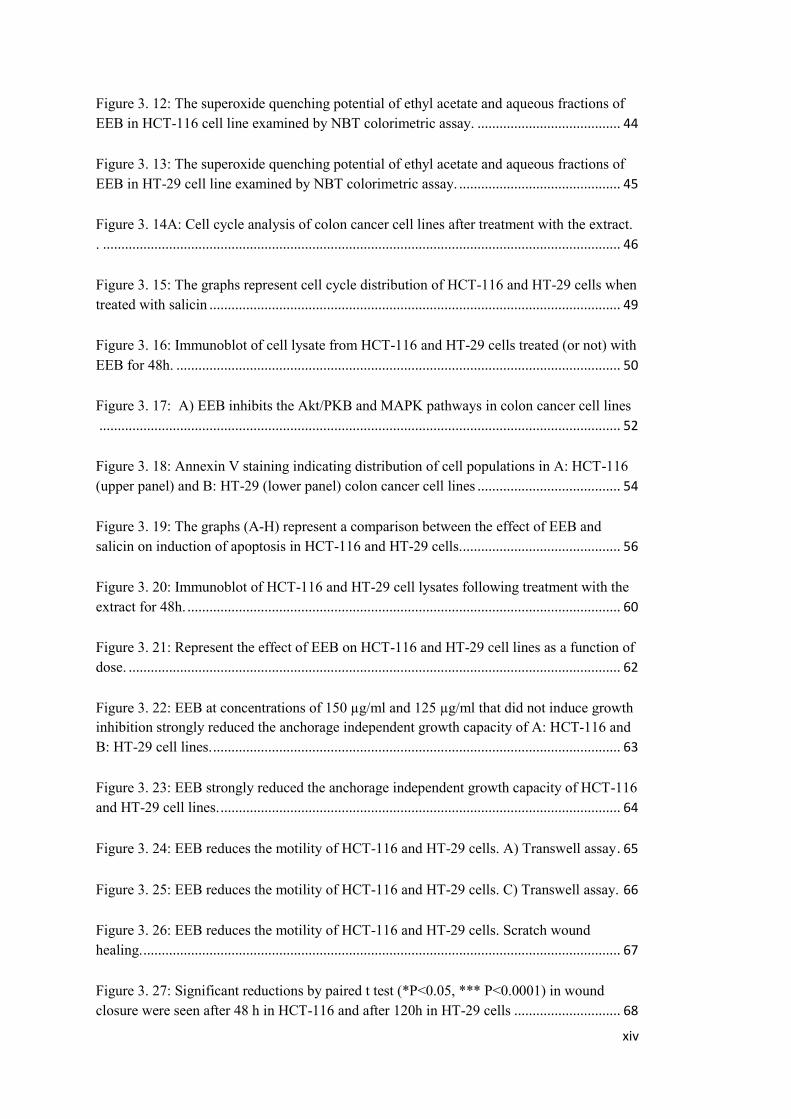
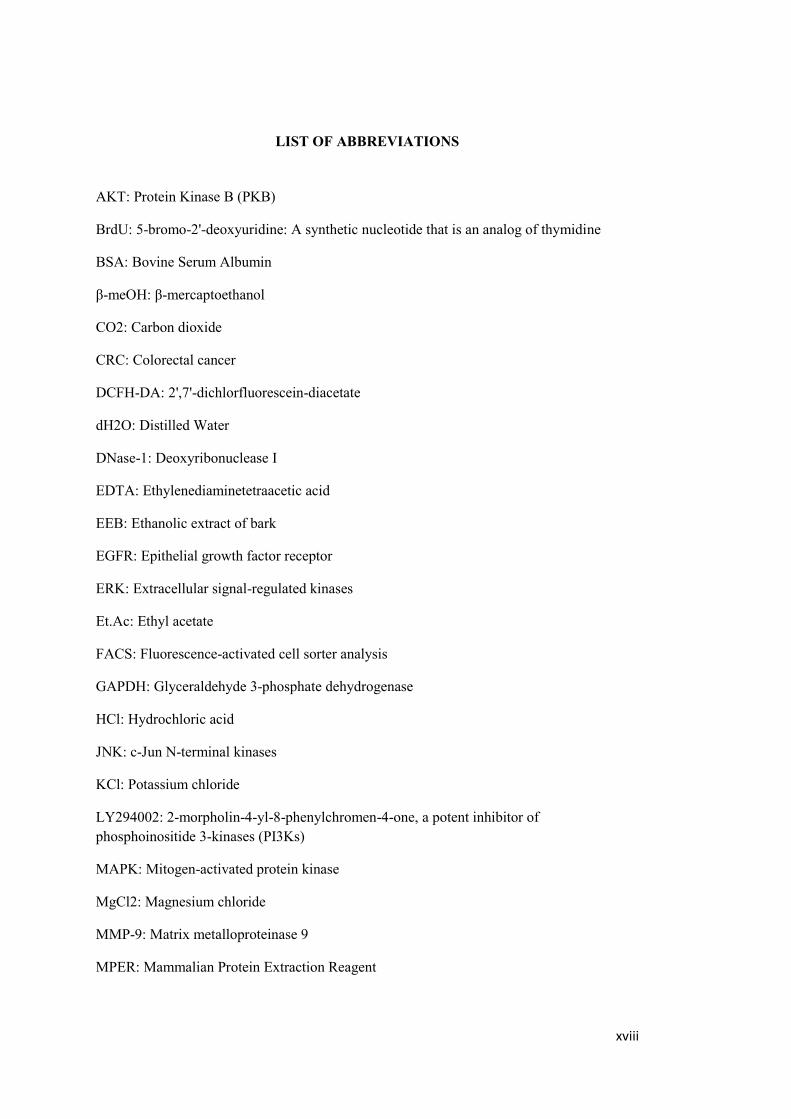
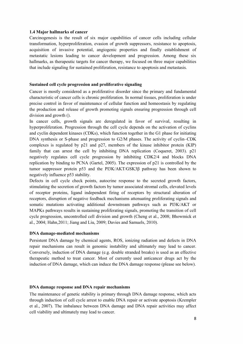
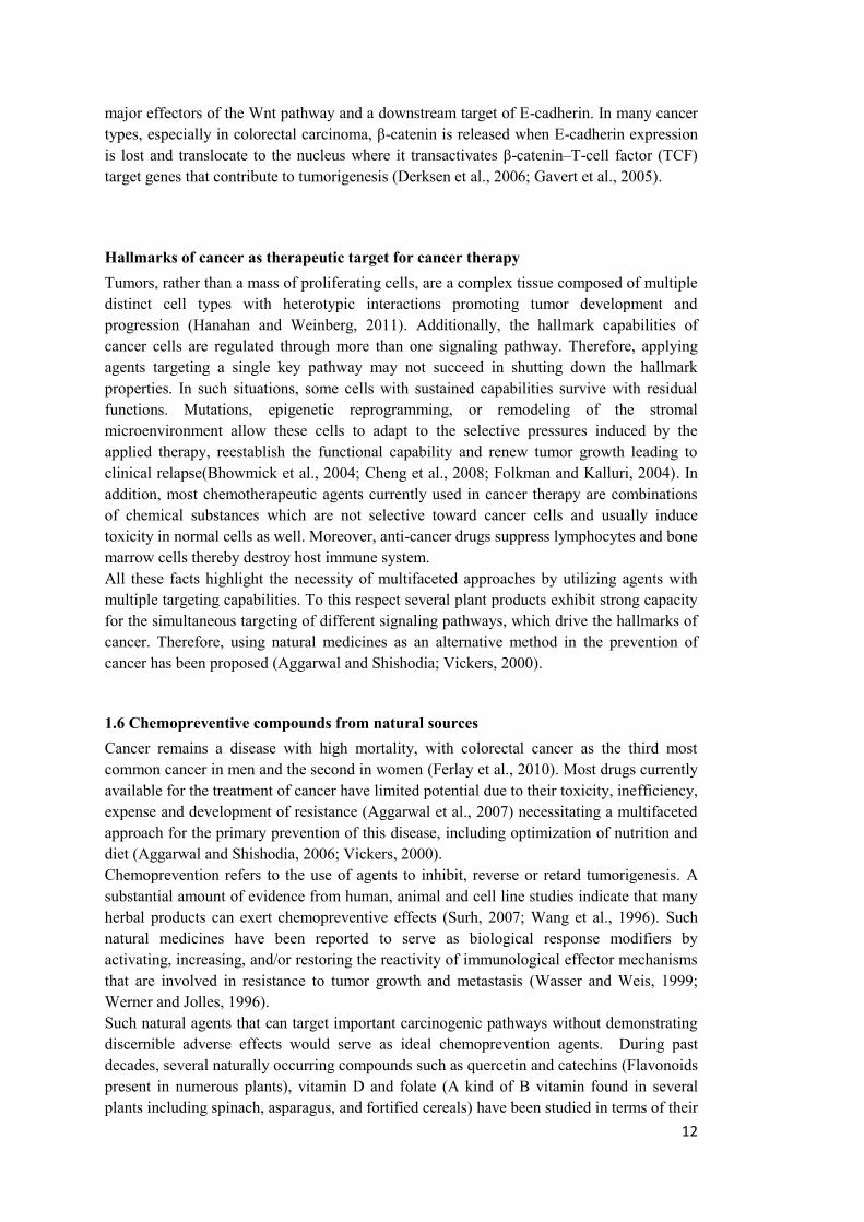
1.1 An overview of the history of cancer; ancient and modern treatment methods Several historical documents reported by ancient physicians demonstrate the presence of human diseases with symptoms similar or related to cancer. These are evidence of the presence of cancer even in ancient populations dating back to the times of the Pharaohs in Egypt and the classical world. According to same reports, although surgery was the common method to cure these kinds of diseases, physicians recommended the use of natural and herbal products as complementary therapy (Nobili et al., 2009). Today, carcinogenesis is defined as a multistep process consisting of tumor initiation, promotion and progression (Dorai and Aggarwal, 2004). The process begins with cellular transformation, progresses to hyperproliferation, evading growth suppressors, resistance to programmed cell death (apoptosis), acquisition of invasive potential, angiogenic properties and finally establishment of metastatic lesions. These six capabilities are considered as the major hallmarks of cancer (Figure 1.1) (Hahn and Weinberg, 2002) (Hanahan and Weinberg, 2011).
Figure 1.1: The six biological alterations considered as the main hallmarks of cancer (Hanahan and Weinberg, 2011)
2
1.2 Carcinogens and therapeutic methods As of today more than a hundred carcinogens from different chemical, physical, and biological sources have been identified. Various environmental carcinogens (such as cigarette smoke, industrial emissions, gasoline vapors), inflammatory agents (such as tumor necrosis factor (TNF) and H2O2), tumor promoters (such as phorbol esters) are able to activate carcinogenesis (Aggarwal and Shishodia, 2006; Vickers, 2000). Surgery, radiation-therapy, immunotherapy, and/or chemotherapy are often applicable medical treatments against cancer. Currently, most chemotherapeutic agents used in cancer therapy are combinations of chemical substances, which are not selective toward cancer cells and usually induce toxicity in normal cells as well. Anti-cancer drugs suppress lymphocytes and bone marrow cells, thereby destroying the host immune system. Drug resistance and high rates of disease recurrence are associated with many of the currently-used chemotherapeutic drugs (Chau and Cunningham, 2006). Therefore, using natural medicines as an alternative method in the prevention of cancer has been proposed (Aggarwal and Shishodia; Vickers, 2000).
1.3 Major signaling pathways involved in cancer: potent targets for cancer therapy Cancer arises from changes in signaling pathways controlling the morphological and biological properties of cells which, therefore, could be good targets for cancer therapy. Among these, deregulation of MAPK/ERK, PI3K/AKT, and NF-κB signal transduction pathways play a prominent role in the initiation and maintenance of human cancer, since many components of these pathways have been found to be mutated or amplified in a broad range of tumors. Additionaly, in most cancer types, elevated levels of reactive oxygen species production play a major role in the induction and sustainment of these signaling pathways (Engelman, 2009) (Benhar et al., 2002; Karin et al., 2002) (Ko and Auyeung, 2013).
Oxidative stress mediated mechanisms: Involvement of Reactive Oxygen Species (ROS) In normal cells, low-level concentrations of ROS are implicated in the activation of several signal transduction pathways involved in cell proliferation. However, in cancer cells, elevated levels of ROS including free radicals (e.g. superoxide (∙O2-), hydroxyl radicals (∙OH), and the non-radical H2O2) result in constitutive activation of stress-related signaling pathways and maintainance of high proliferation rates. Multiple external and internal sources such as the electron transport chain in mitochondria, ionizing radiations, enzymes producing superoxide anion such as NADPH oxidases, lipoxygenases (LOX) and cyclooxygenases (COX) result in the formation and accumulation of ROS in the cells. However, a major source of ROS is the mitochondria. It could occur through electron leakage from mitochondrial respiratory chain, which reacts with cellular oxygen to form superoxide, following by subsequent conversion to other ROS (Tin et al., 2007). The formation and presence of ROS under normal and pathologic conditions is necessary in defense against pathogens and to regulate diverse cellular functions, including intracellular
3
signaling. However, in excess, ROS can cause severe damage to cellular macromolecules including lipids, proteins and DNA leading to tumorigenesis and cell death. In tumors, especially in the advanced stages, an increase in metabolism and the metabolic switch to aerobic glycolysis alters the redox status of the cells and induce elevated levels of ROS (Au-Yeung et al., 2008; Shrikant and Lee, 2010; Surh, 2003). These biochemical properties of cancer cells can be used as a therapeutic tool for selective targeting of cancer cells as cancer cells with elevated oxidative stress are likely to be more vulnerable to high levels of ROS induced by exogenous agents (Shrikant and Lee, 2010; Surh, 2003). There are several phytochemicals that inhibit cell proliferation and induce apoptosis in cancer cells through the induction of ROS. Epigallocatechin gallate EGCG is a well-known example which induces apoptosis in cancer cells via generation of ROS and activation of p38 stress response protein (For more details please see section: MAPK/ERK pathway).
Nuclear factor-kappaB (NF-κB) signaling pathway: a bridge between inflammation and cancer The association of inflammation and cancer is a well-established phenomenon. Pro-inflammatory stimuli such as ROS, TNF and Toll Like Receptor (TLR) ligands activate Inhibitor of Kappa Kinase (IKK)α/β Ser/Thr kinases. This complex, upon activation, phosphorylates the protein inhibitor of kappa B (IκB), resulting in its polyubiquitination and proteasomal degradation. Removal of the inhibitor permits the dissociation and translocation of p50 and p65 (RelA) heterodimer subunits of NF-κB complex to the nucleus and binding to DNA elements at target genes. This cascade of events is known as the classical pathway of NF-κB activation (Gilmore and Wolenski, 2012; Hoffmann et al., 2002). NF-κB, with regulatory functions in immunity, inflammation and proliferation, is at the helm of one of the most important signaling pathways associated with cell survival. In cancer, NF-κB contributes to all major oncogenic processes, including inhibition of apoptosis, proliferation, angiogenesis, inflammation, epithelial to mesenchymal transition (EMT), invasion and metastasis (Basseres and Baldwin, 2006). NF-kB promotes EMT via activation of the mesenchymal transcription factors Twist (Horikawa et al., 2007) or Snail (Julien et al., 2007) and stimulates tumor cell invasion through transcriptional activation of the matrix metalloproteases MMP-3 (Borghaei et al., 2004)and MMP-9 (Yan et al., 2004). NF-κB activates the expression of anti-apoptotic genes including Bcl-2, Bcl-xL, Mcl-1 and c-FLIP (Auyeung et al., 2012; Singh and Aggarwal, 1995). NF-κB is also a well-known regulator of cellular redox status since several direct or indirect activators of NF-κB are kinases that are activated by ROS as second messengers (Adachi et al., 2009; Colombo et al., 2001). In tumor cells, elevated levels of ROS lead to the constitutive activity of NF-κB and secretion of inflammatory cytokines. Flavonoids with antioxidant activity are capable of chelating redox-active metal ions, which can thereby neutralize free radicals and inhibit NF-κB signaling pathway (RiceEvans et al., 1996).
Mitogen Activated Protein Kinase/Extracellular Related Kinase (MAPKs/ERK) signaling pathway The RAS/RAF/MEK/ERK pathway regulated by receptor tyrosine kinases, cytokines, and heterotrimeric G-protein-coupled receptors (Goodall et al., 2004) play critical roles in
4
mediating and maintaining the homeostasis of most cell types by controlling cell growth, differentiation and apoptosis. RAS is a small G-protein localized in the plasma membrane that activates the downstream protein RAF followed by phosphorylation activation of MEK and ERK, which finally transduce the signal to the nucleus for regulation of transcription (Gray-Schopfer et al., 2007). Oncogenic RAS mutation leads to cellular transformation and cancer due to the constitutive activation of MEK/ERK, which prevents apoptosis due to the activation of Bcl family proteins, (Ding et al., 2001; Jost et al., 2001; Pan et al., 2011; Pardo et al., 2002). The other two important members of MAPK family are c-Jun N-terminal kinases (JNKs) and p38 mitogen activated protein kinases, which are involved in stress response pathways. In the presence of upstream stress stimuli such as ROS, UV radiation, cytokines and growth factors, JNK is activated and translocated to the nucleus where it induces the phosphorylation and activation of downstream c-Jun target protein and the formation of the Activator Protein (AP)1 complex. AP1 promotes the transcription of a wide range of genes encoding proapoptotic proteins such as TNF-α, Fas-L, p53 and Bak. On the other hand, JNK activation could also induce cell proliferation but whether this activation leads to apoptosis or induce proliferating signals is dependent on the type of stimulus (JNK and apoptosis paper: Reddy, 2008; Lin & Dibling, 2002; Liu & Lin, 2005). p38 is another stress-related kinase that is activated when the cell is exposed to various stresses such as ROS, UVA radiation and DNA damage. Activation of p38 upregulates the oncogenic COX-2 gene and inhibits apoptosis through the stabilization of the antiapoptotic protein Bcl-xl. Recent studies demonstrate that some phytochemicals such as apigenin, acacetin, indole-3-carbinol (I3C) found in the human diet could abrogate cell proliferation of cancer cells through the down regulation of MAPKs including the stress response related protein p38 (Liu et al., 2011; Shen et al., 2010).
PI3K/Akt/mTOR Pathway The PI3K/Akt/mTOR signaling pathway, commonly known as PI3K signaling pathway, is an important pathway whose aberrant activation plays a central role in cancer growth, survival, and motility as well as resistance to targeted therapy. Phosphoinositide 3-kinases (PI3K) are heterodimeric lipid kinases containing a catalytic and a regulatory subunit. Among PI3K family members, PI3K class IA is widely implicated in the pathogenesis of several cancer types. Its catalytic subunits are p110α, p110β, p110γ, and p110δ (Katso et al., 2001; Vanhaesebroeck et al., 2010). PI3K is activated through ligand binding of growth factors to receptor tyrosine kinases (RTKs), which then phosphorylate the 3′-hydroxyl group of phosphatidylinositols (Escobedo et al., 1991; Myers et al., 1992). PI3K could also be activated by G-protein coupled receptors (GPCRs) and the small GTPase Ras (Guillermet-Guibert et al., 2008; Shaw and Cantley, 2006). PI(3,4,5)P3 is then generated, which then recruits PDK-1 (3′-phosphoinositide-dependent kinase 1) and the serine/threonine kinase AKT/PKB (protein kinase B) (Burgering and Coffer, 1995). Phosphorylation and activation of AKT is considered as the central node of the pathway as the activated AKT translocates to either distinct areas within the cytosol or into the nucleus where it directly or indirectly, by phosphorylation of downstream host substrates, regulates a wide range of molecular functions within the cell, such as cell cycle progression, apoptosis, transcription, and translation.
5
Downstream targets of PI3K/Akt signaling A: Regulation of protein synthesis and translation One of the major downstream targets of AKT is the serine/threonine kinase mammalian target of rapamycin (mTOR). mTOR, in association with other proteins in a complex known as mTORC1, phosphorylate and activate p70S6K (70 kDa ribosomal protein S6 kinase) and 4E-BP1 (eIF4E-binding protein) to mediate translation and synthesis of cell cycle regulating and ribosomal proteins (Beretta et al., 1996; Chung et al., 1992). On the other hand, p70S6K negatively regulates PI3K pathway by interrupting the signaling between ligand stimulated IGF-R1 and PI3K (Harrington et al., 2004; Shah et al., 2004).
B: Cell cycle regulation
Activated AKT promotes cell proliferation and survival directly by inhibiting the Cyclin dependent kinase (CDK) inhibitors p21 and p27, which negatively regulate the cell cycle and therefore have tumor suppressive properties. AKT also promotes survival indirectly by the activation of pro-survival transcription factor nuclear factor-КB (NF-КB) through the phosphorylation of IKK. Finally, activated AKT inhibits the expression and activity of the tumor suppressor p53 by phosphorylating and activating its negative regulator MDM2, which in turn targets the p53 protein for ubiquitination and degradation (Mayo and Donner, 2001; Ozes et al., 1999; Romashkova and Makarov, 1999; Zhou et al., 2001).
C: Involvement in EMT
Activated AKT increases the motility and invasive potential of cancer cells by activating epithelial mesenchymal transition (EMT). Activation of the AKT pathway has been shown to promote the expression of the mesenchymal proteins MMP-9, MMP-2 and SNAI1/2 and repression of the epithelial protein E-cadherin, leading to EMT and metastasis (Adya et al., 2008; Gialeli et al., 2013; Wu et al., 2013; Zuo et al., 2011) (For more detail please see section 1.5 under the title: Metastasis of epithelial tumors).
This multifunctional characteristic of PI3K/AKT pathway makes it a very important target for therapeutic purposes. Moreover, since the hyperactivation of this pathway has been observed in a wide range of cancer types, drugs and therapies targeting this pathway can be used against a wide spectrum of cancers (Luo et al., 2003). In this respect several PI3K pathway inhibitors have been developed, such as LY294002 and Wortmannin.
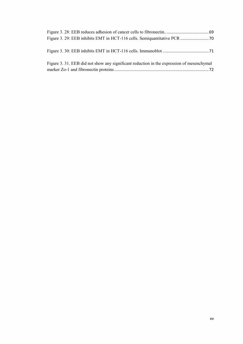
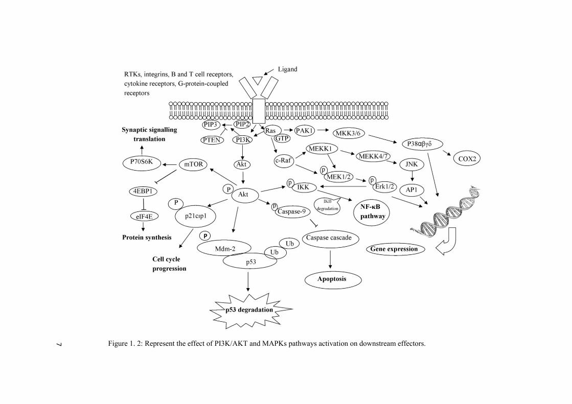
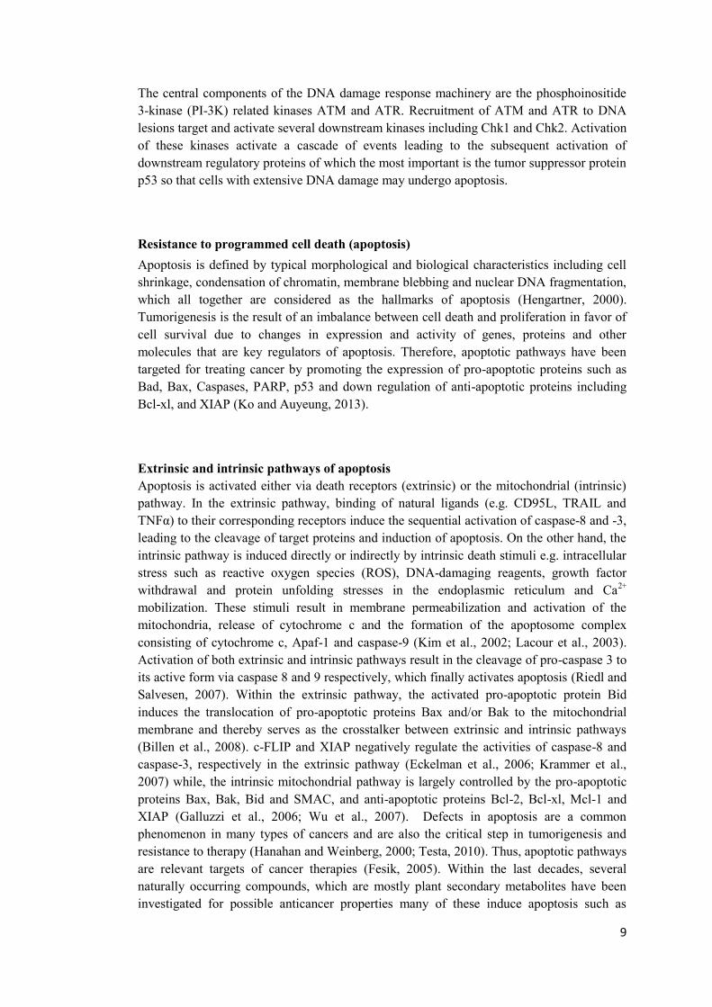
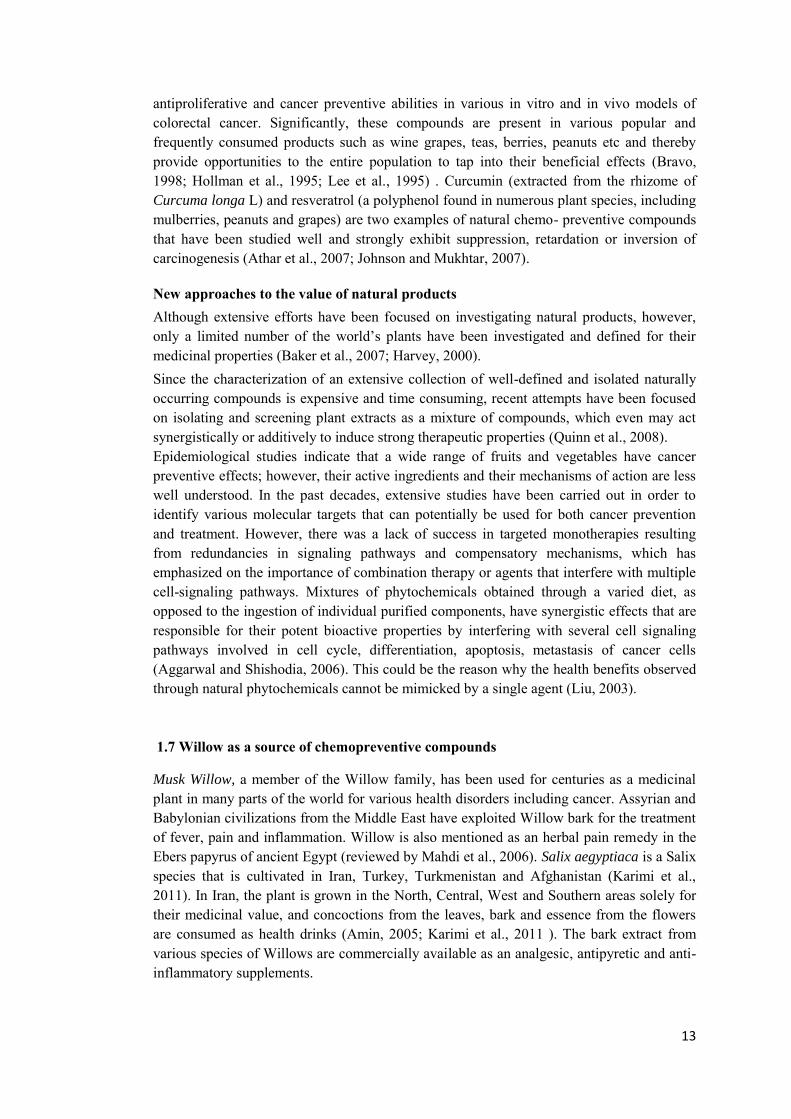
Cross Talk between these pathways The extraordinary complexity of the PI3K pathway is due to multiple feedback control loops and crosstalk with other signaling pathways such as MAPKs and NF-КB pathways (Figure 1. 2). This ensures homeostatic control of cell growth in response to mitogenic signals and to prevent unwanted cell growth under specific cellular states. However, in cancer cells these pathways could also result in the evolution of compensatory mechanisms in response to

6
single agent therapies, thereby demonstrating the necessity of utilizing multiple therapeutic agents for the dual targeting of PI3K/AKT and MAPKs pathways (Chappell et al., 2011; Oda et al., 2008; Turke and Engelman, 2010). A schematic figure representing the overall cellular response to the activation of PI3K/AKT and MAPKs pathways has been presented in figure 1.2.
7
Figure 1. 2: Represent the effect of PI3K/AKT and MAPKs pathways activation on downstream effectors.
8
1.4 Major hallmarks of cancer
Carcinogenesis is the result of six major capabilities of cancer cells including cellular transformation, hyperproliferation, evasion of growth suppressors, resistance to apoptosis, acquisition of invasive potential, angiogenic properties and finally establishment of metastatic lesions leading to cancer development and progression. Among these six hallmarks, as therapeutic targets for cancer therapy, we focused on three major capabilities that include signaling for sustained proliferation, resistance to apoptosis and metastasis.
Sustained cell cycle progression and proliferative signaling Cancer is mostly considered as a proliferative disorder since the primary and fundamental characteristic of cancer cells is chronic proliferation. In normal tissues, proliferation is under precise control in favor of maintenance of cellular function and homeostasis by regulating the production and release of growth promoting signals ensuring progression through cell division and growth (). In cancer cells, growth signals are deregulated in favor of survival, resulting in hyperproliferation. Progression through the cell cycle depends on the activation of cyclins and cyclin dependent kinases (CDKs), which function together in the G1 phase for initiating DNA synthesis or S-phase and progression to G2/M phases. The activity of cyclin–CDK complexes is regulated by p21 and p27, members of the kinase inhibitor protein (KIP) family that can arrest the cell by inhibiting DNA replication (Coqueret, 2003). p21 negatively regulates cell cycle progression by inhibiting CDK2/4 and blocks DNA replication by binding to PCNA (Gartel, 2005). The expression of p21 is controlled by the tumor suppressor protein p53 and the PI3K/AKT/GSK3β pathway has been shown to negatively influence p53 stability. Defects in cell cycle check points, autocrine response to the secreted growth factors, stimulating the secretion of growth factors by tumor associated stromal cells, elevated levels of receptor proteins, ligand independent firing of receptors by structural alteration of receptors, disruption of negative feedback mechanisms attenuating proliferating signals and somatic mutations activating additional downstream pathways such as PI3K/AKT or MAPKs pathways results in sustaining proliferating signals, promoting the transition of cell cycle progression, uncontrolled cell division and growth (Cheng et al., 2008; Bhowmick et al., 2004; Hahn,2011; Jiang and Liu, 2009; Davies and Samuels, 2010).
DNA damage-mediated mechanisms Persistent DNA damage by chemical agents, ROS, ionizing radiation and defects in DNA repair mechanisms can result in genomic instability and ultimately may lead to cancer. Conversely, induction of DNA damage (e.g. double stranded breaks) is used as an effective therapeutic method to treat cancer. Most of currently used anticancer drugs act by the induction of DNA damage, which can induce the DNA damage response (please see below).
DNA damage response and DNA repair mechanisms The maintenance of genetic stability is primary through DNA damage response, which acts through induction of cell cycle arrest to enable DNA repair or activate apoptosis (Krempler et al., 2007). The imbalance between DNA damage and DNA repair activities may affect cell viability and ultimately may lead to cancer.
9
The central components of the DNA damage response machinery are the phosphoinositide 3-kinase (PI-3K) related kinases ATM and ATR. Recruitment of ATM and ATR to DNA lesions target and activate several downstream kinases including Chk1 and Chk2. Activation of these kinases activate a cascade of events leading to the subsequent activation of downstream regulatory proteins of which the most important is the tumor suppressor protein p53 so that cells with extensive DNA damage may undergo apoptosis.
Resistance to programmed cell death (apoptosis) Apoptosis is defined by typical morphological and biological characteristics including cell shrinkage, condensation of chromatin, membrane blebbing and nuclear DNA fragmentation, which all together are considered as the hallmarks of apoptosis (Hengartner, 2000). Tumorigenesis is the result of an imbalance between cell death and proliferation in favor of cell survival due to changes in expression and activity of genes, proteins and other molecules that are key regulators of apoptosis. Therefore, apoptotic pathways have been targeted for treating cancer by promoting the expression of pro-apoptotic proteins such as Bad, Bax, Caspases, PARP, p53 and down regulation of anti-apoptotic proteins including Bcl-xl, and XIAP (Ko and Auyeung, 2013).
Extrinsic and intrinsic pathways of apoptosis Apoptosis is activated either via death receptors (extrinsic) or the mitochondrial (intrinsic) pathway. In the extrinsic pathway, binding of natural ligands (e.g. CD95L, TRAIL and TNFα) to their corresponding receptors induce the sequential activation of caspase-8 and -3, leading to the cleavage of target proteins and induction of apoptosis. On the other hand, the intrinsic pathway is induced directly or indirectly by intrinsic death stimuli e.g. intracellular stress such as reactive oxygen species (ROS), DNA-damaging reagents, growth factor withdrawal and protein unfolding stresses in the endoplasmic reticulum and Ca2+ mobilization. These stimuli result in membrane permeabilization and activation of the mitochondria, release of cytochrome c and the formation of the apoptosome complex consisting of cytochrome c, Apaf-1 and caspase-9 (Kim et al., 2002; Lacour et al., 2003). Activation of both extrinsic and intrinsic pathways result in the cleavage of pro-caspase 3 to its active form via caspase 8 and 9 respectively, which finally activates apoptosis (Riedl and Salvesen, 2007). Within the extrinsic pathway, the activated pro-apoptotic protein Bid induces the translocation of pro-apoptotic proteins Bax and/or Bak to the mitochondrial membrane and thereby serves as the crosstalker between extrinsic and intrinsic pathways (Billen et al., 2008). c-FLIP and XIAP negatively regulate the activities of caspase-8 and caspase-3, respectively in the extrinsic pathway (Eckelman et al., 2006; Krammer et al., 2007) while, the intrinsic mitochondrial pathway is largely controlled by the pro-apoptotic proteins Bax, Bak, Bid and SMAC, and anti-apoptotic proteins Bcl-2, Bcl-xl, Mcl-1 and XIAP (Galluzzi et al., 2006; Wu et al., 2007). Defects in apoptosis are a common phenomenon in many types of cancers and are also the critical step in tumorigenesis and resistance to therapy (Hanahan and Weinberg, 2000; Testa, 2010). Thus, apoptotic pathways are relevant targets of cancer therapies (Fesik, 2005). Within the last decades, several naturally occurring compounds, which are mostly plant secondary metabolites have been investigated for possible anticancer properties many of these induce apoptosis such as
10
curcumin, resveratrol, fisetin, EGCG, luteolin, quercetin, etc.(Braicu and Gherman, 2013; Gokbulut et al., 2013; Guo et al., 2013; Park et al., 2013; Ying et al., 2012; Zheng et al., 2012).
p53-mediated cell cycle arrest and apoptosis Activated p53 regulates the expression and function of downstream effector proteins such as p21, Cdc25A and cyclin-dependent kinases (CDKs), which finally result in a transient and reversible cell cycle arrest at the transition from the G1 to S phase or from the G2 to M phase of the cell cycle. This allows the cells to repair the DNA damage or respond to other stimuli such as starvation of DNA precursors (Agarwal et al., 2006; Hastak et al., 2008). p53 can also induce apoptosis in response to stimuli such as prolonged unrepaired DNA damage. Therefore p53 is considered as the central node in the maintenance of cellular homeostasis, during both normal and stressed conditions, so that the disruption of or defect in either functional p53 or any components of p53 network could result in the development of virtually all cancer types. One of the major downstream targets of p53 is the p53 upregulated modulator of apoptosis (PUMA), which binds to and inhibits Bcl-2 and Bcl-xl antiapoptotic proteins and promotes apoptosis. Exogenous expression of PUMA causes an extremely rapid induction of apoptosis that occurs much earlier than that resulting from exogenous expression of TP53 (Agarwal et al., 2006; Amin et al., 2007; Furgason and Bahassi, 2013).
1.5 Metastasis of epithelial tumor Tumors are classified as benign and malignant. Although benign tumors cause significant organ dysfunction and even death, they are mostly less harmful, localized and not able to spread throughout the body. In contrast, malignant tumors more commonly known as cancer, have gained motility and metastatic potential due to a loss or change in adhesive requirements and dependency on growth factors (Rendon et al., 2007) and cause most (~90%) human cancer deaths. Excessive epithelial cell proliferation and angiogenesis are hallmarks of the initiation and early growth of primary epithelial cancers (Maniatis, 1999). Metastasis includes complex events, starting with tumor cells detachment, motility, invasion to adjacent tissues, adhesion to endothelial cells, thereby gaining access to vascular or lymphatic channels, transition through the bloodstream and finally extravasation and reestablishment of growth at a distant site (Lee et al., 2010). The outer edge of solid tumors that invades the basement membrane and the surrounding tissue, often called the invasive front, is represented as either a sheet of advancing cells or as individual or small groups of cells ‘pushing forward’.
Regulation of metastasis Invasive and metastatic potential of cancer cells raise through malfunction of regulatory processes controlling cell development and homeostasis. Among these regulatory processes, the TGFβ superfamily, Wnt/β-catenin/TCF (T-cell specific transcription factor), and Notch
11
signaling pathways play a major role in the development, differentiation and maintenance of homeostasis of cells. Mutations or aberrant regulation of these pathways often contribute to tumor initiation and progression, especially in the colon.4
Motility and invasion into adjacent tissues is a key requirement for the progression of primary tumors toward metastatic lesions. The tumor cells gain metastatic properties through a cascade of biological and morphological changes called epithelial– mesenchymal transition (EMT). Most of these alterations, including motility and migration, are also observed during developmental processes and recent studies have shown that several master regulators of development play critical roles during EMT as well.
Epithelial–mesenchymal transition and the invasive potential of tumors A primary characteristic of epithelial cells is the tight interaction between neighboring cells, ensured by strong adhesive junctions between cells and between cells and the extracellular matrix (ECM). This property prevents movement of individual cells and enables the formation of uniform epithelial cell sheets with polarized apical and basal surfaces (Hay, 2005). As the process of EMT is initiated, epithelial cells lose most of their biological and morphological properties and gain the characteristics of mesenchymal cells. These include weakening and losing adherence junctions due to the suppression of epithelial biomarkers such as the loss of E-cadherin, plakoglobin and cytokeratins and the dismantling of adherence junctions and desmosomes. Simultaneously, the cells also acquire mesenchymal characteristics, including expression of vimentin, alpha-smooth muscle actin, N-cadherin, specific myosin isoforms, fibronectin, metalloproteinases and an elongated ‘fibroblast-like’ morphology, which all together provide the invasive motility of transformed cells. These morphogenic changes characterizing EMT are under the control of the master transcription regulators Slug, Snail, ZEB and Twist, which are also induced in some invasive cancers (Thiery, 2002; Yang et al., 2004). Therefore, EMT is the result of alteration in various regulatory and signaling pathways, which enhance tumor invasion and/or metastasis.
E-cadherin a major cell adhesion molecule involved in the progression of EMT E-cadherin is one of the main transmembrane adhesive molecules, whose homophilic binding to each other on the surface of adjacent cells provide and maintain adherence junctions while its suppression is a major characteristic of cells undergoing EMT (Birchmeier and Behrens, 1994; Gumbiner, 2005; Peinado et al., 2004). Loss of E-cadherin expression at the invasive front of solid tumors has been observed during transition from adenoma to carcinoma whereas the restoration of E-cadherin expression can promote mesenchymal to epithelial transition (MET) and restore the epithelial phenotype (Perl et al., 1998). Therefore, an understanding of the transcriptional regulation of E-cadherin is crucial for identifying the mechanisms involved in EMT as well as utilizing methods to inhibit or reverse the process of EMT [28]. There are several transcription factors including SNAI1 and SNAI2 (Slug) members of Snail family, ZEB (ZEB1 and ZEB2), basic helix–loop–helix (bHLH) (E47 and Twist) families, which bind to consensus E-box sequences in the E-cadherin gene promoter and downregulate E-cadherin transcription (Peinado et al., 2007). Activation of extracellular-signal-regulated kinase (ERK) by the epidermal growth factor receptor (EGFR), induces Slug activation and proteolytic cleavage of E-cadherin by MMP-2 and MMP-9 (Conacci-Sorrell et al., 2003; Miyaki et al., 1999). Beta-catenin is one of the
12
major effectors of the Wnt pathway and a downstream target of E-cadherin. In many cancer types, especially in colorectal carcinoma, β-catenin is released when E-cadherin expression is lost and translocate to the nucleus where it transactivates β-catenin–T-cell factor (TCF) target genes that contribute to tumorigenesis (Derksen et al., 2006; Gavert et al., 2005).
Hallmarks of cancer as therapeutic target for cancer therapy Tumors, rather than a mass of proliferating cells, are a complex tissue composed of multiple distinct cell types with heterotypic interactions promoting tumor development and progression (Hanahan and Weinberg, 2011). Additionally, the hallmark capabilities of cancer cells are regulated through more than one signaling pathway. Therefore, applying agents targeting a single key pathway may not succeed in shutting down the hallmark properties. In such situations, some cells with sustained capabilities survive with residual functions. Mutations, epigenetic reprogramming, or remodeling of the stromal microenvironment allow these cells to adapt to the selective pressures induced by the applied therapy, reestablish the functional capability and renew tumor growth leading to clinical relapse(Bhowmick et al., 2004; Cheng et al., 2008; Folkman and Kalluri, 2004). In addition, most chemotherapeutic agents currently used in cancer therapy are combinations of chemical substances which are not selective toward cancer cells and usually induce toxicity in normal cells as well. Moreover, anti-cancer drugs suppress lymphocytes and bone marrow cells thereby destroy host immune system. All these facts highlight the necessity of multifaceted approaches by utilizing agents with multiple targeting capabilities. To this respect several plant products exhibit strong capacity for the simultaneous targeting of different signaling pathways, which drive the hallmarks of cancer. Therefore, using natural medicines as an alternative method in the prevention of cancer has been proposed (Aggarwal and Shishodia; Vickers, 2000).
1.6 Chemopreventive compounds from natural sources Cancer remains a disease with high mortality, with colorectal cancer as the third most common cancer in men and the second in women (Ferlay et al., 2010). Most drugs currently available for the treatment of cancer have limited potential due to their toxicity, inefficiency, expense and development of resistance (Aggarwal et al., 2007) necessitating a multifaceted approach for the primary prevention of this disease, including optimization of nutrition and diet (Aggarwal and Shishodia, 2006; Vickers, 2000). Chemoprevention refers to the use of agents to inhibit, reverse or retard tumorigenesis. A substantial amount of evidence from human, animal and cell line studies indicate that many herbal products can exert chemopreventive effects (Surh, 2007; Wang et al., 1996). Such natural medicines have been reported to serve as biological response modifiers by activating, increasing, and/or restoring the reactivity of immunological effector mechanisms that are involved in resistance to tumor growth and metastasis (Wasser and Weis, 1999; Werner and Jolles, 1996). Such natural agents that can target important carcinogenic pathways without demonstrating discernible adverse effects would serve as ideal chemoprevention agents. During past decades, several naturally occurring compounds such as quercetin and catechins (Flavonoids present in numerous plants), vitamin D and folate (A kind of B vitamin found in several plants including spinach, asparagus, and fortified cereals) have been studied in terms of their
13
antiproliferative and cancer preventive abilities in various in vitro and in vivo models of colorectal cancer. Significantly, these compounds are present in various popular and frequently consumed products such as wine grapes, teas, berries, peanuts etc and thereby provide opportunities to the entire population to tap into their beneficial effects (Bravo, 1998; Hollman et al., 1995; Lee et al., 1995) . Curcumin (extracted from the rhizome of Curcuma longa L) and resveratrol (a polyphenol found in numerous plant species, including mulberries, peanuts and grapes) are two examples of natural chemo- preventive compounds that have been studied well and strongly exhibit suppression, retardation or inversion of carcinogenesis (Athar et al., 2007; Johnson and Mukhtar, 2007).
New approaches to the value of natural products Although extensive efforts have been focused on investigating natural products, however, only a limited number of the world’s plants have been investigated and defined for their medicinal properties (Baker et al., 2007; Harvey, 2000). Since the characterization of an extensive collection of well-defined and isolated naturally occurring compounds is expensive and time consuming, recent attempts have been focused on isolating and screening plant extracts as a mixture of compounds, which even may act synergistically or additively to induce strong therapeutic properties (Quinn et al., 2008). Epidemiological studies indicate that a wide range of fruits and vegetables have cancer preventive effects; however, their active ingredients and their mechanisms of action are less well understood. In the past decades, extensive studies have been carried out in order to identify various molecular targets that can potentially be used for both cancer prevention and treatment. However, there was a lack of success in targeted monotherapies resulting from redundancies in signaling pathways and compensatory mechanisms, which has emphasized on the importance of combination therapy or agents that interfere with multiple cell-signaling pathways. Mixtures of phytochemicals obtained through a varied diet, as opposed to the ingestion of individual purified components, have synergistic effects that are responsible for their potent bioactive properties by interfering with several cell signaling pathways involved in cell cycle, differentiation, apoptosis, metastasis of cancer cells (Aggarwal and Shishodia, 2006). This could be the reason why the health benefits observed through natural phytochemicals cannot be mimicked by a single agent (Liu, 2003). 1.7 Willow as a source of chemopreventive compounds
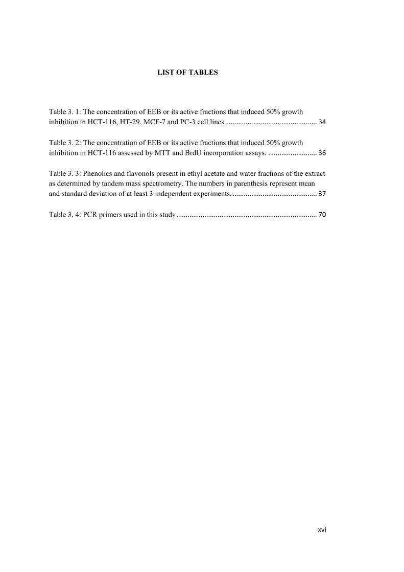
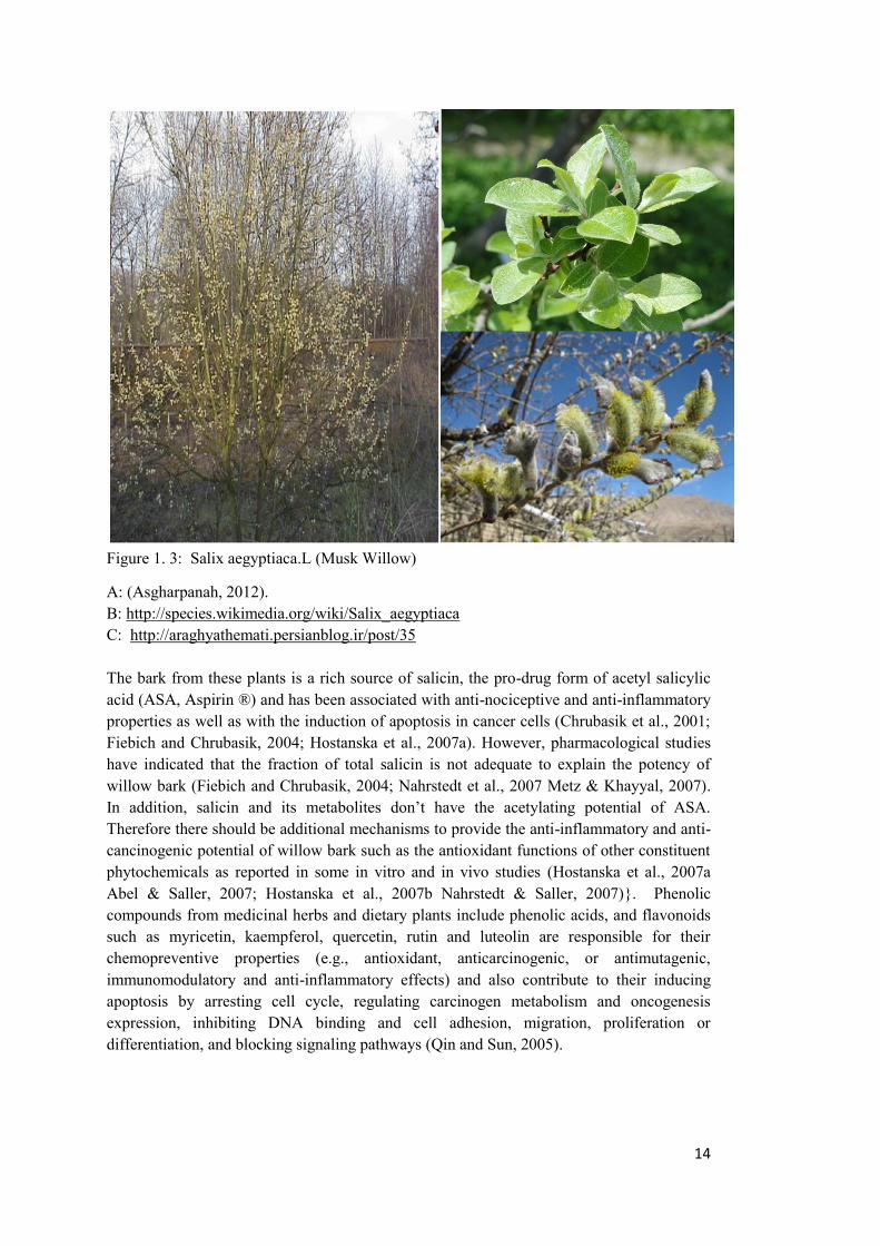
Musk Willow, a member of the Willow family, has been used for centuries as a medicinal plant in many parts of the world for various health disorders including cancer. Assyrian and Babylonian civilizations from the Middle East have exploited Willow bark for the treatment of fever, pain and inflammation. Willow is also mentioned as an herbal pain remedy in the Ebers papyrus of ancient Egypt (reviewed by Mahdi et al., 2006). Salix aegyptiaca is a Salix species that is cultivated in Iran, Turkey, Turkmenistan and Afghanistan (Karimi et al., 2011). In Iran, the plant is grown in the North, Central, West and Southern areas solely for their medicinal value, and concoctions from the leaves, bark and essence from the flowers are consumed as health drinks (Amin, 2005; Karimi et al., 2011 ). The bark extract from various species of Willows are commercially available as an analgesic, antipyretic and anti-inflammatory supplements.
14
Figure 1. 3: Salix aegyptiaca.L (Musk Willow)
A: (Asgharpanah, 2012). B: http://species.wikimedia.org/wiki/Salix_aegyptiaca C: http://araghyathemati.persianblog.ir/post/35 The bark from these plants is a rich source of salicin, the pro-drug form of acetyl salicylic acid (ASA, Aspirin ®) and has been associated with anti-nociceptive and anti-inflammatory properties as well as with the induction of apoptosis in cancer cells (Chrubasik et al., 2001; Fiebich and Chrubasik, 2004; Hostanska et al., 2007a). However, pharmacological studies have indicated that the fraction of total salicin is not adequate to explain the potency of willow bark (Fiebich and Chrubasik, 2004; Nahrstedt et al., 2007 Metz & Khayyal, 2007). In addition, salicin and its metabolites don’t have the acetylating potential of ASA. Therefore there should be additional mechanisms to provide the anti-inflammatory and anti-cancinogenic potential of willow bark such as the antioxidant functions of other constituent phytochemicals as reported in some in vitro and in vivo studies (Hostanska et al., 2007a Abel & Saller, 2007; Hostanska et al., 2007b Nahrstedt & Saller, 2007)}. Phenolic compounds from medicinal herbs and dietary plants include phenolic acids, and flavonoids such as myricetin, kaempferol, quercetin, rutin and luteolin are responsible for their chemopreventive properties (e.g., antioxidant, anticarcinogenic, or antimutagenic, immunomodulatory and anti-inflammatory effects) and also contribute to their inducing apoptosis by arresting cell cycle, regulating carcinogen metabolism and oncogenesis expression, inhibiting DNA binding and cell adhesion, migration, proliferation or differentiation, and blocking signaling pathways (Qin and Sun, 2005).
15
1.7 Aims of the research Extracts of leaves, bark and catkins in various solvents of varying polarity from Salix
aegyptiaca or musk willow have previously been shown to have high antioxidant activity with the ethanolic extract from bark as the most potent (Enayat and Banerjee, 2009). Since the polyphenols and flavonoids that contribute towards the high antioxidant activity of the willow extracts also have anticancer properties, we hypothesized that the ethanolic extract from the bark (EEB) of Salix aegyptiaca could be explored for its anti-cancer properties. The intestines and colon are continuously exposed to dietary components; therefore, prevention of cancer of the colon through the diet is an attractive option. Colorectal cancer is one of the leading causes of premature death worldwide. A multifaceted approach for the primary prevention of this disease, with emphasis on nutrition and diet has curried favor amongst many experts. During past decades, several naturally occurring compounds have been studied in terms of their antiproliferative and cancer preventive abilities in various in vitro and in vivo models of colorectal cancer. Significantly, these compounds are present in various popular and frequently consumed products such as wine grapes, teas, berries, peanuts etc and thereby provide opportunities to the entire population to tap into their beneficial effects (Bravo, 1998; Hollman et al., 1995; Lee et al., 1995). Our main objective is to determine the effects of the ethanolic extract of bark (EEB) of Salix
aegyptiaca on apoptotic pathways, cell cycle, as well as metastatic potential of HCT-116 and HT-29 colon cancer cell lines. Towards this objective, we have:
Determined the cytotoxicity of EEB on the colon cancer cells lines HCT-116 and HT-29 and the non-transformed colon fibroblast CCD-18Co cell line.
Determined the effects EEB on the cell cycle and cellular oxidative stress as well as the biochemical pathways related to these functional effects.
Evaluated the effect of EEB on the anchorage independent growth, determined the antimetastatic potential of EEB by determining its effects on migration and induction of epithelial to mesenchymal transition.
Partitioned EEB into four fractions of n-hexane, ethyl acetate, n-butanol and water. Determined the effects of each fraction on proliferation of HCT-116 and HT-29 colon
cancer cell lines. Analyzed the active constituents of the fractions with the most bioactivity using
chromatographic methods and confirmed the effects of the extract on apoptosis.
Our studies indicate that EEB is a mixture of very potent bioactive compounds that has multifaceted anti-carcinogenic properties. The bountiful resources of nature, so well exploited in the past for various disease states, can now be scientifically characterized with the use of modern biological techniques. Studies such as this go a long way in delineating the benefits of use of ancient plant species such as Willow on cancer, one of the most dreaded diseases of modern times.

16
17
CHAPTER 2
METHODS
2. Material and Methods
2.1 Chemicals and Reagents MTT (3-(4,5-Dimethylthiazol-2-yl)-2,5-diphenyltetrazolium bromide) reagent was purchased from Invitrogen (Carlsbad, CA, USA), Caspase-3 Activity Assay Kit was from Biovision (Mountain View, CA, USA). AnnexinV-FITC Apoptosis Detection Kit and Cell proliferation ELISA, BrdU (Chemiluminescence) kits were purchased from Roche (Mannheim, Germany). Propidium Iodide (PI) and NBT were purchased from Sigma-Aldrich (Taufkirchen, Germany). Antibodies against Bcl-xl (1/300), Caspase 9 (1/150), Procaspase 3 (1/150), p21 (1/350), Puma (1/200), Chk2 (1/150), Akt (1/150), P70S6K (1/150), E-cadherin (1/200 dilution), MMP-9 (1/150 dilution) MMP-2 (1/150 dilution), EGFR ((1/300 dilution), transgelin (1/300 dilution), SNAI1 (1/200 dilutions), COX-2 (1/150 dilutions) and GAPDH (1/ 1000 dilution) were purchased from Santa Cruz Biotechnology, (Santa Cruz, CA, USA) and p-Chk2 (Thr 68) (1/1000) antibody were from Cell Signaling (Danvers, MA, USA). The p53 (1/1000) and p-Akt (Ser 473) (1/2000), mTOR (1/1000) and p-P70S6K (1/1000) antibodies (Cell Signaling) were obtained from Dr Ozlen Konu, Bilkent University, Ankara.
Primers for Twist1 were obtained from Iontec, Istanbul, Turkey. SNAI2 primers were kindly provided by Dr. Ali Güre, Bilkent University, Ankara. ThinCerts TM, 8.0 µm translucent wells were obtained from Greiner Bio-One (Frickenhausen, Germany). Fibronectin was purchased from Invitrogen (Carlsbad, California, USA).
2.1 Plant selection Plant collection was carried out according to published guidelines (N'Guessan et al., 2007). Briefly, barks of Musk Willow were collected from Ghaene ghom, Iran, during the 2008 harvest season. The plant materials were identified morphologically at the herbarium of the Medicinal Plants Research Institute of Shahid Beheshti University of Tehran, Iran. The fresh barks (1 kg) were air dried at room temperature for 1 week giving 250 g dried barks. The samples were then pulverized to a powder form using a steel blender (Sinbo, Istanbul, Turkey) and stored in a desiccator at 4°C in the dark until analysis.
18
2.2 Extract preparation The ethanolic extract of bark from Salix aegyptiaca was prepared from the dried pulverized powder of the bark using the procedure of (Gezer et al., 2006; Kim et al., 2003) with some modifications. The extraction was carried out by using a Soxhlet apparatus where 10 g of dried pulverized bark was extracted in ethanol (EtOH) with the ratio of 1 to 30 (w/v). After extraction, the sample was lyophilized and stored in the dark at -20˚C. Before use, a stock solution of 100 mg/ml of lyophilized bark powder in DMSO was prepared and further diluted by complete RPMI1640 medium to obtain a concentration of 5 mg/ml. This solution was filtered through 0.2 µm syringe filter, and stored at -20˚ C. The final DMSO concentration was kept at below 0.3%-0.4%. DMSO was used throughout the experiment as vehicle control.
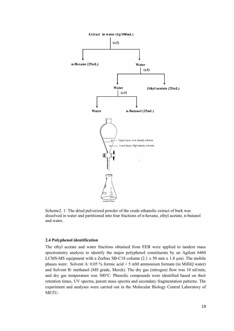
2.3 Solvent-solvent fractionation In order to separate the constituent phytochemicals, the ethanolic extract of bark from S.aegyptiaca (EEB) was partitioned into four fractions of n-hexane, ethyl acetate, n-butanol and water by solvent-solvent fractionation in a separating funnel as shown in the Scheme 1 (Paul et al., 2011). Lyophilized EEB (1.5g) was dissolved into 100mL of water and added to a separating funnel followed by the addition of 25ml n-hexane. The n-hexane fraction was stored in a separate Erlenmeyer flask while the water solution was applied into the funnel for the next round of partitioning with ethyl acetate (Et.Ac) and n-butanol respectively. In order to increase the purity and concentration of the constituents in each fraction, partitioning with each solvent was carried out three times by addition of extra 25mL of corresponding solvent into the separating funnel containing water solution.
19
Scheme2. 1: The dried pulverized powder of the crude ethanolic extract of bark was dissolved in water and partitioned into four fractions of n-hexane, ethyl acetate, n-butanol and water.
2.4 Polyphenol identification The ethyl acetate and water fractions obtained from EEB were applied to tandem mass spectrometry analysis to identify the major polyphenol constituents by an Agilent 6460 LCMS-MS equipment with a Zorbax SB-C18 column (2.1 x 50 mm x 1.8 µm). The mobile phases were: Solvent A: 0.05 % formic acid + 5 mM ammonium formate (in MilliQ water) and Solvent B: methanol (MS grade, Merck). The dry gas (nitrogen) flow was 10 ml/min, and dry gas temperature was 300°C. Phenolic compounds were identified based on their retention times, UV spectra, parent mass spectra and secondary fragmentation patterns. The experiment and analyses were carried out in the Molecular Biology Central Laboratory of METU.
20
2.5 Cell Culture HCT-116 human colorectal cancer cell line was purchased from Deutsche Sammlung von Mikroorganismen und Zellkulturen (Braunschweig, Germany), HT-29 colon cancer cell line and MCF-7 breast cancer cell line were purchased from ŞAP Enstitüsü (Ankara, Turkey) and the non-transformed colon fibroblast cell line CCD-18Co was purchased from ATCC (Teddington, Middlesex, UK). PC3 prostate cancer cell line was obtained from Dr Uddhav Kelavkar, Mercer University School of Medicine – Savannah. HCT-116, HT-29, MCF-7 and PC3 cells were cultured under ATCC recommended conditions in McCoy's 5A medium supplemented with 2mM L-Glutamine, 10% fetal bovine serum (FBS) and 1% penicillin/streptomycin. CCD-18Co cells were cultured in Eagle's Minimum Essential Medium (EMEM) supplemented with 10% FBS and 1% penicillin/ streptomycin. The cells were grown at 37°C in a humidified incubator with 5% CO2. All cell culture reagents were purchased from Biochrom (Berlin, Germany).
2.6 Transfections To overexpress PI3-kinase, HCT-116 cells (1 × 10⁶ cells/well) were transfected with 1µg of rCD2p110, a membrane-localized p110 construct, containing a chimera of the extracellular and transmembrane domains of the rCD2 antigen fused to the p110α catalytic domain of PI3-kinase for 24h in OptiMEM. Following transfection, the medium was removed and changed with fresh complete McCoy 5A medium containing 10% FBS. Transfected cells were treated with EEB for 48h. The whole protein lysate from both treated and non-treated vehicle control were collected for further analysis by western blotting. The vector was kindly provided by Dr K. Reif (Reif et al., 1997).
2.6.1 Tranformation of E.Coli with rCD2p110 plasmid and plasmid purification The filter paper containing dried plasmid was cut into 4 pieces, soaked into TE buffer and kept overnight for release. For every 10 µg/ml of plasmid 150 µl buffer was used. The day after 50 ng of plasmid was mixed with 100 µl aliquot of chilled Escherichia coli (E. coli) competent cells and incubated for 30 min on ice following by one minute of heat shock at 42°C. After heat shock the cells were kept on ice for 2 min. Then 100 µl of LB at 42°C was added to the cells and incubated at 37°C for 1h. A volume of 200 µl of the mixture was added to the already prepared LB/ampicilin containing plates and incubated at 37°C for overnight. Only E. coli cells received plasmid containing ampicilin resistance gene were able to grow and make colony. Small amounts of colonies were picked up using a sterile pipette tip and transferred into a falcon tube containing 5 ml LB medium and 5 µl of 1000X ampicilin and incubated for overnight at 37°C. The rCD2p110 plasmid was isolated using a Qiagen plasmid isolation kit according to manufacturer’s instructions. The concentration of purified plasmid and its purity was determined using a Nanodrop reader at 260 and 280 nm. The ratio of OD260/ OD280 ~ 1.8 is an indicator of pure DNA with minimum levels of protein contamination.
21
2.6.2 Preparation of LB medium LB medium (25 g) and agar (20 g) were dissolved in 1000 ml of distilled water and autoclaved. When the temperature reached to ~ 50-55°C 100 µl of 1000X ampicilin was added to the mixture and immediately poured into plates near the flame.
2.7 Cell viability assay The growth inhibitory effects of EEB, its fractions and salicin on several cell lines from breast, colon and prostate cancers as well as a non-transformed cell line were determined by colorimetric MTT and BrdU incorporation assays according to the manufacturer’s instructions.
2.7.1 MTT Assay For the MTT assay, the HCT-116, HT-29, MCF-7, PC3 and CCD18Co cells were seeded in 96-well plates at a density of 104cells/well and allowed to attach to the wells overnight at 37°C. The day after, cells were treated with different dilutions of the EEB (50µg/ml - 450µg/ml) for 48h. At the end of the incubation, the medium was changed and 10µl 12M MTT was added to each well and incubated at 37°C for an additional 4h. In viable and actively metabolizing cells, enzymes convert the soluble tetrazolium dye (MTT) to insoluble formazan. Formazan was solublized by addition of a solubilization solution containing 10% SDS in 0.01 M HCL (100µl) to each well and incubated for a further 18h at 37°C in a humidified incubator. The absorbance of each well was read at 570 nm in a microplate reader (BioRad, Hercules, CA, USA). The medium with MTT reagent was used as blank. Each assay was carried out in triplicate and the experiment was repeated at least 3 times. The growth inhibitory percentage (GI%) for each concentration of the extracts was calculated as follows:
The concentration of substances that reduced cell growth by 50% (GI50) was calculated from the dose–response relationship by regression analysis using Microsoft Excel computer software.
2.8 BrdU incorporation assay The growth inhibitory effect of the extract on cell lines will be measured by the chemiluminescence based BrdU incorporation assay according to manufacturer‘s instructions. The technique is based on the incorporation of the pyrimidine analogue BrdU (instead of thymidine) into the DNA of proliferating cells. HCT-116, HT-29, MCF-7 and PC3 cells were seeded in 96-well plates at a density of 104cells/well following by an overnight incubation at 37°C. Cells were treated in triplicate with different dilutions of the EEB, fractions or salicin for 48h. At the end of treatment, the medium was changed and
22
fresh medium containing the BrdU labling reagent was added to each well and incubated for additional 1 to 2h. After its incorporation into DNA, BrdU is detected by an immunoassay via addition of anti-BrdU-POD solution to each well and keeping at 15 – 25°C for 90 min. Wells were washed three times with washing solution and incubated with the chemiluminescence substrate for 3-10 minutes. The chemiluminescence signal was measured using a microplate luminometer. Empty wells containing the culture medium, BrdU and anti-BrdU-POD solution but no cells was used as a blank. In addition, in some wells with cells the BrdU labaling reagent was not added but the anti-BdrU-POD solution was added in order to subtract the background signal.
2.9 Annexin V assay Annexin V staining assay is a fluorescence based assay for accurate and sensitive detection of cellular apoptosis, which detects and quantifies viable and dead cell populations with the capability of discriminating cell populations in early and late stages of apoptosis. To this respect HCT-116 and HT-29 cells were treated with EEB or salicin for 48h and stained with propidium iodide (PI) and fluorescein isothiocyanate (FITC)-conjugated annexin V using the Annexin V-FITC Apoptosis Detection Kit (Roche) according to the manufacturer’s instructions. Briefly, treated cells as well as non-treated control cells (1 × 10⁶) were washed with PBS and spun down at 200 x g for 5 min. The pellets were resuspended and stained by the addition of annexin V labeling solution followed by incubation for 10 to 15 min at room temperature. The labeling solution contains 10mM HEPES/NaOH PH 7.4, 140 mM NaCl, 5mM Cacl2, 20µL annexin V solution and 20µl of 5mg/ml PI. The stained cells were incubated on ice and subjected to fluorescence-activated cell sorter analysis (FACS) using a FACScan flow cytometer (Becton–Dickinson, NJ, USA). The FITC fluorescence between 515 and 545 nm and the PI fluorescence between 564 and 606 nm were measured. The analysis reported the results as number of cells with either (+) annexin V (early apoptotic phase) or (+) PI (necrotic phase) or both (+/+, late apoptotic phase) or none (-/-, indicating viability). The FACScan experiments were carried out at the Department of Molecular Biology and Genetics, Bilkent University, under the guidance of Dr Mayda Gürsel.
2.10 RNA extraction and RT-PCR The differential expression of some of the transcripts involved in metastasis and EMT progression as a result of treatment with EEB was monitored by reverse transcriptase polymerase chain reaction (RT-PCR). Total cellular RNA was isolated from HCT-116 cells using an RNeasy RNA extraction kit (Qiagen, Hilden, Germany). The quantity and purity of RNA were determined using a Nanodrop reader. After isolation, 1µg RNA was treated with Dnase I enzyme (Fermentas, Vilnius, Lithuania) to remove genomic DNA contamination according to the manufacturers’ instructions. Reverse transcription was carried out by using Revert Aid First Strand cDNA Synthesis kit (Fermentas) using oligo(dT) primers. The Twist 1 and SNAI2 transcripts were assessed semi-quantitatively in duplex PCR reactions using GAPDH as the internal control. PCR was performed with 1U Taq polymerase in 1x buffer with ammonium sulfate (Fermentas), 1 mM MgCl2, 0.2 mM dNTP mix and 0.5 µmol of each primer set.
23
2.11 Immunoblotting Cell lysates from HCT-116 and HT-29 cells treated or not with the extract for 48h, were isolated using the M-PER assay buffer (Pierce) containing protease inhibitors (Roche) and measured for protein content using the Coomassie Plus protein assay reagent (Pierce). Total cell extracts (50-70µg) were resolved by a 10% SDS-PAGE gel and transferred to a nitrocellulose membrane and blocked overnight at 4°C in 3% BSA (Bovine Serum Albumin) for the detection of phosphorylated proteins or 5% skim milk for the detection of non-phosphorylated proteins. The blots were incubated with the antibodies against; Bcl-xl, caspase 9, procaspase 3, p53, Puma α/β, p21, Chk2, p-Chk2 (Thr 68), Akt, p-Akt (Ser 473), mTOR, P70S6K, p-P70S6K, E-cadherin, MMP-9, MMP-2, EGFR, transgelin, SNAI1, Fibronectin, Vimentin, Zo-1 and GAPDH at appropriate dilutions mentioned in section 2.1, followed by incubation for 1 h with the appropriate secondary antibody. The bands were visualized using an enhanced chemiluminescence kit (ECL Plus; Pierce) according to the manufacturer’s instructions. The membranes were stripped and probed against GAPDH for equal protein loading of the total cell extracts.
2.12 Cell cycle analysis Cell cycle analysis was carried as described previously with some modifications (Nomura et al., 2010; Szczepanski et al., 2009). HCT-116 and HT-29 cells treated with the extract or salicin for 48h, along with non-treated controls, were washed with Hanks solution containing 10 µg/ml RNase I for 2-3 min, trypsinized and fixed with 70% ethanol (500 µl). The fixed cells were then centrifuged at 200 x g for 5 min and the cell pellets were resuspended in 1 ml PI/TritonX-100 staining solution (0.1% (v/v) TritonX100 in PBS, 2 mg DNase free Rnase A, 0.02 mg/ml PI from 1 mg/ml PI stock solution) and analyzed by flow cytometry (FACScan, Becton–Dickinson). For each sample, the singlet cells were gated and 50,000 cells were acquired for cell cycle distribution analysis. According to the DNA content, the percentages of cells in G0/G1, S, G2/M phases, as well as in subG1 (<G1), content were determined using CellQuest software (Becton–Dickinson) (Nomura et al., 2010; Szczepanski et al., 2009). The cell cycle experiments were carried out at the Department of Molecular Biology and Genetics, Bilkent University, under the guidance of Dr Mayda Gürsel.
2.13 Measurement of superoxide anion using a modified colorimetric NBT assay HCT-116, HT-29 and CCD-18Co cells (2 × 10⁴ cell/well) were seeded in 96 well plates and treated separately with 100-750 µg/ml of the extract or salicin along with Nitro blue tetrazolium (NBT) solution in PBS at 0.2 mg/ml per well. The cells were incubated for 6 h, fixed with 70% methanol and air-dried, followed by the addition of 120 µl of 2 M KOH and 140 µl of dimethylsulfoxide (DMSO) to each well. The solution was mixed properly to ensure the complete dissolution of the insoluble blue formazan crystals and the absorbance was read at 620 nm in an ELISA microplate reader (Biorad, Hercules, CA, USA). A well without cells which it was incubated with the NBT solution and subjected to similar fixation and solubilization processes, was used as a blank.
24
2.14 Cellular Antioxidant Activity (CAA) Assay The total cellular free radical quenching and antioxidant potential of EEB was assayed by a cellular antioxidant activity assay. Briefly cover slips were incubated with 2N NAOH for 2 h. The NaOH was completely removed by repeated washings with dH2O. Cover slips could be stored for a long time in a container containing 70% EtOH. For complete sterilization, the cover slips were wrapped with aluminum foil and autoclaved. HCT-116 cells were seeded at a density of 5 × 10⁵/well on pretreated cover slips in a 12-well tissue culture plates. After an overnight incubation at 37◦C, the seeded cells were treated for 6 h with 300µg/ml and 750 µg/ml of EEB followed by incubation with 50 μM Dichloro-dihydro-fluorescein diacetate (DCFH-DA) dissolved in treatment medium for 30 min. N-acetyl cysteine, a well-known antioxidant, was used as a positive control while wells treated with DMSO followed by DCFH-DA treatment were considered as vehicle controls. At the end of the treatment the medium was removed and the cells were fixed with 4% PFA (paraformaldehyde) for 10 min followed by washings with PBS (3x). The cover slips were carefully removed and placed on a slide. Antifade reagent was used to reduce the fading of DCFH-DA. The prepared slides were observed and photographed under an epiflourescent Leica microscope with emission at 538 nm and excitation at 485 nm.
2.15 Colony formation in soft agar The anchorage-independent growth ability of the cancer cells was examined by soft agar colony formation assay. For this, HCT-116 and HT-29 cells (1x10⁵), treated with or without EEB were grown on noble agar (Difco; BD Biosciences, San Jose, CA, USA). The agarose layer was prepared by adding 1 mL complete RPMI-1640 medium containing 0.6% agar to the bottom of the wells and allowed to solidify for 1 h at room temperature in a six-well plate. HCT-116 and HT-29 cells were suspended separately in 400 μL RPMI-1640 containing 0.33% agarose. For treatment with EEB, the extract (150μg/ml for HCT-116 and 125μg/ml for HT-29) was added at a concentration that did not show any inhibition of proliferation to the respective cells in their cell suspension solution. Complete medium (200μl) was added to the wells every 2-3 days for 2 weeks after which the plates were stained with crystal violet (0.005% w/v) and the images were captured under a Leica light microscope with 10× objective. The colonies were counted manually.
2.16 Cell adhesion assay To assess the adherence of cancer cells to ECM proteins such as fibronectin, tissue culture treated 96-well plates were incubated with 75 μL fibronectin (50 μg/mL) (Biological Industries, Kibbutz Bert Haemek, Israel) for 45 min at 37°C and blocked with 1% BSA. HCT-116 and HT-29 cells (4 × 10⁴) treated with EEB (300 μg/ml for HCT-116 cells and 250 μg/ml for HT-29 cells) or DMSO were added to each well and allowed to attach for 2 h at 37°C. The wells were washed twice with PBS to remove non-adherent cells gently, while a second plate prepared in the same manner retained all the cells to represent the total number of cells plated. MTT reagent (10 μL) was added to the wells and the plates were incubated for 4 h. A volume of 100 μL from solubilization solution containing SDS (0.1%) was added for 18 h to solubilize the formazan crystals. At the end of the incubation the absorbance was read at 570 nm in a Bio-Rad microplate reader. The data were represented as percentage adherent cells with respect to the total number of cells plated.
25
2.17 In vitro scratch wound healing assay An in vitro scratch wound healing assay was carried out to measure the cellular motility. HCT-116 and HT-29 cells were seeded in 6-well plates and allowed to create a confluent monolayer. Following this, the monolayer of cells was scratched in a straight line with a sterile pipette tip to create a "scratch" after which the wells were washed twice with PBS to remove the debris and incubated in complete RPMI-1640 medium. The cells in some of the wells from both cell lines were treated with EEB (300μg/ml for HCT-116 cells and 250μg/ml for HT-29 cells) and the controls were treated with DMSO. Immediately after wounding, the image of each well was captured with an inverted microscope with 10× objective (Olympus, Hamburg, Germany). An ocular ruler was used to verify the wound sizes to ensure that the scratches are in similar size at the beginning of the experiment. Wound closure was monitored with microscopy after the wound was formed for 72-120 h. The distances between the wound edges were measured from images of the wound using the ImageJ 1.42 program.
2.18 Cell migration assay The migratory capacities of HCT-116 and HT-29 cells treated with EEB or DMSO was examined by an in vitro Boyden chamber type assay. The cells were treated with EEB at a concentration that did not show any inhibition of proliferation (150μg/ml for HCT-116 and 125μg/ml for HT-29) for 24 h. The treatment was refreshed after 24 h in medium containing 1% serum and incubated for another 24 h. Following this, 100,000 cells were counted and then re-suspended in serum-free RPMI-1640 medium and added to the upper chamber of Transwells containing membranes with 8 μm pores (Sigma Aldrich Chemie GmbH, Munich, Germany). The cells were incubated at 37◦C and allowed to migrate for 24-72 h. Non-migrated cells were removed by scrubbing with sterile cotton swabs. The chambers were then fixed in 100% methanol for 10 min, stained with modified Giemsa staining solution for 1 min and washed three times in distilled water. The migration of HCT-116 and HT-29 cells was quantified under a Leica light microscope with 4× objective by counting five fields per membrane.
2.19 Statistical analysis. Data analysis and graphing was carried out using the GraphPad Prism 5 software package (La Jolla, CA, USA). Each assay was repeated at least 3 times independently and statistical analysis between experimental results was based on either one-way ANOVA, with post hoc Tukey’s multiple comparison test or paired t test. A value of P<0.05 was considered as statistically significant.

26
27
CHAPTER 3
RESULTS
3. RESULTS
3.1 Fractionation of EEB and the effect of the fractions on cellular proliferation To identify and characterize the constituents of the ethanolic extract from bark of Salix aegyptiaca (EEB), it was subjected to solvent-solvent partitioning and four fractions were obtained, namely, in n-hexane, n-butanol, ethyl acetate and water. To determine the cytotoxicity of each fraction, HCT-116 and HT-29 colon cancer cell lines as well as MCF-7 breast cancer and PC-3 prostate cancer cell lines were treated with each fraction separately and analyzed with the MTT and BrdU incorporation assays.
A: MTT cell proliferation assay The cytotoxicity of EEB and its four fractions were examined on several cancer cell lines from colon, breast and prostate using the MTT cell proliferation assay. The results indicated that EEB strongly inhibited the growth of all four tested cell lines with IC50 values of 296 ± 8, 240 ± 8, 170.3 ± 1.7 and 427 ± 17 µg/ml in HCT-116, HT-29, MCF-7 and PC-3 cell lines respectively (Figure 3.5 and 3.8). Among the fractions of EEB, the ethyl acetate fraction was the most effective in reducing proliferation of the four cell lines with IC50 value of 208.6 ± 2.2, 311 ± 11.4, 157.9 ± 2.5, 206.4 ± 9 µg/ml in HCT-116, HT-29, MCF-7 and PC-3 cell lines respectively (Table 3.1 and Figure .3.1-4). No inhibition in cell proliferation was observed with n-hexane and negligible reduction was observed with n-butanol fractions. The aqueous fraction showed no inhibition of proliferation in HT-29 cells, although it could inhibit the proliferation of HCT-116 cells. The aqueous fraction again had varying efficacies with IC50 values of 181.9 ± 1.6 µg/ml in MCF-7 cells, 556 ± 61 µg/ml in PC-3 cells whereas the ethyl acetate fraction could reduce the proliferation of the four cell types to the same extent (Table 3.1 and Figure 1-4).
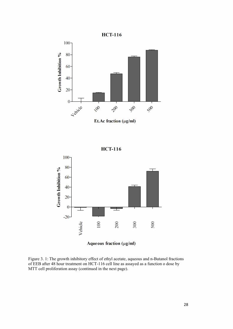
28
Figure 3. 1: The growth inhibitory effect of ethyl acetate, aqueous and n-Butanol fractions of EEB after 48 hour treatment on HCT-116 cell line as assayed as a function o dose by MTT cell proliferation assay (continued in the next page).
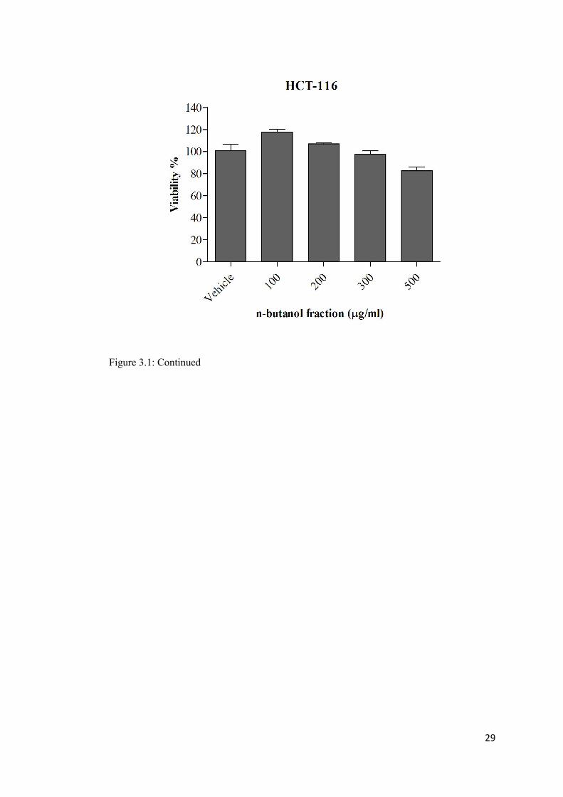
29
Figure 3.1: Continued
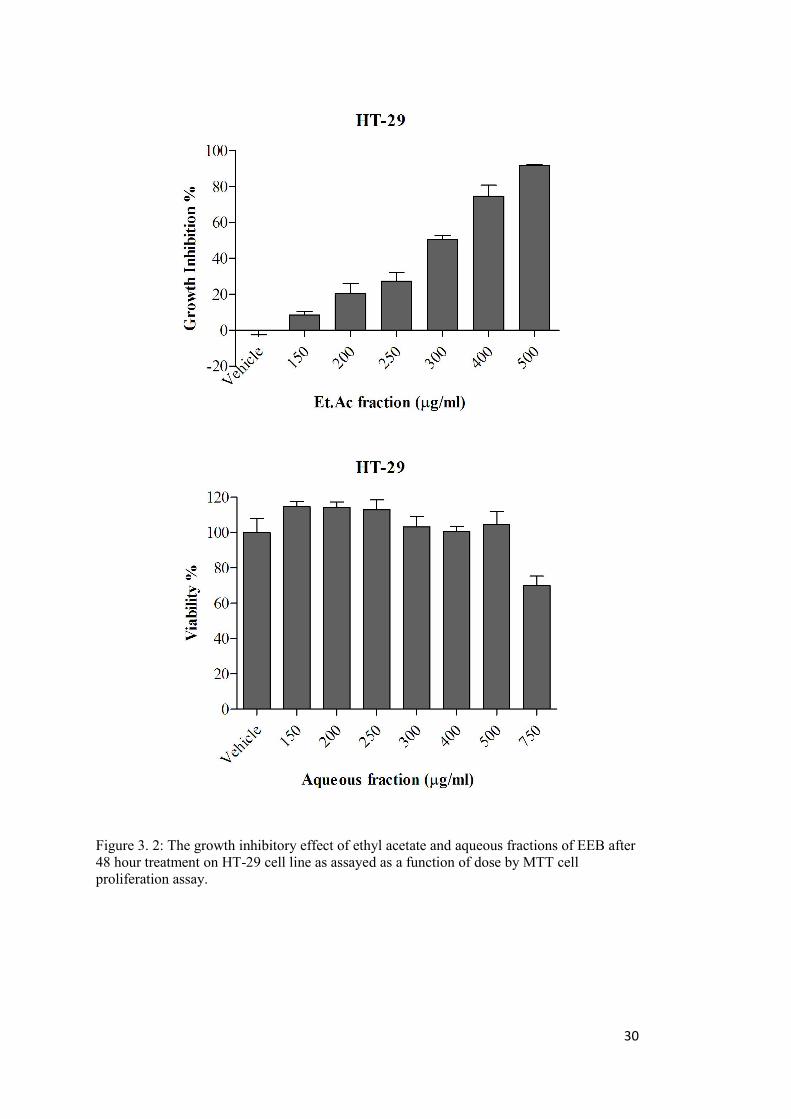
30
Figure 3. 2: The growth inhibitory effect of ethyl acetate and aqueous fractions of EEB after 48 hour treatment on HT-29 cell line as assayed as a function of dose by MTT cell proliferation assay.
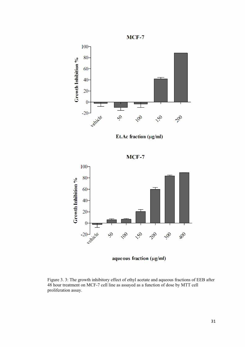
31
Figure 3. 3: The growth inhibitory effect of ethyl acetate and aqueous fractions of EEB after 48 hour treatment on MCF-7 cell line as assayed as a function of dose by MTT cell proliferation assay.
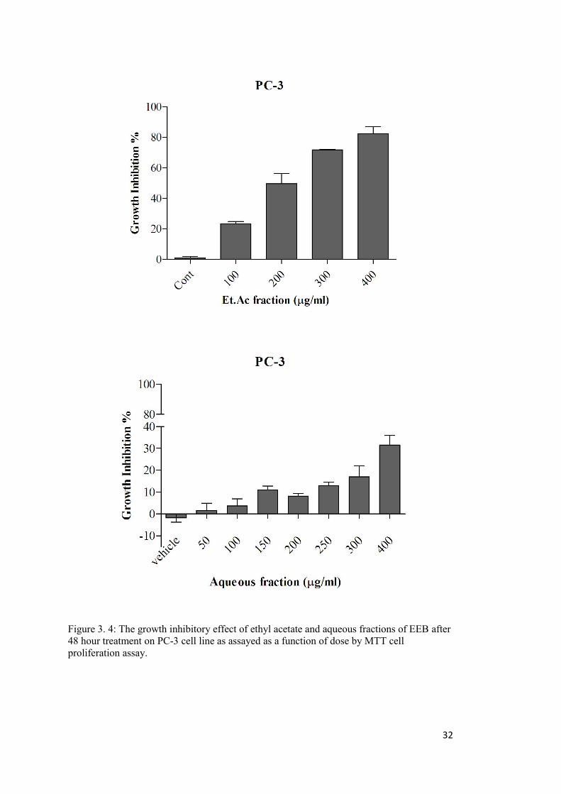
32
Figure 3. 4: The growth inhibitory effect of ethyl acetate and aqueous fractions of EEB after 48 hour treatment on PC-3 cell line as assayed as a function of dose by MTT cell proliferation assay.
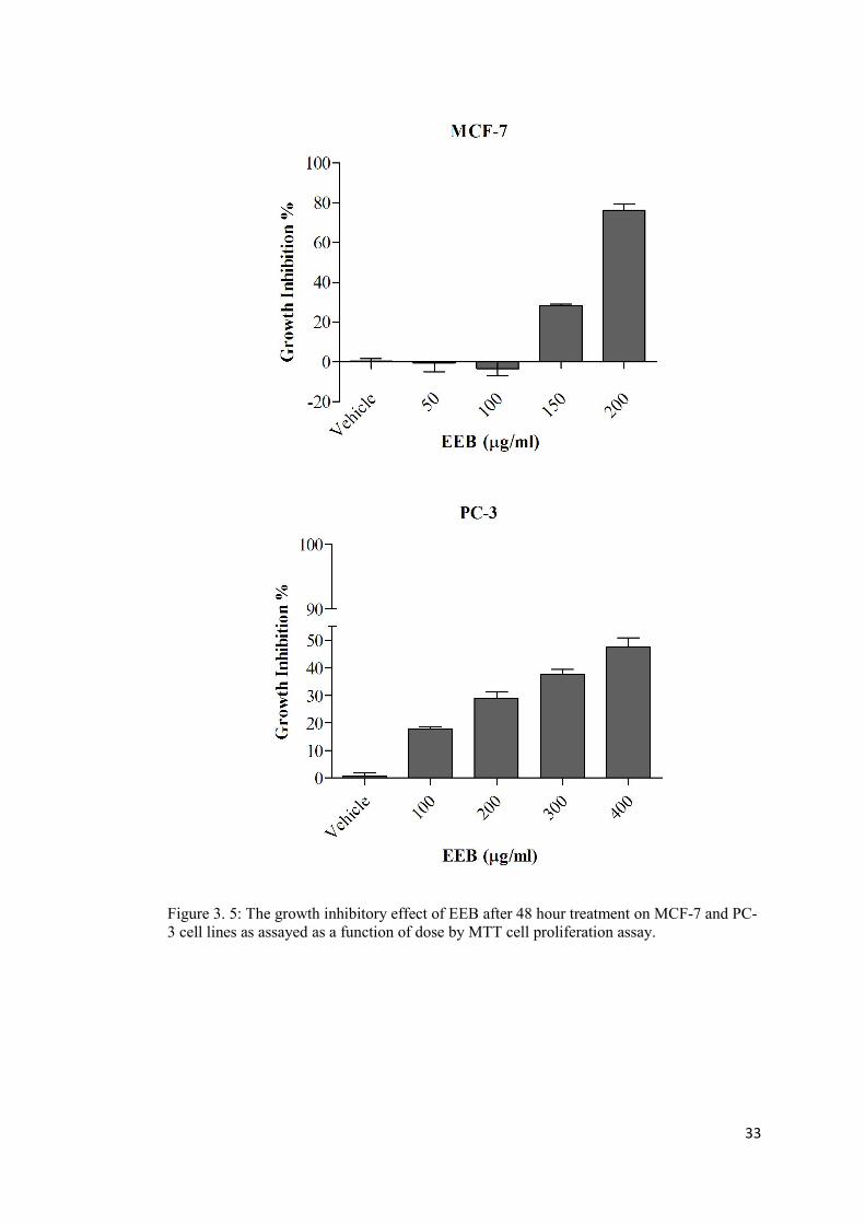
33
Figure 3. 5: The growth inhibitory effect of EEB after 48 hour treatment on MCF-7 and PC-3 cell lines as assayed as a function of dose by MTT cell proliferation assay.
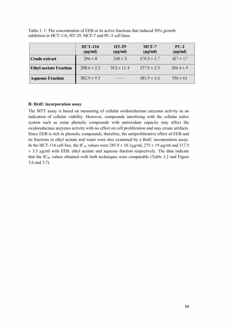
34
Table 3. 1: The concentration of EEB or its active fractions that induced 50% growth inhibition in HCT-116, HT-29, MCF-7 and PC-3 cell lines.
B: BrdU incorporation assay The MTT assay is based on measuring of cellular oxidoreductase enzymes activity as an indication of cellular viability. However, compounds interfering with the cellular redox system such as some phenolic compounds with antioxidant capacity may affect the oxidoreductase anzymes activity with no effect on cell proliferation and may create artifacts. Since EEB is rich in phenolic compounds, therefore, the antiproliferative effect of EEB and its fractions in ethyl acetate and water were also examined by a BrdU incorporation assay. In the HCT-116 cell line, the IC50 values were 285.9 ± 10.1µg/ml, 275 ± 19 µg/ml and 317.5 ± 3.5 µg/ml with EEB, ethyl acetate and aqueous fraction respectively. The data indicate that the IC50 values obtained with both techniques were comparable (Table 3.2 and Figure 3.6 and 3.7).
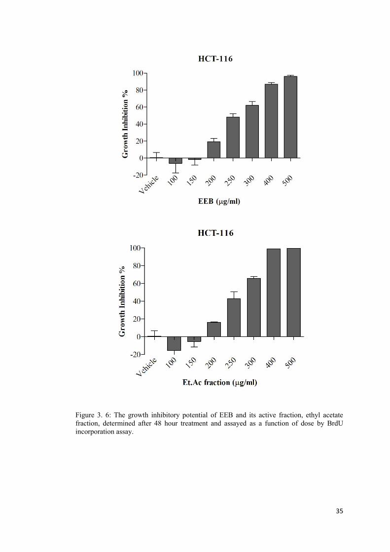
35
Figure 3. 6: The growth inhibitory potential of EEB and its active fraction, ethyl acetate fraction, determined after 48 hour treatment and assayed as a function of dose by BrdU incorporation assay.
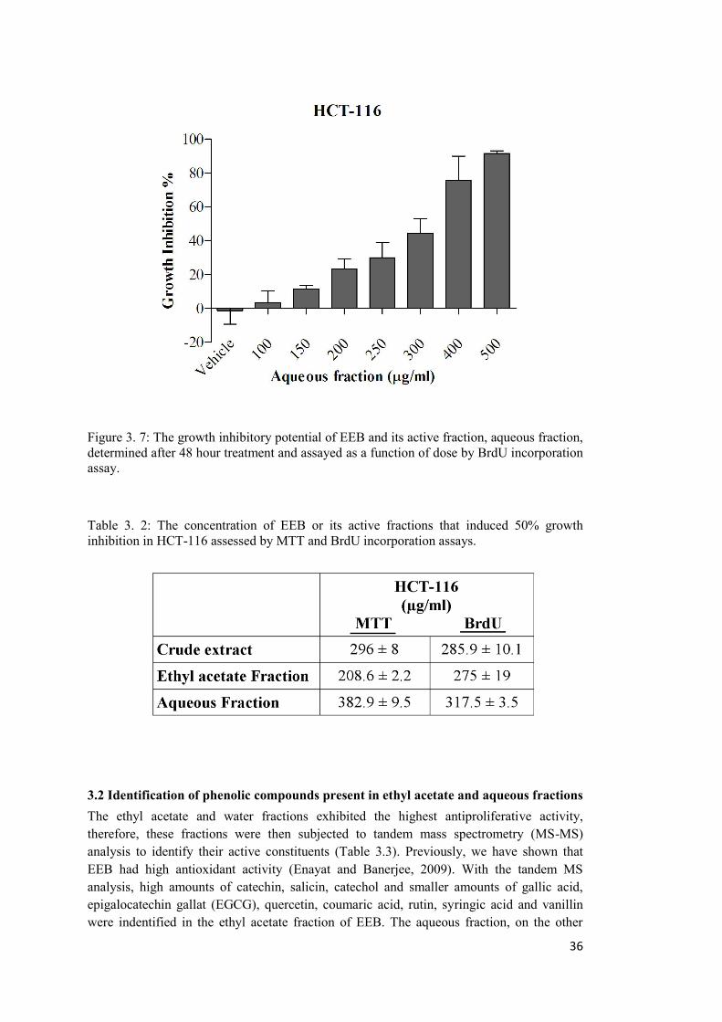
36
Figure 3. 7: The growth inhibitory potential of EEB and its active fraction, aqueous fraction, determined after 48 hour treatment and assayed as a function of dose by BrdU incorporation assay.
Table 3. 2: The concentration of EEB or its active fractions that induced 50% growth inhibition in HCT-116 assessed by MTT and BrdU incorporation assays.
3.2 Identification of phenolic compounds present in ethyl acetate and aqueous fractions The ethyl acetate and water fractions exhibited the highest antiproliferative activity, therefore, these fractions were then subjected to tandem mass spectrometry (MS-MS) analysis to identify their active constituents (Table 3.3). Previously, we have shown that EEB had high antioxidant activity (Enayat and Banerjee, 2009). With the tandem MS analysis, high amounts of catechin, salicin, catechol and smaller amounts of gallic acid, epigalocatechin gallat (EGCG), quercetin, coumaric acid, rutin, syringic acid and vanillin were indentified in the ethyl acetate fraction of EEB. The aqueous fraction, on the other
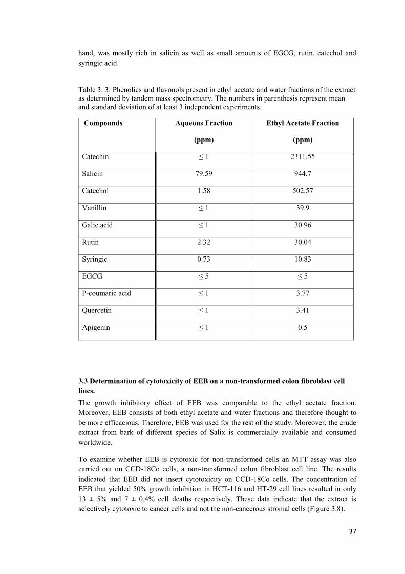
37
hand, was mostly rich in salicin as well as small amounts of EGCG, rutin, catechol and syringic acid.
Table 3. 3: Phenolics and flavonols present in ethyl acetate and water fractions of the extract as determined by tandem mass spectrometry. The numbers in parenthesis represent mean and standard deviation of at least 3 independent experiments.
Compounds Aqueous Fraction
(ppm)
Ethyl Acetate Fraction
(ppm)
Catechin ≤ 1 2311.55
Salicin 79.59 944.7
Catechol 1.58 502.57
Vanillin ≤ 1 39.9
Galic acid ≤ 1 30.96
Rutin 2.32 30.04
Syringic 0.73 10.83
EGCG ≤ 5 ≤ 5
P-coumaric acid ≤ 1 3.77
Quercetin ≤ 1 3.41
Apigenin ≤ 1 0.5
3.3 Determination of cytotoxicity of EEB on a non-transformed colon fibroblast cell lines. The growth inhibitory effect of EEB was comparable to the ethyl acetate fraction. Moreover, EEB consists of both ethyl acetate and water fractions and therefore thought to be more efficacious. Therefore, EEB was used for the rest of the study. Moreover, the crude extract from bark of different species of Salix is commercially available and consumed worldwide.
To examine whether EEB is cytotoxic for non-transformed cells an MTT assay was also carried out on CCD-18Co cells, a non-transformed colon fibroblast cell line. The results indicated that EEB did not insert cytotoxicity on CCD-18Co cells. The concentration of EEB that yielded 50% growth inhibition in HCT-116 and HT-29 cell lines resulted in only 13 ± 5% and 7 ± 0.4% cell deaths respectively. These data indicate that the extract is selectively cytotoxic to cancer cells and not the non-cancerous stromal cells (Figure 3.8).
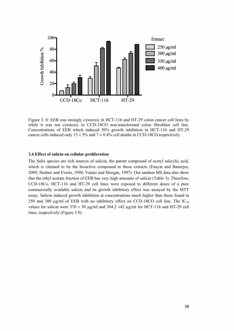
38
Figure 3. 8: EEB was strongly cytotoxic in HCT-116 and HT-29 colon cancer cell lines by while it was not cytotoxic in CCD-18CO non-transformed colon fibroblast cell line. Concentrations of EEB which induced 50% growth inhibition in HCT-116 and HT-29 cancer cells induced only 13 ± 5% and 7 ± 0.4% cell deaths in CCD-18CO respectively.
3.4 Effect of salicin on cellular proliferation The Salix species are rich sources of salicin, the parent compound of acetyl salicylic acid, which is claimed to be the bioactive compound in these extracts (Enayat and Banerjee, 2009; Hedner and Everts, 1998; Vainio and Morgan, 1997). Our tandem MS data also show that the ethyl acetate fraction of EEB has very high amounts of salicin (Table 3). Therefore, CCD-18Co, HCT-116 and HT-29 cell lines were exposed to different doses of a pure commercially available salicin and its growth inhibitory effect was assayed by the MTT assay. Salicin induced growth inhibition at concentrations much higher than those found in 250 and 300 µg/ml of EEB with no inhibitory effect on CCD-18CO cell line. The IC50
values for salicin were 370 ± 30 µg/ml and 304.2 ±42 µg/ml for HCT-116 and HT-29 cell lines, respectively (Figure 3.9).
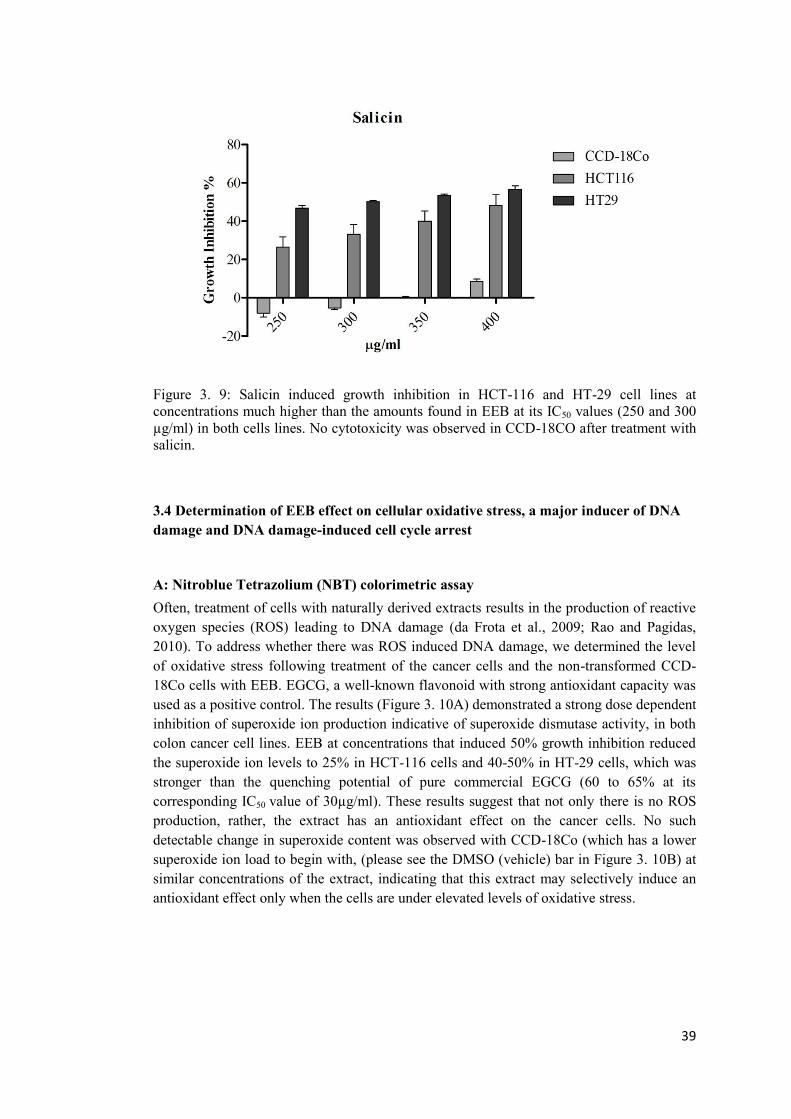
39
Figure 3. 9: Salicin induced growth inhibition in HCT-116 and HT-29 cell lines at concentrations much higher than the amounts found in EEB at its IC50 values (250 and 300 µg/ml) in both cells lines. No cytotoxicity was observed in CCD-18CO after treatment with salicin.
3.4 Determination of EEB effect on cellular oxidative stress, a major inducer of DNA damage and DNA damage-induced cell cycle arrest
A: Nitroblue Tetrazolium (NBT) colorimetric assay Often, treatment of cells with naturally derived extracts results in the production of reactive oxygen species (ROS) leading to DNA damage (da Frota et al., 2009; Rao and Pagidas, 2010). To address whether there was ROS induced DNA damage, we determined the level of oxidative stress following treatment of the cancer cells and the non-transformed CCD-18Co cells with EEB. EGCG, a well-known flavonoid with strong antioxidant capacity was used as a positive control. The results (Figure 3. 10A) demonstrated a strong dose dependent inhibition of superoxide ion production indicative of superoxide dismutase activity, in both colon cancer cell lines. EEB at concentrations that induced 50% growth inhibition reduced the superoxide ion levels to 25% in HCT-116 cells and 40-50% in HT-29 cells, which was stronger than the quenching potential of pure commercial EGCG (60 to 65% at its corresponding IC50 value of 30µg/ml). These results suggest that not only there is no ROS production, rather, the extract has an antioxidant effect on the cancer cells. No such detectable change in superoxide content was observed with CCD-18Co (which has a lower superoxide ion load to begin with, (please see the DMSO (vehicle) bar in Figure 3. 10B) at similar concentrations of the extract, indicating that this extract may selectively induce an antioxidant effect only when the cells are under elevated levels of oxidative stress.
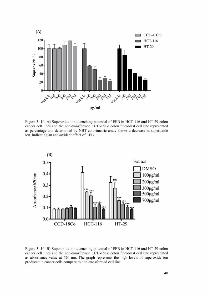
40
Figure 3. 10: A) Superoxide ion quenching potential of EEB in HCT-116 and HT-29 colon cancer cell lines and the non-transformed CCD-18Co colon fibroblast cell line represented as percentage and determined by NBT colorimetric assay shows a decrease in superoxide ion, indicating an anti-oxidant effect of EEB.
Figure 3. 10: B) Superoxide ion quenching potential of EEB in HCT-116 and HT-29 colon cancer cell lines and the non-transformed CCD-18Co colon fibroblast cell line represented as absorbance value at 620 nm. The graph represents the high levels of superoxide ion produced in cancer cells compare to non-transformed cell line.
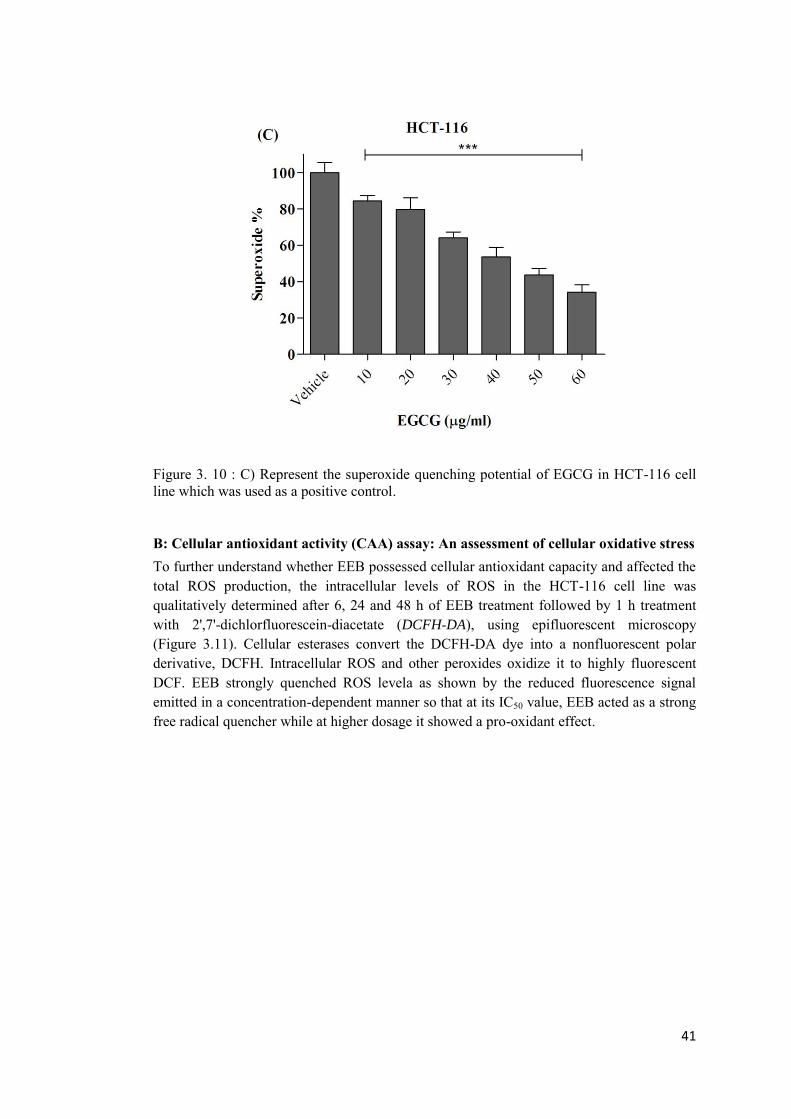
41
Figure 3. 10 : C) Represent the superoxide quenching potential of EGCG in HCT-116 cell line which was used as a positive control.
B: Cellular antioxidant activity (CAA) assay: An assessment of cellular oxidative stress To further understand whether EEB possessed cellular antioxidant capacity and affected the total ROS production, the intracellular levels of ROS in the HCT-116 cell line was qualitatively determined after 6, 24 and 48 h of EEB treatment followed by 1 h treatment with 2',7'-dichlorfluorescein-diacetate (DCFH-DA), using epifluorescent microscopy (Figure 3.11). Cellular esterases convert the DCFH-DA dye into a nonfluorescent polar derivative, DCFH. Intracellular ROS and other peroxides oxidize it to highly fluorescent DCF. EEB strongly quenched ROS levela as shown by the reduced fluorescence signal emitted in a concentration-dependent manner so that at its IC50 value, EEB acted as a strong free radical quencher while at higher dosage it showed a pro-oxidant effect.
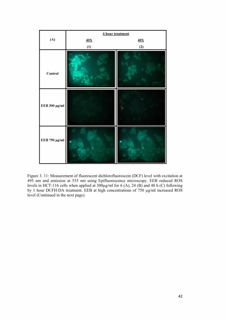
42
Figure 3. 11: Measurement of fluorescent dichlorofluorescein (DCF) level with excitation at 495 nm and emission at 535 nm using Epifluorescence microscopy. EEB reduced ROS levels in HCT-116 cells when applied at 300µg/ml for 6 (A), 24 (B) and 48 h (C) following by 1 hour DCFH-DA treatment. EEB at high concentrations of 750 µg/ml increased ROS level (Continued in the next page).
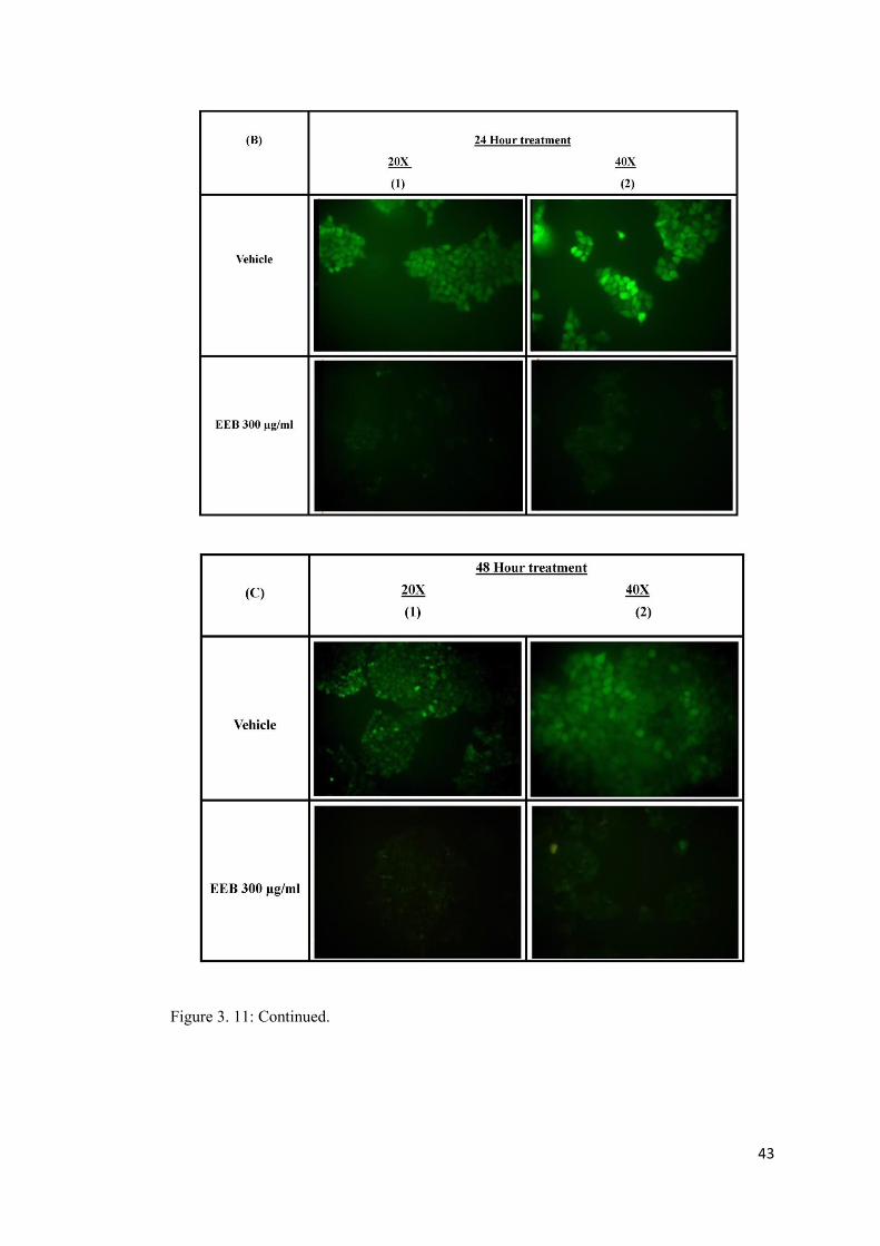
43
Figure 3. 11: Continued.
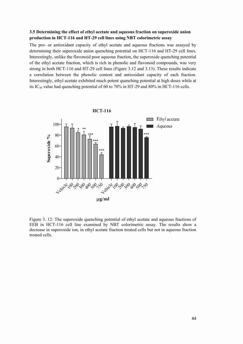
44
3.5 Determining the effect of ethyl acetate and aqueous fraction on superoxide anion production in HCT-116 and HT-29 cell lines using NBT colorimetric assay The pro- or antioxidant capacity of ethyl acetate and aqueous fractions was assayed by determining their superoxide anion quenching potential on HCT-116 and HT-29 cell lines. Interestingly, unlike the flavonoid poor aqueous fraction, the superoxide quenching potential of the ethyl acetate fraction, which is rich in phenolic and flavonoid compounds, was very strong in both HCT-116 and HT-29 cell lines (Figure 3.12 and 3.13). These results indicate a correlation between the phenolic content and antioxidant capacity of each fraction. Interestingly, ethyl acetate exhibited much potent quenching potential at high doses while at its IC50 value had quenching potential of 60 to 70% in HT-29 and 80% in HCT-116 cells.
Figure 3. 12: The superoxide quenching potential of ethyl acetate and aqueous fractions of EEB in HCT-116 cell line examined by NBT colorimetric assay. The results show a decrease in superoxide ion, in ethyl acetate fraction treated cells but not in aqueous fraction treated cells.
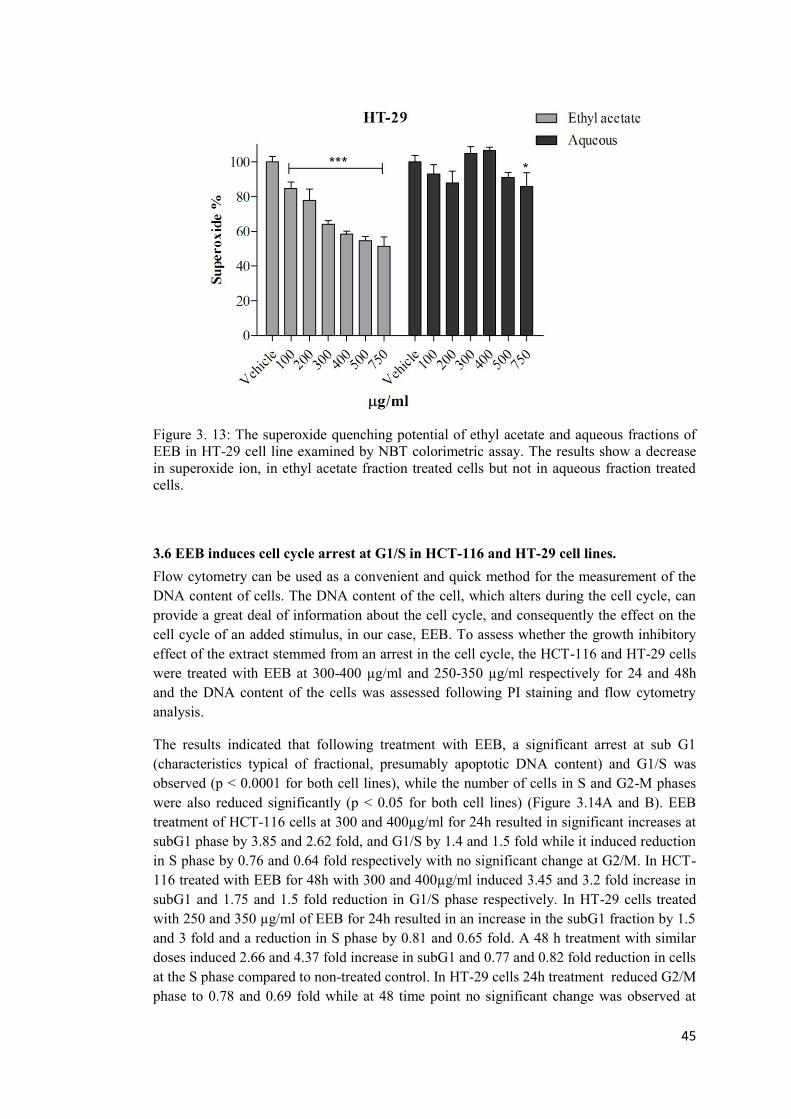
45
Figure 3. 13: The superoxide quenching potential of ethyl acetate and aqueous fractions of EEB in HT-29 cell line examined by NBT colorimetric assay. The results show a decrease in superoxide ion, in ethyl acetate fraction treated cells but not in aqueous fraction treated cells.
3.6 EEB induces cell cycle arrest at G1/S in HCT-116 and HT-29 cell lines. Flow cytometry can be used as a convenient and quick method for the measurement of the DNA content of cells. The DNA content of the cell, which alters during the cell cycle, can provide a great deal of information about the cell cycle, and consequently the effect on the cell cycle of an added stimulus, in our case, EEB. To assess whether the growth inhibitory effect of the extract stemmed from an arrest in the cell cycle, the HCT-116 and HT-29 cells were treated with EEB at 300-400 µg/ml and 250-350 µg/ml respectively for 24 and 48h and the DNA content of the cells was assessed following PI staining and flow cytometry analysis.
The results indicated that following treatment with EEB, a significant arrest at sub G1 (characteristics typical of fractional, presumably apoptotic DNA content) and G1/S was observed (p < 0.0001 for both cell lines), while the number of cells in S and G2-M phases were also reduced significantly (p < 0.05 for both cell lines) (Figure 3.14A and B). EEB treatment of HCT-116 cells at 300 and 400µg/ml for 24h resulted in significant increases at subG1 phase by 3.85 and 2.62 fold, and G1/S by 1.4 and 1.5 fold while it induced reduction in S phase by 0.76 and 0.64 fold respectively with no significant change at G2/M. In HCT-116 treated with EEB for 48h with 300 and 400µg/ml induced 3.45 and 3.2 fold increase in subG1 and 1.75 and 1.5 fold reduction in G1/S phase respectively. In HT-29 cells treated with 250 and 350 µg/ml of EEB for 24h resulted in an increase in the subG1 fraction by 1.5 and 3 fold and a reduction in S phase by 0.81 and 0.65 fold. A 48 h treatment with similar doses induced 2.66 and 4.37 fold increase in subG1 and 0.77 and 0.82 fold reduction in cells at the S phase compared to non-treated control. In HT-29 cells 24h treatment reduced G2/M phase to 0.78 and 0.69 fold while at 48 time point no significant change was observed at
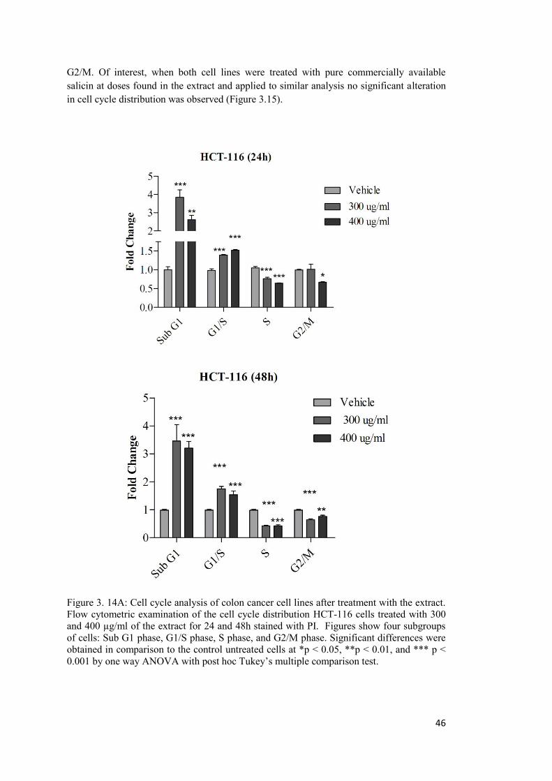
46
G2/M. Of interest, when both cell lines were treated with pure commercially available salicin at doses found in the extract and applied to similar analysis no significant alteration in cell cycle distribution was observed (Figure 3.15).
Figure 3. 14A: Cell cycle analysis of colon cancer cell lines after treatment with the extract. Flow cytometric examination of the cell cycle distribution HCT-116 cells treated with 300 and 400 µg/ml of the extract for 24 and 48h stained with PI. Figures show four subgroups of cells: Sub G1 phase, G1/S phase, S phase, and G2/M phase. Significant differences were obtained in comparison to the control untreated cells at *p < 0.05, **p < 0.01, and *** p < 0.001 by one way ANOVA with post hoc Tukey’s multiple comparison test.
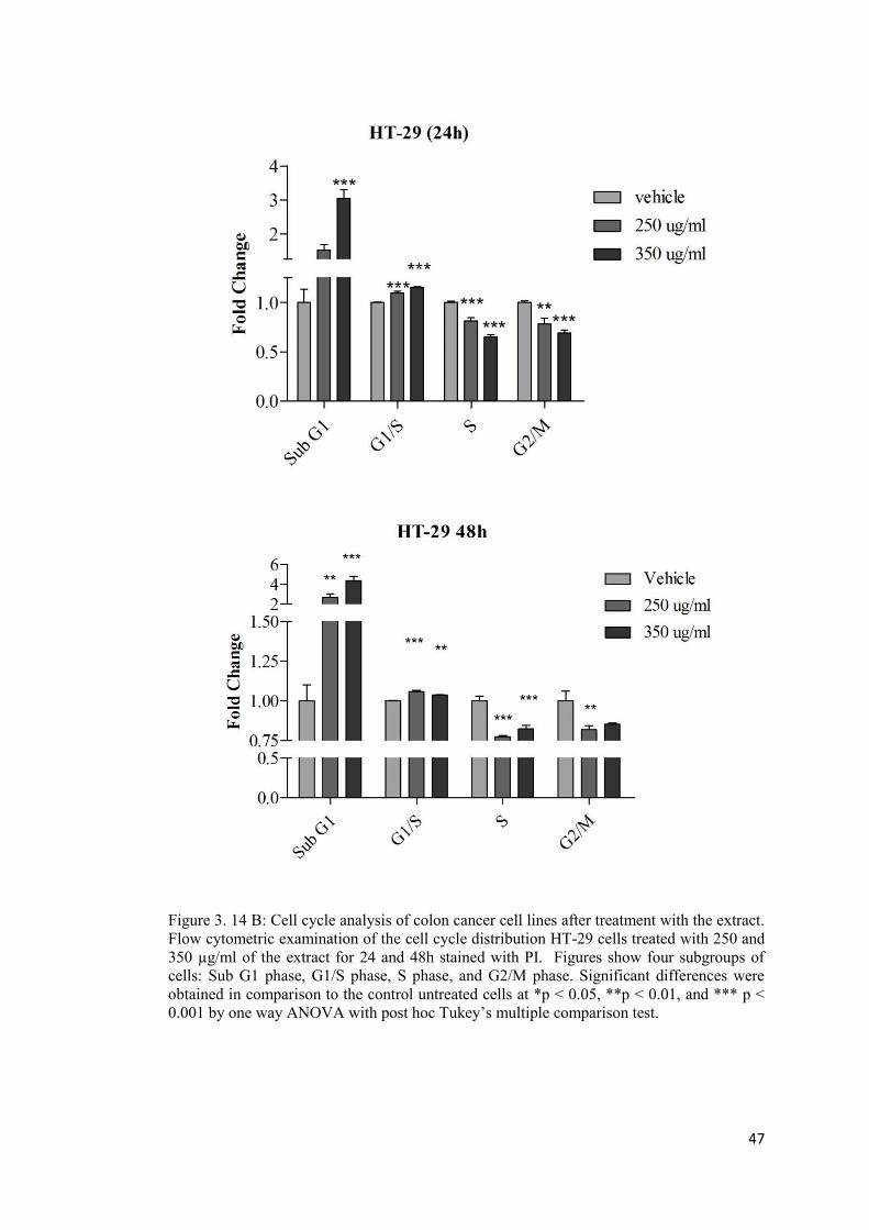
47
Figure 3. 14 B: Cell cycle analysis of colon cancer cell lines after treatment with the extract. Flow cytometric examination of the cell cycle distribution HT-29 cells treated with 250 and 350 µg/ml of the extract for 24 and 48h stained with PI. Figures show four subgroups of cells: Sub G1 phase, G1/S phase, S phase, and G2/M phase. Significant differences were obtained in comparison to the control untreated cells at *p < 0.05, **p < 0.01, and *** p < 0.001 by one way ANOVA with post hoc Tukey’s multiple comparison test.
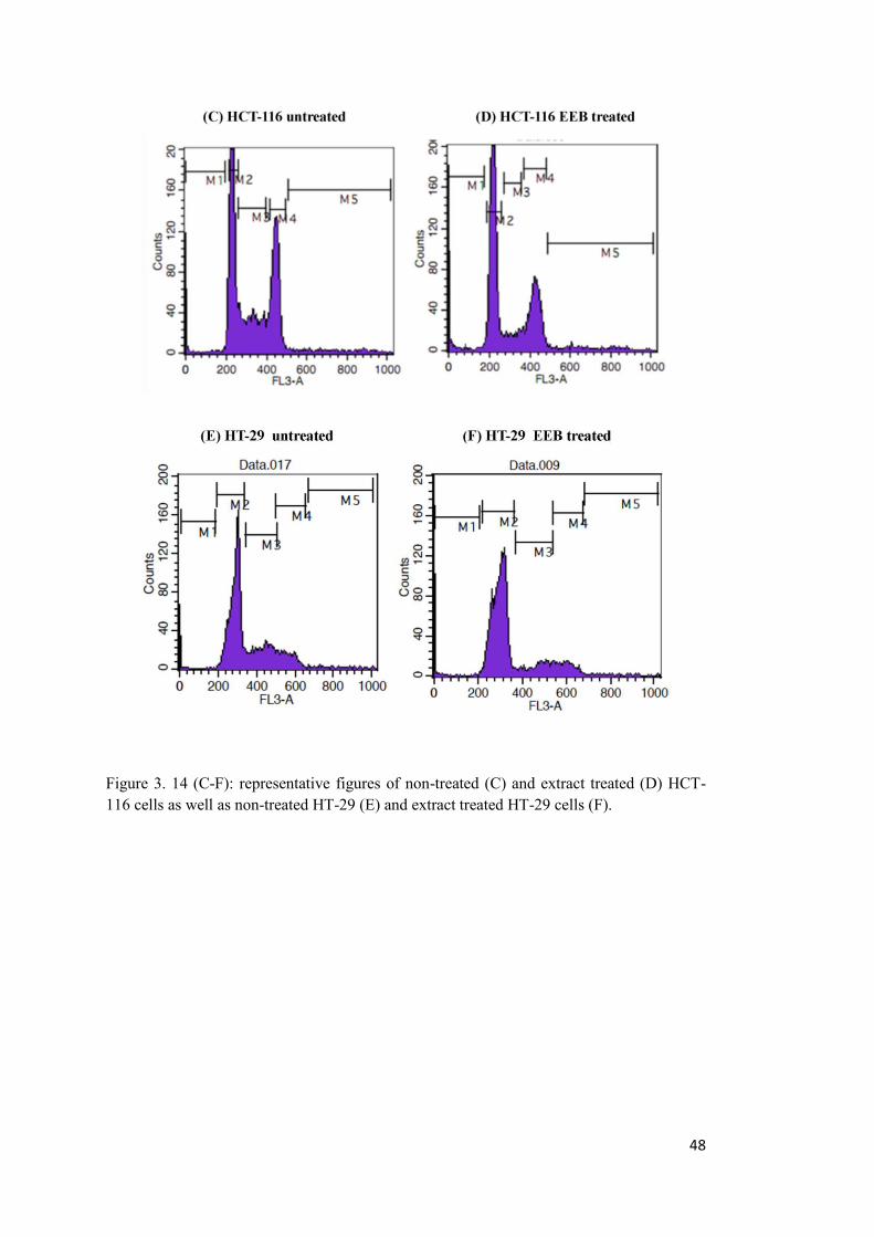
48
Figure 3. 14 (C-F): representative figures of non-treated (C) and extract treated (D) HCT-116 cells as well as non-treated HT-29 (E) and extract treated HT-29 cells (F).
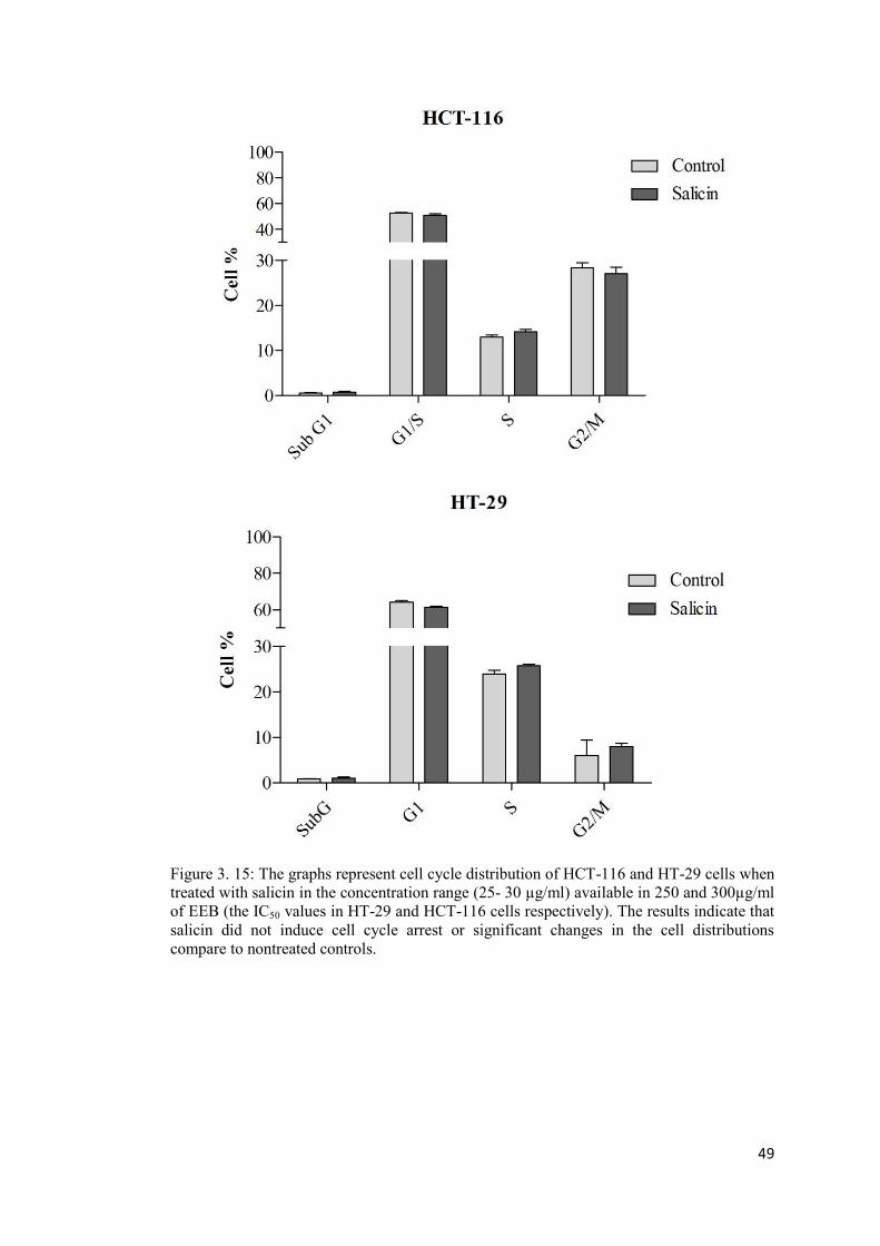
49
Figure 3. 15: The graphs represent cell cycle distribution of HCT-116 and HT-29 cells when treated with salicin in the concentration range (25- 30 µg/ml) available in 250 and 300µg/ml of EEB (the IC50 values in HT-29 and HCT-116 cells respectively). The results indicate that salicin did not induce cell cycle arrest or significant changes in the cell distributions compare to nontreated controls.
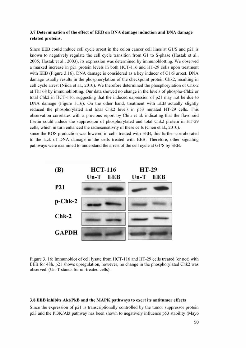
50
3.7 Determination of the effect of EEB on DNA damage induction and DNA damage related proteins.
Since EEB could induce cell cycle arrest in the colon cancer cell lines at G1/S and p21 is known to negatively regulate the cell cycle transition from G1 to S-phase (Hastak et al., 2005; Hastak et al., 2003), its expression was determined by immunoblotting. We observed a marked increase in p21 protein levels in both HCT-116 and HT-29 cells upon treatment with EEB (Figure 3.16). DNA damage is considered as a key inducer of G1/S arrest. DNA damage usually results in the phosphorylation of the checkpoint protein Chk2, resulting in cell cycle arrest (Niida et al., 2010). We therefore determined the phosphorylation of Chk-2 at Thr 68 by immunoblotting. Our data showed no change in the levels of phospho-Chk2 or total Chk2 in HCT-116, suggesting that the induced expression of p21 may not be due to DNA damage (Figure 3.16). On the other hand, treatment with EEB actually slightly reduced the phosphorylated and total Chk2 levels in p53 mutated HT-29 cells. This observation correlates with a previous report by Chiu et al. indicating that the flavonoid fisetin could induce the suppression of phosphorylated and total Chk2 protein in HT-29 cells, which in turn enhanced the radiosensitivity of these cells (Chen et al., 2010). since the ROS production was lowered in cells treated with EEB, this further corroborated to the lack of DNA damage in the cells treated with EEB: Therefore, other signaling pathways were examined to understand the arrest of the cell cycle at G1/S by EEB.
Figure 3. 16: Immunoblot of cell lysate from HCT-116 and HT-29 cells treated (or not) with EEB for 48h. p21 shows upregulation, however, no change in the phosphorylated Chk2 was observed. (Un-T stands for un-treated cells).
3.8 EEB inhibits Akt/PkB and the MAPK pathways to exert its antitumor effects Since the expression of p21 is transcriptionally controlled by the tumor suppressor protein p53 and the PI3K/Akt pathway has been shown to negatively influence p53 stability (Mayo
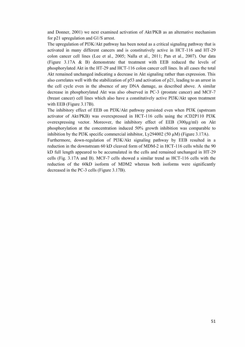
51
and Donner, 2001) we next examined activation of Akt/PKB as an alternative mechanism for p21 upregulation and G1/S arrest. The upregulation of PI3K/Akt pathway has been noted as a critical signaling pathway that is activated in many different cancers and is constitutively active in HCT-116 and HT-29 colon cancer cell lines (Lee et al., 2005; Nalla et al., 2011; Pan et al., 2007). Our data (Figure 3.17A & B) demonstrate that treatment with EEB reduced the levels of phosphorylated Akt in the HT-29 and HCT-116 colon cancer cell lines. In all cases the total Akt remained unchanged indicating a decrease in Akt signaling rather than expression. This also correlates well with the stabilization of p53 and activation of p21, leading to an arrest in the cell cycle even in the absence of any DNA damage, as described above. A similar decrease in phosphorylated Akt was also observed in PC-3 (prostate cancer) and MCF-7 (breast cancer) cell lines which also have a constitutively active PI3K/Akt upon treatment with EEB (Figure 3.17B). The inhibitory effect of EEB on PI3K/Akt pathway persisted even when PI3K (upstream activator of Akt/PKB) was overexpressed in HCT-116 cells using the rCD2P110 PI3K overexpressing vector. Moreover, the inhibitory effect of EEB (300µg/ml) on Akt phosphorylation at the concentration induced 50% growth inhibition was comparable to inhibition by the PI3K specific commercial inhibitor, Ly294002 (50 µM) (Figure 3.17A). Furthermore, down-regulation of PI3K/Akt signaling pathway by EEB resulted in a reduction in the downstream 60 kD cleaved form of MDM-2 in HCT-116 cells while the 90 kD full length appeared to be accumulated in the cells and remained unchanged in HT-29 cells (Fig. 3.17A and B). MCF-7 cells showed a similar trend as HCT-116 cells with the reduction of the 60kD isoform of MDM2 whereas both isoforms were significantly decreased in the PC-3 cells (Figure 3.17B).
52
Figure 3. 17: A) EEB inhibits the Akt/PKB and MAPK pathways in colon cancer cell lines A) Immunoblot showing the inhibition of the activation of Akt in HCT-116 cells by EEB. PI3K was transiently overexpressed with a rDC2P110 plasmid, LY294002 is a specific inhibitor of PI3K.(B) Immunoblot showing the inhibition of Akt in HT-29, MCF-7 breast and PC3 prostate cancer cell lines. A marked reduction in the oncogenic 60 kD isoform of MDM2 was observed in all cell lines.
53
3.9 EEB induces apoptosis in HCT-116 and HT-29 cells. Based on the observation that EEB induced an increase in the sub-G1 fraction in the cancer cells, we next wanted to assess the induction of apoptosis by EEB. For this, the HCT-116 and HT-29 cells were treated with EEB (concentration corresponding to the IC50 value in that cell line) and the induction of apoptosis was assessed using Annexin V staining, and expression of pro and anti-apoptotic proteins.
A: Annexin V staining assay In order to quantitatively assess whether treatment of the colon cancer cells with EEB resulted in the induction of apoptosis, Annexin V/PI staining followed by flow cytometry was carried out. Treatment of HCT-116 cells with EEB resulted in a statistically significant (p<0.0001) increase in the fraction of apoptotic cells (30%) after 48h of treatment compared to the untreated cells (6%) (Figure 3.18A). This increase in the apoptotic fraction was comparable to treatment with 5 mM sodium butyrate (NaBt) for 48h, a well-known inducer of apoptosis, which resulted in 36% apoptotic population in cells (Figure 3.18C). When HT-29 cells were treated with EEB, a statistically significant (p<0.0001) increase in the proportion of apoptotic cells (17%) was observed after 48h of treatment compared to the control untreated cells (6%) (Figure 3.18B). Additionally, for both HCT-116 and HT-29 cells, this increase in the number of apoptotic cells following treatment with EEB correlated with a 1.6 and 2 fold increases in the enzymatic activity of caspase-3 in HCT-116 and HT-29 cells respectively as reported previously (Enayat, 2013). Of note, when both cell lines were treated with salicin no significant induction of apoptosis were observed in either of the cell lines (Figure 3.19 A-H).
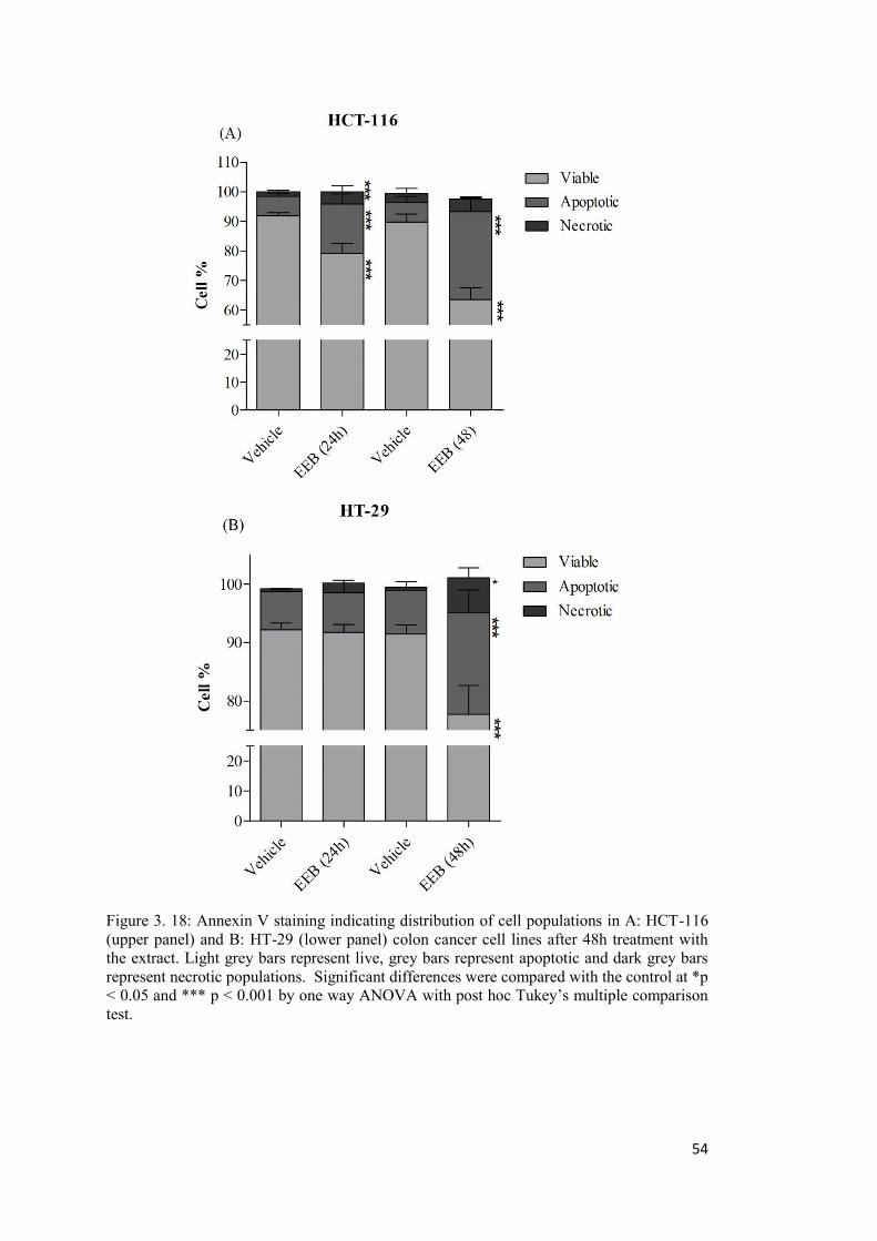
54
Figure 3. 18: Annexin V staining indicating distribution of cell populations in A: HCT-116 (upper panel) and B: HT-29 (lower panel) colon cancer cell lines after 48h treatment with the extract. Light grey bars represent live, grey bars represent apoptotic and dark grey bars represent necrotic populations. Significant differences were compared with the control at *p < 0.05 and *** p < 0.001 by one way ANOVA with post hoc Tukey’s multiple comparison test.
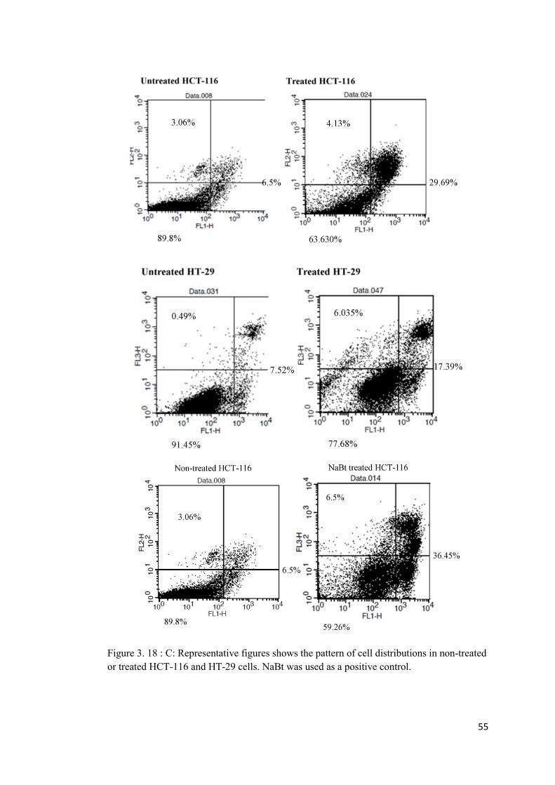
55
Figure 3. 18 : C: Representative figures shows the pattern of cell distributions in non-treated or treated HCT-116 and HT-29 cells. NaBt was used as a positive control.
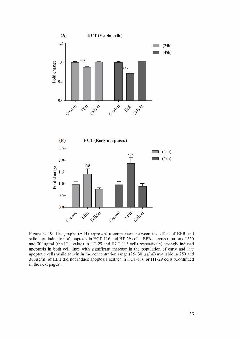
56
Figure 3. 19: The graphs (A-H) represent a comparison between the effect of EEB and salicin on induction of apoptosis in HCT-116 and HT-29 cells. EEB at concentration of 250 and 300µg/ml (the IC50 values in HT-29 and HCT-116 cells respectively) strongly induced apoptosis in both cell lines with significant increase in the population of early and late apoptotic cells while salicin in the concentration range (25- 30 µg/ml) available in 250 and 300µg/ml of EEB did not induce apoptosis neither in HCT-116 or HT-29 cells (Continued in the next pages).
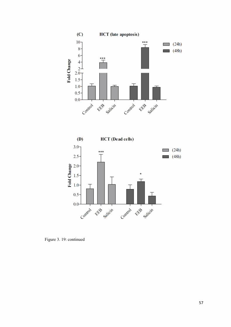
57
Figure 3. 19: continued

58
Figure 3. 19: continued
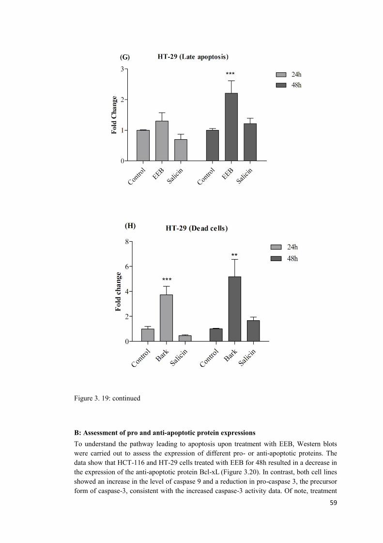
59
Figure 3. 19: continued
B: Assessment of pro and anti-apoptotic protein expressions To understand the pathway leading to apoptosis upon treatment with EEB, Western blots were carried out to assess the expression of different pro- or anti-apoptotic proteins. The data show that HCT-116 and HT-29 cells treated with EEB for 48h resulted in a decrease in the expression of the anti-apoptotic protein Bcl-xL (Figure 3.20). In contrast, both cell lines showed an increase in the level of caspase 9 and a reduction in pro-caspase 3, the precursor form of caspase-3, consistent with the increased caspase-3 activity data. Of note, treatment
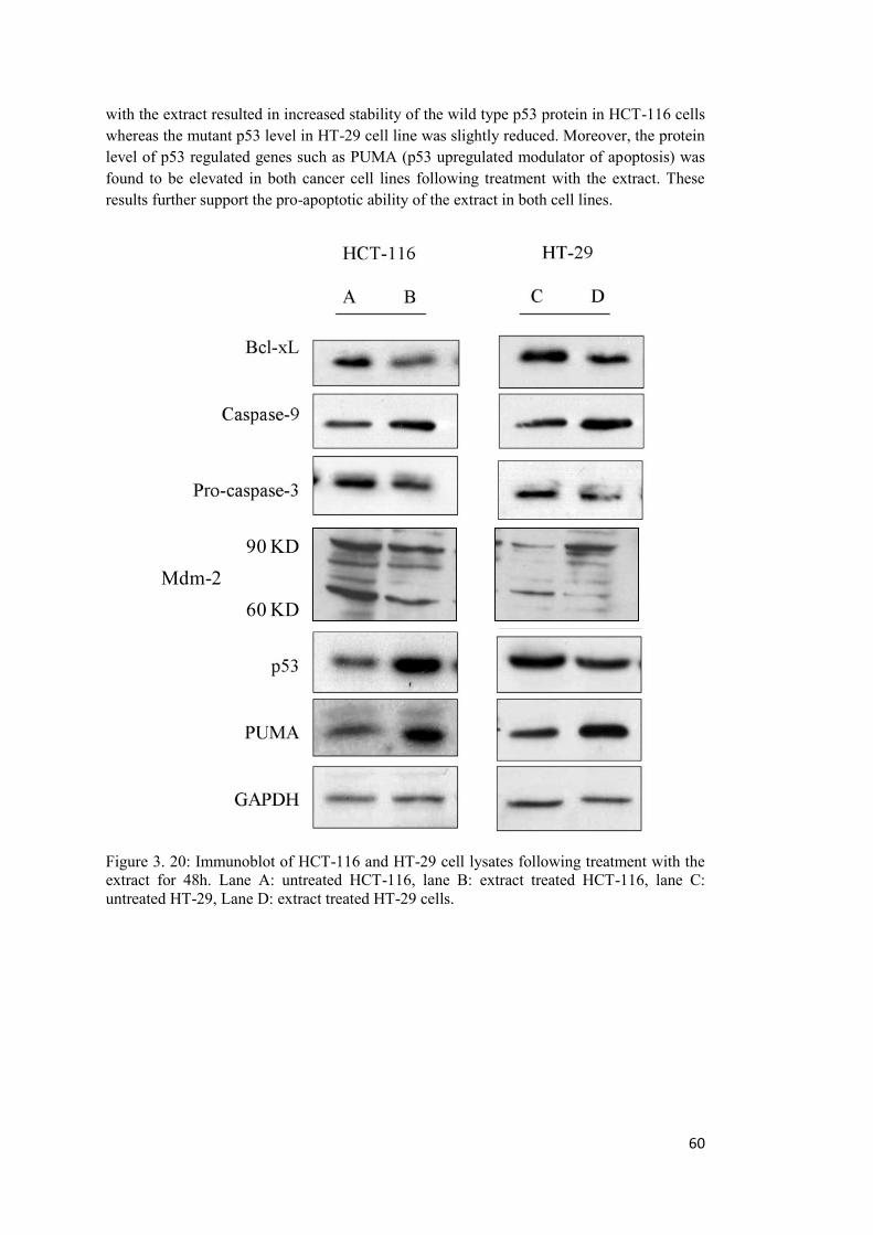
60
with the extract resulted in increased stability of the wild type p53 protein in HCT-116 cells whereas the mutant p53 level in HT-29 cell line was slightly reduced. Moreover, the protein level of p53 regulated genes such as PUMA (p53 upregulated modulator of apoptosis) was found to be elevated in both cancer cell lines following treatment with the extract. These results further support the pro-apoptotic ability of the extract in both cell lines.
90 KD
60 KDMdm-2
Figure 3. 20: Immunoblot of HCT-116 and HT-29 cell lysates following treatment with the extract for 48h. Lane A: untreated HCT-116, lane B: extract treated HCT-116, lane C: untreated HT-29, Lane D: extract treated HT-29 cells.
61
SECTION TWO
3.10 The effect of EEB on motility of cancer cells as an indicator of metastatic potential
3.10.1 Effect of EEB on anchorage independent growth Anchorage-independent growth is one of the hallmarks of cancer cells. Anoikis resistance and anchorage-independency allow tumor cells to expand and invade adjacent tissues, and to disseminate through the body, giving rise to metastasis. Soft agar colony-forming assay was carried out to determine the effect of EEB on anchorage independent growth of HCT-116 and HT-29 cells. Treatment with EEB at a low concentration (150 μg/ml for HCT-116 cells and 125 μg/ml for HT-29 cells) that did not result in inhibition of proliferation strongly reduced the ability of cells to grow in an anchorage independent manner in both cell lines (Figure 3.21- 23). The results clearly show a significant (p<0.0001) reduction in both the number and the size of colonies formed in treated cells compared to the control cells.
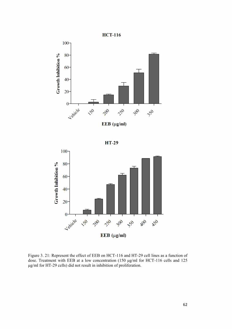
62
Figure 3. 21: Represent the effect of EEB on HCT-116 and HT-29 cell lines as a function of dose. Treatment with EEB at a low concentration (150 μg/ml for HCT-116 cells and 125 μg/ml for HT-29 cells) did not result in inhibition of proliferation.

63
Figure 3. 22: EEB at concentrations of 150 µg/ml and 125 µg/ml that did not induce growth inhibition strongly reduced the anchorage independent growth capacity of A: HCT-116 and B: HT-29 cell lines.
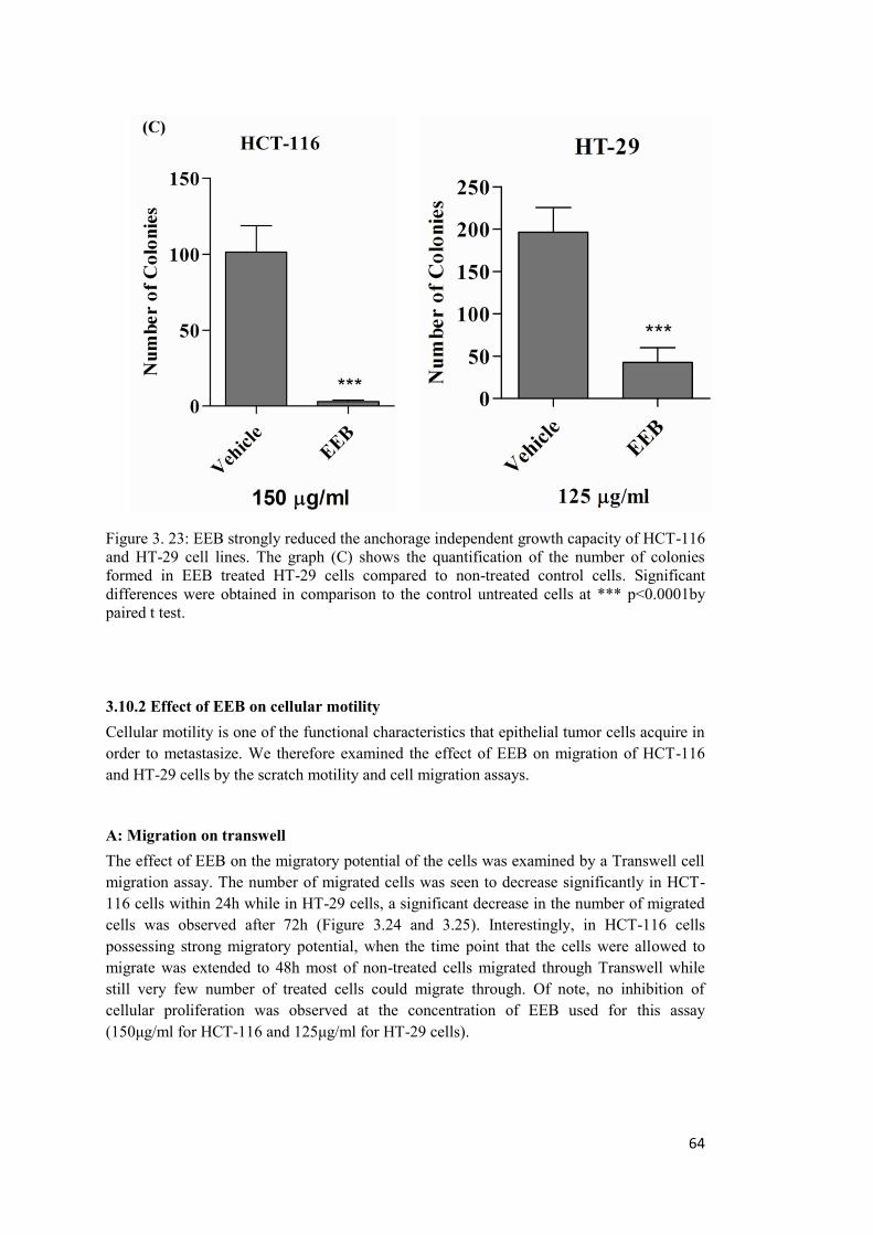
64
Figure 3. 23: EEB strongly reduced the anchorage independent growth capacity of HCT-116 and HT-29 cell lines. The graph (C) shows the quantification of the number of colonies formed in EEB treated HT-29 cells compared to non-treated control cells. Significant differences were obtained in comparison to the control untreated cells at *** p<0.0001by paired t test.
3.10.2 Effect of EEB on cellular motility Cellular motility is one of the functional characteristics that epithelial tumor cells acquire in order to metastasize. We therefore examined the effect of EEB on migration of HCT-116 and HT-29 cells by the scratch motility and cell migration assays.
A: Migration on transwell The effect of EEB on the migratory potential of the cells was examined by a Transwell cell migration assay. The number of migrated cells was seen to decrease significantly in HCT-116 cells within 24h while in HT-29 cells, a significant decrease in the number of migrated cells was observed after 72h (Figure 3.24 and 3.25). Interestingly, in HCT-116 cells possessing strong migratory potential, when the time point that the cells were allowed to migrate was extended to 48h most of non-treated cells migrated through Transwell while still very few number of treated cells could migrate through. Of note, no inhibition of cellular proliferation was observed at the concentration of EEB used for this assay (150μg/ml for HCT-116 and 125μg/ml for HT-29 cells).
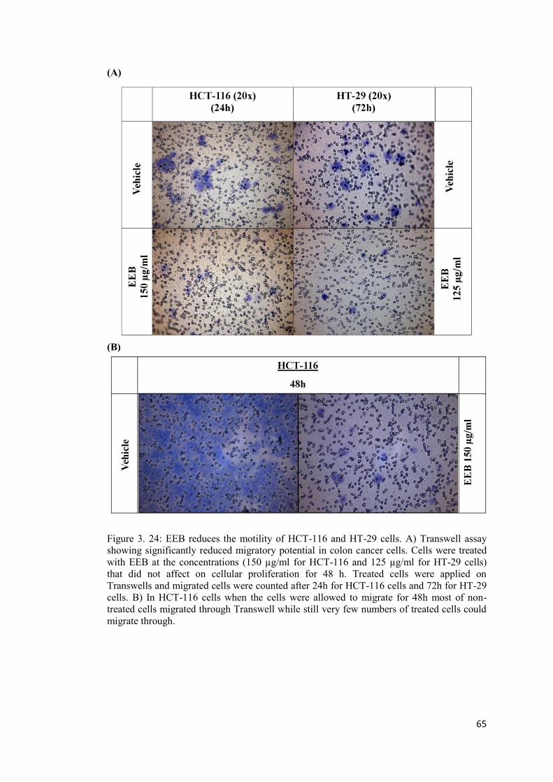
65
(A)
(B)
Figure 3. 24: EEB reduces the motility of HCT-116 and HT-29 cells. A) Transwell assay showing significantly reduced migratory potential in colon cancer cells. Cells were treated with EEB at the concentrations (150 µg/ml for HCT-116 and 125 µg/ml for HT-29 cells) that did not affect on cellular proliferation for 48 h. Treated cells were applied on Transwells and migrated cells were counted after 24h for HCT-116 cells and 72h for HT-29 cells. B) In HCT-116 cells when the cells were allowed to migrate for 48h most of non-treated cells migrated through Transwell while still very few numbers of treated cells could migrate through.
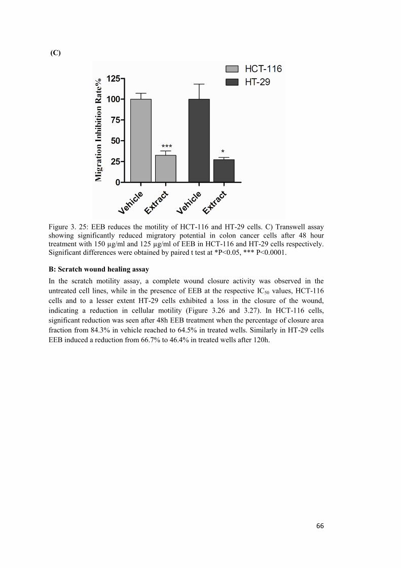
66
(C)
Figure 3. 25: EEB reduces the motility of HCT-116 and HT-29 cells. C) Transwell assay showing significantly reduced migratory potential in colon cancer cells after 48 hour treatment with 150 µg/ml and 125 µg/ml of EEB in HCT-116 and HT-29 cells respectively. Significant differences were obtained by paired t test at *P<0.05, *** P<0.0001.
B: Scratch wound healing assay In the scratch motility assay, a complete wound closure activity was observed in the untreated cell lines, while in the presence of EEB at the respective IC50 values, HCT-116 cells and to a lesser extent HT-29 cells exhibited a loss in the closure of the wound, indicating a reduction in cellular motility (Figure 3.26 and 3.27). In HCT-116 cells, significant reduction was seen after 48h EEB treatment when the percentage of closure area fraction from 84.3% in vehicle reached to 64.5% in treated wells. Similarly in HT-29 cells EEB induced a reduction from 66.7% to 46.4% in treated wells after 120h.
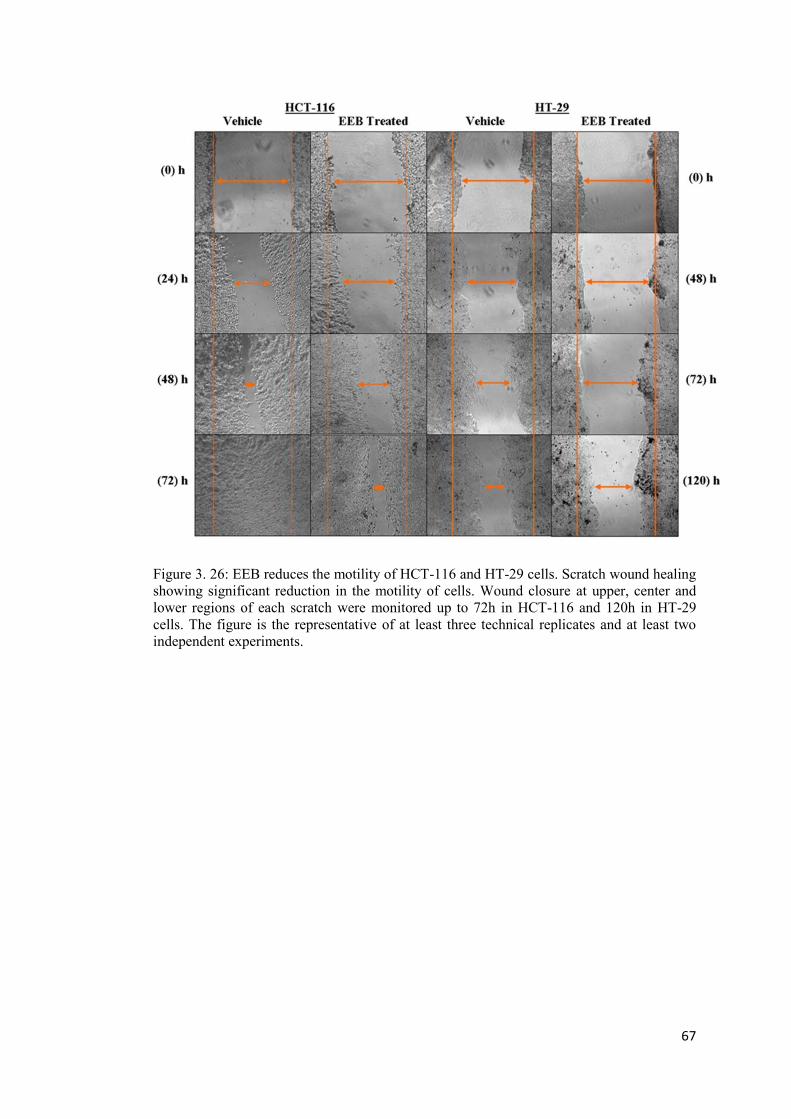
67
Figure 3. 26: EEB reduces the motility of HCT-116 and HT-29 cells. Scratch wound healing showing significant reduction in the motility of cells. Wound closure at upper, center and lower regions of each scratch were monitored up to 72h in HCT-116 and 120h in HT-29 cells. The figure is the representative of at least three technical replicates and at least two independent experiments.
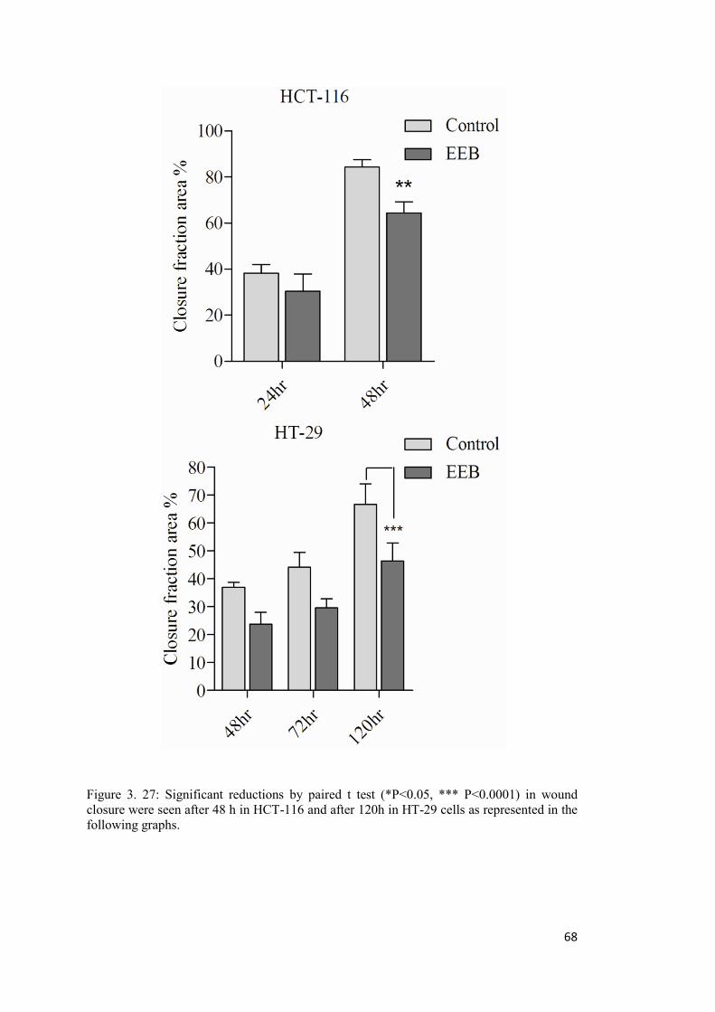
68
Figure 3. 27: Significant reductions by paired t test (*P<0.05, *** P<0.0001) in wound closure were seen after 48 h in HCT-116 and after 120h in HT-29 cells as represented in the following graphs.
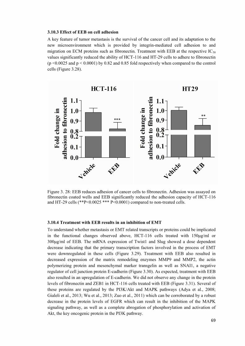
69
3.10.3 Effect of EEB on cell adhesion A key feature of tumor metastasis is the survival of the cancer cell and its adaptation to the new microenvironment which is provided by integrin-mediated cell adhesion to and migration on ECM proteins such as fibronectin. Treatment with EEB at the respective IC50 values significantly reduced the ability of HCT-116 and HT-29 cells to adhere to fibronectin (p <0.0025 and p < 0.0001) by 0.82 and 0.85 fold respectively when compared to the control cells (Figure 3.28).
Figure 3. 28: EEB reduces adhesion of cancer cells to fibronectin. Adhesion was assayed on fibronectin coated wells and EEB significantly reduced the adhesion capacity of HCT-116 and HT-29 cells (**P<0.0025 *** P<0.0001) compared to non-treated cells.
3.10.4 Treatment with EEB results in an inhibition of EMT To understand whether metastasis or EMT related transcripts or proteins could be implicated in the functional changes observed above, HCT-116 cells treated with 150μg/ml or 300μg/ml of EEB. The mRNA expression of Twist1 and Slug showed a dose dependent decrease indicating that the primary transcription factors involved in the process of EMT were downregulated in these cells (Figure 3.29). Treatment with EEB also resulted in decreased expression of the matrix remodeling enzymes MMP9 and MMP2, the actin polymerizing protein and mesenchymal marker transgelin as well as SNAI1, a negative regulator of cell junction protein E-cadherin (Figure 3.30). As expected, treatment with EEB also resulted in an upregulation of E-cadherin. We did not observe any change in the protein levels of fibronectin and ZEB1 in HCT-116 cells treated with EEB (Figure 3.31). Several of these proteins are regulated by the PI3K/Akt and MAPK pathways (Adya et al., 2008; Gialeli et al., 2013; Wu et al., 2013; Zuo et al., 2011) which can be corroborated by a robust decrease in the protein levels of EGFR which can result in the inhibition of the MAPK signaling pathway, as well as a complete abrogation of phosphorylation and activation of Akt, the key oncogenic protein in the PI3K pathway.
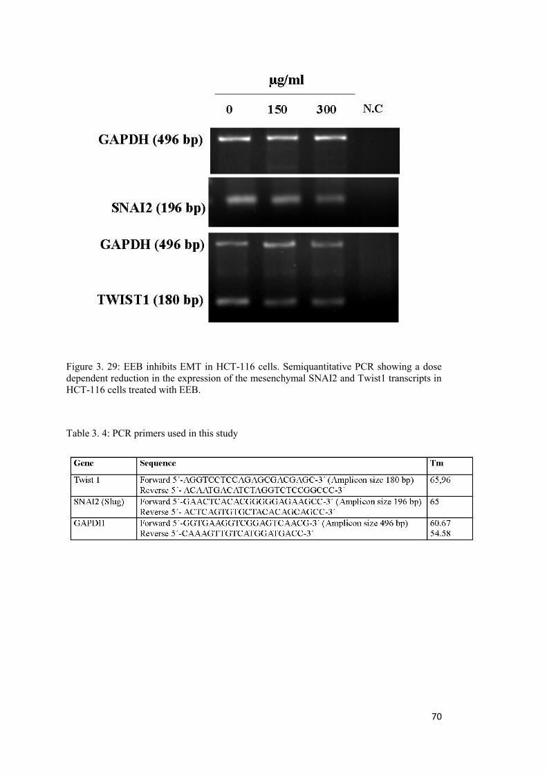
70
Figure 3. 29: EEB inhibits EMT in HCT-116 cells. Semiquantitative PCR showing a dose dependent reduction in the expression of the mesenchymal SNAI2 and Twist1 transcripts in HCT-116 cells treated with EEB.
Table 3. 4: PCR primers used in this study

71
Figure 3. 30: EEB inhibits EMT in HCT-116 cells. Immunoblot showing a reduction in the mesenchymal markers transgelin, SNAI1 and MMP9 along with an increase in the epithelial marker E-cadherin after treatment with EEB. A decrease in the phosphorylation of Akt and total levels of EGFR implicate the involvement of the PI3K/Akt and MAPK pathways in the reversal of EMT.
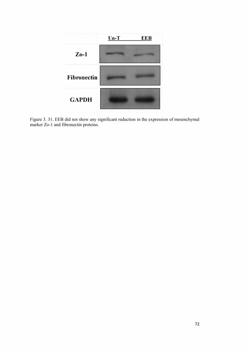
72
Figure 3. 31. EEB did not show any significant reduction in the expression of mesenchymal marker Zo-1 and fibronectin proteins.
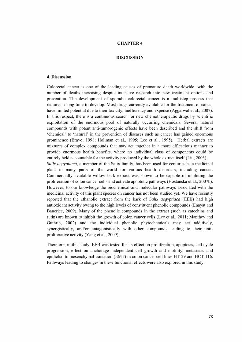
73
CHAPTER 4
DISCUSSION
4. Discussion
Colorectal cancer is one of the leading causes of premature death worldwide, with the number of deaths increasing despite intensive research into new treatment options and prevention. The development of sporadic colorectal cancer is a multistep process that requires a long time to develop. Most drugs currently available for the treatment of cancer have limited potential due to their toxicity, inefficiency and expense (Aggarwal et al., 2007). In this respect, there is a continuous search for new chemotherapeutic drugs by scientific exploitation of the enormous pool of naturally occurring chemicals. Several natural compounds with potent anti-tumorogenic effects have been described and the shift from ‘chemical’ to ‘natural’ in the prevention of diseases such as cancer has gained enormous prominence (Bravo, 1998; Hollman et al., 1995; Lee et al., 1995). Herbal extracts are mixtures of complex compounds that may act together in a more efficacious manner to provide enormous health benefits, where no individual class of components could be entirely held accountable for the activity produced by the whole extract itself (Liu, 2003). Salix aegyptiaca, a member of the Salix family, has been used for centuries as a medicinal plant in many parts of the world for various health disorders, including cancer. Commercially available willow bark extract was shown to be capable of inhibiting the proliferation of colon cancer cells and activate apoptotic pathways (Hostanska et al., 2007b). However, to our knowledge the biochemical and molecular pathways associated with the medicinal activity of this plant species on cancer has not been studied yet. We have recently reported that the ethanolic extract from the bark of Salix aegyptiaca (EEB) had high antioxidant activity owing to the high levels of constituent phenolic compounds (Enayat and Banerjee, 2009). Many of the phenolic compounds in the extract (such as catechins and rutin) are known to inhibit the growth of colon cancer cells (Lee et al., 2011; Manthey and Guthrie, 2002) and the individual phenolic phytochemicals may act additively, synergistically, and/or antagonistically with other compounds leading to their anti-proliferative activity (Yang et al., 2009).
Therefore, in this study, EEB was tested for its effect on proliferation, apoptosis, cell cycle progression, effect on anchorage independent cell growth and motility, metastasis and epithelial to mesenchymal transition (EMT) in colon cancer cell lines HT-29 and HCT-116. Pathways leading to changes in these functional effects were also explored in this study.
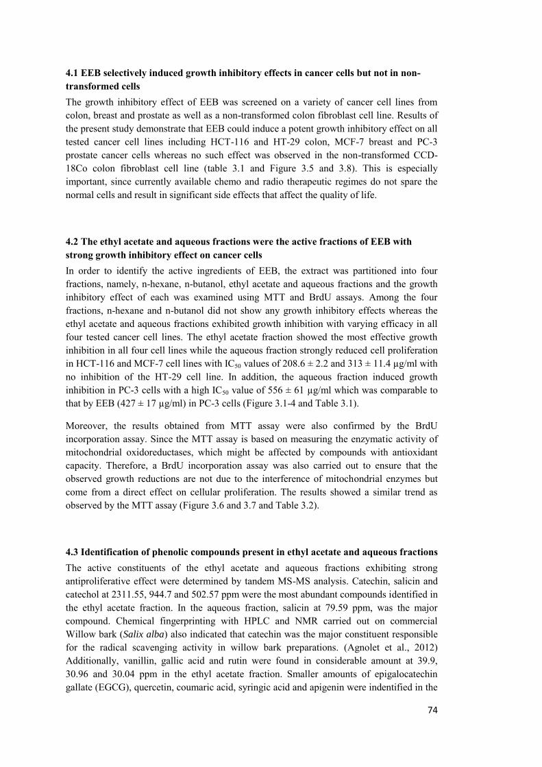
74
4.1 EEB selectively induced growth inhibitory effects in cancer cells but not in non-transformed cells The growth inhibitory effect of EEB was screened on a variety of cancer cell lines from colon, breast and prostate as well as a non-transformed colon fibroblast cell line. Results of the present study demonstrate that EEB could induce a potent growth inhibitory effect on all tested cancer cell lines including HCT-116 and HT-29 colon, MCF-7 breast and PC-3 prostate cancer cells whereas no such effect was observed in the non-transformed CCD-18Co colon fibroblast cell line (table 3.1 and Figure 3.5 and 3.8). This is especially important, since currently available chemo and radio therapeutic regimes do not spare the normal cells and result in significant side effects that affect the quality of life.
4.2 The ethyl acetate and aqueous fractions were the active fractions of EEB with strong growth inhibitory effect on cancer cells In order to identify the active ingredients of EEB, the extract was partitioned into four fractions, namely, n-hexane, n-butanol, ethyl acetate and aqueous fractions and the growth inhibitory effect of each was examined using MTT and BrdU assays. Among the four fractions, n-hexane and n-butanol did not show any growth inhibitory effects whereas the ethyl acetate and aqueous fractions exhibited growth inhibition with varying efficacy in all four tested cancer cell lines. The ethyl acetate fraction showed the most effective growth inhibition in all four cell lines while the aqueous fraction strongly reduced cell proliferation in HCT-116 and MCF-7 cell lines with IC50 values of 208.6 ± 2.2 and 313 ± 11.4 µg/ml with no inhibition of the HT-29 cell line. In addition, the aqueous fraction induced growth inhibition in PC-3 cells with a high IC50 value of 556 ± 61 µg/ml which was comparable to that by EEB (427 ± 17 µg/ml) in PC-3 cells (Figure 3.1-4 and Table 3.1).
Moreover, the results obtained from MTT assay were also confirmed by the BrdU incorporation assay. Since the MTT assay is based on measuring the enzymatic activity of mitochondrial oxidoreductases, which might be affected by compounds with antioxidant capacity. Therefore, a BrdU incorporation assay was also carried out to ensure that the observed growth reductions are not due to the interference of mitochondrial enzymes but come from a direct effect on cellular proliferation. The results showed a similar trend as observed by the MTT assay (Figure 3.6 and 3.7 and Table 3.2).
4.3 Identification of phenolic compounds present in ethyl acetate and aqueous fractions The active constituents of the ethyl acetate and aqueous fractions exhibiting strong antiproliferative effect were determined by tandem MS-MS analysis. Catechin, salicin and catechol at 2311.55, 944.7 and 502.57 ppm were the most abundant compounds identified in the ethyl acetate fraction. In the aqueous fraction, salicin at 79.59 ppm, was the major compound. Chemical fingerprinting with HPLC and NMR carried out on commercial Willow bark (Salix alba) also indicated that catechin was the major constituent responsible for the radical scavenging activity in willow bark preparations. (Agnolet et al., 2012) Additionally, vanillin, gallic acid and rutin were found in considerable amount at 39.9, 30.96 and 30.04 ppm in the ethyl acetate fraction. Smaller amounts of epigalocatechin gallate (EGCG), quercetin, coumaric acid, syringic acid and apigenin were indentified in the
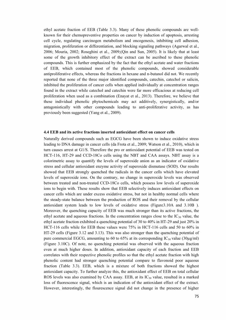
75
ethyl acetate fraction of EEB (Table 3.3). Many of these phenolic compounds are well-known for their chemopreventive properties on cancer by induction of apoptosis, arresting cell cycle, regulating carcinogen metabolism and oncogenesis, inhibiting cell adhesion, migration, proliferation or differentiation, and blocking signaling pathways (Agarwal et al., 2006; Mouria, 2002; Roseghini et al., 2009;(Qin and Sun, 2005). It is likely that at least some of the growth inhibitory effect of the extract can be ascribed to these phenolic compounds. This is further emphasized by the fact that the ethyl acetate and water fractions of EEB, which contained most of the phenolic compounds, showed considerable antiproliferative effects, whereas the fractions in hexane and n-butanol did not. We recently reported that none of the three major identified compounds, catechin, catechol or salicin, inhibited the proliferation of cancer cells when applied individually at concentration ranges found in the extract while catechol and catechin were far more efficacious at reducing cell proliferation when used as a combination (Enayat et al., 2013). Therefore, we believe that these individual phenolic phytochemicals may act additively, synergistically, and/or antagonistically with other compounds leading to anti-proliferative activity, as has previously been suggested (Yang et al., 2009).
4.4 EEB and its active fractions inserted antioxidant effect on cancer cells Naturally derived compounds such as EGCG have been shown to induce oxidative stress leading to DNA damage in cancer cells (da Frota et al., 2009; Watson et al., 2010), which in turn causes arrest at G1/S. Therefore the pro or antioxidant potential of EEB was tested on HCT-116, HT-29 and CCD-18Co cells using the NBT and CAA assays. NBT assay is a colorimetric assay to quantify the levels of superoxide anion as an indicator of oxidative stress and cellular antioxidant enzyme activity of superoxide dismutase (SOD). Our results showed that EEB strongly quenched the radicals in the cancer cells which have elevated levels of superoxide ions. On the contrary, no change in superoxide levels was observed between treated and non-treated CCD-18Co cells, which possess low levels of superoxide ions to begin with. These results show that EEB selectively induces antioxidant effects on cancer cells which are under excess oxidative stress, but not in healthy normal cells where the steady-state balance between the production of ROS and their removal by the cellular antioxidant system leads to low levels of oxidative stress (Figure3.10A and 3.10B ). Moreover, the quenching capacity of EEB was much stronger than its active fractions, the ethyl acetate and aqueous fractions. In the concentration ranges close to the IC50 value, the ethyl acetate fraction exhibited a quenching potential of 30 to 40% in HT-29 and just 20% in HCT-116 cells while for EEB these values were 75% in HCT-116 cells and 50 to 60% in HT-29 cells (Figure 3.12 and 3.13). This was also stronger than the quenching potential of pure commercial EGCG, amounting to 60 to 65% at its corresponding IC50 value (30µg/ml) (Figure 3.10C). Of note, no quenching potential was observed with the aqueous fraction even at much higher doses. In addition, antioxidant capacity of each fraction and EEB correlates with their respective phenolic profiles so that the ethyl acetate fraction with high phenolic content had stronger quenching potential compare to flavonoid poor aqueous fraction (Table 3.3). EEB, which is a mixture of both fractions showed the highest antioxidant capacity. To further analyze this, the antioxidant effect of EEB on total cellular ROS levels was also examined by CAA assay. EEB, at its IC50 value, resulted in a marked loss of fluorescence signal, which is an indication of the antioxidant effect of the extract. However, interestingly, the flourescence signal did not change in the presence of higher
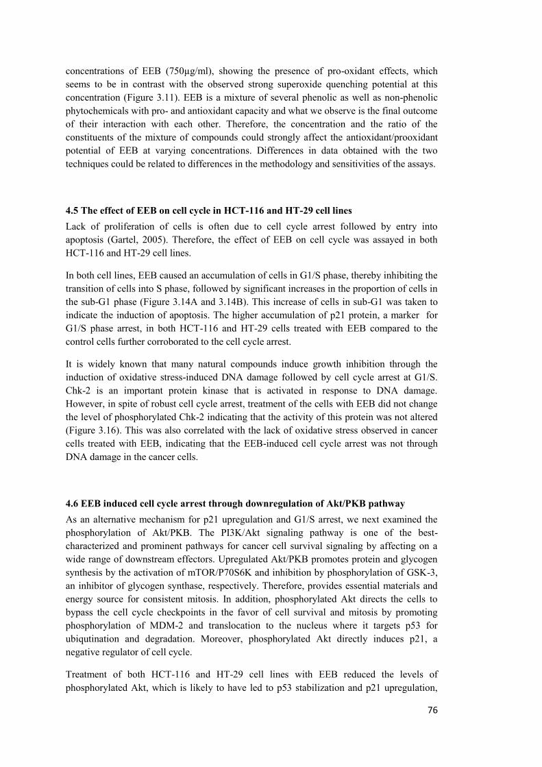
76
concentrations of EEB (750µg/ml), showing the presence of pro-oxidant effects, which seems to be in contrast with the observed strong superoxide quenching potential at this concentration (Figure 3.11). EEB is a mixture of several phenolic as well as non-phenolic phytochemicals with pro- and antioxidant capacity and what we observe is the final outcome of their interaction with each other. Therefore, the concentration and the ratio of the constituents of the mixture of compounds could strongly affect the antioxidant/prooxidant potential of EEB at varying concentrations. Differences in data obtained with the two techniques could be related to differences in the methodology and sensitivities of the assays.
4.5 The effect of EEB on cell cycle in HCT-116 and HT-29 cell lines Lack of proliferation of cells is often due to cell cycle arrest followed by entry into apoptosis (Gartel, 2005). Therefore, the effect of EEB on cell cycle was assayed in both HCT-116 and HT-29 cell lines.
In both cell lines, EEB caused an accumulation of cells in G1/S phase, thereby inhibiting the transition of cells into S phase, followed by significant increases in the proportion of cells in the sub-G1 phase (Figure 3.14A and 3.14B). This increase of cells in sub-G1 was taken to indicate the induction of apoptosis. The higher accumulation of p21 protein, a marker for G1/S phase arrest, in both HCT-116 and HT-29 cells treated with EEB compared to the control cells further corroborated to the cell cycle arrest.
It is widely known that many natural compounds induce growth inhibition through the induction of oxidative stress-induced DNA damage followed by cell cycle arrest at G1/S. Chk-2 is an important protein kinase that is activated in response to DNA damage. However, in spite of robust cell cycle arrest, treatment of the cells with EEB did not change the level of phosphorylated Chk-2 indicating that the activity of this protein was not altered (Figure 3.16). This was also correlated with the lack of oxidative stress observed in cancer cells treated with EEB, indicating that the EEB-induced cell cycle arrest was not through DNA damage in the cancer cells.
4.6 EEB induced cell cycle arrest through downregulation of Akt/PKB pathway As an alternative mechanism for p21 upregulation and G1/S arrest, we next examined the phosphorylation of Akt/PKB. The PI3K/Akt signaling pathway is one of the best-characterized and prominent pathways for cancer cell survival signaling by affecting on a wide range of downstream effectors. Upregulated Akt/PKB promotes protein and glycogen synthesis by the activation of mTOR/P70S6K and inhibition by phosphorylation of GSK-3, an inhibitor of glycogen synthase, respectively. Therefore, provides essential materials and energy source for consistent mitosis. In addition, phosphorylated Akt directs the cells to bypass the cell cycle checkpoints in the favor of cell survival and mitosis by promoting phosphorylation of MDM-2 and translocation to the nucleus where it targets p53 for ubiqutination and degradation. Moreover, phosphorylated Akt directly induces p21, a negative regulator of cell cycle.
Treatment of both HCT-116 and HT-29 cell lines with EEB reduced the levels of phosphorylated Akt, which is likely to have led to p53 stabilization and p21 upregulation,
77
thereby causing cell cycle arrest (Figure 3.17). Activation of the PI3K/Akt pathway by various mitogens or growth factors inhibit apoptosis by phosphorylating MDM2 at Ser-166 and -186, inducing ubiquitination and degradation of p53 (Mayo and Donner, 2001; Zhou et al., 2001). In addition, in humans, the 60 kD fragment of MDM2 has been found to be upregulated in several cancer types and is generated through a nonapoptotic proteolytic cleavage of the larger 90kD fragment (Pochampally et al., 1998). This p90 isoform of MDM2 is involved in the p53/MDM2 feedback control loop when p53 is activated. Therefore, the upregulation of p90, observed upon treatment with EEB, is likely to have resulted from the stabilization and activation of p53 (Cheng and Cohen, 2007). Moreover, the reduction observed in the oncogenic p60 fragment upon treatment with EEB further confirms its anticarcinogenic nature (Figure 3.17).
4.7 The dual targeting of Akt/PKB and MAPKs pathways by EEB Several recent studies have emphasized significant cross-talk between the Akt/PKB and MAPK pathways in tumors and there has been an enormous interest in the identification of molecules that dually target both pathways (Gruson et al., 2010; Liu et al., 2009; Onan et al., 2004). Ras is one of the most frequently mutated oncogenes found in several cancers, which leads to the deregulation of several effector pathways controlling cell proliferation, survival, and migration thus promoting malignant transformation (Downward, 2003). The best-characterized Ras effector pathway is the RAF-mitogen-activated protein kinase (MAPK)/extracellular signal-regulated kinase (ERK) kinase (MEK) MAPK cascade. ERK1/2 activation leads to phosphorylation and activation of various cytosolic and nuclear proteins, including transcription factors that control the G1/S cell cycle transition (Hoshino et al., 1999). Our group has recently shown that treatment of cancer cells with EEB resulted in decreased phosphorylation and activity of ERK1/2, JNK and p38(Enayat et al., 2013). The JNK cascade is one of the main stress-activated protein kinases that is involved in the pathology of several cancer types by modulating cell proliferation and apoptosis. Different growth factors (Hibi et al., 1993), cytokines (Uehara et al., 2005), and stress factors (Cano et al., 1994), can activate JNKs. Another class of stress activated kinases is the p38 mitogen-activated protein kinases (p38s), which can be activated by osmotic shock, inflammatory cytokines, growth factors or reactive oxidative species (deLaat et al., 1997; Dolado et al., 2007). The decreased phosphorylation of JNK and p38 observed upon treatment with EEB further corroborates with the decreased superoxide dismutase activity and thereby low ROS levels in the cells (Enayat et al., 2013).
4.8 EEB induced apoptosis in both colon cancer cell lines via a p53 dependent pathway The induction of apoptosis, indicated by arrested cell cycle progression, was assayed by Annexin V-FITC staining and protein expression analysis of independent markers of the apoptotic cascade: Bcl-xl, caspase 9, procaspase 3, MDM-2, p53 and PUMA.
The principle of Annexin V staining is its binding to phosphatidylserine (PS), a membrane phospholipid, that is externalized in cells undergoing apoptosis. Flow cytometry analysis of the FITC conjugated Annexin V showed a significant increase in the early and late apoptotic
78
fractions of cells as well as necrotic cells in the cells treated with EEB compared to their corresponding non-treated HCT-116 and HT-29 controls. When the results from treatments with EEB for 24h and 48h were compared, the apoptotic to necrotic cells ratio were seen to increase in 48h compared to 24h treatment. This finding indicates that the effect of the extract 24 h after treatment is more non selective by inducing more necrotic death while after 48 h the effect is more selective through the activation of apoptotic pathways resulting in an increased ratio of apoptotic fraction to necrotic fraction (Figure .3.18).
Western blotting analyses of pro and anti-apoptotic proteins indicate that exposure to EEB led to increased protein levels of caspase 9 as well as reduced expression of Bcl-xl and procaspase 3 proteins in both cell lines (Figure 3.20). The reduction in the protein levels of procaspase3 correlated with the elevated caspase 3 activity upon treatment with EEB as previously shown (Enayat et al., 2013). In addition, treatment with EEB resulted in increased levels of the wild type p53 protein in HCT-116 cells while interestingly the mutant form of p53 protein was reduced in HT-29 cells. These events were accompanied by a strong induction of p53 upregulated modulator of apoptosis (PUMA) (Figure 3.20). PUMA is a specific downstream target of p53, which upon activation binds to the antiapoptotic Bcl-2 protein family, liberates proapoptotic BAK and BAX proteins and stimulates mitochondrial apoptosis cascade. Reduced procaspase 3 and activation of caspase-3 and -9 are early biomarkers of apoptosis, which act downstream to the release of cytochrome c from the mitochondria. Activated caspase-3 is responsible for DNA fragmentation and other morphological changes associated with apoptosis (Gupta et al., 2009).
4.9 Salicin did not effectively contribute in the cell cycle arrest or proapoptotic function of EEB Salicin is an active ingredient of Willow bark extracts and constitutes a significant proportion of compounds identified from EEB (Table 3.2) as well as other ethanolic extracts from other species. Annexin V staining of the HCT-116 and HT-29 cells after exposure to the corresponding concentrations of salicin induced 50% growth inhibition did not show significant induction of apoptosis in any of the cell types (Figure 19). Based on the salicin content of the ethanolic extract of bark as determined by HPLC (3 mg/ml) (Enayat & Banerjee, 2009), the amount of salicin available in 250 and 300 µg/ml of the extract induced 50% growth inhibition in HT-29 and HCT-116 Cells are 75 and 90 µg/ml respectively. Additionally, no detectable change was observed in the cell cycle distribution in salicin treated cells compared to the non-treated controls in both cell lines (Figure 3.15). These data indicate that probably salicin does not contribute to the G1/S arrest and apoptosis observed in the EEB treated HCT-116 and HT-29 cells.
Therefore, it might be concluded that salicin is not a significant factor determining the strong proapoptotic effects of EEB observed in the two colon cancer cell lines. Even the concentrations of salicin (370µg/ml for HCT-116 and 304µg/ml for HT-29) which accounts for 50% growth inhibitory effects could not behave as effective as the bark extract (Figure 12, 3.15 and 3.9). These data are in contrast with the conventional belief that salicin is the main component for the antiproliferative and anti-tumorigenic effects involved with bark treatment.
79
It was recently proposed by Vlachojannis et al. that the anti-nociceptive and anti- inflammatory functions of Willow bark are not because of the constituent salicin, rather, it results from the unique combination of polyphenols and flavonoids in the extract (Vlachojannis et al., 2011). All of these point to the fact that salicin is most likely not the main contributor in the anti-cancer properties of EEB. On the other hand, EEB, as well as its fractions in ethyl acetate and water are rich in several phenolic compounds (Table 3.3), which, in combination, could contribute very significantly towards the anti-carcinogenic properties of the extract. Therefore, we propose that the strong antioxidant capacity and remarkably high level of total phenolic and flavonoid contents as well as the presence of several potent polyphenols in the extracts that have known anti-carcinogenic properties could contribute to the antiproliferative effects of the extracts. Several reports about anticarcinogenic potency of phenolic and flavonoid compounds support this hypothesis (Hostanska et al., 2007a Abel & Saller, 2007; Hostanska et al., 2007b Nahrstedt & Saller, 2007); (Fresco et al., 2010; Qin and Sun, 2005).
4.10 EEB induced strong antimetastatic effects on colon cancer cell lines Cellular motility and altered adhesion to the extra cellular matrix are frequently acquired by cells that undergo neoplastic changes and are critical steps in metastasis of the primary tumor cell to a distant site (Rendon et al., 2007).
In this study, using an in vitro assay system, we have examined whether EEB could have any effect on the metastatic potential of HCT-116 and HT-29 cells and whether such changes were accompanied by an inhibition of EMT.
4.11 EEB strongly reduced the anchorage independent growth of cancer cells Attachment to the extracellular matrix and the stimulation of growth factor signaling cascades generally promotes the survival of epithelial cells by suppressing the activity of apoptotic factors leading to anoikis (Taddei et al., 2012) One of the primary signs of cellular transformation is the ability of cancer cells to grow in an anchorage independent manner whereby, altered adhesion to the extra cellular matrix is frequently acquired by cells that undergo neoplastic changes (Reddig and Juliano, 2005; Rendon et al., 2007) The activation of the PI3K/Akt pathway and its downstream proteins play a central role in resistance to anoikis and the anchorage independent growth of cancer cells, a hallmark feature of tumor cells (Reddig and Juliano, 2005). Treatment with EEB resulted in a drastic decrease in the anchorage independent growth of HCT-116 and HT-29 cells at concentrations that did not decrease cellular proliferation which could have resulted from the inhibition of the PI3K/Akt pathway by EEB (Figure 3.21, 3.22 and 3.23).
4.12 EEB effectively reduced the motility and migration of cancer cells Cells motility and migration ability are major factors that influence tumor metastasis. Therefore, the role of EEB in the motility and migration of cancer cells was determined by cellular motility through Transwells as well as with a scratch wound healing assay. Treatment of HCT-116 and HT-29 cells with EEB at concentrations that did not reduce
80
cellular proliferation could dramatically reduce cellular migration through Transwells as well as motility after a scratch wound indicating that the extract could significantly reduce the motility of the cancer cells (Figure 3.24, 3.25, 3.26 and 3.27). PI3K/Akt signaling pathway is considered as the most important pathway involved in modulation of tumor metastasis. Recent studies indicate that inhibition of PI3K activity by selective inhibitors such as LY294002 led to reduced motility and migration (Hsu et al., 2012). Therefore, the potent inhibitory effect of EEB on cell motility and migration could have resulted from the strong inhibitory effect of EEB on PI3K/AKT pathway (Figure 3.17).
4.13 EEB strongly reduced the integrin-mediated adhesion to ECM proteins The ability of a cancer cell to metastasize stems from a wide spectrum of changes that include anchorage independent growth, resistance to anoikis (Ng et al., 2012), motility, integrin-mediated adhesion to ECM proteins such as fibronectin and migration of the transformed cells (Cimen et al., 2009). Interaction with and adhesion to ECM is one of the key mechanisms necessary for cell survival which is often mediated by the PI3K pathway (Reddig and Juliano, 2005). The transmembrane receptors of integrin family members play a critical role in sensing and responding to ECM properties as well as promoting growth factor signaling cascades. Moreover, integrins allow cells to adhere to a variety of ECM structures such as fibronectin-containing matrices which is associated with wound healing and enriched during angiogenesis and cancer progression (Xiong et al., 2013). Therefore, imbalance in integrin-mediated cell adhesion may leads to the pathology of several diseases especially cancer. Treatment of HCT-116 and HT-29 cells with the extract significantly reduced the ability of the cancer cells to adhere to fibronectin, a component of the ECM with critical role on metastasis, which again could have resulted from the inhibition of the PI3K pathway (Figure 3.28).
4.14 EEB inhibits the epithelial to mesenchymal transition of colon cancer cell lines. The process of the de-differentiation of epithelial cells toward a more mesenchymal phenotype is a critical step for acquisition of metastatic potential in cancer cells which is mediated by a variety of inter and intracellular factors, such as intra-tumoural hypoxia, growth factor signaling and cell-cell interaction (Friedl and Wolf, 2003; Jiang et al., 2009). This transition directs the tumor to more aggressive states which is accompanied by the loss of epithelial characters, gain of mesenchymal marker expression, acquisition of migration, invasive activity and capability to pass through the basement membrane and migration throughout the body. E-cadherin is one of the most important adhesive molecules in epithelial cells whose suppression is a main characteristic of cells undergoing EMT. SNAI1 and SNAI2 (Slug) members of snail family, ZEB (ZEB1 and ZEB2), basic helix–loop–helix (bHLH) (E47 and Twist) families are considered as the major suppressors of E-cadherin whose activation result in transcriptional suppression of E-cadherin and its proteolytic cleavage by MMP-2 and MMP-9 (Usami et al., 2008).
Therefore, in this study, the effect of EEB on the expression of various epithelial and mesenchymal markers was explored by western blotting and semiquantitative PCR analysis. We observed a decrease in the transcripts of the mesenchymal markers Slug and Twist as a
81
result of EEB treatment (Figure 3.29). Moreover, the protein levels of SNAI1 and Transgelin (TAGLN) were also reduced in cells treated with EEB accompanied by a restoration of E-cadherin (Figure 3.30). Transgelin is a 22-kDa protein, which, by virtue of its actin binding property, is involved in podocyte formation in smooth muscle cells and their motility. (Gimona et al., 2003) However, it has subsequently been shown that in colon cancer cells, transgelin is localized to the nucleus and can play a role in the transcriptional upregulation of fibronectin and vimentin, thereby making it a mesenchymal marker. (Lin et al., 2009) It has also been reported that the expression of transgelin is under the control of TGFβ, (Adam et al., 2000) another key protein that also orchestrates the process of EMT (Xu et al., 2009). Therefore, the reduction in transgelin protein expression could also contribute towards the reversal of EMT observed in cells treated with EEB. From these observations we could conclude that EEB may slowdown or reverse the EMT process with the cells acquiring a more epithelial phenotype.
The activated PI3K/Akt and MAPK pathways can directly mediate a reduction in E-cadherin through the induction of SNAI1/2, thereby initiating EMT (Scheme 2) (Jang et al., 2009); (Peinado et al., 2004; Schmalhofer et al., 2009). Recent studies also indicate that pharmacologic inhibition of EGFR, MEK and PI3K in colorectal carcinoma led to a reduction in the EGF induced production of MMP-9, cell migration and invasion and expression changes of EMT markers (Adya et al., 2008; Gialeli et al., 2013; Wu et al., 2013; Zuo et al., 2011). In addition, focal adhesion kinase (FAK) mediated activation of PI3K/Akt and MAPK/Erk pathways may also result in the transduction of external stimuli from the extracellular matrix to the nucleus resulting in the activation of the promoters of MMP-9 and MMP-2, facilitating the invasion to the extracellular matrix and metastasis of cancer cells (Adya et al., 2008; Hwangbo et al., 2010; Lai et al., 2010; Liao et al., 2003; Meng et al., 2009). Recent studies have also implicated JNK and p38 in regulation of MMP-9 and MMP-2 (Hsu et al., 2012; Kumar et al., 2010). We have shown that EEB could strongly reduce the phosphorylation of Akt, Erk1/2, JNK and p38 (Enayat et al., 2013) as well as a robust reduction in the levels of EGFR as a result of treatment with EEB (Figure 3.30). Moreover, we have also observed a reduction in the expression of MMP-9 and to a less extent MMP-2 (Figure 3.30). MMP-9 and MMP-2 proteins are abundantly expressed in several malignancies with a critical role in tumor invasion and angiogenesis, and is considered as a biomarker for the metastatic potential of cancer cells (Pories et al., 2008). Thus, the inhibition of MMP-9 by EEB could have resulted in a reduction in the motility and migratory capacity of the cancer cells.

82
83
CHAPTER 5
CONCLUSION
5.CONCLUSION
The ethanolic extract of bark from Salix aegyptiaca is a potent mixture of bioactive phytochemicals that can reduce the proliferation of colon cancer cells through the induction of cell cycle arrest and apoptosis. Our data provide, for the first time, detailed molecular evidence on the potent antitumorogenic effects of an ethanolic extract from bark (EEB) of Salix aegyptiaca, a plant species that has been in use from ancient times mostly as an anti-inflammatory agent. EEB could inhibit the proliferation of cancer cells and halt cell cycle progression at G1/S. This extract can also inhibit one of the most important signaling pathway associated with survival signals in cancer cells: the PI3K/Akt at levels comparable to known commercial inhibitors. The results of current study from the comparative cell cycle distribution analysis as well as apoptosis assays of pure salicin indicate that salicin is not the main contributor in the health beneficial effects observed in the willow bark extracts. Here we have also shown, for the first time, evidence of the anti-metastatic effects of EEB, most likely resulting from the inhibition of the PI3K/Akt and the MAPKinase signaling pathways. The extract could also induce a reversal of epithelial to mesenchymal transition in the cancer cells by upregulating epithelial markers and down regulating mesenchymal markers.
The conclusive schematic figures 1 and 2 represent the determined overall effects of EEB on the biochemical pathways and functional characteristic of cancer cells.
Salix species have been known for centuries to be beneficial against a number of ailments, including cancer. Understanding of the molecular mechanisms behind such beneficial effects will further justify the use of these extracts for the chemoprevention of cancer. In addition, since EEB is a phytochemical rich extract therefore identification of a broader range of compounds using fingerprinting methods as well as isolating the major compounds will provide a better understanding of the active constituents of the extract.
Moreover, a comprehensive in vivo study on the cytotoxicy, chemopreventive and anticarcinogenic effect of EEB on animal models would be essentail to confirm the merit anticacinogenic potential of this extract observed in our in vitro study and will be a step forward for future application of this extract in the clinical trial phases.
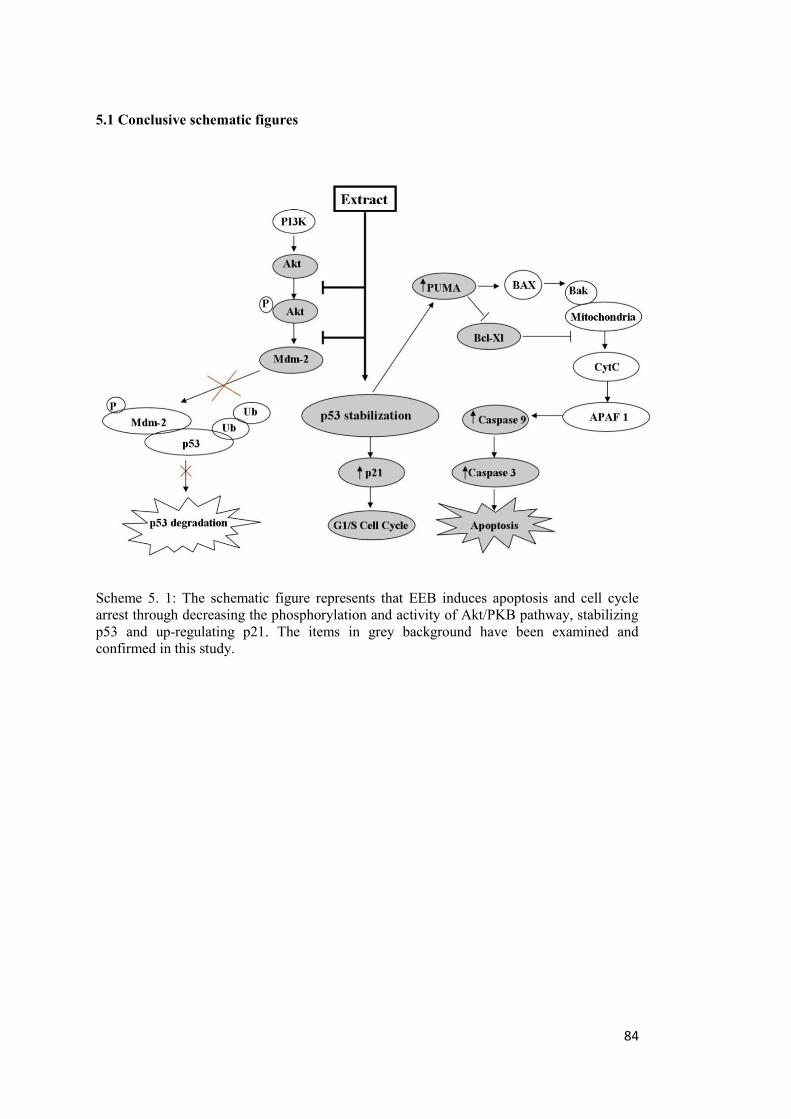
84
5.1 Conclusive schematic figures
Scheme 5. 1: The schematic figure represents that EEB induces apoptosis and cell cycle arrest through decreasing the phosphorylation and activity of Akt/PKB pathway, stabilizing p53 and up-regulating p21. The items in grey background have been examined and confirmed in this study.
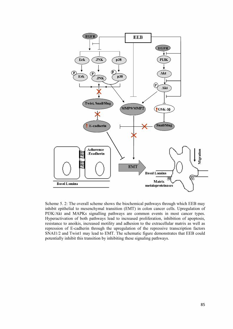
85
Scheme 5. 2: The overall scheme shows the biochemical pathways through which EEB may inhibit epithelial to mesenchymal transition (EMT) in colon cancer cells. Upregulation of PI3K/Akt and MAPKs signalling pathways are common events in most cancer types. Hyperactivation of both pathways lead to increased proliferation, inhibition of apoptosis, resistance to anoikis, increased motility and adhesion to the extracellular matrix as well as repression of E-cadherin through the upregulation of the repressive transcription factors SNAI1/2 and Twist1 may lead to EMT. The schematic figure demonstrates that EEB could potentially inhibit this transition by inhibiting these signaling pathways.

86
87
REFERENCES
Adachi, S., Shimizu, M., Shirakami, Y., Yamauchi, J., Natsume, H., Matsushima-Nishiwaki, R., To, S., Weinstein, I.B., Moriwaki, H., Kozawa, O., 2009. (-)-Epigallocatechin gallate downregulates EGF receptor via phosphorylation at Ser1046/1047 by p38 MAPK in colon cancer cells. Carcinogenesis 30, 1544-1552. Adam, P.J., Regan, C.P., Hautmann, M.B., Owens, G.K., 2000. Positive- and negative-acting Kruppel-like transcription factors bind a transforming growth factor beta control element required for expression of the smooth muscle cell differentiation marker SM22alpha in vivo. J Biol Chem 275, 37798-37806. Adya, R., Tan, B.K., Punn, A., Chen, J., Randeva, H.S., 2008. Visfatin induces human endothelial VEGF and MMP-2/9 production via MAPK and PI3K/Akt signalling pathways: novel insights into visfatin-induced angiogenesis. Cardiovascular Research 78, 356-365. Agarwal, M.K., Hastak, K., Jackson, M.W., Breit, S.N., Stark, G.R., Agarwal, M.L., 2006. Macrophage inhibitory cytokine 1 mediates a p53-dependent protective arrest in S phase in response to starvation for DNA precursors. Proceedings of the National Academy of Sciences of the United States of America 103, 16278-16283. Aggarwal, B.B., Banerjee, S., Bharadwaj, U., Sung, B., Shishodia, S., Sethi, G., 2007. Curcumin induces the degradation of cyclin E expression through ubiquitin-dependent pathway and up-regulates cyclin-dependent kinase inhibitors p21 and p27 in multiple human tumor cell lines. Biochemical Pharmacology 73, 1024-1032. Aggarwal, B.B., Shishodia, S., 2006. Molecular targets of dietary agents for prevention and therapy of cancer. Biochemical Pharmacology 71, 1397-1421. Agnolet, S., Wiese, S., Verpoorte, R., Staerk, D., 2012. Comprehensive analysis of commercial willow bark extracts by new technology platform: Combined use of metabolomics, high-performance liquid chromatography-solid-phase extraction-nuclear magnetic resonance spectroscopy and high-resolution radical scavenging assay. Journal of Chromatography A 1262, 130-137. Amin, A., Paul, R.K., Thakur, V.S., Agarwal, M.L., 2007. A novel role for p73 in the regulation of Akt-Foxo1a-Bim signaling and apoptosis induced by the plant lectin, concanavalin A. Cancer Research 67, 5617-5621. Athar, M., Back, J.H., Tang, X., Kim, K.H., Kopelovich, L., Bickers, D.R., Kim, A.L., 2007. Resveratrol: A review of preclinical studies for human cancer prevention. Toxicology and Applied Pharmacology 224, 274-283. Au-Yeung, K.K.W., Liu, P.L., Chan, C., Wu, W.Y., Lee, S.S.T., Ko, J.K.S., 2008. Herbal isoprenols induce apoptosis in human colon cancer cells through transcriptional activation of PPAR gamma. Cancer Investigation 26, 708-717. Auyeung, K.K., Woo, P.K., Law, P.C., Ko, J.K., 2012. Astragalus saponins modulate cell invasiveness and angiogenesis in human gastric adenocarcinoma cells. Journal of Ethnopharmacology 141, 635-641.
88
Baker, D.D., Chu, M., Oza, U., Rajgarhia, V., 2007. The value of natural products to future pharmaceutical discovery. Natural Product Reports 24, 1225-1244. Basseres, D.S., Baldwin, A.S., 2006. Nuclear factor-kappa B and inhibitor of kappa B kinase pathways in oncogenic initiation and progression. Oncogene 25, 6817-6830. Benhar, M., Engelberg, D., Levitzki, A., 2002. ROS, stress-activated kinases and stress signaling in cancer. Embo Reports 3, 420-425. Beretta, L., Gingras, A.C., Svitkin, Y.V., Hall, M.N., Sonenberg, N., 1996. Rapamycin blocks the phosphorylation of 4E-BP1 and inhibits cap-dependent initiation of translation. Embo Journal 15, 658-664. Bhowmick, N.A., Neilson, E.G., Moses, H.L., 2004. Stromal fibroblasts in cancer initiation and progression. Nature 432, 332-337. Billen, L.P., Shamas-Din, A., Andrews, D.W., 2008. Bid: a Bax-like BH3 protein. Oncogene 27, S93-S104. Birchmeier, W., Behrens, J., 1994. Cadherin expression in carcinomas - role in the formation of cell-junctions and the prevention of invasiveness. Biochimica Et Biophysica Acta-Reviews on Cancer 1198, 11-26. Borghaei, R.C., Rawlings, P.L., Javadi, M., Woloshin, J., 2004. NF-kappa B binds to a polymorphic repressor element in the MMP-3 promoter. Biochemical and Biophysical Research Communications 316, 182-188. Braicu, C., Gherman, C., 2013. Epigallocatechin gallate induce cell death and apoptosis in triple negative breast cancer cells Hs578T. Journal of Drug Targeting 21, 250-256. Bravo, L., 1998. Polyphenols: Chemistry, dietary sources, metabolism, and nutritional significance. Nutrition Reviews 56, 317-333. Burgering, B.M.T., Coffer, P.J., 1995. Protein-kinase-b (c-Akt) in phosphatidylinositol-3-oh inase signal-transduction. Nature 376, 599-602. Cano, E., Hazzalin, C.A., Mahadevan, L.C., 1994. Anisomycin-activated protein kinase-p45 and kinase-p55 but not mitogen-activated protein kinase-erk-1 and kinase-erk-2 are implicated in the induction of c-FOS and c-JUN. Molecular and Cellular Biology 14, 7352-7362. Chappell, W.H., Steelman, L.S., Long, J.M., Kempf, R.C., Abrams, S.L., Franklin, R.A., Basecke, J., Stivala, F., Donia, M., Fagone, P., Malaponte, G., Mazzarino, M.C., Nicoletti, F., Libra, M., Maksimovic-Ivanic, D., Mijatovic, S., Montalto, G., Cervello, M., Laidler, P., Milella, M., Tafuri, A., Bonati, A., Evangelisti, C., Cocco, L., Martelli, A.M., McCubrey, J.A., 2011. Ras/Raf/MEK/ERK and PI3K/PTEN/Akt/mTOR Inhibitors: Rationale and Importance to Inhibiting These Pathways in Human Health. Oncotarget 2, 135-164. Chau, I., Cunningham, D., 2006. Adjuvant therapy in colon cancer - what, when and how? Annals of Oncology 17, 1347-1359. Chen, W.S., Lee, Y.J., Yu, Y.C., Hsaio, C.H., Yen, J.H., Yu, S.H., Tsai, Y.J., Chiu, S.J., 2010. Enhancement of p53-mutant human colorectal cancer cells radiosensitivity by flavonoid fisetin. International Journal of Radiation Oncology Biology Physics 77, 1527-1535.
89
Cheng, N., Chytil, A., Shyr, Y., Joly, A., Moses, H.L., 2008. Transforming Growth Factor-beta Signaling-Deficient Fibroblasts Enhance Hepatocyte Growth Factor Signaling in Mammary Carcinoma Cells to Promote Scattering and Invasion. Molecular Cancer Research 6, 1521-1533. Cheng, T.H., Cohen, S.N., 2007. Human MDM2 isoforms translated differentially on constitutive versus p53-regulated transcripts have distinct functions in the p53/MDM2 and TSG101/MDM2 feedback control loops. Molecular and Cellular Biology 27, 111-119. Chung, J., Kuo, C.J., Crabtree, G.R., Blenis, J., 1992. Rapamycin fkbp specifically blocks growth-dependent activation of and signaling by the 70 kD S6 protein-kinases. Cell 69, 1227-1236. Cimen, I., Tuncay, S., Banerjee, S., 2009. 15-Lipoxygenase-1 expression suppresses the invasive properties of colorectal carcinoma cell lines HCT-116 and HT-29. Cancer Sci 100, 2283-2291. Colombo, M.L., Bugatti, C., Mossa, A., Pescalli, N., Piazzoni, L., Pezzoni, G., Menta, E., Spinelli, S., Johnson, F., Gupta, R.C., Dasaradhi, L., 2001. Cytotoxicity evaluation of natural coptisine and synthesis of coptisine from berberine. Farmaco 56, 403-409. Conacci-Sorrell, M., Simcha, I., Ben-Yedidia, T., Blechman, J., Savagner, P., Ben-Ze'ev, A., 2003. Autoregulation of E-cadherin expression by cadherin-cadherin interactions: the roles of beta-catenin signaling, Slug, and MAPK. Journal of Cell Biology 163, 847-857. Coqueret, O., 2003. New roles for p21 and p27 cell-cycle inhibitors: a function for each cell compartment? Trends in Cell Biology 13, 65-70. da Frota, M.L., Jr., Braganhol, E., Canedo, A.D., Klamt, F., Apel, M.A., Mothes, B., Lerner, C., Battastini, A.M., Henriques, A.T., Moreira, J.C., 2009. Extracts of marine sponge Polymastia janeirensis induce oxidative cell death through a caspase-9 apoptotic pathway in human U138MG glioma cell line. Invest New Drugs 27, 440-446. deLaat, A., vanderLeun, J.C., deGruijl, F.R., 1997. Carcinogenesis induced by UVA (365-nm) radiation: The dose-time dependence of tumor formation in hairless mice. Carcinogenesis 18, 1013-1020. Derksen, P.W.B., Liu, X.L., Saridin, F., van der Gulden, H., Zevenhoven, J., Evers, B., van Beijnum, J.R., Griffioen, A.W., Vink, J., Krimpenfort, P., Peterse, J.L., Cardiff, R.D., Berns, A., Jonkers, J., 2006. Somatic inactivation of E-cadherin and p53 in mice leads to metastatic lobular mammary carcinoma through induction of anoikis resistance and angiogenesis. Cancer Cell 10, 437-449. Ding, Q.M., Wang, Q.D., Evers, B.M., 2001. Alterations of MAPK activities associated with intestinal cell differentiation. Biochemical and Biophysical Research Communications 284, 282-288. Dolado, I., Swat, A., Ajenjo, N., De Vita, G., Cuadrado, A., Nebreda, A.R., 2007. p38 alpha MAP kinase as a sensor of reactive oxygen species in tumorigenesis. Cancer Cell 11, 191-205. Dorai, T., Aggarwal, B.B., 2004. Role of chemopreventive agents in cancer therapy. Cancer Letters 215, 129-140.

90
Downward, J., 2003. Targeting ras signalling pathways in cancer therapy. Nature Reviews Cancer 3, 11-22. Eckelman, B.P., Salvesen, G.S., Scott, F.L., 2006. Human inhibitor of apoptosis proteins: why XIAP is the black sheep of the family. Embo Reports 7, 988-994. Enayat, S., Banerjee, S., 2009. Comparative antioxidant activity of extracts from leaves, bark and catkins of Salix aegyptiaca sp. Food Chemistry 116, 23-28. Enayat, S., Ceyhan, M., Basaran, A., Gursel, M., Banerjee, S., 2013. Anticarcinogenic effects of the ethanolic extract of Salix aegyptiaca in colon cancer cells: involvement of Akt/PKB and MAPK pathways. Nutrition and Cancer: An International Journal. Engelman, J.A., 2009. Targeting PI3K signalling in cancer: opportunities, challenges and limitations. Nature Reviews Cancer 9, 550-562. Escobedo, J.A., Navankasattusas, S., Kavanaugh, W.M., Milfay, D., Fried, V.A., Williams, L.T., 1991. cDNA cloning of a novel 85kD protein that has SH2 domains and regulates binding of PI3-kinase to the PDGF beta-receptor. Cell 65, 75-82. Ferlay, J., Shin, H.R., Bray, F., Forman, D., Mathers, C., Parkin, D.M., 2010. Estimates of worldwide burden of cancer in 2008: Globocan 2008. International Journal of Cancer 127, 2893-2917. Fesik, S.W., 2005. Promoting apoptosis as a strategy for cancer drug discovery. Nature Reviews Cancer 5, 876-885. Fiebich, B.L., Chrubasik, S., 2004. Effects of an ethanolic Salix extract on the release of selected inflammatory mediators in vitro. Phytomedicine 11, 135-138. Folkman, J., Kalluri, R., 2004. Cancer without disease. Nature 427, 787-787. Fresco, P., Borges, F., Marques, M.P.M., Diniz, C., 2010. The Anticancer Properties of Dietary Polyphenols and its Relation with Apoptosis. Current Pharmaceutical Design 16, 114-134. Friedl, P., Wolf, K., 2003. Tumour-cell invasion and migration: Diversity and escape mechanisms. Nature Reviews Cancer 3, 362-374. Furgason, J.M., Bahassi, E., 2013. Targeting DNA repair mechanisms in cancer. Pharmacology & Therapeutics 137, 298-308. Galluzzi, L., Larochette, N., Zamzami, N., Kroemer, G., 2006. Mitochondria as therapeutic targets for cancer chemotherapy. Oncogene 25, 4812-4830. Gartel, A.L., 2005. The conflicting roles of the cdk inhibitor p21((CIP1/WAF1)) in apoptosis. Leukemia Research 29, 1237-1238. Gavert, N., Conacci-Sorrell, M., Gast, D., Schneider, A., Altevogt, P., Brabletz, T., Ben-Ze'ev, A., 2005. L1, a novel target of beta-catenin signaling, transforms cells and is expressed at the invasive front of colon cancers. Journal of Cell Biology 168, 633-642. Gezer, K., Duru, M.E., Kivrak, I., Turkoglu, A., Mercan, N., Turkoglu, H., Gulcan, S., 2006. Free-radical scavenging capacity and antimicrobial activity of wild edible mushroom from Turkey. African Journal of Biotechnology 5, 1924-1928.
91
Gialeli, C., Theocharis, A.D., Kletsas, D., Tzanakakis, G.N., Karamanos, N.K., 2013. Expression of matrix macromolecules and functional properties of EGF-responsive colon cancer cells are inhibited by panitumumab. Investigational New Drugs 31, 516-524. Gilmore, T.D., Wolenski, F.S., 2012. NF-kappa B: where did it come from and why? Immunological Reviews 246, 14-35. Gimona, M., Kaverina, I., Resch, G.P., Vignal, E., Burgstaller, G., 2003. Calponin repeats regulate actin filament stability and formation of podosomes in smooth muscle cells. Mol Biol Cell 14, 2482-2491. Gokbulut, A.A., Apohan, E., Baran, Y., 2013. Resveratrol and quercetin-induced apoptosis of human 232B4 chronic lymphocytic leukemia cells by activation of caspase-3 and cell cycle arrest. Hematology 18, 144-150. Goodall, J., Wellbrock, C., Dexter, T.J., Roberts, K., Marais, R., Goding, C.R., 2004. The Brn-2 transcription factor links activated BRAF to melanoma proliferation. Molecular and Cellular Biology 24, 2923-2931. Gray-Schopfer, V., Wellbrock, C., Marais, R., 2007. Melanoma biology and new targeted therapy. Nature 445, 851-857. Gruson, D., Ginion, A., Decroly, N., Lause, P., Vanoverschelde, J.L., Ketelslegers, J.M., Bertrand, L., Thissen, J.P., 2010. Urotensin II induction of adult cardiomyocytes hypertrophy involves the Akt/GSK-3 beta signaling pathway. Peptides 31, 1326-1333. Guillermet-Guibert, J., Bjorklof, K., Salpekar, A., Gonella, C., Ramadani, F., Bilancio, A., Meek, S., Smith, A.J.H., Okkenhaug, K., Vanhaesebroeck, B., 2008. The p110 beta isoform of phosphoinositide 3-kinase signals downstream of G protein-coupled receptors and is functionally redundant with p110 gamma. Proceedings of the National Academy of Sciences of the United States of America 105, 8292-8297. Gumbiner, B.M., 2005. Regulation of cadherin-mediated adhesion in morphogenesis. Nature Reviews Molecular Cell Biology 6, 622-634. Guo, L.D., Chen, X.J., Hu, Y.H., Yu, Z.J., Wang, D., Liu, J.Z., 2013. Curcumin Inhibits Proliferation and Induces Apoptosis of Human Colorectal Cancer Cells by Activating the Mitochondria Apoptotic Pathway. Phytotherapy Research 27, 422-430. Hahn, W.C., Weinberg, R.A., 2002. Mechanisms of disease: Rules for making human tumor cells. New England Journal of Medicine 347, 1593-1603. Hanahan, D., Weinberg, R.A., 2000. The hallmarks of cancer. Cell 100, 57-70. Hanahan, D., Weinberg, R.A., 2011. Hallmarks of Cancer: The Next Generation. Cell 144, 646-674. Harrington, L.S., Findlay, G.M., Gray, A., Tolkacheva, T., Wigfield, S., Rebholz, H., Barnett, J., Leslie, N.R., Cheng, S., Shepherd, P.R., Gout, I., Downes, C.P., Lamb, R.E., 2004. The TSC1-2 tumor suppressor controls insulin-PI3K signaling via regulation of IRS proteins. Journal of Cell Biology 166, 213-223. Harvey, A., 2000. Strategies for discovering drugs from previously unexplored natural products. Drug Discovery Today 5, 294-300.

92
Hastak, K., Agarwal, M.K., Mukhtar, H., Agarwal, M.L., 2005. Ablation of either p21 or Bax prevents p53-dependent apoptosis induced by green tea polyphenol epigallocatechin-3-gallate. Faseb Journal 19, 789-+. Hastak, K., Gupta, S., Ahmad, N., Agarwal, M.K., Agarwal, M.L., Mukhtar, H., 2003. Role of p53 and NF-kappa B in epigallocatechin-3-gallate-induced apoptosis of LNCaP cells. Oncogene 22, 4851-4859. Hastak, K., Paul, R.K., Agarwal, M.K., Thakur, V.S., Amin, A., Agrawal, S., Sramkoski, R.M., Jacobberger, J.W., Jackson, M.W., Stark, G.R., Agarwal, M.L., 2008. DNA synthesis from unbalanced nucleotide pools causes limited DNA damage that triggers ATR-CHK1-dependent p53 activation. Proceedings of the National Academy of Sciences of the United States of America 105, 6314-6319. Hay, E.D., 2005. The mesenchymal cell, its role in the embryo, and the remarkable signaling mechanisms that create it. Developmental Dynamics 233, 706-720. Hedner, T., Everts, B., 1998. The early clinical history of salicylates in rheumatology and pain. Clinical Rheumatology 17, 17-25. Hengartner, M.O., 2000. The biochemistry of apoptosis. Nature 407, 770-776. Hibi, M., Lin, A.N., Smeal, T., Minden, A., Karin, M., 1993. Identification of an oncoprotein-responsive and UV-responsive protein-kinase that binds and potentiates the c-JUN activation domain. Genes & Development 7, 2135-2148. Hoffmann, A., Levchenko, A., Scott, M.L., Baltimore, D., 2002. The I kappa B-NF-kappa B signaling module: Temporal control and selective gene activation. Science 298, 1241-1245. Hollman, P.C.H., Devries, J.H.M., Vanleeuwen, S.D., Mengelers, M.J.B., Katan, M.B., 1995. Absorption of dietary quercetin glycosides and quercetin in healthy ileostomy volunteers. American Journal of Clinical Nutrition 62, 1276-1282. Horikawa, T., Yang, J., Kondo, S., Yoshizaki, T., Joab, I., Furukawa, N., Pagano, J.S., 2007. Twist and epithelial-mesenchymal transition are induced by the EBV oncoprotein latent membrane protein 1 and are associated with metastatic nasopharyngeal carcinoma. Cancer Research 67, 1970-1978. Hoshino, R., Chatani, Y., Yamori, T., Tsuruo, T., Oka, H., Yoshida, O., Shimada, Y., Ari-i, S., Wada, H., Fujimoto, J., Kohno, M., 1999. Constitutive activation of the 41-/43-kDa mitogen-activated protein kinase signaling pathway in human tumors. Oncogene 18, 813-822. Hostanska, K., Jurgenliemk, G., Abel, G., Nahrstedt, A., Saller, R., 2007a. Willow bark extract (BNO1455) and its fractions suppress growth and induce apoptosis in human colon and lung cancer cells. Cancer Detection and Prevention 31, 129-139. Hostanska, K., Jurgenliemk, G., Nahrstedt, A., Abel, G., Saller, R., 2007b. Willow bark extract (BNO 1455) and its fraction of salicyl alcohol derivates promote apoptosis in human colon cancer cell lines irrespective their cyclooxygenase-2 status. Annals of Oncology 18, VII38-VII38. Hsu, H.H., Liu, C.J., Shen, C.Y., Chen, Y.J., Chen, L.M., Kuo, W.H., Lin, Y.M., Chen, R.J., Tsai, C.H., Tsai, F.J., Huang, C.Y., 2012. p38 alpha MAPK mediates 17 beta-estradiol
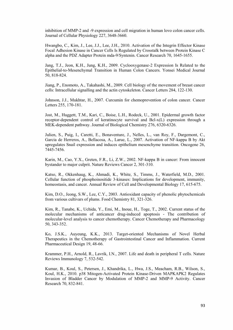
93
inhibition of MMP-2 and -9 expression and cell migration in human lovo colon cancer cells. Journal of Cellular Physiology 227, 3648-3660. Hwangbo, C., Kim, J., Lee, J.J., Lee, J.H., 2010. Activation of the Integrin Effector Kinase Focal Adhesion Kinase in Cancer Cells Is Regulated by Crosstalk between Protein Kinase C alpha and the PDZ Adapter Protein mda-9/Syntenin. Cancer Research 70, 1645-1655. Jang, T.J., Jeon, K.H., Jung, K.H., 2009. Cyclooxygenase-2 Expression Is Related to the Epithelial-to-Mesenchymal Transition in Human Colon Cancers. Yonsei Medical Journal 50, 818-824. Jiang, P., Enomoto, A., Takahashi, M., 2009. Cell biology of the movement of breast cancer cells: Intracellular signalling and the actin cytoskeleton. Cancer Letters 284, 122-130. Johnson, J.J., Mukhtar, H., 2007. Curcumin for chemoprevention of colon cancer. Cancer Letters 255, 170-181. Jost, M., Huggett, T.M., Kari, C., Boise, L.H., Rodeck, U., 2001. Epidermal growth factor receptor-dependent control of keratinocyte survival and Bcl-x(L) expression through a MEK-dependent pathway. Journal of Biological Chemistry 276, 6320-6326. Julien, S., Puig, I., Caretti, E., Bonaventure, J., Nelles, L., van Roy, F., Dargemont, C., Garcia de Herreros, A., Bellacosa, A., Larue, L., 2007. Activation of NF-kappa B by Akt upregulates Snail expression and induces epithelium mesenchyme transition. Oncogene 26, 7445-7456. Karin, M., Cao, Y.X., Greten, F.R., Li, Z.W., 2002. NF-kappa B in cancer: From innocent bystander to major culprit. Nature Reviews Cancer 2, 301-310. Katso, R., Okkenhaug, K., Ahmadi, K., White, S., Timms, J., Waterfield, M.D., 2001. Cellular function of phosphoinositide 3-kinases: Implications for development, immunity, homeostasis, and cancer. Annual Review of Cell and Developmental Biology 17, 615-675. Kim, D.O., Jeong, S.W., Lee, C.Y., 2003. Antioxidant capacity of phenolic phytochemicals from various cultivars of plums. Food Chemistry 81, 321-326. Kim, R., Tanabe, K., Uchida, Y., Emi, M., Inoue, H., Toge, T., 2002. Current status of the molecular mechanisms of anticancer drug-induced apoptosis - The contribution of molecular-level analysis to cancer chemotherapy. Cancer Chemotherapy and Pharmacology 50, 343-352. Ko, J.S.K., Auyeung, K.K., 2013. Target-oriented Mechanisms of Novel Herbal Therapeutics in the Chemotherapy of Gastrointestinal Cancer and Inflammation. Current Pharmaceutical Design 19, 48-66. Krammer, P.H., Arnold, R., Lavrik, I.N., 2007. Life and death in peripheral T cells. Nature Reviews Immunology 7, 532-542. Kumar, B., Koul, S., Petersen, J., Khandrika, L., Hwa, J.S., Meacham, R.B., Wilson, S., Koul, H.K., 2010. p38 Mitogen-Activated Protein Kinase-Driven MAPKAPK2 Regulates Invasion of Bladder Cancer by Modulation of MMP-2 and MMP-9 Activity. Cancer Research 70, 832-841.
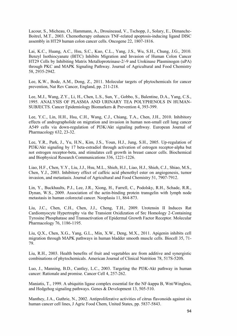
94
Lacour, S., Micheau, O., Hammann, A., Drouineaud, V., Tschopp, J., Solary, E., Dimanche-Boitrel, M.T., 2003. Chemotherapy enhances TNF-related apoptosis-inducing ligand DISC assembly in HT29 human colon cancer cells. Oncogene 22, 1807-1816. Lai, K.C., Huang, A.C., Hsu, S.C., Kuo, C.L., Yang, J.S., Wu, S.H., Chung, J.G., 2010. Benzyl Isothiocyanate (BITC) Inhibits Migration and Invasion of Human Colon Cancer HT29 Cells by Inhibiting Matrix Metalloproteinase-2/-9 and Urokinase Plasminogen (uPA) through PKC and MAPK Signaling Pathway. Journal of Agricultural and Food Chemistry 58, 2935-2942. Lee, K.W., Bode, A.M., Dong, Z., 2011. Molecular targets of phytochemicals for cancer prevention, Nat Rev Cancer, England, pp. 211-218. Lee, M.J., Wang, Z.Y., Li, H., Chen, L.S., Sun, Y., Gobbo, S., Balentine, D.A., Yang, C.S., 1995. ANALYSIS OF PLASMA AND URINARY TEA POLYPHENOLS IN HUMAN-SUBJECTS. Cancer Epidemiology Biomarkers & Prevention 4, 393-399. Lee, Y.C., Lin, H.H., Hsu, C.H., Wang, C.J., Chiang, T.A., Chen, J.H., 2010. Inhibitory effects of andrographolide on migration and invasion in human non-small cell lung cancer A549 cells via down-regulation of PI3K/Akt signaling pathway. European Journal of Pharmacology 632, 23-32. Lee, Y.R., Park, J., Yu, H.N., Kim, J.S., Youn, H.J., Jung, S.H., 2005. Up-regulation of PI3K/Akt signaling by 17 beta-estradiol through activation of estrogen receptor-alpha but not estrogen receptor-beta, and stimulates cell growth in breast cancer cells. Biochemical and Biophysical Research Communications 336, 1221-1226. Liao, H.F., Chen, Y.Y., Liu, J.J., Hsu, M.L., Shieh, H.J., Liao, H.J., Shieh, C.J., Shiao, M.S., Chen, Y.J., 2003. Inhibitory effect of caffeic acid phenethyl ester on angiogenesis, tumor invasion, and metastasis. Journal of Agricultural and Food Chemistry 51, 7907-7912. Lin, Y., Buckhaults, P.J., Lee, J.R., Xiong, H., Farrell, C., Podolsky, R.H., Schade, R.R., Dynan, W.S., 2009. Association of the actin-binding protein transgelin with lymph node metastasis in human colorectal cancer. Neoplasia 11, 864-873. Liu, J.C., Chen, C.H., Chen, J.J., Cheng, T.H., 2009. Urotensin II Induces Rat Cardiomyocyte Hypertrophy via the Transient Oxidization of Src Homology 2-Containing Tyrosine Phosphatase and Transactivation of Epidermal Growth Factor Receptor. Molecular Pharmacology 76, 1186-1195. Liu, Q.X., Chen, X.G., Yang, G.L., Min, X.W., Deng, M.X., 2011. Apigenin inhibits cell migration through MAPK pathways in human bladder smooth muscle cells. Biocell 35, 71-79. Liu, R.H., 2003. Health benefits of fruit and vegetables are from additive and synergistic combinations of phytochemicals. American Journal of Clinical Nutrition 78, 517S-520S. Luo, J., Manning, B.D., Cantley, L.C., 2003. Targeting the PI3K-Akt pathway in human cancer: Rationale and promise. Cancer Cell 4, 257-262. Maniatis, T., 1999. A ubiquitin ligase complex essential for the NF-kappa B, Wnt/Wingless, and Hedgehog signaling pathways. Genes & Development 13, 505-510. Manthey, J.A., Guthrie, N., 2002. Antiproliferative activities of citrus flavonoids against six human cancer cell lines, J Agric Food Chem, United States, pp. 5837-5843.
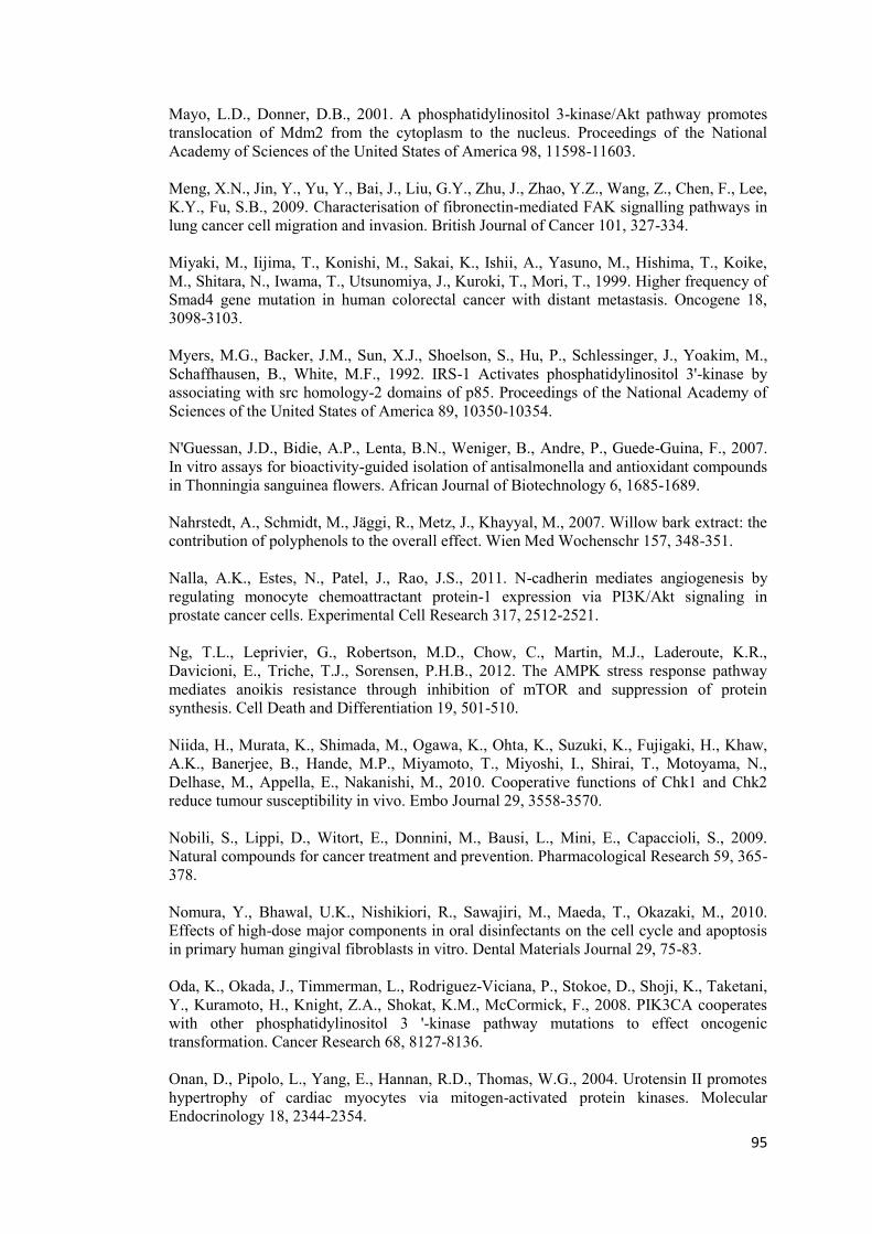
95
Mayo, L.D., Donner, D.B., 2001. A phosphatidylinositol 3-kinase/Akt pathway promotes translocation of Mdm2 from the cytoplasm to the nucleus. Proceedings of the National Academy of Sciences of the United States of America 98, 11598-11603. Meng, X.N., Jin, Y., Yu, Y., Bai, J., Liu, G.Y., Zhu, J., Zhao, Y.Z., Wang, Z., Chen, F., Lee, K.Y., Fu, S.B., 2009. Characterisation of fibronectin-mediated FAK signalling pathways in lung cancer cell migration and invasion. British Journal of Cancer 101, 327-334. Miyaki, M., Iijima, T., Konishi, M., Sakai, K., Ishii, A., Yasuno, M., Hishima, T., Koike, M., Shitara, N., Iwama, T., Utsunomiya, J., Kuroki, T., Mori, T., 1999. Higher frequency of Smad4 gene mutation in human colorectal cancer with distant metastasis. Oncogene 18, 3098-3103. Myers, M.G., Backer, J.M., Sun, X.J., Shoelson, S., Hu, P., Schlessinger, J., Yoakim, M., Schaffhausen, B., White, M.F., 1992. IRS-1 Activates phosphatidylinositol 3'-kinase by associating with src homology-2 domains of p85. Proceedings of the National Academy of Sciences of the United States of America 89, 10350-10354. N'Guessan, J.D., Bidie, A.P., Lenta, B.N., Weniger, B., Andre, P., Guede-Guina, F., 2007. In vitro assays for bioactivity-guided isolation of antisalmonella and antioxidant compounds in Thonningia sanguinea flowers. African Journal of Biotechnology 6, 1685-1689. Nahrstedt, A., Schmidt, M., Jäggi, R., Metz, J., Khayyal, M., 2007. Willow bark extract: the contribution of polyphenols to the overall effect. Wien Med Wochenschr 157, 348-351. Nalla, A.K., Estes, N., Patel, J., Rao, J.S., 2011. N-cadherin mediates angiogenesis by regulating monocyte chemoattractant protein-1 expression via PI3K/Akt signaling in prostate cancer cells. Experimental Cell Research 317, 2512-2521. Ng, T.L., Leprivier, G., Robertson, M.D., Chow, C., Martin, M.J., Laderoute, K.R., Davicioni, E., Triche, T.J., Sorensen, P.H.B., 2012. The AMPK stress response pathway mediates anoikis resistance through inhibition of mTOR and suppression of protein synthesis. Cell Death and Differentiation 19, 501-510. Niida, H., Murata, K., Shimada, M., Ogawa, K., Ohta, K., Suzuki, K., Fujigaki, H., Khaw, A.K., Banerjee, B., Hande, M.P., Miyamoto, T., Miyoshi, I., Shirai, T., Motoyama, N., Delhase, M., Appella, E., Nakanishi, M., 2010. Cooperative functions of Chk1 and Chk2 reduce tumour susceptibility in vivo. Embo Journal 29, 3558-3570. Nobili, S., Lippi, D., Witort, E., Donnini, M., Bausi, L., Mini, E., Capaccioli, S., 2009. Natural compounds for cancer treatment and prevention. Pharmacological Research 59, 365-378. Nomura, Y., Bhawal, U.K., Nishikiori, R., Sawajiri, M., Maeda, T., Okazaki, M., 2010. Effects of high-dose major components in oral disinfectants on the cell cycle and apoptosis in primary human gingival fibroblasts in vitro. Dental Materials Journal 29, 75-83. Oda, K., Okada, J., Timmerman, L., Rodriguez-Viciana, P., Stokoe, D., Shoji, K., Taketani, Y., Kuramoto, H., Knight, Z.A., Shokat, K.M., McCormick, F., 2008. PIK3CA cooperates with other phosphatidylinositol 3 '-kinase pathway mutations to effect oncogenic transformation. Cancer Research 68, 8127-8136. Onan, D., Pipolo, L., Yang, E., Hannan, R.D., Thomas, W.G., 2004. Urotensin II promotes hypertrophy of cardiac myocytes via mitogen-activated protein kinases. Molecular Endocrinology 18, 2344-2354.
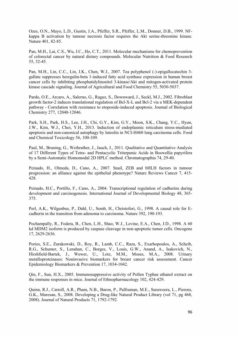
96
Ozes, O.N., Mayo, L.D., Gustin, J.A., Pfeffer, S.R., Pfeffer, L.M., Donner, D.B., 1999. NF-kappa B activation by tumour necrosis factor requires the Akt serine-threonine kinase. Nature 401, 82-85. Pan, M.H., Lai, C.S., Wu, J.C., Ho, C.T., 2011. Molecular mechanisms for chemoprevention of colorectal cancer by natural dietary compounds. Molecular Nutrition & Food Research 55, 32-45. Pan, M.H., Lin, C.C., Lin, J.K., Chen, W.J., 2007. Tea polyphenol (-)-epigallocatechin 3-gallate suppresses heregulin-beta 1-induced fatty acid synthase expression in human breast cancer cells by inhibiting phosphatidylinositol 3-kinase/Akt and mitogen-activated protein kinase cascade signaling. Journal of Agricultural and Food Chemistry 55, 5030-5037. Pardo, O.E., Arcaro, A., Salerno, G., Raguz, S., Downward, J., Seckl, M.J., 2002. Fibroblast growth factor-2 induces translational regulation of Bcl-X-L and Bcl-2 via a MEK-dependent pathway - Correlation with resistance to etoposide-induced apoptosis. Journal of Biological Chemistry 277, 12040-12046. Park, S.H., Park, H.S., Lee, J.H., Chi, G.Y., Kim, G.Y., Moon, S.K., Chang, Y.C., Hyun, J.W., Kim, W.J., Choi, Y.H., 2013. Induction of endoplasmic reticulum stress-mediated apoptosis and non-canonical autophagy by luteolin in NCI-H460 lung carcinoma cells. Food and Chemical Toxicology 56, 100-109. Paul, M., Bruning, G., Weihrather, J., Jauch, J., 2011. Qualitative and Quantitative Analysis of 17 Different Types of Tetra- and Pentacyclic Triterpenic Acids in Boswellia papyrifera by a Semi-Automatic Homomodal 2D HPLC method. Chromatographia 74, 29-40. Peinado, H., Olmeda, D., Cano, A., 2007. Snail, ZEB and bHLH factors in tumour progression: an alliance against the epithelial phenotype? Nature Reviews Cancer 7, 415-428. Peinado, H.C., Portillo, F., Cano, A., 2004. Transcriptional regulation of cadherins during development and carcinogenesis. International Journal of Developmental Biology 48, 365-375. Perl, A.K., Wilgenbus, P., Dahl, U., Semb, H., Christofori, G., 1998. A causal role for E-cadherin in the transition from adenoma to carcinoma. Nature 392, 190-193. Pochampally, R., Fodera, B., Chen, L.H., Shao, W.J., Levine, E.A., Chen, J.D., 1998. A 60 kd MDM2 isoform is produced by caspase cleavage in non-apoptotic tumor cells. Oncogene 17, 2629-2636. Pories, S.E., Zurakowski, D., Roy, R., Lamb, C.C., Raza, S., Exarhopoulos, A., Scheib, R.G., Schumer, S., Lenahan, C., Borges, V., Louis, G.W., Anand, A., Isakovich, N., Hirshfield-Bartek, J., Wewer, U., Lotz, M.M., Moses, M.A., 2008. Urinary metalloproteinases: Noninvasive biomarkers for breast cancer risk assessment. Cancer Epidemiology Biomarkers & Prevention 17, 1034-1042. Qin, F., Sun, H.X., 2005. Immunosuppressive activity of Pollen Typhae ethanol extract on the immune responses in mice. Journal of Ethnopharmacology 102, 424-429. Quinn, R.J., Carroll, A.R., Pham, N.B., Baron, P., Palframan, M.E., Suraweera, L., Pierens, G.K., Muresan, S., 2008. Developing a Drug-like Natural Product Library (vol 71, pg 468, 2008). Journal of Natural Products 71, 1792-1792.
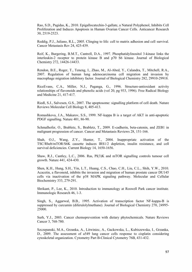
97
Rao, S.D., Pagidas, K., 2010. Epigallocatechin-3-gallate, a Natural Polyphenol, Inhibits Cell Proliferation and Induces Apoptosis in Human Ovarian Cancer Cells. Anticancer Research 30, 2519-2523. Reddig, P.J., Juliano, R.L., 2005. Clinging to life: cell to matrix adhesion and cell survival. Cancer Metastasis Rev 24, 425-439. Reif, K., Burgering, B.M.T., Cantrell, D.A., 1997. Phosphatidylinositol 3-kinase links the interleukin-2 receptor to protein kinase B and p70 S6 kinase. Journal of Biological Chemistry 272, 14426-14433. Rendon, B.E., Roger, T., Teneng, I., Zhao, M., Al-Abed, Y., Calandra, T., Mitchell, R.A., 2007. Regulation of human lung adenocarcinoma cell migration and invasion by macrophage migration inhibitory factor. Journal of Biological Chemistry 282, 29910-29918. RiceEvans, C.A., Miller, N.J., Paganga, G., 1996. Structure-antioxidant activity relationships of flavonoids and phenolic acids (vol 20, pg 933, 1996). Free Radical Biology and Medicine 21, 417-417. Riedl, S.J., Salvesen, G.S., 2007. The apoptosome: signalling platform of cell death. Nature Reviews Molecular Cell Biology 8, 405-413. Romashkova, J.A., Makarov, S.S., 1999. NF-kappa B is a target of AKT in anti-apoptotic PDGF signalling. Nature 401, 86-90. Schmalhofer, O., Brabletz, S., Brabletz, T., 2009. E-cadherin, beta-catenin, and ZEB1 in malignant progression of cancer. Cancer and Metastasis Reviews 28, 151-166. Shah, O.J., Wang, Z.Y., Hunter, T., 2004. Inappropriate activation of the TSC/Rheb/mTOR/S6K cassette induces IRS1/2 depletion, insulin resistance, and cell survival deficiencies. Current Biology 14, 1650-1656. Shaw, R.J., Cantley, L.C., 2006. Ras, PI(3)K and mTOR signalling controls tumour cell growth. Nature 441, 424-430. Shen, K.H., Hung, S.H., Yin, L.T., Huang, C.S., Chao, C.H., Liu, C.L., Shih, Y.W., 2010. Acacetin, a flavonoid, inhibits the invasion and migration of human prostate cancer DU145 cells via inactivation of the p38 MAPK signaling pathway. Molecular and Cellular Biochemistry 333, 279-291. Shrikant, P., Lee, K., 2010. Introduction to immunology at Roswell Park cancer institute. Immunologic Research 46, 1-3. Singh, S., Aggarwal, B.B., 1995. Activation of transcription factor NF-kappa-B is suppressed by curcumin (diferulolylmethane). Journal of Biological Chemistry 270, 24995-25000. Surh, Y.J., 2003. Cancer chemoprevention with dietary phytochemicals. Nature Reviews Cancer 3, 768-780. Szczepanski, M.A., Grzanka, A., Litwiniec, A., Gackowska, L., Kubiszewska, I., Grzanka, D., 2009. The assessment of a549 lung cancer cells response to cisplatin considering cytoskeletal organization. Cytometry Part B-Clinical Cytometry 76B, 431-432.
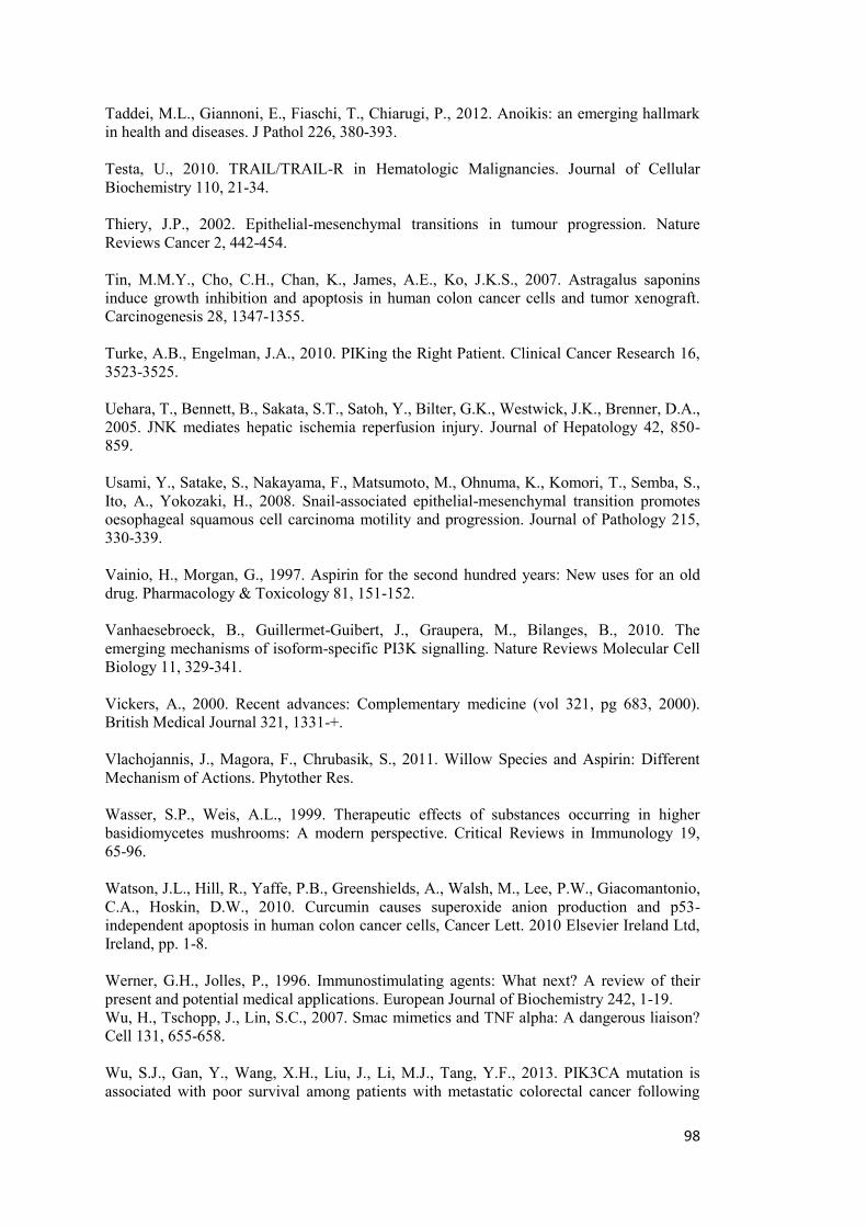
98
Taddei, M.L., Giannoni, E., Fiaschi, T., Chiarugi, P., 2012. Anoikis: an emerging hallmark in health and diseases. J Pathol 226, 380-393. Testa, U., 2010. TRAIL/TRAIL-R in Hematologic Malignancies. Journal of Cellular Biochemistry 110, 21-34. Thiery, J.P., 2002. Epithelial-mesenchymal transitions in tumour progression. Nature Reviews Cancer 2, 442-454. Tin, M.M.Y., Cho, C.H., Chan, K., James, A.E., Ko, J.K.S., 2007. Astragalus saponins induce growth inhibition and apoptosis in human colon cancer cells and tumor xenograft. Carcinogenesis 28, 1347-1355. Turke, A.B., Engelman, J.A., 2010. PIKing the Right Patient. Clinical Cancer Research 16, 3523-3525. Uehara, T., Bennett, B., Sakata, S.T., Satoh, Y., Bilter, G.K., Westwick, J.K., Brenner, D.A., 2005. JNK mediates hepatic ischemia reperfusion injury. Journal of Hepatology 42, 850-859. Usami, Y., Satake, S., Nakayama, F., Matsumoto, M., Ohnuma, K., Komori, T., Semba, S., Ito, A., Yokozaki, H., 2008. Snail-associated epithelial-mesenchymal transition promotes oesophageal squamous cell carcinoma motility and progression. Journal of Pathology 215, 330-339. Vainio, H., Morgan, G., 1997. Aspirin for the second hundred years: New uses for an old drug. Pharmacology & Toxicology 81, 151-152. Vanhaesebroeck, B., Guillermet-Guibert, J., Graupera, M., Bilanges, B., 2010. The emerging mechanisms of isoform-specific PI3K signalling. Nature Reviews Molecular Cell Biology 11, 329-341. Vickers, A., 2000. Recent advances: Complementary medicine (vol 321, pg 683, 2000). British Medical Journal 321, 1331-+. Vlachojannis, J., Magora, F., Chrubasik, S., 2011. Willow Species and Aspirin: Different Mechanism of Actions. Phytother Res. Wasser, S.P., Weis, A.L., 1999. Therapeutic effects of substances occurring in higher basidiomycetes mushrooms: A modern perspective. Critical Reviews in Immunology 19, 65-96. Watson, J.L., Hill, R., Yaffe, P.B., Greenshields, A., Walsh, M., Lee, P.W., Giacomantonio, C.A., Hoskin, D.W., 2010. Curcumin causes superoxide anion production and p53-independent apoptosis in human colon cancer cells, Cancer Lett. 2010 Elsevier Ireland Ltd, Ireland, pp. 1-8. Werner, G.H., Jolles, P., 1996. Immunostimulating agents: What next? A review of their present and potential medical applications. European Journal of Biochemistry 242, 1-19. Wu, H., Tschopp, J., Lin, S.C., 2007. Smac mimetics and TNF alpha: A dangerous liaison? Cell 131, 655-658. Wu, S.J., Gan, Y., Wang, X.H., Liu, J., Li, M.J., Tang, Y.F., 2013. PIK3CA mutation is associated with poor survival among patients with metastatic colorectal cancer following
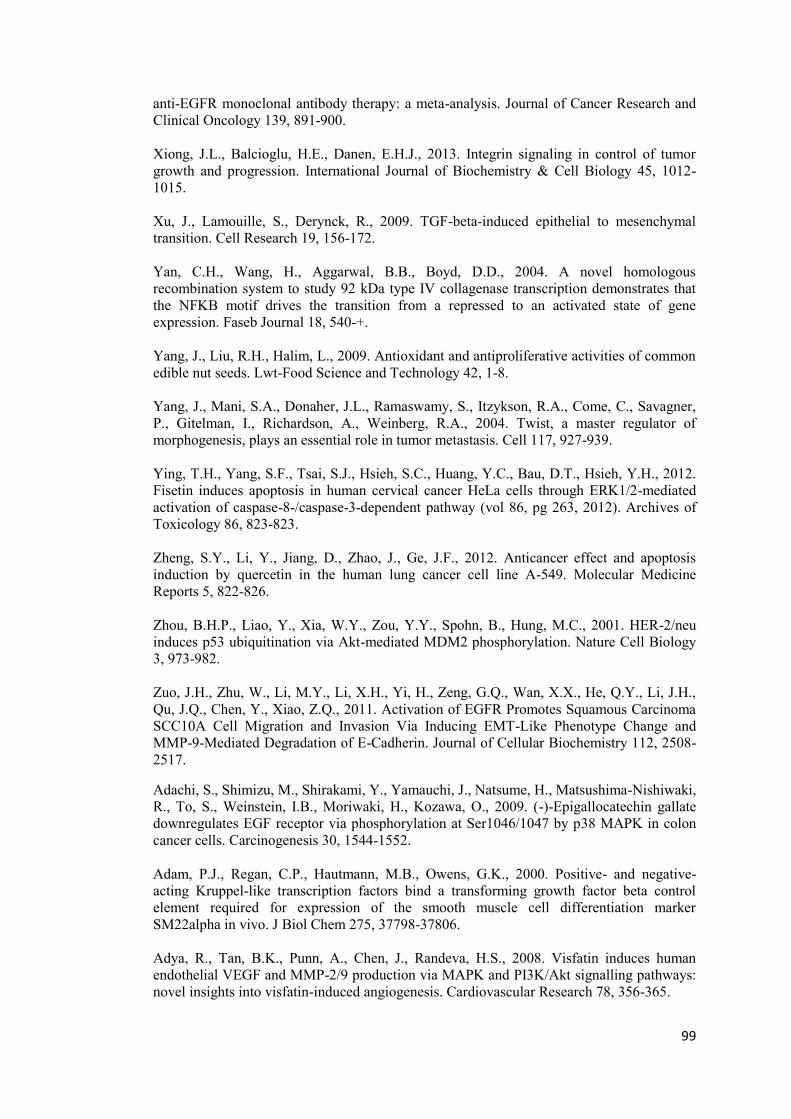
99
anti-EGFR monoclonal antibody therapy: a meta-analysis. Journal of Cancer Research and Clinical Oncology 139, 891-900. Xiong, J.L., Balcioglu, H.E., Danen, E.H.J., 2013. Integrin signaling in control of tumor growth and progression. International Journal of Biochemistry & Cell Biology 45, 1012-1015. Xu, J., Lamouille, S., Derynck, R., 2009. TGF-beta-induced epithelial to mesenchymal transition. Cell Research 19, 156-172. Yan, C.H., Wang, H., Aggarwal, B.B., Boyd, D.D., 2004. A novel homologous recombination system to study 92 kDa type IV collagenase transcription demonstrates that the NFKB motif drives the transition from a repressed to an activated state of gene expression. Faseb Journal 18, 540-+. Yang, J., Liu, R.H., Halim, L., 2009. Antioxidant and antiproliferative activities of common edible nut seeds. Lwt-Food Science and Technology 42, 1-8. Yang, J., Mani, S.A., Donaher, J.L., Ramaswamy, S., Itzykson, R.A., Come, C., Savagner, P., Gitelman, I., Richardson, A., Weinberg, R.A., 2004. Twist, a master regulator of morphogenesis, plays an essential role in tumor metastasis. Cell 117, 927-939. Ying, T.H., Yang, S.F., Tsai, S.J., Hsieh, S.C., Huang, Y.C., Bau, D.T., Hsieh, Y.H., 2012. Fisetin induces apoptosis in human cervical cancer HeLa cells through ERK1/2-mediated activation of caspase-8-/caspase-3-dependent pathway (vol 86, pg 263, 2012). Archives of Toxicology 86, 823-823. Zheng, S.Y., Li, Y., Jiang, D., Zhao, J., Ge, J.F., 2012. Anticancer effect and apoptosis induction by quercetin in the human lung cancer cell line A-549. Molecular Medicine Reports 5, 822-826. Zhou, B.H.P., Liao, Y., Xia, W.Y., Zou, Y.Y., Spohn, B., Hung, M.C., 2001. HER-2/neu induces p53 ubiquitination via Akt-mediated MDM2 phosphorylation. Nature Cell Biology 3, 973-982. Zuo, J.H., Zhu, W., Li, M.Y., Li, X.H., Yi, H., Zeng, G.Q., Wan, X.X., He, Q.Y., Li, J.H., Qu, J.Q., Chen, Y., Xiao, Z.Q., 2011. Activation of EGFR Promotes Squamous Carcinoma SCC10A Cell Migration and Invasion Via Inducing EMT-Like Phenotype Change and MMP-9-Mediated Degradation of E-Cadherin. Journal of Cellular Biochemistry 112, 2508-2517.
Adachi, S., Shimizu, M., Shirakami, Y., Yamauchi, J., Natsume, H., Matsushima-Nishiwaki, R., To, S., Weinstein, I.B., Moriwaki, H., Kozawa, O., 2009. (-)-Epigallocatechin gallate downregulates EGF receptor via phosphorylation at Ser1046/1047 by p38 MAPK in colon cancer cells. Carcinogenesis 30, 1544-1552. Adam, P.J., Regan, C.P., Hautmann, M.B., Owens, G.K., 2000. Positive- and negative-acting Kruppel-like transcription factors bind a transforming growth factor beta control element required for expression of the smooth muscle cell differentiation marker SM22alpha in vivo. J Biol Chem 275, 37798-37806. Adya, R., Tan, B.K., Punn, A., Chen, J., Randeva, H.S., 2008. Visfatin induces human endothelial VEGF and MMP-2/9 production via MAPK and PI3K/Akt signalling pathways: novel insights into visfatin-induced angiogenesis. Cardiovascular Research 78, 356-365.
100
Agarwal, M.K., Hastak, K., Jackson, M.W., Breit, S.N., Stark, G.R., Agarwal, M.L., 2006. Macrophage inhibitory cytokine 1 mediates a p53-dependent protective arrest in S phase in response to starvation for DNA precursors. Proceedings of the National Academy of Sciences of the United States of America 103, 16278-16283. Aggarwal, B.B., Banerjee, S., Bharadwaj, U., Sung, B., Shishodia, S., Sethi, G., 2007. Curcumin induces the degradation of cyclin E expression through ubiquitin-dependent pathway and up-regulates cyclin-dependent kinase inhibitors p21 and p27 in multiple human tumor cell lines. Biochemical Pharmacology 73, 1024-1032. Aggarwal, B.B., Shishodia, S., 2006. Molecular targets of dietary agents for prevention and therapy of cancer. Biochemical Pharmacology 71, 1397-1421. Agnolet, S., Wiese, S., Verpoorte, R., Staerk, D., 2012. Comprehensive analysis of commercial willow bark extracts by new technology platform: Combined use of metabolomics, high-performance liquid chromatography-solid-phase extraction-nuclear magnetic resonance spectroscopy and high-resolution radical scavenging assay. Journal of Chromatography A 1262, 130-137. Amin, A., Paul, R.K., Thakur, V.S., Agarwal, M.L., 2007. A novel role for p73 in the regulation of Akt-Foxo1a-Bim signaling and apoptosis induced by the plant lectin, concanavalin A. Cancer Research 67, 5617-5621. Athar, M., Back, J.H., Tang, X., Kim, K.H., Kopelovich, L., Bickers, D.R., Kim, A.L., 2007. Resveratrol: A review of preclinical studies for human cancer prevention. Toxicology and Applied Pharmacology 224, 274-283. Au-Yeung, K.K.W., Liu, P.L., Chan, C., Wu, W.Y., Lee, S.S.T., Ko, J.K.S., 2008. Herbal isoprenols induce apoptosis in human colon cancer cells through transcriptional activation of PPAR gamma. Cancer Investigation 26, 708-717. Auyeung, K.K., Woo, P.K., Law, P.C., Ko, J.K., 2012. Astragalus saponins modulate cell invasiveness and angiogenesis in human gastric adenocarcinoma cells. Journal of Ethnopharmacology 141, 635-641. Baker, D.D., Chu, M., Oza, U., Rajgarhia, V., 2007. The value of natural products to future pharmaceutical discovery. Natural Product Reports 24, 1225-1244. Basseres, D.S., Baldwin, A.S., 2006. Nuclear factor-kappa B and inhibitor of kappa B kinase pathways in oncogenic initiation and progression. Oncogene 25, 6817-6830. Benhar, M., Engelberg, D., Levitzki, A., 2002. ROS, stress-activated kinases and stress signaling in cancer. Embo Reports 3, 420-425. Beretta, L., Gingras, A.C., Svitkin, Y.V., Hall, M.N., Sonenberg, N., 1996. Rapamycin blocks the phosphorylation of 4E-BP1 and inhibits cap-dependent initiation of translation. Embo Journal 15, 658-664. Bhowmick, N.A., Neilson, E.G., Moses, H.L., 2004. Stromal fibroblasts in cancer initiation and progression. Nature 432, 332-337. Billen, L.P., Shamas-Din, A., Andrews, D.W., 2008. Bid: a Bax-like BH3 protein. Oncogene 27, S93-S104.
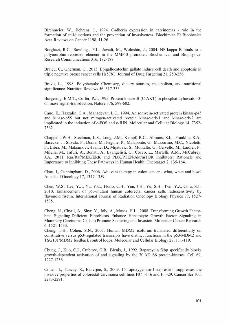
101
Birchmeier, W., Behrens, J., 1994. Cadherin expression in carcinomas - role in the formation of cell-junctions and the prevention of invasiveness. Biochimica Et Biophysica Acta-Reviews on Cancer 1198, 11-26. Borghaei, R.C., Rawlings, P.L., Javadi, M., Woloshin, J., 2004. NF-kappa B binds to a polymorphic repressor element in the MMP-3 promoter. Biochemical and Biophysical Research Communications 316, 182-188. Braicu, C., Gherman, C., 2013. Epigallocatechin gallate induce cell death and apoptosis in triple negative breast cancer cells Hs578T. Journal of Drug Targeting 21, 250-256. Bravo, L., 1998. Polyphenols: Chemistry, dietary sources, metabolism, and nutritional significance. Nutrition Reviews 56, 317-333. Burgering, B.M.T., Coffer, P.J., 1995. Protein-kinase-B (C-AKT) in phosphatidylinositol-3-oh inase signal-transduction. Nature 376, 599-602. Cano, E., Hazzalin, C.A., Mahadevan, L.C., 1994. Anisomycin-activated protein kinase-p45 and kinase-p55 but not mitogen-activated protein kinase-erk-1 and kinase-erk-2 are implicated in the induction of c-FOS and c-JUN. Molecular and Cellular Biology 14, 7352-7362. Chappell, W.H., Steelman, L.S., Long, J.M., Kempf, R.C., Abrams, S.L., Franklin, R.A., Basecke, J., Stivala, F., Donia, M., Fagone, P., Malaponte, G., Mazzarino, M.C., Nicoletti, F., Libra, M., Maksimovic-Ivanic, D., Mijatovic, S., Montalto, G., Cervello, M., Laidler, P., Milella, M., Tafuri, A., Bonati, A., Evangelisti, C., Cocco, L., Martelli, A.M., McCubrey, J.A., 2011. Ras/Raf/MEK/ERK and PI3K/PTEN/Akt/mTOR Inhibitors: Rationale and Importance to Inhibiting These Pathways in Human Health. Oncotarget 2, 135-164. Chau, I., Cunningham, D., 2006. Adjuvant therapy in colon cancer - what, when and how? Annals of Oncology 17, 1347-1359. Chen, W.S., Lee, Y.J., Yu, Y.C., Hsaio, C.H., Yen, J.H., Yu, S.H., Tsai, Y.J., Chiu, S.J., 2010. Enhancement of p53-mutant human colorectal cancer cells radiosensitivity by flavonoid fisetin. International Journal of Radiation Oncology Biology Physics 77, 1527-1535. Cheng, N., Chytil, A., Shyr, Y., Joly, A., Moses, H.L., 2008. Transforming Growth Factor-beta Signaling-Deficient Fibroblasts Enhance Hepatocyte Growth Factor Signaling in Mammary Carcinoma Cells to Promote Scattering and Invasion. Molecular Cancer Research 6, 1521-1533. Cheng, T.H., Cohen, S.N., 2007. Human MDM2 isoforms translated differentially on constitutive versus p53-regulated transcripts have distinct functions in the p53/MDM2 and TSG101/MDM2 feedback control loops. Molecular and Cellular Biology 27, 111-119. Chung, J., Kuo, C.J., Crabtree, G.R., Blenis, J., 1992. Rapamycin fkbp specifically blocks growth-dependent activation of and signaling by the 70 kD S6 protein-kinases. Cell 69, 1227-1236. Cimen, I., Tuncay, S., Banerjee, S., 2009. 15-Lipoxygenase-1 expression suppresses the invasive properties of colorectal carcinoma cell lines HCT-116 and HT-29. Cancer Sci 100, 2283-2291.
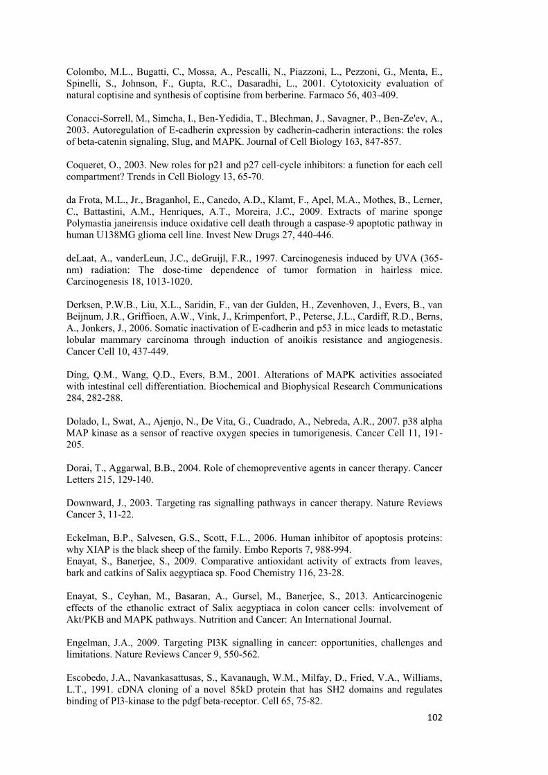
102
Colombo, M.L., Bugatti, C., Mossa, A., Pescalli, N., Piazzoni, L., Pezzoni, G., Menta, E., Spinelli, S., Johnson, F., Gupta, R.C., Dasaradhi, L., 2001. Cytotoxicity evaluation of natural coptisine and synthesis of coptisine from berberine. Farmaco 56, 403-409. Conacci-Sorrell, M., Simcha, I., Ben-Yedidia, T., Blechman, J., Savagner, P., Ben-Ze'ev, A., 2003. Autoregulation of E-cadherin expression by cadherin-cadherin interactions: the roles of beta-catenin signaling, Slug, and MAPK. Journal of Cell Biology 163, 847-857. Coqueret, O., 2003. New roles for p21 and p27 cell-cycle inhibitors: a function for each cell compartment? Trends in Cell Biology 13, 65-70. da Frota, M.L., Jr., Braganhol, E., Canedo, A.D., Klamt, F., Apel, M.A., Mothes, B., Lerner, C., Battastini, A.M., Henriques, A.T., Moreira, J.C., 2009. Extracts of marine sponge Polymastia janeirensis induce oxidative cell death through a caspase-9 apoptotic pathway in human U138MG glioma cell line. Invest New Drugs 27, 440-446. deLaat, A., vanderLeun, J.C., deGruijl, F.R., 1997. Carcinogenesis induced by UVA (365-nm) radiation: The dose-time dependence of tumor formation in hairless mice. Carcinogenesis 18, 1013-1020. Derksen, P.W.B., Liu, X.L., Saridin, F., van der Gulden, H., Zevenhoven, J., Evers, B., van Beijnum, J.R., Griffioen, A.W., Vink, J., Krimpenfort, P., Peterse, J.L., Cardiff, R.D., Berns, A., Jonkers, J., 2006. Somatic inactivation of E-cadherin and p53 in mice leads to metastatic lobular mammary carcinoma through induction of anoikis resistance and angiogenesis. Cancer Cell 10, 437-449. Ding, Q.M., Wang, Q.D., Evers, B.M., 2001. Alterations of MAPK activities associated with intestinal cell differentiation. Biochemical and Biophysical Research Communications 284, 282-288. Dolado, I., Swat, A., Ajenjo, N., De Vita, G., Cuadrado, A., Nebreda, A.R., 2007. p38 alpha MAP kinase as a sensor of reactive oxygen species in tumorigenesis. Cancer Cell 11, 191-205. Dorai, T., Aggarwal, B.B., 2004. Role of chemopreventive agents in cancer therapy. Cancer Letters 215, 129-140. Downward, J., 2003. Targeting ras signalling pathways in cancer therapy. Nature Reviews Cancer 3, 11-22. Eckelman, B.P., Salvesen, G.S., Scott, F.L., 2006. Human inhibitor of apoptosis proteins: why XIAP is the black sheep of the family. Embo Reports 7, 988-994. Enayat, S., Banerjee, S., 2009. Comparative antioxidant activity of extracts from leaves, bark and catkins of Salix aegyptiaca sp. Food Chemistry 116, 23-28. Enayat, S., Ceyhan, M., Basaran, A., Gursel, M., Banerjee, S., 2013. Anticarcinogenic effects of the ethanolic extract of Salix aegyptiaca in colon cancer cells: involvement of Akt/PKB and MAPK pathways. Nutrition and Cancer: An International Journal. Engelman, J.A., 2009. Targeting PI3K signalling in cancer: opportunities, challenges and limitations. Nature Reviews Cancer 9, 550-562. Escobedo, J.A., Navankasattusas, S., Kavanaugh, W.M., Milfay, D., Fried, V.A., Williams, L.T., 1991. cDNA cloning of a novel 85kD protein that has SH2 domains and regulates binding of PI3-kinase to the pdgf beta-receptor. Cell 65, 75-82.
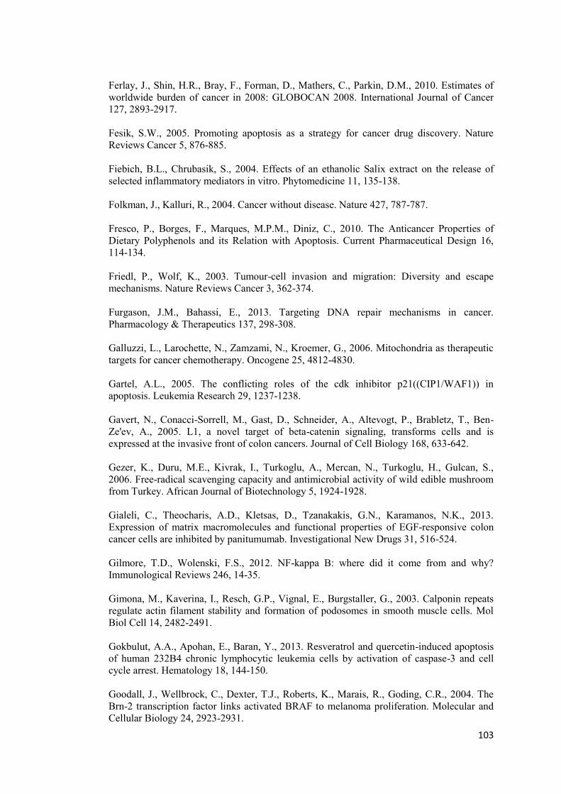
103
Ferlay, J., Shin, H.R., Bray, F., Forman, D., Mathers, C., Parkin, D.M., 2010. Estimates of worldwide burden of cancer in 2008: GLOBOCAN 2008. International Journal of Cancer 127, 2893-2917. Fesik, S.W., 2005. Promoting apoptosis as a strategy for cancer drug discovery. Nature Reviews Cancer 5, 876-885. Fiebich, B.L., Chrubasik, S., 2004. Effects of an ethanolic Salix extract on the release of selected inflammatory mediators in vitro. Phytomedicine 11, 135-138. Folkman, J., Kalluri, R., 2004. Cancer without disease. Nature 427, 787-787. Fresco, P., Borges, F., Marques, M.P.M., Diniz, C., 2010. The Anticancer Properties of Dietary Polyphenols and its Relation with Apoptosis. Current Pharmaceutical Design 16, 114-134. Friedl, P., Wolf, K., 2003. Tumour-cell invasion and migration: Diversity and escape mechanisms. Nature Reviews Cancer 3, 362-374. Furgason, J.M., Bahassi, E., 2013. Targeting DNA repair mechanisms in cancer. Pharmacology & Therapeutics 137, 298-308. Galluzzi, L., Larochette, N., Zamzami, N., Kroemer, G., 2006. Mitochondria as therapeutic targets for cancer chemotherapy. Oncogene 25, 4812-4830. Gartel, A.L., 2005. The conflicting roles of the cdk inhibitor p21((CIP1/WAF1)) in apoptosis. Leukemia Research 29, 1237-1238. Gavert, N., Conacci-Sorrell, M., Gast, D., Schneider, A., Altevogt, P., Brabletz, T., Ben-Ze'ev, A., 2005. L1, a novel target of beta-catenin signaling, transforms cells and is expressed at the invasive front of colon cancers. Journal of Cell Biology 168, 633-642. Gezer, K., Duru, M.E., Kivrak, I., Turkoglu, A., Mercan, N., Turkoglu, H., Gulcan, S., 2006. Free-radical scavenging capacity and antimicrobial activity of wild edible mushroom from Turkey. African Journal of Biotechnology 5, 1924-1928. Gialeli, C., Theocharis, A.D., Kletsas, D., Tzanakakis, G.N., Karamanos, N.K., 2013. Expression of matrix macromolecules and functional properties of EGF-responsive colon cancer cells are inhibited by panitumumab. Investigational New Drugs 31, 516-524. Gilmore, T.D., Wolenski, F.S., 2012. NF-kappa B: where did it come from and why? Immunological Reviews 246, 14-35. Gimona, M., Kaverina, I., Resch, G.P., Vignal, E., Burgstaller, G., 2003. Calponin repeats regulate actin filament stability and formation of podosomes in smooth muscle cells. Mol Biol Cell 14, 2482-2491. Gokbulut, A.A., Apohan, E., Baran, Y., 2013. Resveratrol and quercetin-induced apoptosis of human 232B4 chronic lymphocytic leukemia cells by activation of caspase-3 and cell cycle arrest. Hematology 18, 144-150. Goodall, J., Wellbrock, C., Dexter, T.J., Roberts, K., Marais, R., Goding, C.R., 2004. The Brn-2 transcription factor links activated BRAF to melanoma proliferation. Molecular and Cellular Biology 24, 2923-2931.
104
Gray-Schopfer, V., Wellbrock, C., Marais, R., 2007. Melanoma biology and new targeted therapy. Nature 445, 851-857. Gruson, D., Ginion, A., Decroly, N., Lause, P., Vanoverschelde, J.L., Ketelslegers, J.M., Bertrand, L., Thissen, J.P., 2010. Urotensin II induction of adult cardiomyocytes hypertrophy involves the Akt/GSK-3 beta signaling pathway. Peptides 31, 1326-1333. Guillermet-Guibert, J., Bjorklof, K., Salpekar, A., Gonella, C., Ramadani, F., Bilancio, A., Meek, S., Smith, A.J.H., Okkenhaug, K., Vanhaesebroeck, B., 2008. The p110 beta isoform of phosphoinositide 3-kinase signals downstream of G protein-coupled receptors and is functionally redundant with p110 gamma. Proceedings of the National Academy of Sciences of the United States of America 105, 8292-8297. Gumbiner, B.M., 2005. Regulation of cadherin-mediated adhesion in morphogenesis. Nature Reviews Molecular Cell Biology 6, 622-634. Guo, L.D., Chen, X.J., Hu, Y.H., Yu, Z.J., Wang, D., Liu, J.Z., 2013. Curcumin Inhibits Proliferation and Induces Apoptosis of Human Colorectal Cancer Cells by Activating the Mitochondria Apoptotic Pathway. Phytotherapy Research 27, 422-430. Hahn, W.C., Weinberg, R.A., 2002. Mechanisms of disease: Rules for making human tumor cells. New England Journal of Medicine 347, 1593-1603. Hanahan, D., Weinberg, R.A., 2000. The hallmarks of cancer. Cell 100, 57-70. Hanahan, D., Weinberg, R.A., 2011. Hallmarks of Cancer: The Next Generation. Cell 144, 646-674. Harrington, L.S., Findlay, G.M., Gray, A., Tolkacheva, T., Wigfield, S., Rebholz, H., Barnett, J., Leslie, N.R., Cheng, S., Shepherd, P.R., Gout, I., Downes, C.P., Lamb, R.E., 2004. The TSC1-2 tumor suppressor controls insulin-PI3K signaling via regulation of IRS proteins. Journal of Cell Biology 166, 213-223. Harvey, A., 2000. Strategies for discovering drugs from previously unexplored natural products. Drug Discovery Today 5, 294-300. Hastak, K., Agarwal, M.K., Mukhtar, H., Agarwal, M.L., 2005. Ablation of either p21 or Bax prevents p53-dependent apoptosis induced by green tea polyphenol epigallocatechin-3-gallate. Faseb Journal 19, 789-+. Hastak, K., Gupta, S., Ahmad, N., Agarwal, M.K., Agarwal, M.L., Mukhtar, H., 2003. Role of p53 and NF-kappa B in epigallocatechin-3-gallate-induced apoptosis of LNCaP cells. Oncogene 22, 4851-4859. Hastak, K., Paul, R.K., Agarwal, M.K., Thakur, V.S., Amin, A., Agrawal, S., Sramkoski, R.M., Jacobberger, J.W., Jackson, M.W., Stark, G.R., Agarwal, M.L., 2008. DNA synthesis from unbalanced nucleotide pools causes limited DNA damage that triggers ATR-CHK1-dependent p53 activation. Proceedings of the National Academy of Sciences of the United States of America 105, 6314-6319. Hay, E.D., 2005. The mesenchymal cell, its role in the embryo, and the remarkable signaling mechanisms that create it. Developmental Dynamics 233, 706-720. Hedner, T., Everts, B., 1998. The early clinical history of salicylates in rheumatology and pain. Clinical Rheumatology 17, 17-25.
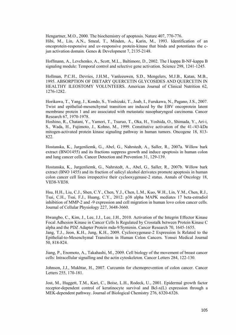
105
Hengartner, M.O., 2000. The biochemistry of apoptosis. Nature 407, 770-776. Hibi, M., Lin, A.N., Smeal, T., Minden, A., Karin, M., 1993. Identification of an oncoprotein-responsive and uv-responsive protein-kinase that binds and potentiates the c-jun activation domain. Genes & Development 7, 2135-2148. Hoffmann, A., Levchenko, A., Scott, M.L., Baltimore, D., 2002. The I kappa B-NF-kappa B signaling module: Temporal control and selective gene activation. Science 298, 1241-1245. Hollman, P.C.H., Devries, J.H.M., Vanleeuwen, S.D., Mengelers, M.J.B., Katan, M.B., 1995. ABSORPTION OF DIETARY QUERCETIN GLYCOSIDES AND QUERCETIN IN HEALTHY ILEOSTOMY VOLUNTEERS. American Journal of Clinical Nutrition 62, 1276-1282. Horikawa, T., Yang, J., Kondo, S., Yoshizaki, T., Joab, I., Furukawa, N., Pagano, J.S., 2007. Twist and epithelial-mesenchymal transition are induced by the EBV oncoprotein latent membrane protein 1 and are associated with metastatic nasopharyngeal carcinoma. Cancer Research 67, 1970-1978. Hoshino, R., Chatani, Y., Yamori, T., Tsuruo, T., Oka, H., Yoshida, O., Shimada, Y., Ari-i, S., Wada, H., Fujimoto, J., Kohno, M., 1999. Constitutive activation of the 41-/43-kDa mitogen-activated protein kinase signaling pathway in human tumors. Oncogene 18, 813-822. Hostanska, K., Jurgenliemk, G., Abel, G., Nahrstedt, A., Saller, R., 2007a. Willow bark extract (BNO1455) and its fractions suppress growth and induce apoptosis in human colon and lung cancer cells. Cancer Detection and Prevention 31, 129-139. Hostanska, K., Jurgenliemk, G., Nahrstedt, A., Abel, G., Saller, R., 2007b. Willow bark extract (BNO 1455) and its fraction of salicyl alcohol derivates promote apoptosis in human colon cancer cell lines irrespective their cyclooxygenase-2 status. Annals of Oncology 18, VII38-VII38. Hsu, H.H., Liu, C.J., Shen, C.Y., Chen, Y.J., Chen, L.M., Kuo, W.H., Lin, Y.M., Chen, R.J., Tsai, C.H., Tsai, F.J., Huang, C.Y., 2012. p38 alpha MAPK mediates 17 beta-estradiol inhibition of MMP-2 and -9 expression and cell migration in human lovo colon cancer cells. Journal of Cellular Physiology 227, 3648-3660. Hwangbo, C., Kim, J., Lee, J.J., Lee, J.H., 2010. Activation of the Integrin Effector Kinase Focal Adhesion Kinase in Cancer Cells Is Regulated by Crosstalk between Protein Kinase C alpha and the PDZ Adapter Protein mda-9/Syntenin. Cancer Research 70, 1645-1655. Jang, T.J., Jeon, K.H., Jung, K.H., 2009. Cyclooxygenase-2 Expression Is Related to the Epithelial-to-Mesenchymal Transition in Human Colon Cancers. Yonsei Medical Journal 50, 818-824. Jiang, P., Enomoto, A., Takahashi, M., 2009. Cell biology of the movement of breast cancer cells: Intracellular signalling and the actin cytoskeleton. Cancer Letters 284, 122-130. Johnson, J.J., Mukhtar, H., 2007. Curcumin for chemoprevention of colon cancer. Cancer Letters 255, 170-181. Jost, M., Huggett, T.M., Kari, C., Boise, L.H., Rodeck, U., 2001. Epidermal growth factor receptor-dependent control of keratinocyte survival and Bcl-x(L) expression through a MEK-dependent pathway. Journal of Biological Chemistry 276, 6320-6326.
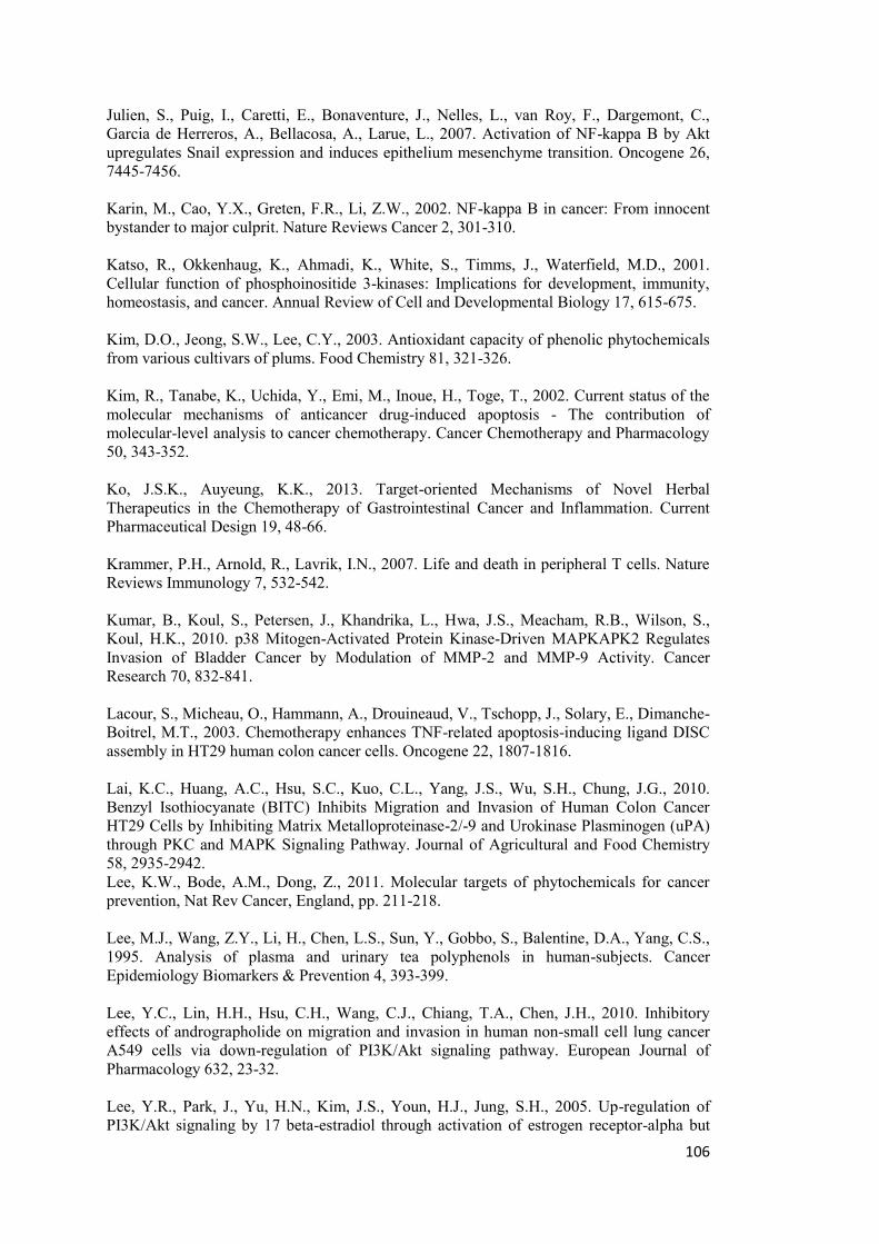
106
Julien, S., Puig, I., Caretti, E., Bonaventure, J., Nelles, L., van Roy, F., Dargemont, C., Garcia de Herreros, A., Bellacosa, A., Larue, L., 2007. Activation of NF-kappa B by Akt upregulates Snail expression and induces epithelium mesenchyme transition. Oncogene 26, 7445-7456. Karin, M., Cao, Y.X., Greten, F.R., Li, Z.W., 2002. NF-kappa B in cancer: From innocent bystander to major culprit. Nature Reviews Cancer 2, 301-310. Katso, R., Okkenhaug, K., Ahmadi, K., White, S., Timms, J., Waterfield, M.D., 2001. Cellular function of phosphoinositide 3-kinases: Implications for development, immunity, homeostasis, and cancer. Annual Review of Cell and Developmental Biology 17, 615-675. Kim, D.O., Jeong, S.W., Lee, C.Y., 2003. Antioxidant capacity of phenolic phytochemicals from various cultivars of plums. Food Chemistry 81, 321-326. Kim, R., Tanabe, K., Uchida, Y., Emi, M., Inoue, H., Toge, T., 2002. Current status of the molecular mechanisms of anticancer drug-induced apoptosis - The contribution of molecular-level analysis to cancer chemotherapy. Cancer Chemotherapy and Pharmacology 50, 343-352. Ko, J.S.K., Auyeung, K.K., 2013. Target-oriented Mechanisms of Novel Herbal Therapeutics in the Chemotherapy of Gastrointestinal Cancer and Inflammation. Current Pharmaceutical Design 19, 48-66. Krammer, P.H., Arnold, R., Lavrik, I.N., 2007. Life and death in peripheral T cells. Nature Reviews Immunology 7, 532-542. Kumar, B., Koul, S., Petersen, J., Khandrika, L., Hwa, J.S., Meacham, R.B., Wilson, S., Koul, H.K., 2010. p38 Mitogen-Activated Protein Kinase-Driven MAPKAPK2 Regulates Invasion of Bladder Cancer by Modulation of MMP-2 and MMP-9 Activity. Cancer Research 70, 832-841. Lacour, S., Micheau, O., Hammann, A., Drouineaud, V., Tschopp, J., Solary, E., Dimanche-Boitrel, M.T., 2003. Chemotherapy enhances TNF-related apoptosis-inducing ligand DISC assembly in HT29 human colon cancer cells. Oncogene 22, 1807-1816. Lai, K.C., Huang, A.C., Hsu, S.C., Kuo, C.L., Yang, J.S., Wu, S.H., Chung, J.G., 2010. Benzyl Isothiocyanate (BITC) Inhibits Migration and Invasion of Human Colon Cancer HT29 Cells by Inhibiting Matrix Metalloproteinase-2/-9 and Urokinase Plasminogen (uPA) through PKC and MAPK Signaling Pathway. Journal of Agricultural and Food Chemistry 58, 2935-2942. Lee, K.W., Bode, A.M., Dong, Z., 2011. Molecular targets of phytochemicals for cancer prevention, Nat Rev Cancer, England, pp. 211-218. Lee, M.J., Wang, Z.Y., Li, H., Chen, L.S., Sun, Y., Gobbo, S., Balentine, D.A., Yang, C.S., 1995. Analysis of plasma and urinary tea polyphenols in human-subjects. Cancer Epidemiology Biomarkers & Prevention 4, 393-399. Lee, Y.C., Lin, H.H., Hsu, C.H., Wang, C.J., Chiang, T.A., Chen, J.H., 2010. Inhibitory effects of andrographolide on migration and invasion in human non-small cell lung cancer A549 cells via down-regulation of PI3K/Akt signaling pathway. European Journal of Pharmacology 632, 23-32. Lee, Y.R., Park, J., Yu, H.N., Kim, J.S., Youn, H.J., Jung, S.H., 2005. Up-regulation of PI3K/Akt signaling by 17 beta-estradiol through activation of estrogen receptor-alpha but
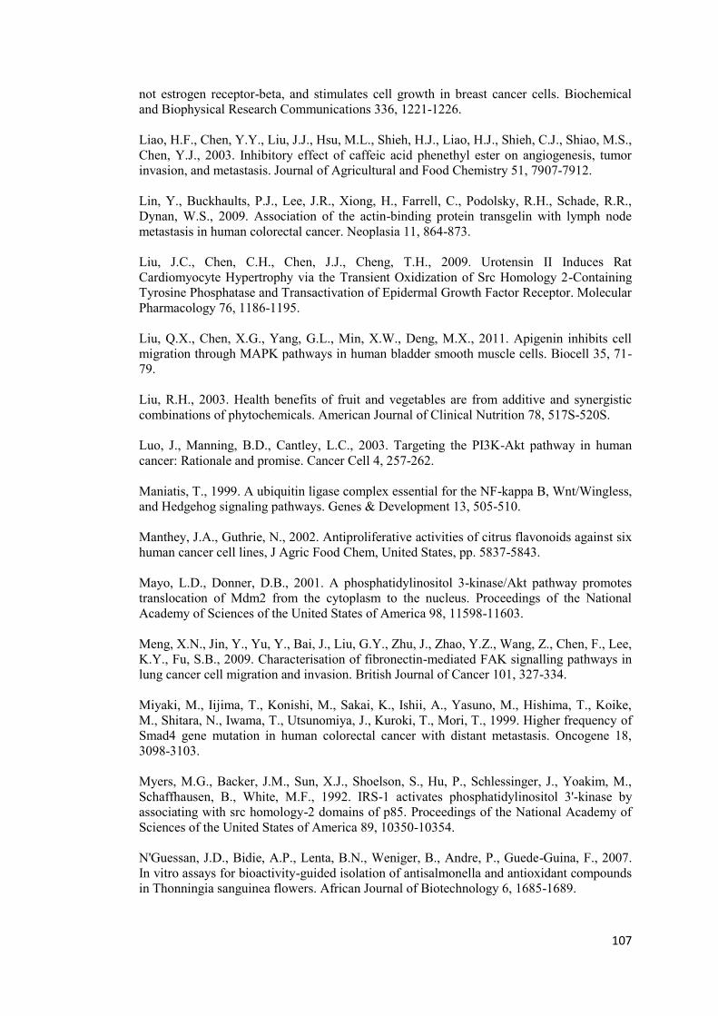
107
not estrogen receptor-beta, and stimulates cell growth in breast cancer cells. Biochemical and Biophysical Research Communications 336, 1221-1226. Liao, H.F., Chen, Y.Y., Liu, J.J., Hsu, M.L., Shieh, H.J., Liao, H.J., Shieh, C.J., Shiao, M.S., Chen, Y.J., 2003. Inhibitory effect of caffeic acid phenethyl ester on angiogenesis, tumor invasion, and metastasis. Journal of Agricultural and Food Chemistry 51, 7907-7912. Lin, Y., Buckhaults, P.J., Lee, J.R., Xiong, H., Farrell, C., Podolsky, R.H., Schade, R.R., Dynan, W.S., 2009. Association of the actin-binding protein transgelin with lymph node metastasis in human colorectal cancer. Neoplasia 11, 864-873. Liu, J.C., Chen, C.H., Chen, J.J., Cheng, T.H., 2009. Urotensin II Induces Rat Cardiomyocyte Hypertrophy via the Transient Oxidization of Src Homology 2-Containing Tyrosine Phosphatase and Transactivation of Epidermal Growth Factor Receptor. Molecular Pharmacology 76, 1186-1195. Liu, Q.X., Chen, X.G., Yang, G.L., Min, X.W., Deng, M.X., 2011. Apigenin inhibits cell migration through MAPK pathways in human bladder smooth muscle cells. Biocell 35, 71-79. Liu, R.H., 2003. Health benefits of fruit and vegetables are from additive and synergistic combinations of phytochemicals. American Journal of Clinical Nutrition 78, 517S-520S. Luo, J., Manning, B.D., Cantley, L.C., 2003. Targeting the PI3K-Akt pathway in human cancer: Rationale and promise. Cancer Cell 4, 257-262. Maniatis, T., 1999. A ubiquitin ligase complex essential for the NF-kappa B, Wnt/Wingless, and Hedgehog signaling pathways. Genes & Development 13, 505-510. Manthey, J.A., Guthrie, N., 2002. Antiproliferative activities of citrus flavonoids against six human cancer cell lines, J Agric Food Chem, United States, pp. 5837-5843. Mayo, L.D., Donner, D.B., 2001. A phosphatidylinositol 3-kinase/Akt pathway promotes translocation of Mdm2 from the cytoplasm to the nucleus. Proceedings of the National Academy of Sciences of the United States of America 98, 11598-11603. Meng, X.N., Jin, Y., Yu, Y., Bai, J., Liu, G.Y., Zhu, J., Zhao, Y.Z., Wang, Z., Chen, F., Lee, K.Y., Fu, S.B., 2009. Characterisation of fibronectin-mediated FAK signalling pathways in lung cancer cell migration and invasion. British Journal of Cancer 101, 327-334. Miyaki, M., Iijima, T., Konishi, M., Sakai, K., Ishii, A., Yasuno, M., Hishima, T., Koike, M., Shitara, N., Iwama, T., Utsunomiya, J., Kuroki, T., Mori, T., 1999. Higher frequency of Smad4 gene mutation in human colorectal cancer with distant metastasis. Oncogene 18, 3098-3103. Myers, M.G., Backer, J.M., Sun, X.J., Shoelson, S., Hu, P., Schlessinger, J., Yoakim, M., Schaffhausen, B., White, M.F., 1992. IRS-1 activates phosphatidylinositol 3'-kinase by associating with src homology-2 domains of p85. Proceedings of the National Academy of Sciences of the United States of America 89, 10350-10354. N'Guessan, J.D., Bidie, A.P., Lenta, B.N., Weniger, B., Andre, P., Guede-Guina, F., 2007. In vitro assays for bioactivity-guided isolation of antisalmonella and antioxidant compounds in Thonningia sanguinea flowers. African Journal of Biotechnology 6, 1685-1689.
108
Nahrstedt, A., Schmidt, M., Jäggi, R., Metz, J., Khayyal, M., 2007. Willow bark extract: the contribution of polyphenols to the overall effect. Wien Med Wochenschr 157, 348-351. Nalla, A.K., Estes, N., Patel, J., Rao, J.S., 2011. N-cadherin mediates angiogenesis by regulating monocyte chemoattractant protein-1 expression via PI3K/Akt signaling in prostate cancer cells. Experimental Cell Research 317, 2512-2521. Ng, T.L., Leprivier, G., Robertson, M.D., Chow, C., Martin, M.J., Laderoute, K.R., Davicioni, E., Triche, T.J., Sorensen, P.H.B., 2012. The AMPK stress response pathway mediates anoikis resistance through inhibition of mTOR and suppression of protein synthesis. Cell Death and Differentiation 19, 501-510. Niida, H., Murata, K., Shimada, M., Ogawa, K., Ohta, K., Suzuki, K., Fujigaki, H., Khaw, A.K., Banerjee, B., Hande, M.P., Miyamoto, T., Miyoshi, I., Shirai, T., Motoyama, N., Delhase, M., Appella, E., Nakanishi, M., 2010. Cooperative functions of Chk1 and Chk2 reduce tumour susceptibility in vivo. Embo Journal 29, 3558-3570. Nobili, S., Lippi, D., Witort, E., Donnini, M., Bausi, L., Mini, E., Capaccioli, S., 2009. Natural compounds for cancer treatment and prevention. Pharmacological Research 59, 365-378. Nomura, Y., Bhawal, U.K., Nishikiori, R., Sawajiri, M., Maeda, T., Okazaki, M., 2010. Effects of high-dose major components in oral disinfectants on the cell cycle and apoptosis in primary human gingival fibroblasts in vitro. Dental Materials Journal 29, 75-83. Oda, K., Okada, J., Timmerman, L., Rodriguez-Viciana, P., Stokoe, D., Shoji, K., Taketani, Y., Kuramoto, H., Knight, Z.A., Shokat, K.M., McCormick, F., 2008. PIK3CA cooperates with other phosphatidylinositol 3 '-kinase pathway mutations to effect oncogenic transformation. Cancer Research 68, 8127-8136. Onan, D., Pipolo, L., Yang, E., Hannan, R.D., Thomas, W.G., 2004. Urotensin II promotes hypertrophy of cardiac myocytes via mitogen-activated protein kinases. Molecular Endocrinology 18, 2344-2354. Ozes, O.N., Mayo, L.D., Gustin, J.A., Pfeffer, S.R., Pfeffer, L.M., Donner, D.B., 1999. NF-kappa B activation by tumour necrosis factor requires the Akt serine-threonine kinase. Nature 401, 82-85. Pan, M.H., Lai, C.S., Wu, J.C., Ho, C.T., 2011. Molecular mechanisms for chemoprevention of colorectal cancer by natural dietary compounds. Molecular Nutrition & Food Research 55, 32-45. Pan, M.H., Lin, C.C., Lin, J.K., Chen, W.J., 2007. Tea polyphenol (-)-epigallocatechin 3-gallate suppresses heregulin-beta 1-induced fatty acid synthase expression in human breast cancer cells by inhibiting phosphatidylinositol 3-kinase/Akt and mitogen-activated protein kinase cascade signaling. Journal of Agricultural and Food Chemistry 55, 5030-5037. Pardo, O.E., Arcaro, A., Salerno, G., Raguz, S., Downward, J., Seckl, M.J., 2002. Fibroblast growth factor-2 induces translational regulation of Bcl-X-L and Bcl-2 via a MEK-dependent pathway - Correlation with resistance to etoposide-induced apoptosis. Journal of Biological Chemistry 277, 12040-12046. Park, S.H., Park, H.S., Lee, J.H., Chi, G.Y., Kim, G.Y., Moon, S.K., Chang, Y.C., Hyun, J.W., Kim, W.J., Choi, Y.H., 2013. Induction of endoplasmic reticulum stress-mediated apoptosis and non-canonical autophagy by luteolin in NCI-H460 lung carcinoma cells. Food and Chemical Toxicology 56, 100-109.
109
Paul, M., Bruning, G., Weihrather, J., Jauch, J., 2011. Qualitative and Quantitative Analysis of 17 Different Types of Tetra- and Pentacyclic Triterpenic Acids in Boswellia papyrifera by a Semi-Automatic Homomodal 2D HPLC method. Chromatographia 74, 29-40. Peinado, H., Olmeda, D., Cano, A., 2007. Snail, ZEB and bHLH factors in tumour progression: an alliance against the epithelial phenotype? Nature Reviews Cancer 7, 415-428. Peinado, H.C., Portillo, F., Cano, A., 2004. Transcriptional regulation of cadherins during development and carcinogenesis. International Journal of Developmental Biology 48, 365-375. Perl, A.K., Wilgenbus, P., Dahl, U., Semb, H., Christofori, G., 1998. A causal role for E-cadherin in the transition from adenoma to carcinoma. Nature 392, 190-193. Pochampally, R., Fodera, B., Chen, L.H., Shao, W.J., Levine, E.A., Chen, J.D., 1998. A 60 kd MDM2 isoform is produced by caspase cleavage in non-apoptotic tumor cells. Oncogene 17, 2629-2636. Pories, S.E., Zurakowski, D., Roy, R., Lamb, C.C., Raza, S., Exarhopoulos, A., Scheib, R.G., Schumer, S., Lenahan, C., Borges, V., Louis, G.W., Anand, A., Isakovich, N., Hirshfield-Bartek, J., Wewer, U., Lotz, M.M., Moses, M.A., 2008. Urinary metalloproteinases: Noninvasive biomarkers for breast cancer risk assessment. Cancer Epidemiology Biomarkers & Prevention 17, 1034-1042. Qin, F., Sun, H.X., 2005. Immunosuppressive activity of Pollen Typhae ethanol extract on the immune responses in mice. Journal of Ethnopharmacology 102, 424-429. Quinn, R.J., Carroll, A.R., Pham, N.B., Baron, P., Palframan, M.E., Suraweera, L., Pierens, G.K., Muresan, S., 2008. Developing a Drug-like Natural Product Library (vol 71, pg 468, 2008). Journal of Natural Products 71, 1792-1792. Rao, S.D., Pagidas, K., 2010. Epigallocatechin-3-gallate, a Natural Polyphenol, Inhibits Cell Proliferation and Induces Apoptosis in Human Ovarian Cancer Cells. Anticancer Research 30, 2519-2523. Reddig, P.J., Juliano, R.L., 2005. Clinging to life: cell to matrix adhesion and cell survival. Cancer Metastasis Rev 24, 425-439. Reif, K., Burgering, B.M.T., Cantrell, D.A., 1997. Phosphatidylinositol 3-kinase links the interleukin-2 receptor to protein kinase B and p70 S6 kinase. Journal of Biological Chemistry 272, 14426-14433. Rendon, B.E., Roger, T., Teneng, I., Zhao, M., Al-Abed, Y., Calandra, T., Mitchell, R.A., 2007. Regulation of human lung adenocarcinoma cell migration and invasion by macrophage migration inhibitory factor. Journal of Biological Chemistry 282, 29910-29918. RiceEvans, C.A., Miller, N.J., Paganga, G., 1996. Structure-antioxidant activity relationships of flavonoids and phenolic acids (vol 20, pg 933, 1996). Free Radical Biology and Medicine 21, 417-417. Riedl, S.J., Salvesen, G.S., 2007. The apoptosome: signalling platform of cell death. Nature Reviews Molecular Cell Biology 8, 405-413. Romashkova, J.A., Makarov, S.S., 1999. NF-kappa B is a target of AKT in anti-apoptotic PDGF signalling. Nature 401, 86-90.
110
Schmalhofer, O., Brabletz, S., Brabletz, T., 2009. E-cadherin, beta-catenin, and ZEB1 in malignant progression of cancer. Cancer and Metastasis Reviews 28, 151-166. Shah, O.J., Wang, Z.Y., Hunter, T., 2004. Inappropriate activation of the TSC/Rheb/mTOR/S6K cassette induces IRS1/2 depletion, insulin resistance, and cell survival deficiencies. Current Biology 14, 1650-1656. Shaw, R.J., Cantley, L.C., 2006. Ras, PI(3)K and mTOR signalling controls tumour cell growth. Nature 441, 424-430. Shen, K.H., Hung, S.H., Yin, L.T., Huang, C.S., Chao, C.H., Liu, C.L., Shih, Y.W., 2010. Acacetin, a flavonoid, inhibits the invasion and migration of human prostate cancer DU145 cells via inactivation of the p38 MAPK signaling pathway. Molecular and Cellular Biochemistry 333, 279-291. Shrikant, P., Lee, K., 2010. Introduction to immunology at Roswell Park cancer institute. Immunologic Research 46, 1-3. Singh, S., Aggarwal, B.B., 1995. Activation of transcription factor nf-kappa-b is suppressed by curcumin (diferulolylmethane). Journal of Biological Chemistry 270, 24995-25000. Surh, Y.J., 2003. Cancer chemoprevention with dietary phytochemicals. Nature Reviews Cancer 3, 768-780. Szczepanski, M.A., Grzanka, A., Litwiniec, A., Gackowska, L., Kubiszewska, I., Grzanka, D., 2009. The assessment of a549 lung cancer cells response to cisplatin considering cytoskeletal organization. Cytometry Part B-Clinical Cytometry 76B, 431-432. Taddei, M.L., Giannoni, E., Fiaschi, T., Chiarugi, P., 2012. Anoikis: an emerging hallmark in health and diseases. J Pathol 226, 380-393. Testa, U., 2010. TRAIL/TRAIL-R in Hematologic Malignancies. Journal of Cellular Biochemistry 110, 21-34. Thiery, J.P., 2002. Epithelial-mesenchymal transitions in tumour progression. Nature Reviews Cancer 2, 442-454. Tin, M.M.Y., Cho, C.H., Chan, K., James, A.E., Ko, J.K.S., 2007. Astragalus saponins induce growth inhibition and apoptosis in human colon cancer cells and tumor xenograft. Carcinogenesis 28, 1347-1355. Turke, A.B., Engelman, J.A., 2010. PIKing the Right Patient. Clinical Cancer Research 16, 3523-3525. Uehara, T., Bennett, B., Sakata, S.T., Satoh, Y., Bilter, G.K., Westwick, J.K., Brenner, D.A., 2005. JNK mediates hepatic ischemia reperfusion injury. Journal of Hepatology 42, 850-859. Usami, Y., Satake, S., Nakayama, F., Matsumoto, M., Ohnuma, K., Komori, T., Semba, S., Ito, A., Yokozaki, H., 2008. Snail-associated epithelial-mesenchymal transition promotes oesophageal squamous cell carcinoma motility and progression. Journal of Pathology 215, 330-339. Vainio, H., Morgan, G., 1997. Aspirin for the second hundred years: New uses for an old drug. Pharmacology & Toxicology 81, 151-152.
111
Vanhaesebroeck, B., Guillermet-Guibert, J., Graupera, M., Bilanges, B., 2010. The emerging mechanisms of isoform-specific PI3K signalling. Nature Reviews Molecular Cell Biology 11, 329-341. Vickers, A., 2000. Recent advances: Complementary medicine (vol 321, pg 683, 2000). British Medical Journal 321, 1331-+. Vlachojannis, J., Magora, F., Chrubasik, S., 2011. Willow Species and Aspirin: Different Mechanism of Actions. Phytother Res. Wasser, S.P., Weis, A.L., 1999. Therapeutic effects of substances occurring in higher basidiomycetes mushrooms: A modern perspective. Critical Reviews in Immunology 19, 65-96. Watson, J.L., Hill, R., Yaffe, P.B., Greenshields, A., Walsh, M., Lee, P.W., Giacomantonio, C.A., Hoskin, D.W., 2010. Curcumin causes superoxide anion production and p53-independent apoptosis in human colon cancer cells, Cancer Lett. 2010 Elsevier Ireland Ltd, Ireland, pp. 1-8. Werner, G.H., Jolles, P., 1996. Immunostimulating agents: What next? A review of their present and potential medical applications. European Journal of Biochemistry 242, 1-19. Wu, H., Tschopp, J., Lin, S.C., 2007. Smac mimetics and TNF alpha: A dangerous liaison? Cell 131, 655-658. Wu, S.J., Gan, Y., Wang, X.H., Liu, J., Li, M.J., Tang, Y.F., 2013. PIK3CA mutation is associated with poor survival among patients with metastatic colorectal cancer following anti-EGFR monoclonal antibody therapy: a meta-analysis. Journal of Cancer Research and Clinical Oncology 139, 891-900. Xiong, J.L., Balcioglu, H.E., Danen, E.H.J., 2013. Integrin signaling in control of tumor growth and progression. International Journal of Biochemistry & Cell Biology 45, 1012-1015. Xu, J., Lamouille, S., Derynck, R., 2009. TGF-beta-induced epithelial to mesenchymal transition. Cell Research 19, 156-172. Yan, C.H., Wang, H., Aggarwal, B.B., Boyd, D.D., 2004. A novel homologous recombination system to study 92 kDa type IV collagenase transcription demonstrates that the NFKB motif drives the transition from a repressed to an activated state of gene expression. Faseb Journal 18, 540-+. Yang, J., Liu, R.H., Halim, L., 2009. Antioxidant and antiproliferative activities of common edible nut seeds. Lwt-Food Science and Technology 42, 1-8. Yang, J., Mani, S.A., Donaher, J.L., Ramaswamy, S., Itzykson, R.A., Come, C., Savagner, P., Gitelman, I., Richardson, A., Weinberg, R.A., 2004. Twist, a master regulator of morphogenesis, plays an essential role in tumor metastasis. Cell 117, 927-939. Ying, T.H., Yang, S.F., Tsai, S.J., Hsieh, S.C., Huang, Y.C., Bau, D.T., Hsieh, Y.H., 2012. Fisetin induces apoptosis in human cervical cancer HeLa cells through ERK1/2-mediated activation of caspase-8-/caspase-3-dependent pathway (vol 86, pg 263, 2012). Archives of Toxicology 86, 823-823.

112
Zheng, S.Y., Li, Y., Jiang, D., Zhao, J., Ge, J.F., 2012. Anticancer effect and apoptosis induction by quercetin in the human lung cancer cell line A-549. Molecular Medicine Reports 5, 822-826. Zhou, B.H.P., Liao, Y., Xia, W.Y., Zou, Y.Y., Spohn, B., Hung, M.C., 2001. HER-2/neu induces p53 ubiquitination via Akt-mediated MDM2 phosphorylation. Nature Cell Biology 3, 973-982. Zuo, J.H., Zhu, W., Li, M.Y., Li, X.H., Yi, H., Zeng, G.Q., Wan, X.X., He, Q.Y., Li, J.H., Qu, J.Q., Chen, Y., Xiao, Z.Q., 2011. Activation of EGFR Promotes Squamous Carcinoma SCC10A Cell Migration and Invasion Via Inducing EMT-Like Phenotype Change and MMP-9-Mediated Degradation of E-Cadherin. Journal of Cellular Biochemistry 112, 2508-2517.
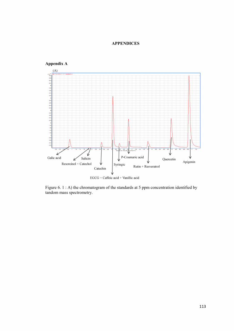
113
APPENDICES
Appendix A
Figure 6. 1 : A) the chromatogram of the standards at 5 ppm concentration identified by tandom mass spectrometry.
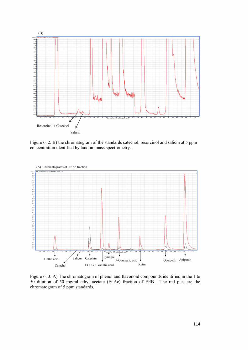
114
Figure 6. 2: B) the chromatogram of the standards catechol, resorcinol and salicin at 5 ppm concentration identified by tandom mass spectrometry.
Figure 6. 3: A) The chromatogram of phenol and flavonoid compounds identified in the 1 to 50 dilution of 50 mg/ml ethyl acetate (Et.Ac) fraction of EEB . The red pics are the chromatogram of 5 ppm standards.
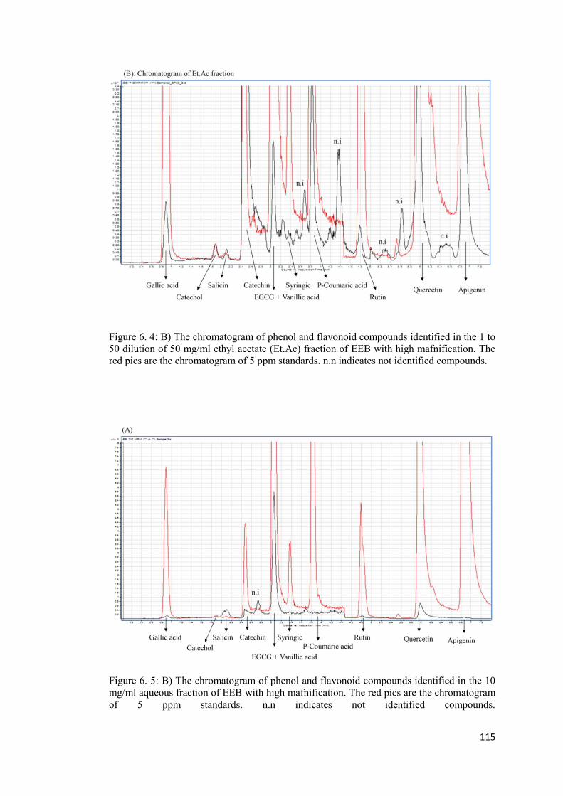
115
Figure 6. 4: B) The chromatogram of phenol and flavonoid compounds identified in the 1 to 50 dilution of 50 mg/ml ethyl acetate (Et.Ac) fraction of EEB with high mafnification. The red pics are the chromatogram of 5 ppm standards. n.n indicates not identified compounds.
Figure 6. 5: B) The chromatogram of phenol and flavonoid compounds identified in the 10 mg/ml aqueous fraction of EEB with high mafnification. The red pics are the chromatogram of 5 ppm standards. n.n indicates not identified compounds.
116
Appendix B.
0 500 1000 1500 20000.0
0.5
1.0
1.5
Y= 0.0008X + 0.1019R2= 0.9907
Concentration of Standards g/ml
595n
m
Figure 6. 6: Represent the protein concentration standard curve obtainedused for measuring protein contents.
117
Appendix C. Mammelian expression vector
Figure 6. 7. Mammalien expression vector pEF-BOS used for expression cloning of p110 subunit of PI3K. SV40 ori (311 bp), human EF-1alpha promoter (1.2 kb), stuffer fragment from CDM8 vector (450 bp, cDNA to be expressed can be inserted by removing the stuffer using BstXI or XbaI), poly-A from human G-CSF (700 bp Eco81I-EcoRI fragment), into HindIII-EcoRI site of pUC119. http://www.addgene.org/vector-database/2450/
118
CURRICULUM VITAE
PERSONAL INFORMATION Surname, Name: Enayat, Shabnam Nationality: Iranian Date and Place of Birth: 5 June 1983, Tehran Marital Status: Single Phone: +90 312 210 6482 email: [email protected] EDUCATION Degree Institution Year of Graduation PhD METU Biology 2013 MS METU Biology 2009 BS Alzahra University of Tehran Biology 2005 Awards 2009-2013 Phd Fellowships For Foreign Citizens, TÜBİTAK-BİDEB Volunteer Research Activities: Year Place 2005-2006 Pharmaceutical Materials and Plants Research Center of Shahid Beheshti
University, Tehran, Iran 2006 May Endocrine Research Center, Shahid Beheshti University of Medical
Sciences, Tehran, Iran FOREIGN LANGUAGES Advanced English, Native Persian, Fluent Turkish PUBLICATIONS
1. Enayat S, Ceyhan S. M, Basaran A. A, Gürsel M and Banerjee S. Anticarcinogenic effects of the ethanolic extract of Salix aegyptiaca in colon cancer cells: involvement of Akt/PKB and MAPK pathways. Nutrition and Cancer: An International Journal – Manuscript, 2013, ID N&C-12-12-1580.R1,accepted.
2. Enayat, S and Banerjee, S. Comparative antioxidant activity of extracts from leaves,
bark and catkins of Salix aegyptiaca sp. Food Chemistry, 2009, 116, 23-28. 3. Enayat S and Banerjee S. The ethanolic extract of bark (EEB) from Salix. Aegyptiaca.L
inhibits epithelial to mesenchymal transition and metastasis of colorectal cancer through PI3K/Akt and MAPKs signalling pathways. Nutrition and Cancer: An International Journal – Manuscript under review, 2013, ID N&C-08-13-1809