Embed Size (px)
Citation preview

ANTIDIABETIC AND HEPATOPROTECTIVE EFFECTS OF Moringa oleifera LEAF
EXTRACTS IN STREPTOZOTOCIN-INDUCED DIABETES IN RATS.
BY
TAMBWE WILLY MUZUMBUKILWA
217040227
2018

i
ANTIDIABETIC AND HEPATOPROTECTIVE EFFECTS OF Moringa oleifera LEAF
EXTRACTS IN STREPTOZOTOCIN-INDUCED DIABETES IN RATS.
BY
WILLY TAMBWE MUZUMBUKILWA
217040227
A Dissertation submitted in fulfillment of the requirements of the degree of Master of Medical
Science (Pharmacology), in the Department of Pharmacology, Discipline of Pharmaceutical
Sciences, College of Health Sciences, University of KwaZulu-Natal, Durban 4000. South Africa.
Supervisor: Dr. PETER OWIRA
Co-supervisor: Dr. MANIMBULU NLOOTO
2018

ii
ANTIDIABETIC AND HEPATOPROTECTIVE EFFECTS OF Moringa oleifera LEAF
EXTRACTS IN STREPTOZOTOCIN-INDUCED DIABETES IN RATS.
BY
WILLY TAMBWE MUZUMBUKILWA
217040227
Submitted in fulfillment of the requirements of the degree of masters of pharmacy (pharmacology),
Department of Pharmacology, Discipline of Pharmaceutical Sciences, College of Health Sciences,
University of KwaZulu-Natal.
As the candidate‘s supervisors, we have approved this dissertation for submission.
Signed: Name: Date:
Signed: Name: Date:

iii
PREFACE
The experimental work described in this dissertation was carried out in the Department of
Pharmacology, Discipline of Pharmaceutical Sciences, College of Health Science, University of
KwaZulu-Natal, Westville, from January 2017 to October 2018, under the supervision of Dr.
Owira P.M.O and Dr. Manimbulu Nlooto.
This dissertation is presented in a manuscript format and consists of three chapters.
Chapter one: This chapter provides a general introduction, comprehensive literature review,
stating the epidemiology and prevalence of diabetes mellitus. This chapter also provides the
pathophysiological mechanisms of diabetes-induced liver damage. The chapter also includes the
medicinal use of Moringa oleifera in managing diabetes. General objectives, materials, and
methods used in the study are also stated in this chapter.
Chapter two: This chapter provides a manuscript for publication presented in the required format
of the journal.
Manuscript title: Hepatoprotective effects of Moringa oleifera Lam (Moringaceae) leaf extracts in
streptozotocin-induced diabetes. Journal of Functional Foods.
Authors: Muzumbukilwa W.T., Nlooto M., Owira P.M.O.
Chapter three: This is a chapter which describes the general discussion, conclusion, and
recommendations.
………………………………………
Willy Tambwe M (217040227)
……………………………………….
Dr P.M.O owira (Supervisor)
………………………………………..
Dr. Manimbulu Nlooto (Co-supervisor)

iv
DECLARATIONS
DECLARATION 1 – PLAGIARISM
I, Tambwe Muzumbukilwa Willy declare that
1. The research reported in this thesis, except where otherwise indicated, is my original research.
2. This thesis has not been submitted for any degree or examination at any other university.
3. This thesis does not contain other persons’ data, pictures, graphs or other information unless
specifically acknowledged as being sourced from other persons.
4. This thesis does not contain other persons' writing unless specifically acknowledged as being
sourced from other researchers. Where other written sources have been quoted, then:
a) Their words have been re-written but the general information attributed to them has been
referenced.
b) Where their exact words have been used, then their writing has been placed in italics and inside
quotation marks and referenced.
5. This thesis does not contain text, graphics or tables copied and pasted from the internet, unless
specifically acknowledged, and the source being detailed in the thesis and in the References
sections.
Signed
………………………………………………………………………………....

v
DECLARATION 2 – MANUSCRIPT PUBLICATION
Article title 1: Hepatoprotective effects of Moringa oleifera Lam (Moringaceae) leaf extracts
in streptozotocin-induced diabetes.
Authors: Muzumbukilwa W.T., Nlooto M., Owira P.M.O.
Submitted to the Journal of Functional Foods.
Supplementary Paper: Mapping the evidence of hepatoprotective properties of Moringa
oleifera from sub-Saharan African countries: a systematic review protocol.
Authors: Muzumbukilwa W.T., Kadima M.G., Nlooto M., and Owira P.M.O.
Submitted to BMC Systematic reviews.
These articles are presented in the required format of the journal.
Article 1 describes the general findings and discussion of the results of the study.
CONFERENCES:
1. Oral presentation ̏ Hepatoprotective effects of Moringa oleifera Lam (Moringaceae) leaf
extracts in streptozotocin-induced diabetes ̏- Echohealth international-African chapter
conference, Durban, South Africa, 13-14 November 2018.
Signed:

vi
Acknowledgments
First and for most I would like to thank God, the beginner and the finisher of all things, through it
all He remained faithful, indeed I can do all through Him who strengthens me (Colossians 4: 13).
I would like to acknowledge and thank my supervisors, Dr. Peter Owira and Dr. Manimbulu Nlooto
who carefully supervised the study and offered guidance, support, and patience whenever needed.
I also wish to express my appreciation to my colleagues at the pharmacology department; Aganze
Glory, Edith Mofo, Kadima Gedeon, and Ntsoaki Anna, I am grateful for the support and
encouragement throughout the years. The endless discussions and great times we had are truly
appreciated. My gratitude to Shoohana Singh from the Department of Physiology (Westville Campus) for
her expertise and assistance during the histopathological studies. The Biochemistry Department in UKZN,
for their kind technical assistance. I also wish to appreciate the staff of the Biomedical Resource Unit (BRU)
at the Westville Campus UKZN, for their technical assistance and expertise with regards to experimental
animals.
My cordial gratitude and appreciation go to my wife Jolie Kangele for the endless support,
motivation, and forbearance that she gave me throughout the course of my study. I would also like
to thank my parents, my children, sisters, and brothers especially Job Wilondja and Fredy Musaka
for their motivation, support, and spiritual guidance.
I would like to acknowledge the UKZN’s College of Health Sciences for providing financial
assistance during this study.

vii
LIST OF ABBREVIATIONS
ACE
AGEs
Angiotensin-converting enzyme
Advance Glycation End products
ALAT
AMPK
Alanine Aminotransaminase
Adenosine Monophosphate-Activated Protein
Kinase
ANG II Angiotensin II
ANOVA Analysis of Variance
ARI Aldose reductase inhibitors
ASAT Aspartate Aminotransaminase
AUC Area Under the Curve
BMI Body Mass Index
BRU Biomedical Resource Unit
BW Body Weight
CCL4 Carbon Tetrachloride
CD4 Cluster of differentiation4
CD8 Cluster of differentiation8
DCM Diabetic Cardio Myopathy
DKA Diabetic ketoacidosis
DM Diabetes Mellitus
DPP-4 Dipeptidyl peptidase
FBG Fasting Blood Glucose
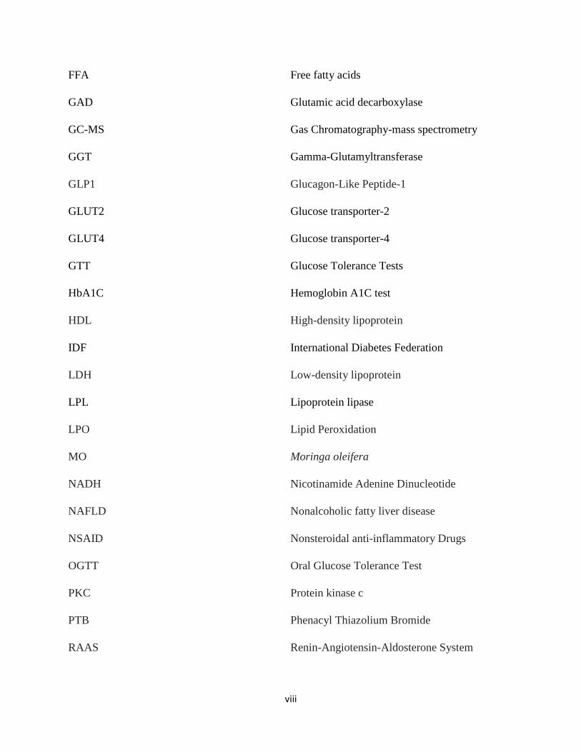
viii
FFA Free fatty acids
GAD
GC-MS
Glutamic acid decarboxylase
Gas Chromatography-mass spectrometry
GGT Gamma-Glutamyltransferase
GLP1 Glucagon-Like Peptide-1
GLUT2
GLUT4
Glucose transporter-2
Glucose transporter-4
GTT
HbA1C
Glucose Tolerance Tests
Hemoglobin A1C test
HDL High-density lipoprotein
IDF International Diabetes Federation
LDH Low-density lipoprotein
LPL Lipoprotein lipase
LPO Lipid Peroxidation
MO Moringa oleifera
NADH Nicotinamide Adenine Dinucleotide
NAFLD Nonalcoholic fatty liver disease
NSAID Nonsteroidal anti-inflammatory Drugs
OGTT Oral Glucose Tolerance Test
PKC Protein kinase c
PTB
RAAS
Phenacyl Thiazolium Bromide
Renin-Angiotensin-Aldosterone System

ix
RBX
RNS
RPM
Ruboxistaurin
Reactive Nitrogen Species
Revolutions per minute
SGOT Serum Glutamate-oxaloacetate
SGPT serum glutamic-pyruvic transaminase
SOD Superoxide Dismutase
STZ Streptozotocin
T1DM Type1 Diabetes Mellitus
T2DM Type2 Diabetes Mellitus
TBARS Thiobarbituric Acid Reactive Substance
TCF7L2 Transcription factor 7-like 2
UKZN University of KwaZulu-Natal
UNE Ulnar Neuropathy at the Elbow
VEGF Vascular endothelial growth factor
VLDL Very low-density lipoproteins
WHO World Health Organization
β Beta

x
LIST OF FIGURES
Page
Chapter 1
Figure 1.1: Pathophysiology of Type 2 diabetes mellitus…………………………………..9
Figure 1.2: Genetic and environmental factors of T2DM…………………………………..11
Figure 1.3: South African Moringa oleifera tree……………………………………….…..27
Chapter 2
Figure 2.1: Weight change graph……………………………………………………………64
Figure 2.2: Water consumption graph……………………………………………………….65
Figure 2.3: Fasting blood glucose graph…………………………………………………….66
Figure 2.4: Fasting plasma insulin graph…………………………………………………....67
Figure 2.5: Calculated Area Under the Curve graph………………………………………...68
Figure 2.6.A: ALAT concentration graph……………………………………………………69
Figure 2.6.B: ASAT concentration graph…………………………………………………....70
Figure 2.6.C: GGT concentration graph……………………………………………………..70
Figure 2.7: Plasma albumin concentration graph…………………………………………….71
Figure 2.8: Microphotograph of liver section………………………………………………..72
APPENDIX-C: GC-MS chromatogram for Moringa oleifera………………………………88
APPENDIX-D: PRISMA-P flow-chart of study selection procedure……………………....99
xi
LIST OF TABLES
Page
Chapter 1
Table 1.1: The definition of metabolic syndrome by different criteria……………………...6
Table 1.2: Treatment of diabetic retinopathy………………………………………………..13
Table 1.3: Criteria for the diagnosis of diabetes mellitus……………………………………17
Table 1.4: Drugs approved by the FDA for the treatment of Type 2 diabetes mellitus….…19
Table 1.5: Pharmacological actions of Moringa oleifera…………………………………....30
Table 1.6: Animal treatment protocol………………………………………………………..34
Chapter 2
Table 2.1: Animal treatment protocol……………………………………………………..…59
Table 2.2: Representative compounds of Moringa oleifera identified by GC- MS……….....62
Table 2.3: Average weight of animals…………..……………………………………….…...65
APPENDIX-D: Search strategy in PubMed……………………………………………….....97

xii
TABLE OF CONTENTS
Contents Page number.
PREFACE ………………………………………………………………………………...iii
DECLARATION 1- PLAGIARISM ……………………………………………....……..iv
DECLARATION 2- PUBLICATIONS ………………………………………….……….v
Acknowledgments…………………………………………………………………...……vi
LIST OF ABBREVIATIONS ……………………………………………………...…….vii
LIST OF FIGURES ……………………………………………………………………….x
LIST OF TABLES ……………………………………………………………...………...xi
LIST OF CONTENTS……………………………………………………………...……..xii
ABSTRACT ………………………………………………………………………..……. 1
CHAPTER 1: GENERAL INTRODUCTION AND
LITERATURE REVIEW …………………………………………..…….3
1.1 DIABETES MELLITUS ………………………………………………………..……3
1.1.1 Epidemiology of diabetes mellitus ……………………………………..……... 3
1.1.2 Definition and classification of diabetes mellitus ……………………….……..4
1.1.3 Pathophysiology and major risk factors ………………………………….…… 8
1.1.4 Complications of diabetes………………………………………………….…. 12
1.1.5 Diagnosis of DM ………………………………………………………….….. 17
1.1.6 Management and current treatment for diabetes mellitus………………….…. 18
1.2 LIVER DISEASE AND DIABETES ………………………………...……….…… 21
1.2.1 Structure of liver …………………………………………...………….……… 21
1.2.2 Function of liver ………………………………………………………………. 21
1.2.3 Clinical signs of liver diseases ………………………………………...……… 22
1.2.4 Mechanisms of diabetes-induced liver damage ……………………...……….. 24
1.2.5 Assessment of liver damage …………………………………………...……….25

xiii
1.3 MEDICINAL PLANTS AS SOURCES OF PUTATIVE
THERAPEUTICS AGENTS. ………………………………………………………..26
1.3.1 Medicinal plants used in diabetes management in South Africa……………. 26
1.3.2 The plant: Moringa oleifera…………………………………………………. 27
1.3.2.1 Classification …………………………………………………………….... 28
1.3.2.2 Botanical description; Synonyms ……………………………………….... .28
1.3.2.3 Geographical source and morphology……………………………….……. 28
1.3.2.4 Bioactive of Moringa oleifera………………………………………….…. .29
1.3.2.5 Hepatoprotective activity of Moringa oleifera………………………….… 31
1.4 HYPOTHESIS, AIM AND OBJECTIVES…………………………………….…....32
1.4.1 Hypothesis ……………………………………………………………......…....32
1.4.2 Aim ………………………………………………………………………….....32
1.4.3 Objectives…………………………………………………………………..…..32
1.5 MATERIALS AND METHODS ……………………………………………..……..33
1.5.1 Drugs and Chemicals…………………………………………………….… …33
1.5.2 Animals …………………………………………………………….……........ 33
1.5.3 Preparation of the plant………………………………………….……….…….33
1.5.4 Experimental design…………………………………………………….……..34
1.5.5 Induction of diabetes…………………………………………………….…….35
Preparation of citrate buffer and STZ solutions. ………………………….…...35
1.5.6 Glucose Tolerance Test (GTT). ……………………………………………...35
1.5.7 Plasma insulin concentration and
Calculation of HOMA-IR score ………………………………………..….. .36
1.5.8 Liver function tests ………………………………………………….…….…37
1.5.9 Histological analysis of liver………………………………………..………..38
1.5.10 Statistical analysis……………………………………………………..........38
REFERENCES ……………………………………………………………...…….…….40
xiv
CHAPTER 2: RESULTS. ……………………….……………………………….……..53
Hepatoprotective effects of Moringa oleifera Lam (Moringaceae) leaf extracts in streptozotocin-
induced diabetes. ………………………………………………...………………………54
Abstract…………………………………………………………………………………. 55
2.1 Introduction…………………………………………………………………………..56
2.2 Materials and methods ………………………………………………………………58
2.3 Results ………………. ……………………………………………………………..61
2.4 Discussions ………………………………………………………………………….73
2.5 Conclusion …………………………………………………………………………..75
References ………………………………………………………………………………..76
CHAPTER 3: GENERAL DISCUSSION, CONCLUSION AND
RECOMMENDATION ....................................................................................................84
3.1 GENERAL DISCUSSION AND CONCLUSION. ……………………….…………84
3.2 RECOMMENDATION ………………………………………….……….…………85
REFERENCES ……………………………………………………….……….……….…85
APPENDIX A- Ethical approval letter obtained from the University of KwaZulu-Natal.86
APPENDIX-B- Proof of animal training……………………………………………...….87
APPENDIX C- GC-MS chromatogram for Moringa oleifera………………………...….88
APPENDIX D- Supplementary paper: Mapping the evidence of hepatoprotective properties of
Moringa oleifera from sub-Saharan African countries: a systematic review
protocol……………………………………………………………………………………..….…90

1
ABSTRACT
Introduction
Diabetes Mellitus is one of the major causes of degenerative diseases worldwide. Long term
complications of diabetes include hepatic injury characterized by cirrhosis, inflammation,
apoptosis, and microvascular and macrovascular aberrations. Mechanisms by which diabetes
induces liver damage include the development of lipotoxicity-induced mitochondrial dysfunction
and activation of inflammatory pathways that lead to progressive liver damage. Previous studies
have reported that the leading cause of death in patients with diabetes mellitus is chronic liver
disease. The liver is a metabolically active organ involved in many vital life functions. It performs
many activities that are critical for survival. Due to its important activities, the liver is exposed to
a number of insults and is one of the body's organs most subject to injury.
Despite considerable progress in modern medicine, there are very few therapeutic agents that can
protect the liver from hyperglycemia-induced oxidative damage and restore normal liver functions.
As a result, the search for novel therapies that would be cheaper and effective in the management
of liver diseases is paramount. Moringa oleifera (MO) is a multipurpose plant traditionally used
for its medicinal and nutritional properties in many countries, especially in Durban, KwaZulu-
Natal/ South Africa where the material for this study has been harvested. It has been shown to
possess antihyperglycemic, antioxidant and anti-inflammatory properties and could possibly
prevent liver injury. This study, therefore, investigated whether MO leaf extracts could mitigate
hepatotoxicity associated with diabetes mellitus.
Methods
Male Wistar rats (250-300 g) were divided into six groups (n=7). Group A was orally treated daily
with 3.0 ml/kg body weight (BW) of distilled water; group C was similarly treated with MO (500
mg/kg/BW) daily. Groups B, D, E, and F were rendered diabetic by a single intraperitoneal
injection of streptozotocin (STZ) (45 mg/kg/BW in 0.1M citrate buffer, pH4.5). Diabetes was
confirmed 3 days later. Additionally, group D was treated with subcutaneous insulin (2 U/kg/BW,
bid) while groups E and F were orally treated daily with MO 250 mg/kg/BW and 500 mg/kg/BW,
respectively. Glucose tolerance tests (GTT) were done on day 47 of the animal treatment. After an

2
overnight fast for 8 hours, rats in all groups were intraperitoneally dosed with a D-glucose solution
(3.0 g/kg BW) in 0.9% normal saline. This solution was prepared by dissolving 45 g of D-glucose
anhydrous in 60 ml distilled water (0.75 g/ml). Blood glucose concentrations were measured by
tail pricking at 0, 30, 60, 90, and 120 minutes, using glucometer (OneTouch select®; Lifescan Inc.,
Milpitas, California, USA). On day 54 of treatment, animals were sacrificed by halothane
overdose. Blood was collected by cardiac puncture in heparinized tubes then separated into plasma
and stored at -80˚C for further biochemical analysis. Livers were excised, snap-frozen in liquid
nitrogen and similarly stored for histological analysis.
Results: Diabetic animals had significantly (p<0.05) elevated Fasting Blood Glucose (FBG) and
reduced insulin levels compared to controls. Treatment with either insulin or MO significantly
(p<0.05) reduced FBG compared to non-treated diabetic rats. Treatment with 500 mg/kgBW
significantly reduced FBG compared to treatment with 250 mg/kgBW. Calculated Areas-Under-
the Curve (AUCs) from OGTT suggested that untreated diabetic rats exhibited glucose intolerance
but treatment with either insulin or MO extracts significantly (p<0.05) reversed this. Liver function
tests defined by Aspartate Aminotransaminase (ASAT), Alanine Aminotransaminase
(ALAT),gamma-glutamyl aminotransaminase (GGT) and albumin were significantly (p<0.05)
elevated in untreated diabetic group but treatment with either insulin or MO extracts significantly
(p<0.05) reversed this. Treatment with 500 mg/kgBW of MO significantly (p<0.05) reduced GGT
levels compared to treatment with 250 mg/kgBW. Untreated diabetic, unlike MO-treated rats,
exhibited degeneration of hepatocytes and inflammatory cells infiltration with the fragmentation
of the nucleus and cell lysis, necrotic hepatocytes, hepatic vein congestion, and vesicular
cytoplasm compared to normal controls.
Conclusion: This study has shown that methanolic leaf extracts of MO have dose-dependent
antidiabetic effects. Liver function tests (ASAT, ALAT, GGT) and albumin were significantly
elevated in untreated diabetic rats than those treated with MO extracts. This may justify the
hepatoprotective effects of MO extracts in streptozotocin-induced diabetic rats.
Keywords: Moringa oleifera, hepatocytes, streptozotocin, diabetes.

3
CHAPTER 1: GENERAL INTRODUCTION AND LITERATURE REVIEW
1.1 DIABETES MELLITUS
1.1.1 Epidemiology of Diabetes Mellitus.
In 2017, the International Diabetes Federation (IDF) estimated that 425 million adults aged
between 20 and 79 years were living with diabetes worldwide (Ogurtsova et al., 2017). Globally,
422 million people were living with diabetes in 2014 and 1.6 million deaths were attributed to
diabetes (World Health Organization, 2016). IDF estimates that 75% of people with diabetes live
in low-and-middle-income countries (Whiting et al., 2011). Total global health expenditure due to
diabetes in 2017 was estimated at 727 billion US dollars (Ogurtsova et al., 2017).
The National Diabetes Statistics Report estimates that 30.3 million people with diabetes (9.4% of
the USA population) including 23.1 million people diagnosed and 7.2 million people undiagnosed
were living with diabetes in the United States in 2017 (Centers for Disease Control and Prevention,
2017).
Public health burden of diabetes in Africa has been recognized (Motala et al., 2003). The
prevalence of diabetes increased from 0-1% in the 1980s to 20% in the 2000s and is predicted to
be affecting between 18.6 to 23.9 million Africans by the year 2030 (Mbanya et al., 2010). More
so, 14.2 million Africans were estimated to be living with diabetes in 2015, and this was projected
to increase to 34.2 million by 2040 (Mutyambizi et al., 2018). It has further been estimated that
15.5 million adults aged 20-79 years were living with diabetes in Africa in 2017, representing a
regional prevalence of 3.3% (Ogurtsova et al., 2017).
In South Africa, 3.5 million people (approximately 6% of the total population) are living with
diabetes and many others remain undiagnosed (World Health Organization, 2016). The high
incidence of T2DM in South Africa, like in the rest of the world has been attributed to sedentary
lifestyle, lack of physical activities and increased urbanization (Menghani et al.,2010). In the year
2000, the South African National Burden of Disease Study reported that diabetes was the tenth
leading cause of morbidity and mortality, accounting for about 2.6% of all deaths, which represents
20,000 deaths or 4.3% of the total population (Bradshaw et al., 2007).

4
1.1.2 Definition and classification of diabetes mellitus
Diabetes mellitus is a disorder of carbohydrate, lipid and protein metabolism, characterized by
high blood glucose levels resulting from defects in insulin secretion, insulin action, or both. Long-
term complications of untreated DM include cardiomyopathy, nephropathy, neuropathy and
retinopathy (Alberti and Zimmet, 1998). Classical symptoms of DM include polydipsia, polyuria,
blurring of vision and weight loss. As reported by the American Diabetes Association, 4 categories
of DM are recognized: Type 1 Diabetes mellitus, Type 2 Diabetes mellitus, gestational diabetes
and diabetes due to secondary disease processes or drugs. Maturity-onset diabetes of the young
(MODY) is a rare form of diabetes caused by mutations in nuclear transcription factors and
glucokinase genes which result in pancreatic β-cell dysfunction in the production of insulin
hormone (Cydulka and Maloney., 2002; Navale, 2017).
1.1.2.1 Type 1 Diabetes
Type 1 diabetes mellitus (T1DM) is autoimmune-mediated destruction of pancreatic β-cells. The
dendritic cells and macrophages in the pancreatic islets possess autoantigens and present them to
autoreactive CD4+ T cells. CD4+ T cells secrete cytokines which can activate cytotoxic T cells
(CD8+). These cells activate macrophages and other T cells leading to beta cells destruction
ultimately causing absolute insulin deficiency which predisposes individuals to ketoacidosis
(Lambert et al., 2004). Genetic and environmental factors have been identified as leading for the
autoimmune destruction of the β-cells. Human leukocyte antigen (HLA) is a common genetic risk
factor associated with the development of diabetes (Nokoff and Rewers, 2013). Environmental
factors involved in the pathogenesis of T1DM include parasites, viruses, and bacteria which
mediate the direct infection of the pancreatic β-cells or shape the immune system to mutually
benefit the parasite and the host (David et al., 2004).
T1DM leads to complete pancreatic ß cell destruction and eventually all patients with T1DM will
require insulin therapy to maintain glycemic control. Patients with T1DM are rarely obese and are
prone to other autoimmune conditions such as Grave’s or Addison's disease (Krzewska and Ben-
skowronek, 2016).

5
All patients with T1DM present with classical symptoms of hyperglycemia and signs of
ketoacidosis (David, M. and Nathan., 2009). When the diagnosis is uncertain, other tests can be
used including the measurement of plasma C-peptide levels or markers of immune destruction
such as islet cell antibodies, autoantibodies to insulin, autoantibodies to glutamic acid
decarboxylase (GAD) and autoantibodies to tyrosine phosphatases IA-1 and IA-2 (Casqueiro et
al., 2012). T1DM usually develops in younger and teenage population (adolescent) although it can
also occur in adults (Devendra et al., 2004). It has been shown that human leukocyte antigen
chromosome 6 is a genetic predisposition that is linked to T1DM (Lie et al., 1999). Type 1 diabetes
accounts for 10% of all diagnosed cases of diabetes in adults worldwide (Adeeyo et al., 2013).
1.1.2.2 Type 2 Diabetes
T2DM is insulin independent and develops when there is impaired glucose tolerance, obesity,
dyslipidemia, insulin resistance (IR), increased hepatic glucose production, pancreatic β-cell
dysfunction and hyperglycemia (Wu et al., 2014). Insulin usually controls glucose homeostasis by
stimulation of glucose uptake into peripheral tissues, and by signaling the liver to take up glucose
and store it as glycogen. Production of advanced glycation end products (AGE), increased
stimulation of hemodynamic regulation systems such as the renin-angiotensin system (RAS) and
elevated production of reactive oxygen species (ROS) which are associated with chronic
hyperglycemia plays a pivotal role in the initiation of diabetic vascular complications (Jakus,
2004). However, the pancreas lacks antioxidant defense systems antioxidant (due to the absence
of glutathione peroxidase-1 gene) against ROS (Rahal et al., 2014). In this regard, dyslipidemia is
initiated by insulin resistance which activates lipoprotein lipase (LPL) in adipocytes. The
lipoprotein lipases catalyze the release of free fatty acids (FFA) which are taken up by hepatocytes
leading to the production of triglycerides. Triglycerides then stimulate the production of very low-
Density Lipoproteins (VLDL) which contribute to the increased risk of cardiovascular diseases (
Ginsberg, H. and Sung, H, 2012). The specific causes of Type 2 diabetes are not well defined, but
some factors are incriminated such as environmental, genetic as well as lifestyle changes (Zheng
et al., 2017). In "prediabetes", blood glucose levels are higher than normal but not yet high enough
to be diagnosed as diabetes. Research has shown that some long-term damage to the body organs
especially the heart and circulatory system, may already be occurring during prediabetes (Silva et
al., 2008). Cardiovascular disease is the foremost killer of patients with diabetes.

6
The metabolic syndrome is a clustering of risk factors that often accompany obesity and associated
with increased risk for both cardiovascular diseases and type 2 diabetes (Ford et al., 2008). This
cluster of risk factors led to diabetes and cardiovascular diseases. Such factors include
hypertension, insulin resistance, defective glucose metabolism (impaired fasting glycemia or
glucose intolerance), increased LDL-c, triglycerides, obesity and low HDL-c concentrations (Paula
et al., 2013a). European Group for Study of Insulin Resistance (EGIR), International Diabetes
Federation (IDF), American Association of Clinical Endocrinologists (AACE), WHO clinical
criteria (WHO 1999) and the National Cholesterol Education Program Adult Treatment Panel III
(NCEP/ATP III) have defined the criteria classification of metabolic syndrome (Table 1) (Paula et
al., 2013b). These groups have a harmonized definition of the metabolic syndrome with criteria
defined in Table 1 below.
Table 1: The definition of metabolic syndrome by different criteria (Alberti and Zimmet., 1998;
Einhorn et al., 2003). Legend: BMI: body mass index; DM: diabetes mellitus; FBG: fasting
blood glucose; IFG; impaired fasting glucose; IGT: impaired glucose tolerance; IR: insulin
resistance; HDL: high-density lipoprotein; TG; triglyceride; WC: Waist circumference.
Clinical
measures
World of
Health
Organization
(1998)
European
Group for
Study of
Insulin
Resistance
(1999)
National
Cholesterol
Education
Program
Adult
Treatment
Panel (2001)
American
Association of
Clinical
Endocrinologists
(2003)
International
Diabetes
Federation
(2005)
Insulin
resistance
IGT, IFG,
T2DM,
or Reduced
insulin
Sensitivity
Plasma
insulin
>75th
percentile
None IGT or IFG None
Body weight Male: waist-
tohip
> 0.90
Female:
Waistto-
hip > 0.85
and/or
BMI > 30
kg/m2
Male: WC ≥
94 cm
Female: WC
≥
80 cm
Male: WC
≥102
cm
Female: WC
≥
88 cm
BMI ≥ 25 kg/m2 Increased WC
(populace
specific)

7
Dyslipidemia TGs ≥ 150
mg/dl
and/or HDL-c
< 35 mg/dl in
males or < 39
mg/dl in
females
TGs ≥150
mg/dl
and/or HDL-c
< 39 mg/dl in
males or
females
TGs ≥ 150
mg/dl
HDL-c < 40
mg/dl in
males or < 50
mg/dl in
females
TGs ≥ 150 mg/dl
and
HDL-c < 40
mg/dl in
males or < 50
mg/dl in
females
TGs ≥ 150
mg/dl
HDL-c < 40
mg/dl
in males or
<50
mg/dl in
females or
on HDL-c
treatment
Blood
pressure
≥ 140/90 mm
Hg
≥ 140/90 mm
Hg or on
hypertension
treatment
≥ 130/85 mm
Hg
≥ 130/85 mm Hg ≥ 130 mm Hg
systolic or ≥
85 mm
Hg
diastolic or on
hypertension
treatment
Glucose
Metabolism
IGT, IFG, or
T2DM
IGT or IFG
(None
diabetes)
>110 mg/dL
(comprises
diabetes)
IGT or IFG
(None
diabetes)
≥ 100 mg/dL
(comprises
diabetes)
The major causes of metabolic syndrome include genetic and environmental factors which
influence both lipid and glucose metabolism. An estimated 20-25% of the adult individuals have
been clinically diagnosed with metabolic syndrome globally (Moreira et al., 2014).
Genes that control the expression of lipases, adiponectin, uncoupling proteins , β-Adrenergic
receptors, PPAR-γ, glycoprotein PC-1, skeletal muscle glycogen synthase 1, IRS-1, Calpain-10,
CD36, fatty acid binding protein-2, apolipoprotein E, upstream transcription factor 1 and 11 β-
hydroxysteroid dehydrogenase type 1 have associated with increased incidence of metabolic
syndrome (Stancakova and Laakso., 2014). Pro-inflammatory cytokines like tumor necrosis factor
(TNF) and Interleukin 1-β (IL1-β), C-reactive protein and neuropeptide Y(NPY), leptin receptor,
pro-opiomelanocortin (POMC) and melanocortin receptors are also involved in metabolic
syndrome (Groop, 2007).
Environmental factors that lead to metabolic syndrome include physical inactivity, obesity,
smoking and aging, which have been suggested to induce insulin resistance, cardiovascular
disorders, inflammation, hyperinsulinemia and endothelial dysfunction (Takashi et al., 2006).
These factors are associated with dysfunctions of adipocytes leading to altered production of
8
hormones and cytokines such as interleukin-6 (IL-6), TNF-α, resistin, adiponectin, and leptin
(Takashi et al., 2006).
1.1.2.3 Gestational Diabetes
Gestational diabetes is defined as glucose intolerance with first recognition pregnancy. Women
who are overweight, had gestational diabetes before or have a family history of diabetes are at a
higher risk of developing gestational diabetes. Untreated gestational diabetes may cause problems
to the fetus such as breathing difficulties and low blood sugar levels. Gestational diabetes is
associated with increased risk of maternal and fetal complications. Fetal macrosomia is a common
complication of diabetes in pregnancy. Pregnant woman of a macrosomic fetus is at increased risk
for preeclampsia, labor abnormalities, severe perineal lacerations, risks of preterm birth and
cesarean section. The fetus is at risk for stillbirth, intracranial hemorrhage, shoulder dystocia and
malformations (Susa JB, 1988). The neonate is at risk for hyperbilirubinemia, hypocalcemia,
hypoglycemia, hypomagnesemia, polycythemia and neonatal cardiomyopathy (Macintosh MC et
al., 2006). Fetal macrosomia is caused by fetal hyperinsulinemia that occurs as a physiological
response to maternal hyperglycemia (Moreli et al., 2016).
Later in life, the baby may be at an increased risk for obesity and T2DM for the rest of their lives
(Reece, 2010).
1.1.3 Pathophysiology and major risk factors for T2DM
Type 2 diabetes is caused by a combination of genetic factors related to impaired insulin secretion
and insulin resistance and environmental factors such as lack of exercise, aging, stress as well as
obesity. Impaired insulin secretion and insulin resistance contribute jointly to the development of
pathophysiological conditions. Although the pathogenesis of T2DM has been attributed mainly on
insulin and beta-cell dysfunction, an abnormal increase in alpha-cell function which results in
hyperglucagonemia and consequent hepatic glucose production has long been recognized as a
contributory factor to hyperglycemia in diabetic patients. Postprandial state in T2DM patients is
characterized by elevated fasting concentrations of glucagon, impaired glucose-induced glucagon

9
suppression, and disrupted insulin–glucagon interaction against those of healthy subjects.
Alterations in the cross-talk between beta- and alpha-cells may underlie diminished mass of insulin
pulses and hyperglucagonemia in T2DM patients (Menge et al., 2011). Because of resistance to
the actions of insulin and reduced insulin secretion, tissues such as skeletal muscles, liver, and
adipose tissues are affected, leading to elevated blood glucose levels (Stumvoll et al., 2005). In
T2DM, insulin resistance plays an important role due to increased gluconeogenesis and decreased
glucose uptake in the skeletal muscles and adipose tissues at a regular plasma insulin level. In
addition, β-cell dysfunction results in reduced insulin release, which is not enough for maintaining
normal glucose levels (Weyer et al., 1999).
Figure 1: Consequences of insulin action/inaction in Type 2 diabetes mellitus.

10
Insulin secretion from the β-cells in the pancreas normally suppresses gluconeogenesis and
increases glucose uptake by skeletal muscles and adipose tissues. Diminished pancreatic β-cells
function leads to hyperglycemia (Zheng et al., 2017).
Major risk factors for T2DM
There are many factors that contribute to T2DM such as:
Older age
The family history of type 2 diabetes mellitus (T2DM)
Low socioeconomic status
Genetic factors (for example, carrying risk alleles in the TCF7L2 gene)
Components of the metabolic syndrome (increased waist circumference,
increased blood pressure, increased plasma levels of triglycerides, low plasma
levels of HDL cholesterol and small, dense LDL cholesterol particles)
Overweight or obese (BMI ≥25 kg/m2)
Abdominal or central obesity (independent of BMI)
Cigarette smoking
Sedentary lifestyle
History of gestational diabetes mellitus or delivery of neonates >4 kg in weight.
Some medications, such as statins, thiazides, and beta-blockers
Psychosocial stress and depression (Spiegel et al., 2005).

11
Figure 2: Genetic and environmental factors of T2DM.
Genetic factors Environmental factors
Obesity susceptibility, diabetes
history, race, age.
Viruses, psychology, smoking,
exercise, culture, stress, diet,
alcohol.
Metabolic syndrome
Insulin resistance
T2DM

12
1.1.4 Complications of diabetes.
Persistent hyperglycemia leads to both acute and chronic complications. Major tissues affected by
hyperglycemia are nervous system, the retina of eye, kidney, adipose tissue, liver and pancreatic
β cells. T1DM and T2DM both result in long term macro and microvascular complications.
Microvascular complications include diabetic retinopathy, neuropathy, and nephropathy, while
macrovascular complications include coronary artery diseases, peripheral vascular disease, and
stroke (Casqueiro, et al., 2012). Diabetic Ketoacidosis (DKA) is one of the major acute
complications of T1DM characterized by ketoacidosis and electrolyte imbalances, caused by
absolute lack of insulin; characterized by dehydration and lipolysis (Cydulka et al.,2002). In DKA,
there is a deficiency of insulin, which results in increased plasma catabolic hormones, that cause
overproduction of glucose (Aronson and Ferner, 2003). In renal tubes, excess glucose draws water
and electrolytes such as calcium, magnesium, potassium, and sodium into the urine, from the
circulation. This osmotic diuresis causes dehydration and other electrolyte imbalances associated
with diabetic ketoacidosis (Cydulka et al.,2002).
1.1.4.1 Diabetic Retinopathy.
Diabetic retinopathy is one of the most common complications of untreated DM and leads to visual
impairment and blindness in adults aged between 20-74 years ( Lilly et al., 2006; Forbes and
Cooper, 2013). Lesions in the retina cause alterations of retinal structures such as changes in
vascular permeability, capillary degeneration, capillary microaneurysms, and neovascularization
by increased formation of new blood vessels (Lilly et al., 2006). Functional impairment of the
neural retina alters retinal electrophysiology which results in an inability to differentiate between
colors (Forbes & Cooper, 2013). The development and progression of DM are dependent on the
severity and duration of hyperglycemia.
Clinically, diabetic retinopathy is divided into non- proliferative and proliferative disease phases.
In the initial phase, hyperglycemia can lead to intramural pericyte (cells that wrap around the
endothelial cells of capillaries and venules throughout the body) death and thickening of the
basement membrane, which contribute to changes in the integrity of blood vessels within the

13
retina, altering the blood-retinal barrier and vascular permeability (Watkins, 2003). During this
primary phase of nonproliferative diabetic retinopathy (NPDR), no visual impairment is noticed.
Degeneration or occlusion of retinal capillaries is associated with failing prognosis, which is most
likely the result of ischemia followed by subsequent release of angiogenic factors including those
related to hypoxia (Osborne N.et al., 2004).
In the proliferative phase neovascularization and accumulation of fluid within the retina, called
macular edema, contribute to visual damage. In more severe cases, there can be bleeding with
associated distortion of the retinal architecture including the development of a fibrovascular
membrane which can subsequently lead to retinal detachment (Sikorski et al., 2013).
Diabetic retinopathy can be prevented with early diagnosis and treatment.
In order to maintain glycemic control and blood pressure, treatments with an injection of the steroid
triamcinolone, laser photocoagulation, and vascular endothelial growth factor (VEGF) antagonists
into the eye, and vitrectomy, to remove the vitreous (Forbes & Cooper, 2013). However, newer
agents have been developed (Table 2).
Table 2: Potential therapies in the treatment of diabetic retinopathy (Nawaz et al., 2013).
Targets Drug
Inhibitor of AGE formation Pyridoxamine, Aminoguanidine, OPB-9195, ALT-
946, ALT-711, LR-90, N-PheracylThiazolium
Bromide (PTB), Alagebrium.
Protein kinase C (PKCs) inhibitors.
Ruboxistaurin (RBX), PKC412.
Aldose reductase Inhibitors (ARIs).
Sorbinil, Tolrestat, Epalrestat, Lidorestat, Zenarestat,
Ranirestat, Ponalrestat, Zopolrestat, ARI-809,
Fidarestat.
Nonsteroidal anti-inflammatory drugs
(NSAIDs)
Aspirin, Nepafenac, Sodium salicylate, Sulfasalazine,
Baicalein, Genistein, Nepafenac, Celecoxib

14
1.1.4.2 Diabetic Neuropathy
Diabetic neuropathy is characterized by a progressive loss of nerve fibers affecting both the
autonomic and somatic divisions. It is defined by impaired damage healing due to a decrease in
oxygen in tissues because of glycated hemoglobin and altered immune system (Schalkwijk, 2008).
As with other microvascular complications, the risk of developing diabetic neuropathy is relative
to chronic glucose intolerance even though some individuals may have a genetic predisposition
(Dobretsov et al., 2007).
There are two classes of diabetic neuropathies: diabetic polyneuropathy in which hyperglycemia
causes diffuse damage to the peripheral nervous system; and mononeuropathy, where nerve
damage is focal (Boulton, A.J., 2005). Polyneuropathy is the most common form of diabetic
neuropathy and may present as cranial, autonomic neuropathy and peripheral motor neuropathy.
While mononeuropathies include Carpal Tunnel Syndrome (CTS) and Ulnar Neuropathy at the
Elbow (UNE). Both polyneuropathy and mononeuropathy play significant roles in sensory and
motor deficits and are both associated with debility in patients (Zochodne, 2007).
The precise nature of the injury to the peripheral nerves from hyperglycemia is not known but it
may be related to mechanisms such as polyol accumulation, injury from AGEs, and oxidative
stress. AGEs play significant roles in sensory and motor deficits; they induce expression of
inflammatory genes resulting in neurologic dysfunction and altered pain sensation (Herold et al.,
2007).
Up to 80% of amputations occur after foot ulceration or injury, which can result from diabetic
neuropathy (Doupis, J. and Alexiadou, K., 2012).
Neurological dysfunction can affect gastrointestinal system, cardiovascular system or
reproductive system; and can be manifested by neuropathy signs such as bladder dysfunction,
constipation, diarrhea, erectile dysfunction, exercise intolerance, resting tachycardia, silent
ischemia, and sudden cardiac arrest (Kempler et al., 2011).
Current management of diabetic neuropathy is mainly based on symptoms. Analgesics are used to
tranquilize pain. However, in severe conditions, opiates such as amitriptyline, gabapentin,
duloxetine, and pregabalin may be required (Boulton et al., 2005). Management of peripheral
neuropathy requires holistic approach and patients should be informed about the importance of
foot care and regular checkups (Navale, 2017). Autonomic neuropathy manifests as gastroparesis

15
and erectile dysfunction and has devastating effects on lifestyle. Cases of gastroparesis are usually
treated with domperidone, erythromycin, and metoclopramide, as well as dietary changes.
However, severe cases may require implantation of electrodes to stimulate gastric contractions
(Boulton, A.J. et al., 2005).
1.1.4.3 Diabetic Nephropathy (DN).
Diabetic nephropathy is defined as “microalbuminuria or macroalbuminuria and impaired renal
function characterized by elevated serum creatinine, reduced creatinine clearance, or increased
glomerular filtration rate”(Navale, 2017).
Predisposing factors to DN include hemodynamic changes, metabolic changes, as well as
environmental and genetic factors. Elevated serum AGEs levels that occur as a result of
hyperglycemia have been found in nephropathic kidneys (Lilly et al., 2006). Hemodynamic
changes occur early in diabetic nephropathy and are often characterized by glomerular
hyperfiltration (Forbes et al.,2013). DN is the leading cause of chronic kidney disease and serves
as a risk factor for diabetic cardiomyopathy (Ronco et al., 2010). DN is defined by the presence of
proteinuria >0.5 g/24 h and is categorized into stages based on urinary albumin excretion (ADA,
2005). The cutoff values (timed, 24-h, and spot urine collection) for the diagnosis of
microalbuminuria and macroalbuminuria is 30-299 mg/24h and ≥300 mg/24h respectively (ADA,
2004). DN is clinically characterized by the thickening of the glomerular basement membrane, and
accumulation of matrix material in the mesangium. This is followed by nodular deposits, and
glomerulosclerosis. At the terminal stage, the glomeruli are lost, hence the function of the kidney
declines (Marshall, 2016).
Treatment is focused on control in blood glucose, dyslipidemia, hypertension, microalbuminuria,
smoking cessation, and weight management. However, anemia, acidosis and bone disease, as well
as malnutrition are common conditions secondary to renal disease, which also need to be treated
(Levey and Coresh, 2012). Reduction in blood pressure is vital in the prevention and treatment of
DN. The target blood pressure of 120-130 mm Hg is recommended for patients of T2DM to
prevent loss of renal function and prevention of diabetic nephropathy ( Grassi, G. et al., 2016). In
DN initiation of treatment is recommended for patients with urinary albumin excretion of >30
mg/day.
16
Treatment of DN includes blockade of Renin-Angiotensin-Aldosterone System (RAAS) with ACE
inhibitors or angiotensin receptor blocker drugs with maximum tolerable doses. Treatment with
ACE inhibitors in clinical studies has demonstrated a reduction in the progression of nephropathy
in diabetic patients (Lewis et al., 1993).
1.1.4.4 Diabetic cardiomyopathy (DCM).
Diabetic cardiomyopathy is defined as functional and structural changes in myocardium associated
with diabetes without coronary artery disease. It is one of the causes of heart failure in diabetic
patients besides myocardial ischemia and coronary artery disease. Diabetes mellitus affects the
heart by mechanisms including cardiac autonomic dysfunction, inflammation, maladaptive
immune response, metabolic disturbance, microvascular impairment, and subcellular
abnormalities (Lee and Kim, 2017). Activation of the Renin-Angiotensin-Aldosterone System
(RAAS) and oxidative stress are also proposed mechanisms of DCM ( Pappachan et al., 2013; Jia
et al., 2016). Hyperglycemia, hyperlipidemia, and inflammation stimulate the production of
Reactive Nitrogen Species (RNS) or ROS that cause diabetic complications, including DCM
(Inoguchi et al., 2000). Adaptive responses to these metabolic abnormalities result in cardiac
dysfunction and heart failure (Pappachan et al., 2013).
DCM can be asymptomatic in first phases, however, in later phases it may appear as overt heart
failure. Patients develop symptoms related to advanced heart failure or previous heart failure or
both due to lack of pumping ability or impaired venous dumping due to congestion. Angina
pectoris, dyspnea, edema in lower extremities, fatigue, hepatomegaly, increased jugular vein
pressure, syncope and weakness may appear as symptoms of failing cardiac function (Miki et al.,
2013).
Echocardiography can show diastolic dysfunction even in asymptomatic patients and those without
any cardiac hypertrophic modifications. Vital aspects of treatment are the minimization of cardiac
risk factors, lifestyle changes, regulation of blood glucose levels, and therapy of heart failure in a
patient with overt cardiac damage (Ogihara et al., 2009).
Many clinical trials showed that an improvement of glycemic control has been shown to be
associated with improved parameters of outcome in diabetic cardiomyopathy. Management of
T2DM is associated with improved management options for patients with DCM. Metformin has

17
been shown to up-regulate cardiomyocyte autophagy that has a role in the prevention of DCM in
animal models. Glucagon-like Peptide-1 (GLP-1) is a peptide hormone, secreted by the L-cells of
jejunum and ileum of the small intestine that stimulates meal-related endogenous insulin secretion.
The use of this drug in obese T2DM patients is associated with improvement in glycemic control
and weight loss. This drugs may emerge as a promising treatment option in obese T2DM patients
with DCM (Xie et al., 2011).
1.1.5 Diagnosis of Diabetes Mellitus
Diabetes can be diagnosed based on blood glucose criteria, either the Fasting Blood Glucose or
the 2-hours Blood glucose (2-h BG) value after a 75 g oral glucose tolerance test (OGTT) or
HbA1C criteria (Eric, S., 2009). These tests are used for diagnosis of DM in addition to clinical
features and family history of the patient. However, it is essential to consider age, anemia or
hemoglobinopathies and race in patients while interpreting the results. Table 3 describes the
criteria for classification and diagnosis of the diabetic state of the patient (American Diabetes
Association, 2017).
Table 3: Criteria for diagnosis of DM (American Diabetes Association, 2017).
Normal Prediabetes Diabetes
Random plasma glucose levels < 11.1 mmol/l
N/A
≥ 11.1 mmol/l
Fasting plasma glucose < 5.6 mmol/l
5.6-6.9 mmol/l
≥ 7.0 mmol/l
2-hours blood glucose value
after a 75g oral glucose
tolerance test
< 7.8 mmol/l
7.8-11.0 mmol/l
≥ 11.1 mmol/l
HbAlC <39 mmol/mol 39-47 mmol/mol ≥48 mmol/mol

18
1.1.6 Management and current treatment for diabetes mellitus.
Insulin is the main drug for the management of T1DM. Biotechnologically produced insulin is less
likely to cause allergic reactions in diabetics. This form of insulin is absorbed more rapidly than
animal-derived insulin thus showing its effectiveness in a shorter duration. Table 4 describes
various oral drugs for T2DM.

19
Table 4: Drugs approved by the FDA for the treatment of Type 2 DM (Navale, 2017).
Class Drug Mechanism of action Adverse effects
Alpha
glucosidase
inhibitors
Acarbose,
Miglitol
Reduce postprandial blood
glucose level by delaying
complex carbohydrate
absorption.
Gastrointestinal
disturbances.
Biguanides Metformin Decrease in hepatic
gluconeogenesis and increased
skeletal muscle glucose uptake
through activation of AMPK.
Diarrhea, flatulence,
nausea, and
vomiting.
Dopamine
agonist
Bromocriptine Reduces insulin resistance
mediated by a central
mechanism.
Nausea, vomiting,
dizziness, headache,
diarrhea.
DPP-4
inhibitors
Sitagliptin,
Saxagliptin,
Linagliptin
Inhibits enzyme
dipeptidyl peptidase reducing
degradation of incretin
hormone and an increase in
insulin secretion.
Rare clinically
significant
side-effects
GLP 1
analogs
Exenatide,
Liraglutide
Stimulate insulin secretion,
slows gastric emptying,
suppresses glucagon levels and
leads to weight loss, induce
satiety.
Nausea,
vomiting,
diarrhea
Non
sulfonylureas
secretogogues
Repaglinide,
Nateglinide
Stimulate insulin secretion
from β cells.
Risk of
hypoglycemia
Sulfonylureas:
first generation
Glibenclamide
Chlorpropamide,
Tolazamide,
Stimulate insulin
Secretion from β cells
Hypoglycemia
Thiazolidinediones Pioglitazone Agonist at PPAR-γ
receptors, improves insulin
sensitivity in peripheral
tissues.
Edema
Amylin analogs Pramlintide
acetate
Slows gastric emptying
promotes satiety.
Nausea
Meglitinides Repaglinide,
Nateglinide
Blocks ATP-dependent
potassium channels, stimulates
insulin release from pancreatic
β cells.
Hypoglycemia,
associated with rare
hepatotoxicity

20
SGLT2 inhibitors Dapagliflozin,
Canagliflozin,
Empagliflozin
Decrease renal glucose
reabsorption and thereby
enhancing urinary glucose
excretion and subsequent
reductions in plasma glucose
and glycosylated hemoglobin
concentration.
Urinary and genital
tract infections more
common with
SGLT2 inhibitors,
hypotension
Insulin Rapid-acting
(aspart, glulisine,
lispro),
long-acting
(glargine,
detemir)
Binds to the insulin receptor
activates glucose transport
GLUT4. PIP3 and tyrosine
phosphorylated guanine
nucleotide exchange proteins,
facilitates GLUT4 translocation
from the cytosol to the plasma
membrane.
Hypoglycemia,
weight gain

21
1.2 LIVER DISEASE AND DIABETES.
1.2.1 Structure of liver
The liver is the largest organ in the human body which is located below the diaphragm in the right
upper quadrant of the abdominal cavity. The liver is roughly triangular and consists of two lobes:
a larger right lobe and a smaller left lobe. The lobes are separated by the falciform ligament, a
band of tissue that keeps it anchored to the diaphragm. An adult's liver weighs around 1.5 kg and
extends approximately from the right 5th rib to the lower border of the rib cage. The internal
structure of the liver is made of around 100,000 small hexagonal functional units known as lobules
(Ramadori et al., 2008). Each lobule consists of a central vein surrounded by 6 hepatic portal veins
and 6 hepatic arteries. These blood vessels are connected by many capillary-like tubes called
sinusoids. Each sinusoid passes through liver tissue containing 2 main cell types including Kupffer
cells and hepatocytes. Kupffer cells are a type of macrophage that capture and break down old,
worn out red blood cells passing through the sinusoids whereas; hepatocytes are the working cells
of the liver that have a unique capacity to reproduce in response to liver injury. The hepatocytes
of the liver are tasked with many of the essential metabolic functions that support the cells of the
body (Ramadori et al., 2008).
1.2.2 Functions of liver
The liver plays a major role in the regulation of many of physiological processes in our bodies,
which include metabolic, vascular, immunological, storing, secretory and excretory functions.
Furthermore, it is involved in detoxification of a variety of drugs and xenobiotics. Also, it plays a
key role in the carbohydrate, protein and fat metabolism in the human body (El-bakry et al., 2016).
The digestive system breaks down carbohydrates into monosaccharides, which cells use as a
primary energy source. Blood which enters the liver through hepatic portal vein is rich in glucose
from digested food. Hepatocytes receive this glucose and store it as the macromolecule glycogen,
a branched polysaccharide that allows the hepatocytes to collect large amounts of glucose and
quickly release glucose between meals. The absorption and release of glucose by the hepatocytes

22
help to maintain homeostasis and protects the rest of the body from elevation and drops in the
blood glucose level (Jensen et al., 2011).
When amino acids enter the liver, they require metabolic processing before they can be used as an
energy source. Hepatocytes remove the amine groups of the amino acids and transform them into
ammoniac and eventually urea. Urea is less toxic than ammonia and can be excreted in urine as a
waste product of metabolism. The remaining parts of the amino acids can be broken down into
ATP or transformed into new glucose molecules (Wu, 2009).
The liver synthesizes and excretes bile necessary for digestion and absorption of fats. It also helps
in the absorption of vitamin K from the diet. The emulsification of fats by bile turns the large
clumps of fat into smaller pieces that have more surface area and are therefore easier for the body
to digest (Panjaliya et al., 2013).
Liver stores vitamins such as vitamins A, D, E, K, and B12, and the minerals iron and copper in
order to provide a continuous supply of these important substances to body tissues.
The liver plays a crucial role in detoxifying substances that are harmful to the body; as blood from
the digestive organs passes through the hepatic portal circulation, the hepatocytes of the liver
monitor the contents of the blood and remove many potentially toxic substances before they can
reach the rest of the body (Panjaliya et al., 2013).
The liver function is an organ immune system through the function of the Kupffer cells that line
its sinusoids. Kupffer cells are a type of fixed macrophage that forms part of the mononuclear
phagocyte system along with macrophages in the spleen and lymph nodes. Kupffer cells play an
important role by trapping parasites, bacteria, fungi, cellular debris and worn-out blood cells
(Ramadori et al., 2008).
The liver produces several vital protein components of blood plasma including albumins,
fibrinogen, and prothrombin. Albumins are proteins that maintain colloidal osmotic pressure
(Yang and Monti., 2017).
1.2.3 Clinical signs of liver diseases
Jaundice: Jaundice refers to the staining of tissue with bilirubin or bilirubin complexes. It is
detectable when the plasma bilirubin exceeds 50 mmol/L (3 mg/dl). The staining is much more

23
pronounced with conjugated bilirubin (direct), than with unconjugated bilirubin (Ullah et al.,
2016).
The usual divisions of jaundice are:
• Hemolytic jaundice (pre hepatic) – increased bilirubin load for the liver cells.
• Hepatocellular jaundice (hepatic or toxic) – defects in conjugation.
• Cholestatic jaundice (obstructive or post-hepatic), including hepatocellular (parenchymal) liver
disease and large duct obstruction (Ullah et al., 2016)
Hepatic encephalopathy:
Hepatic encephalopathy (HE) is a term used to describe a spectrum of neuropsychiatric
abnormalities in patients with significant liver disease. HE is classified as type A, B, or C based
on the underlying mechanism causing the encephalopathy. Type A is caused by acute liver failure,
whereas type B is primarily caused by portosystemic shunting of blood in the absence of liver
disease and type C is resulting from cirrhosis of the liver with portal hypertension (Bajaj et al.,
2009). Lactulose is used to treat or to prevent HE.
In a patient with known or suspected of liver disease, the following clinical features are key to
diagnosing overt HE: impaired orientation; agitation; reduced awareness of surroundings
succeeded by drowsiness, stupor, and coma; changes in consciousness, rigidities, tremors, and
alteration in reflexes (Salgado, M., 2013). Asterixis (flapping tremor) is observed as rhythmic
bursts (3-4 per second) of flexion-extension movements of the metacarpophalangeal joints, along
with the side-to-side movement of fingers with the active maintenance of posture. These
movements are also associated with flexion-extension and radial-ulnar deviation of the wrists
(Salgado, M., 2013).
Edema and emaciation: In liver failure, there is a low metabolism protein and amino acid which
is manifested by tissue wasting and a fall of plasma protein causing edema as a result of lowering
the osmotic pressure of the plasma.
Diarrhea and constipation: Bile salts have laxative and disinfectant effects, so if they are low or
absent in the alimentary tract due to hepatitis, hepatic fibrosis or obstruction of the biliary system,
constipation punctuated by attacks of diarrhea may ensue. The feces are pale in color with high-
fat contents (steatorrhea).

24
Hemorrhagic diathesis: This occurs in severe diseases of the liver due to a deficiency in
prothrombin formation and retardation of the fat-soluble vitamin production (Vit K) (Per H et al.,
2013).
Abdominal pain: Abdominal pain in liver diseases is caused by enlargement of the liver with
increased tension and lesions of the capsule.
1.2.4 Mechanisms of diabetes-associated liver damage
To appreciate the many physical and biochemical alterations that occur in the liver in the presence
of diabetes, and to recognize how the liver disease may affect glucose metabolism, understanding
of the role of the liver in the regulation of carbohydrate homeostasis is essential. The liver uses
glucose and also has the ability to store it as glycogen and synthesize it from non-carbohydrate
precursors (gluconeogenesis) (Mohamed, J. et al., 2016). Approximately 70% of persons with
T2DM have fatty liver, with necro-inflammation and fibrosis. Evidence suggests that it is not
steatosis but the development of lipotoxicity induced-mitochondrial dysfunction and activation of
inflammatory pathways that lead to progressive liver damage (Cusi, 2009).
Insulin resistance appears to be a critical contributing factor in the pathogenesis of nonalcoholic
fatty liver disease (NAFLD), with obesity as the most common cause of the insulin resistant state.
As body fat stores expand with calorie excess and progressive obesity, alterations in lipid
metabolism together with inflammation in adipose tissue and ectopic sites of fat deposition lead to
insulin resistance predominantly secondary to post-receptor abnormalities in insulin signaling
pathways (Andreas and Gerald., 2014). Synthesis of excess triglyceride in the liver is driven by
the supply of fatty acids and the accumulation of excess liver fat which is further exacerbated by
impaired hepatic fatty acid oxidation secondary to insulin resistance (Bhatt and Smith, 2015).
Insulin resistance most commonly is associated with NAFLD in the context of obesity, and the
development and progression of NAFLD usually occur in association with both insulin resistance
and a state of ongoing excess calorie intake. There may not only be an increased risk for NAFLD
secondary to diabetes, but there also is evidence suggesting that NAFLD conversely may be a risk
factor for the development of T2DM (Bhatt and Smith, 2015).

25
1.2.5 Assessment of liver damage
Liver function tests are commonly used in clinical practice to screen for liver complications and
disease, monitor the progression of the known disease, and the effects of potentially hepatotoxic
drugs. The most common liver function tests include measurement of plasma/serum Alanine
Aminotransaminase (ALAT), Aspartate Aminotransaminase (ASAT), Gama-Glutamyltransferase
(GGT) and albumin (Harris, 2005).
ALAT and ASAT catalyze the reductive transfer of an amino group from alanine or aspartate, to
alpha-ketoglutarate to become glutamate and pyruvate or oxaloacetate, respectively. Damaged
hepatocytes release their contents including ALAT and ASAT into the extracellular space. The
released enzymes ultimately enter into circulation. ASAT has a broader tissue distribution than
ALAT and perturbations in ASAT levels can occur in response to diseases or injuries in multiple
tissues including skeletal muscles, brain, liver, and heart. ALAT is primarily found in the liver
with lower enzymatic activities in the heart tissue and skeletal muscles (Ozer et al., 2008). GGT
is present on the surface of most cell types and highly active in liver, kidney, and pancreas. It has
been considered the most specific marker for liver injury. GGT is responsible for the catabolism
of extracellular glutathione and may be linked to oxidative stress and chronic inflammation, which
are also important pathways for Type 2 diabetes development (Wang et al 2016).
Albumins is the most abundant protein in plasma and is synthesized in the liver. Serum albumin is
a reliable prognostic indicator for liver disease, nephritic syndrome, malnutrition, and protein-
losing enteropathies (Levitt DG and Levitt MD., 2016). The main function of albumins is to
maintain colloidal osmotic pressure in plasma. In addition, albumin assists in the transport of
different materials, such as vitamins and certain molecules and drugs (Yavuz et al., 2017).

26
1.3 MEDICINAL PLANTS AS SOURCES OF PUTATIVE THERAPEUTIC AGENTS.
Plants as medicines are mentioned in historic documents dating back many thousands of years.
The plants have been used for years to treat diseases (Petrovska, 2012).
It is estimated that 80% of the world population is dependent on traditional medicines for primary
healthcare (WHO, 2013). This dependence is significantly due to the fact that plants are considered
the only available, affordable and trusted medicine to bring about sustainable solutions to health
problems. In spite of considerable progress in modern medicine, there are very few therapeutic
agents that can protect the liver from damage and stimulate liver functions (Pathan M et al., 2014).
Patients with chronic liver disease are liable to have liver transplantation, which is not only costly
but has long-term consequences of immunosuppressive agents, especially hyperlipidemia,
hypertension and renal disease (Detlef S and Nezam, 2009). For these reasons, many patients with
liver disease use herbal remedies to solve their health problems (Simon P et al., 2010).
In South Africa, there is growing interest in natural plant-based remedies as a source for treatment.
Up to 80% of the South African population use traditional medicines to meet their primary health
care needs (Street and Prinsloo, 2013).
1.3.1 Medicinal plants used in diabetes management in South Africa.
A study conducted by Oyedemi et al., (2009) in South Africa especially in the Eastern Cape
province, showed that 15 different plant species belonging to thirteen families are used by the
traditional healers where Strychnos henningsii and Leonotis leonorus were the most cited plants in
the study. Another study from Limpopo Province South Africa reported that 52 traditional healers
interviewed recommended 24 plant species belonging to 20 families for the treatment of diabetes
mellitus (Semenya et al., 2012). The most commonly used plants were Aloe marlothii,
Helichrysum caespititium, Hypoxis iridifolia, Mimusops zeyheri, Plumeria obtuse and Moringa
oleifera.

27
1.3.2 The plant: Moringa oleifera
Figure 3: Moringa oleifera tree with leaves

28
1.3.2.1 Classification
Plant Name: Moringa oleifera
Family: Moringaceae
Genus: Moringa
Species: Oleifera L
1.3.2.2 Botanical description: synonyms (Mishra et al., 2011).
Latin : Moringa oleifera
Sanskrit : Subhanjana
Hindi : Saguna, Sainjna
Gujarati : Suragavo
Tamil : Morigkai
Telugu : Mulaga, Munaga
Malayalam : Murinna, Sigru
Punjabi : Sainjna, Soanjna
Unani : Sahajan
Ayurvedic : Akshiva, Haritashaaka, Raktaka, Tikshnagandhaa
Arabian : Rawag
French : Moringe à graine ailée, Morungue
Spanish : Ángela, Ben, Moringa
Portuguese : Moringa, Moringueiro
Chinese : La ken
English : Drumstick tree, Horseradish tree, Ben tree
1.3.2.3 Geographical source and morphology.
Moringa oleifera is the most widely used and Moringaceae family with 13 species. It is a fast-
growing, drought-resistant tree that is native to the sub-Himalayan tracts of Afghanistan,
Bangladesh, India, and Pakistan; but now dispersed worldwide in the tropics and subtropics.
Moringa grows best in dry sandy soil, it tolerates poor soil, including coastal areas (Ray et al.,

29
2006). The plant is a domestic tree in South Africa and is commonly planted in Durban and
surrounding areas. Moringa oleifera is a small, fast-growing evergreen or deciduous tree that
usually grows as high as 9 m, with soft and white wood and corky and gummy bark. Roots have
the taste of horseradish. Leaves are longitudinally cracked, 30-75 cm long main axis and its branch
jointed, glandular at joints, leaflets are glabrous and entire. The leaflets are finely hairy, green and
almost hairless on the upper surface, paler and hairless beneath, with red-tinged mid-veins, with
entire (not toothed) margins, and are rounded or blunt-pointed at the apex and short-pointed at the
base. The twigs are finely hairy and green. Flowers are white, scented in large axillary down
panicles, pods are pendulous, ribbed, seeds are 3-angled (Mishra et al., 2011).
1.3.2.4 Bioactive compounds of Moringa oleifera.
Moringa oleifera has an impressive range of medicinal uses with high nutritional value and
medicinal benefits. Different parts of Moringa tree contain protein, vitamins A, B1, B2, B3, C, and
minerals such as calcium, iron, magnesium, and phosphorus ( Gowrishankar et al., 2010). Bioactive
nutritional and medicinal compounds are found in its roots, bark, leaves, flowers, fruits and seeds.
Phytochemical analysis has shown that the leaves, flowers and pods are good sources of protein,
vitamins A, B and C, riboflavin, ascorbic acid, nicotinic acid, folic acid, pyridoxine, beta-carotene,
amino acids, iron, calcium, alpha-tocopherol, flavonoids (quercetin, kaempferol) and various
phenols (Mbikay, 2012; Chinedu et al., 2015).
The root and bark contain two alkaloids: moringine and moringinine. Other isolated compounds
from plants are 4 - (4'-Oacetyl – α - L- rhamnopyranosyloxy) benzyl, 4 - (-L rhamnopyranosyloxy)
benzyl isothiocyanate, niazimicin, pterygospermin, benzyl isothiocyanate, and 4 - (α-L-
rhamnopyranosyloxy) benzylglucosinolate4 (Mishra et al., 2011).
Pharmacological studies of Moringa oleifera.
Pharmacological studies have suggested that MO have antioxidant, antiatherosclerotic,
hypolipidaemic, antitumoral, antipyretic, anti-inflammatory, antiulcer, antispasmodic, diuretic,
antibacterial and antifungal properties (See table 4 for references). MO leaf is known to be non-
toxic to animals at high doses. A study performed by Ekundina et al., (2015) testing the

30
hepatotoxicity and nephrotoxicity of ethanolic extract of Moringa oleifera on liver and kidney at
doses of 400, 600 and 800 mg/kg did not show any visible lesions or disease.
Table 5: Pharmacological actions of Moringa oleifera.
Uses of the plant References.
Morphological
part used
Anti-spasmodic.
(Anwar et al., 2007) Aqueous leaf extracts
Anti-atherosclerotic.
(Chumark et al., 2008) Aqueous leaf extracts
Anti‐bacteria
(Rahman & Sheikh, 2009) Aqueous leaf extracts
Anti-cancer
(Vasanth et al., 2014) Stem bark
Anti‐diabetic
(Gupta et al., 2012; Aja et al.,
2015)
Aqueous leaf extracts
Anti-fungal.
(Kekuda et al., 2010; Al-
Malki & El Rabey, 2015)
Leaf powder
Diuretic.
(Anwar et al., 2007) Aqueous leaf extracts
Antihyperglycemic
( Jaiswal et al., 2009; Edoga et
al., 2013)
Aqueous leaf extracts
Anti-pyretic.
(Aney et al., 2009) Aqueous leaf extracts
Anti-inflammatory.
(Anwar et al., 2007) Aqueous leaf extracts
Anti-oxidant.
(Sreelatha & Padma, 2009;
Ndhlala et al., 2014)
Aqueous leaf extracts
anti-tumor
(Dawood and Fathy, 2014) Aqueous leaf extracts
Anti-ulcer.
(Verma et al., 2012;
Charoensin, 2014)
Aqueous leaf extracts
Anti-dyslipidemia.
(Chumark et al., 2008;
Dawood & Fathy, 2014)
Aqueous leaf extracts
hepatoprotective
(Abou-Zeid et al., 2016; El-
bakry et al., 2016).
Aqueous leaf extracts

31
1.3.2.5 Hepatoprotective activity of Moringa oleifera.
MO has extensively been studied pharmacologically to justify its broad traditional medicinal use,
from which it could have hepatoprotective properties. The study by El-bakry et al., (2016) showed
that aqueous extracts have protected against CCl4-induced acute hepatotoxicity through the
restoration of the anti-oxidative defense system and down-regulation of the pro-inflammatory
pathway.
A previous study showed that MO has hepatoprotective activity against the hepatotoxic effects of
acetaminophen (Abou-zeid et al., 2016). In that study, the protective effects of MO alcoholic leaf
extracts against the hepatotoxicity induced by acetaminophen were examined on isolated rat
hepatocytes (Abou-zeid et al., 2016). The first set of hepatocytes was exposed to acetaminophen.
Other sets of hepatocytes were preincubated with MO extract for 30 minutes before exposure to
acetaminophen. Cell viability and enzyme (Lactate Dehydrogenase (LDH), ASAT & ALAT)
leakage were assessed. As a result, acetaminophen reduced the hepatocyte viability and increased
leakage of LDH, ASAT, and ALAT into incubation medium in a time-dependent manner.
However, pretreatment of hepatocytes with MO extract showed increased cell viability, reduced
ASAT and ALAT leakage and reduced lipid peroxide levels (Abou-zeid et al., 2016).
MO has shown protection against cadmium-induced hepatotoxicity. Oral administration of
cadmium chloride to Wistar albino rats for 28 days showed a significant increase in alkaline
phosphatase (ALP), ALAT, ASAT, lipid peroxidation (LPO) and a decrease in superoxide
dismutase (SOD), an increase in cadmium accumulation in the liver. Treatment with MO 500
mg/kg significantly decreased the elevated ALAT, ASAT, ALP, LPO levels and an increase of
SOD levels, as compared to cadmium chloride treated group (Toppo et al., 2015).
In the study conducted by Saalu et al., (2012), alcohol was administered to induce hepatotoxicity
in Wistar rats. Assessment of the liver histology, liver oxidative stress and the activities of liver
biomarker enzymes was done in order to investigate the capabilities of polyphenolic antioxidant-
rich MO leaf extract to ameliorate liver derangement caused by alcohol. As outcomes, post-
treatment with MO leaf extract attenuated liver morphological, dysfunctional and oxidative status
changes mediated by alcohol ingestion.
These hepatoprotective properties have been shown to be due to the antioxidant activity of
Moringa oleifera (phenolic compounds including flavonoids: kaempferol, rhamnetin, quercetin,
chlorogenic acid, rutin, apigenin).

32
It has been reported that oxidative stress plays a main role in the initiation and progression of a
variety of liver diseases, and many natural antioxidants have been tried to prevent oxidative stress-
mediated liver injury (Xie et al., 2012).
In the absence of reliable therapeutic agents that can protect the liver from damage in modern
medicine, remedies from medicinal plants are recommended to stimulate liver functions. Anti-
diabetic effects of MO have previously been demonstrated (Aja et al., 2015). Diabetes is associated
with hepatotoxicity. This study aims to investigate whether Moringa oleifera could mitigate
hepatotoxicity associated with diabetes mellitus.
1.4 HYPOTHESIS, AIM AND OBJECTIVES.
1.4.1 Hypothesis:
Moringa oleifera leaf extracts have hepatoprotective effects in streptozotocin-induced diabetes.
1.4.2 Aim:
To investigate the hepatoprotective effects of Moringa oleifera in streptozotocin-induced
diabetes.
1.4.3 Objectives:
To determine the effects of MO extracts on glucose intolerance in streptozotocin-induced
diabetes.
To determine the effects of MO extracts on liver function tests in diabetic rats.

33
1.5 MATERIALS AND METHODS
1.5.1 Drugs and chemicals.
All chemicals and reagents were purchased from Sigma Aldrich Pty. Ltd., Johannesburg, South
Africa. Streptozotocin, citric acid, sodium phosphate, potassium chloride, methanol, phosphate
buffer, sodium citrate, sodium chloride, ALAT activity assay kit, ASAT activity assay kit, GGT
activity calorimetric assay, insulin ELISA kit, and BCP albumin assay kit were used in the study.
Normal saline, insulin, glucometer, and glucometer test strips were purchased from a local
pharmacy in Durban-South Africa. Two thousand grams of fresh leaves of fully grown Moringa
oleifera were locally collected in Durban, South Africa. The harvested leaves and parts of the plant
were identified and confirmed by Professor Himansu Baijnath of School of Life Sciences,
University of KwaZulu-Natal, South Africa.
1.5.2 Animals.
Male Wistar rats (250-300 g) were procured from the Biomedical Resource Unit (BRU) of the
University of KwaZulu-Natal, Durban, South Africa. They were kept in cages and housed in the
animal holding facility; and exposed to a 12-hour light, 12-hour darkness cycle in a controlled
room (temperature 24±1˚c, humidity 55%±5%). Animals had free access to standard commercial
chow and drinking tap water. They were treated with humane care according to the guidelines of
the Animal Research Ethics Committee of the University of KwaZulu- Natal; Ethical clearance
number: AREC/038/017M.
1.5.3 Preparation of the plant.
The fresh plant samples (leaves) collected were washed under running tap water to remove soil
particles and other dirt. The leaves of Moringa oleifera were air-dried at room temperature (23 ±
2⁰C) for 12 days. The dried leaves were pulverized to fine granules using an electric grinder.
Method of Anwar et al., (2013) was adopted for the preparation of water and aqueous-methanol
extracts. For the aqueous extract, 100 g of dry MO leaf powder was weighed and soaked into a
500 ml of distilled water in volumetric flasks. Water was gradually added to this flask and the flask
moderately shaken until of uniform consistency was formed. The constituents were then stirred by
use of a magnetic stirrer for 48 h and thereafter centrifuged at 3000 revolutions per minute (RPM)
for 10 min. The extract was then passed through a Whatman N⁰1 filter paper (24 cm). The residues

34
were extracted by the same procedure. A mixture of water and methanol (20:80 v/v) was gradually
added to this flask and the flask was gently shaken until a slurry of uniform consistency was
formed. Supernatant collected was poured into a round-bottomed flask.
Due to the different boiling points of water and methanol, the round-bottomed flask was then
attached to a rotary evaporator (EYELA, SB-651, Rik.kikai co.Ltd.Tokyo, Japan) at 40 °C. After
running for 8h, methanol was removed. Finally, the extract was weighted and stored at -4˚C until
use.
1.5.4 Experimental design.
After one week acclimatization period, the rats were randomly assigned into six 6 groups (n=7) as
follows: Group A was orally treated daily with 3.0 ml/kg/body weight (BW) of distilled water;
group C was similarly treated with MO (500 mg/kg/BW) daily. Groups B, D, E, and F were
rendered diabetic by a single intraperitoneal injection of STZ (45 mg/kg/BW in 0.1M citrate buffer,
pH4.5). Diabetes was confirmed 3 days later. Additionally, group D was treated with subcutaneous
insulin (2 U/kg/BW, bid) while groups E and F were orally treated daily with MO 250 mg/kg/BW
and 500 mg/kg/BW, respectively. Administration of STZ was done on day 1. Administration of
MO/insulin started three days later when hyperglycemia was established.
Table 6: Animal treatment protocol
Treatment Distilled Water MO MO STZ Insulin
Groups ml/kg BW mg/kg BW mg/kg BW mg/kg BW IU/kg BW
A 3.0 Nil Nil Nil Nil
B Nil Nil Nil 45 Nil
C Nil Nil 500 Nil Nil
D Nil Nil Nil 45 2.0
E Nil 250 Nil 45 Nil
F Nil Nil 500 45 Nil

35
Only 500 mg/kg BW of MO was used to treat non-diabetic rats. This dose displayed the maximum of
effects that could have been displayed when using 250 mg/kg BW.
1.5.5 Induction of diabetes
Preparation of citrate buffer and STZ solutions.
Citrate buffer (0.1 M, pH 4.4): To prepare 0.1 M citrate buffer (pH 4.4), 1.92 g of citric acid
crystals (MW of 192.12, Sigma Aldrich, Johannesburg, South Africa) and 2.94 g of sodium citrate
dihydrate (M.W. of 294.10, BDH Merck Ltd., Poole, England) were separately dissolved in 100
ml of distilled water. Then, these two solutions were mixed together to adjust the pH 4.4 by using
a pH meter (Lasec SA (Pty) Ltd).
Normal saline (0.9% NaCl) solution: A 0.9 g of sodium chloride (M.W. of 58.44, Merck
Chemicals (PTY) LTD) was dissolved in 100 ml of distilled water.
Streptozotocin solution: 333 mg of STZ was dissolved in 26 ml of citrate buffer (12 mg/ml) and
administered at 45 mg/kg BW to the rats.
Diabetes was induced in animals of groups B, D, E and F by single intraperitoneal injection of
STZ (45.0 mg/kg body weight) dissolved in 0.2 mL of 0.1 M citrate buffer, pH 4.5. Three days
after STZ administration, development of hyperglycemia was confirmed by tail picking to analyze
blood glucose levels using glucometer machine with trips. Rats with fasting blood glucose (FBG)
more than 6.0 mmol/L were included in the study.
Body weights were measured daily. Body weights were measured daily. Seven rats were housed
per cage. Graduated bottles of water were placed on animal cages. Calculation of average
consumption per cage was expressed by kilogram of body weight.
1.5.6 Glucose Tolerance Tests (GTT).
Glucose tolerance tests (GTT) were done on day 47 of the animal treatment. After an overnight
fast for 8 hours, rats in all groups were intraperitoneally dosed with a D-glucose solution (3.0 g/kg
BW) in 0.9% normal saline. This solution was prepared by dissolving 45 g of D-glucose anhydrous

36
in 60 ml distilled water (0.75 g/ml). Blood glucose concentrations were measured by tail pricking
at 0, 30, 60, 90, and 120 minutes, using glucometer (OneTouch select®; Lifescan Inc., Milpitas,
California, USA).
The Area under the Curve (AUC) was calculated from blood glucose-time curves and presented
as AUC units (mg/dl/min) (Scheff et al., 2012). On day 54 of treatment, animals were sacrificed
by halothane overdose. Blood was collected by cardiac puncture in heparinized tubes then
separated into plasma and stored at -80˚C for further biochemical analysis. Livers were excised,
snap-frozen in liquid nitrogen and similarly stored for histological analysis.
1.5.7 Plasma Insulin concentrations and calculation of HOMA-IR score.
An ultrasensitive rat insulin ELISA kit was used to determine the concentration of insulin as per
manufacturer’s instructions. Calibrators (25 µl), and the plasma samples in triplicate were pipetted
into the 96 well microtiter-plate. One hundred (100) µl of the enzyme conjugate solution was then
added into each well. This was then followed by incubation on a plate shaker at 700-900 rpm for
2 hours at room temperature. The plate was then washed 6 times with 700 µl of washing buffer
solution per well. After the final wash, the plate was inverted and tapped on dry absorbent paper
in order to remove excess washing buffer. Two hundred (200) µl of substrate IMP was then added
into each well. The plate was then incubated at room temperature for 15 minutes. Fifty (50) µl of
the stop solution was then added and then the microtiter plate was placed on the plate shaker for 5
seconds in order to ensure a maximum reaction. The microtiter plate was then measured at 450
nm.
Insulin resistance was calculated by using the Homeostasis Model Assessment of Insulin
resistance (HOMA-IR) equation (Gayoso-diz et al., 2013):
Insulin Resistance = [Fasting Plasma Insulin (μU/ml) x Fasting plasma glucose (mmol/L)]/22.5].
IR =(FPI x FPG)
22,5
Where IR is Insulin Resistance, FPI is the Fasting Plasma Insulin and FPG is the Fasting Plasma
Glucose.
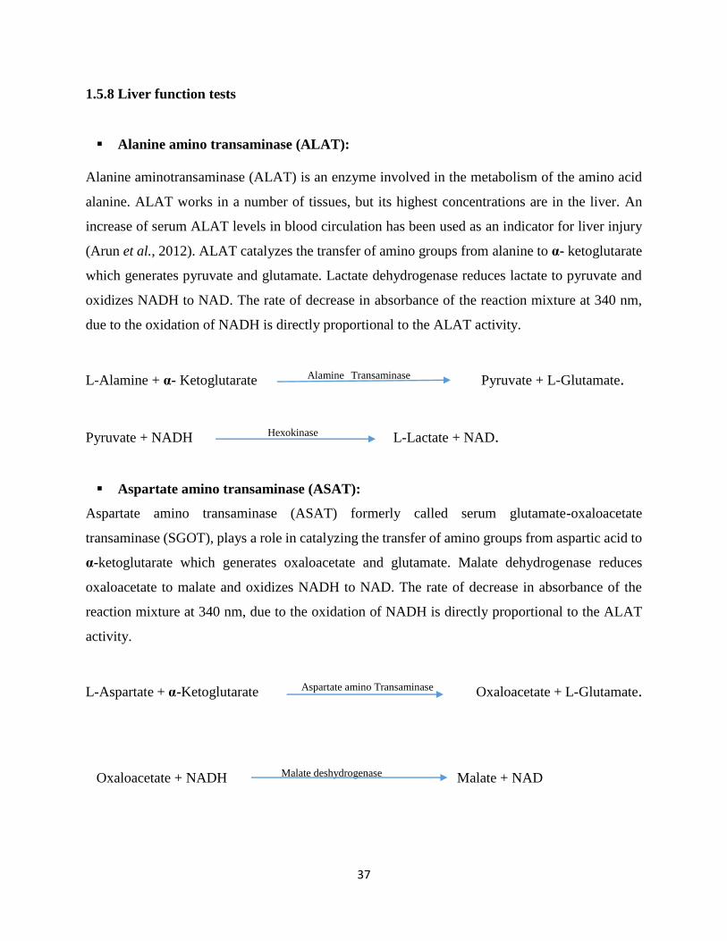
37
1.5.8 Liver function tests
Alanine amino transaminase (ALAT):
Alanine aminotransaminase (ALAT) is an enzyme involved in the metabolism of the amino acid
alanine. ALAT works in a number of tissues, but its highest concentrations are in the liver. An
increase of serum ALAT levels in blood circulation has been used as an indicator for liver injury
(Arun et al., 2012). ALAT catalyzes the transfer of amino groups from alanine to α- ketoglutarate
which generates pyruvate and glutamate. Lactate dehydrogenase reduces lactate to pyruvate and
oxidizes NADH to NAD. The rate of decrease in absorbance of the reaction mixture at 340 nm,
due to the oxidation of NADH is directly proportional to the ALAT activity.
L-Alamine + α- Ketoglutarate Alamine Transaminase Pyruvate + L-Glutamate.
Pyruvate + NADH Hexokinase L-Lactate + NAD.
Aspartate amino transaminase (ASAT):
Aspartate amino transaminase (ASAT) formerly called serum glutamate-oxaloacetate
transaminase (SGOT), plays a role in catalyzing the transfer of amino groups from aspartic acid to
α-ketoglutarate which generates oxaloacetate and glutamate. Malate dehydrogenase reduces
oxaloacetate to malate and oxidizes NADH to NAD. The rate of decrease in absorbance of the
reaction mixture at 340 nm, due to the oxidation of NADH is directly proportional to the ALAT
activity.
L-Aspartate + α-Ketoglutarate Aspartate amino Transaminase Oxaloacetate + L-Glutamate.
Oxaloacetate + NADH Malate deshydrogenase Malate + NAD

38
Gamma Glutamyl transferase (GGT).
Gamma-Glutamyltransferase is membrane-bound protein that catalyze the transfer of Gamma-
glutamyl moiety from Gamma-glutamyl peptides to other peptides such as glutathione and to L-
amino acids. Serum GGT levels can be elevated in many pathophysiological conditions such as
cardiovascular disease, metabolic syndrome, and chronic liver disease. Elevated serum levels of
GGT can also be indicative of oxidative stress. GGT is the most specific marker for liver injury.
Amino acid + Glutathione Gamma-glutamyl transferase- Glutamyl amino acid + Cysteinylglycine.
Albumin
Albumin is produced in the liver and makes up 60% of plasma protein. Albumin plays an important
physiological role in maintaining the osmotic pressure of plasma. Also, albumin binds and
transports drugs, vitamins, and other molecules. Albumin is measured as an endpoint reaction with
albumin binding to Bromocresol green (BCG), which is an anionic dye, in an acidic environment.
The increase in absorbance at 620 nm of the green colored product is relative to the albumin
concentrations in the sample (Moman and Bhimji, S., 2018 ).
1.5.9 Histological analysis.
A standard laboratory protocol for paraffin embedding was followed in processing the formalin-
preserved liver tissues. Liver specimens excised from experimental animals were washed and fixed
in 10% buffered formalin. Tissues were processed by embedding in paraffin and sectioned (4 μm
thickness). Thereafter, p-xylene was used to deparaffinize the slides which were then rehydrated
in ethanol concentration gradient (100%, 80%, 70%, and 50%) and rinsed with tap water (Essawy
et al., 2012). After, the hematoxylin and eosin were used to stain the slides for 5 minutes which
were rinsed with water. As a final point, the slides were mounted in DPX, cover-slipped and
viewed with Leica slide scanner (SCN 4000, Leica Biosystems Germany) (Yahya et al., 2013).

39
1.5.10 Statistical analysis
Data were expressed as mean ± standard deviation. One-way ANOVA or student t-tests were used
to determine statistical significance for normal distribution data, while KRUSKAL-WALLIS H or
Mann-WHITNEY U (Nonparametric tests) were used for non-normal distribution. GraphPad
Prism Software Version 5.0. (GraphPad Prism® Software, Inc. San Diego, CA, USA) was used to
compare the statistical difference between control means and treatment groups. Values of p < 0.05
were considered statistically significant.

40
REFERENCES.
Abou-Zeid, S. M. et al., 2016. In vitro hepatoprotective effects of Moringa oleifera extract against
Acetaminophen-induced hepatotoxicity. World Journal of Pharmacy and Pharmaceutical Sciences,
5(12): 50–70. doi: 10.20959/wjpps201612-8155.
Adeeyo, A. O. et al., 2013. Antihyperglycemic effects of aqueous leaf extracts of Mistletoe and
Moringa Oleifera in streptozotocin-induced diabetes Wistar rats. Diabetologia Croatica, 42(3),
Aja et al., 2014.Chemical Constituents of Moringa oleifera Leaves and Seeds from Abakaliki,
Nigeria. American Journal of Phytomedicine and Clinical Therapeutics, 2(3): 310-321.
Aja P. et al., 2015. Evaluation of anti-diabetic effect and liver function indices of ethanol extracts
of Moringa oleifera and Cajanus cajan Leaves in alloxan induced diabetic albino rats. Global
Veterinaria, 14(3): 439–447. doi: 10.5829/idosi.gv.2015.14.03.93129.
Albert Reece, 2010. The fetal and maternal consequences of gestational diabetes mellitus. The
Journal of Maternal-Fetal and Neonatal Medicine, 23(3): 199-203.
https://doi.org/10.3109/14767050903550659.
Alberti, K. G. M. M., and Zimmet, P. Z. 1998 Definition, diagnosis and classification of diabetes
mellitus and its Complications Part 1 : Diagnosis and classification of diabetes mellitus
provisional report of a WHO consultation. Diabet Med. 15(7): 539–553. DOI:
10.1002/(SICI)1096-9136(199807)15:7<539::AID-DIA668>3.0.CO;2-S.
Al-Malki, A. L. and El Rabey, H. A., 2015.The antidiabetic effect of low doses of Moringa oleifera
Lam. Seeds on streptozotocin-induced diabetes and diabetic nephropathy in male rats. BioMed
Research International, 2015: 1-13. doi: 10.1155/2015/381040.
American Diabetes Association, 2004. Nephropathy in diabetes (Position Statement). Diabetes
Care 27 (Suppl.1): S79 – S83, 2004.
American Diabetes Association, 2005. Diabetic nephropathy: diagnosis, prevention, and
treatment. Diabetes Care.8 (1): 164-176. https://doi.org/10.2337/diacare.28.1.164.

41
American Diabetes Association, 2010. Standards of medical care in diabetes-2010. Diabetes
Care, 33(Suppl 1): S11–S61. doi: 10.2337/dc10-S011.
American Diabetes Association, 2017. Classification and diagnosis of diabetes. Diabetes Care,
40(supplement 1); S11–S24. doi: 10.2337/dc17-S005.
Andreas L. and Gerald I., 2014. Non-alcoholic fatty liver disease, hepatic insulin resistance, and
Type 2 diabetes. Hepatology, 59(2): 713–723. doi: 10.1002/hep.26672.
Aney, J. S. et al., 2009. The pharmacological and pharmaceutical potential of Moringa oleifera :
A Review. Journal of Pharmacy Research, 2(9): 1424–1426.
Anwar, F. et al., 2007. Moringa oleifera : A food plant with multiple medicinal uses. Phytotherapy
Research, 25(1): 17–25. doi: 10.1002/ptr.2023.
Anwar F. et al., 2013. Effect of drying method on the total phenolics and antioxidant Activity of
Cauliflower (Brassica oleraceae L) extracts. International Food Research Journal, 20(2): 653-659.
Aronson, J. K., and Ferner, R. E. 2003. Joining the DoTS : New approach to classifying adverse
drug reactions. BMJ, 327(7425): 1222–1225. DOI: 10.1136/bmj.327.7425.1222.
Bajaj JS, Wade JB, Sanyal AJ. Spectrum of neurocognitive impairment in cirrhosis: implications
for the assessment of hepatic encephalopathy. Hepatology. 2009;50(6):2014–2021.
Bhatt, H. B., and Smith, R. J., 2015. Fatty liver disease in diabetes mellitus. Hepatobiliary Surgery
and Nutrition, 4(2): 101–108. doi: 10.3978/j.issn.2304-3881.2015.01.03.
Boulton, A.J. et al., 2005. Diabetes Neuropathies. Diabetes Care, 28(4): 956-
962.https://doi.org/10.2337/diacare.28.4.956.
Bradshaw et al., 2007. Estimating the burden of disease attributable to diabetes in South Africa in
2000, the South African Medical Journal. 97(8Pt2): 700-706.
Casqueiro, J. and Alves, C. 2012. Infections in patients with diabetes mellitus : A review of
pathogenesis. Indian Journal of Endocrinology and Metabolism, 16(7), 27-36. doi: 10.4103/2230-
8210.94253.
Centers for Disease Control and Prevention (CDC), 2017. National Diabetes Statistics Report,
42
2017. Estimates of diabetes and its burden in the United States. CDC.
https://www.cdc.gov/diabetes/data/statistics/statistics-report.html.
Chang. L et al., 2005. Insulin signaling and the regulation of glucose transport. Journal of
Molecular Medicine, 10(7): 65–71. doi: 10.2119/2005-00029.Saltiel.
Charoensin, S., 2014. Antioxidant and anticancer activities of Moringa oleifera leaves. Journal of
Medicinal Plant Research, 8(7): 318–325. doi: 10.5897/JMPR2013.5353.
Chinedu, A. et al., 2015. Effect of the ethanolic leaf extract of Moringa oleifera on insulin
resistance in streptozotocin-induced diabetic rats. Journal of Plant Sciences, 2(6-1): 5–12. doi:
10.11648/j.jps.s.2014020601.12.
Chumark, P. et al., 2008. he in vitro and ex vivo antioxidant properties, hypolipidaemic and
antiatherosclerotic activities of water extract of Moringa oleifera Lam. Leaves. Journal of
Ethnopharmacology, 116(3): 439–446. doi: 10.1016/j.jep.2007.12.010.
Cusi, K., 2009. Nonalcoholic fatty liver disease in type 2 diabetes mellitus. Current Opinion in
Endocrinology, Diabetes, and Obesity, 16(2): 141-149. doi: 10.1097/MED.0b013e3283293015.
Cydulka, R. K. and Maloney, G. E. (2002). ‘Diabetes Mellitus and Disorders of Glucose
Homeostasis’. Eighth Edition, Rosen’s Emergency Medicine - Concepts and Clinical Practice, 2-
Volume Set. Eighth Edition. Elsevier Inc. doi: 10.1016/B978-1-4557-0605-1.00126-3.
David, M. and Nathan., 2009. International expert committee report on the role of the A1C assay
in the diagnosis of diabetes. Diabetes Care, 32(7): 1327-1334. doi: 10.2337/dc09-9033.
David T, Thomas C, Zaccone P, Dunne DW, Cooke A (2004). The impact of infection on the
incidence of autoimmune disease. Curr. Top. Med. Chem., 4: 521–529.
Dawood, H. and Fathy, A., 2014. Extract of Moringa oleifera leaves ameliorates streptozotocin-
induced diabetes mellitus in adult rats. Acta Histochemica. Elsevier GmbH., 116(5): 844–854. doi:
10.1016/j.acthis.2014.02.002.
Detlef Schuppan and Nezam H Afdhal. Liver cirrhosis. Lancet, 2009; 371(9615): 838–851.
Doi:10.1016/S0140-6736(08)60383-9.
Devendra, D. et al., 2004. Clinical review Type 1 diabetes : recent developments. BMJ, 328(7442) :

43
750-754. doi: 10.1136/bmj.328.7442.750.
Dobretsov, M. et al., 2007. Early diabetic neuropathy : Triggers and mechanisms. World Journal
of Gastroenterology, 13(2): 175–191. doi: 10.3748/wjg.v13.i2.175.
Doupis, J. and Alexiadou, K., 2012. Management of diabetic foot ulcers. Diabetes Therapy, 3(1):
4.doi: 10.1007/s13300-012-0004-9.
Edoga, C. O. et al., 2013. Blood sugar lowering effect of Moringa Oleifera Lam in Albino rats’,
International Journal of Science and Technology, 3(1): 88–90.
Einhorn, D., et al., American College of Endocrinology position statement on the insulin resistance
syndrome. Endocr Pract, 2003. 9(3): 237-52.
Ekundina, V. O. et al., 2015. Hepatotoxic and nephrotoxic effects of Moringa Oleifera Leaves
extract in adult Wistar rats. Journal of Natural Sciences Research, 5(3): 110–118.
El-bakry, K. et al., 2016. Hepatoprotective effect of Moringa Oleifera leaves extract against carbon
tetrachloride-induced liver damage in rats. World Journal of Pharmacy and Pharmaceutical
Sciences, 5(5): 76–89. doi: 10.20959/wjpps20165-6638.
Eric, S. et al., 2009. International expert committee report on the role of the A1C assay in the
diagnosis of diabetes. Diabetes Care, 32(12): e159-e159. https://doi.org/10.2337/dc09-1231.
Essawy, A. E. et al., 2012. Nigella sativa seeds protect against hepatotoxicity and dyslipidemia
induced by carbon tetrachloride in mice. Journal of Applied Pharmaceutical Science, 2(10): 21–
25. doi: 10.7324/JAPS.2012.21004.
Forbes, J. M. and Cooper, M. E. 2013. Mechanisms of diabetic complications. Physiological
Reviews, 93(1): 137–188. doi: 10.1152/physrev.00045.2011.
Ford et al., 2008. Metabolic syndrome and incident diabetes. Diabetes Care, 31(9): 1898-1904.
Gayoso-diz, P. et al., 2013. Insulin resistance ( HOMA-IR ) cut-off values and the metabolic
syndrome in a general adult population : effect of gender and age : EPIRCE cross-sectional study.
BMC Endocrine Disorders 2013, 13:47. http://www.biomedcentral.com/1472-6823/13/47
Ginsberg, H. and Sung, H, 2012. Increased very low-density lipoprotein secretion, hepatic

44
steatosis, and insulin resistance. Trends in Endocrinology and Metabolism-Journal, 22(9): 353–
363. doi: 10.1016/j.tem.2011.04.007.
Gowrishankar, R., 2010. Trace element studies on Tinospora cordifolia (Menispermaceae),
Ocimum sanctum (Lamiaceae), moringa oleifera (Moringaceae), and Phyllanthus niruri
(Euphorbiaceae) using PIXE. Biological Trace Element Research, 133(3): 357–363. doi:
10.1007/s12011-009-8439-1.
Grassi, G. et al., 2016. Specific blood pressure targets for patients with diabetic nephropathy?.
Diabetes Care, 39(supplement 2); S228-S233. https://doi.org/10.2337/dcS15-3020.
Groop, L., Genetics of the metabolic syndrome. British Journal of Nutrition, 2007. 83(S1): S39-
S48.
Gupta, R. et al., 2012. Evaluation of antidiabetic and antioxidant activity of Moringa oleifera in
experimental diabetes. Journal of Diabetes, 4(2): 164–171. doi: 10.1111/j.1753-
0407.2011.00173.x.
Gupta et al., 2014. Evaluation of antioxidant and hepatoprotective activities of Moringa oleifera
Lam. Leaves in carbon tetrachloride-intoxicated rats. Antioxidants, 3, 569-591;
doi:10.3390/antiox3030569.
Harris, E. H., 2005. Elevated Liver function tests in Type 2 diabetes. Clinical Diabetes, 23(3):
115–119. doi: 10.2337/diaclin.23.3.115.
Helmy M. and Robert M., 2010. Role of the Intrarenal Renin-Angiotensin-Aldosterone System in
chronic kidney disease. American Journal of Nephrology, 31(6): 541–550. doi:
10.1159/000313363.
Herold, K. et al., 2007. Receptor for advanced glycation end products ( RAGE ) in a dash to the
rescue : inflammatory signals gone awry in the primal response to stress. Journal of Leukocyte
Biology, 82(2): 204-212. doi: 10.1189/jlb.1206751.
Inoguchi, T. et al., 2000. High glucose level and free fatty acid stimulate reactive oxygen species
production through protein kinase C – dependent activation of NAD ( P ) H oxidase in cultured
vascular Cells. Diabetes, 49(11): 1939–1945.

45
Jaiswal, D. et al., 2009. Effect of Moringa oleifera Lam. leaves aqueous extract therapy on
hyperglycemic rats. Journal of Ethnopharmacology, 123(3): 392–396. doi:
10.1016/j.jep.2009.03.036.
Jakus, V. 2004. Advanced glycation end-products and the progress of diabetic vascular
complications.Physiological Research, 53(2): 131-142.
Jensen, J. et al., 2011. The role of skeletal muscle glycogen breakdown for the regulation of insulin
sensitivity by exercise. Frontiers in Physiology, 2: 1–11. doi: 10.3389/fphys.2011.00112.
Jia, G. et al., 2016. Insulin resistance and hyperinsulinaemia in diabetic cardiomyopathyNature
Reviews Endocrinology, 12(3): 144–153. doi: 10.1038/nrendo.2015.216.
Jusciele B. Moreli et al., 2016. Hyperglycemia differentially affects maternal and fetal DNA
integrity and DNA damage response. Int J Biol Sci, 12(4): 466-477. Doi: [10.7150/ijbs.12815].
Kekuda, T. et al., 2010. Antibacterial and Antifungal efficacy of steam distillate of Moringa
oleifera Lam. Journal of Pharmaceutical Sciences and Research, 2(1): 34–37.
Kempler, G.P., 2011. Management strategies for gastrointestinal, erectile, bladder, and sudomotor
dysfunction in patients with diabetes. Diabetes Metabolism Research and Reviews, 27(7): 665–
677. doi: 10.1002/dmrr.1223.
Krzewska, A. and Ben-Skowronek, I. 2016. Effect of associated autoimmune diseases on Type 1
diabetes mellitus incidence and metabolic control in children and adolescents. Hindawi Publishing
Corporation, 2016. doi: 10.1155/2016/6219730.
Lambert, A. P. et al., 2004. The absolute risk of childhood-onset Type 1 diabetes defined by
Human Leukocyte Antigen Class II Genotype : A population-based study in the United Kingdom.
The Journal of Clinical Endocrinology and Metabolism, 89(8): 4037–4043. doi: 10.1210/jc.2003-
032084.
Lee, W. and Kim, J., 2017. Diabetic cardiomyopathy : where we are and where we are going.
Korean Journal of Internal Medicine, 32(3): 404–421. doi: 10.3904/kjim.2016.208.
Levey, A. S, and Coresh, J. 2012. Chronic kidney disease. The Lancet. Elsevier Ltd, 379(9811):
165–180. doi: 10.1016/S0140-6736(11)60178-5.
46
Levitt, DG, and Levitt, MD., 2016. Human serum albumin homeostasis: a new look at the roles of
synthesis, catabolism, renal and gastrointestinal excretion, and the clinical value of serum albumin
measurements. Int Gen Med. 9: 229-255. doi: [10.2147/IJGM.S102819].
Lewis, E., Hunsicker, L., Bain, R. and Rohde, R. (1993). The Effect of Angiotensin-converting
enzyme Inhibition on Diabetic Nephropathy. New England Journal of Medicine, 329(20): 1456-
1462.
Lie, B. et al., 1999. The predisposition to Type 1 diabetes linked to the human leukocyte antigen
complex includes at least one in – class II gene. The American Journal of Human Genetics, 64(3):
793–800.
Lilly, E. et al., 2006. Diabetic microvascular complications : can patients at risk be identified ? A
review. International Journal of Clinical Practice, 60(11): 1471–1483. doi: 10.1111/j.1742-
1241.2006.01175.
Macintosh MC, Fleming KM, Bailey JA, Doyle P, Modder J, Acolet D, Golightly S, Miller A.
Perinatal mortality and congenital anomalies in babies of women with type 1 or type 2 diabetes in
England, Wales, and Northern Ireland: population based study. BMJ 2006;333:177 7.
Marshall, C. B. 2016. Rethinking glomerular basement membrane thickening in diabetic
nephropathy : adaptive or pathogenic ?, American Journal of Physiology-Renal Physiology,
331(5): F831-F843. doi: 10.1152/ajprenal.00313.2016.
Mbanya et al., 2010. Diabetes in sub-Saharan Africa. The Lancet. Elsevier Ltd, 375(9733): 2254–
2266. doi: 10.1016/S0140-6736(10)60550-8.
Mbikay, M., 2012. Therapeutic potential of Moringa oleifera leaves in chronic hyperglycemia and
dyslipidemia: A review. Frontiers in Pharmacology, 3 MAR(March): 1–12. doi:
10.3389/fphar.2012.00024.
Menge BA, Gruber L, Jorgensen SM, Deacon CF, Schmidt WE, Veldhuis JD, Holst JJ, Meier JJ.
Loss of inverse relationship between pulsatile insulin and glucagon secretion in patients with type
2 diabetes. Diabetes. 2011; 60:2160–2168. doi: 10.2337/db11-0251.
Menghani et al., 2010 ‘Antidiabetic potentials of various ethnomedicinal plants of Rajasthan.
Ethnobotanical Leaflets, 2010(5). Available at: https://opensiuc.lib.siu.edu/ebl/vol2010/iss5/3.
47
Miki, T. et al., 2013. Diabetic cardiomyopathy: Pathophysiology and clinical features. Heart
Failure Reviews, 18(2): 149-166. doi: 10.1007/s10741-012-9313-3.
Mishra, G. et al., 2011. Traditional uses, phytochemistry and pharmacological properties of
Moringa oleifera plant: An overview. Der Pharmacia Lettre, 3(2): 141–164.
Mohamed, J. et al., 2016. Mechanisms of diabetes-induced liver damage. The role of oxidative
stress and inflammation. Sultan Qaboos University Medical Journal, 16(2): e132-
e141. doi: 10.18295/squmj.2016.16.02.002.
Moman RN, Bhimji SS. Physiology, Albumin. [Updated 2018 Sep 12]. In: StatPearls [Internet].
Treasure Island (FL): StatPearls Publishing; 2018 Jan.
Moreira, G.C., et al., Prevalence of metabolic syndrome: association with risk factors and
cardiovascular complications in an urban population. PLoS One, 2014. 9(9): e105056.
Motala et al., 2003. Diabetes in Africa. Epidemiology of type 1 and type 2 diabetes in Africa.
Journal of cardiovascular risk, 10(2): 77–83. doi: 10.1097/01.hjr.0000060843.48106.31.
Mutyambizi et al., 2018. Cost of diabetes mellitus in Africa : a systematic review of existing
literature. Globalization and Health, 14: 1–13. doi: 10.1186/s12992-017-0318-5.
Navale, A. M. 2017. Evaluation of anogeissus acuminata (roxb. ex dc.) in diabetes mellitus and its
complications, Ph.D. in pharmacy, Gujarat Technological University Ahmedabad, western Indian
districts of Gujarat.
National Cholesterol Education Program (NCEP) Expert Panel on Detection, Evaluation, and
Treatment of High Blood Cholesterol in Adults (Adult Treatment Panel III). Third report of the
National Cholesterol Education Program (NCEP) Expert Panel on Detection, Evaluation, and
Treatment of High Blood Cholesterol in Adults (Adult Treatment Panel III) final report.
Circulation 2002; 106:3143– 421.
Ndhlala, A. R. et al., 2014. Antioxidant, antimicrobial and phytochemical variations in thirteen
Moringa oleifera Lam. Cultivars. Molecules, 19(7): 10480–10494. doi:
10.3390/molecules190710480.
Nokoff N, Rewers M (2013). Pathogenesis of type 1 diabetes: lessons from natural history
studies of high-risk individuals. Ann NY Acad Sci., doi: 10.1111/nyas.12021.

48
Ogihara, T. et al., 2009. The Japanese society of hypertension guidelines for the management of
hypertension ( JSH 2009 ). Hypertension Research, 32(1): 3–107.
Ogurtsova et al., 2017. ‘IDF Diabetes Atlas : Global estimates for the prevalence of diabetes for
2015 and 2040’, Diabetes Research and Clinical Practice. Elsevier B.V., 128: 40–50. doi:
10.1016/j.diabres.2017.03.024.
Osborne N. et al., 2004. Retinal ischemia: Mechanisms of damage and potential therapeutic
strategies. Progress in Retinal and Eye Research, 23(1): 91-147.
https://doi.org/10.1016/j.preteyeres.2003.12.001.
Oyedemi, S. O. et al., 2009. Ethnobotanical survey of medicinal plants used for the management
of diabetes mellitus in the Nkonkobe municipality of South Africa. Journal of Medicinal Plants
Research, 3(12): 1040–1044.
Ozer, J. et al., 2008. The Current state of serum biomarkers of hepatotoxicity. Toxicology, 245(3):
194-205. doi: 10.1016/j.tox.2007.11.021.
Panjaliya, R. K. et al., 2013. Association of insertion/deletion polymorphism of Alu angiotensin-
converting enzyme insertion/deletion genotype with type 2 diabetes mellitus and hypertension in
J & K population : A case-control study. Global Journal of Molecular Evolution and Genomic,
1(1): 016–020.
Pathan MA, Khan, AP, Somkuward N Z G. Hepatoprotective activity of Maytenus emarginata
against paracetamol-induced liver injury in male Wistar rats. Int. J. Pharm. Pharm. Sci. 2014;
6(8): 320–323.
Pappachan, J. M. et al., 2013. Diabetic cardiomyopathy : Pathophysiology, diagnostic evaluation,
and management. World Journal of Diabetes, 4(5): 177–189. doi: 10.4239/wjd.v4.i5.177.
Paula, A.A., M.C.N. Falcão, and A.G. Pacheco, Metabolic syndrome in HIV-infected individuals:
underlying mechanisms and epidemiological aspects. AIDS Research and Therapy, 2013. 10: 32-
32.
Per H et al., 2013. Intracranial hemorrhages and late hemorrhagic disease associated with
cholestatic liver disease. Neurol Sci. 34(1): 51-56. doi: [10.1007/s10072-012-0965-5].

49
Petrovska, B.B, 2012. Historical review of medicinal plants’ usage. Pharmacognosy Review.
6(11): 1-5. doi: 10.4103/0973-7847.95849
Rahal, A. et al., 2014. Oxidative stress, prooxidants, and antioxidants : The interplay. Journal of
Biomedicine and Biotechnology, 2014. doi: 10.1155/2014/761264.
Rahman, M. M., and Sheikh, M. M. I. (2009) ‘Antibacterial Activity of Leaf Juice and Extracts of
Moringa oleifera Lam . against Some Human Pathogenic Bacteria. CMU Journal of Natural of
Sciences, 8(2): 219–228.
Ramadori, G. et al., 2008. Physiology and pathophysiology of liver inflammation, damage, and
repair. Journal of Physiology and Pharmacology, 59 suppl 1: 107-11.
Ray RY, Tsou SCS, Lee TC, Chang LC, Kuo G, Lai PY (2006). Moringa, a novel plant rich in
antioxidants, bioavailable iron, and nutrients. In Wang M (ed), Herbs: Challenges in Chemistry
and Biology of Herbs, Am. Chem. Soc. US: 224-239.
Ronco, C. et al., 2010. Cardio-renal syndromes : The report from the consensus conference of the
acute dialysis quality initiative. European Heart Journal, 31(6): 703–711. doi:
10.1093/eurheartj/ehp507.
Saalu, L. et al., 2012. The hepato-protective potentials of Moringa oleifera leaf extract on alcohol-
induced hepato-toxicity in Wistar rat. American Journal of Biotechnology and Molecular Sciences,
2(1): 6–14. doi: 10.5251/ajbms.2012.2.1.6.14.
Salgado, M., 2013. Hepatic Encephalopathy : Etiology, pathogenesis, and clinical signs. Compend
Contin Educ Vet, 35(6): 1–9.
Schalkwijk, C. G. 2008. Advanced glycation end products and diabetic foot disease. Diabetes
Metab Res Rev, 24 Suppl 1: S19-24. doi: 10.1002/dmrr.861.
Scheff, J. D. et al., 2012. Assessment of pharmacologic Area Under the Curve when Baselines are
Variable. Pharmaceutical Research, 28(5): 1081–1089. doi: 10.1007/s11095-010-0363-8.
Semenya, S. et al., 2012. Ethnobotanical survey of medicinal plants used by Bapedi healers to treat
diabetes mellitus in the Limpopo Province, South Africa. Journal of Ethnopharmacology. Elsevier
Ireland Ltd, 141(1): 440–445. doi: 10.1016/j.jep.2012.03.008.

50
Sikorski, B. et al., 2013. The diagnostic function of OCT in diabetic maculopathy. Mediators of
Inflammation, 2013. http://dx.doi.org/10.1155/2013/434560.
Silva, B. R. et al., 2008. Laparoscopic treatment of metabolic syndrome in patients with type 2
diabetes mellitus. Surgical Endoscopy, 22: 2670–2678. doi: 10.1007/s00464-008-9808-0.
Simon Rama Parmar, Patel Hitesh Vashrambhai, Karan Kalia. Hepatoprotective activity of some
plants extracts against paracetamol-induced hepatotoxicity in rats. J. Herb. Med. Toxicol. 2010;
4(2): 101–106.
Spiegel, K. et al., 2005. Sleep loss : a novel risk factor for insulin resistance and Type 2 diabetes.
Journal of Applied Physiology, 99: 2008–2019. doi: 10.1152/japplphysiol.00660.2005.
Sreelatha, S. and Padma, P. R., 2009. Antioxidant activity and total phenolic content of Moringa
oleifera leaves in two stages of maturity. Plant Foods Hum Nutr, 64(4): 303–311. doi:
10.1007/s11130-009-0141-0.
Stančáková, A. and M. Laakso, Genetics of metabolic syndrome. Reviews in Endocrine and
Metabolic Disorders, 2014. 15(4): 243-252.
Street, R. A., and Prinsloo, G., 2013. Commercially important medicinal plants of South Africa:
A Review. Journal of Chemistry, 2013: 1–16. Available at: dx.doi.org/10.1155/2013/205048.
Stumvoll, M. et al., 2005. Type 2 diabetes : principles of pathogenesis and therapy. Lancet,
365(9467): 1333-1346. https://doi.org/10.1016/S0140-6736(05)61032-X.
Susa JB: Effects of diabetes on fetal growth. In Diabetes Mellitus in Pregnancy, Principles, and
Practice. Reece EA, Coustan DR, Eds. New York, Churchill Livingstone, 1988: 105-22 2.
Takashi K et al., 2006. Adiponectin and adiponectin receptors in insulin resistance, diabetes, and
metabolic syndrome. J Clin Invest. 2006; 116(7):1784-1792. https://doi.org/10.1172/JCI29126.
Toppo, R. et al., 2015. Hepatoprotective activity of Moringa oleifera against cadmium toxicity in
rats. Veterinary World Journal, 8(4); 537-540. doi: 10.14202/vetworld.2015.537-540.
Ullah, S. et al., 2016. Hyperbilirubinemia in neonates : Types, causes, clinical examinations,
preventive measures, and treatments : A narrative review article. Iranian Journal of Public Health,

51
45(5): 558–568.
Vasanth, K. et al., 2014. Anticancer activity of Moringa oleifera mediated silver nanoparticles on
human cervical carcinoma cells by apoptosis induction. Colloids and Surfaces B: Biointerfaces.
Elsevier B.V, 117: 354–359. doi: 10.1016/j.colsurfb.2014.02.052.
Verma, V. K. et al., 2012. Anti-ulcer and antioxidant activity of Moringa Oleifera ( Lam ) leaves
against Aspirin and Ethanol-induced gastric ulcer in rats. Int. Res.J. of Pharmaceutical, 2(2): 46–
57.
Wang, Y. et al., 2016. Association between liver enzymes and incident type 2 diabetes in Singapore
Chinese men and women. BMJ Open Diabetes Research and Care, 4(1). doi: 10.1136/bmjdrc-
2016-000296.
Watkins, P. J. 2003. ABC of diabetic retinopathy. BMJ, 326(7395): 924-926.
doi: 10.1136/bmj.326.7395.924.
Weyer, C. et al., 1999. The natural history of insulin secretory dysfunction and insulin resistance
in the pathogenesis of type 2 diabetes mellitus. Journal of Clinical Investigation, 104(6):787-
794. https://doi.org/10.1172/JCI7231.
Whiting et al., 2011. IDF Diabetes Atlas : Global estimates of the prevalence of diabetes for 2011
and 2030, Diabetes Research and Clinical Practice. Elsevier Ireland Ltd, 94(3): 311–321. doi:
10.1016/j.diabres.2011.10.029.
World Health Organization (2013). WHO Traditional Medicine Strategy: 2014-2023. World
Health Organization. doi: www.who.int.
World Health Organization., 2016 Global Report on Diabetes, Isbn, 978: 88. doi: ISBN 978 92 4
156525 7.
World Health Organization., 2016. Global Report on Diabetes. Isbn, 978: 88. doi: ISBN 978 92 4
156525 7.
Wu, G. 2009. Amino acids: metabolism, functions, and nutrition. Journal of Amino Acids, 37(1):
1–17. doi: 10.1007/s00726-009-0269-0.
Wu, Y. et al., 2014.Risk factors contributing to type 2 diabetes and recent advances in the treatment
52
and prevention, International Journal of Medical Sciences, 11(11): 1185–1200. doi:
10.7150/ijms.10001.
Xie, Q. et al., 2012. Protective effects of cassia seed ethanol extract against carbon tetrachloride-
induced liver injury in mice. Acta Biochim Pol, 59(2): 265–270.
Xie Z, Lau K, Eby B, Lozano P, He C, Pennington B, Li H, Rathi S, Dong Y, Tian R, Kem D, Zou
MH. Improvement of cardiac functions by chronic metformin treatment is associated with
enhanced cardiac autophagy in diabetic OVE26 mice. Diabetes 2011; 60: 1770-1778 [PMID:
21562078 DOI: 10.2337/db10-0351].
Yahya, et al., 2013. Hepatoprotective activity of methanolic extract of Bauhinia purpurea leaves
against paracetamol-induced hepatic damage in rats. Evidence-Based Complementary and
Alternative Medicine, 2013, 10. https://doi.org/http://dx.doi.org/10.1155/2013/636580.
Yang, J. and Monti, D.A., (2017). Clinical acupuncture and ancient Chinese medicine. United
States of American, Oxford University.
Yavuz, F. et al., 2017. Serum ischemic modified albumin ( IMA ) concentration and IMA/albumin
ratio in patients with hepatitis B-related chronic liver diseases. Turkish Journal of Medical
Sciences, 47(3): 947–953. doi: 10.3906/sag-1611-66.
Zheng, Y., Ley, S. H. and Hu, F. B. 2017. Global etiology and epidemiology of type 2 diabetes
mellitus and its complications, Nature Reviews Endocrinology, 14(2): 88–98. doi:
10.1038/nrendo.2017.151.
Zochodne, D. W., 2007. Diabetes mellitus and the peripheral nervous system : manifestations and
mechanisms. Muscle and Nerve, 36(2): 144–166. doi: 10.1002/mus.20785.

53
CHAPTER 2: RESULTS.
After presenting the literature review, methods, aim and objectives of the study in
chapter one, the general findings, discussion and results of the study were further
reported in chapter two. A paper was presented in the form of a manuscript titled
“Hepatoprotective effects of Moringa oleifera leaf extracts in streptozotocin-induced
diabetes”. This manuscript is submitted to Journal of Functional Foods for publication.

54
Hepatoprotective effects of Moringa oleifera Lam (Moringaceae) leaf extracts in
streptozotocin-induced diabetes.
Authors: Willy Tambwe Muzumbukilwa1, Manimbulu Nlooto1, Peter Mark Oroma Owira1*.
*: Corresponding author, email address: [email protected]; Tel: +27312607720
1. The discipline of Pharmaceutical Sciences, School of Health Sciences, Westville campus,
University of KwaZulu-Natal, University Road, Durban 4001/South Africa.
Emails: [email protected]; Tel: +27620163854
[email protected]; Tel: +27760911427

55
Abstract
Effects of methanolic leaf extracts of Moringa oleifera (MO) on hepatic injury in streptozotocin
(STZ)-induced diabetes were investigated.
Male Wistar rats were divided into 6 groups (n=7). Animals in group A were orally treated daily
with 3.0 ml/kg/body weight (BW) of distilled water, while groups C, E and F were similarly treated
with 500, 250 and 500 mg/kg/BW, respectively with MO. Groups B, D, E and F were rendered
diabetic by single intraperitoneal injections of 45 mg/kg/BW of STZ. Additionally, group D was
treated with subcutaneous insulin (2.0 U/kg/BW, bid).
Diabetic animals exhibited significant weight loss, polydipsia, impaired glucose tolerance, fasting
hypoinsulinemia and impaired liver function tests compared to controls. Treatment with MO
methanolic leaf extracts significantly improved weight loss, polydipsia, glucose tolerance and also
liver function tests in diabetic animals.
MO has dose-dependent antidiabetic and hepatoprotective effects.
Keywords
Moringa oleifera, diabetes, streptozotocin, liver.
Abbreviations
ADA, American diabetes association; ANOVA, one way analysis of variance; ALAT, Alanine
Aminotransaminase; ASAT, Aspartate Aminotransaminase; AUC, areas under the curve; BW,
body weight; FBG, fasting blood glucose; FPI, fasting plasma insulin; GC-MS chromatography-
mass spectrometry; GGT, gamma-glutamyl aminotransaminase; MO, Moringa oleifera; OGGT,
oral glucose tolerance tests; STZ, streptozotocin.

56
1.0 Introduction
Diabetes mellitus is associated with increased morbidity and mortality globally and 425 million
adults aged between 20 and 79 years were diagnosed with diabetes in 2017 (IDF, 2018; Ogurtsova
et al., 2017). Due to defects in insulin secretion, insulin action, or both persistent hyperglycemia
affects carbohydrate, lipid and protein metabolism (ADA, 2017). Long term complications include
hepatic injury characterized by cirrhosis, inflammation, apoptosis, and microvascular and
macrovascular aberrations (ADA 2009 ).
Liver function tests are commonly used in clinical practice to diagnose and monitor liver diseases.
Liver enzymes such as Alanine Aminotransferase (ALAT; EC 2.6.1.2), Aspartate
Aminotransferase (ASAT; EC 2.6.1.1), Gama-glutamylaminotransferase (GGT; EC 2.3.2.2) and
albumin are routinely used as biomarkers for hepatocyte integrity (Harris, 2005). GGT is more
specific to liver injury, is responsible for the catabolism of extracellular glutathione and is elevated
in conditions of increased oxidative stress and inflammation such as diabetes (Wang et al, 2016).
ALAT is specific for hepatocyte necrosis and although ASAT levels may be elevated in other
conditions such as heart failure or myocardial infarction, increased levels are also indicative of
liver disease. The liver synthesizes albumin, and hepatocellular inflammation and increased
oxidative stress which are common conditions in persistent hyperglycemia is known to cause
hypoalbuminemia (Levitt &Levitt, 2016).
It has been reported that 80% of the world’s population use traditional medicines in primary
healthcare due to their abundance, affordability, safety, and efficacy (WHO Traditional Medicine
& Strategy, 2014). In South Africa for example, 80% of the population uses some sort of traditional
medicines to meet their primary health care needs (van Wyk, 2008). One of such medicinal plants
is MO which is commonly called “drumstick tree” in English, "Zogale" and "George," in Hausa
and Igala languages of Nigeria, respectively (Atawodi et al., 2010). MO was first discovered in
India, Asia and some parts of Western Africa in countries such as Ghana and Senegal (Booth,
1988). In the Limpopo province of South Africa, MO is grown by small scale farmers and the
leaves processed into a powder that is sold commercially in local markets (Mabapa et al, 2017).
The leaves can be harvested as early as 3 months post-planting for domestic use as a food source
or dried to concentrate nutrients and facilitate preservation, then ground by traditional methods
into fine powder that acts as feed source for both humans and domestic animals (Mabapa et al,

57
2017; Moyo et al., 2011; Maroyi, 2006; Gadziray at al., 2013). MO has 13 drought resistant sub-
species that are fast growing (up to 6-7 m/year) in different parts of the world (Mabapa et al., 2017;
Odee, 1998). In the province of KwaZulu-Natal, as in the rest of the country, traditional healers
use different morphological parts of the plant to treat infectious, chronic and acute degenerative
ailments.
It has since emerged that MO has human and animal nutritive value owing to the high contents of
proteins, flavonoids, vitamins and minerals such as calcium, iron, magnesium, and phosphorus
(Gowrishankar et al., 2010; Chinedu et al., 2014; Mishra et al., 2011). Furthermore, MO extracts
have been credited with pharmacological effects such as antioxidant (Charoensin, 2014; Ndhlala
et al., 2014), antiatherosclerotic and hypolipidemic (Chumark et al., 2008; Yassa & Tohamy,
2014), antineoplastic (Charoensin, 2014; Yassa & Tohamy, 2014; Vasanth et al, 2014 ), antipyretic
(Aney et al., 2009), anti-inflammatory, antispasmodic and diuretic (Anwar et al., 2007),
antibacterial (Ndhlala et al., 2014), antifungal (Kekuda et al., 2010), anti-hyperglycemic (Edoga
et al., 2013) and hepatoprotective effects (Abou-Zeid et al., 2016; El-bakry et al., 2016).
Glucotoxicity associated with diabetes is known to cause inflammation and oxidative stress which
interfere with lipid and glucose metabolism (Mota et al, 2016). A Japanese Society of Diabetes
study previously reported that the leading cause of death in patients with diabetes mellitus was a
chronic liver disease (Shima et al, 2013). It is envisaged that with the high prevalence of type 2
diabetes globally (Ogurtsova et al., 2017), a significant number of people could be afflicted with
hepatic dysfunctions secondary to chronic hyperglycemia.
To the best of our knowledge, the effects of MO leaf extracts on hyperglycemia-related
hepatotoxicity have not previously been studied. It is envisaged in this study that besides its known
antihyperglycemic effects, MO leaf extracts could mitigate the development of hyperglycemia-
induced hepatic injury in experimental animals.

58
2.0 Materials and Methods
2.1 Reagents
All chemicals and reagents unless otherwise stated were purchased from Sigma Aldrich Pty. Ltd.,
Johannesburg, South Africa. Diagnostic kits for insulin, ALAT, ASAT, GGT and albumin,
streptozotocin, citric acid, sodium phosphate, potassium chloride, methanol, phosphate buffer,
sodium citrate, sodium chloride were used in the study. Normal saline, insulin, glucometer, and
glucometer test strips were purchased from a local pharmacy in Durban, South Africa.
2.2 Plant material and preparation of methanol extracts.
Two thousand grams fresh leaves of MO were collected in Durban, South Africa and identified by
an expert Botanist at the herbarium in the School of Life Sciences, University of KwaZulu-Natal.
The leaves were then washed with running tap water to remove soil particles and other dirt, and
air-dried under room temperature (30 ± 2⁰ C) for 12 days. The dried leaves were pulverized to fine
granules using an electric grinder. Water and aqueous-methanol extracts were prepared according
to the modified methods of Makkar et al, (1996) and Sultana and Anwar, (2007). Briefly, 100 g of
dry MO leaf powder was weighed and soaked in 500 ml of distilled water in a volumetric flask.
Water was gradually added to the flask which was then shaken with a magnetic stirrer for 48 hours
until a slurry of uniform consistency was formed and then centrifuged at 3000 rpm for 10 min. The
extract was then filtered by Whatman filter paper (24 cm). The residue was re-extracted and a
mixture of water and methanol (20:80 v/v) gradually added to this flask with gentle shaking until
a slurry of uniform consistency was again formed. Supernatants collected were dispensed into
round-bottomed flasks which were then rotary evaporated at 40 °C. Finally, the dried extract
residues were weighed and stored at -4˚C until use.
2.3 Gas Chromatography-Mass Spectrometry (GC-MS) analysis
A PerkinEmler® Gas Chromatography (Clarus® 580) equipped with MSD mass spectrometer
(Clarus® SQ8S) instrument with built-in autosampler was used carry out GC-MS analysis.
Analytical parameters were set as Elite-5MS (30 m x 0.25 mm id x 0.25 μm) column; oven
temperature 37 to 320°C at a rate of 18-25°C/min and held for 0.5 and 1.85 mins at 18 and 320
°C, respectively; injector temperature was 250°C; MS Ion Source temperature was 280°C with full
scan and solvent delay of 0–2.30 min; ms scan Range was m/z 35 – 500 in 0.10 sec ; carrier gas
was Heat split flow of 20 ml/min; sample volume was 1.0 µl.

59
2.4 Animals treatment.
Male Wistar rats (250-300 g) were procured from the Biomedical Resource Unit (BRU) of the
University of KwaZulu-Natal. They were kept in cages and housed in the animal holding room
facility and exposed to a 12-hour light/dark cycle in a controlled room (temperature 24±1ᴼC,
humidity 55±5%). The animals had free access to standard commercial chow and tap drinking
water. They were treated with humane care according to the guidelines of the Animal Research
Ethics Committee of the University of KwaZulu-Natal; Ethical clearance number:
AREC/038/017M.
2.5 Experimental design and protocol
After one week of acclimatization, the rats were randomly divided into 6 groups (n=7). Rats in
group A served as normal control and were daily orally dosed with 3.0 ml/kg body weight (BW)
of distilled water. Groups B, D, E, and F were rendered diabetic by a single intraperitoneal injection
of 45 mg/kg BW of streptozotocin (STZ) in 0.2 ml of 0.1 M citrate buffer pH 4.5, after an overnight
fast. Rats in groups C and F were orally treated daily with 500 mg/kg BW of MO. Group E was
orally treated daily with 250 mg/kg BW of MO. Additionally, group D animals were treated with
subcutaneous injections of insulin (2.0 IU bid), (Table 1). Administration of STZ was done on day
1. Administration of MO/insulin started three days later when hyperglycemia was established.
Table 1. Animal treatment protocol. The animals were orally treated daily with distilled water,
MO methanolic leaf extracts, regular insulin and a single intraperitoneal injection of STZ in citrate
buffer, respectively. (BW = Body Weight).
Treatment Distilled Water MO MO STZ Insulin
Groups ml/kg BW mg/kg BW mg/kg BW mg/kg BW IU/kg BW
A 3.0 Nil Nil Nil Nil
B Nil Nil Nil 45 Nil
C Nil Nil 500 Nil Nil
D Nil Nil Nil 45 2.0
E Nil 250 Nil 45 Nil
F Nil Nil 500 45 Nil

60
Three days after STZ administration, development of diabetes was confirmed by tail pricking to
analyze blood glucose levels using hand-held glucometer with trips. Rats with fasting blood
glucose (FBG) more than 6.0 mmol/L were included in the study. Administration of MO/insulin
starts immediately after confirmation of diabetes and ends 24 hours before the sacrifice of animals.
Animals were treated consecutively for 53 days, they were sacrificed 24 hours after the last
treatment or dose of MO/insulin. GTT was done by intraperitoneal injection of 3.0 g/kg BW of D-
glucose in 0.9% normal saline on day 47 after an overnight fast. Blood glucose concentrations
were similarly measured at 0, 30, 60, 90, and 120 minutes. Areas under the Curve (AUC) were
calculated from blood glucose-time plots and expressed as AUC units (millimoles /liter X
minutes). The animals were then sacrificed by halothane overdose after 54 days and blood drawn
by cardiac puncture in heparinized tubes and separated into plasma that was kept at -80ᴼC for
further biochemical analysis. Livers were excised, divided into equal halves which were either
snap-frozen in liquid nitrogen for further biochemical analysis or kept in phosphate buffered-
formalin for histological analysis.
2.5.1 Liver function tests
Liver function biomarkers ALAT, ASAT, GGT, and Albumin were measured by commercial kits
as per the manufacturer’s instructions.
2.5.2 Fasting plasma insulin
Plasma insulin levels were analyzed by ultrasensitive rat insulin enzyme-linked immunoassay kit
(DRG Instruments GmbH., Marburg, Germany) as per the manufacturer’s instructions.
2.5.3 Histological analysis
Standard laboratory protocol for paraffin embedding was followed in the processing of the
formalin-preserved liver tissues. The livers were washed and fixed in 10% phosphate-buffered
formalin. The tissues were then embedded in paraffin and sectioned into slices of 4 μm thickness.
Thereafter, p-xylene was used to deparaffinize the slides which were then rehydrated in ethanol
concentration gradient (100%, 80%, 70%, and 50%) and rinsed with tap water (Essawy et al,
2012). Hematoxylin and eosin were used to stain the slides for 5 minutes then rinsed with water.

61
The slides were then mounted in Distyrene, Plasticizer and Xylene (DPX) resin, cover-slipped and
viewed with Leica slide scanner (SCN 4000, Leicabiosystems Germany) (Yahya et al., 2013).
2.6 Statistical analysis
Data were expressed as mean ± standard deviation. One-way ANOVA or Student‘s t-tests were
used to determine statistical significance for normal distribution data, while KRUSKAL-WALLIS
H or Mann-WHITNEY U (Nonparametric tests) were used for non-normal distribution. GraphPad
Prism Software Version 5.0. (GraphPad Prism® Software, Inc. San Diego, CA, USA) was used to
compare the statistical differences between treatment groups. Values of P< 0.05 were considered
statistically significant.
3.0 Results
3.1 GC-MS analysis
Potentially bioactive constituents of the MO leaf extracts were separated and identified by GC-MS
with an in-built automated mass spectral software that directly compared the peaks and retention
times with those of standard compounds in the library following characteristic fragmentation
patterns of mass spectra of the identified compounds.
Identified compounds included hydrocarbons, acids, esters, alcohols, and aromatics. Most
prominent peaks were identified as acetic acid, octyl ester, 2-pyrrolidinone, furan-2-carboxylic
acid, malic acid, glycine, benzene acetic acid, 4-hydroxy-, ethyl ester, D-galactopyranoside, D-
glucopyranoside, 9H-pyrido[3,4-b]indole, cyclopentane, 1-isopropylidene-2-trimethylsilyl, cis-
7,10,13-Hexadecatrienal, benzyl-β-D-glucoside, octadecanoic acid, (2-phenyl-1,3-dioxolan-4-yl,
2-(2-Bromoethyl)-3-methyl-oxirane (Table 2).

62
Table 2
Representative compounds identified by MS scans from integrated chromatogram peaks. The most
prominent peaks are highlighted in bold. R.T (Retention Time), A/H (Area/Height ratio).
Peak# R. Time Area Area% Height Height% A/H Name
3 3.028 231622 0.77 2639841 4.47 0.85 1-Propanamine, 2-methyl-N-(2-
methylpropylidene
6 3.661 746671 0.94 1952843 3.30 1.41 Butyrolactone
9 3.802 5289671 1.82 2484839 4.20 2.13 N-(2-Methylbutylidene)isobutylami
10 3.877 1443668 0.50 740235 1.25 1.95 N-methylene-n-octylimine
13 4.418 1961438 0.67 716172 1.21 2.74 Pyrazine, 2-ethyl-3-methyl-
14 4.488 1035382 0.36 250872 0.42 4.13 2-Hexenoic acid
15 4.606 2798953 0.96 593850 1.00 4.71 Acetic acid, octyl ester
16 4.670 5652995 1.94 2934702 4.96 1.93 1-Butanamine, 2-methyl-N-(2-
methylbutylidene
17 4.759 4000179 1.37 1448581 2.45 2.76 1-Butanamine, 3-methyl-N-(3-
methylbutylidene)-
18 4.802 5501102 1.89 1410588 2.39 3.90 2-Propenoic acid, 2-methyl-, 1-
methylethyl ester
20 5.095 5248693 1.80 1076697 1.82 4.87 2-Pyrrolidinone
21 5.156 1785509 0.61 785709 1.33 2.27 [1,3]Diazepan-2,4-dione
25 5.704 7071155 2.43 3259889 5.51 2.17 4H-Pyran-4-one, 2,3-dihydro-3,5-
dihydroxy-6
26 5.834 2443005 0.84 849869 1.44 2.87 Ethyl hydrogen succinate
28 6.005 1778374 0.61 422370 0.71 4.21 Furan-2-carboxylic acid [2,2,2-
trifluoro-1-(2-o
30 6.274 3321809 1.14 944318 1.60 3.52 Benzofuran, 2,3-dihydro-
32 6.502 1166600 0.40 338981 0.57 3.44 Benzeneacetic acid
33 6.763 2078329 0.71 479400 0.81 4.34 Malic Acid
35 7.034 1631466 0.56 565238 0.96 2.89 2-Methoxy-4-vinylphenol
40 7.940 1095895 0.38 167187 0.28 6.55 Glycine, N-(trifluoroacetyl)-, 1-
methylbutyl ester
42 8.282 2611355 0.90 874437 1.48 2.99 Benzeneacetonitrile, 4-hydroxy-

63
45 8.762 5525823 1.90 696614 1.18 7.93 Benzeneacetic acid, 4-hydroxy-, ethyl
ester
46 8.803 1382837 0.47 554126 0.94 2.50 1H-Pyrrole, 2-phenyl-
48 9.003 1370724
0.47 890099 1.51 1.54 1H-Pyrrole, 2-(2,4,6-cycloheptatrienyl)-
51 9.325 1640494 0.56 617500 1.04 2.66 Megastigmatrienone
52 9.388 1123407 0.39 410069 0.69 2.74 Ethyl 9-decenoate
53 9.593 21509411 7.38 2913249 4.93 7.38 alpha.-D-Galactopyranoside, methyl
54 9.720 47517811 16.31 3338788 5.65 14.23 Ethyl.alpha.-d-glucopyranoside
55 9.943 15564596 5.34 1501540 2.54 10.37 Ethyl .alpha.-d-glucopyranoside
60 10.982 18914584 6.49 1645304 2.78 11.50 Octyl-.beta.-D-glucopyranoside
64 11.727 13643205 4.68 1876552 3.17 7.27 9H-Pyrido[3,4-b]indole
65 11.807 28277500 9.70 2209710 3.74 12.80 Cyclopentane, 1-isopropylidene-2-
trimethylsilyl
66 12.223 1560703 0.54 258284 0.44 6.04 cis,cis,cis-7,10,13-Hexadecatrienal
68 12.985 3087096 1.06 490810 0.83 6.29 Benzyl .beta.-d-glucoside
69 13.431 1589967 0.55 263882 0.45 6.03 Octadecanoic acid, (2-phenyl-1,3-
dioxolan-4-yl
70 14.018 2609788 0.90 713437 1.21 3.66 2-Dimethyl(trimethylsilylmethyl)silyloxy
methyltetrahydrofuran
71 14.203 1251510 0.43 288339 0.49 4.34 3,5-O-Benzylidene-1-deoxy-d-gluco-d-
guloheptitol
72 14.302 1402047 0.48 427768 0.72 3.28 beta.-l-Rhamnofuranoside, 5-O-acetyl-
thio-octy
75 15.433 1294605 0.44 101719 0.17 12.73 2-(2-Bromoethyl)-3-methyl-oxirane

64
3.2.0 Evidence of diabetes
3.2.1 Animal weights
Diabetic rats exhibited significantly (p<0.05) reduced weight gain compared to controls.
Treatment with either MO (500 mg/kg BW) or insulin significantly (p<0.05) reduced weight loss
in diabetic rats compared to untreated diabetic group (Fig 1). There were no significant differences
between MO treated non-diabetic and control rats or between MO-treated (250 mg/kg BW) and
non-treated diabetic rats. There was a weight loss in the diabetic groups (Groups B, D, E, and F).
A C B D E F
-100
-50
0
50
100
150
200
***
# #
WEI
GHT
CHA
NGE(
g)
Fig. 1. Change in animal weights between day 0 and day 54 of treatment. A = controls; C= MO
(500 mg/kg BW)-treated, non-diabetic; B= Non-treated, diabetic; D= Insulin (2.0 IU/kg BW, bid)-
treated diabetic; E= MO (250 mg/kg BW)-treated, diabetic; F= MO (500 mg/kg BW)-treated,
diabetic. (***, # p<0.05 compared to control (A) and non-treated diabetic group (B), respectively)

65
3.2.2 Water consumption
Average water intake was significantly (p<0.05) higher in non-treated diabetic animals compared
to controls. Treatment with either MO (500 mg/kg BW) or insulin (2.0 IU/kg BW, bid)
significantly (p<0.05) reduced water intake in diabetic rats compared to non-treated diabetic rats
(Fig. 2). There were no significant differences between MO treated non-diabetic and control rats
or between MO-treated (250 mg/kg BW) and non-treated diabetic rats.
A C B D E F
0
1
2
3
4
5
Avera
ge da
ily w
ater c
onsum
ption
(ml/k
g BW
)
***
##
Fig. 2. Average daily water consumption in treatment groups. A = controls; C= MO (500 mg/kg
BW)-treated, non-diabetic; B= Non-treated, diabetic; D= Insulin (2.0 IU/kg BW, bid)-treated,
diabetic; E= MO (250 mg/kg BW)-treated, diabetic; F= MO (500 mg/kg BW)-treated, diabetic.
(***, # p<0.05 compared to control (A) and non-treated diabetic group (B), respectively)

66
3. 2.3 Fasting blood glucose
FBG concentrations were significantly (p<0.05) elevated in non-treated diabetic animals compared
to controls. Treatment with either MO (500 mg/kg BW) or insulin (2.0 IU/kg BW, bid)
significantly (p<0.05) reduced FBG compared to non-treated diabetic animals (Fig. 3). There were
no significant differences between MO treated non-diabetic and control rats or between MO-
treated (250 mg/kg BW) and non-treated diabetic rats.
A C B D E F
0
10
20
30
40
***
#
#
Fast
ing
Bloo
d G
luco
se (m
M)
Fig. 3. FBG concentrations measured after the animals were fasted overnight and blood drawn by
tail pricking into a test strip and measured by a glucometer. A = controls; C= MO (500 mg/kg
BW)-treated, non-diabetic; B= Non-treated, diabetic; D= Insulin (2.0 IU/kg BW, bid)-treated
diabetic; E= MO (250 mg/kg BW)-treated diabetic; F= MO (500 mg/kg BW)-treated, diabetic. (***,
# p<0.05 compared to control (A) and non-treated diabetic group (B), respectively).

67
3.2.4 Fasting plasma insulin
Fasting plasma insulin concentrations were significantly (p<0.05) reduced in non-treated diabetic
rats compared to controls. Treatment with either MO or insulin (2.0 IU/kg BW, bid) significantly
(p<0.05) improved fasting plasma insulin concentration in diabetic compared to non-treated
diabetic rats. There were no significant differences between MO-treated non-diabetic and control
rats (Fig. 4).
A C B D E F
0
10
20
30
***
# #
#
^
Fas
tin
g p
lasm
a in
suli
n (
U/m
l)
Fig. 4. Fasting plasma insulin measured in heparinized blood that was drawn by cardiac puncture
after the animals were euthanized by halothane overdose. A = controls; C= MO (500 mg/kg BW)-
treated, non-diabetic; B= Non-treated, diabetic; D= Insulin (2.0 IU/kg BW, bid)-treated diabetic;
E= MO (250 mg/kg BW)-treated diabetic; F= MO (500 mg/kg BW)-treated, diabetic. (***, #, ^
p<0.05 compared to control (A), non-treated diabetic group (B) and MO (250 mg/kg BW)-treated
diabetic group (E), respectively).

68
3.2.5 Glucose tolerance
Calculated AUC units from GTT suggested that untreated diabetic rats had significantly (p<0.05)
increased glucose intolerance compared to controls. Treatment with MO or insulin (2.0 IU/kg BW)
significantly reduced glucose intolerance in diabetic animals compared to the untreated diabetic
group. There were no significant differences between MO treated non-diabetic and control rats
(Fig. 5).
A C B D E F
0
1000
2000
3000
4000
5000
***
##
#
Cal
cula
ted
AU
C u
nits
Fig. 5. Calculated AUCs from glucose tolerance curves in GTT. A = controls; C= MO (500 mg/kg
BW)-treated, non-diabetic; B= Non-treated, diabetic; D= Insulin (2.0 IU/kg BW, bid)-treated
diabetic; E= MO (250 mg/kg BW)-treated diabetic; F= MO (500 mg/kg BW)-treated, diabetic.
(***, # p<0.05 compared to control (A) and non-treated diabetic group (B), respectively).

69
3.3.0 Liver function tests
3.3.1 Liver enzymes
Fasting plasma ALAT, ASAT and GGT concentrations were significantly (p<0.05) elevated in
non-treated diabetic rats compared to controls (Fig 6A-C). Treatment with MO (either 500 or 250
mg/kg BW) or insulin (2.0 IU/kg BW) significantly reduced plasma ALAT, ASAT and GGT levels
in diabetic compared to non-treated diabetic rats. Treatment with MO (500 mg/kg BW)
significantly (p<0.05) reduced plasma GGT concentrations in diabetic rats compared to MO (250
mg/kg BW) (Fig 6C). There were no significant differences in liver enzymes between MO-treated
non-diabetic and control rats.
A
B
A C B D E F
0
20
40
60
80
100
***
# # #
AL
AT
co
nc
en
tra
tio
ns
(IU
/L)

70
C
Fig 6. Fasting plasma A: ALAT; B: ASAT and C: GGT concentrations after the animals were
euthanized by halothane overdose at the end of the treatment period and blood collected in
heparinized tubes by cardiac puncture. A = controls; C= MO (500 mg/kg BW)-treated, non-
diabetic; B= Non-treated, diabetic; D= Insulin (2.0 IU/kg BW, bid)-treated diabetic; E= MO (250
mg/kg BW)-treated diabetic; F= MO (500 mg/kg BW)-treated, diabetic. (***, #, $ p<0.05
compared to control (A), non-treated diabetic group (B), and MO (250 mg/kg BW)-treated diabetic
group (E), respectively.
A C B D E F
0
20
40
60
80
100
***
# #
#
AS
AT
Co
ncen
trat
ion
s (I
U/L
)
A C B D E F
0
20
40
60
80
100
***
#
#
#
$
GG
T C
on
cen
trat
ion
s (I
U/L
)

71
3.3.2 Plasma albumin
Plasma albumin concentrations were significantly (p<0.05) reduced in non-treated diabetic rats
compared to controls. Treatment with MO (either 500 or 250 mg/kg BW) or insulin (2.0 IU/kg
BW) significantly (p<0.05) increased plasma albumin levels in diabetic compared to non-treated
diabetic rats. There were no significant differences in plasma albumin levels between MO-treated
non-diabetic and control rats (Fig 7).
A C B D E F
0
20
40
60
***
# # #
Alb
um
in (
g/l
)
Fig. 7. Plasma albumin concentrations in blood that was collected by cardiac puncture in
heparinized tubes at the end of the treatment period. A = controls; C= MO (500 mg/kg BW)-
treated, non-diabetic; B= Non-treated, diabetic; D= Insulin (2.0 IU/kg BW, bid)-treated diabetic;
E= MO (250 mg/kg BW)-treated diabetic; F= MO (500 mg/kg BW)-treated, diabetic. (***, #
p<0.05 compared to control (A) and non-treated diabetic group (B), respectively).
3.4.0 Histological analysis
The histopathological examination of liver sections from the control group (panel A) showed
normal liver structure including central vein (CV) and hepatic cells (hp). However, non-treated
diabetic rats (panel B) had livers with histopathological alterations such as degeneration of
hepatocytes and inflammatory infiltrates with the fragmentation of the nucleus and cell lysis (F),
necrotic hepatocytes (N), hepatic vein congestion (C), as well as vesicular cytoplasm (V). These
pathological aberrations were absent in diabetic rats that were treated with either MO extracts or

72
insulin (panels E, F, D respectively) (Fig 8).
Fig. 8. Hematoxylin and eosin stains of sections of liver tissues at the end of the treatment period.
Panel A = controls showing normal liver structure including central vein (CV) and hepatic cells
(hp); Panel B= Non-treated, diabetic livers with degeneration of hepatocytes and inflammatory
infiltrates with fragmentation of the nucleus and cell lysis (F), necrotic hepatocytes (N), hepatic
vein congestion (C), as well as vesicular cytoplasm (V); Panel C= MO (500 mg/kg BW)-treated,

73
non-diabetic showing normal histopathology similar to controls; Pane D= Insulin (2.0 IU/kg BW,
bid)-treated diabetic showing improved hepatocyte architecture; Panel E= MO (250 mg/kg BW)-
treated diabetic livers showing improved hepatocyte architecture similar to the insulin-treated
diabetic group and Panel F= MO (500 mg/kg BW)-treated, diabetic livers with prominent central
vein (CV), reduced hepatic vein congestion (C), necrotic hepatocytes (N), fragmentation of
nucleus and cell lysis (F) and vesicular cytoplasm (V).
4.0 Discussion
This study was designed to investigate the hepatoprotective effects of MO methanolic leaf extracts
in diabetic rats. Diabetes was induced by a single intraperitoneal injection of 45 mg/kg BW of STZ
which partially destroyed pancreatic β-cells leading to hyperglycemia (El-mahmoudy et al, 2005;
Szkudelski, 2001). Our results here indicate that the animals were indeed diabetic with reduced
weight gain (Fig. 1), polydipsia (Fig. 2), increased FBG (Fig. 3), reduced fasting plasma insulin
(Fig. 4) and impaired glucose tolerance (Fig. 5). Treatment with MO methanolic leaf extracts (500
mg/kg BW) significantly mitigated these symptoms of diabetes (Figs. 1-5). That treatment with
250 mg/kg BW of MO methanolic leaf extracts only led to partial antidiabetic effects suggests that
these pharmacological effects of MO are dose-dependent. Moringa oleifera contains alkaloids,
flavonoids, glycoside, stilbenes and terpenoids as its bioactive compounds, which elicit their anti-
diabetic effect by causing an increase in insulin sensitivity or by inhibition of the intestinal
absorption glucose or to the facilitation of metabolites in insulin-dependent processes (Gupta and
Misra, 2006).
This study found that insulin levels were increased in diabetic rats treated with MO extracts at
different dosages (250 mg and 500 mg). This finding is in agreement with another study done by
Anthanont et al., (2016).
Hepatic injury associated with increased hyperglycemia has previously been documented (Mota
et al., 2016; Sokolovska et al, 2015; Shima et al, 2013). In our study, degeneration of hepatocytes
and inflammatory infiltrates with the fragmentation of the nucleus and cell lysis, necrotic
hepatocytes, hepatic vein congestion, as well as vesicular cytoplasm were evident in non-treated
diabetic rats (Fig. 8) similarly to previous reports (Kamalakkannan and Prince, 2006; Omodanisi

74
et al, 2017). However, treatment with MO just like insulin, restored some of these
histopathological aberrations, suggesting a protective role of MO in hyperglycemia-associated
hepatic injury. We have presented evidence of hepatocellular injury by measuring plasma levels
of key enzymes that are used clinically in liver function tests. ALAT, ASAT, and GGT were all
elevated in untreated diabetic rats but treatment with either MO methanolic leaf extracts or insulin
significantly reduced plasma levels of these enzymes in diabetic animals (Fig. 6) yet there were no
significant differences in these enzymes’ concentrations between MO-treated non-diabetic and
control rats. That response in GGT levels was significantly lower with 500 than with 250 mg/kg
BW of MO in diabetic animals suggests yet again that hepatoprotective effects of MO could be
dose-dependent considering that, clinically GGT is a more reliable marker of hepatic injury than
the other enzymes (Fig. 6C), (Wang et al, 2016). ). The reduction of ALAT, ASAT, and GGT
concentration after MO treatment observed in this study is in agreement with another study by
Gupta et al., (2014).
The increased activities of these enzymes in diabetic rats in this study may be a result of
hepatocellular damage caused by oxidative stress that results in the leakage of ALAT, ASAT, and
GGT from liver cells. MO may contain many bioactive chemical constituents like carotenoids,
coumarins, essential oil, flavonoids, glycosides, lignans, monoterpenes, reducing sugars, resins,
stibeans, and saponins which could be responsible for the decrease of these enzymes (Aja et al.,
2014). Potentially, extract of MO could be increasing regenerative capacity of the β-cells to restore
some of them to recover from damage caused by STZ.
Our results further show that untreated diabetic animals had significant hypoalbuminemia
compared to controls suggesting hyperglycemia-associated hepatocellular injury. Treatment with
either MO methanolic leaf extracts or insulin significantly restored plasma albumin levels
compared to untreated diabetic animals yet there were no significant differences between MO-
treated non-diabetic animals and controls (Fig. 7). Hypoalbuminemia is a strong clinical biomarker
for hepatic inflammation and injury (Levitt and Levitt, 2016).
Although our study has not clearly shown the mechanisms by which MO methanolic leaf extracts
could be exerting hepatoprotective effects, it has previously been suggested that phenolic
compounds (apigenin, kaempferol, quercitrin, chlorogenic acid, rutin, and rhamnetin) and
flavonoids abundantly present in MO extracts could exert free radical-scavenging effects that

75
reduce inflammation and oxidative stress (Sreelatha and Padma 2009; Ndhlala et al., 2014; Ahmed
and Urooj, 2009; Ramadan and Khalil, 2013). We have identified similar compounds to be present
in our extracts, (Table 2) and therefore posit that MO ethanolic leaf extracts that we used in this
study reduced inflammation and oxidative stress hence mitigated hyperglycemia-associated
hepatic injury. Representative compounds identified by MS scans from integrated chromatogram
peaks are presented in table 2. Prominent peaks are highlighted in bold. These compounds could
have hepatoprotective activity. In this particular study, it is not clear if the activity resulted from a
mixture of or individual compounds. Their significance as the bioactive chemical compound has
not been previously determined. Hence future studies need to isolate them individually and test in
vitro if they will have hepatoprotective activity.
5.0 Conclusions
Our study reported herein has shown that methanolic leaf extracts of MO have dose-dependent
antidiabetic and hepatoprotective effects which could be attributed to its phenolic bioactive
compounds. These findings, therefore, lend credence to the folkloric medicinal claims of
hepatoprotective effects of MO leaf extracts by African traditional healers. Even though MO was
originally from Asia, this plant has been grown in South Africa and also used in traditional
medicine for the management of certain diseases, including diabetes. Further studies are needed
to investigate phytochemical compounds responsible for the effects and the molecular
mechanisms involved in hepatoprotective properties of MO.
Acknowledgments
We are indebted to the staff of Biomedical Resource Unit (BRU) of the University of KwaZulu-
Natal for technical assistance. Many thanks to the University of KwaZulu-Natal, College of Health
Sciences for Scholarship awarded to TMW and for running expenses for this study.
Conflict of interest
None to declare
Authors' contributions
WTM did the experiments and drafted the initial manuscript. MN and PO conceptualized the study
and revised the manuscript for its intellectual content.

76
REFERENCES
Abolfathi, A.C., Mohajeri, D., Rezaie, A. and Nazeri, M. (2012). Protective effects of Green Tea
extract against hepatic tissue injury in streptozotocin-induced diabetic rats. Evidence-Based
Complementary and Alternative Medicine. https://doi.org/10.1155/2012/740671.
Abou-Zeid, S.M., Osama, S.E., Mahmoud, A.G. (2016). In vitro hepatoprotective effects of
Moringa Oleifera extract against acetaminophen-induced hepatotoxicity. World Journal of
Pharmacy and Pharmaceutical Sciences, 5(12), 50–70.
https://doi.org/10.20959/wjpps201612-8155.
Ahmed, F., & Urooj, A. (2009). Glucose-lowering, hepatoprotective and hypolipidemic activities
of stem bark of Ficus racemosa in Streptozotocin-induced diabetic rats. J Young Pharm, 1(2),
1–5. https://doi.org/10.4103/0975-1483.55749.
American Diabetes Association (ADA), (2017). Classification and diagnosis of diabetes. Diabetes
Care, 40, s11–s24. https://doi.org/10.2337/dc17-S005.
American Diabetes Association (ADA), (2009). Classification and diagnosis of diabetes. Diabetes
Care. 2009 Jan;32 Suppl 1:S62-7. doi: 10.2337/dc09-S062.
Aney, J.S., Rashmi, T., Maushumi, K., Kiran, B. (2009). The pharmacological and pharmaceutical
potential of Moringa oleifera : A Review. Journal of Pharmacy Research, 2(9), 1424–1426.
Anthanont et al., 2016. Moringa oleifera leaf increases insulin secretion after single dose
administration: A preliminary study in healthy subjects.J Med Assoc Thai; 99(3): 303-313.
Anwar, F., Latif, S., Ashraf, M., Gilani, A.H. (2007). Moringa oleifera: A food plant with multiple
medicinal uses. Phytotherapy Research, 21,17–25. https://doi.org/10.1002/ptr.

77
Anyanwu, A.C., Salako, O.A., Adeyemi, O.O. (2014). Effect of the ethanolic leaf extract of
Moringa oleifera on insulin resistance in streptozotocin-induced diabetic rats. Journal of
Plant Sciences, 2, 5–12. https://doi.org/10.11648/j.jps.s.2014020601.12.
Atawodi, S.E., Atawodi, J.C., Idakwo, G.A., Pfundstein, B., Wurtele, G., Bartsch, H., Owen, R.W.
(2010). Evaluation of the polyphenol content and antioxidant properties of methanol extracts
of the leaves, stem, and root barks of Moringa oleifera Lam. J Med Food, 13(3), 710-6. doi:
10.1089/jmf.2009.0057.
Booth F.E.M. and Wickens G.E, (1988). Non-timber uses of selected arid zone trees and shrubs in
Africa. In: FAO Conservation Guide. Rome: Food and Agriculture Organization; p. 176
Charoensin, S. (2014). Antioxidant and anticancer activities of Moringa oleifera leaves. Journal
of Medicinal Plant Research, 8(7), 318–325. https://doi.org/10.5897/JMPR2013.5353.
Chumark, P., Khunawat, P., Sanvarinda, Y., Phornchirasilp, S., Morales, N.P., Phivthong-Ngam,
L., Ratanachamnong, P., Srisawat, S., Pongrapeeporn, K.U. (2008). The in vitro and ex vivo
antioxidant properties, hypolipidaemic and antiatherosclerotic activities of water extract of
Moringa oleifera Lam. leaves. Journal of Ethnopharmacology, 116(3), 439–446.
https://doi.org/10.1016/j.jep.2007.12.010.
Edoga, O.C., Njoku, O.O., Okeke, J. (2013). Blood sugar lowering effect of Moringa Oleifera
Lam in Albino rats. International Journal of Science and Technology, 3(1), 88–90.
El-bakry, K., El-shahat, T., Mamdouh, S., Magy, A. (2016). Hepatoprotective effect of Moringa
Oleifera leaves extract against carbon tetrachloride-induced liver damage in rats. World
Journal of Pharmacy and Pharmaceutical Sciences, 5(5), 76–89.
https://doi.org/10.20959/wjpps20165-6638.

78
El-mahmoudy, A., Shimizu, Y., Shiina, T., Matsuyama, H., El-sayed, M., Takewaki, T. (2005).
Successful abrogation by thymoquinone against induction of diabetes mellitus with
streptozotocin via nitric oxide inhibitory mechanism. International Immunopharmacology, 5,
195–207. https://doi.org/10.1016/j.intimp.2004.09.001.
Essawy, E.A., Ashraf, M.A., Latifa, I.K., Aglal, A.E. (2012). Nigella sativa seeds protect against
hepatotoxicity and dyslipidemia induced by carbon tetrachloride in mice. Journal of Applied
Pharmaceutical Science, 2(10), 21–25. https://doi.org/10.7324/JAPS.2012.21004.
Gadzirayi, C.T., Mudyiwa, S.M., Mupangwa, J.F., Gotosa, J. (2013). Cultivation practices and
utilization of Moringa oleifera provenances by smallholder farmers: Case of Zimbabwe.
Asian J. Agric. Extension Econ. Sociol., 2(2), 152-162.
Gowrishankar, R., Kumar, M., Menon, V., Divi, S.M., Saravanan, M., Magudapathy, P., Panigrahi,
B.K., Nair, K.G., Venkataramaniah, K. (2010). Trace element studies on Tinospora cordifolia
(Menispermaceae), Ocimum sanctum (Lamiaceae), Moringa oleifera (Moringaceae), and
Phyllanthus niruri (Euphorbiaceae) using PIXE. Biological Trace Element Research, 133(3),
357–363. https://doi.org/10.1007/s12011-009-8439-1.
Gupta, A.K. and N. Misra, 2006. Hepatoprotective activity of aqueous ethanolic extract of
Chamomile capitula in paracetamol intoxicated albino rats. American Journal of Pharmacological
Toxicology, 1: 17-20.
Harris, E. H., (2005). Elevated liver function tests in type 2 diabetes. Clinical Diabetes, 23(3),
115–119. https://doi.org/10.2337/diaclin.23.3.115.
International Diabetes Federation (IDF), 2018. www.idf.org
Sokolovska, J., Isajevs, S., Rostoka, E., Sjakste, T., Trapina, I., Osina, K., Paramonova, N.,

79
Sjakste, N. (2015). Changes in glucose transporter expression and nitric oxide production are
associated with liver injury in diabetes. Cell Biochem Funct, 33(6), 367–374.
https://doi.org/10.1002/cbf.3123
Kamalakkannan, N., & Prince, P. S. M., (2006). Rutin improves the antioxidant status in
streptozotocin-induced diabetic rat tissues. Molecular and Cellular Biochemistry, 293, 211–
219. https://doi.org/10.1007/s11010-006-9244-1.
Kekuda, T.R., Mallikarjun, N., Swathi, D., Nayana, K.V., Meera, B.a., Rohini, T.R. (2010).
Antibacterial and antifungal efficacy of steam distillate of Moringa oleifera Lam. Journal of
Pharmaceutical Sciences and Research, 2(1), 34–37.
Levitt, D. G., & Levitt, M. D., (2016). Human serum albumin homeostasis : a new look at the roles
of synthesis, catabolism, renal and gastrointestinal excretion, and the clinical value of serum
albumin measurements. Int J Gen Med, 9, 229–255.
Mabapa, M.P., Ayisi, K.K., Magira, I.K., Mohlabi, R.C., Chuene, R.S. (2017). Production and
Utilization of Moringa by Farmers in Limpopo Province, South Africa. Int. J. Agric. Res., 12
(4), 160-171. Doi: 10.3923/ijar.2017.160.171.
Makkar HPS, Becker K. (1996). Nutritional value and antinutritional components of whole and
ethanol extracted Moringa oleifera leaves. Anim Feed Sci Technol, 6, 211–228.
Maroyi, A. (2006). The utilization of Moringa oleifera in Zimbabwe: A sustainable livelihood
approach. J. Sustain. Dev.Africa., 8: 172-185.
Mishra, G., Singh, P., Verma, R., Kumar, S., Srivastav, S., Jha1, K.K., and Khosa, R.L (2011).
Traditional uses, phytochemistry and pharmacological properties of Moringa oleifera plant:

80
An overview. Der Pharmacia Lettre, 3(2), 141–164.
Mota, M., Banini, B.A., Cazanave, S.C., Sanyal, A.J. (2016). Molecular mechanisms of
lipotoxicity and glucotoxicity in nonalcoholic fatty liver disease. Metabolism, 65(8), 1049-
1061. https://doi.org/10.1016/j.metabol.2016.02.014.
Moyo, B., Masika, P.J., Hugo, A., Muchenje, V. (2011). Nutritional characterization of Moringa
(Moringa oleifera Lam.) leaves. Afr. J. Biotechnol., 10(60), 12925-12933. DOI:
10.5897/AJB10.1599.
Ndhlala, A.R., Mulaudzi, R., Ncube, B., Abdelgadir, H.A., du Plooy, C.P., Van Staden, J. (2014).
Antioxidant, antimicrobial and phytochemical variations in thirteen Moringa oleifera Lam.
Cultivars. Molecules,19(7),10480–10494. https://doi.org/10.3390/molecules190710480.
Odee, D. (1998). Forest biotechnology research in drylands of Kenya: The development of
Moringa species. Dryland Biodivers., 2, 7-8.
Ogurtsova, K., Fernandes, J.D., Huang, Y., Linnenkamp, U., Guariguata, L., Cho, N.H., Cavan,
D., Shaw, J.E., Makoroff, L.E. (2017). IDF Diabetes Atlas : Global estimates for the
prevalence of diabetes for 2015 and 2040. Diabetes Research and Clinical Practice, 128, 40–
50. https://doi.org/10.1016/j.diabres.2017.03.024.
Omodanisi, El., Aboua, Y.G., Chegou, N.N., Oguntibeju, O.O. (2017). Hepatoprotective,
antihyperlipidemic, and anti-inflammatory activity of Moringa oleifera in diabetic-induced
damage in male Wistar rats. Pharmacognosy Research, 9(2), 182–187.
https://doi.org/10.4103/0974-8490.204651.
Ramadan, K. S., & Khalil, O. A. (2013). Hypoglycemic and hepatoprotective activity of

81
Rosmarinus officinalis extracts in diabetic rats. J Physiol Biochem, 69, 779–783.
https://doi.org/10.1007/s13105-013-0253-8.
Rwegerera, G.M., Moshomo, T., Gaenamong, M., Oyewo, T.B., Gollakota, S., Rivera, Y.P.,
Masaka, A., Godman, B., Shimwela, M., Habte, D. (2018). Health-related quality of life and
associated factors among patients with diabetes mellitus in Botswana. Alexandria Journal of
Medicine, 54(2), 111–118. https://doi.org/10.1016/j.ajme.2017.05.010.
Shima, T., Uto, H., Ueki, K., Takamura, T., Kohgo, Y., Kawata, S., Yasui, K., Park, H., Nakamura,
N., Nakatou, T., Tanaka, N., Umemura, A., Mizuno, M., Tanaka, J., Okanoue, T. (2013).
Clinicopathological features of liver injury in patients with type 2 diabetes mellitus and
comparative study of histologically proven nonalcoholic fatty liver diseases with or without
type 2 diabetes mellitus. J Gastroenterol, 48(4), 515–525. https://doi.org/10.1007/s00535-
012-0653-5.
Sokolovska, J., Isajevs, S., Rostoka, E., Sjakste, T., Trapina, I., Osina, K., Paramonova, N.,
Sjakste, N. (2015). Changes in glucose transporter expression and nitric oxide production are
associated with liver injury in diabetes. Cell Biochem Funct, 33(6), 367–374.
https://doi.org/10.1002/cbf.3123
Sreelatha, S., & Padma, P. R. (2009). Antioxidant activity and total phenolic content of Moringa
oleifera leaves in two stages of maturity. Plant Foods Hum Nutr, 64, 303–311.
https://doi.org/10.1007/s11130-009-0141-0.
Sultana, B. and Anwar, F. (2008). Flavonols (kaempferol, quercetin, myricetin) contents of
selected fruits, vegetables, and medicinal plants. Food Chem, 108, 879-884.
http://dx.doi.org/10.1016/j.foodchem.2007.11.053

82
van Wyk B.E. (2008). A broad review of commercially important southern African medicinal
plants. Journal of Ethnopharmacology, 119(3), 342–355. Doi: 10.1016/j.jep.2008.05.029.
Szkudelski, T. (2001). The mechanism of alloxan and streptozotocin action in B cells of the rat
pancreas. Physiological Research, 50, 537–546.
Vasanth, K., Ilango, K., & Mohankumar, R. (2014). Colloids and surfaces B : Biointerfaces
anticancer activity of Moringa oleifera mediated silver nanoparticles on human cervical
carcinoma cells by apoptosis induction. Colloids and Surfaces B: Biointerfaces, 117, 354–
359. https://doi.org/10.1016/j.colsurfb.2014.02.052.
Wang, Ye-li., Koh, W., Yuan, J., Pan, A. (2016). Association between liver enzymes and incident
type 2 diabetes in Singapore Chinese men and women. BMJ Open Diabetes Research & Care,
4(1), e000296. https://doi.org/10.1136/bmjdrc-2016-000296.
World Health Organization. (2013). WHO traditional medicine strategy: 2014-2023. World Health
Organization 2013. https://doi.org/www.who.int.
Yahya, F., Mamat, S.S., Kamarolzaman, M.F.F., Seyedan, A.A., Jakius, K.F., Mahmood,
N.D., Shahril, M.S., Suhaili, Z., Mohtarrudin, N., Susanti, D., Somchit, M.N., Teh,
L.K., Salleh, M.Z., and Zakaria, Z.A. (2013). Hepatoprotective activity of methanolic extract
of Bauhinia purpurea leaves against paracetamol-induced hepatic damage in rats. Evidence-
Based Complementary and Alternative Medicine, 2013, 10.
https://doi.org/http://dx.doi.org/10.1155/2013/636580.
Yassa, H.D., Tohamy, A.F. (2014). Extract of Moringa oleifera leaves ameliorates streptozotocin-
induced diabetes mellitus in adult rats. Acta Histochemica, 116(5), 844–854.
https://doi.org/10.1016/j.acthis.2014.02.002.

83
CHAPTER 3: GENERAL DISCUSSION, CONCLUSION AND RECOMMANDATION.
3.1 GENERAL DISCUSSION AND CONCLUSION
This study was carried out to investigate whether Moringa oleifera could mitigate hepatotoxicity
in diabetes mellitus.
Because of its functional roles in the regulation of physiological processes in the body, the liver is
prone to toxicity. Injury to the liver may affect the integrity of hepatocytes leading to the release
of liver enzymes such as ALAT, ASAT, and GGT since these enzymes are commonly used as
markers of liver injuries (Harris, 2005).
In the present study, damage to the liver caused by diabetes was evident in plasma levels of ALAT,
ASAT, GGT and albumin concentrations. Induction of diabetes significantly increased the plasma
levels of liver enzymes (ALAT, ASAT, and GGT) and significantly decreased the plasma levels
of albumin. This result indicates that acute hepatotoxicity and liver dysfunction may arise due to
diabetes. After that, increase in the enzyme activities may mostly be due to leakage of these
enzymes from the liver cells into the bloodstream which screen for the hepatotoxic effect of
diabetes-mediated toxicity (Paremes et al., 2015).
In conclusion, fasting plasma ALAT, ASAT and GGT concentrations were significantly (p<0.05)
increased in non-treated diabetic rats compared to controls. Treatment with MO significantly
decreased plasma ALAT, ASAT and GGT levels in diabetic compared to non-treated diabetic rats.
Plasma albumin concentrations were significantly (p<0.05) decreased in non-treated diabetic rats
compared to controls. Treatment with MO significantly (p<0.05) increased plasma albumin levels
in diabetic compared to non-treated diabetic rats.
Therefore, this result as shown by biochemical and histological analysis, support the hypothesis of
this study and shows that the extract of Moringa oleifera grown in South Africa could mitigate
hepatotoxicity associated with diabetes mellitus. This effect could be attributed to the presence of
flavonoids and phenolic compounds (apigenin, kaempferol, quercitrin, chlorogenic acid, rutin,
and rhamnetin) and their antioxidant effects, besides the free radical scavenging property of this
plant.
84
3.2 RECOMMENDATIONS FOR FUTURE RESEARCH
Further studies are needed to investigate phytochemical compounds responsible for the effects
and the molecular mechanisms involved in hepatoprotective properties of MO.
REFERENCES
Harris, E. H., 2005. Elevated Liver function tests in Type 2 diabetes. Clinical Diabetes, 23(3):
115–119. doi: 10.2337/diaclin.23.3.115.
Paremes C. et al., 2015. Curcumin protects rat liver from streptozotocin-induced diabetic
pathophysiology by counteracting reactive oxygen species and inhibiting the activation of p53 and
MAPKs mediated stress response pathways. Toxicology Reports, 2(2015): 365-376.
doi: 10.1016/j.toxrep.2014.12.017.

85
APPENDIX A: Ethical approval letter.

86
APPENDIX-B: Proof of animal training.

87
APPENDIX-C: GC-MS chromatogram for Moringa oleifera.

88
Representative’s chromatogram.

89
APPENDIX-D: SUPPLEMENTARY PAPER
Title: Mapping the evidence of hepatoprotective properties of Moringa oleifera from sub-Saharan
African countries: a systematic review protocol.
Authors: Willy Tambwe Muzumbukilwa1*, Mukanda Gedeon Kadima1, Manimbulu Nlooto1,
Peter Mark Oroma Owira1.
*: Corresponding author, email address: [email protected]; +27731552804; +27620163854.
1. The discipline of Pharmaceutical Sciences, School of Health Sciences, Westville campus,
University of KwaZulu-Natal, University Road, Durban 4001/South Africa.
Emails: [email protected]; Telephone: +27760911427
[email protected]; Tel : +27793476920.
[email protected]; +27810966106

90
Abstract
Background: One of the most challenging health problems is liver disease, which can be caused
by medications, toxic substances, and excessive consumption of alcohol. Liver problems can be
also caused by infections, autoimmune disorders, and food. This study aims to establish evidence
of the use of Moringa oleifera sub-Saharan African countries to manage liver damage, conditions
in animals.
Methods: In vivo studies will include those in which the activity of the serum levels of hepatic
enzymes Alanine Transaminase (ALT) or serum glutamate-pyruvate transaminase (SGPT),
Aspartate Transaminase (AST) or Serum Glutamic oxaloacetic transaminase (SGOT), Gama-
Glutamyltransferase (GGT) and/or Alkaline phosphatase (ALP) were measured after
administering substances that induced liver injury as a primary outcome.
The secondary outcome will include studies that measure the serum levels of hepatic enzymes
(ALT, AST, GGT, and ALP) after administering the sub-Saharan Moringa oleifera, decreases in
their levels indicating the improvement of their activity.
Articles written in languages other than English and French will be excluded. Search engines will
include MEDLINE, CINAHL, PubMed and Google Scholar. The screened results will be grouped
according to any noteworthy grouping variable, such as study characteristics.
Discussion: The results will give scientists new insight into the possible use of this plant to develop
effective hepatoprotective drugs treat liver damage, and indicate areas of research for
consideration.
Systematic review registration: CRD42018084698.
Keywords: Moringa oleifera, alanine aminotransferase, aspartate aminotransferase, Gamma-
glutamyltransferase, alkaline phosphatase, sub-Saharan Africa countries.
Background
The liver plays a major role in regulating many of the body’s physiological processes such as its
metabolic, vascular, immunological, storing, secretory and excretory functions. Furthermore, it is
involved in detoxifying a variety of drugs and xenobiotics; and plays a key role in metabolizing

91
carbohydrate, protein, and fat in the human body [1]. However, the ability of the liver to achieve
these functions is often inhibited by many substances which may cause liver diseases. Liver diseases
are some of the most challenging health problems, which can be caused by the undesirable effects
of some medications, toxic substances, excessive consumption of alcohol, infections and
autoimmune disorders [2]. Liver diseases, which can cause death include cirrhosis, liver cancer,
viral hepatitis (B, C, and D), non-alcoholic fatty liver diseases (NALD), alcoholic liver disease,
autoimmune and drug-induced liver diseases [3]. The prevalence of non-alcoholic fatty liver
disease was estimated to 20% worldwide in the general population in the year 2012 [4]. Two billion
people were infected by hepatitis B worldwide in 2015, and 520,000 deaths were reported, with
170 million being affected by hepatitis C [5]. In India, almost 20,000 deaths are reportedly due to
liver diseases every year [6]. Europe reported 63,500 cases of liver cancer in the year 2012, with
29 million people suffering from liver diseases in 2013. In the United States of America, 30 million
people suffered from liver diseases versus 2013, with 170,000 deaths being reported due to liver
cirrhosis [5].
Alanine Transaminase (ALT), Aspartate Transaminase (AST) and Gama-Glutamyltransferase
(GGT) are serum of hepatocellular enzymes that are normally found in the liver. ALT is the
specific marker for liver injury, AST can provide information about other tissues including the
heart and skeletal, while GGT is found in the kidney and pancreas [7]. The elevated levels of these
enzymes in the serum is a sign of their increased entrance in serum from damaged liver cells 6,
with injury being identified using enzymes as hepatic markers [8]. Studies have shown that the
chances of the damaged liver being able to heal are reduced in the acute phase which often leads
to chronic diseases with complications [9]. In spite of considerable progress in modern medicine,
there are very few therapeutic agents that can protect the liver from damage and stimulate liver
functions [10]. Patients with chronic liver disease are liable to have liver transplantation, which is
not only costly but has long-term consequences of immunosuppressive agents, especially
hyperlipidemia, hypertension and renal disease [11]. For these reasons, many patients with liver
disease use herbal remedies to solve their health problems [12].
Moringa oleifera is cultivated worldwide, but native of India, Bangladesh, and Pakistan [13], being
a wild and domestic tree in many sub-Sahara African countries. Phytochemical analyses have
shown that its leaves contain various minerals, such as calcium, iron, phosphorous, potassium and
vitamins (A, C and D) [14]. Its bark, flowers, fruits, leaves, pods, and roots possess many

92
biological and pharmacological properties, such as antibacterial, antidiabetic, anti-inflammatory,
antifungal, antihypertensive, antioxidant, antipyretic, antispasmodic, antitumor, antiulcer,
cholesterol lowering, diuretic and hepatoprotective activities [9]. The leaf extracts reportedly
reduce serum intracellular enzymes levels by stabilizing the cell membrane, which prevents
enzymes leakages [15]. While the agro-climatic conditions of vegetable materials may vary from
one place to another; little is known about the different species of Moringa in sub-Saharan
countries. This study, therefore, aims to establish the evidence of Moringa oleifera from sub-
Saharan Africa being used to manage liver damage, specifically in animals. The results will give
scientists new insights into the possible use of this plant to develop effective hepatoprotective
drugs against liver damage.
Research questions
For the purpose of this study, the general research question is as follows: Does Moringa oleifera
from sub-Saharan African manage liver damage? To answer this general question, specific
research questions have been developed:
1. Does the administration of substances inducing liver damage increase the levels of hepatic
enzyme activities (ALT, AST, GGT and/or ALP);
2 Does Moringa oleifera from sub-Saharan African decrease the activity of hepatic
enzymatic markers?
The study objectives are:
1 To establish whether substances inducing liver damage increase the levels of hepatic
enzyme activities (ALT, AST, GGT and/or ALP).
2 To determine whether Moringa oleifera decrease the activity of the hepatic enzymatic
markers.
Methods/ design
This systematic review is reported following the Preferred Reporting Items for Systematic
Reviews and Meta-analyses Protocols (PRISMA-P) 2015 checklist (Additional File 1) [16]. This
review protocol having been registered in the international prospective register of the systematic
review (PROSPERO), register number CRD42018084698.

93
Population
Studies using rats and mice will be selected. Also, studies using Moringa oleifera growing in sub-
Sahara African countries will be selected.
Intervention
The intervention will include studies in which administration of substance was done to induce liver
damage in rats and mice. The intervention group will include also studies in which Moringa
oleifera from sub-Saharan African was administered to treat the induced liver damage.
Comparison
The comparison will include studies in which groups of animals that have and have not been treated
with Moringa oleifera from within African were compared. This comparison will be evaluated
according to the level of liver markers such as ALT, AST, GGT and/or ALP.
Types of outcomes
Primary outcome
The primary outcome will consist of studies in which the levels of hepatic enzymes activities of
ALT, AST, GGT and ALP in the serum after administration of substances that induced liver
damage were measured. Increase in the levels of those hepatic enzymes in serum would indicate
damaged liver possibly caused by these substances.
Secondary outcome
The secondary outcome will consist of studies in which the levels of hepatic enzymes activities of
ALT, AST, GGT and/or ALP in serum after administration of sub-Saharan African Moringa
oleifera were measured. This can have positive, negative, or no effects when compared to standard
treatment before and after the intervention. Decreases in the levels of those hepatic enzymes in the
serum would indicate an improvement of their activity after the administration of Moringa oleifera
(positive effect). Increases in the levels of those hepatic enzymes in the serum would indicate no
improvement in their activity after the administration of Moringa oleifera (negative effect). Lastly,
when there is no change of hepatic enzymes levels when compared to standard treatment before,
the case effects will be regarded as static.

94
Study eligibility criteria
Inclusion Criteria
Studies will be included in the systematic review if they satisfied all of the following:
• English and French research published before December 31, 2018.
• In vivo research on rats and mice treated with sub-Saharan African Moringa oleifera to
manage the liver damage.
• Where AST, ALT, GGT, and ALP are measured in the animals’ serum.
• Case-control groups of treatment with and without Moringa oleifera.
Exclusion criteria
• Articles on human or animal study other than rats and mice.
• Letters, reviews, case report and comments.
• All articles without available full text.
• Articles not addressing the hepatoprotective activity of Moringa oleifera.
• Articles using co-administration with another plant.
• Articles using Moringa oleifera not harvested in sub-Saharan Africa.
• Case-control group of treatment with another plant than Moringa oleifera.
Information sources and search strategy for identifying relevant studies
A comprehensive and exhaustive search of CINAHL, Google Scholar, Medline, and PubMed
Database will be performed to identify all relevant articles published on hepatoprotective
properties of Moringa oleifera from sub-Saharan African countries before December 31, 2017,
regardless of the language of publication. A search strategy based on the combination of relevant
terms will be conceived and applied. The following terms and their variants will be used for
Moringa oleifera: “Moringa oleifera”, “Morungue”, “Drumstick tree”, “Horseradish tree”, “Ben
tree”, “Moringueiro” and “Rawag”. For hepatoprotective, we will use the terms “Alanine
Transaminase”, “Alamine Amino Transferase”, “serum glutamate-pyruvate transaminase”,
“Aspartate Transaminase”, “Aspartate Amino Transferase”, “Serum Glutamic oxaloacetic
transaminase”, “Gama-Glutamyltransferase” and “Alkaline phosphatase”. Individual country
names for the 54 sub-Saharan African countries will also be used as additional key search terms
for more abstracts on the subject. African country names will be introduced both in English and

95
languages relevant to each country for example, “Ivory Coast” and “Côte d’Ivoire”. Where country
names have changed over time, old and new names will be included, such as “Zaire” and
“Democratic Republic of Congo”. Abstracts of all eligible papers will be reviewed and full texts
of articles will be accessed through CINAHL, Google Scholar, MEDLINE and PubMed. The main
search strategy conducted in PubMed is shown in Table 1. This search strategy will be adapted for
searching other databases.
A manual search which consists of scanning the reference lists of eligible papers and other relevant
review articles will be also conducted.

96
Table 1: Search strategy in PubMed
Search Area Search terms
#1 Moringa Moringa oleifera OR Morungue OR Drumstick tree OR
Horseradish tree OR Ben tree OR Moringueiro OR Rawag
#2 Hepatoprotective Alanine Transaminase OR Alamine Amino Transferase OR serum
glutamate-pyruvate transaminase OR Aspartate Transaminase OR
Aspartate Amino Transferase OR Serum Glutamic oxaloacetic
transaminase OR Gama-Glutamyltransferase OR Alkaline
phosphatase.
#3
Sub-Saharan
Africa
(Algeria OR Angola OR Benin OR Botswana OR Burkina Faso
OR Burundi OR Cameroon OR Canary Islands OR Cape Verde
OR Central African Republic OR Chad OR Comoros OR Congo
OR Democratic Republic of Congo OR Djibouti OR Egypt OR
Equatorial Guinea OR Eritrea OR Ethiopia OR Gabon OR
Gambia OR Ghana OR Guinea OR Guinea Bissau OR Ivory
Coast OR Cote d'Ivoire OR Jamahiriya OR Kenya OR Lesotho
OR Liberia OR Libya OR Madagascar OR Malawi OR Mali OR
Mauritania OR Mauritius OR Mayotte OR Morocco OR
Mozambique OR Namibia OR Niger OR Nigeria OR Rwanda
OR Sao Tome OR Senegal OR Seychelles OR Sierra Leone OR
Somalia OR South Africa OR St Helena OR Sudan OR
Swaziland OR Tanzania OR Togo OR Tunisia OR Uganda OR
Western Sahara OR Zaire OR Zambia OR Zimbabwe OR sub
Saharan Africa).
#4 #1 AND #2 AND #3

97
Selection of studies
All studies identified using selected keywords will be screened after removing duplicated articles.
The first choice of inclusion criteria for this study will be based on titles and thereafter on abstracts
if the title does not clearly give the information related to our study. Full articles will be found for
detailed appreciation against the inclusion criteria before screened of articles. Finally, included
articles for the study will be selected in using the above eligibility criteria. Assessment of studies
for possible inclusion will be done by two reviewers, and any changes of views will be fixed
through discussion until a compromise is reached. Any papers that are not unanimously excluded
or included by both reviewers will be examined by both reviewers until an outcome is agreed. If
necessary, a third person may be consulted. This will reduce the risk of bias. The review study
selection process will be followed by a flow diagram showing the number of all excluded studies
as well as reasons for their exclusion (Figure 1) [17].
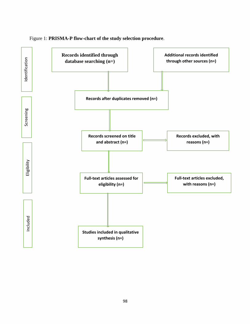
98
Figure 1: PRISMA-P flow-chart of the study selection procedure.
Records identified through
database searching (n=)
Additional records identified
through other sources (n=)
Records after duplicates removed (n=)
Records screened on title
and abstract (n=)
Full-text articles assessed for
eligibility (n=)
Studies included in qualitative
synthesis (n=)
Records excluded, with
reasons (n=)
Full-text articles excluded,
with reasons (n=)
Iden
tifi
cati
on
Sc
reen
ing
Elig
ibili
ty
Incl
ud
ed

99
Data extraction and management.
A data extraction form will be created by the principal author (WTM) in Microsoft Excel and
modified by feedback from three independent reviewers (MGK, MN, and PMOO) to ensure that
complete data is obtained. Two review authors (WTM and MGK) will independently extract data.
Data will be collected on the first author name; animal species (rats, mice), gender (male and/or
female), age (mature adult, middle-aged, old adult), weight; nature of acute liver damage (drug,
toxic chemical, alcohol, food, and infection or autoimmune induced damage); part of Moringa
used (bark, flour, leaves, seed), African country of harvest, dose of Moringa administrated,
duration of treatment; and primary and secondary outcome measurements.
Disagreements between authors will be resolved through discussion and consensus, or arbitration
by a third author whenever necessary. For managing missing data, we will contact the
corresponding author of the respective studies in order to obtain the required details. If no
correspondence is received, the study will be included in the systematic review and discussed in
the narrative summary.
Data analysis including an assessment of heterogeneity
Data will be analyzed using Stata statistical software (Stata Corp V.14, Texas, USA).
Study data will be quantitatively synthesized by first assessing heterogeneity to examine whether
the estimates from included studies could be pooled. Heterogeneity will be assessed by the Chi-
squared test on Cochrane’s Q statistic, which will be quantified by I-squared values. Values of 25,
50, and 75% for I-squared will be represented low, medium, and high heterogeneity, respectively
[18]. When substantial heterogeneity will be detected, we will perform meta-regression and
subgroup analyses to investigate the possible sources of heterogeneity using the following
grouping variables: animal species (rat , mice), age (mature adult, middle-aged, old adult), gender
(male, female, combination of genders used), weight; nature of acute liver damage (drug, toxic
chemical, alcohol, food, and infection or autoimmune induced damage); part of Moringa used
(Bark, flour, leaves, seed), African country of harvested, dose of Moringa administrated, duration
of treatment; and outcome measurement results. The heterogeneity between subgroups will be
detected by using the X2 test on Cochrane’s Q statistic. In cases where quantitative synthesis is not
appropriate because it is not possible to conduct meta-analysis due to the heterogeneity of
estimates reported by studies, a qualitative narrative synthesis of the evidence will be performed.

100
This will summarize the studies characteristics, animal’s features, nature of liver damage, Moringa
features an outcome result.
Assessment of risk of bias
The study quality and the presence of potential bias within individual studies will be done at both
the outcome level and the study level. Two reviewers (WTM and MGK) will complete the
assessment independently. The Cochrane risk of bias tool will be used to assess publication bias,
addressing assessments for random sequence generation, concealment of allocation, blinding and
the outcome measurements, and completeness of outcome reporting [19]. Each bias criterion will
be assigned a value of low, high, or unclear risk of bias for each included study.
Visual assessment of the funnel plot and the Egger’s statistic will be used to assess for both the
presence and statistical significance of publication bias across studies [20].
Assessment of methodological and evidence qualities.
We will use the Grading of Recommendations Assessment, Development, and Evaluation
(GRADE) methodology to assess the quality of evidence for each outcome as recommended in
the Cochrane Handbook for Systematic Reviews of Interventions [21]. Quality rating of overall
evidence will be downgraded according to the following factors: Risk of bias, the inconsistency
of result, indirectness, imprecision, and publication bias. In addition and where appropriate, the
reasons to upgrade the evidence quality will include a large magnitude of effects, a dose-
response gradient, and plausible residual confounding that would reduce a demonstrated effect or
suggest a spurious effect when results show no effect. We will integrate downgrading and
upgrading factors to obtain an overall quality of evidence for each outcome of interest. Overall
quality of evidence will be then ranked as high, moderate, low, or very low as specified by the
GRADE approach [21; 22].
Discussion
Moringa oleifera (MO) is a plant traditionally used for its medicinal and nutritional properties in
Africa. Many studies have been done on MO, and some of them showed that MO possesses
hepatoprotective activity [1]. But then, not many studies have focused on MO from sub-Saharan
countries in managing acute liver damage induced. This protocol states the plan for a systematic
review and meta-synthesis of the effectiveness of Moringa oleifera from sub-Saharan African

101
countries to manage liver damage. Therefore, this review will fill this gap by revealing evidence
that Moringa oleifera from different countries of sub-Saharan Africa have the same effects on
induced acute liver damage, so agro-climatic conditions of vegetable materials may vary from one
place to another [23]. Results will be shared with the scientific community through publishing in
a peer-reviewed journal. The findings could give scientists new insight into the possible use of this
plant to develop effective hepatoprotective drugs that treat liver damage and can serve as valuable
inputs for future research in this area.
Abbreviations
ALP: alkaline phosphatase, ALT: alanine Transaminase, AST: aspartate Transaminase, GGT:
Gamma-glutamyltransferase, NALD: Non-alcoholic liver diseases, PRISMA: Preferred
Reporting Items for Systematic Review and Meta-analyses, PRISMA-P: Preferred Reporting
Items for Systematic Review and Meta-Analysis Protocol, SGOT: Serum Glutamic oxaloacetic
transaminase, SGPT: serum glutamate-pyruvate transaminase (SGPT), WHO: world health
organisation.
Declarations
Acknowledgments
The acknowledgments are addressed to the University of KwaZulu-Natal, College of Health
Sciences for Scholarship Award which funded the study. The authors are grateful to Ms Carrin
Martin for editing this manuscript.
Funding
The University of KwaZulu-Natal, College of Health Sciences Scholarship Award (CHS
Scholarship) will fund this research study.
Availability of data and material
All data generated or analyzed during this study will be included in the published systematic
review.

102
Authors’ contributions
WTM conceived and designed the protocol and drafted and revised the manuscript for intellectual
content. MGK designed the protocol and revised the manuscript for intellectual content. MN
supervised and revised the manuscript for intellectual content. PMOO supervised and revised the
manuscript for intellectual content. All authors had full access to the data and they gave their
approval before the submission of the final version to the journal.
Competing interests
No, any competing interests are declared by the authors.
Consent for publication
Not applicable
Ethics approval and consent to participate
Not applicable
Authors’ Affiliations
1Discipline of Pharmaceutical Sciences, School of Health Sciences, Westville Campus,
University of KwaZulu-Natal.
REFERENCES
1. El-bakry, Toson E, Serag M and Aboser M. Hepatoprotective effect of Moringa Oleifera
leaves extract against carbon tetrachloride-induced liver damage in rats. World Journal of
Pharmacy and Pharmaceutical Sciences, 2016;5(5):76–89,
https://doi.org/10.20959/wjpps20165-6638.
2. Krishnaveni J & Ananthi T. Hepatoprotective effect of Moringa oleifera in isoniazid-induced
rats. Res. J. Pharm. Technol. 2011;4(12): 1901–1903.
3. Desai N A, Kotak K P & Patel S S. Investigation of epidemiology and etiology of liver
diseases and characterization of its association with various factors. Asian J Pharm Clin
Res. 2015; 8(2): 346–349.
4. Sattar N, Forrest E & Preiss D. Non-alcoholic fatty liver disease. BMJ, 2014; 349.

103
doi:10.1136/bmj.g4596.
5. Baseke J, Musenero M & Mayanja-kizza H. Prevalence of hepatitis B and C and relationship
to liver damage in HIV infected patients attending the Joint Clinical Research Centre Clinic
( JCRC ), Kampala, Uganda. African Health Sciences. 2015; 15(2): 323–327.
6. Ezejindu D N, Chinweife KC & Ihentuge C J. The effects of Moringa extract on liver
enzymes of carbon tetrachloride-induced hepatotoxicity in adult Wistar rats.The Journal of
Engineering and Science. 2013; 2(7): 54–59.
7. Wang Y L, Woon-Puay Koh, Jian-Min Yuan, An Pan. Association between liver enzymes
and incident type 2 diabetes in Singapore Chinese men and women. BMJ Open Diabetes
Research & Care. 2016; 4(1). e000296. https://doi.org/10.1136/bmjdrc-2016-000296.
8. Olatosin T M, Akinduko DS, Uchec C Z, Bardi J. Effects of Moringa oleifera seed oil on
acetaminophen-induced oxidative stress and liver damage in Wistar albino rats. IOSR J.
Pharm. Biol. Sci. 2014; 9(2): 53–59.
9. Anwar F, Latif S, Ashraf M. Moringa oleifera: A food plant with multiple medicinal uses.
Phytother. Res. 2007; 21: 17–25. DOI: 10.1002/ptr.2023.
10. Pathan MA, Khan, AP, Somkuward N Z G. Hepatoprotective activity of Maytenus
emarginata against paracetamol-induced liver injury in male Wistar rats. Int. J. Pharm.
Pharm. Sci. 2014; 6(8): 320–323.
11. Detlef Schuppan and Nezam H Afdhal. Liver cirrhosis. Lancet, 2009; 371(9615): 838–851.
Doi:10.1016/S0140-6736(08)60383-9.
12. Simon Rama Parmar, Patel Hitesh Vashrambhai, Karan Kalia. Hepatoprotective activity of
some plants extracts against paracetamol-induced hepatotoxicity in rats. J. Herb. Med.
Toxicol. 2010; 4(2): 101–106.
13. Omotoso O D, Adelakun S A, Amedu N O & Idoko U P. Antioxidant properties of Moringa
oleifera oil and Anacardium occidentale oil on cadmium-induced liver damage in adult
Wistar rats. Journal of Advances in Medical and Pharmaceutical Sciences. 2016; 7(4): 1–7.
DOI:10.9734/JAMPS/2016/24250.
104
14. Gowrishankar R, Kumar M, Menon V, Divi SM, Saravanan M, Magudapathy P, Panigrahi
BK, Nair KG, Venkataramaniah K R. Trace element studies on Tinospora cordifolia
(Menispermaceae), Ocimum sanctum (Lamiaceae), Moringa oleifera (Moringaceae), and
Phyllanthus niruri (Euphorbiaceae) using PIXE. Biol. Trace Elem. Res. 2010; 133(3): 357–
363. Doi: 10.1007/s12011-009-8439-1.
15. Saalu LC, Ogunlade, B, Ajayi GO, Oyewopo AO, Akunna GG, Ogunmodede O S. The
hepato-protective potentials of Moringa oleifera leaf extract on alcohol-induced hepato-
toxicity in wistar rat. Am. J. Biotechnol. Mol. Sci. 2012;2(1):6–14.
Doi:10.5251/ajbms.2012.2.1.6.14.
16. Shamseer L, Moher D, Clarke M, Ghersi D, Liberati A, Petticrew M., Shekelle P, Lesley A.
the PRISMA-P Group. Preferred reporting items for systematic review and meta-analysis
protocols (PRISMA-P)2015: elaboration and explanation. BMJ. 2015; 349.
Doi:https://doi.org/10.1136/bmj.g7647.
17. Moher D, Liberati A, Tetzlaff J, Altman DG, the PRISMA Group. Preferred Reporting
Items for Systematic Reviews and Meta-Analyses: The PRISMA Statement. PLoS Med
2009; 6(7): e1000097. https://doi.org/10.1371/journal.pmed.1000097.
18. Higgins J P T & Thompson, S G. Quantifying heterogeneity in a meta-analysis. Statistics in
Medicine. 2002; 21(11): 1539–1558. https://doi.org/10.1002/sim.1186.
19. Higgins JPT, Atman DG, Cotzsche PC, Juni P, Moher D, Oxman A.D, Savocic J, Schulz
KF, Weeks L, Sterne JA. The Cochrane Collaboration ’ s tool for assessing the risk of bias
in randomized trials. BMJ, 2011; 343. Doi:https://doi.org/10.1136/bmj.d5928.
20. Egger M, Smith G.D, Schneider M, Minder C. Bias in a meta-analysis
detected by a simple, graphical test. BMJ, 1997; 315(7109):629–34.
doi: https://doi.org/10.1136/bmj.315.7109.629.
21. Higgins JPT, editors. Cochrane Handbook for Systematic Reviews of Interventions, Version
5.1.0 [Updated March 2011]. The Cochrane Collaboration, 2011.
http://handbook.cochrane.org.
22 Guyatt G, Oxman AD, Akl EA, Kunz R, Vist G, Brozek J, et al. GRADE guidelines: 1.

105
Introduction-GRADE evidence profiles and summary of findings tables. J Clin
Epidemiol. 2011; 64:383–94.
23. Ochieng, J., Kirimi, L. & Mathenge, M. Effects of climate variability and change on
agricultural production : The case of small scale farmers in Kenya. NJAS - Wageningen
Journal of Life Sciences. 2016; 77: 71–78. https://doi.org/10.1016/j.njas.2016.03.005.