Embed Size (px)
Citation preview

CELLULARIMMUNOLOGY 122,122-130 (1989)
Antigen-Specific Inhibition of the IL-2-Driven Proliferation of Myelin Basic Protein-Reactive, Human T Cells
JAMESBURNS,* L. JILLKFCASNER,PANDKIMBERLYLITTLEFIELD*
*Department ofNeurology, Veterans Administration Medical Center, University of Utah, Salt Lake City, Utah 84148, and TTemple University School ofMedicine, Philadelphia, Pennsylvania 19122
Received November 28, 1988; accepted April 24,1989
This report examines the antigen-specific inhibition of the IL-2-driven proliferation of autoan- tigen-reactive, human T cells. Human, myelin basic protein (MBP)-reactive CD4+ cell lines and clones were isolated and maintained in culture by use of IL2 and periodic antigen stimulation. When freshly isolated antigen-presenting cells (APC) were present, MBP induced proliferation of MBP-reactive T cell populations. However, under different culture conditions, MBP reduced the ILZ-driven proliferation of some MBP-reactive T cell populations. The inhibition of IL-2- driven proliferation did not appear to require CD8+ or OKM I+ cells since these were not de- tected when inhibition studies were performed at least 9 days after the last restimulation by irradiated APC and MBP. Supraoptimal concentrations of MBP were not required for inhibition of proliferation. Some heterogeneity of response was apparent since MBP inhibited the IL-2- driven proliferation of some T cell clones while for others MBP had either no effect or produced slight enhancement of proliferation. These results demonstrate an antigen-specific, in vitro im- mune mechanism that reduces the IL-2-dependent proliferation of autoantigen-reactive, human T Celk. 0 1989 Academic Press. Inc.
INTRODUCTION
T lymphocyte activation and proliferation are mediated through the interaction of specific ligands with T cell surface receptors. Among the most thoroughly studied of these surface receptors is the T cell antigen/MHC recognition complex ( 1,2). Appro- priate ligand binding to this complex usually initiates activation of lymphocytes with production of IL-2 and expression of IL-2 receptors on the T cell surface (3-5). In addition to activation by the antigen/MHC complex, “antigen-like” stimulation may be seen with anti-T cell receptor antibody and anti-CD3 antibody (6-8). While prolif- eration is the usual result of these interactions, some investigators have noted inhibi- tion following interaction of the antigen/MHC receptor complex with anti-CD3 anti- bodies (9), anti-T cell receptor antibodies (10, 1 l), or the appropriate antigen and MHC antigens (12-14). These inhibitory effects have included inhibition of IL-2- driven proliferation ( 11) and induction of tolerance to subsequent antigen restimula- tion (9, 12, 14). It is unknown whether these in vitro inhibitory effects mimic in vivo mechanisms that control T cell proliferation or maintain tolerance to autoantigens.
An autoantigen frequently studied in experimental systems is myelin basic protein (MBP). MBP is a normal protein component of central nervous system (CNS) myelin and constitutes approximately 30% of the protein in myelin ( 15). CD4+ T cells that
122
0008-8749189 $3.00 Copyright 0 1989 by Academic Press, Inc. All rizghts of reproduction in any form reserved.

INHIBITION OF IL-2-DRIVEN PROLIFERATION 123
react with human MBP may be isolated from the blood of normal subjects as well as patients with neurologic disease ( 16- 18). These autoantigen-reactive cells are main- tained in long-term culture by the use of IL-2 and periodic antigen restimulation (18). During experiments to determine the optimal conditions for maintaining MBP- reactive cells in long-term culture, we noted that the IL-2-dependent proliferation of MBP-reactive cells was often reduced when MBP was present in the medium. The current study was undertaken to determine the characteristics of this inhibition. We have demonstrated that the inhibition of IL-2-driven proliferation by MBP is specific for MBP-reactive T cells and is concentration dependent. This inhibition does not require the participation of CDS+ T cells.
METHODS
Cell separations. Peripheral blood mononuclear cells (PBMC) were obtained by density gradient separation of heparinized blood on Ficoll-Hypaque gradients (Phar- macia Fine Chemicals, Piscataway, NJ). Neurologically normal subjects were studied.
Antigens. Whole human myelin was prepared by the method of Norton and Po- duslo ( 19). SDS-polyacrylamide gel electrophoresis of this myelin preparation con- firmed that MBP and the other major protein components of myelin were present (data not shown). Human MBP was prepared by the method of Deibler et al. (20) and was generously provided by Drs. D. Pleasure and W. F. Hickey. Tuberculin, purified protein derivative (PPD), was obtained from Connaught Laboratories, To- ronto, Canada.
T cell lines and clones. T cell lines specific for MBP and PPD were isolated from the PBMC of normal subjects as previously described (18). Freshly isolated PBMC (5 X 10⁶) were placed in 2.0-ml culture wells in complete medium (RPM1 1640 sup- plemented with 2 mM glutamine, 1% nonessential amino acids (GIBCO, Grand Is- land, NY), 1 mMsodium pyruvate, antibiotics, and 10% autologous human serum). Human MBP (30 pg/ml) or PPD (2 pg/ml) was added and after 7 days the cells were collected, washed twice, and placed in culture medium containing IL-2 (supernatant from the MLA-144 cell line, (2 I)). The cultures were refed with IL-2-containing me- dium at least every other day for an additional 5 days. The cells were then washed and 2 X lo5 cells were restimulated using 5 X lo6 irradiated (3000 rad) autologous PBMC (as a source of antigen presenting cells, APC) and the appropriate antigen (MBP, 30 @g/ml or PPD, 2 yg/ml) in 2.0-ml cultures in complete medium without added IL-2. After 3 days the cells were washed twice and placed again in culture medium supplemented with IL-2 but without antigen. The proliferating cell cultures were then refed with IL-2-supplemented medium at least every third day. Antigen restimulation with irradiated APC and antigen was required every other week. T cell lines and clones were maintained in culture for up to 6 months by this protocol. T cells were cloned as previously described (22) by limiting dilution in 96-well round- bottom microtiter plates at 0.2 T cells per well with 5 X lo4 irradiated APC and an optimal concentration of antigen. After 2 days, IL-2 was added and the cultures were refed with IL-2 every other day until cell growth was apparent after 10 to 14 days. Clones were then maintained as described above. Cloning efficiency was approxi- mately 40%. T cell phenotype was determined by epifluorescent microscopy as pre- viously described using a panel of monoclonal antibodies purchased from Ortho
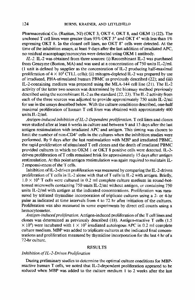
124 BURNS, KRASNER, AND LITTLEFIELD
Pharmaceutical Co. (Raritan, NJ) (OKT 3, OKT 4, OKT 8, and OKM 1) (22). The uncloned T cell lines were greater than 95% OKT 3+ and OKT 4+ with less than 1% expressing OKT 8. In the cloned cell lines, no OKT 8+ cells were detected. At the time of the inhibition assays, at least 9 days after the last addition of irradiated APC, no residual macrophage/monocytes were detected using OKM 1 antibody.
IL-2. IL-2 was obtained from three sources: (i) Recombinant IL-2 was purchased from Genzyme (Boston, MA) and was used at a concentration of 750 units IL-2/ml. (1 unit is defined by supplier as the concentration of IL-2 producing half-maximal proliferation of 4 X lo3 CTLL cells); (ii) mitogen-depleted IL-2 was prepared by use of irradiated, PHA-stimulated human PBMC as previously described (22); and (iii) IL-2-containing medium was prepared using the MLA-144 cell line (21). The IL-2 activity of the latter two sources was determined by the bioassay method previously described using the recombinant IL-2 as the standard (22,23). The IL-2 activity from each of the three sources was adjusted to provide approximately 750 units IL-2/ml for use in the assays described below. With the culture conditions described, one-half maximal proliferation of human T cell lines was obtained with approximately 400 units IL-2/ml.
Antigen-induced inhibition of IL-d-Dependent proliferation. T cell lines and clones were studied after at least 6 weeks in culture and between 9 and 15 days after the last antigen restimulation with irradiated APC and antigen. This timing was chosen to limit the number of non-CD4+ cells in the cultures when the inhibition studies were performed. By 9 days after the last restimulation with MBP and irradiated PBMC, the rapid proliferation of stimulated T cell clones and the death of irradiated PBMC provided cultures in which no OKM 1 or OKT 8 positive cells were detected. IL-2- driven proliferation of T cells remained brisk for approximately 15 days after antigen restimulation. At this point antigen restimulation was again required to maintain IL- 2 responsiveness of the T cells.
Inhibition of IL-Zdriven proliferation was measured by comparing the IL-2-driven proliferation of T cells in IL-2 alone with that of T cells in IL-2 with antigen. Briefly, 1.0 X lo4 T cells were cultured in 0.2 ml complete culture medium in round-bot- tomed microwells containing 750 units IL-2/ml without antigen, or containing 750 units IL-2/ml with antigen at the indicated concentrations. Proliferation was mea- sured by tritiated thymidine incorporation of triplicate cultures using a 2- or 4-hr pulse as indicated at time intervals from 4 to 72 hr after initiation of the cultures. Proliferation was also measured in some experiments by direct cell counts using a hemocytometer.
Antigen-inducedproliferation. Antigen-induced proliferation of the T cell lines and clones was determined as previously described (18). Antigen-reactive T cells (1.5 X 10⁴) were incubated with 1 X lo5 irradiated autologous APC in 0.2 ml complete culture medium. MBP was added to triplicate cultures at the indicated final concen- trations and proliferation measured by thymidine incorporation for the last 4 hr of a 72-hr culture.
RESULTS Inhibition of IL-2-Driven Proliferation
During preliminary studies to determine the optimal culture conditions for MBP- reactive human T cells, we noted that IGZdependent proliferation appeared to be reduced when MBP was added to the culture medium 1 to 2 weeks after the last

INHIBITION OF IL-2-DRIVEN PROLIFERATION 125
5 12000
8 - 10ooo
.s aom
E 6000 z t 4000
IL-2 Alone
IL- 2 + MBP
2coO IL- 2 + MBP IL- 2 * MBP
0 0 4 I4 24
Tome (Houn)
FIG. 1. Antigen-specific inhibition of IL-2-driven proliferation. A CD4+ T cell line reactive with human MBP was established and maintained in culture for 3 months as described under Methods. Antigen-in- duced inhibition of IL-2driven proliferation was measured 10 days after the last antigen restimulation with MBP and irradiated APC. T cells (1 .O X IO“) were placed in round-bottomed microwells with IL-2 alone or IL-2 plus human MBP at the indicated concentrations. Four identical plates were established in parallel. Tritiated thymidine (2 pCi/microwelI) was added at 0, 4, 14, and 24 hr and the plates were har- vested 2 hr later for liquid scintillation counting. Results are expressed as mean cpm. Tritiated thymidine incorporation for cells in medium alone, without added IL-2 or MBP, was 5876 cpm at 4 hr, 1756 at 14 hr, and 1441 at 24 hr.
antigen restimulation. To determine the degree of inhibition, we studied the IL-2- driven proliferation, as measured by thymidine incorporation, of an MBP-reactive T cell line in the presence of human MBP (Fig. 1). This cell line was studied 10 days after the last antigen restimulation. IL-2-driven proliferation was examined at four time points during a 24-hr culture period using four different concentrations of hu- man MBP. Inhibition of proliferation was noted with 50 pg/ml MBP as early as 4 hr after initiation of these cultures and by 24 hr inhibition was apparent with all concentrations of MBP tested. Similar results were noted in identical 24-hr experi- ments with two MBP-reactive T cell clones (data not shown).
In other experiments, a dose-response assay was performed to examine the effect of increasing concentrations of MBP on the IL-2-driven proliferation of seven addi- tional MBP-specific T cell populations (five T cell clones and two T cell lines). Each T cell population was assayed for 48 hr in IL-2 alone or IL-2 plus MBP at concentra- tions from 0.1 to 50 pg/ml. The IL-2-driven proliferation of both of the MBP-reactive T cell lines and three of the five clones was reduced by MBP. Figure 2 shows the mean inhibition of proliferation observed for these five T cell populations with each value representing the mean of five experiments using each of these five T cell populations. Two clones were not inhibited by MBP and are discussed below. Greater inhibition of proliferation occurred with increasing concentrations of MBP.
The two clones that were not inhibited by MBP were derived from parental cell lines that, by contrast, were sensitive to this effect of MBP. Both clones were CD4+ and proliferated in response to MBP when peripheral blood APC were present (data not shown). A comparison between these two clones (8a and la) and another clone that was inhibited by MBP (6) is presented in Table 1. Clones 6 and 8a were cloned from the same parental line. The pattern of response shown in this representative experiment was characteristic of each clone. The IL-2-driven proliferation of clone 6 was inhibited by 50 pg/ml MBP while that of clone 8a was essentially unchanged by a similar concentration of MBP. By contrast, the IL-2-driven proliferation of clone la was slightly enhanced when MBP was added to the culture medium. In three

126 BURNS, KRASNER, AND LITTLEFIELD
0.1 I 5 IO 50
MBP Concentration(yg/ml)
FIG. 2. Antigen-specific inhibition of IL-Z-driven proliferation. Two MBP-specific T cell lines and three MBP-specific T cell clones were isolated and maintained as described under Methods. The IL-2driven proliferation of each of these five T cell populations was noted to be reduced by MBP. MBP-induced inhibition of IL-2-driven proliferation was measured with each T cell population over a range of MBP concentrations. T cells (1 .O X 104) were incubated in 0.2 ml of culture medium containing IL-2 without MBP or IL2 with MBP at the concentrations indicated. Proliferation was measured by thymidine incorpo- ration during the final 4 hr of a 48-hr culture period. For individual experiments, the percentage inhibition was determined by the formula: (1 - (cpm of T cells in IL-2 plus MBP)/(cpm of T cells in IL-2 alone)) X 100. The values shown are the mean of the percentage inhibition + SEM at each concentration of MBP for each of five different cell lines or clones.
separate experiments the mean inhibition of IL-2-driven proliferation of clone 6 was 63% and that of clone 8a was 6%. The proliferation of clone la was enhanced 59%. The factors responsible for these differences are not known. The majority of these experiments were performed using MLA 144-derived IL-2; however, similar results were obtained with recombinant IL-2 and IL-2 from mitogen-stimulated hu- man PBMC.
To assure that the reduction in thymidine incorporation reflected an actual reduc- tion of cell division, the number of viable T cells recovered from cultures containing IL-2 alone, or IL-2 with a specific antigen was determined by counting viable cells with a hemocytometer. Antigen specificity was examined by studying two T cell clones, one reactive with MBP and the other recognizing the control antigen, PPD. In the experiment shown in Fig. 3, each of the two T cell clones was incubated with
TABLE 1
Differential Susceptibility of MBP-Reactive T Cell Clones to Inhibition of IL-2 Driven Proliferation”
Clone IL-2 alone IL2 plus
MBP Percentage inhibition
MBP-6 9,409 -c 99 3,631 + 100 61 MBP-8a 13,521 + 605 13,021 f 482 4
MBP-la 4,162 + 83 1,179 + 649 -51
a MBP reactive T cell clones were incubated in IL-2 alone or IL-2 plus MBP, 50 pg/ml, in round-bot- tomed microwells for 48 hr. Tritiated thymidine was added for the final 4 hr to determine the IL-2-driven proliferation. The values shown are the mean cpm + SEM for triplicate cultures and are representative of three separate experiments.

INHIBITION OF IL-Z-DRIVEN PROLIFERATION 127
MBP clone6 PPfJ clone3 T Cell Clone
FIG. 3. T cell proliferation following culture with IL-2 alone or IL-2 plus antigen. Two T cell clones, one recognizing MBP (MBP clone 6) and the other reactive with PPD (PPD clone 3), were isolated as described under Methods and collected 10 days after the last antigen restimulation. Cells (2.5 X 10⁵) were incubated in flat-bottomed wells in 2.5 ml of culture medium to which was added (i) IL-2 alone; (ii) IL-2 plus MBP, 30 &ml; or (iii) IL-2 plus PPD, 100 rg/ml. After 72 hr the cells in each well were collected and the number of viable cells determined by trypan blue exclusion. The values shown represent the percentage recovery from each of the’six separate wells with the initial cell number in each well (2.5 X IO’) representing 100%. Similar results were obtained in two separate experiments using these T cell clones.
IL-2 alone, IL-2 plus 30 pg/ml MBP, or IL-2 plus 100 pg/ml PPD. For both T cell populations in IL-2 alone, there was a 400-500% increase in the total viable cell number after 72 hr confirming active IL-2-driven proliferation. There was no reduc- tion of proliferation or percentage recovery when the MBP-reactive clone was incu- bated with PPD, or when the PPD-reactive clone was incubated with MBP. By con- trast, the IL-2-driven proliferation of the MBP-reactive clone was inhibited by ap proximately 70% by 30 pg/ml MBP, and proliferation of the PPD-reactive clone was inhibited by about 50% by 100 &ml PPD. These results demonstrate the following points: (i) The inhibition of IL-Zdriven proliferation in these experiments is antigen specific; (ii) the inhibition of IL-Zdriven proliferation is not restricted to autoantigen- reactive T cell populations; and (iii) the reduction of thymidine incorporation by T cells in these assays reflects an actual reduction of proliferation. In experiments which are not shown, proliferation was not inhibited by this concentration of PPD for a PPD-reactive cell line and two other PPD-reactive clones. These experiments con- firmed an actual antigen-specific reduction in IL-Zdriven proliferation.
Inhibition Does Not Require Supraoptimal MBP Concentrations
Previous investigators have noted that exposure of T cells to concentrations of antigen well above the optimal for antigen stimulation may inhibit subsequent re- sponsiveness to that antigen ( 12, 14). To determine whether the concentrations of MBP that produced inhibition of IL-2-driven proliferation were in a supraoptimal range, a dose-response determination for antigen stimulation was performed. As shown in a representative experiment in Table 2, there was minimal response at 0.1 pg/ml MBP, with the optimal proliferation occurring at 50 pg/ml. Above this range the proliferation began to diminish, but at 500 pg/ml remained approximately 75% of the maximal response.
Inhibition of IL-2-Driven Proliferation by Whole Human Myelin
Myelin basic protein represents approximately 10% of the dry weight of whole my- elin ( 15). In preliminary experiments we noted that human MBP-specific T cells will

128 BURNS, KRASNER, AND LITTLEPIELD
TABLE 2
T Cell Proliferation following Stimulation with MBP”
0 0.1
MBP concentration (*g/ml)
1 50 100 500
230 3555 44,182 106,97 1 100,308 79,925
’ A T cell line reactive with MBP was isolated as described. T cells ( 1.5 X 10⁴) were cultured with irradi- ated APC in triplicate microcultures with the indicated concentration of MBP. Proliferation was deter- mined by incorporation of tritiated thymidine following a 72-hr culture period. Values represent mean cpm of triplicate cultures (SEM was generally less than 15% of the mean). A similar pattern of response was seen in separate experiments using three different cell lines.
proliferate when cultured with whole human myelin and APC indicating that MBP present in whole myelin is accessible in culture to MBP-reactive T cells (data not shown). MBP-reactive and PPD-reactive cells, recovered 10 days after the last antigen restimulation, were incubated in IL-2 alone, IL-2 with MBP, or IL-2 with whole hu- man myelin. The proliferation of the MBP-reactive T cells was inhibited by both MBP and by whole myelin while the proliferation of the PPD-reactive cells in parallel cultures was unchanged (Table 3).
DISCUSSION
Previous studies have documented that the interaction of various ligands with the T3/Ti MHC receptor complex will under some conditions lead to inhibition of re- sponsiveness rather than stimulation (9-14). The current study demonstrates that the IL-Zdriven proliferation of human MBP-reactive T cells may be reduced by the presence of MBP. The inhibition was dose dependent and occurred at concentrations of MBP which were optimal for the stimulation of proliferation if freshly isolated
TABLE 3
Effect of MBP and Whole Myelin on the IL-2-Driven Proliferation of MBP- and PPD-Reactive T Cells”
Antigen specificity of cell line
IL2 alone MBP 5
IL-2 plus
MBP 50 Myelin 200
MBP 11,399 3770 2389 5834 (67%) (79%) (37%)
PPD 10,289 9868 9894 10,786 (4%) (4%) (0%)
a T cell clones reactive with MBP or PPD were isolated and maintained in culture as described under Methods. Ten days after the last antigen restimulation, 1.0 X lo4 T cells/microwell were incubated with IL2 alone, or IL-2 plus MBP or whole myelin in round-bottomed microwells for 48 hr. The concentrations shown are micrograms per milliliter. Proliferation was determined by thymidine incorporation for the final 4 hr of the culture period. The values shown represent the mean cpm with the percentage inhibition of proliferation shown in parentheses.

INHIBITION OF IL-ZDRIVEN PROLIFERATION 129
APC were present. There was heterogeneity in the response of MBP-reactive T cell clones under these culture conditions. Some clones displayed reduced IL-2-driven proliferation in the presence of MBP while the proliferation of others was unchanged or enhanced. This inhibition of proliferation was not an antigen-nonspecific or toxic effect since MBP had no effect on the proliferation of T cells not reactive with MBP. This effect was studied between 9 and 14 days after the last antigen restimulation when no CD8+ cells or OKM I+ cells could be detected in the cell cultures.
The mechanism of this inhibition does not appear to require the presence of CD8+ cells since the T cell lines were predominantly CD4+, and the clones were exclusively CD4+. Since T cell clones were studied, it is unlikely that a separate CD4+ subset of suppressor cells could have been generated. Similar inhibition of IL-2-driven prolifer- ation has been reported by Nau et al. (1 I) to be induced by stimulation of the T cell receptor of murine T cell clones by immobilized anti-TCR monoclonal antibodies. These investigators were not able to detect transferrable lymphokines responsible for the inhibition, and this effect was noted with CD4+ T cell clones as well as CD8+ T cells. In addition, Nau et al. noted increased, rather than reduced, expression of IL-2 receptors and transferrin receptors on T cell populations exposed to immobilized anti-T cell receptor monoclonal antibody. We have not yet determined whether sim- ilar changes occur with MBP-reactive cells exposed to MBP.
The inhibition of IL-Zdriven proliferation described in this report is different from the tolerance induction to antigen restimulation that has been reported by other groups of investigators (9, 12, 14, 26). The studies of Lamb and colleagues demon- strated that tolerance to antigen restimulation could be induced in human CD4+ T cell clones reactive with influenza antigens by exposure to supraoptimal concentra- tions of these antigens in the absence of APC (14). Following a brief exposure to high concentrations of the appropriate viral antigen, the T cells continued to respond to IL-2 but did not respond by proliferation to antigen plus APC. These investigators noted that tolerance induction could be blocked by anti-MHC class II antibodies (25) and that tolerance persisted for at least 96 hr (14). Preliminary studies with MBP- reactive cells suggest that the inhibition of IL-2-driven proliferation can be partially blocked by anti-MHC class II antibodies and, in contrast to tolerance induction, is reversible within 24 hr (data not shown). Antigen-induced tolerance in murine T cell lines and clones also has been reported (24).
Myelin basic protein is a CNS antigen that has been implicated as the target antigen in the encephalomyelitis in humans which may follow rabies vaccination with the CNS antigen-containing Semple vaccine (27). In addition MBP is frequently studied as an autoantigen that induces experimental allergic encephalomyelitis (EAE) (28). Thus an obvious question is whether conditions in vivo can duplicate the inhibitory effects noted in vitro in these experiments. If this occurs in vivo, inhibition of IL-2- driven proliferation could reduce the intensity of an autoimmune disease by limiting the number of autoantigen-reactive cells present within the target organ. Induction of tolerance to antigen restimulation, as described by Lamb and colleagues (14), might further reduce the intensity of the inflammatory response and contribute to recovery.
The mechanism responsible for the spontaneous recovery that normally follows the induction of either active or adoptively induced EAE is unknown. Immunohisto- chemical studies have identified increased numbers of CD8+ cells present in the CNS during recovery from EAE suggesting that these cells play some role in promoting recovery (29). However, another study has documented that nude mice will recover

130 BURNS, KRASNER, AND LITTLEFIELD
normally from EAE adoptively induced with cloned MBP-reactive, CD4+ T cells (30). This observation and an additional study documenting normal recovery from adoptively induced EAE in Lewis rats depleted of CD8+ cells suggests that recovery from adoptive EAE may not require cell-mediated suppressor activity (31). In the present study we have demonstrated that whole myelin, as well as purified MBP, will reduce the IL-Zdriven proliferation of human MBP-reactive T cells in vitro. Whether similar events occur in vivo and contribute to the spontaneous recovery from EAE induced by MBP-reactive T cells is unknown.
ACKNOWLEDGMENTS
This work was supported by Grant RG 1894-A-2 from the National Multiple Sclerosis Society and by support from the V. A. Medical Research Service.
REFERENCES
1. Haskins, K., Kappler, J., and Marrack, P., Annu. Rev. Zmmunol. 2,5 1, 1984. 2. Acute, O., and Reinherz, E., N. Engl. J Med. 312, 1100, 1985. 3. Morgan, D. A., Ruscetti, F. W., and Gallo, R. C., Science 193, 1007, 1976.
4. Meuer, S., Hussey, R., Cantrell, D., Hodgdon, J., Schlossman, S., Smith, K., and Reinherz, E., Proc. Natl. Acad. Sci. USA 81, 1509, 1984.
5. Cantrell, D. A., and Smith, K. A., Science 224, 13 12, 1984. 6. Meuer, S., Hodgdon, J., Hussey, R., Protentis, J., Schlossman, S., and Reinhetz, E., J. Exp. Med. 158,
988,1983. 7. Van Wauwe, J., DeMey, J., and Goossens, J., J. Zmmunol. 124,2708, 1980.
8. Kaye, K., Porcelli, S., Tite, J., Jones, B., and Janeway, C., J. Exp. Med. 158,836, 1983. 9. Pantaleo, G., Olive, D., Poggi, A., Pozzan, T., Moretta, L., and Moretta, A., J. Exp. Med. 166,619,
1987. 10. Breitmeyer, J. B., Oppenheim, S. O., Daley, J., Levine, H. B., and Schlossman, S. F., J. Zmmunol. 138,
726, 1987. 11. Nau, G. J., Moldwin, R. L., Lancki, D. W., Kim, D.-K., and Fitch, F. W., J. Zmmunol. 139, 114, 1987. 12. Quill, H., and Schwartz, R. H., J. Zmmunol. 138,3704, 1987. 13. Ashwell, J. D., Cunningham, R. E., Noguchi, P. D., and Hernandez, D., J. Exp. Med. 165,173, 1987.
14. Lamb, J. R., Skidmore, B. J., Green, N., Chiller, J. M., and Feldmann, M., J. Exp. Med. 157, 1434, 1983.
15. Carnegie, P. R., and Moore, W. J., In “Proteins of the Nervous System” (R. A. Bradshaw and D. M. Schneider, Eds.), pp. 145-160. Raven Press, New York, 1980.
16. Richert, J., Reuben-Bumside, C., Deibler, G., and Kies, M. W., Neurology38,739, 1988. 17. Hafler, D. A., Buchsbaum, M., Johnson, D., and Weiner, H., Ann. Neural. 18,45 1,1985. 18. Bums, J., Rosenzweig, A., Zweiman, B., and Lisak, R., Cell. Zmmunol. 81,435, 1983.
19. Norton, W. T., and Poduslo, S. E., J. Neurochem. 21,749, 1973. 20. Deibler, G. E., Martenson, R. E., and Kies, M. W., Prep. Biochem. 2, 139, 1972. 2 1. Rabin, H., Hopkins, R., Ruscetti, F., Neubauer, R. H., Brown, R. L., and Kawakami, T. G., J. Zmmu-
nol. 127, 1852, 1981. 22. Bums, J., Rosenzweig, A., Zweiman, B., and Lisak, R., Cell. Zmmunol. 77,363, 1983. 23. Gillis, S., Ferm, M. W., Ou, W., and Smith, K. A., J. Zmmunol. 120,2027, 1978. 24. Levich, J. D., Signorella, A. P., Wittenberg, G., and Weigle, W. O., J. Zmmunol. 138,3675, 1987. 25. Lamb, J., and Feldmann, M., Nature (London) 308,72, 1984. 26. Zanders, E., Lamb, J., Feldmann, M., Green, N., and Beverley, P., Nature (London) 303,625, 1983. 27. Hemachudha, T., Griffin, D. E., Giffels, J. J., Johnson, R. T., Moser, A. B., and Phanuphak, P., N.
Engl. J. Med. 316,369, 1987.
28. Raine, C. S., Lab. Invest. SO, 608, 1984. 29. Hickey, W. F., and Gonatas, N., Cell. Zmmunol. 85,284, 1984. 30. Sakai, K., Namikawa, T., Kunishita, T., Yamanouchi, K., and Tabira, T., J Zmmunol. 137, 1527,
1986. 3 I. Willenborg, D. O., Sjollema, P., and Danta, G., J. Zmmunol. 136, 1676, 1986.