Embed Size (px)
Citation preview

ENGLISH
ANTIGENSANTISERABACTERIAL STRAINS
2 3
CONTENT
GENERAL INFORMATION ......................................................................................................4ANTIGENS .........................................................................................................................................6
PNEUMOCOCCAL CELL WALL POLYSACCHARIDES ....................................................6CWPS AND CWPS MULTI ............................................................................................................6
PNEUMOCOCCAL CAPSULAR POLYSACCHARIDES ......................................................8PNEUMOCOCCAL F-ANTIGEN .......................................................................................................9PERTUSSIS TOXIN ................................................................................................................................. 10
ANTISERA ....................................................................................................................................... 12GENERAL INFORMATION .................................................................................................................12E. COLI .......................................................................................................................................................... 14
OK O ANTISERA ................................................................................................................................ 14O ANTISERA ....................................................................................................................................... 16H ANTISERA ........................................................................................................................................ 19K ANTISERA.........................................................................................................................................22K9 ANTISERUM ................................................................................................................................ 24F ANTISERA .........................................................................................................................................25PHAGE SUSPENSIONS ............................................................................................................... 26
PNEUMOCOCCUS .............................................................................................................................. 28NEUFELD ANTISERA .................................................................................................................... 28PNEUMOTEST KIT .......................................................................................................................... 30
SALMONELLA ..........................................................................................................................................32O AND H ANTISERA .......................................................................................................................32VI MONOCLONAL ANTIBODY ................................................................................................32SERO-QUICK ID KIT ...................................................................................................................... 36SERO-QUICK GROUP KIT ......................................................................................................... 39SALMONELLA BIG FIVE KIT – ISO 6579-3 ...................................................................... 42
STREPTOCOCCUS................................................................................................................................46NEUFELD ANTISERA OR ANTISERA FOR LANCEFIELD TEST .........................46
H. INFLUENZAE ....................................................................................................................................49TYPE B ANTISERUM .....................................................................................................................49
SHIGELLA ....................................................................................................................................................51O POOL AND PHASE ANTISERA............................................................................................51
Y. ENTEROCOLITICA ............................................................................................................................52O ANTISERA ........................................................................................................................................52
BACTERIAL STRAINS .............................................................................................................54STRAINS IN AGAR ..........................................................................................................................54FREEZE-DRIED STRAINS .......................................................................................................... 56
REFERENCES ..............................................................................................................................58
AN
TIG
EN
SA
NTI
SER
AB
AC
TER
IAL
STR
AIN
SR
EFE
RE
NC
ES
4 5
GENERAL INFORMATION
Quality CertificateSSI Diagnostica is quality assured and certified in accordance with ISO 13485.Certificate of analysis can be downloaded from our website www.ssidiagnostica.com
Abbreviation listB. pertussis Bordetella pertussis CWPS Cell Wall PolySaccharide ETEC EnteroToxigenic E. coli ELISA Enzyme Linked ImmunoSorbent Assay E. coli Escherichia coli H. influenzae Haemophilus influenzae PT Pertussis Toxin PBS Phosphate Buffered Saline CPS Pneumococcal capsular polysaccharides 22F Pneumococcal serotype 22F capsule PCR Polymerase Chain Reaction S. Enteritidis Salmonella Enteritidis S. Hadar Salmonella Hadar S. Hilligdon Salmonella Hillingdon S. Infantis Salmonella Infantis S. Parathyphi Salmonella Parathyphi S. Typhi Salmonella Typhi S. Typhimurium Salmonella Typhimurium S. Virchow Salmonella Virchow S. sonnei Shigella sonnei S. suis Streptococus suis Y. enterocolitica Yersinia enterocolitica
6 7
ProcedureAbsorption of human serum sample: 1. Prepare a stock solution by dissolving 10 mg of either CWPS or CWPS Multi
in 1 mL sterile Type I ultrapure water (stock solution: 10 mg/mL). 2. Further dilutions to 1 mg/mL are made in sterile Type I ultrapure water.3. The absorption dilution is made in antibody buffer to the final
concentration of 10 µg/mL3. 4. See the further procedure at: http://www.vaccine.uab.edu/uploads/
mdocs/ELISAProtocol(007sp).pdf3.
Storage and shelf lifeStore the unopened CWPS and CWPS Multi vials at 8-25°C. Expiry date of the sealed vial is printed on the label. The stability of the stock solution (10 mg/mL) is 1-2 weeks at 2-8°C and can be prolonged by addition of sodium azide. The dilution of 1 mg/mL can be stored at -80°C for 10 years.
ANTIGENS
PNEUMOCOCCAL CELL WALL POLYSACCHARIDES CWPS AND CWPS MULTI
Intended useCWPS and CWPS Multi are intended for preabsorbing human serum samples before quantitation of specific pneumococcal capsular polysaccharide antibodies1-5. CWPS and CWPS Multi may also be used as a coating agent during performance of an ELISA test.
DescriptionCWPS is a pneumococcal antigen common to all pneumococcal serotypes.CWPS Multi is a 1:1 mixture of CWPS and the purified pneumococcal cell wall polysaccharide antigen from the pneumococcal serotype 22F capsule (CWPS/22F).CWPS and CWPS Multi are supplied in vials containing 10 mg lyophilized purified antigen.
PrincipleTo measure a specific antibody response to a specific pneumococcal CPS, it is necessary to remove the antibodies against CWPS and the CWPS/22F since they offer no protection against pneumococcal invasive diseases. By addition of CWPS to the human serum sample the antibodies against CWPS are removed. By addition of CWPS Multi to the human serum sample the antibodies against CWPS and CWPS/22F are removed. Consult this website for the WHO protocol http://www.vaccine.uab.edu/uploads/mdocs/ELISAProtocol(007sp).pdf3.
If these absorption steps are not performed, a false high antibody titer in the human serum will be measured.
Materials required but not provided • Sterile Type I ultrapure water, conforming to ASTM, CAP, NCCLS, USP and ISO
specifications • Mixer
8 9
PNEUMOCOCCAL F-ANTIGEN
Intended useThe purified pneumococcal F-antigen (Forssman antigen, LTA) is intended to be used in immune/immunological research6.
DescriptionF antigen is supplied in a vial containing 10 mg lyophilized purified antigen. Lipid contamination is not assayed and may vary from batch to batch.
PrincipleAll pneumococci both virulent and avirulent strains possess a common polysaccharide, the F-antigen. This antigen is a lipoteichoic acid with a polysaccharide part that has the same repeat as CWPS. This part is then linked to a diacylated glycerol residue via a glucose residue6.
Materials Required but not Provided • Sterile Type I ultrapure water, conforming to ASTM, CAP, NCCLS, USP and ISO
specifications • Mixer
ProcedureAs for Pneumococcal capsular polysaccharides described on page 8.
Storage and Shelf LifeStore the unopened F-antigen vial at 8-25°C. Expiry date of the sealed vial is printed on the label. The stability of the stock solution (10 mg/mL) is 1-2 weeks at 2-8° C and can be prolonged by addition of sodium azide.
PNEUMOCOCCAL CAPSULAR POLYSACCHARIDES
Intended useThe purified pneumococcal CPS are intended to be used in assays for type-specific antibodies. The antigens can also be used in analysis for other immune responses and immunodeficiency studies.
DescriptionPneumococcal CPS are supplied in vials containing 10 mg lyophilized purified antigen.
The purified polysaccharides are contaminated with approx. 5% cell wall polysaccharides (CWPS and CWPS/22F).
PrincipleSSI Diagnostica offers CPS antigens specific to all 92 pneumococcal serotypes. Each specific pneumococcal serotype has been propagated, purified and the specificity verified.
Materials Required but not Provided • Sterile Type I ultrapure water, conforming to ASTM, CAP, NCCLS, USP and
ISO specifications • Mixer
Procedure1. Add 1 mL sterile Type I ultrapure water to the vial (stock solution: 10 mg/mL).2. Replace the rubber bung and gently shake the vial to reconstitute the
material.3. Place the vial in a mixer at 2-8°C overnight to fully dissolve the
polysaccharide.4. Aliquot the polysaccharide stock solution.5. The stock solution can be diluted to a working concentration in a buffer of
own choice.
Storage and Shelf LifeStore the unopened CPS vials at 8-25°C . Expiry date of the sealed vial is printed on the label. The stability of the stock and aliquoted solution (10 mg/mL) is 1-2 weeks at 2-8°C and can be prolonged by addition of sodium azide.
10 11
PERTUSSIS TOXIN
Intended usePT is intended to be used for the quantitative measurement of antibodies against B. pertussis in human serum samples7,8.
DescriptionPT is available in a volume of 100 µL in concentration ranging between 200 - 500 µg/mL. This PT is used in child vaccinations and is therefore highly purified. The PT is in its active form.
PrincipleB. pertussis is only present in the airways for the first few weeks after the infection, but the symptoms last for a longer period due to presence of the toxin and several other virulence factors. Diagnostic methods detecting the bacterium itself (culture and PCR) are therefore only useful early in the infection. Serology is however very efficient for diagnosis of patients with prolonged cough. PT is the recommended antigen for pertussis ELISA as there is no risk of cross-reactions with other bacterial antigens.
Materials Required but not ProvidedDependent of chosen protocol.
ProcedureSSI Diagnostica has experience with the ELISA protocol described in reference 7, but a range of other protocols can be used.
Storage and Shelf LifeStore at 2-8°C (in a dark place). Expiry date is printed on the package. Do not freeze (if PT has accidentally been frozen, it should not be used).
12 13
Pneumococcal, streptococcal and H. influenzae antisera:Phosphate buffered saline pH 7.4 is used as a negative control and must be negative. If the negative control is positive (agglutinates), the strain is auto agglutinating and cannot be serotyped.
Storage and Shelf LifeSodium azide has been added as preservative to a final concentration of 0.0975 %.All antisera must be stored at 2-8°C in a dark place. Expiry date is printed on the label. Turbidity due to lipoprotein precipitation can occur after prolonged storage. Precipitation and/or contamination can be removed by centrifugation (10,000 g) followed by sterile filtration (0.22 μM).
SupportAdditional information about the antisera as well as serotyping guidelines are available at our homepage www.ssidiagnostica.com.If you have any difficulties using the products, please contact SSI Diagnostica at [email protected].
ANTISERA
GENERAL INFORMATION
Intended useAll diagnostic antisera are for in vitro diagnostic use only. The intended use for all antisera is bacterial serotyping. It is important only to use pure culture isolates for determination of bacterial antigens.
DescriptionAll antisera are polyclonal, raised in rabbits, except for the Salmonella Vi antibody and the S. sonnei antibody, which are monoclonal.Cross-reactions have been removed by absorption when necessary to make the antisera specific.All products are sold as ready-to-use. Some antisera are also available as high titer products.
E. coli antiseraFalse positive and false negative results might occur if bacteria from a selective medium are used. Some selective media (e.g. SSI Diagnostica Enteric Medium, art. no. 34121) do not influence the agglutination.
Salmonella, Shigella, H. influenzae and Y. enterocolitica antiseraFalse positive and false negative results might occur if bacteria from a selective medium are used.
PrincipleWhen a bacterial culture is mixed with the corresponding specific antiserum directed against the bacterial surface components, the bacteria cells are bound together through antigen-antibody bonds to form aggregates (agglutination). All SSI Diagnostica antisera are based on antigen-antibody bonds and depending on the assay, it appears differently.
E. coli, Salmonella, Shigella and Y. enterocolitica antisera:Physiological saline pH 7.4 is used as a negative control and must be negative. If the negative control is positive (agglutinates), the strain is auto agglutinating, i.e. rough.
14 15
Procedure1. The E. coli is grown overnight at 35-37°C on a suitable growth medium.2. Apply a small drop of antiserum (approx. 20 μL) on a glass slide.3. Transfer culture from 3-5 colonies to the drop of antiserum and mix well.
The amount of culture should be sufficient to give a distinct milky turbidity. Use an inoculating loop.
4. Tilt the slide for 5-10 sec.5. The reaction is read with the naked eye by holding the slide in front of a
light source against a black background (indirect illumination).6. A positive reaction is seen as visible agglutination. A negative reaction
is persistence of the homogeneous milky turbidity. A late or weak agglutination (after 10 sec.) should be considered negative.
7. A positive reaction indicates that one of the 3-5 colonies is positive. It is important to identify the positive colony for further analyses to avoid working with mixed cultures.
E. COLI OK O ANTISERA
Intended useThe E. coli diagnostic OK O antisera are intended for partial serotyping by slide agglutination of live culture9.
DescriptionE. coli OK O antisera from SSI Diagnostica are supplied in vials with 1 and/or 3 mL. OK O antisera are for screening of live cultures from a non- selective agar plate. The pools have been absorbed free for cross reactions against each other (pool 1 will not cross react with serotypes from pool 2 and 3 etc.), while the single antisera have been absorbed free for cross reactions within the serotypes present in the same pool. The OK O result is a screening result. It is important to have the O group confirmed by agglutination with O antisera using a boiled culture (see pages 16-18).
PrincipleE. coli OK O antisera are intended for slide agglutination. By mixing a specific antiserum with an E. coli culture, the O antigen is determined. The available OK O antisera are used to detect the most frequent and pathogenic E. coli for humans.
Material Required but not Provided • Non-selective agar medium (eg. beef extract agar) • Physiological saline pH 7.4 • Inoculating loop • Glass slide • Incubator (35-37°C)
16 17
Material Required but not Provided • Non-selective agar plate (eg. beef extract agar) • Infusion broth (e.g. E. coli broth, art. no. 89496) • Physiological saline pH 7.4 • Inoculating loop • Pipettes • Round bottom microtiter plate • Foil bag • Incubator (35-37°C) • Incubator (50-52°C) • Water bath (100°C)
ProcedurePreparing a boiled E. coli culture1. It is important to use a broth which does not agglutinate with the antisera
alone giving false positive results. It is recommended to test the broth before use since different broths agglutinate differently (see figure 1).
2. The positive colony from the OK O screening or 3-5 colonies from a non-selective agar plate are selected for inoculation in infusion broth and incubated overnight at 35-37°C.
3. Next day the broth culture is boiled (>90°C) for 1 hour. After boiling, the culture must be left on the table for 1 hour to allow sedimentation. Boiled cultures can be stored at 2-8°C for 1 week but must be mixed again by turning the broth op-side-down a few times and left for 1 hour on the table before use.
E. COLI O ANTISERA
Intended useThe E. coli diagnostic O antisera are intended for complete or partial serotyping by agglutination with boiled culture9.
DescriptionE. coli O antisera from SSI Diagnostica are supplied in vials with 1 and/or 3 mL. O antisera are for serotyping of boiled cultures. Boiling of an E. coli culture removes possible cross-reactions against other antigens than O antigens. All O single and pool antisera are absorbed free for cross reactions against other O antigens.Some strains (e.g. O8, O9, O20 and O101) have heat-stable acidic capsules (K antigens) resembling lipopolysaccharide O antigens and these may sometimes cover the O antigen. Such strains can only be serotyped after autoclaving a broth culture at 121°C for 2 hours.
PrincipleE. coli O antisera are intended for overnight agglutination in round bottom microtiter plates. By mixing a specific antiserum with an E. coli culture, the O antigen is determined. SSI Diagnostica offers E. coli O antisera against all E. coli O antigens (O1-O188).
18 19
E. COLI H ANTISERA
Intended useThe E. coli diagnostic H antisera are intended for complete or partial serotyping
by agglutination with formaline fixed culture9.
DescriptionE. coli H antisera from SSI Diagnostica are supplied in vials with 5 mL. H antisera are for H serotyping of formalin killed culture. The formalin fixates the H antigen, making an agglutination easier to see. Use a fume cupboard when working with formalin.
PrincipleE. coli H antisera are intended for agglutination in widal tubes. By mixing a specific H antiserum with a formaline fixed E. coli culture, the H antigens are determined.SSI Diagnostica offers E. coli H antisera against all E. coli H antigens (H1-H56).
Material Required but not Provided • Non-selective agar plate (eg. beef extract agar) • Infusion broth (e.g. E. coli broth, art. no. 89496) • Semisolid agar • Physiological saline pH 7.4 • Inoculating loop • Transfer pipettes • Pipettes • Glass tubes (Widal tubes) • Incubator (35-37°C) • Water bath or incubator (50-52°C)
ProcedurePreparing test material1. Day 1: 3-5 colonies from a non-selective agar plate are selected for
inoculation in a semisolid agar (approx. 2-3 cm deep) and incubated over-night at 35-37°C.
2. Day 2: Use a transfer pipette to pass 3-5 drops of the culture (which has grown furthest down towards the bottom of the tube) on to another semisolid agar and incubate overnight at 35-37°C.
Performing the O serotyping1. Mix 80 μL O antiserum with 80 µL boiled culture in one well of the
microtiter plate.2. Perform a negative control by mixing 80 μL of physiological saline pH 7.4
with 80 µL boiled culture.3. The lid is placed on the microtiter plate, and the plate is sealed in a foil bag
and incubated at 50-52°C overnight.4. Next day the reaction is read against artificial light against a black background.
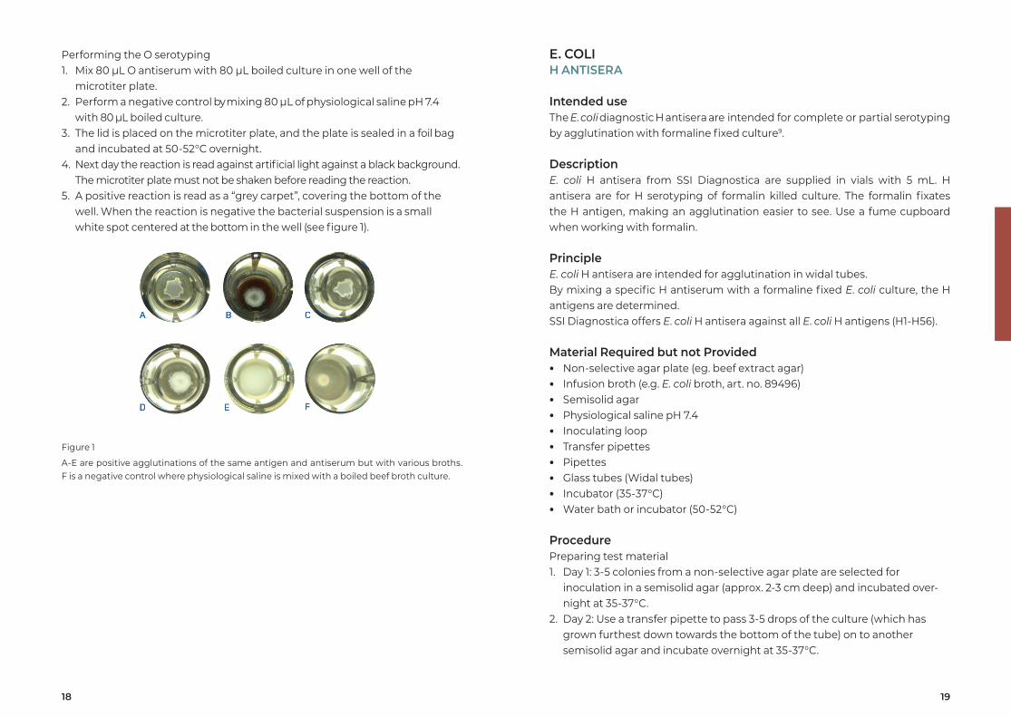
The microtiter plate must not be shaken before reading the reaction.5. A positive reaction is read as a “grey carpet”, covering the bottom of the
well. When the reaction is negative the bacterial suspension is a small white spot centered at the bottom in the well (see figure 1).
Figure 1
A-E are positive agglutinations of the same antigen and antiserum but with various broths. F is a negative control where physiological saline is mixed with a boiled beef broth culture.
20 21
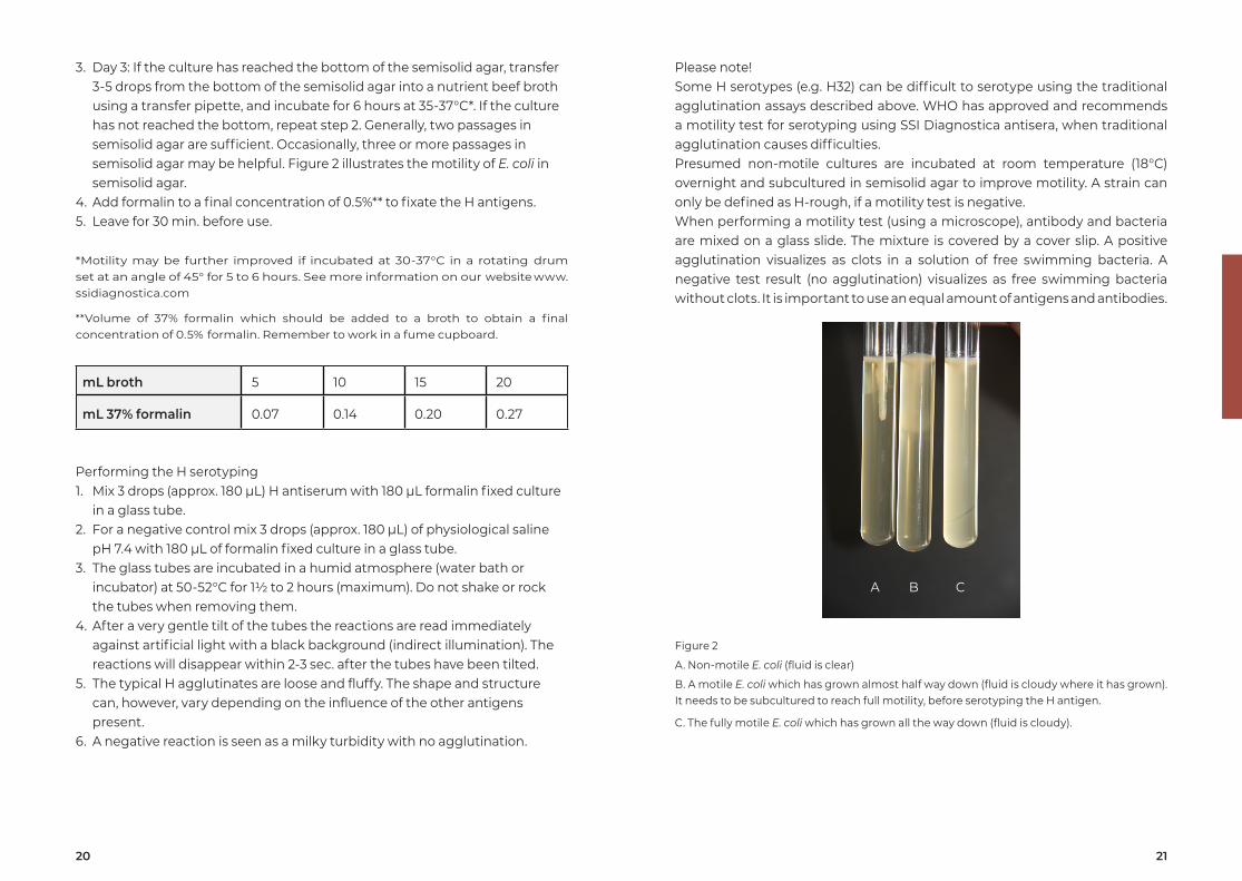
Please note!Some H serotypes (e.g. H32) can be difficult to serotype using the traditional agglutination assays described above. WHO has approved and recommends a motility test for serotyping using SSI Diagnostica antisera, when traditional agglutination causes difficulties. Presumed non-motile cultures are incubated at room temperature (18°C) overnight and subcultured in semisolid agar to improve motility. A strain can only be defined as H-rough, if a motility test is negative.When performing a motility test (using a microscope), antibody and bacteria are mixed on a glass slide. The mixture is covered by a cover slip. A positive agglutination visualizes as clots in a solution of free swimming bacteria. A negative test result (no agglutination) visualizes as free swimming bacteria without clots. It is important to use an equal amount of antigens and antibodies.
Figure 2
A. Non-motile E. coli (fluid is clear)
B. A motile E. coli which has grown almost half way down (fluid is cloudy where it has grown). It needs to be subcultured to reach full motility, before serotyping the H antigen.
C. The fully motile E. coli which has grown all the way down (fluid is cloudy).
3. Day 3: If the culture has reached the bottom of the semisolid agar, transfer 3-5 drops from the bottom of the semisolid agar into a nutrient beef broth using a transfer pipette, and incubate for 6 hours at 35-37°C*. If the culture has not reached the bottom, repeat step 2. Generally, two passages in semisolid agar are sufficient. Occasionally, three or more passages in semisolid agar may be helpful. Figure 2 illustrates the motility of E. coli in semisolid agar.
4. Add formalin to a final concentration of 0.5%** to fixate the H antigens.5. Leave for 30 min. before use.
*Motility may be further improved if incubated at 30-37°C in a rotating drum set at an angle of 45° for 5 to 6 hours. See more information on our website www.ssidiagnostica.com
**Volume of 37% formalin which should be added to a broth to obtain a final concentration of 0.5% formalin. Remember to work in a fume cupboard.
mL broth 5 10 15 20
mL 37% formalin 0.07 0.14 0.20 0.27
Performing the H serotyping1. Mix 3 drops (approx. 180 μL) H antiserum with 180 µL formalin fixed culture
in a glass tube.2. For a negative control mix 3 drops (approx. 180 μL) of physiological saline
pH 7.4 with 180 µL of formalin fixed culture in a glass tube.3. The glass tubes are incubated in a humid atmosphere (water bath or
incubator) at 50-52°C for 1½ to 2 hours (maximum). Do not shake or rock the tubes when removing them.
4. After a very gentle tilt of the tubes the reactions are read immediately against artificial light with a black background (indirect illumination). The reactions will disappear within 2-3 sec. after the tubes have been tilted.
5. The typical H agglutinates are loose and fluffy. The shape and structure can, however, vary depending on the influence of the other antigens present.
6. A negative reaction is seen as a milky turbidity with no agglutination.
A B C
22 23
Procedure1. The bacterial layer from an overnight culture grown at 35-37 °C on a beef
heart agar plate is suspended in 1 mL PBS pH 7.4 and incubated for 20 min. at 60°C to liberate the polysaccharides.
2. After centrifugation for 30 min. at 15,000 rpm, the supernatant is transferred to another tube and heated for 1 hour at ≥90°C. Subsequently the extract is kept at 2-8°C.
3. 1.5% LSA agarose gel is melted and approx. 19 mL are applied on a glass plate (70x100 mm). After cooling, the gel can be kept in a moist jar at 2-8°C.
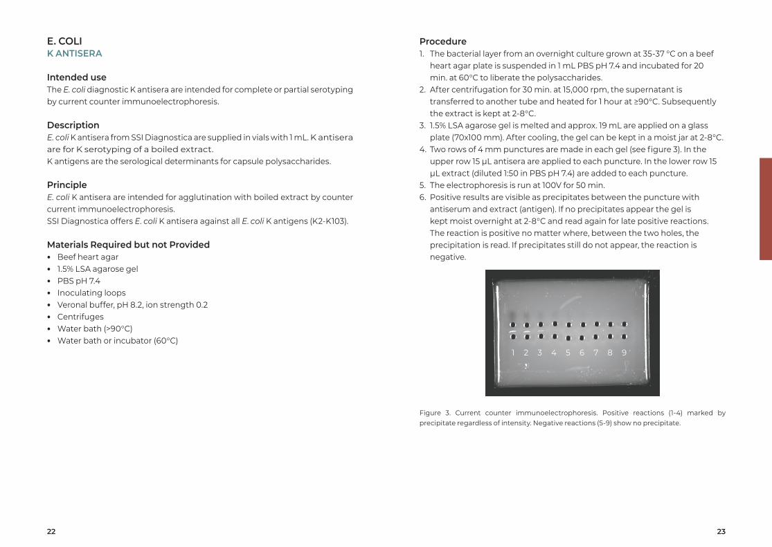
4. Two rows of 4 mm punctures are made in each gel (see figure 3). In the upper row 15 µL antisera are applied to each puncture. In the lower row 15 µL extract (diluted 1:50 in PBS pH 7.4) are added to each puncture.
5. The electrophoresis is run at 100V for 50 min.6. Positive results are visible as precipitates between the puncture with
antiserum and extract (antigen). If no precipitates appear the gel is kept moist overnight at 2-8°C and read again for late positive reactions. The reaction is positive no matter where, between the two holes, the precipitation is read. If precipitates still do not appear, the reaction is negative.
Figure 3. Current counter immunoelectrophoresis. Positive reactions (1-4) marked by precipitate regardless of intensity. Negative reactions (5-9) show no precipitate.
E. COLIK ANTISERA
Intended useThe E. coli diagnostic K antisera are intended for complete or partial serotyping by current counter immunoelectrophoresis.
DescriptionE. coli K antisera from SSI Diagnostica are supplied in vials with 1 mL. K antisera are for K serotyping of a boiled extract. K antigens are the serological determinants for capsule polysaccharides.
PrincipleE. coli K antisera are intended for agglutination with boiled extract by counter current immunoelectrophoresis. SSI Diagnostica offers E. coli K antisera against all E. coli K antigens (K2-K103).
Materials Required but not Provided • Beef heart agar • 1.5% LSA agarose gel • PBS pH 7.4 • Inoculating loops • Veronal buffer, pH 8.2, ion strength 0.2 • Centrifuges • Water bath (>90°C) • Water bath or incubator (60°C)
1 52 63 74 8 9
24 25
E. COLI F ANTISERA
Intended useThe E. coli diagnostic F antisera are intended for partial serotyping of flagella by agglutination of live culture9.
DescriptionE. coli F antisera from SSI Diagnostica are supplied in vials with 5 mL. F antisera are for screening of live cultures from a non-selective agar plate (for detection of F4 antigen) or from a Minca IS agar plate (for detection of F5 antigen). False positive and false negative results might occur if bacteria from another selective medium is used.The F4 and F5 antisera are the most common fimbrial adhesins, which help ETEC to bind to the intestinal mucosa.
PrincipleE. coli F antisera are intended for slide agglutination. By mixing a specific antiserum with an E. coli culture, the F antigen is determined.
Material required but not provided • Non-selective agar medium (e.g. beef extract agar) • Minca IS agar plates (IS = Iso Vitalex) • Physiological saline pH 7.4 • Inoculating loop or toothpick • Pipettes • Glass slides • Incubator (35-37°C)
Procedure1. The E. coli is grown over night at 35-37°C on a suitable agar medium not
inhibiting motility and on a Minca IS agar plate (F5 antiserum requires this special medium).
2-7. As for OK O antisera described on page 15.
E. COLIK9 ANTISERUM
Intended useThe E. coli diagnostic K9 antiserum for slide agglutination is intended for screening of E. coli O104 strains. It is important to confirm the result with an O104 antiserum for use with boiled culture.
DescriptionE. coli K9 antiserum from SSI Diagnostica is supplied in vials with 1 mL. K9 antiserum is for screening of live cultures9.
PrincipleE. coli K9 antiserum is intended for slide agglutination. By mixing the antiserum with an E. coli culture, the O104 antigen is determined.
Materials required but not provided • Non-selective agar plate (eg. beef extract agar) • Physiological saline pH 7.4 • Inoculating loop or toothpick • Pipettes • Glass slides • Incubator (35-37°C)
Procedure1. The E. coli is grown over night at 35-37°C on a suitable agar plate not
inhibiting motility.2. Apply a drop (approx. 20 μL) of antiserum on a glass slide. 3. Transfer culture from a single colony to the drop of antiserum and mix well.
Use an inoculating loop or a toothpick. The amount of culture should be sufficient to give a distinct milky turbidity.
4. Tilt the slide forth and back for no more than 5 - 10 sec.5. The reaction is read with the naked eye by holding the slide in front of a
light source against a black background (indirect illumination).6. A positive reaction is seen as a visible agglutination. A negative reaction
is persistence of the homogeneous milky turbidity. A late or weak agglutination (after 10 sec.) should be considered negative.
7. A positive result should be confirmed by a positive O104 antiserum reaction.
26 27
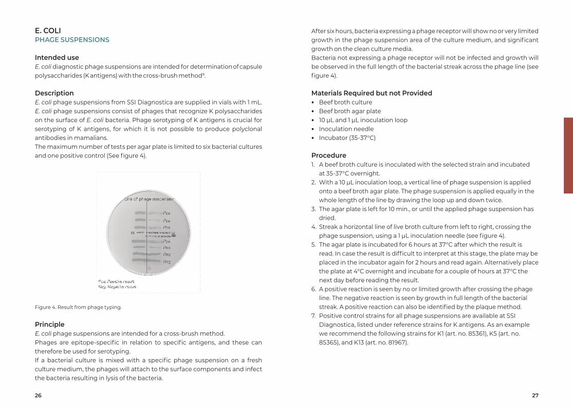
After six hours, bacteria expressing a phage receptor will show no or very limited growth in the phage suspension area of the culture medium, and significant growth on the clean culture media.Bacteria not expressing a phage receptor will not be infected and growth will be observed in the full length of the bacterial streak across the phage line (see figure 4).
Materials Required but not Provided • Beef broth culture • Beef broth agar plate • 10 µL and 1 µL inoculation loop • Inoculation needle • Incubator (35-37°C)
Procedure1. A beef broth culture is inoculated with the selected strain and incubated
at 35-37°C overnight.2. With a 10 µL inoculation loop, a vertical line of phage suspension is applied
onto a beef broth agar plate. The phage suspension is applied equally in the whole length of the line by drawing the loop up and down twice.
3. The agar plate is left for 10 min., or until the applied phage suspension has dried.
4. Streak a horizontal line of live broth culture from left to right, crossing the phage suspension, using a 1 µL inoculation needle (see figure 4).
5. The agar plate is incubated for 6 hours at 37°C after which the result is read. In case the result is difficult to interpret at this stage, the plate may be placed in the incubator again for 2 hours and read again. Alternatively place the plate at 4°C overnight and incubate for a couple of hours at 37°C the next day before reading the result.
6. A positive reaction is seen by no or limited growth after crossing the phage line. The negative reaction is seen by growth in full length of the bacterial streak. A positive reaction can also be identified by the plaque method.
7. Positive control strains for all phage suspensions are available at SSI Diagnostica, listed under reference strains for K antigens. As an example we recommend the following strains for K1 (art. no. 85361), K5 (art. no. 85365), and K13 (art. no. 81967).
E. COLI PHAGE SUSPENSIONS
Intended useE. coli diagnostic phage suspensions are intended for determination of capsule polysaccharides (K antigens) with the cross-brush method9.
DescriptionE. coli phage suspensions from SSI Diagnostica are supplied in vials with 1 mL. E. coli phage suspensions consist of phages that recognize K polysaccharides on the surface of E. coli bacteria. Phage serotyping of K antigens is crucial for serotyping of K antigens, for which it is not possible to produce polyclonal antibodies in mamalians. The maximum number of tests per agar plate is limited to six bacterial cultures and one positive control (See figure 4).
Figure 4. Result from phage typing.
PrincipleE. coli phage suspensions are intended for a cross-brush method. Phages are epitope-specific in relation to specific antigens, and these can therefore be used for serotyping. If a bacterial culture is mixed with a specific phage suspension on a fresh culture medium, the phages will attach to the surface components and infect the bacteria resulting in lysis of the bacteria.
28 29
Materials required but not provided • Serum broth, Todd Hewitt broth or 5-10% blood agar plate • Phosphate buffered saline pH 7.4 • Pipette • 1 µL inoculation loop • Glass slide and cover slip • Immersion oil • Phase contrast microscope (100 x magnification, oil immersion lens) • Incubator (35-37 °C)
ProcedureThe result is often more evident when compared with a negative control.1. Dispense 1 droplet (2-4 µL) of an overnight broth culture (grown at 35-37
°C) on a glass slide. Alternatively, colonies from a blood agar plate are mixed with phosphate buffered saline. It is preferable to have relatively few bacteria per microscope field.
2. Add an equal amount of antiserum and mix thoroughly.3. Immediately place a cover slip on top of the mixture (must not dry out).4. Examine the mixture under a phase contrast microscope within 5 min.5. If the capsule becomes visible (the bacterium appears swollen) the
reaction is positive.
PNEUMOCOCCUS NEUFELD ANTISERA
Intended useThe pneumococcal diagnostic antisera are intended for qualitative identification and typing of pneumococci by use of the capsular reaction test (Neufeld test).
DescriptionPneumococcal antisera from SSI Diagnostica are supplied in vials with 1 mL. Pneumoccal antisera are for serotyping of pure cultures of capsulated pneumococci using the Neufeld test.For factor antisera cross-reactions have been removed within the group.For antiserum Group 25, Type 29, Group 35 and Type 42 all cross-reactions cannot be removed without simultaneously absorbing too much of the homologous antibody. Consequently, the four antisera are sometimes not group or type specific (see table 1 below):
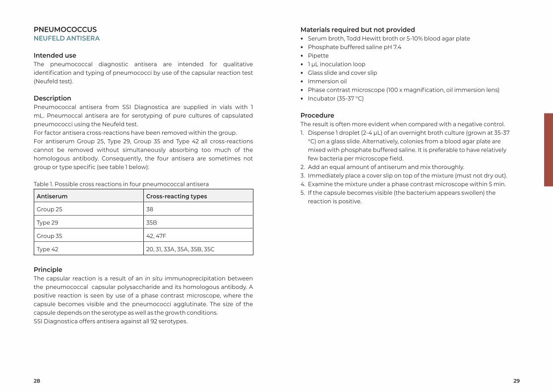
Table 1. Possible cross reactions in four pneumococcal antisera
Antiserum Cross-reacting types
Group 25 38
Type 29 35B
Group 35 42, 47F
Type 42 20, 31, 33A, 35A, 35B, 35C
PrincipleThe capsular reaction is a result of an in situ immunoprecipitation between the pneumococcal capsular polysaccharide and its homologous antibody. A positive reaction is seen by use of a phase contrast microscope, where the capsule becomes visible and the pneumococci agglutinate. The size of the capsule depends on the serotype as well as the growth conditions.SSI Diagnostica offers antisera against all 92 serotypes.
30 31
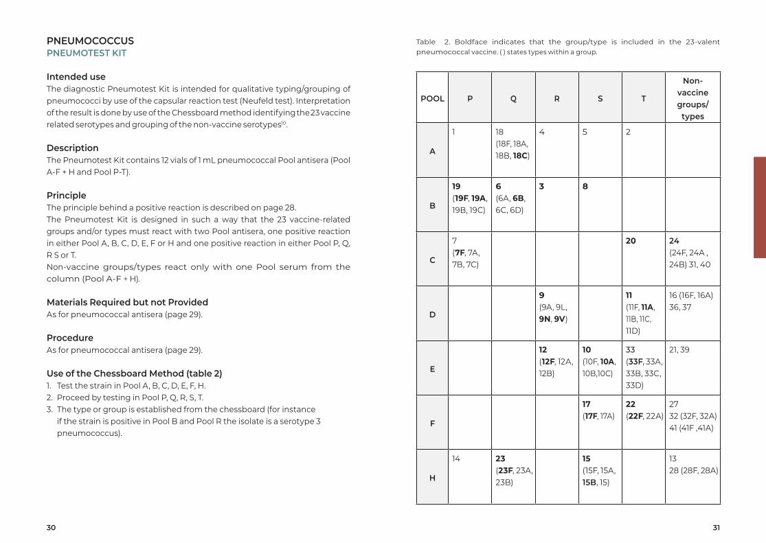
Table 2. Boldface indicates that the group/type is included in the 23-valent pneumococcal vaccine. ( ) states types within a group.
POOL P Q R S T
Non-vaccinegroups/
types
A
1 18(18F, 18A, 18B, 18C)
4 5 2
B
19(19F, 19A, 19B, 19C)
6(6A, 6B, 6C, 6D)
3 8
C
7(7F, 7A,7B, 7C)
20 24(24F, 24A , 24B) 31, 40
D
9(9A, 9L,9N, 9V)
11(11F, 11A,11B, 11C, 11D)
16 (16F, 16A)36, 37
E
12(12F, 12A,12B)
10(10F, 10A,10B,10C)
33(33F, 33A, 33B, 33C, 33D)
21, 39
F
17(17F, 17A)
22(22F, 22A)
2732 (32F, 32A)41 (41F ,41A)
H
14 23 (23F, 23A,23B)
15(15F, 15A,15B, 15)
1328 (28F, 28A)
PNEUMOCOCCUSPNEUMOTEST KIT
Intended useThe diagnostic Pneumotest Kit is intended for qualitative typing/grouping of pneumococci by use of the capsular reaction test (Neufeld test). Interpretation of the result is done by use of the Chessboard method identifying the 23 vaccine related serotypes and grouping of the non-vaccine serotypes10.
DescriptionThe Pneumotest Kit contains 12 vials of 1 mL pneumococcal Pool antisera (Pool A-F + H and Pool P-T).
PrincipleThe principle behind a positive reaction is described on page 28.The Pneumotest Kit is designed in such a way that the 23 vaccine-related groups and/or types must react with two Pool antisera, one positive reaction in either Pool A, B, C, D, E, F or H and one positive reaction in either Pool P, Q, R S or T. Non-vaccine groups/types react only with one Pool serum from the column (Pool A-F + H).
Materials Required but not ProvidedAs for pneumococcal antisera (page 29).
ProcedureAs for pneumococcal antisera (page 29).
Use of the Chessboard Method (table 2)1. Test the strain in Pool A, B, C, D, E, F, H.2. Proceed by testing in Pool P, Q, R, S, T.3. The type or group is established from the chessboard (for instance
if the strain is positive in Pool B and Pool R the isolate is a serotype 3 pneumococcus).
32 33
ProcedureSlide agglutination with O and H antisera1. The Salmonella is grown over night at 35-37 °C on a non-selective agar
medium. Swarm agar is the best suited medium for growing cultures for H typing, but H antigens can be serotyped from a non-selective agar medium if the H antigens are well expressed.
2. Apply a small drop of antiserum (approx. 20 µL) on the glass slide.3. Transfer culture from several colonies to the drop of antiserum and mix
well. The amount of culture should be sufficient to give a distinct milky turbidity. Use an inoculating loop or a toothpick.
4. Tilt the slide for 5-10 sec.5. The reaction is read with the naked eye by holding the slide in front of a
light source against a black background (indirect illumination).6. A positive reaction is seen as a visible agglutination. A negative reaction
is persistence of the homogeneous milky turbidity. A late or weak agglutination (after 10 sec.) should be considered negative.
Absence of reactions may be due to a strain expressing the Vi antigen (see below), to a strain not covered by the antisera used or to a strain not being Salmonella.
Demonstration of the Vi antigenThe presence of the Vi antigen may interfere with or prevent agglutination in O antisera. Negative isolates must therefore be examined for Vi antigen. Due to form variation in the Vi antigen it is important to select single colonies as colony forms expressing the Vi antigen are more opaque than Vi negative colonies.
H phase inversion on swarm agar plates (S. Gard method)12
1. Melt the swarm agar in a microwave oven or in a water bath (>90 °C) and cool to 45°C.
2. Apply 100 µL of H antiserum for phase inversion (corresponding to the phase which has already been identified) in the center of a small, sterile petri dish (diameter 6 cm).
3. Pour 10 mL of the swarm agar onto the antiserum resulting in a final dilution of 1:100.
4. Leave the plate for solidification at the site of pouring at room temperature (22-25°C) for 10-15 min.
5. Inoculate the plate in the center with a loop full of fresh bacterial culture from an agar plate or a broth culture.
SALMONELLAO AND H ANTISERAVI MONOCLONAL ANTIBODY
Intended useThe Salmonella diagnostic antisera are intended for complete or partial serotyping by slide agglutination and H phase inversion.
DescriptionSalmonella O and H antisera from SSI Diagnostica are supplied in vials with 1 and/or 3 mL. Salmonella O Group and O Factor and H Phase and H Factor antisera are for screening of live cultures from a non- selective agar plate. Salmonella Phase Inversion antisera are for inversion of H phases.All Salmonella antisera are absorbed free of cross-reactions except for Poly A-E+Vi, Poly A-I+Vi, Poly A-S+Vi, Poly 42-67 and Poly H.
PrincipleSalmonella O Group and O Factor and H Phase and H Factor antisera are intended for slide agglutination. By mixing a specific antiserum with a Salmonella culture, the O and H antigens are determined. Salmonella Phase Inversion antisera are intended for inversion of the H phases. By mixing Phase Inversion antiserum and swarm agar and growing a Salmonella culture on the solidified medium, the H phase is inverted. On the basis of the observed agglutination pattern the serotype is determined using the Kauffmann-White Scheme11.
Material required but not provided • Non-selective agar medium (eg. beef extract agar) • Physiological saline pH 7.4 • Inoculating loop or toothpick • Glass slides • Incubator (35-37°C) • Kauffmann-White Scheme
Additional material for phase inversion • Sterile petri dishes (diameter 6 cm) • A microwave oven or a water bath (>90 °C) • Swarm agar • Pipette
34 35
6. Incubate overnight at 35-37°C.7. Use culture material from the edge of the growth zone for slide
agglutination. Select the relevant H antisera by using the Kauffmann-White Scheme.
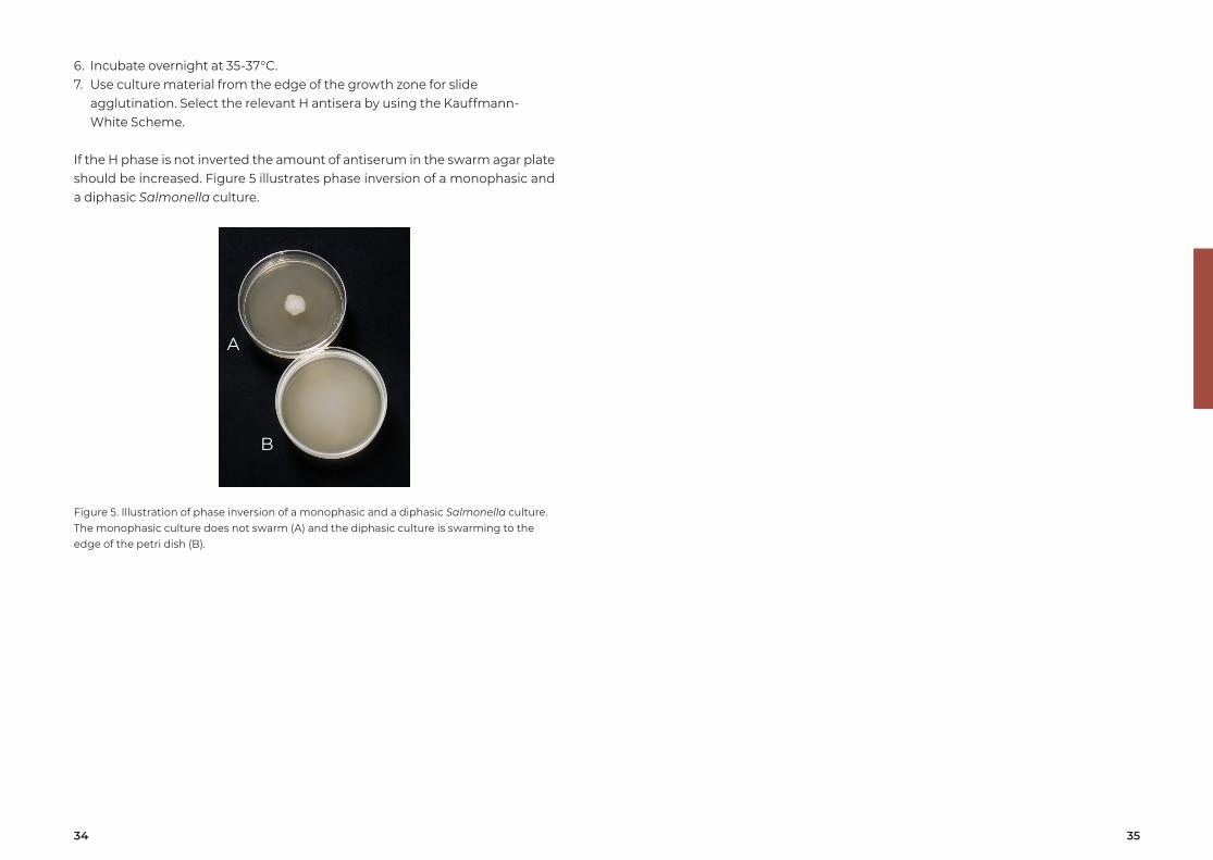
If the H phase is not inverted the amount of antiserum in the swarm agar plate should be increased. Figure 5 illustrates phase inversion of a monophasic and a diphasic Salmonella culture.
Figure 5. Illustration of phase inversion of a monophasic and a diphasic Salmonella culture. The monophasic culture does not swarm (A) and the diphasic culture is swarming to the edge of the petri dish (B).
A
B
36 37
H:ipositive
H:2positive
SG2in agar
SG6in agar
Agar
or
O typing
Agar orsoft agar
H typing1 phase
Soft agarPhase
inversion
Agar orsoft agar
S. Typhimurium1,4,[5],12:i:1,2
H typing2 phase
O:4positive
H:2positive
H:ipositive
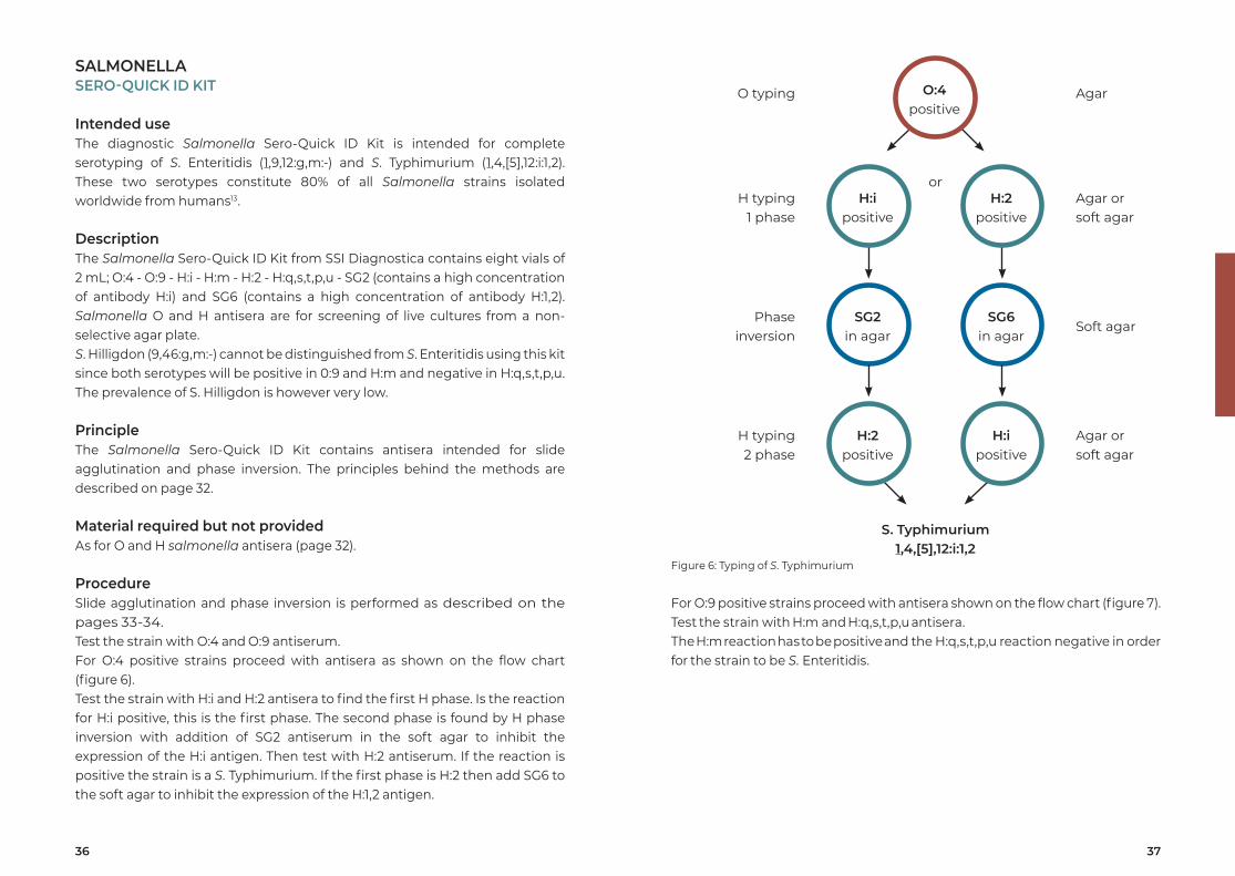
Figure 6: Typing of S. Typhimurium
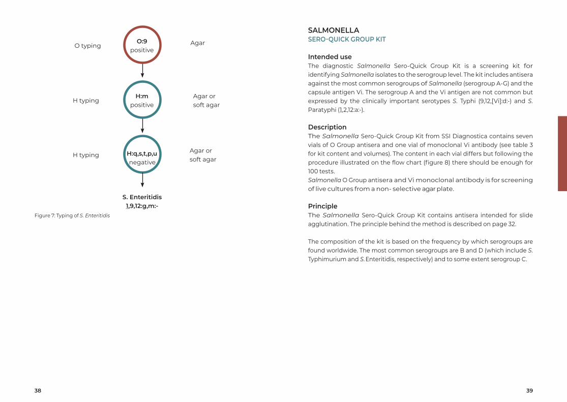
For O:9 positive strains proceed with antisera shown on the flow chart (figure 7).Test the strain with H:m and H:q,s,t,p,u antisera. The H:m reaction has to be positive and the H:q,s,t,p,u reaction negative in order for the strain to be S. Enteritidis.
SALMONELLASERO-QUICK ID KIT
Intended useThe diagnostic Salmonella Sero-Quick ID Kit is intended for complete serotyping of S. Enteritidis (1,9,12:g,m:-) and S. Typhimurium (1,4,[5],12:i:1,2). These two serotypes constitute 80% of all Salmonella strains isolated worldwide from humans13.
DescriptionThe Salmonella Sero-Quick ID Kit from SSI Diagnostica contains eight vials of 2 mL; O:4 - O:9 - H:i - H:m - H:2 - H:q,s,t,p,u - SG2 (contains a high concentration of antibody H:i) and SG6 (contains a high concentration of antibody H:1,2). Salmonella O and H antisera are for screening of live cultures from a non- selective agar plate. S. Hilligdon (9,46:g,m:-) cannot be distinguished from S. Enteritidis using this kit since both serotypes will be positive in 0:9 and H:m and negative in H:q,s,t,p,u. The prevalence of S. Hilligdon is however very low.
PrincipleThe Salmonella Sero-Quick ID Kit contains antisera intended for slide agglutination and phase inversion. The principles behind the methods are described on page 32.
Material required but not providedAs for O and H salmonella antisera (page 32).
ProcedureSlide agglutination and phase inversion is performed as described on the pages 33-34.Test the strain with O:4 and O:9 antiserum.For O:4 positive strains proceed with antisera as shown on the flow chart (figure 6).Test the strain with H:i and H:2 antisera to find the first H phase. Is the reaction for H:i positive, this is the first phase. The second phase is found by H phase inversion with addition of SG2 antiserum in the soft agar to inhibit the expression of the H:i antigen. Then test with H:2 antiserum. If the reaction is positive the strain is a S. Typhimurium. If the first phase is H:2 then add SG6 to the soft agar to inhibit the expression of the H:1,2 antigen.
38 39
SALMONELLASERO-QUICK GROUP KIT
Intended useThe diagnostic Salmonella Sero-Quick Group Kit is a screening kit for identifying Salmonella isolates to the serogroup level. The kit includes antisera against the most common serogroups of Salmonella (serogroup A-G) and the capsule antigen Vi. The serogroup A and the Vi antigen are not common but expressed by the clinically important serotypes S. Typhi (9,12,[Vi]:d:-) and S. Paratyphi (1,2,12:a:-).
DescriptionThe Salmonella Sero-Quick Group Kit from SSI Diagnostica contains seven vials of O Group antisera and one vial of monoclonal Vi antibody (see table 3 for kit content and volumes). The content in each vial differs but following the procedure illustrated on the flow chart (figure 8) there should be enough for 100 tests. Salmonella O Group antisera and Vi monoclonal antibody is for screening of live cultures from a non- selective agar plate.
PrincipleThe Salmonella Sero-Quick Group Kit contains antisera intended for slide agglutination. The principle behind the method is described on page 32.
The composition of the kit is based on the frequency by which serogroups are found worldwide. The most common serogroups are B and D (which include S. Typhimurium and S. Enteritidis, respectively) and to some extent serogroup C.
H:mpositive
H:q,s,t,p,unegative
AgarO typing
Agar orsoft agar
H typing
H typingAgar orsoft agar
S. Enteritidis1,9,12:g,m:-
O:9positive
Figure 7: Typing of S. Enteritidis
40 41
Gr. D
VI
Gr. C
Gr. G
Gr. A
Control
Gr. B
Gr. E
Gr. F
Rough
Serogroup B
Serogroup E
Serogroup F
Serogroup D
Serogroup C
Serogroup G
Serogroup A
Both negative
Both negative
Both negative
All negative
Send the strain to a reference laboratory for serotyping
Positive
Positive
Possibly S. Typhi
Positive
Positive
Positive
Positive
Positive
Positive
Positive
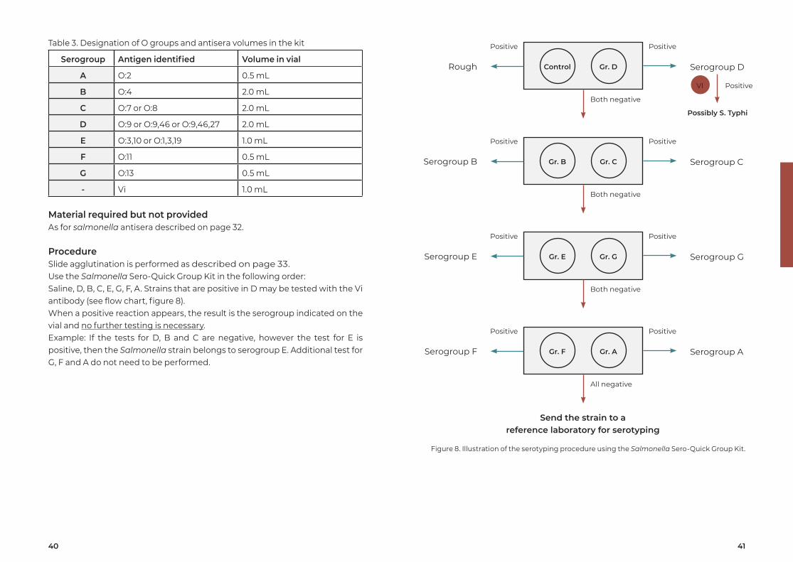
Figure 8. Illustration of the serotyping procedure using the Salmonella Sero-Quick Group Kit.
Table 3. Designation of O groups and antisera volumes in the kit
Serogroup Antigen identified Volume in vial
A O:2 0.5 mL
B O:4 2.0 mL
C O:7 or O:8 2.0 mL
D O:9 or O:9,46 or O:9,46,27 2.0 mL
E O:3,10 or O:1,3,19 1.0 mL
F O:11 0.5 mL
G O:13 0.5 mL
- Vi 1.0 mL
Material required but not providedAs for salmonella antisera described on page 32.
ProcedureSlide agglutination is performed as described on page 33.Use the Salmonella Sero-Quick Group Kit in the following order: Saline, D, B, C, E, G, F, A. Strains that are positive in D may be tested with the Vi antibody (see flow chart, figure 8).When a positive reaction appears, the result is the serogroup indicated on the vial and no further testing is necessary. Example: If the tests for D, B and C are negative, however the test for E is positive, then the Salmonella strain belongs to serogroup E. Additional test for G, F and A do not need to be performed.
42 43
Table 4. The kit contains the following Salmonella antisera
O antisera (3 mL) H antisera (3 mL) H phase inversion antisera (3 mL)
O:4 H:q,s,t,p,u SG1
O:8 H:i SG2
O:6,7,8 H:r SG5
O:9 H:z10 SG6
O:61 H:m H:r phase inversion
O:7 H:x
O:46 H:2
H:5
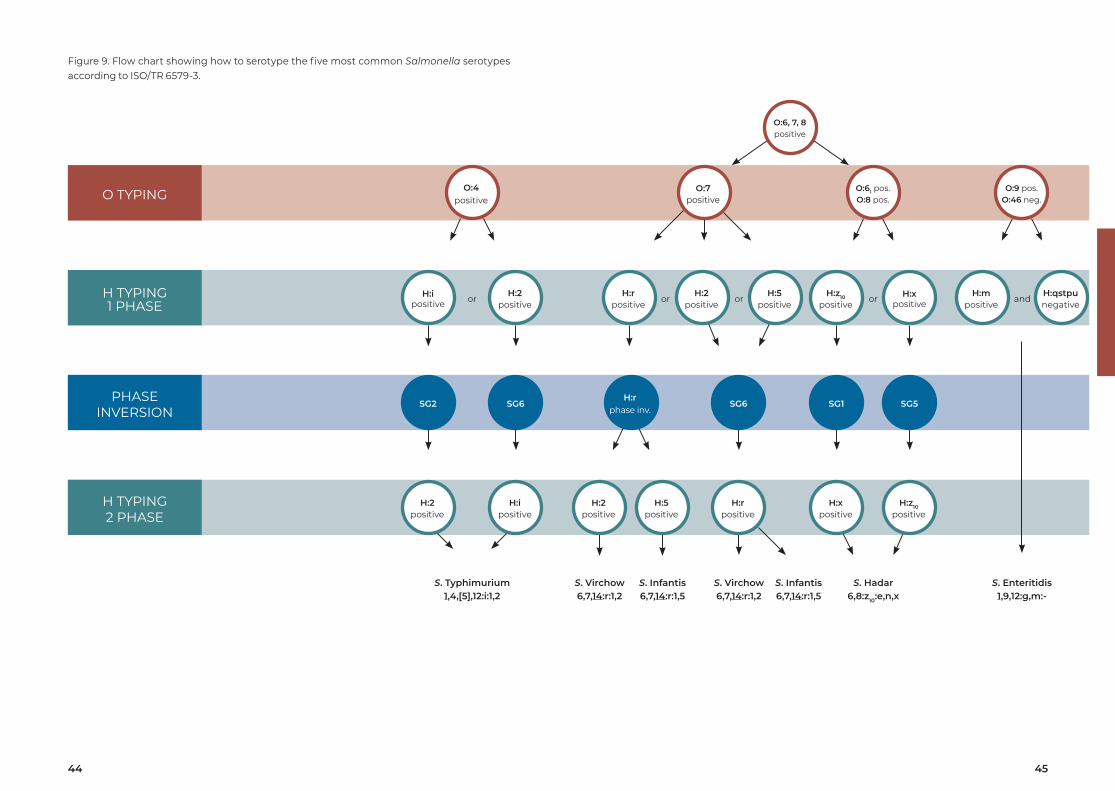
PrincipleThe Salmonella “BIG FIVE” kit – ISO 6579-3 contains O Group and O Factor as well as H Phase and H Factor antisera intended for slide agglutination. In addition, it contains H Phase Inversion antisera for phase inversion. The principles behind the two methods are described on page 32.
Material required but not providedAs for O and H salmonella antisera described on page 32.
ProcedureSlide agglutination and phase inversion is performed as described on the pages 33-34.A guideline for serotyping of the 5 serotypes is shown in the flow chart (figure 9).
SALMONELLA SALMONELLA BIG FIVE KIT – ISO 6579-3
Intended useThe diagnostic Salmonella “BIG FIVE” kit – ISO 6579-3 contains all the Salmonella antisera needed to serotype the five most common human related Salmonella serotypes which cause food poisoning according to ISO/TR 6579-3 ”Microbiology of food and animal feed - Horizontal method for the detection, enumeration and serotyping of Salmonella”.
The 5 Salmonella serotypes are: • S. Typhimurium • S. Enteritidis • S. Infantis • S. Virchow • S. Hadar
DescriptionThe Salmonella “BIG FIVE” kit – ISO 6579-3 from SSI Diagnostica contains 20 vials of 3 mL antisera (see table 4). For description of the Salmonella O and H antisera see page 32.
44 45
PHASEINVERSION
O TYPING
H TYPING2 PHASE
H TYPING1 PHASE
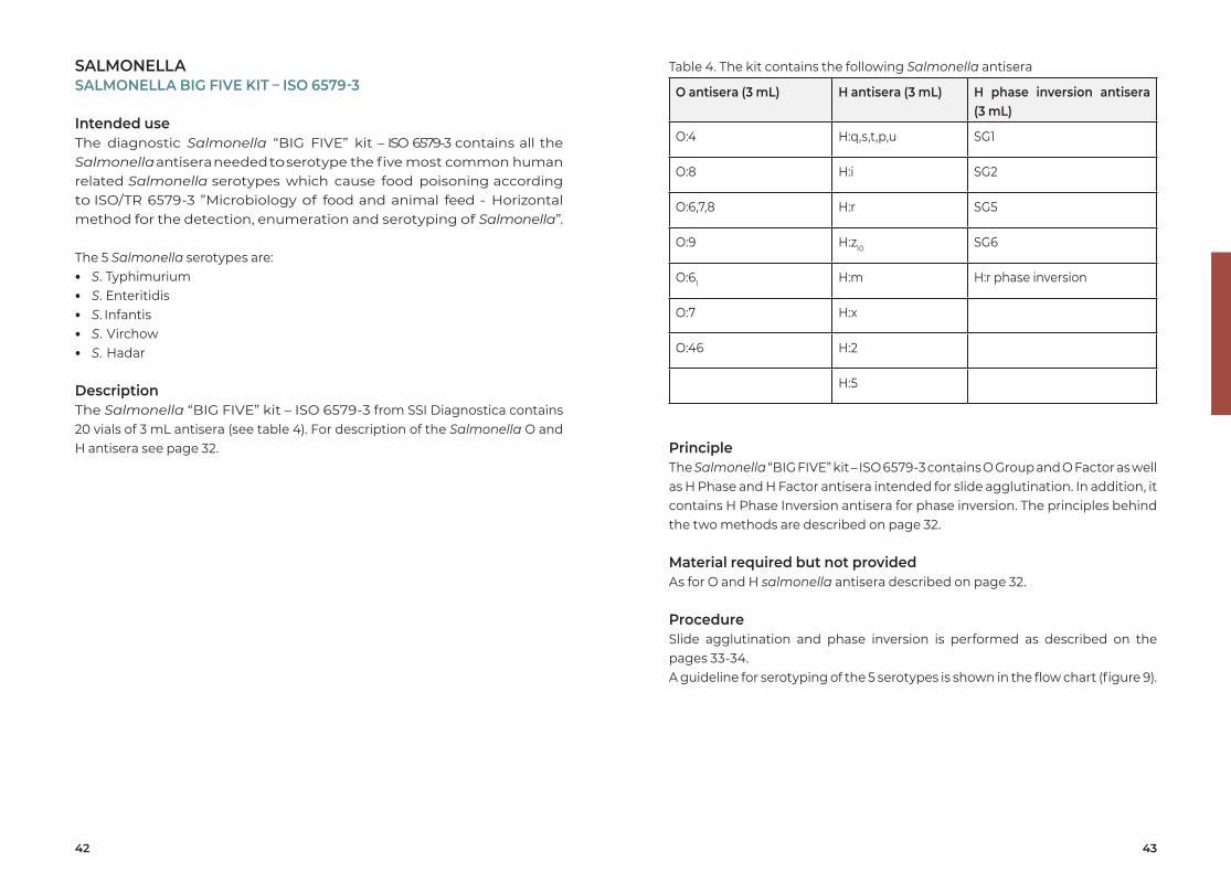
Figure 9. Flow chart showing how to serotype the five most common Salmonella serotypes according to ISO/TR 6579-3.
O:6, 7, 8 positive
S. Virchow6,7,14:r:1,2
S. Infantis6,7,14:r:1,5
S. Hadar6,8:z10:e,n,x
S. Enteritidis1,9,12:g,m:-
S. Virchow6,7,14:r:1,2
S. Infantis6,7,14:r:1,5
SG6 SG1 SG5H:r
phase inv.SG2 SG6
S. Typhimurium1,4,[5],12:i:1,2
O:7positive
O:61 pos.O:8 pos.
O:9 pos.O:46 neg.
O:4positive
H:rpositive
H:xpositive
H:z10
positiveH:2
positiveH:5
positiveH:2
positiveH:i
positive
H:2positive
H:5positive
orH:z10
positiveH:x
positiveorH:m
positiveH:qstpunegative
andH:r
positiveH:i
positiveH:2
positiveor or
46 47
Lancefield test: • 5-10% blood agar plate • Glucose broth • 1 µL Inoculation loop • Centrifuge • Pipette • 0.06N, 0.1N and 0.2N HCl • Phenol red (indicator) • 0.2N NaOH • Glass tube • Capillary tubes • Incubator (35-37°C) • Water bath (100°C)
ProcedureEach serotype will react positive either in the Lancefield test or the Neufeld test (the preferred method will be indicated on the certificate of analysis). The result is often more evident when compared with a negative control.
Neufeld test:1. The streptococci are grown overnight at 35-37 °C on a 5-10% blood agar
plate.2. Apply a small drop (3-6 µL) of saline on a glass slide.3. Transfer a small amount of culture from the blood agar plate with an
inoculating loop and mix well.4. An equal amount of antiserum is added and mixed thoroughly with the
droplet.5. Immediately place a cover slip on top of the mixture (must not dry out).6. Examine the mixture under a phase contrast microscope. The reaction is
stable for half an hour (provided no dry out).7. If the capsule becomes visible (the bacterium appears swollen) the
reaction is positive.
STREPTOCOCCUSNEUFELD ANTISERA OR ANTISERA FOR LANCEFIELD TEST
Intended useThe streptococcal diagnostic antisera are intended for serotyping of streptococci by means of the Neufeld test14 or the Lancefield test15,16.
DescriptionStreptococcal antisera from SSI Diagnostica are supplied in vials with 1 mL. Streptococcal antisera are for serotyping directly from a blood agar plate using the Neufeldt test or from an extract using the Lancefield test.Streptococcal antisera can be used for identification of Group A, B, C, D, F, G and L streptococci, serotyping of Group B streptococci and serotyping of S. suis.
PrincipleStreptococcal antisera are intended for serotyping using the Neufeldt or the Lancefield test. Neufeld test: by mixing specific antisera with a Streptococcus culture, the capsular antigens are determined. The capsular reaction is a result of an in situ immune precipitation between the streptococcal capsular polysaccharide and its homologous antibody. A positive reaction is seen by use of a phase contrast microscope where the capsule becomes visible and the streptococci agglutinate. The size of the capsule depends on the serotype as well as the growth conditions.Lancefield test: when an acid antigen extract is mixed with a specific antiserum directed against bacterial surface components, the cells are bound together through antigen-antibody bonds to form aggregates (precipitation). This is visible to the naked eye as snow in the capillary tube.
Materials Required but not ProvidedNeufeld test: • 5-10% blood agar plate • Physiological saline pH 7.4 • 1 µL inoculation loop • Pipette • Glass slides and cover slip • Immersion oil • Phase contrast microscope (100 x magnification, oil immersion lens) • Incubator (35-37 °C)
48 49
H. INFLUENZAE TYPE B ANTISERUM
Intended useThe H. influenzae type b diagnostic antiserum is intended for serotyping of H. influenzae type b.
DescriptionH. influenzae type b antiserum from SSI Diagnostica is supplied in vials with 1 mL. H. influenzae type b antiserum is for serotyping of live cultures from a non- selective agar plate.
PrincipleH. influenzae type b antiserum is intended for Neufeldt reaction using a phase contrast microscope. By mixing the antiserum with a H. influenzae type b culture, the serotype is determined. The available H. influenzae type b antiserum detects the most frequent and pathogenic H. influenzae for humans.The capsular reaction is a result of the interaction between the H. influenzae capsular polysaccharide and its homologous antibody14. If the capsule becomes visible in a phase contrast microscope, the reaction is positive. A positive reaction is the result of an in situ immunoprecipitation leading to a change in the refractive index. In addition, the bacteria agglutinates.
Materials required but not provided • Chocolate agar plate • Phosphate buffered saline (pH 7.4) • Pipette or any other utility that can make a droplet • 1 µL inoculation loop • Glass slide • Cover slip • Immersion oil • Phase contrast microscope (100 x magnification, oil immersion lens) • Incubator (35-37 °C)
Lancefield test1. The streptococci are grown overnight at 35-37 °C on a 5-10% blood agar
plate.2. Add a few colonies into 6 mL glucose broth and incubate at 35-37°C
overnight.3. Centrifuge the suspension for 10 min. at 3,000 rpm and remove the
supernatant.4. Add 0.1 mL of either 0.06N, 0.1N or 0.2N HCl to the bacteria pellet (the
preferred method will be indicated on the antiserum label).5. The acid suspension is placed in a water bath (100°C) for 15 min.6. Cool the acid suspension under tab water.7. The pH-value is adjusted to approx. 7 by addition of droplets of 0.2N NaOH
until the color is brown/orange (use phenol red as pH-indicator, red (pH > 8.2) - yellow (pH < 6.4)).
8. Centrifuge the suspension for 10 min. at 3.000 rpm and transfer the supernatant (acid antigen extract) to a new glass.
9. Equal amount of the antiserum (first) and the acid antigen extract (second) are sucked up with the capillary tube. The antiserum must be in the upper part of the capillary tube to diffuse down through the acid extract.
10. Read the result against a light source.11. Precipitation looking like snowfall will occur if positive.
50 51
SHIGELLAO POOL AND PHASE ANTISERA
Intended useThe Shigella diagnostic antisera are intended for partial serotyping of Shigella by slide agglutination.
DescriptionShigella O Pool and Phase antisera from SSI Diagnostica are supplied in vials with 3 mL. Shigella O Pool and Phase antisera are for screening of live cultures from a non- selective agar plate.
PrincipleShigella O Pool and Phase antisera are intended for slide agglutination. By mixing antiserum with a Shigella culture, the Shigella species and the phases for S. sonnei are determined.
Materials Required but not Provided • Non-selective agar plate (eg. beef extract agar) • Physiological saline, pH 7.4 • Inoculating loop or toothpick • Glass slides • Incubator (35-37°C)
Procedure1. The Shigella strain is grown overnight at 35-37°C on a non-selective or
selective agar medium.2. Apply a small drop of antisera (approx. 20 µL) on the glass slide.3. Transfer culture from several colonies to the drop of antiserum and mix
well. The amount of culture should be sufficient to give a distinct milky turbidity. Use an inoculating loop or a toothpick.
4. Tilt the slide gently for 5-10 sec.5. The reaction is read with the naked eye by holding the slide in front of a
light source against a black background (indirect illumination).6. A positive reaction is seen as a visible agglutination. A negative reaction
is persistence of the homogeneous milky turbidity. A late or weak agglutination (after 10 sec.) should be considered negative.
ProcedureThe result is often more evident when compared with a negative control.
1. The H. influenzae culture should be incubated overnight at 35-37 °C on a chocolate agar plate.
2. A small amount of bacterial culture is transferred from the plate and mixed into a droplet (2 - 4 µl) of phosphate buffered saline which has been placed on the glass slide. It is preferable to have relatively few organisms per microscope field.
3. An equal amount of antiserum is added and mixed thoroughly with the droplet on the slide.
4. Immediately place a cover slip on top of the mixture. It is important that the preparation does not dry out.
5. Examine the mixture under the phase contrast microscope using an oil immersion lens, magnification x 100 within 5 min.
6. If the capsule becomes visible (the bacteria appears swollen) the reaction is positive.
52 53
Y. ENTEROCOLITICAO ANTISERA
Intended useY. enterocolitica diagnostic antisera are intended for serotyping by slide agglutination.
DescriptionY. enterocolitica O antisera from SSI Diagnostica are supplied in vials with 3 mL. Y. enterocolitica O antisera are for screening of live cultures from a non- selective agar plate.
PrincipleY. enterocolitica antisera are intended for slide agglutination. By mixing a specific antiserum with a Y. enterocolitica culture, the O antigen is determined. The available Y. enterocolitica antisera detect the most frequent and pathogenic Y. enterocolitica for humans.
Materials required but not provided • Non-selective agar medium (eg. beef extract agar) • Physiological saline, pH 7.4 • Inoculating loop or toothpick • Glass slides • Incubator (20-25°C)
Procedure1. The Y. enterocolitica strain is grown at 20-25ºC for 24-48 hours on a non-
selective agar medium.2. Apply a small drop of antiserum (approx. 20 µL) on a glass slide.3. Transfer culture from a single colony to the drop of antiserum and mix well.
The amount of culture should be sufficient to give a distinct milky turbidity. Use an inoculating loop or a toothpick.
4. Tilt the slide gently for 5-10 sec.5. The reaction is read with the naked eye by holding the slide in front of a
light source against a black background (indirect illumination).6. A positive reaction is seen as a visible agglutination. A negative reaction
is persistence of the homogeneous milky turbidity. A late or weak agglutination (after 10 sec.) should be considered negative.
54 55
Streptococcus • 5-10% Blood agar plates • Agar plate (selective) • Storage medium • 5% CO2 incubator 35-37°C (CO2 is recommended but a normal incubator
can be used)
ProcedureAll strains and subsequent cultures must be regarded as potentially pathogen, and must be handled by persons trained in microbiological techniques. The working facilities should be classified according to the biosafety level 2.
1. Work in a biological safety cabinet and wear gloves. 2. All waste should be handled and discarded as infected material.3. Let the container reach room temperature.4. Remove the lid from the container.5. Transfer aseptically bacterial material to a non-selective agar plate and a
selective agar plate, by leading an inoculation needle/loop into the extract agar. For Stuart Transport medium use the inoculation pen from the tube.
6. Incubate the agar plates for 18-24 hours at 35-37°C.7. Inspect the agar plates for pure growth and serotype the isolate.8. From the pure agar plate culture spread bacterial material on a new non-
selective agar plate.9. Incubate the agar plate for 18-24 hours at 35-37°C.10. Subsequent subcultures can be made from this agar plate.
BACTERIAL STRAINS
STRAINS IN AGARE. coliSalmonellaStreptococcus
Intended use The bacterial strains are intended to be used for research or as negative/positive controls for serotyping of antigens.
DescriptionE. coli and Salmonella strains are delivered in extract agar in either a sealed glass tube or in a cryogenic vial.Streptococcal strains are delivered in Stuart transport medium.Each strain has been grown on a non-selective agar plate overnight and the serotype verified by SSI Diagnostica specific antisera.
PrincipleThe bacterial strains have been QC tested as negative/positive controls for serotyping. The strains can be subcultured directly from the transport medium.Paraffin sealed glass tube: the strain can be stored for up to 10 years at 8-25°C.Stuart transport medium: the strain should be subcultured or transferred to a storage medium immediately on receipt.
Materials Required but not Provided • Biological safety cabinet • Gloves • Inoculation needle or loop
E. coli and Salmonella • Non-selective agar plate (eg. beef extract agar) • Incubator (35-37°C)
56 57
ProcedureAll strains and subsequent cultures must be regarded as potentially pathogen, and must be handled by persons trained in microbiological techniques. The working facilities should be classified according to the biosafety level 2.
1. Work in a biological safety cabinet and wear gloves. 2. All waste should be handled and discarded as infected material.3. Scratch using a sharp file on the narrow part of the ampoule approx. 0.5 cm
from the top of the ampoule.4. Wet a piece of gauze with 70% ethanol and disinfect the ampoule.5. Wrap the gauze around the ampoule and break the glass at the scratched
area. Be careful as this might leave sharp glass edges.6. Discard the top of the ampoule as biological waste. 7. Add aseptically 1 mL TH or BHI broth using a sterile pipette to the freeze-
dried material in the ampoule and mix well.8. Transfer the entire bacterial suspension to 7 mL preheated (35-37°C) TH or
BHI broth.9. Incubate the broth overnight at 35-37°C in a 5% CO2 incubator.10. Next day transfer one drop of the bacterial suspension to a 5-10% blood
agar plate and spread.11. Incubate the agar plate for 18-24 hours at 35-37°C in a 5% CO2 incubator.12. Next day inspect the blood agar plate for pure growth and serotype the
isolate.13. Subsequent subcultures can be made from the blood agar plate.
FREEZE-DRIED STRAINSPneumococcusStreptococcus
Intended useThe bacterial strains are intended to be used for research or as negative/positive controls for serotyping of antigens.
DescriptionPneumococus and Streptococcus strains are freeze dried in TH broth or BHI broth. Each strain has been grown on a 5-10% blood agar plate overnight and the serotype verified by SSI Diagnostica specific antisera.Unopened ampoules can be stored for numerous years at room temperature. Once an ampoule has been opened the freeze-dried bacteria cannot be stored but must be used immediately.
PrincipleThe strains are grown in serum broth overnight, and the serotype is verified by SSI Diagnostica specific antisera.1 mL of the broth is afterwards dispensed in an ampoule and freeze-dried.The strains have been QC tested as negative/positive controls for serotyping.
Material required but not provided • TH broth or BHI broth • 5-10% blood agar plate • 70% Ethanol • Gloves • Gauze • Sharp file • Sterile pipette • Biological safety cabinet • 5% CO2 incubator 35-37°C (CO2 is recommended but a normal incubator
can be used)
58 59
11. Grimont, P.A.D. and Weill, F.-X. Antigenic formulae of the Salmonella serovars, WHO Collaborating Centre for Reference and Research on Salmonella, Institut Pasteur, Paris, France, 9th ed., 2007.
12. Gard, S., Das Schwärmphänomen in der Salmonella-gruppe und seine praktische Ausnützung, Zeit. f. Hyg. Inf. Krankh, 120:615-19, 1938.
13. Herikstad et al., Salmonella surveillance: a global survey of public health serotyping, Epidemiol. Infect., 129:1-8, 2002
14. Austrian R. The Quelling Reaction, A neglected Microbiologic Technique. The Mount Sinai Journal of Medicine, 43:669-09, 1976.
15. Lancefield, R.C. A Serological Differentiation of Specific Types of Bovine hemolytic streptococci (group B), J. Exp. Med., 59:441-58, 1934.
16. Slotved, H.C. et al., False-negative results in typing of group B streptococci by the standard l Lancefield antigen extraction method, J. Clin. Microbiol., May;40(5):1882-3, 2002.
Information and ordering
SSI Diagnostica A/S
Herredsvejen 2
3400 Hillerød
Denmark
Tel.: +45 4829 9100
www.ssidiagnostica.com
shop.ssidiagnostica.com
REFERENCES
1. Konradsen, H.B. et al., A modified enzyme-linked immunosorbent assay for measuring type specific anti-pneumococcal capsular polysaccharide antibodies, J. Immunol. Methods, 164(1), 13-20, 1993.
2. Plikaytis, B.D. et al., An analytical model applied to a multicenter pneumococcal enzyme-linked immunosorbent assay study, J. Clin. Microbiol., 38(6), 2043-50, 2000.
3. Training manual for Enzyme linked immunosorbent assay for the quantitation of Streptococcus pneumoniae serotype specific IgG (Pn PS ELISA). Prepared by the World Health Organization Pneumococcal Serology Reference Laboratories at the Institute of Child Health (London, England) and the Department of Pathology (Birmingham Alabama, USA, http://www.vaccine.uab.edu/uploads/mdocs/ELISAProtocol(007sp).pdf 2002/2011.
4. Wernette, C.M. et al., Enzyme-Linked Immosorbent Assay for Quantitation of Human Antibodies to Pneumococcal Polysaccharides, Clin. Diagn. Lab. Immunol., 10(4), 514-19, 2003.
5. Skovsted, I.C. et al., Purification and structure characterization of the active component in the pneumococcal 22F polysaccharide capsule used for adsorption in pneumococcal enzyme-linked immunosorbent assays, Vaccine. Aug 29;25(35):6490-500, 2007.
6. Sørensen, U.B.S. et al., Ultrastructural localization of capsules, cell wall polysaccharide, cell wall proteins, and F antigen in pneumococci, Infect. Immun., 56(8):1890-6, 1988.
7. Dalby, T. et al., Problem solved: a modified enzyme-linked immunosorbent assay for detection of human antibodies to pertussis toxin eliminates false-positive results occurring at analysis of heat-treated sera, Diagnostic. Microbiol. Infect. Dis., 63:354-60, 2009.
8. Guiso, N. et al., What to do and what not to do in serological diagnosis of pertussis: recommendations from EU reference laboratories, Eur. J. Clin. Microbiol. Infect. Dis., 2010.
9. Ørskov F., Ørskov I., Serotyping of Escherichia coli, Methods in Microbiol., 14:44-112, 1984.
10. Sørensen U.B.S., Typing of Pneumococci by Using 12 Pooled Antisera, J. Clin. Microbiol., 31: 2097-100, 1993.
1st edition •February 2018 • 99268

60