Embed Size (px)
Citation preview

Antimicrobial Antimicrobial
Susceptibility Susceptibility
TestingTesting
AimsAims• Aim is to measure susceptibility of an isolate to range of antibiotics.
• At the individual patient level for effective prescribing.
• But also to assess emerging bacterial resistance patterns.

PrecautionsPrecautions

The pH of each batch of Müeller-Hinton agar
should be checked when the medium is
prepared.
The agar medium should have a pH between
7.2 and 7.4 at room temperature.
pHpH
If the pH is too low, certain drugs will appear to
lose potency (e.g., aminoglycosides,
quinolones, and macrolides), while other agents
may appear to have excessive activity (e.g.,
tetracyclines).
If the pH is too high, the opposite effects can be
expected.

Just before use, excess surface moisture is
present, the plates should be placed in an incubator
(35C) or a laminar flow hood at room temperature
with lids ajar until excess surface moisture is lost
by evaporation (usually 10 to 30 minutes).
MoistureMoisture

The surface should be moist, but no droplets
of moisture should be apparent on the surface
of the medium or on the Petri dish covers when
the plates are inoculated.
Inoculum densityInoculum density Usually optimal results are obtained with
an inoculum size that produces near
confluent growth.

If the plates, after being seeded with the test strain,
are left at room temperature for periods longer than
the standard time, multiplication of the inoculum
may take place before the discs are applied.
This causes a reduction in the zone diameter and
may result in a susceptible strain being reported as
resistant.
Timing of disc applicationTiming of disc application
Susceptibility tests are normally incubated at
35 °C for optimal growth.
If the temperature is lowered, the time required
for effective growth is extended and larger
zones result.
At higher temperatures the entire culture
appears to be susceptible.
Temperature of Temperature of
incubationincubation

Most techniques adopt an incubation period of
between 16 and 18 hours.
Incubation TimeIncubation Time

Susceptibility tests are usually carried out
with 9 - 10 cm plates and no more than 6 or 7
antibiotic discs on each plate.
If larger numbers of antibiotics have to be
tested, two plates, or one 14- cm diameter
plate, is to be preferred.
SizeSize of plate, of plate, depthdepth of agar of agar
medium, and medium, and spacingspacing of the of the
antibiotic discsantibiotic discs

Excessively large inhibition zones may be
formed on very thin media; the converse is
true for thick media.
Minor changes in the depth of the agar layer
have negligible effect.
Proper spacing of the discs is essential to
avoid overlapping of the inhibition zones or
deformation near the edge of the plate.

The diameter of the inhibition zone is related
to the amount of drug in the disc.
If the potency of the drug is reduced owing to
deterioration during storage, the inhibition
zone will show a corresponding reduction in
size.
Potency of the antibiotic discsPotency of the antibiotic discs

Methods of Antimicrobial Methods of Antimicrobial
Susceptibility TestingSusceptibility Testing

Diffusion Diffusion
DilutionDilution
Diffusion & Diffusion & DilutionDilution

Disc Disc Diffusion Diffusion MethodsMethods

The Kirby-Bauer methods are usually used
for antimicrobial susceptibility testing, with
the Kirby-Bauer method being recommended
by the NCCLS (National Committee for
Clinical Laboratory Standards guidelines ).
The accuracy and reproducibility of this test
are dependent on maintaining a standard set
of procedures as described here.

Procedure for Procedure for Performing the Performing the Disc Diffusion Disc Diffusion
TestTest

Inoculum Inoculum PreparationPreparation
Growth Method
Direct Colony
Suspension Method

The growth method is performed as follows:
1. At least 3 to 5 well-isolated colonies of the same
morphological type are selected from an agar plate
culture. The top of each colony is touched with
a loop, and the growth is transferred into a tube
containing 4 to 5 ml of a suitable broth medium,
such as tryptic soy broth.
Growth Method

2. The broth culture is incubated at 35C until it
achieves or exceeds the turbidity of the
0.5 McFarland standard (usually 2 to 6 hours).
In microbiology, McFarland standards are used as
a reference to adjust the turbidity of bacterial
suspensions so that the number of bacteria will be
within a given range.

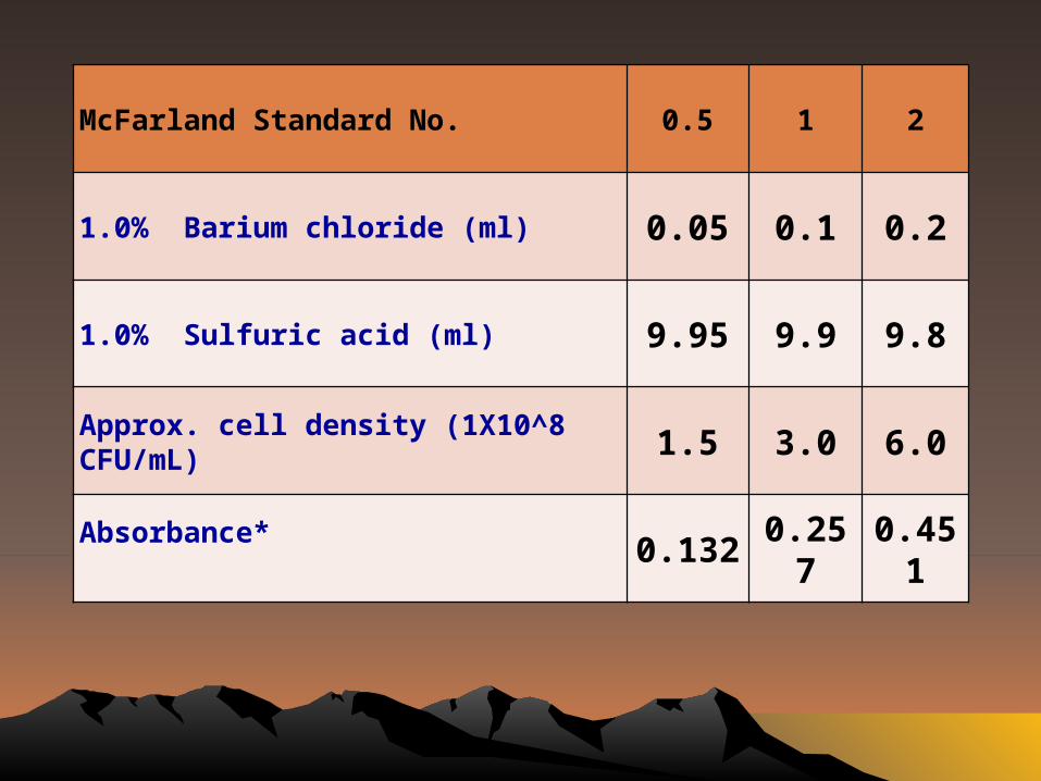
McFarland Standard No. 0.5 1 2
1.0% Barium chloride (ml) 0.05 0.1 0.2
1.0% Sulfuric acid (ml) 9.95 9.9 9.8
Approx. cell density (1X10^8 CFU/mL) 1.5 3.0 6.0
Absorbance* 0.1320.25
70.45
1

3. The turbidity of the actively growing broth culture is
adjusted with sterile saline or broth to obtain a
turbidity optically comparable to that of the 0.5
McFarland standard.
4. To perform this step properly, either
a photometric device can be used or, if done
visually, adequate light is needed to visually compare
the inoculum tube and the 0.5 McFarland standard
against a card with a white background and
contrasting black lines.
• As a convenient alternative to the growth method, the
inoculum can be prepared by making a direct broth or
saline suspension of isolated colonies selected from
a 18- to 24-hour agar plate (a nonselective medium,
such as blood agar, should be used).
• The suspension is adjusted to match the 0.5 McFarland
turbidity standard, using saline and a vortex mixer.
Direct Colony Suspension Method

Optimally, within 15 minutes after adjusting the
turbidity of the inoculum suspension, a sterile
cotton swab is dipped into the adjusted
suspension.
The swab should be rotated several times and
pressed firmly on the inside wall of the tube above
the fluid level. This will remove excess inoculum
from the swab.
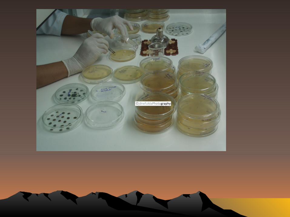
Inoculation of Inoculation of Test PlatesTest Plates
The dried surface of a Müeller-Hinton agar plate
is inoculated by streaking the swab over the
entire sterile agar surface.
This procedure is repeated by streaking two
more times, rotating the plate approximately 60
each time to ensure an even distribution of
inoculum.
As a final step, the rim of the agar is swabbed.
The lid may be left ajar for 3 to 5 minutes, but
no more than 15 minutes, to allow for any
excess surface moisture to be absorbed before
applying the drug impregnated disks.

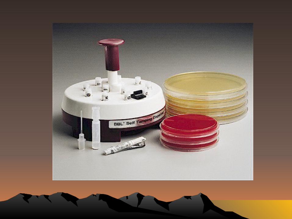
The predetermined battery of antimicrobial discs
is dispensed onto the surface of the inoculated
agar plate.
Each disc must be pressed down to ensure
complete contact with the agar surface. Whether
the discs are placed individually or with
a dispensing apparatus, they must be
distributed evenly so that they are no closer than
24 mm from center to center.
Application of Discs to Application of Discs to Inoculated Agar PlatesInoculated Agar Plates

Ordinarily, no more than 12 discs should be
placed on one 150 mm plate or more than 5
discs on a 100 mm plate.
Because some of the drug diffuses almost
instantaneously, a disc should not be relocated
once it has come into contact with the agar
surface. Instead, place a new disc in another
location on the agar.

After 16 to 18 hours of incubation, each plate is
examined.
If the plate was satisfactorily streaked, and the
inoculum was correct, the resulting zones of
inhibition will be uniformly circular and there will
be a confluent lawn of growth.
If individual colonies are apparent, the inoculum
was too light and the test must be repeated.
Reading Plates and Reading Plates and Interpreting ResultsInterpreting Results
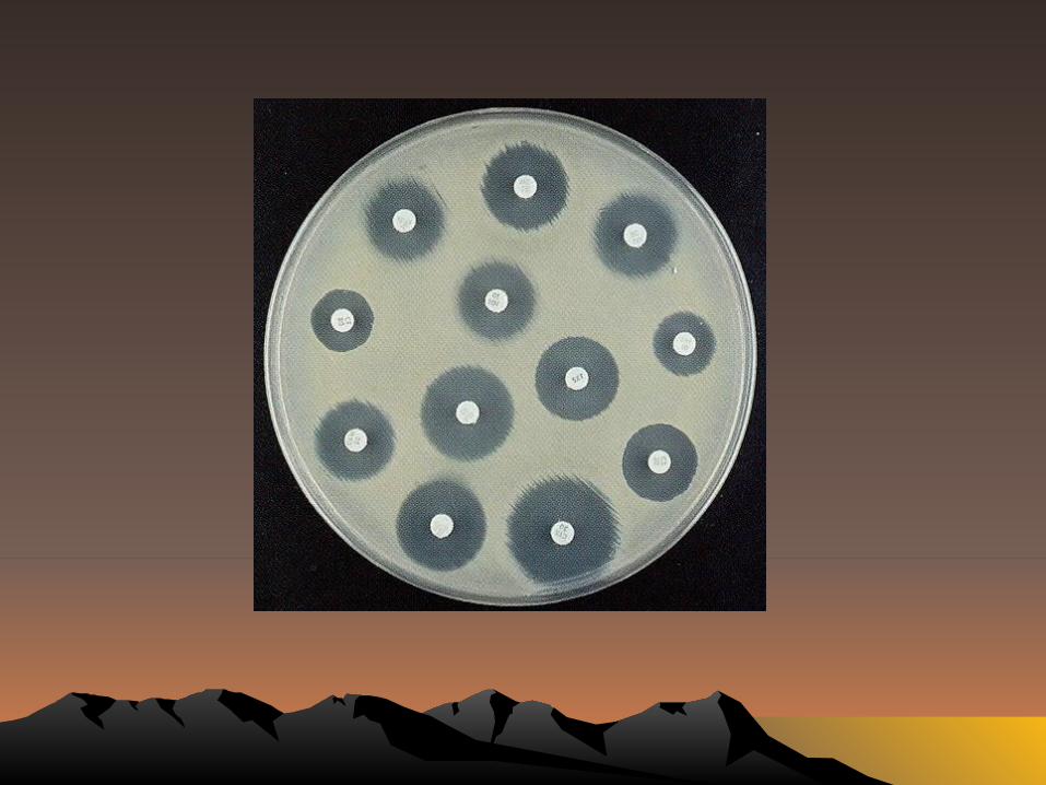

The diameters of the zones of complete
inhibition (as judged by the unaided eye) are
measured, including the diameter of the disc.
Zones are measured to the nearest whole
millimeter, using a ruler, which is held on the
back of the inverted Petri plate.
The Petri plate is held a few inches above a
black, nonreflecting background and
illuminated with reflected light.
If blood was added to the agar base (as with
streptococci), the zones are measured from the
upper surface of the agar illuminated with reflected
light, with the cover removed.
The zone margin should be taken as the area
showing no obvious, visible growth that can be
detected with the unaided eye.
Faint growth of tiny colonies, which can be detected
only with a magnifying lens at the edge of the zone
of inhibited growth, is ignored.
However, discrete colonies growing within a
clear zone of inhibition should be subcultured,
re-identified, and retested.
Strains of Proteus spp. may swarm into areas of
inhibited growth around certain antimicrobial
agents. With Proteus spp., the thin veil of
swarming growth in an otherwise obvious zone
of inhibition should be ignored.
When using blood-supplemented medium for
testing streptococci, the zone of growth
inhibition should be measured, not the zone of
inhibition of hemolysis.