Embed Size (px)
Citation preview
461
POJ 8(5):461-471 (2015) ISSN:1836-3644
Antioxidant enzymes metabolism and cellular differentiation during the developmental stages of
somatic embryogenesis in Torilis japonica (Houtt.) DC
Abinaya Manivannan
1#, Sonali Jana
2#, Prabhakaran Soundararajan
1#, Chung Ho Ko
1, and Byoung
Ryong Jeong1,2,3,
*
1Division of Applied Life Science (BK21 Plus), Graduate School, Gyeongsang National University, Jinju, Korea 660-
701 2Institute of Agriculture and Life Science, Gyeongsang National University, Jinju, Korea 660-701
3Research Institute of Life Science, Gyeongsang National University, Jinju, Korea 660-701
*Corresponding author: [email protected] #Equal contribution as the first author.
Abstract
In the present study, an efficient protocol for high frequency somatic embryogenesis from stem explants of Torilis japonica was
established. Explants were cultured in Murashige and Skoog (MS) medium containing 0-2.0 mg·L-1 2,4- dichlorophenoxy acetic acid
(2,4-D) with or without the combination of indole-3-butyric acid (IBA). The embryogenic callus induction and somatic embryogenesis
frequency were measured at four week intervals. The highest embryogenic callus induction (100±0.0%) was achieved in 2.0 mg·L-1 2,4-
D and 1.0 mg·L-1 IBA. Maximum somatic embryo induction (100±0.0%) was noted on the MS medium augmented with 2.0 mg·L-1 2, 4-
D, 1.0 mg·L-1 IBA, and 1.0 mg·L-1 gibberellic acid (GA3) with an average of 75.8±3.7 somatic embryos per explant. Scanning electron
microscopic investigation and histological analysis demonstrated the systemic development of somatic embryos. Comparatively, the
embryogenic callus induction and plantlet conversion stages possessed more total protein and hydrogen peroxide content than other
stages. As a consequence, the activities of superoxide dismutase (SOD), guaiacol peroxidase (GPX), ascorbate peroxidase (APX), and
catalase (CAT) were also increased during the induction of somatic embryos and bipolar shoot-root axis differentiation stages. Clonal
fidelity assessment by random amplified polymorphic DNA (RAPD) fingerprinting and inter simple sequence specific repeats (ISSR)
markers displayed the monomorphic banding pattern across the micropropagated plantlets. Thus, the RAPD and ISSR markers validated
the genetic homogeneity or the true-to-type nature of the in vitro plants.
Keywords: Auxins; Clonal fidelity; Encapsulation; Hydrogen peroxide; ISSR; Native PAGE; RAPD.
Abbreviations: PGRs_Plant growth regulators, 2,4-D_2,4-dichloropheoxyacetic acid, ROS_Reactive oxygen species, GPX_Guaiacol
peroxidase, APX_Ascorbate peroxidase, CAT_Catalase, RAPD_Random amplification of polymorphic DNA, ISSR_Inter simple
sequence repeat, MS_ Murashige and Skoog, IBA_Indole-3-butyric acid, GA3_Gibberellic acid, SEM_Scanning electron microscopy,
SOD_Superoxide dismutase, PAGE_Polyacrylamide gel electrophoresis.
Introduction
Torilis japonica Houtt is an important aromatic medicinal herb
belonging to the Apiaceae family widely distributed in East
Asia. The fruit of T. japonica consists of torilin, a
pharmaceutically important sesquiterpene prescribed to treat
renal disorders, angiogenesis, cancer, eczema, and
inflammation in Chinese medicine and also used as an
aphrodisiac in Japan (Juinchi et al., 2002). Previously, torilin
has been reported to reverse the multi-drug nature of cancer
cells and prevent the invasive ability of human fibrosarcoma
cells (Kim et al., 1998; Kim et al., 2001). In addition, the
essential oil obtained from the aerial parts of T. japonica was
effective against pathogenic bacteria such as Bacillus subtilis,
Salmonella typhimurium, and Listeria monocytogenes (Cho et
al., 2008; Chen et al., 2013). Although, T. japonica encloses
several pharmaceutical benefits, the conventional propagation
of this plant was hindered by poor seed setting, improper
pollination, sterility, and under developed flowers, which are
the most prevalent problems encountered in the Apiaceae
family (Richa et al., 2014). Hence, the development of in vitro
propagation techniques can rectify the aforementioned
difficulties. Moreover, micropropagation by means of somatic
embryogenesis offers the possibility for the large scale clonal
propagation of T. japonica with elite qualities in short period.
Somatic embryogenesis is one of the complex developmental
processes in plants. In general, the development of somatic
embryos occurs from the somatic cells without fusion of
gametes that pave the way for mass propagation of true-to-type
plants (Bhojwani and Razdan, 1996). During somatic
embryogenesis, the cells may undergo various genetic,
proteomic, and biochemical changes (Singla et al., 2007).
Application of plant growth regulators (PGRs) either induces
somatic embryogenesis either directly from the explants or
indirectly from the embryogenic callus. Several studies suggest
that the synthetic auxin, 2,4-D is an efficient and effective
hormone for somatic embryo induction (Raju et al., 2013). In
general, somatic embryogenesis can be considered as the stress
responses that occur due to the auxin mediated signaling that
enhances the embryogenic competence of the cells. The stress
signaling directed by 2,4-D activate the gene expression and
protein synthesis responsible for somatic embryogenesis
(Mihaljević et al., 2011). Hence, 2,4-D alone or in combination
with other plant growth regulators is frequently used in somatic
462
embryogenesis in many plants (Rathore et al., 2015). However,
during reprogramming of somatic cells for embryogenesis
several signaling molecules are involved. Among the signaling
molecules, the reactive oxygen species (ROS) such as H2O2
mediated signaling plays an important role (Zhang et al., 2010).
H2O2 is one of the vital signaling molecules entailed in the
process of differentiation, growth, and development of plants
(Kairong et al., 1999). In addition H2O2 also acts as a major
hallmark of oxidative stress. Therefore, plants have a
constitutive effect over the H2O2 accumulation i.e., stimulation
of defensive antioxidants to scavenge the excess H2O2
(Christou et al., 2014). Antioxidant enzymes such as guaiacol
peroxidase (GPX), ascorbate peroxidase (APX), and catalase
(CAT) are primarily associated with H2O2 elimination
(Tiryakioglu et al., 2006). Even though, the implication of ROS
metabolism with respect to physiological process has been
dealt for decades still the underlying metabolic networks
remain cryptic.
Therefore, the current study has attempted to investigate the
somatic embryogenesis process in T. japonica and the
consequent biochemical changes that occur during the
induction, development, and germination of somatic embryos.
Additionally, the present endeavor has also attempted to
demonstrate the sodium alginate encapsulation method for
synthetic seed production. Moreover, the genetic homogeneity
of the in vitro plantlets were validated using random amplified
polymorphic DNA fingerprinting (RAPD) and inter simple
sequence repeat (ISSR) markers.
Results and discussion
Induction and development of somatic embryos
The surface sterilized stem explants inoculated on the auxin
augmented Murashige and Skoog (MS) medium bulged at the
cut ends within two weeks denoting the early de-differentiation
stage. However, the explants cultured on the hormone free-MS
medium necrotized after 3 weeks. In general, auxins are
prerequisite for the induction and proliferation of callus,
whereas the combination of auxin and cytokinins is required
for the re-differentiation of callus into organized structures
(Wang et al., 2008). The medium supplemented with 2, 4-D
possessed the most effective embryogenic callus induction in
both alone or in combination with indole-3-butyric acid (IBA).
The explants cultured on the 2,4-D medium formed brownish
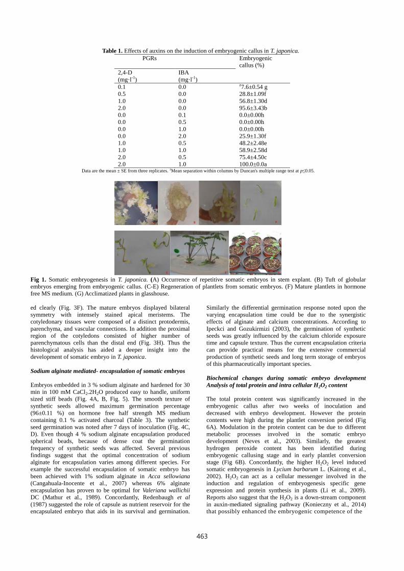
friable embryogenic callus. The greatest embryogenic callus
induction percentage (100.0±0.0 %) was achieved in medium
with 2.0 mg·l-1 2, 4-D and 1.0 mg·l-1 IBA (Table 1). Despite the
presence of various auxins, 2,4-D has been widely employed
for the induction of somatic embryogenesis in several species
(Brown et al., 1995). According to Skupa et al. (2014) 2, 4-D
creates a time-limited endogenous auxin-maxima or an optimal
auxin shock that leads to the induction of somatic embryos.
Furthermore the inclusion of IBA along with 2,4-D could have
synergistically increased the endogenous auxin stress that led to
the enhancement of embryogenic potential of the explants.
Similarly, the positive effect of supplementary auxins like IBA
on somatic embryogenesis has been reported by Filippov et al.,
(2006) in wheat.
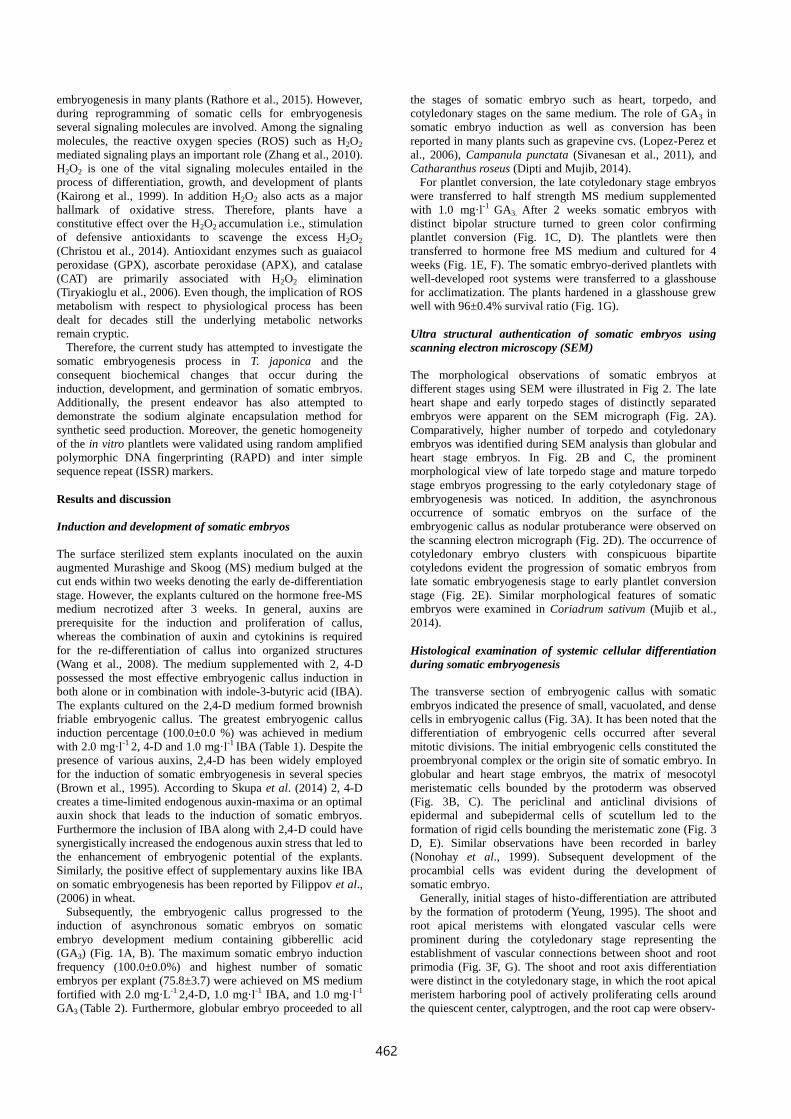
Subsequently, the embryogenic callus progressed to the
induction of asynchronous somatic embryos on somatic
embryo development medium containing gibberellic acid
(GA3) (Fig. 1A, B). The maximum somatic embryo induction
frequency (100.0±0.0%) and highest number of somatic
embryos per explant (75.8±3.7) were achieved on MS medium
fortified with 2.0 mg·L-1 2,4-D, 1.0 mg·l-1 IBA, and 1.0 mg·l-1
GA3 (Table 2). Furthermore, globular embryo proceeded to all
the stages of somatic embryo such as heart, torpedo, and
cotyledonary stages on the same medium. The role of GA3 in
somatic embryo induction as well as conversion has been
reported in many plants such as grapevine cvs. (Lopez-Perez et
al., 2006), Campanula punctata (Sivanesan et al., 2011), and
Catharanthus roseus (Dipti and Mujib, 2014).
For plantlet conversion, the late cotyledonary stage embryos
were transferred to half strength MS medium supplemented
with 1.0 mg·l-1 GA3. After 2 weeks somatic embryos with
distinct bipolar structure turned to green color confirming
plantlet conversion (Fig. 1C, D). The plantlets were then
transferred to hormone free MS medium and cultured for 4
weeks (Fig. 1E, F). The somatic embryo-derived plantlets with
well-developed root systems were transferred to a glasshouse
for acclimatization. The plants hardened in a glasshouse grew
well with 96±0.4% survival ratio (Fig. 1G).
Ultra structural authentication of somatic embryos using
scanning electron microscopy (SEM)
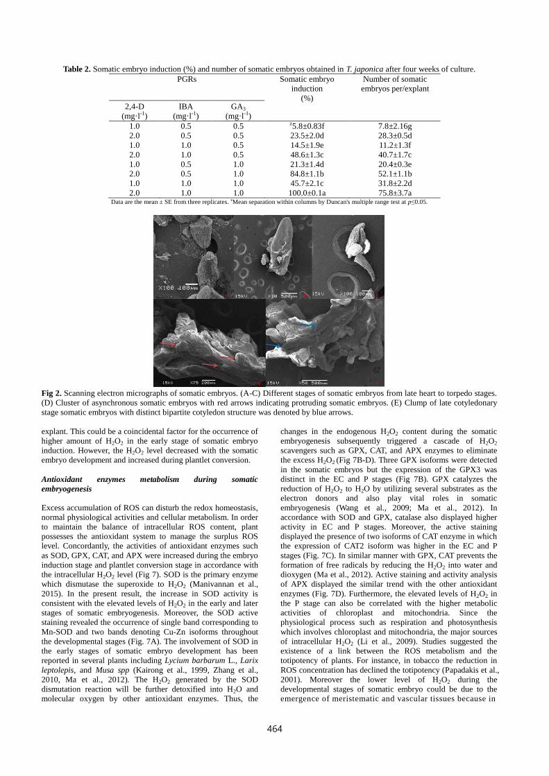
The morphological observations of somatic embryos at
different stages using SEM were illustrated in Fig 2. The late
heart shape and early torpedo stages of distinctly separated
embryos were apparent on the SEM micrograph (Fig. 2A).
Comparatively, higher number of torpedo and cotyledonary
embryos was identified during SEM analysis than globular and
heart stage embryos. In Fig. 2B and C, the prominent
morphological view of late torpedo stage and mature torpedo
stage embryos progressing to the early cotyledonary stage of
embryogenesis was noticed. In addition, the asynchronous
occurrence of somatic embryos on the surface of the
embryogenic callus as nodular protuberance were observed on
the scanning electron micrograph (Fig. 2D). The occurrence of
cotyledonary embryo clusters with conspicuous bipartite
cotyledons evident the progression of somatic embryos from
late somatic embryogenesis stage to early plantlet conversion
stage (Fig. 2E). Similar morphological features of somatic
embryos were examined in Coriadrum sativum (Mujib et al.,
2014).
Histological examination of systemic cellular differentiation
during somatic embryogenesis
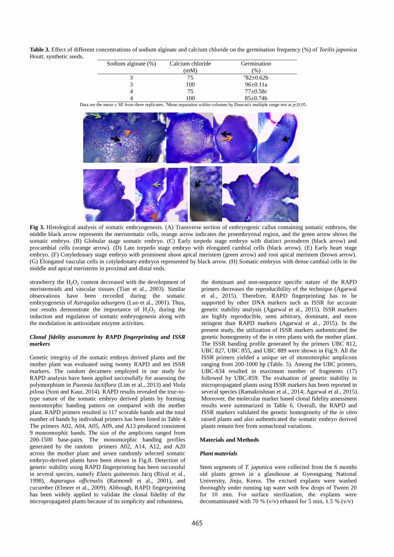
The transverse section of embryogenic callus with somatic
embryos indicated the presence of small, vacuolated, and dense
cells in embryogenic callus (Fig. 3A). It has been noted that the
differentiation of embryogenic cells occurred after several
mitotic divisions. The initial embryogenic cells constituted the
proembryonal complex or the origin site of somatic embryo. In
globular and heart stage embryos, the matrix of mesocotyl
meristematic cells bounded by the protoderm was observed
(Fig. 3B, C). The periclinal and anticlinal divisions of
epidermal and subepidermal cells of scutellum led to the
formation of rigid cells bounding the meristematic zone (Fig. 3
D, E). Similar observations have been recorded in barley
(Nonohay et al., 1999). Subsequent development of the
procambial cells was evident during the development of
somatic embryo.
Generally, initial stages of histo-differentiation are attributed
by the formation of protoderm (Yeung, 1995). The shoot and
root apical meristems with elongated vascular cells were
prominent during the cotyledonary stage representing the
establishment of vascular connections between shoot and root
primodia (Fig. 3F, G). The shoot and root axis differentiation
were distinct in the cotyledonary stage, in which the root apical
meristem harboring pool of actively proliferating cells around
the quiescent center, calyptrogen, and the root cap were observ-
463
Table 1. Effects of auxins on the induction of embryogenic callus in T. japonica.
PGRs Embryogenic
callus (%)
2,4-D
(mg·l-1)
IBA
(mg·l-1)
0.1 0.0 z7.6±0.54 g
0.5 0.0 28.8±1.09f
1.0 0.0 56.8±1.30d
2.0 0.0 95.6±3.43b
0.0 0.1 0.0±0.00h
0.0 0.5 0.0±0.00h
0.0 1.0 0.0±0.00h
0.0 2.0 25.9±1.30f
1.0 0.5 48.2±2.48e
1.0 1.0 58.9±2.58d
2.0 0.5 75.4±4.50c
2.0 1.0 100.0±0.0a Data are the mean ± SE from three replicates. zMean separation within columns by Duncan's multiple range test at p≤0.05.
Fig 1. Somatic embryogenesis in T. japonica. (A) Occurrence of repetitive somatic embryos in stem explant. (B) Tuft of globular
embryos emerging from embryogenic callus. (C-E) Regeneration of plantlets from somatic embryos. (F) Mature plantlets in hormone
free MS medium. (G) Acclimatized plants in glasshouse.
ed clearly (Fig. 3F). The mature embryos displayed bilateral
symmetry with intensely stained apical meristems. The
cotyledonary tissues were composed of a distinct protodermis,
parenchyma, and vascular connections. In addition the proximal
region of the cotyledons consisted of higher number of
parenchymatous cells than the distal end (Fig. 3H). Thus the
histological analysis has aided a deeper insight into the
development of somatic embryo in T. japonica.
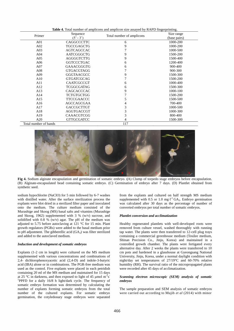
Sodium alginate mediated- encapsulation of somatic embryos
Embryos embedded in 3 % sodium alginate and hardened for 30
min in 100 mM CaCl2.2H2O produced easy to handle, uniform
sized stiff beads (Fig. 4A, B, Fig. 5). The smooth texture of
synthetic seeds allowed maximum germination percentage
(96±0.11 %) on hormone free half strength MS medium
containing 0.1 % activated charcoal (Table 3). The synthetic
seed germination was noted after 7 days of inoculation (Fig. 4C,
D). Even though 4 % sodium alginate encapsulation produced
spherical beads, because of dense coat the germination
frequency of synthetic seeds was affected. Several previous
findings suggest that the optimal concentration of sodium
alginate for encapsulation varies among different species. For
example the successful encapsulation of somatic embryo has
been achieved with 1% sodium alginate in Acca sellowiana
(Cangahuala-Inocente et al., 2007) whereas 6% alginate
encapsulation has proven to be optimal for Valeriana wallichii
DC (Mathur et al., 1989). Concordantly, Redenbaugh et al
(1987) suggested the role of capsule as nutrient reservoir for the
encapsulated embryo that aids in its survival and germination.
Similarly the differential germination response noted upon the
varying encapsulation time could be due to the synergistic
effects of alginate and calcium concentrations. According to
Ipeckci and Gozukirmizi (2003), the germination of synthetic
seeds was greatly influenced by the calcium chloride exposure
time and capsule texture. Thus the current encapsulation criteria
can provide practical means for the extensive commercial
production of synthetic seeds and long term storage of embryos
of this pharmaceutically important species.
Biochemical changes during somatic embryo development
Analysis of total protein and intra cellular H2O2 content
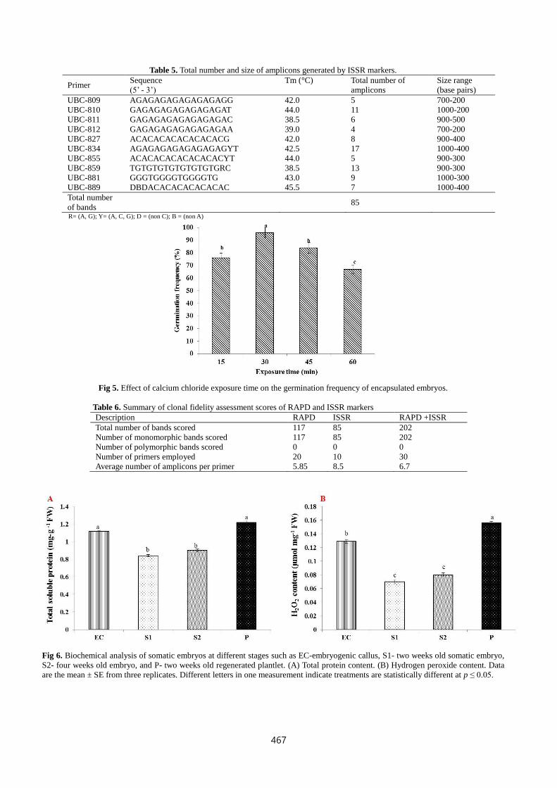
The total protein content was significantly increased in the
embryogenic callus after two weeks of inoculation and
decreased with embryo development. However the protein
contents were high during the plantlet conversion period (Fig
6A). Modulation in the protein content can be due to different
metabolic processes involved in the somatic embryo
development (Neves et al., 2003). Similarly, the greatest
hydrogen peroxide content has been identified during
embryogenic callusing stage and in early plantlet conversion
stage (Fig 6B). Concordantly, the higher H2O2 level induced
somatic embryogenesis in Lycium barbarum L. (Kairong et al.,
2002). H2O2 can act as a cellular messenger involved in the
induction and regulation of embryogenesis specific gene
expression and protein synthesis in plants (Li et al., 2009).
Reports also suggest that the H2O2 is a down-stream component
in auxin-mediated signaling pathway (Konieczny et al., 2014)
that possibly enhanced the embryogenic competence of the
464
Table 2. Somatic embryo induction (%) and number of somatic embryos obtained in T. japonica after four weeks of culture.
PGRs Somatic embryo
induction
(%)
Number of somatic
embryos per/explant
2,4-D
(mg·l-1)
IBA
(mg·l-1)
GA3
(mg·l-1)
1.0 0.5 0.5 z5.8±0.83f 7.8±2.16g
2.0 0.5 0.5 23.5±2.0d 28.3±0.5d
1.0 1.0 0.5 14.5±1.9e 11.2±1.3f
2.0 1.0 0.5 48.6±1.3c 40.7±1.7c
1.0 0.5 1.0 21.3±1.4d 20.4±0.3e
2.0 0.5 1.0 84.8±1.1b 52.1±1.1b
1.0 1.0 1.0 45.7±2.1c 31.8±2.2d
2.0 1.0 1.0 100.0±0.1a 75.8±3.7a Data are the mean ± SE from three replicates. zMean separation within columns by Duncan's multiple range test at p≤0.05.
Fig 2. Scanning electron micrographs of somatic embryos. (A-C) Different stages of somatic embryos from late heart to torpedo stages.
(D) Cluster of asynchronous somatic embryos with red arrows indicating protruding somatic embryos. (E) Clump of late cotyledonary
stage somatic embryos with distinct bipartite cotyledon structure was denoted by blue arrows.
explant. This could be a coincidental factor for the occurrence of
higher amount of H2O2 in the early stage of somatic embryo
induction. However, the H2O2 level decreased with the somatic
embryo development and increased during plantlet conversion.
Antioxidant enzymes metabolism during somatic
embryogenesis
Excess accumulation of ROS can disturb the redox homeostasis,
normal physiological activities and cellular metabolism. In order
to maintain the balance of intracellular ROS content, plant
possesses the antioxidant system to manage the surplus ROS
level. Concordantly, the activities of antioxidant enzymes such
as SOD, GPX, CAT, and APX were increased during the embryo
induction stage and plantlet conversion stage in accordance with
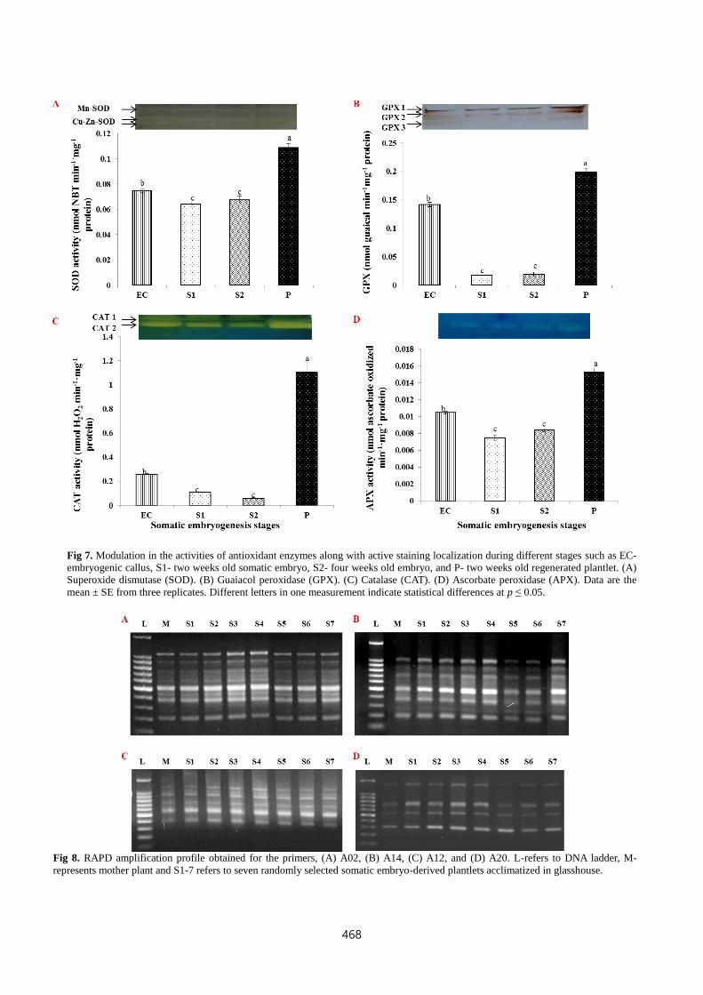
the intracellular H2O2 level (Fig 7). SOD is the primary enzyme
which dismutase the superoxide to H2O2 (Manivannan et al.,
2015). In the present result, the increase in SOD activity is
consistent with the elevated levels of H2O2 in the early and later
stages of somatic embryogenesis. Moreover, the SOD active
staining revealed the occurrence of single band corresponding to
Mn-SOD and two bands denoting Cu-Zn isoforms throughout
the developmental stages (Fig. 7A). The involvement of SOD in
the early stages of somatic embryo development has been
reported in several plants including Lycium barbarum L., Larix
leptolepis, and Musa spp (Kairong et al., 1999, Zhang et al.,
2010, Ma et al., 2012). The H2O2 generated by the SOD
dismutation reaction will be further detoxified into H2O and
molecular oxygen by other antioxidant enzymes. Thus, the
changes in the endogenous H2O2 content during the somatic
embryogenesis subsequently triggered a cascade of H2O2
scavengers such as GPX, CAT, and APX enzymes to eliminate
the excess H2O2 (Fig 7B-D). Three GPX isoforms were detected
in the somatic embryos but the expression of the GPX3 was
distinct in the EC and P stages (Fig 7B). GPX catalyzes the
reduction of H2O2 to H2O by utilizing several substrates as the
electron donors and also play vital roles in somatic
embryogenesis (Wang et al., 2009; Ma et al., 2012). In
accordance with SOD and GPX, catalase also displayed higher
activity in EC and P stages. Moreover, the active staining
displayed the presence of two isoforms of CAT enzyme in which
the expression of CAT2 isoform was higher in the EC and P
stages (Fig. 7C). In similar manner with GPX, CAT prevents the
formation of free radicals by reducing the H2O2 into water and
dioxygen (Ma et al., 2012). Active staining and activity analysis
of APX displayed the similar trend with the other antioxidant
enzymes (Fig. 7D). Furthermore, the elevated levels of H2O2 in
the P stage can also be correlated with the higher metabolic
activities of chloroplast and mitochondria. Since the
physiological process such as respiration and photosynthesis
which involves chloroplast and mitochondria, the major sources
of intracellular H2O2 (Li et al., 2009). Studies suggested the
existence of a link between the ROS metabolism and the
totipotency of plants. For instance, in tobacco the reduction in
ROS concentration has declined the totipotency (Papadakis et al.,
2001). Moreover the lower level of H2O2 during the
developmental stages of somatic embryo could be due to the
emergence of meristematic and vascular tissues because in
465
Table 3. Effect of different concentrations of sodium alginate and calcium chloride on the germination frequency (%) of Torilis japonica
Houtt. synthetic seeds.
Sodium alginate (%) Calcium chloride
(mM)
Germination
(%)
3 75 z82±0.62b
3 100 96±0.11a
4 75 77±0.58c
4 100 85±0.74b Data are the mean ± SE from three replicates. zMean separation within columns by Duncan's multiple range test at p≤0.05.
Fig 3. Histological analysis of somatic embryogenesis. (A) Transverse section of embryogenic callus containing somatic embryos, the
middle black arrow represents the meristematic cells, orange arrow indicates the proembryonal region, and the green arrow shows the
somatic embryo. (B) Globular stage somatic embryo. (C) Early torpedo stage embryo with distinct protoderm (black arrow) and
procambial cells (orange arrow). (D) Late torpedo stage embryo with elongated cambial cells (black arrow). (E) Early heart stage
embryo. (F) Cotyledonary stage embryo with prominent shoot apical meristem (green arrow) and root apical meristem (brown arrow).
(G) Elongated vascular cells in cotyledonary embryos represented by black arrow. (H) Somatic embryos with dense cambial cells in the
middle and apical meristems in proximal and distal ends.
strawberry the H2O2 content decreased with the development of
meristemoids and vascular tissues (Tian et al., 2003). Similar
observations have been recorded during the somatic
embryogenesis of Astragalus adsurgens (Luo et al., 2001). Thus,
our results demonstrate the importance of H2O2 during the
induction and regulation of somatic embryogenesis along with
the modulation in antioxidant enzyme activities.
Clonal fidelity assessment by RAPD fingerprinting and ISSR
markers
Genetic integrity of the somatic embryo derived plants and the
mother plant was evaluated using twenty RAPD and ten ISSR
markers. The random decamers employed in our study for
RAPD analysis have been applied successfully for assessing the
polymorphism in Paeonia lactiflora (Lim et al., 2013) and Viola
pilosa (Soni and Kaur, 2014). RAPD results revealed the true-to-
type nature of the somatic embryo derived plants by forming
monomorphic banding pattern on compared with the mother
plant. RAPD primers resulted in 117 scorable bands and the total
number of bands by individual primers has been listed in Table 4.
The primers A02, A04, A05, A09, and A13 produced consistent
9 monomorphic bands. The size of the amplicons ranged from
200-1500 base-pairs. The monomorphic banding profiles
generated by the random primers A02, A14, A12, and A20
across the mother plant and seven randomly selected somatic
embryo-derived plants have been shown in Fig.8. Detection of
genetic stability using RAPD fingerprinting has been successful
in several species, namely Elaeis guineensis Jacq (Rival et al.,
1998), Asparagus officinalis (Raimondi et al., 2001), and
cucumber (Elmeer et al., 2009). Although, RAPD fingerprinting
has been widely applied to validate the clonal fidelity of the
micropropagated plants because of its simplicity and robustness,
the dominant and non-sequence specific nature of the RAPD
primers decreases the reproducibility of the technique (Agarwal
et al., 2015). Therefore, RAPD fingerprinting has to be
supported by other DNA markers such as ISSR for accurate
genetic stability analysis (Agarwal et al., 2015). ISSR markers
are highly reproducible, semi arbitrary, dominant, and more
stringent than RAPD markers (Agarwal et al., 2015). In the
present study, the utilization of ISSR markers authenticated the
genetic homogeneity of the in vitro plants with the mother plant.
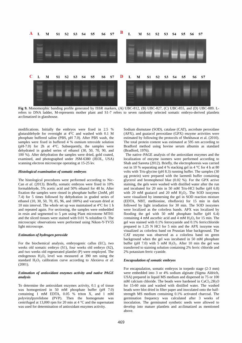
The ISSR banding profile generated by the primers UBC 812,
UBC 827, UBC 855, and UBC 889 were shown in Fig.9. All the
ISSR primers yielded a unique set of monomorphic amplicons
ranging from 200-1000 bp (Table. 5). Among the UBC primers,
UBC-834 resulted in maximum number of fragments (17)
followed by UBC-859. The evaluation of genetic stability in
micropropagated plants using ISSR markers has been reported in
several species (Ramakrishnan et al., 2014; Agarwal et al., 2015).
Moreover, the molecular marker based clonal fidelity assessment
results were summarized in Table 6. Overall, the RAPD and
ISSR markers validated the genetic homogeneity of the in vitro
raised plants and also authenticated the somatic embryo derived
plants remain free from somaclonal variations.
Materials and Methods
Plant materials
Stem segments of T. japonica were collected from the 6 months
old plants grown in a glasshouse at Gyeongsang National
University, Jinju, Korea. The excised explants were washed
thoroughly under running tap water with few drops of Tween 20
for 10 min. For surface sterilization, the explants were
decontaminated with 70 % (v/v) ethanol for 5 min, 1.5 % (v/v)
A B C D
F E H G
466
Table 4. Total number of amplicons and amplicon size assayed by RAPD fingerprinting.
Primer Sequence
(5’ - 3’) Total number of amplicons
Size range
(base pairs)
A01 CAGGCCCTTC 6 1000-200
A02 TGCCGAGCTG 9 1000-200
A03 AGTCAGCCAC 7 1000-500
A04 AATCGGGCTG 9 1500-200
A05 AGGGGTCTTG 9 1500-400
A06 GGTCCCTGAC 6 1200-400
A07 GAAACGGGTG 6 900-400
A08 GTGACGTAGG 7 900-300
A09 GGGTAACGCC 9 1500-300
A10 GTGATCGCAG 7 1500-200
A11 CAATCGCCGT 6 1000-400
A12 TCGGCGATAG 6 1500-300
A13 CAGCACCCAC 9 1000-100
A14 TCTGTGCTGG 7 1500-200
A15 TTCCGAACCC 5 1500-500
A16 AGCCAGCGAA 4 700-400
A17 GACCGCTTGT 3 1000-500
A18 AGGTGACCGT 3 1000-300
A19 CAAACGTCGG 3 800-400
A20 GTTGCGATCC 4 1500-300
Total number of bands 117
Fig 4. Sodium alginate encapsulation and germination of somatic embryo. (A) Clump of torpedo stage embryos before encapsulation.
(B) Alginate-encapsulated bead containing somatic embryo. (C) Germination of embryo after 7 days. (D) Plantlet obtained from
synthetic seed.
sodium hypochlorite (NaClO) for 5 min followed by 6-7 washes
with distilled water. After the surface sterilization process the
explants were blot dried in a sterilized filter paper and inoculated
onto the medium. The culture medium consisted of the
Murashige and Skoog (MS) basal salts and vitamins (Murashige
and Skoog, 1962) supplemented with 3 % (w/v) sucrose, and
solidified with 0.8 % (w/v) agar. The pH of the medium was
adjusted to 5.75 before autoclaving at 121 °C for 15 min. Plant
growth regulators (PGRs) were added to the basal medium prior
to pH adjustment. The gibberellic acid (GA3) was filter sterilized
and added to the autoclaved medium.
Induction and development of somatic embryos
Explants (1-2 cm in length) were cultured on the MS medium
supplemented with various concentrations and combinations of
2,4- dichlorophenoxyacetic acid (2,4-D) and indole-3-butyric
acid (IBA) alone or in combinations. The PGR-free medium was
used as the control. Five explants were placed in each petridish
containing 20 ml of the MS medium and maintained for 15 days
at 25 °C in darkness, and then exposed to light of 45 μmol m-2s-
1PPFD for a daily 16/8 h light/dark cycle. The frequency of
somatic embryo formation was determined by calculating the
number of explants forming somatic embryos from the total
number of the cultured explants. For somatic embryo
germination, the cotyledonary stage embryos were separated
from the explants and cultured on half strength MS medium
supplemented with 0.5 or 1.0 mg·l-1 GA3. Embryo germination
was calculated after 30 days as the percentage of number of
converted embryos per total number of somatic embryos.
Plantlet conversion and acclimatization
Healthy regenerated plantlets with well-developed roots were
removed from culture vessel, washed thoroughly with running
tap water. The plants were then transferred to 12-cell plug trays
containing a commercial greenhouse medium (Tosilee medium,
Shinan Precision Co., Jinju, Korea) and maintained in a
controlled growth chamber. The plants were fertigated every
alternative day. After 2 weeks the plants were transferred to 10
cm pots and hardened in a glasshouse at Gyeongsang National
University, Jinju, Korea, under a normal daylight condition with
night/day set temperatures of 27/19°C and 60-70% relative
humidity (RH). The survival ratio of the micropropagated plants
were recorded after 45 days of acclimatization.
Scanning electron microscopic (SEM) analysis of somatic
embryos
The sample preparation and SEM analysis of somatic embryos
were carried out according to Mujib et al (2014) with minor
467
Table 5. Total number and size of amplicons generated by ISSR markers.
Primer Sequence
(5’ - 3’)
Tm (°C) Total number of
amplicons
Size range
(base pairs)
UBC-809 AGAGAGAGAGAGAGAGG 42.0 5 700-200
UBC-810 GAGAGAGAGAGAGAGAT 44.0 11 1000-200
UBC-811 GAGAGAGAGAGAGAGAC 38.5 6 900-500
UBC-812 GAGAGAGAGAGAGAGAA 39.0 4 700-200
UBC-827 ACACACACACACACACG 42.0 8 900-400
UBC-834 AGAGAGAGAGAGAGAGYT 42.5 17 1000-400
UBC-855 ACACACACACACACACYT 44.0 5 900-300
UBC-859 TGTGTGTGTGTGTGTGRC 38.5 13 900-300
UBC-881 GGGTGGGGTGGGGTG 43.0 9 1000-300
UBC-889 DBDACACACACACACAC 45.5 7 1000-400
Total number
of bands
85
R= (A, G); Y= (A, C, G); D = (non C); B = (non A)
Fig 5. Effect of calcium chloride exposure time on the germination frequency of encapsulated embryos.
Table 6. Summary of clonal fidelity assessment scores of RAPD and ISSR markers
Description RAPD ISSR RAPD +ISSR
Total number of bands scored 117 85 202
Number of monomorphic bands scored 117 85 202
Number of polymorphic bands scored 0 0 0
Number of primers employed 20 10 30
Average number of amplicons per primer 5.85 8.5 6.7
Fig 6. Biochemical analysis of somatic embryos at different stages such as EC-embryogenic callus, S1- two weeks old somatic embryo,
S2- four weeks old embryo, and P- two weeks old regenerated plantlet. (A) Total protein content. (B) Hydrogen peroxide content. Data
are the mean ± SE from three replicates. Different letters in one measurement indicate treatments are statistically different at p ≤ 0.05.
468
Fig 7. Modulation in the activities of antioxidant enzymes along with active staining localization during different stages such as EC-
embryogenic callus, S1- two weeks old somatic embryo, S2- four weeks old embryo, and P- two weeks old regenerated plantlet. (A)
Superoxide dismutase (SOD). (B) Guaiacol peroxidase (GPX). (C) Catalase (CAT). (D) Ascorbate peroxidase (APX). Data are the
mean ± SE from three replicates. Different letters in one measurement indicate statistical differences at p ≤ 0.05.
Fig 8. RAPD amplification profile obtained for the primers, (A) A02, (B) A14, (C) A12, and (D) A20. L-refers to DNA ladder, M-
represents mother plant and S1-7 refers to seven randomly selected somatic embryo-derived plantlets acclimatized in glasshouse.
469
Fig 9. Monomorphic banding profile generated by ISSR markers, (A) UBC-812, (B) UBC-827, (C) UBC-855, and (D) UBC-889. L-
refers to DNA ladder, M-represents mother plant and S1-7 refers to seven randomly selected somatic embryo-derived plantlets
acclimatized in glasshouse.
modifications. Initially the embryos were fixed in 2.5 %
glutaraldehyde for overnight at 4°C and washed with 0.1 M
phosphate buffered saline (PBS, pH 7.0). After PBS wash, the
samples were fixed in buffered 4 % osmium tetroxide solution
(pH-7.0) for 2h at 4°C. Subsequently, the samples were
dehydrated in graded series of ethanol (30, 50, 70, 90, and
100 %). After dehydration the samples were dried, gold coated,
examined, and photographed under JSM-6380 (JEOL, USA)
scanning electron microscope operating at 15-25 kv.
Histological examination of somatic embryos
The histological procedures were performed according to Nic-
Can et al. (2013). Briefly, somatic embryos were fixed in 10%
formaldehyde, 5% acetic acid and 50% ethanol for 48 hr. After
fixation the samples were rinsed in phosphate buffer (2mM, pH
7.4) for 5 times followed by dehydration in graded series of
ethanol (10, 30, 50, 70, 85, 96, and 100%) and vacuum dried at
10 min interval. The whole set up was maintained at 4°C for 1 h
and repeated again. For sectioning, the samples were embedded
in resin and segmented to 5 µm using Plant microtome MTH1
and the sliced tissues were stained with 0.01 % toluidine O. The
microscopic observations were performed using Nikon-Y-TV55
light microscope.
Estimation of hydrogen peroxide
For the biochemical analysis, embryogenic callus (EC), two
weeks old somatic embryo (S1), four weeks old embryo (S2),
and two weeks old regenerated plantlet (P) were employed. The
endogenous H2O2 level was measured at 390 nm using the
standard H2O2 calibration curve according to Alexieva et al.
(2001).
Estimation of antioxidant enzymes activity and native PAGE
analysis
To determine the antioxidant enzymes activity, 0.1 g of tissue
was homogenized in 50 mM phosphate buffer (pH 7.0)
containing 1 mM EDTA, 0.05 % triton X, and 1 mM
polyvinylpyrolidone (PVP). Then the homogenate was
centrifuged at 13,000 rpm for 20 min at 4 °C and the supernatant
was used for determination of antioxidant enzymes activity.
Sodium dismutase (SOD), catalase (CAT), ascrobate peroxidase
(APX), and guaiacol peroxidase (GPX) enzyme activities were
estimated by following the protocols of Shekhawat et al. (2010).
The total protein content was estimated at 595 nm according to
Bradford method using bovine serum albumin as standard
(Bradford, 1976).
The native–PAGE analysis of the antioxidant enzymes and the
localization of enzyme isomers were performed according to
Shah and Sareeta (2012). Briefly, the electrophoresis was carried
out in 10 % separating and 4 % stacking gel in 4 °C for 4 h at 80
volts with Tris-glycine (pH 8.3) running buffer. The samples (30
µg protein) were prepared with the laemmli buffer containing
glycerol and bromophenol blue (0.02 %). For the GPX active
staining, the gels were washed with distilled water after the run
and incubated for 20 min in 50 mM Tris-HCl buffer (pH 6.8)
with 20 mM guaiacol and 20 mM H2O2. The SOD isozymes
were visualized by immersing the gel in SOD reaction mixture
(EDTA, NBT, methionine, riboflavin) for 15 min in dark
followed by light irradiation for 30 min. The SOD isozymes
were localized as the colorless bands. APX was localized by
flooding the gel with 50 mM phosphate buffer (pH 6.4)
containing 4 mM ascorbic acid and 4 mM H2O2 for 15 min. The
gel was stained with 0.1% ferrocyanide and 0.1% ferricchloride
prepared in 1.25 N HCl for 5 min and the APX isozyme was
visualized as colorless band on Prussian blue background. The
CAT enzyme was observed as a colorless band on green
background when the gel was incubated in 50 mM phosphate
buffer (pH 7.0) with 5 mM H2O2. After 10 min the gel was
transferred to staining solution containing 2% ferric chloride and
2% potassium ferric cyanide.
Encapsulation of somatic embryos
For encapsulation, somatic embryos in torpedo stage (2-3 mm)
were embedded into 3 or 4% sodium alginate (Sigma Aldrich,
USA) prepared in liquid MS medium and dispersed in 75 or 100
mM calcium chloride. The beads were hardened in CaCl2.2H2O
for 15-60 min and washed with distilled water. The washed
beads were blot dried in filter paper and inoculated onto the half-
strength MS medium containing 0.1% activated charcoal. The
germination frequency was calculated after 3 weeks of
inoculation. The germinated synthetic seeds were allowed to
develop into mature plantlets and acclimatized as mentioned
above.
470
Clonal fidelity assessment of in vitro plants using RAPD and
ISSR markers
The genomic DNA was isolated from the leaf sample of
randomly selected plants hardened in glasshouse using plant
DNA extraction kit (iNtRON Biotechnology, Seongnam, Korea).
The clonal fidelity of the micropropagated plants were detected
using randomly amplified polymorphic DNA (RAPD)
fingerprinting assay. The assay was carried out with 20 random
decamers (Enotech Co., Daejeon, Korea) as listed in Table 4. For
genetic stability assessment using ISSR markers, ten UBC
primers shown in Table 5 were employed. For all PCR reactions,
the reaction mixture consisted of a total volume of 20 µl with 1X
PCR buffer, 2.5 mM dNTPs, and 2.5 U Taq DNA polymerase
(iNtRON’s Maxime PCR Pre-mix, Seongnam, Korea) with 1 µl
primer, and 2 µl DNA. The PCR amplification was performed in
a thermal cycler (Gene Amp PCR system 9700, Applied
Biosystems, Foster City, CA, U.S.A.). The amplification profile
consisted of initial denaturation (5 min at 94°C),denaturation (1
minute at 94°C), annealing (1 minute at 33°C for RAPD primers
and different annealing temperatures employed for individual
ISSR primers were listed in Table 5), extension (1 minute at
72°C), and final extension (10 minutes at 72°C) with 30 cycles.
Amplified PCR products were separated on 1.5 % agarose gel
and photographed under UV light in a gel documentation system.
Statistical analysis
In each treatment, 30 explants were used and the experiment was
repeated thrice. Data were statistically analyzed by analysis of
variance (ANOVA) followed by Duncan multiple range test at
5% probability level. Data analysis was performed using SAS
computer package (SAS Institute Inc., Cary, NC, USA).
Conclusions
The present work has successfully established an efficient
protocol for high frequency somatic embryogenesis in Torillis
japonica Houtt. In addition, SEM analysis and histological
observations demonstrated the development of somatic embryos.
Moreover, the biochemical modulation during the induction and
development of somatic embryos revealed the vital involvement
of H2O2 and antioxidant enzymes. Further, the efficient alginate
encapsulation for synthetic seed production was demonstrated
and the genetic homogeneity of the in vitro plants was assessed
using RAPD fingerprinting and ISSR markers. In future, the
current endeavor can be extended to determine molecular
rationale behind the somatic embryogenesis in pharmaceutically
valuable Torilis japonica Houtt.
Acknowledgements
Abinaya Manivannan, Prabhakaran Soundararajan, and Chung
Ho Ko were supported by a scholarship from the Brain Korea 21
Plus (BK21 plus) Program, Ministry of Education, Korea. The
authors are thankful to Dr. Yoo Gyeong Park for statistical
assistance.
References
Agarwal T, Gupta A K, Patel A K, Shekhawat N S (2015)
Micropropagation and validation of genetic homogeneity of
Alhagi maurorum using SCoT, ISSR and RAPD markers. Plant
Cell Tiss Org Cult. 120(1):313-323.
Alexieva V, Sergiev I, Mapelli S, Karanov E (2001) The effect of
drought and ultraviolet radiation on growth and stress markers
in pea and wheat. Plant Cell Environ. 24:1337-1344.
Bhojwani SS, Razdan MK (1996) Plant Tissue Culture: Theory
and Practice. Elsevier, New York.
Bradford MM (1976) A rapid and sensitive method for the
quantitation of microgram quantities of protein utilizing the
principle of protein-dye binding. Anal Biochem.72:248-254.
Brown DCW, Finstad KI, Watson EM (1995) Somatic
embryogenesis in herbaceous dicots. In: Thorpe TA (ed) In
vitro embryogenesis in plants. Kluwer, Dordrecht.
Cangahuala-Inocente GC, Vesco LLD, Steinmacher D, Torres
AC, Guerra MP (2007) Improvements in somatic
embryogenesis protocol in Feijoa (Acca sellowiana (Berg)
Burret): induction, conversion and synthetic seeds. Sci Hortic.
111:228-234.
Chen J, Xu XJ, Fang YH, Li S, Zhang YI (2013) Chemical
composition and antibacterial activity of the essential oil from
the aerial parts of Torilis japonica. J Essential Oil Bearing
Plants. 16(4):499-505.
Cho WI, Choi JB, Lee K, Chung MS, Pyun YR (2008)
Antimicrobial activity of torilin isolated from Torilis japonica
fruit against Bacillus subtilis. J Food Sci. 73(2):37-46.
Christou A, Manganaris GA, Fotopoulos V (2014) Systemic
mitigation of salt stress by hydrogen peroxide and sodium
nitroprusside in strawberry plants via transcriptional regulation
of enzymatic and non-enzymatic antioxidants. Environ Exp
Bot. 107:46-54.
Dipti SF, Mujib A (2014) Morphological anomalies in somatic
embryo structure in Catharanthus roseus: Improving embryo
germination by amending plant growth regulators, activated
charcoal and sucrose level. Br Biotechnol J. 4:10-20.
Elmeer KMS, Gallagher TF, Hennerty MJ (2009) RAPD-based
detection of genomic instability in cucumber plants derived
from somatic embryogenesis. Afr J Biotechnol. 8:3219-3222.
Filippov M, Miroshnichenko D, Vernikovskaya D, Dolgov, S
(2006) The effect of auxins, time exposure to auxin and
genotypes on somatic embryogenesis from mature embryos of
wheat. Plant Cell Tiss Org Cult. 84(2):213-222.
Ipekci Z, Gozukirmizi N (2003) Direct somatic embryogenesis
and synthetic seed production from Paulownia elongata. Plant
Cell Rep. 22(1):16-24.
Junichi K, Nobuyuki S, Mituru S, Mitsuo W (2002)
Sesquiterpenoids of Torilis japonica fruit. Phytochem.
59(8):811-815.
Kairong C, Gengsheng X, Xinmin L, Gengmei X, Yafu W (1999)
Effect of hydrogen peroxide on somatic embryogenesis of
Lycium barbarum L. Plant Sci. 146(1):9-16.
Kairong C, Ji L, Gengmei X, Jianlong L, Lihong W, Yafu W
(2002) Effect of hydrogen peroxide on synthesis of proteins
during somatic embryogenesis in Lycium barbarum. Plant Cell
Tiss Org Cult. 68(2):187-193.
Kim MS, Baek JH, Park MT, Sohn TK, Lee JJ, Kim SU, Kim
KW (2001) Anti-invasive activity of torilin, a sesquiterpene
compound isolated from Torilis japonica. Oncol Rep. 8:359-
364.
Kim SE, Kim YH, Kim YC, Lee JJ (1998) Torilin, a
sesquiterpene from Torilis japonica, reverses multidrug-
resistance in cancer cells. Planta Med. 64(4):332-334.
Konieczny R, Banaś AK, Surówka E, Michalec Ż, Miszalski Z,
Libik-Konieczny M (2014) Pattern of antioxidant enzyme
activities and hydrogen peroxide content during developmental
stages of rhizogenesis from hypocotyl explants of
Mesembryanthemum crystallinum L. Plant Cell Rep. 33(1):165-
177.
Li SW, Xue L, Xu S, Feng H, An L (2009) Hydrogen peroxide
acts as a signal molecule in the adventitious root formation of
mung bean seedlings. Environ Exp Bot. 65:63-71.
471
Lim MY, Jana S, Sivanesan I, Park HR, Hwang JH, Park YH,
Jeong BR (2013) Analysis of genetic variability using RAPD
markers in Paeonia spp. grown in Korea. Kor J Hort Sci
Technol. 31:322-327.
Lopez-Perez AJ, Carreno J, Dabauza M (2006) Somatic embryo
germination and plant regeneration of three grapevine cvs:
Effect of IAA, GA3 and embryo morphology. Vitis. 45:141-
143.
Luo JP, Jiang ST, Pan LJ (2001) Enhanced somatic
embryogenesis by salicylic acid of Astragalus adsurgens Pall.:
relationship with H2O2 production and H2O2-metabolizing
enzyme activities. Plant Sci. 161(1):125-132.
Ma L, Xie L, Lin G, Jiang S, Chen H, Li H, Takac T, Samaj J, Xu
C (2012) Histological changes and differences in activities of
some antioxidant enzymes and hydrogen peroxide content
during somatic embryogenesis of Musa AAA cv. Yueyoukang 1.
Sci Hortic. 144:87-92.
Manivannan A, Soundararajan P, Halimah N, Ko CH, Jeong BR
(2015). Blue LED light enhances growth, phytochemical
contents, and antioxidant enzyme activities of Rehmannia
glutinosa cultured in vitro. Hortic Environ Biotechnol.
56(1):105-113.
Mathur J, Ahuja PS, Lal N, Mathur AK (1989) Propagation of
Valeriana wallichii DC using encapsulated apical and axial
shoot buds. Plant Sci. 60:111-116.
Mihaljević S, Radić S, Bauer N, Garić R, Mihaljević B, Horvat
G, Jelaska S (2011) Ammonium-related metabolic changes
affect somatic embryogenesis in pumpkin (Cucurbita pepo L.).
J Plant Physiol. 168(16):1943-1951.
Mujib A, Tonk D, Ali M (2014) Plant regeneration from
protoplasts in Indian local Coriandrum sativum L.: scanning
electron microscopy and histological evidences for somatic
embryogenesis. Plant Cell Tiss Org Cult. 117(3):323-334.
Murashige T, Skoog F (1962) A revised medium for rapid growth
and bio assays with tobacco tissue cultures. Physiol Plantarum.
15(3):473-497.
Neves N, Segura-Nieto M, Blanco MA, Sánchez M, González A,
González JL (2003) Biochemical characterization of
embryogenic and non-embryogenic calluses of sugarcane.
InVitro Cell Dev Biol Plant. 39(3):343-345.
Nic-Can GI, López-Torres A, Barredo-Pool F, Wrobel K,
Loyola-Vargas VM, Rojas-Herrera R, De-la-Peña C (2013)
New insights into somatic embryogenesis: LEAFY
COTYLEDON1, BABY BOOM1 and WUSCHEL-RELATED
HOMEOBOX4 are epigenetically regulated in Coffea
canephora. PloS One. 8(8):e72160.
Nonohay JS, Mariath JEA, Winge H (1999) Histological analysis
of somatic embryogenesis in Brazilian cultivars of barley,
Hordeum vulgare vulgare, Poaceae. Plant Cell Rep.
18(11):929-934.
Papadakis AK, Siminis CI, Roubelakis-Angelakis KA (2001)
Reduced activity of antioxidant machinery is correlated with
suppression of totipotency in plant protoplasts. Plant Physiol.
126(1):434-444.
Raimondi JP, Masuelli RW, Camadro EL (2001) Assessment of
somaclonal variation in asparagus by RAPD fingerprinting and
cytogenetic analyses. Sci Hortic. 90:19-29.
Raju CS, Kathiravan K, Aslam A, Shajahan A (2013) An efficient
regeneration system via somatic embryogenesis in mango
ginger (Curcuma amada Roxb.). Plant Cell Tiss Org Cult.
112(3):387-393.
Ramakrishnan M, Ceasar SA, Duraipandiyan V, Ignacimuthu S
(2014) Efficient plant regeneration from shoot apex explants of
maize (Zea mays) and analysis of genetic fidelity of
regenerated plants by ISSR markers. Plant Cell Tiss Org Cult.
119(1):183-196.
Rathore MS, Paliwal N, Anand KV, Agarwal PK (2015) Somatic
embryogenesis and in vitro plantlet regeneration in Salicornia
brachiata Roxb. Plant Cell Tissue Organ Cult. 120(1):355-360.
Redenbaugh K, Slade D, Viss P, Fujii JA (1987) Encapsulation
of somatic embryos in synthetic seed coats. HortScience.
22:803-809.
Richa D, Shekhawat GS, Alam A (2014) Improved protocol for
somatic embryogenesis and calcium alginate encapsulation in
Anethum graveolens L.: a medicinal herb. Appl Biochem
Biotechnol. 173(8):2267-2278.
Rival A, Bertrand L, Beule T, Combes MC, Trouslot P,
Lashermes P (1998) Suitability of RAPD analysis for the
detection of somaclonal variants in oil palm (Elaeis guineensis
Jacq). Plant Breed. 117:73-76.
Shah K, Sareeta N (2012) Heat exposure alters the expression of
SOD, POD, APX and CAT isozymes and mitigates low
cadmium toxicity in seedlings of sensitive and tolerant rice
cultivars. Plant Physiol Biochem. 57:106-113.
Shekhawat GS, Verma K, Jana S, Singh K, Teotia P, Prasad A
(2010) In vitro biochemical evaluation of cadmium tolerance
mechanism in callus and seedlings of Brassica juncea.
Protoplasma. 239:31-38.
Singla B, Tyagi AK, Khurana JP, Khurana P (2007) Analysis of
expression profile of selected genes expressed during auxin-
induced somatic embryogenesis in leaf base system of wheat
(Triticum aestivum) and their possible interactions. Plant Mol
Biol. 65(5):677-692.
Sivanesan I, Lim MY, Jeong BR (2011) Somatic embryogenesis
and plant regeneration from leaf and petiole explants of
Campanula punctata Lam. var. rubriflora Makino. Plant Cell
Tissue Organ Cult. 107:365-369.
Skůpa P, Opatrný Z, Petrášek J (2014) Auxin biology:
Applications and the mechanisms behind. In: Nick P, Opatrny
Z (eds) Applied Plant Cell Biology. Springer Berlin
Heidelberg. 22:69-102.
Soni M, Kaur R (2014) Rapid in vitro propagation, conservation
and analysis of genetic stability of Viola pilosa. Physiol Mol
Biol Plants. 20:95-101.
Tian M, Gu Q, Zhu M (2003) The involvement of hydrogen
peroxide and antioxidant enzymes in the process of shoot
organogenesis of strawberry callus. Plant Sci. 165(4):701-707.
Tiryakioglu M, Eker S, Ozkutlu F, Husted S, Cakmak I (2006)
Antioxidant defense system and cadmium uptake in barley
genotypes differing in cadmium tolerance. J Trace Elem Med
Biol. 20(3):181-189.
Wang W, Zhao X, Zhuang G, Wang S, Chen F (2008) Simple
hormonal regulation of somatic embryogenesis and/or shoot
organogenesis in caryopsis cultures of Pogonatherum
paniceum (Poaceae). Plant Cell Tiss Org Cult. 95(1):57-67.
Wang WB, Kim YH, Lee HS, Kim KY, Deng X P, Kwak SS
(2009) Analysis of antioxidant enzyme activity during
germination of alfalfa under salt and drought stresses. Plant
Physiol Biochem. 47(7):570-577.
Yeung EC (1995) Structural and developmental patterns in
somatic embryogenesis. In: Thorp TA (ed) In vitro
embryogenesis in plants. Kluwer Academic, Dordrecht.
20:205-247.
Zhang SG, Han SY, Yang WH, Wei HL, Zhang M, Qi LW
(2010) Changes in H2O2 content and antioxidant enzyme gene
expression during the somatic embryogenesis of Larix
leptolepis. Plant Cell Tiss Org Cult. 100(1):21-29.