Embed Size (px)
Citation preview
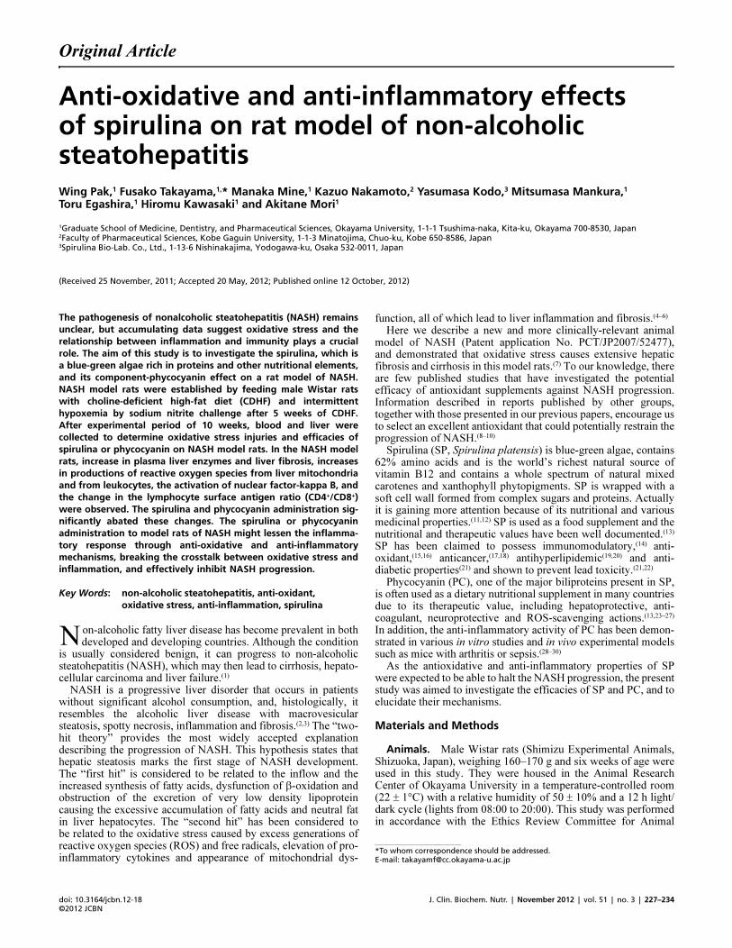
Original Article
J. Clin. Biochem. Nutr. | November 2012 | vol. 51 | no. 3 | 227–234doi: 10.3164/jcbn.12�18©2012 JCBN
JCBNJournal of Clinical Biochemistry and Nutrition0912-00091880-5086the Society for Free Radical Research JapanKyoto, Japanjcbn12-1810.3164/jcbn.12-18Original ArticleAnti�oxidative and anti�inflammatory effects of spirulina on rat model of non�alcoholic steatohepatitisWing Pak,1 Fusako Takayama,1,* Manaka Mine,1 Kazuo Nakamoto,2 Yasumasa Kodo,3 Mitsumasa Mankura,1 Toru Egashira,1 Hiromu Kawasaki1 and Akitane Mori1
1Graduate School of Medicine, Dentistry, and Pharmaceutical Sciences, Okayama University, 1�1�1 Tsushima�naka, Kita�ku, Okayama 700�8530, Japan2Faculty of Pharmaceutical Sciences, Kobe Gaguin University, 1�1�3 Minatojima, Chuo�ku, Kobe 650�8586, Japan3Spirulina Bio�Lab. Co., Ltd., 1�13�6 Nishinakajima, Yodogawa�ku, Osaka 532�0011, Japan
*To whom correspondence should be addressed. E�mail: [email protected]�u.ac.jp
11(Received 25 November, 2011; Accepted 20 May, 2012; Published online 12 October, 2012)
Copyright © 2012 JCBN2012This is an open access article distributed under the terms of theCreative Commons Attribution License, which permits unre-stricted use, distribution, and reproduction in any medium, pro-vided the original work is properly cited.The pathogenesis of nonalcoholic steatohepatitis (NASH) remains
unclear, but accumulating data suggest oxidative stress and the
relationship between inflammation and immunity plays a crucial
role. The aim of this study is to investigate the spirulina, which is
a blue�green algae rich in proteins and other nutritional elements,
and its component�phycocyanin effect on a rat model of NASH.
NASH model rats were established by feeding male Wistar rats
with choline�deficient high�fat diet (CDHF) and intermittent
hypoxemia by sodium nitrite challenge after 5 weeks of CDHF.
After experimental period of 10 weeks, blood and liver were
collected to determine oxidative stress injuries and efficacies of
spirulina or phycocyanin on NASH model rats. In the NASH model
rats, increase in plasma liver enzymes and liver fibrosis, increases
in productions of reactive oxygen species from liver mitochondria
and from leukocytes, the activation of nuclear factor�kappa B, and
the change in the lymphocyte surface antigen ratio (CD4+/CD8+)
were observed. The spirulina and phycocyanin administration sig�
nificantly abated these changes. The spirulina or phycocyanin
administration to model rats of NASH might lessen the inflamma�
tory response through anti�oxidative and anti�inflammatory
mechanisms, breaking the crosstalk between oxidative stress and
inflammation, and effectively inhibit NASH progression.
Key Words: non�alcoholic steatohepatitis, anti�oxidant,
oxidative stress, anti�inflammation, spirulina
IntroductionNon-alcoholic fatty liver disease has become prevalent in bothdeveloped and developing countries. Although the condition
is usually considered benign, it can progress to non-alcoholicsteatohepatitis (NASH), which may then lead to cirrhosis, hepato-cellular carcinoma and liver failure.(1)
NASH is a progressive liver disorder that occurs in patientswithout significant alcohol consumption, and, histologically, itresembles the alcoholic liver disease with macrovesicularsteatosis, spotty necrosis, inflammation and fibrosis.(2,3) The “two-hit theory” provides the most widely accepted explanationdescribing the progression of NASH. This hypothesis states thathepatic steatosis marks the first stage of NASH development.The “first hit” is considered to be related to the inflow and theincreased synthesis of fatty acids, dysfunction of β-oxidation andobstruction of the excretion of very low density lipoproteincausing the excessive accumulation of fatty acids and neutral fatin liver hepatocytes. The “second hit” has been considered tobe related to the oxidative stress caused by excess generations ofreactive oxygen species (ROS) and free radicals, elevation of pro-inflammatory cytokines and appearance of mitochondrial dys-
function, all of which lead to liver inflammation and fibrosis.(4–6)
Here we describe a new and more clinically-relevant animalmodel of NASH (Patent application No. PCT/JP2007/52477),and demonstrated that oxidative stress causes extensive hepaticfibrosis and cirrhosis in this model rats.(7) To our knowledge, thereare few published studies that have investigated the potentialefficacy of antioxidant supplements against NASH progression.Information described in reports published by other groups,together with those presented in our previous papers, encourage usto select an excellent antioxidant that could potentially restrain theprogression of NASH.(8–10)
Spirulina (SP, Spirulina platensis) is blue-green algae, contains62% amino acids and is the world’s richest natural source ofvitamin B12 and contains a whole spectrum of natural mixedcarotenes and xanthophyll phytopigments. SP is wrapped with asoft cell wall formed from complex sugars and proteins. Actuallyit is gaining more attention because of its nutritional and variousmedicinal properties.(11,12) SP is used as a food supplement and thenutritional and therapeutic values have been well documented.(13)
SP has been claimed to possess immunomodulatory,(14) anti-oxidant,(15,16) anticancer,(17,18) antihyperlipidemic(19,20) and anti-diabetic properties(21) and shown to prevent lead toxicity.(21,22)
Phycocyanin (PC), one of the major biliproteins present in SP,is often used as a dietary nutritional supplement in many countriesdue to its therapeutic value, including hepatoprotective, anti-coagulant, neuroprotective and ROS-scavenging actions.(13,23–27)
In addition, the anti-inflammatory activity of PC has been demon-strated in various in vitro studies and in vivo experimental modelssuch as mice with arthritis or sepsis.(28–30)
As the antioxidative and anti-inflammatory properties of SPwere expected to be able to halt the NASH progression, the presentstudy was aimed to investigate the efficacies of SP and PC, and toelucidate their mechanisms.
Materials and Methods
Animals. Male Wistar rats (Shimizu Experimental Animals,Shizuoka, Japan), weighing 160–170 g and six weeks of age wereused in this study. They were housed in the Animal ResearchCenter of Okayama University in a temperature-controlled room(22 ± 1°C) with a relative humidity of 50 ± 10% and a 12 h light/dark cycle (lights from 08:00 to 20:00). This study was performedin accordance with the Ethics Review Committee for Animal
N
doi: 10.3164/jcbn.12�18©2012 JCBN
228
Experimentation of the Graduate School of Medicine, Dentistryand Pharmaceutical Science, Okayama University.
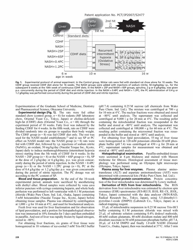
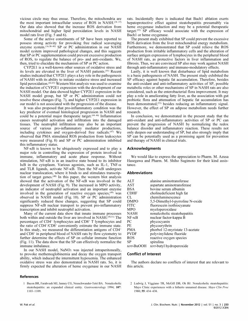
Experimental design (Fig. 1). The rats were fed eitherstandard chow (control group, n = 8) for rodents (MF laboratorychow, Oriental Yeast Co., Tokyo, Japan) or choline-deficienthigh-fat (CDHF) diets (Oriental Yeast Co., n = 48) through theexperiment period of 10 weeks. Fatty liver was induced by thefeeding CDHF for 4 weeks. In the fifth week, rats on CDHF weredivided randomly into six groups to equalize their body weight.The CDHF group (n = 8) was fed CDHF diet only. The rest wasused for the NASH model establishment,(7) and to see SP or PCeffect on NASH model rats: the NASH group (n = 8) rats werefed with CDHF diet, followed by i.p. injections of sodium nitrite(NaNO2), an oxidant, 50 mg/kg/day (Nacalai Tesque Inc., Kyoto,Japan) daily to induce methemoglobinemia (intermittent hypoxiastress) starting from the 5th week of CDHF for 6 weeks. In theNASH + 2SP group (n = 8) or the NASH + 6SP group (n = 8), SPat the dose of 2 g/kg/day or 6 g/kg/day, p.o. was given concur-rently during the period of nitrite injection. In the NASH + 0.4PCgroup (n = 8) or the NASH + 1.2PC group (n = 8), PC at thedose of 0.4 g/kg/day or 1.2 g/kg/day p.o. was given concurrentlyduring the period of nitrite injection. The PC dosage was setaccording to the PC content of SP.
Blood and tissue preparation. At the end of the 10-weekexperimental period, animals were sacrificed by anesthetizingwith diethyl ether. Blood samples were collected by vena cavainferior puncture with syringe containing heparin, and whole bodyperfusion was performed to left ventricular with 0.1 M potassiumchloride (Wako Pure Chem. Ind. Ltd., Osaka, Japan) containing5 mM benzamidine (Nacalai Tesque Inc., Kyoto, Japan) beforeobtaining tissue samples. Plasma was obtained by centrifugationat 1,000 × g for 10 min at 4°C, and used for biochemical analysis.
Fresh liver was used for liver fractionation and for observationof lipid peroxidation, and a portion for histopathological observa-tion was immersed in 10% formalin for 3 days and then embeddedin paraffin. And rest of liver was rapidly frozen by liquid nitrogen,stored in −80°C.
For obtaining liver fractions, six grams of liver tissue werehomogenized in 18 volumes of ice-cooled 5 mM Tris-HCl buffer
(pH 7.4) containing 0.25 M sucrose (all chemicals from WakoPure Chem. Ind. Ltd.). The mixture was centrifuged at 700 × gfor 10 min at 4°C. The nuclear fractions were obtained and storedat −80°C until analysis. The supernatant was collected andcentrifuged at 9,000 × g for 20 min at 4°C. The resulting pelletcontaining the mitochondrial fraction was resuspended in thebuffer and stored at −80°C until analysis. The supernatant wascollected and centrifuged at 100,000 × g for 60 min at 4°C. Theresulting pellet containing the microsomal fraction was resus-pended in the buffer and stored at −80°C until analysis.
For obtaining liver lipid peroxidation, 55 mg of liver tissuewere homogenized in 120 mM potassium chloride-30 mM phos-phate buffer (pH 7.4) was centrifuged at 450 × g for 20 min at4°C, supernatant samples for measurement was obtained andstored at −80°C until analysis.
Histopathological examination. Paraffin-embedded tissueswere sectioned at 4 μm thickness and stained with Massontrichrome for fibrosis. Histological assessment of tissue mor-phology was performed using an Olympus light microscope(Olympus, Tokyo, Japan).
Plasma assay for liver enzymes. Plasma alanine amino-transferase (ALT) and aspartate aminotransferase (AST) weredetermined with commercial kits (Wako Pure Chem. Ind. Ltd.).
Mitochondrial protein concentration. Protein concentra-tion of the liver mitochondria was determined by Lowry method.
Derivation of ROS from liver mitochondria. The ROSderivation from liver mitochondria was estimated by electron spinresonance (ESR) spectrometer (JES-RE1X JEOL RE-1X, JEOL,Tokyo, Japan) connected with the data analyzer (WIN-RAD,Radical Research Inc., Tokyo, Japan), using 5,5-dimethyl-1-pyrroline-1-oxide (DMPO) (Labotech Co., Tokyo, Japan) as aradical-trapping reagent.
35 μL of mitochondria suspension in 0.25 M sucrose Tris-HClbuffer containing 0.1 M potassium chloride was mixed with25 μL of substrate solution containing 0.4% dodesyl maltoside,40 mM sodium glutamate, 40 mM disodium malate and 800 mMdisodium succinate in 50 mM potassium phosphate buffer, pH 7.4,20 μL of 4.6 M DMPO and finally 1.0 mM NADH (OrientalYeast Co., Osaka, Japan), then was incubated at 37°C. After 5 min
Fig. 1. Experimental protocol of animal experiment. In the Control group, Wistar rats were fed with standard rat chow alone for 10 weeks. TheCDHF group received CDHF diet alone for 16 weeks. The NASH groups were added with injections of sodium nitrite, 50 mg/kg/day i.p., for thesubsequent 6 weeks at the 10th week of continuous CDHF diets. In the NASH + 2SP and NASH + 6SP groups, spirulina, 2 g or 6 g/kg/day, was givenp.o. concurrently during the period of CDHF diet and nitrite injection. In the NASH + 0.4PC and NASH + 1.2PC, the PC administration of 0.4 g or1.2 g/kg/day was performed concurrently during the period of CDHF diet and nitrite injection.
J. Clin. Biochem. Nutr. | November 2012 | vol. 51 | no. 3 | 229
©2012 JCBNW. Pak et al.
incubation, 45 μL aliquots of above-mentioned mixture wereplaced into a capillary glass tube, and this was in turn introducedinto a quartz tube, and then measured at room temperature. Theinstrument conditions were the following: about 9.4 GHz with100-kHz modulation frequency, central field ± sweep width of336.0 ± 5 mT, microwave power of 8 mW, field modulation widthof 0.079 mT, time constant of 0.1 s, sweep speed of 5 mT/min.The magnetic field was calculated from the splitting of divalentmanganese (Mn2+), in which the distance from the third to fourthsignal is 8.69 mT.
In this measurement, the characteristic ESR spectrum of thespin adducts of DMPO and hydroxyl radical (DMPO-OH) wasdetected. The ROS derivation was estimated by the ratio of the2nd signal intensity of the DMPO-OH spectrum/the 3rd signal ofMn2+.
Liver lipid peroxidation assay. The liver lipid peroxidationwas determined with chemiluminescence (CL) method. 140 μL ofstored for lipid peroxidation supernatant samples were mixed with20 μL of 130 μg/mL luminol (Wako Pure Chem. Ind. Ltd.) and20 μL of Hanks’ Balanced Salt Solution (HBSS, pH 7.4), thenwas incubated and analyzed in the detector (TriStar LB941,Berthold Japan Co. Ltd., Tokyo, Japan) at 37°C for the baselineCL intensity. After 5 min incubation, 80 μL of 2.2 μg/L tert-butylhydroperoxide (tert-BuOOH; SIGMA, St. Louis, Mo) was beadded. CL intensity was detected from the cleavage of lipidperoxides induced by tert-BuOOH addition, for 120 min withincubating at 37°C. In this measurement, lipid peroxidationlevels estimated by the accumulated CL intensity by addition oftert-BuOOH subtract from the baseline CL intensity for 120 min.
Leukocyte oxygen radical production. 125 μL of 50-folddiluted whole blood samples were mixed with 25 µL of 300 µg/mLluminol and 20 μL of HBSS then was incubated at 37°C. After5 min incubation, 80 μL of 0.0781 μg/mL phorbol 12-myristate13-acetate (PMA; SIGMA) was be added. The intensity of CL wasestimated by measuring the amount of oxidized luminol byoxygen free radicals after PMA stimulation, as the same mannermentioned above.
Nuclear extract and Western blot analyses to determinenuclear transcription factors. The nuclear fractions samplesuspended in 50 mM HEPES buffer (pH 7.4) containing 0.1 Mpotassium chloride, 3 mM magnesium chloride, 1 mM ethylenedi-aminetetraacetic acid, 10% Glycerol, 0.1 mM phenylmethyl-sulfonyl fluoride, 5 μg/mL pepstatin A, 5 μg/mL leapeptin and2 μg/mL aprotinin, and centrifuged at 22,000 × g for 20 min at4°C. The supernatant was used as nucleoprotein samples. Forprotein quantification, Lowry method was used.
The nucleoprotein sample was diluted to 6 mg/mL, then mixedwith sample buffer (62.5 mM Tris-HCl pH 6.8, containing 25%glycerol, 2% sodium dodecyl sulfate, 5% 2-mercaptoethanol,0.01% bromophenol blue) and denatured at 95°C for 5 min. Thesamples (in 30 μg protein/10 μL) were separated on SDS-12.5%polyacrylamide gel (Bio-Rad Laboratories Inc., Berkeley, CA),then transferred to polyvinylidene fluoride (PVDF) membraneusing a transblot apparatus (Bio-Rad Laboratories Inc.). Themembranes were blocked in 5% nonfat milk dissolved in TBS-Tbuffer (25 mM Tris-HCl buffer, pH 7.4, containing 0.15 M sodiumchloride and 0.1% Tween20) for 1 h at room temperature. Themembranes were incubated with primary antibodies as follows:mouse monoclonal anti-rat nuclear factor-kappa B (NF-κB)(1:1000; Santa Cruz Biotechnology, Santa Cruz, CA) or rabbitpolyclonal anti-rat Histone H1 (1:200; Santa Cruz Biotechnology)for 1 h. And then incubated with secondary antibodies for goatanti-mouse IgG-HRP (1:5000; Santa Cruz Biotechnology) or goatanti-rabbit IgG-HRP (1:5000; Santa Cruz Biotechnology) for30 min. Protein bands were visualized using enhanced Chemilu-minescence Luminol Reagent (Santa Cruz Biotechnology). Theresults were standardized by Histone H1.
Preparation of the liver microsomal fraction. Aliquots ofliver microsomes (5 μg) were placed in sample buffer (62.5 mMTris-HCl pH 6.8, containing 25% glycerol, 2% sodium dodecylsulfate, 5% 2-mercaptoethanol, 0.01% bromophenol blue) anddenatured at 95°C for 5 min. Samples were separated on SDS-12.5% polyacrylamide gel and transferred to PVDF membranes,followed by Western blot analyses with the primary rabbit anti-human/rat cytochrome P450 enzyme (CYP2E1) polyclonal anti-bodies (1:1500; Chemicon International, Temecula, CA) or mousemonoclonal anti-rat β-actin (1:2000, Santa Cruz Biotechnology)for 1 h. Proteins were detected with peroxidase-conjugatedsecondary anti-rabbit IgG antibody or goat anti-mouse IgGHRP (1:5000; Santa Cruz Biotechnology) for 1 h and imagedusing enhanced Chemiluminescence Luminol Reagent. The resultswere standardized by Histone H1.
Plasma myeloperoxidase (MPO) activity. 225 μL of theplasma was mix with 315 μL of 10 mM citrate buffer, pH 5.0, thenaliquots of 90 μL of these mixed liquid were pipetted into fourwells. The 540 nm absorbance was monitored about the twowells to which 120 μL of cold stop solution (4 N sulfuric acid)was added to stop the reaction to detect the background absor-bance. 30 µL of MPO substrate solution containing 6 mM 3,3',5,5'-tetramethylbenzidene dihydrochloride, 120 μM resorcinol, and4.4 mM hydrogen peroxide in distilled water was added to eachwell, and the reaction was stopped after 2 min with 120 μL of coldstop solution, subsequently measured by absorbance at 450 nm.
As an additional control, 90 μL of dilution buffer (without liverextract) were pipetted into four wells, 30 μL of substrate bufferwas added, followed by 120 μL of stop solution after 2 min. Nocolor reaction was observed in these control wells. The MPOactivity of the liver sample was calculated by subtracting the meanbackground absorbance and is expressed as change in opticaldensity per minute.
Isolation of leukocytes and flow cytometry analysis oflymphocyte surface antigen CD4, CD8. The peripheral bloodwas divided and erythrocytes were lysed with ammonium chloridesolution, and washed twice in phosphate-buffered saline containing0.1% sodium azide and 0.1% bovine serum albumin (BSA). Thefinal pellet was resuspended in same buffer.
Liver tissue was dissected into 1 mm3 fragments in HBSSmedium containing 2% fetal bovine serum (FBS), 25 mM HEPES,0.6% BSA, 0.125% type IV collagenase and 0.002% deoxyribonu-clease I (DNase I) were added to each sample and the mixturesincubated with gentle agitation at 37°C for 30 min. Undissociatedtissue was removed by filtration through a 30 μm nylon mesh andthe cells were washed twice with HBSS containing 0.001% DNaseI and 25 mM HEPES with centrifugation at 250 × g for 10 min.The hepatocyte-rich matrix was removed by centrifugation for1 min at 30 × g and the final pellet was resuspended in phosphate-buffered saline containing 0.1% sodium azide and 0.1% BSA.
For flow cytometry, the cells were incubated with fluoresceinisothiocyanate (FITC) or phycoerythrin (PE)-conjugated mono-clonal antibodies (antimouse CD4 [FITC], or antimouse CD8[PE]) (all sourced from eBioscience, San Diego, CA) for 20 min,washed twice times. Then cell fixation using 400 μL of 1%paraformaldehyde immediately before determination. At least10,000 cells in fluorescent channel 1 for CD4, and fluorescentchannel 2 for CD8 were detected using FACSCaliburTM flowcytometer (BD Biosciences, Franklin Lakes, NJ). Results wereexpressed as mean fluorescence intensity for a given moleculeper cell.
Statistical analyses. Quantitative data were expressed asthe mean ± SEM of six rats. Differences among groups wereexamined by one-way analysis of variance (ANOVA) followedby Tukey-Kramer multiple comparison test. A value of p<0.05was considered statistically significant.
doi: 10.3164/jcbn.12�18©2012 JCBN
230
Results
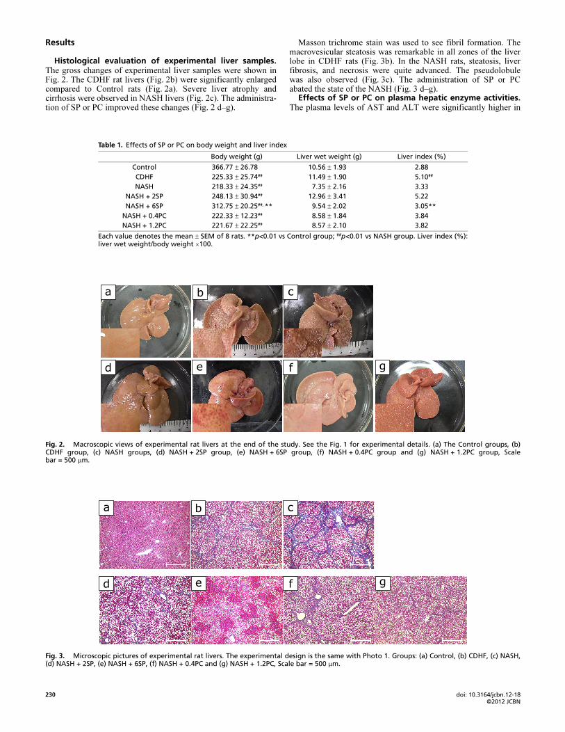
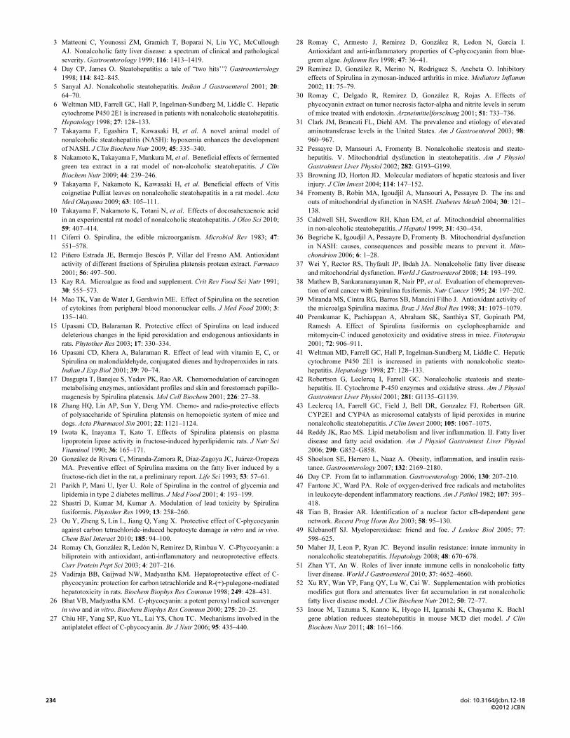
Histological evaluation of experimental liver samples.The gross changes of experimental liver samples were shown inFig. 2. The CDHF rat livers (Fig. 2b) were significantly enlargedcompared to Control rats (Fig. 2a). Severe liver atrophy andcirrhosis were observed in NASH livers (Fig. 2c). The administra-tion of SP or PC improved these changes (Fig. 2 d–g).
Masson trichrome stain was used to see fibril formation. Themacrovesicular steatosis was remarkable in all zones of the liverlobe in CDHF rats (Fig. 3b). In the NASH rats, steatosis, liverfibrosis, and necrosis were quite advanced. The pseudolobulewas also observed (Fig. 3c). The administration of SP or PCabated the state of the NASH (Fig. 3 d–g).
Effects of SP or PC on plasma hepatic enzyme activities.The plasma levels of AST and ALT were significantly higher in
Table 1. Effects of SP or PC on body weight and liver index
Each value denotes the mean ± SEM of 8 rats. **p<0.01 vs Control group; ##p<0.01 vs NASH group. Liver index (%):liver wet weight/body weight ×100.
Body weight (g) Liver wet weight (g) Liver index (%)
Control 366.77 ± 26.78 10.56 ± 1.93 2.88
CDHF 225.33 ± 25.74## 11.49 ± 1.90 5.10##
NASH 218.33 ± 24.35## 7.35 ± 2.16 3.33
NASH + 2SP 248.13 ± 30.94## 12.96 ± 3.41 5.22
NASH + 6SP 312.75 ± 20.25##, ** 9.54 ± 2.02 3.05**
NASH + 0.4PC 222.33 ± 12.23## 8.58 ± 1.84 3.84
NASH + 1.2PC 221.67 ± 22.25## 8.57 ± 2.10 3.82
Fig. 2. Macroscopic views of experimental rat livers at the end of the study. See the Fig. 1 for experimental details. (a) The Control groups, (b)CDHF group, (c) NASH groups, (d) NASH + 2SP group, (e) NASH + 6SP group, (f) NASH + 0.4PC group and (g) NASH + 1.2PC group, Scalebar = 500 μm.
Fig. 3. Microscopic pictures of experimental rat livers. The experimental design is the same with Photo 1. Groups: (a) Control, (b) CDHF, (c) NASH,(d) NASH + 2SP, (e) NASH + 6SP, (f) NASH + 0.4PC and (g) NASH + 1.2PC, Scale bar = 500 μm.
J. Clin. Biochem. Nutr. | November 2012 | vol. 51 | no. 3 | 231
©2012 JCBNW. Pak et al.
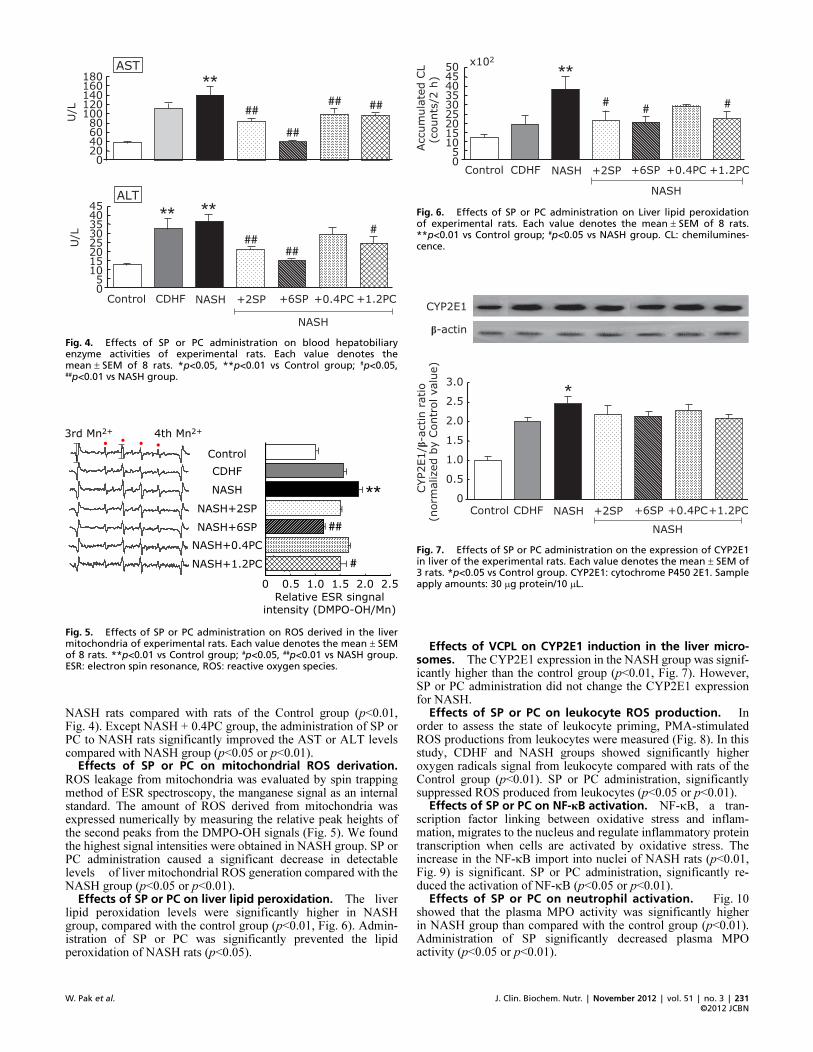
NASH rats compared with rats of the Control group (p<0.01,Fig. 4). Except NASH + 0.4PC group, the administration of SP orPC to NASH rats significantly improved the AST or ALT levelscompared with NASH group (p<0.05 or p<0.01).
Effects of SP or PC on mitochondrial ROS derivation.ROS leakage from mitochondria was evaluated by spin trappingmethod of ESR spectroscopy, the manganese signal as an internalstandard. The amount of ROS derived from mitochondria wasexpressed numerically by measuring the relative peak heights ofthe second peaks from the DMPO-OH signals (Fig. 5). We foundthe highest signal intensities were obtained in NASH group. SP orPC administration caused a significant decrease in detectablelevels of liver mitochondrial ROS generation compared with theNASH group (p<0.05 or p<0.01).
Effects of SP or PC on liver lipid peroxidation. The liverlipid peroxidation levels were significantly higher in NASHgroup, compared with the control group (p<0.01, Fig. 6). Admin-istration of SP or PC was significantly prevented the lipidperoxidation of NASH rats (p<0.05).
Effects of VCPL on CYP2E1 induction in the liver micro�somes. The CYP2E1 expression in the NASH group was signif-icantly higher than the control group (p<0.01, Fig. 7). However,SP or PC administration did not change the CYP2E1 expressionfor NASH.
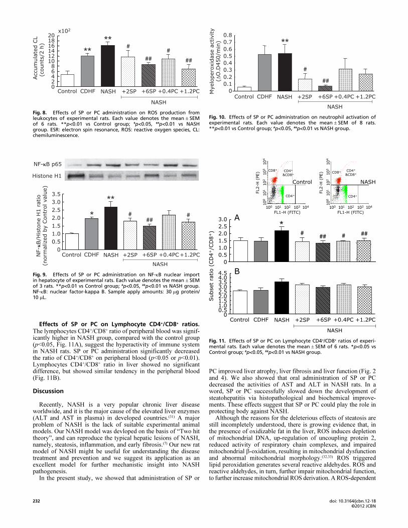
Effects of SP or PC on leukocyte ROS production. In order to assess the state of leukocyte priming, PMA-stimulatedROS productions from leukocytes were measured (Fig. 8). In thisstudy, CDHF and NASH groups showed significantly higheroxygen radicals signal from leukocyte compared with rats of theControl group (p<0.01). SP or PC administration, significantlysuppressed ROS produced from leukocytes (p<0.05 or p<0.01).
Effects of SP or PC on NF�κB activation. NF-κB, a tran-scription factor linking between oxidative stress and inflam-mation, migrates to the nucleus and regulate inflammatory proteintranscription when cells are activated by oxidative stress. Theincrease in the NF-κB import into nuclei of NASH rats (p<0.01,Fig. 9) is significant. SP or PC administration, significantly re-duced the activation of NF-κB (p<0.05 or p<0.01).
Effects of SP or PC on neutrophil activation. Fig. 10 showed that the plasma MPO activity was significantly higherin NASH group than compared with the control group (p<0.01).Administration of SP significantly decreased plasma MPOactivity (p<0.05 or p<0.01).
Fig. 4. Effects of SP or PC administration on blood hepatobiliaryenzyme activities of experimental rats. Each value denotes themean ± SEM of 8 rats. *p<0.05, **p<0.01 vs Control group; #p<0.05,##p<0.01 vs NASH group.
Fig. 5. Effects of SP or PC administration on ROS derived in the livermitochondria of experimental rats. Each value denotes the mean ± SEMof 8 rats. **p<0.01 vs Control group; #p<0.05, ##p<0.01 vs NASH group.ESR: electron spin resonance, ROS: reactive oxygen species.
Fig. 6. Effects of SP or PC administration on Liver lipid peroxidationof experimental rats. Each value denotes the mean ± SEM of 8 rats.**p<0.01 vs Control group; #p<0.05 vs NASH group. CL: chemilumines�cence.
Fig. 7. Effects of SP or PC administration on the expression of CYP2E1in liver of the experimental rats. Each value denotes the mean ± SEM of3 rats. *p<0.05 vs Control group. CYP2E1: cytochrome P450 2E1. Sampleapply amounts: 30 μg protein/10 μL.
doi: 10.3164/jcbn.12�18©2012 JCBN
232
Effects of SP or PC on Lymphocyte CD4+/CD8+ ratios.The lymphocytes CD4+/CD8+ ratio of peripheral blood was signif-icantly higher in NASH group, compared with the control group(p<0.05, Fig. 11A), suggest the hyperactivity of immune systemin NASH rats. SP or PC administration significantly decreasedthe ratio of CD4+/CD8+ on peripheral blood (p<0.05 or p<0.01).Lymphocytes CD4+/CD8+ ratio in liver showed no significantdifference, but showed similar tendency in the peripheral blood(Fig. 11B).
Discussion
Recently, NASH is a very popular chronic liver diseaseworldwide, and it is the major cause of the elevated liver enzymes(ALT and AST in plasma) in developed countries.(31) A majorproblem of NASH is the lack of suitable experimental animalmodels. Our NASH model was devloped on the basis of “Two hittheory”, and can reproduce the typical hepatic lesions of NASH,namely, steatosis, inflammation, and early fibrosis.(7) Our new ratmodel of NASH might be useful for understanding the diseasetreatment and prevention and we suggest its application as anexcellent model for further mechanistic insight into NASHpathogenesis.
In the present study, we showed that administration of SP or
PC improved liver atrophy, liver fibrosis and liver function (Fig. 2and 4). We also showed that oral administration of SP or PCdecreased the activities of AST and ALT in NASH rats. In aword, SP or PC successfully slowed down the development ofsteatohepatitis via histopathological and biochemical improve-ments. These effects suggest that SP or PC could play the role inprotecting body against NASH.
Although the reasons for the deleterious effects of steatosis arestill incompletely understood, there is growing evidence that, inthe presence of oxidizable fat in the liver, ROS induces depletionof mitochondrial DNA, up-regulation of uncoupling protein 2,reduced activity of respiratory chain complexes, and impairedmitochondrial β-oxidation, resulting in mitochondrial dysfunctionand abnormal mitochondrial morphology.(32,33) ROS triggeredlipid peroxidation generates several reactive aldehydes. ROS andreactive aldehydes, in turn, further impair mitochondrial function,to further increase mitochondrial ROS derivation. A ROS-dependent
Fig. 8. Effects of SP or PC administration on ROS production fromleukocytes of experimental rats. Each value denotes the mean ± SEMof 6 rats. **p<0.01 vs Control group; #p<0.05, ##p<0.01 vs NASHgroup. ESR: electron spin resonance, ROS: reactive oxygen species, CL:chemiluminescence.
Fig. 9. Effects of SP or PC administration on NF�κB nuclear importin hepatocyte of experimental rats. Each value denotes the mean ± SEMof 3 rats. **p<0.01 vs Control group; #p<0.05, ##p<0.01 vs NASH group.NF�κB: nuclear factor�kappa B. Sample apply amounts: 30 μg protein/10 μL.
Fig. 10. Effects of SP or PC administration on neutrophil activation ofexperimental rats. Each value denotes the mean ± SEM of 8 rats.**p<0.01 vs Control group; #p<0.05, ##p<0.01 vs NASH group.
Fig. 11. Effects of SP or PC on Lymphocyte CD4+/CD8+ ratios of experi�mental rats. Each value denotes the mean ± SEM of 6 rats. *p<0.05 vsControl group; #p<0.05, ##p<0.01 vs NASH group.
J. Clin. Biochem. Nutr. | November 2012 | vol. 51 | no. 3 | 233
©2012 JCBNW. Pak et al.
vicious circle may thus ensue. Therefore, the mitochondria arethe most important intracellular source of ROS in NASH.(34–37)
Our data also showed that the increase ROS derivation frommitochondrial and higher lipid peroxidation levels in NASHmodel rats liver (Fig. 5 and 6).
Some of the active constituents of SP have been reported topossess strong activity for provoking free radical scavengingenzyme system.(16,38–40) SP or PC administration in our NASHmodel system improved pathological changes, and this suggeststhat SP or PC supplementation could prevent excessive productionof ROS, to regulate the balance of pro- and anti-oxidants. We,then, tried to elucidate the mechanism of SP or PC action.
CYP2E1 is a well-known other sources of oxidative stress andits levels are elevated in the liver of NASH patients.(41) Recentstudies indicated that CYP2E1 plays a key role in the pathogenesisof NASH with its ability to initiate oxidative stress and increasedlipid peroxidation.(42,43) Western blot analysis was used to examinethe induction of CYP2E1 expression with the development of ourNASH model. Our data showed higher CYP2E1 expression in theNASH model group, but SP or PC administration could notresolve these changes (Fig. 7), and higher CYP2E1 expression inour model is not associated with the progression of the disease.
It was also proposed that pro-inflammatory condition was thekey predictor of eventual histological progression in NASH andcould be a potential major therapeutic target.(44–46) Inflammationcauses neutrophil activation and infiltration into the damagedtissues. The neutrophil infiltration may also be an importantsource of various pro-inflammatory mediator productions,including cytokines and oxygen-derived free radicals.(47) Weobserved that PMA stimulated ROS production from leukocyteswere measured (Fig. 8) and SP or PC administration inhibitedthis inflammatory status.
NF-κB is known to be ubiquitously expressed and to play amajor role in controlling the expression of protein involved inimmune, inflammatory and acute phase response. Withoutstimulation, NF-κB is in an inactive state bound to its inhibitorIκB in the cytoplasm. Various agonists, such as IL-1, TNF-αand TLR ligands, activate NF-κB. Then the NF-κB undergoesnuclear translocation, where it binds to and stimulates transcrip-tion of target genes.(48) In this paper, the western blot analysisshowed that the activation of the NF-κB was involved in thedevelopment of NASH (Fig. 9). The increased in MPO activity,an indicator of neutrophil activation and an important enzymeinvolved in the generation of reactive oxygen species,(49) wasobserved in NASH model (Fig. 10). SP or PC administrationsignificantly reduced these changes, suggesting that SP couldsuppress NF-κB nuclear transport to prevent pro-inflammatorytranscription and inhibit neutrophil activation.
Many of the current data show that innate immune processesboth within and outside the liver are involved in NASH.(50,51) Thepercentages of CD4+ lymphocytes and CD8+ T lymphocytes andthe ratio of CD4+/CD8+ conveniently estimate the immune state.In this study, we measured the differentiation antigens of CD4+
and CD8+ in peripheral blood of NASH rats by flow cytometry tofurther determine the effects of SP on cellular immune function(Fig. 11). The data show that the SP can effectively normalize theimmune imbalance.
In our NASH model, NaNO2 was injected intraperitoneally,to provoke methemoglobinemia and decay the oxygen transportability, which induced the intermittent hypoxemia. The enhancedoxidative stress was also demonstrated in NASH rats. So, it isfirmly expected the alteration of heme oxygenase in our NASH
rats. Incidentally there is indicated that Bach1 ablation exertshepatoprotective effect against steatohepatitis presumably viaheme oxygenase induction and may be a potential therapeutictarget.(52) SP efficacy would associate with the expression ofBach1 or heme oxygenase.
The present study exhibited that SP could prevent the excessiveROS derivation from the liver mitochondrial energy metabolism.Furthermore, we demonstrated that SP could relieve the ROSproduction from irritable inflammatory cells and the alteration ofsurface antigen expression of lymphocytes in the peripheral bloodof NASH rats, as protective factors in liver inflammation andfibrosis. Thus, we are convinced SP also may work against NASHthrough anti-inflammatory and immuno-modulatory effects.
Lastly, it is well known that disturbance of lipid metabolismis a basic pathogenesis of NASH. The present study exhibited theSP efficacy against hepatic fat accumulation. Therefore, besidesthe anti-oxidant and anti-inflammatory activities of SP, possiblemetabolic roles or other mechanisms of SP in NASH rats are alsoconsidered, such as the enterobacterial flora improvement. It mayplay a role in ameliorating fatty liver, as the association with gutprobiotic flora and attenuation of hepatic fat accumulation hasbeen demonstrated,(53) besides reducing an inflammatory signal.However, the effect of SP on adipose metabolism needs furtherresearch.
In conclusion, we demonstrated in the present study that theanti-oxidant and anti-inflammatory activities of SP or PC canprevent the progression of NASH by normalizing the redoxbalance disorder and inflammatory reaction. These results notonly deepen our understanding of SP, but also strongly imply thatSP can be potentially used as a promising agent for preventionand therapy of NASH in clinical trials.
Acknowledgments
We would like to express the appreciation to Pharm. M. AzusaHasegawa and Pharm. M. Shiho Sugimoto for their kind assis-tance.
Abbreviations
ALT alanine aminotransferaseAST aspartate aminotransferaseBSA bovine serum albuminCDHF cholinedeficient high fatCL chemiluminescenceDMPO 5,5-Dimethyl-l-pyrroline N-oxideFITC fluorescein isothiocyanateMPO myeloperoxidaseNASH nonalcoholic steatohepatitisNF-κB nuclear factor-kappa BPC phycocyaninPE phycoerythrinPMA phorbol 12-myristate 13-acetatePVDF polyvinylidene fluorideROS reactive oxygen speciesSP spirulinatert-BuOOH tert-butyl hydroperoxide
Conflict of Interest
The authors declare no conflicts of interest that are relevant tothis article.
References
1 Bacon BR, Farahvash MJ, Janney CG, Neuschwander-Tetri BA. Nonalcoholic
steatohepatitis: an expanded clinical entity. Gastroenterology 1994; 107:
1103–1109.
2 Ludwig J, Viggiano TR, McGill DB, Oh BJ. Nonalcoholic steatohepatitis:
Mayo Clinic experiences with a hitherto unnamed disease. Mayo Clin Proc
1980; 55: 434–438.
doi: 10.3164/jcbn.12�18©2012 JCBN
234
3 Matteoni C, Younossi ZM, Gramich T, Boparai N, Liu YC, McCullough
AJ. Nonalcoholic fatty liver disease: a spectrum of clinical and pathological
severity. Gastroenterology 1999; 116: 1413–1419.
4 Day CP, James O. Steatohepatitis: a tale of “two hits’’? Gastroenterology
1998; 114: 842–845.
5 Sanyal AJ. Nonalcoholic steatohepatitis. Indian J Gastroenterol 2001; 20:
64–70.
6 Weltman MD, Farrell GC, Hall P, Ingelman-Sundberg M, Liddle C. Hepatic
cytochrome P450 2E1 is increased in patients with nonalcoholic steatohepatitis.
Hepatology 1998; 27: 128–133.
7 Takayama F, Egashira T, Kawasaki H, et al. A novel animal model of
nonalcoholic steatohepatitis (NASH): hypoxemia enhances the development
of NASH. J Clin Biochem Nutr 2009; 45: 335–340.
8 Nakamoto K, Takayama F, Mankura M, et al. Beneficial effects of fermented
green tea extract in a rat model of non-alcoholic steatohepatitis. J Clin
Biochem Nutr 2009; 44: 239–246.
9 Takayama F, Nakamoto K, Kawasaki H, et al. Beneficial effects of Vitis
coignetiae Pulliat leaves on nonalcoholic steatohepatitis in a rat model. Acta
Med Okayama 2009; 63: 105–111.
10 Takayama F, Nakamoto K, Totani N, et al. Effects of docosahexaenoic acid
in an experimental rat model of nonalcoholic steatohepatitis. J Oleo Sci 2010;
59: 407–414.
11 Ciferri O. Spirulina, the edible microorganism. Microbiol Rev 1983; 47:
551–578.
12 Piñero Estrada JE, Bermejo Bescós P, Villar del Fresno AM. Antioxidant
activity of different fractions of Spirulina platensis protean extract. Farmaco
2001; 56: 497–500.
13 Kay RA. Microalgae as food and supplement. Crit Rev Food Sci Nutr 1991;
30: 555–573.
14 Mao TK, Van de Water J, Gershwin ME. Effect of Spirulina on the secretion
of cytokines from peripheral blood mononuclear cells. J Med Food 2000; 3:
135–140.
15 Upasani CD, Balaraman R. Protective effect of Spirulina on lead induced
deleterious changes in the lipid peroxidation and endogenous antioxidants in
rats. Phytother Res 2003; 17: 330–334.
16 Upasani CD, Khera A, Balaraman R. Effect of lead with vitamin E, C, or
Spirulina on malondialdehyde, conjugated dienes and hydroperoxides in rats.
Indian J Exp Biol 2001; 39: 70–74.
17 Dasgupta T, Banejee S, Yadav PK, Rao AR. Chemomodulation of carcinogen
metabolising enzymes, antioxidant profiles and skin and forestomach papillo-
magenesis by Spirulina platensis. Mol Cell Biochem 2001; 226: 27–38.
18 Zhang HQ, Lin AP, Sun Y, Deng YM. Chemo- and radio-protective effects
of polysaccharide of Spirulina platensis on hemopoietic system of mice and
dogs. Acta Pharmacol Sin 2001; 22: 1121–1124.
19 Iwata K, Inayama T, Kato T. Effects of Spirulina platensis on plasma
lipoprotein lipase activity in fructose-induced hyperlipidemic rats. J Nutr Sci
Vitaminol 1990; 36: 165–171.
20 González de Rivera C, Miranda-Zamora R, Díaz-Zagoya JC, Juárez-Oropeza
MA. Preventive effect of Spirulina maxima on the fatty liver induced by a
fructose-rich diet in the rat, a preliminary report. Life Sci 1993; 53: 57–61.
21 Parikh P, Mani U, Iyer U. Role of Spirulina in the control of glycemia and
lipidemia in type 2 diabetes mellitus. J Med Food 2001; 4: 193–199.
22 Shastri D, Kumar M, Kumar A. Modulation of lead toxicity by Spirulina
fusiformis. Phytother Res 1999; 13: 258–260.
23 Ou Y, Zheng S, Lin L, Jiang Q, Yang X. Protective effect of C-phycocyanin
against carbon tetrachloride-induced hepatocyte damage in vitro and in vivo.
Chem Biol Interact 2010; 185: 94–100.
24 Romay Ch, González R, Ledón N, Remirez D, Rimbau V. C-Phycocyanin: a
biliprotein with antioxidant, anti-inflammatory and neuroprotective effects.
Curr Protein Pept Sci 2003; 4: 207–216.
25 Vadiraja BB, Gaijwad NW, Madyastha KM. Hepatoprotective effect of C-
phycocyanin: protection for carbon tetrachloride and R-(+)-pulegone-mediated
hepatotoxicity in rats. Biochem Biophys Res Commun 1998; 249: 428–431.
26 Bhat VB, Madyastha KM. C-phycocyanin: a potent peroxyl radical scavenger
in vivo and in vitro. Biochem Biophys Res Commun 2000; 275: 20–25.
27 Chiu HF, Yang SP, Kuo YL, Lai YS, Chou TC. Mechanisms involved in the
antiplatelet effect of C-phycocyanin. Br J Nutr 2006; 95: 435–440.
28 Romay C, Armesto J, Remirez D, González R, Ledon N, García I.
Antioxidant and anti-inflammatory properties of C-phycocyanin from blue-
green algae. Inflamm Res 1998; 47: 36–41.
29 Remirez D, González R, Merino N, Rodriguez S, Ancheta O. Inhibitory
effects of Spirulina in zymosan-induced arthritis in mice. Mediators Inflamm
2002; 11: 75–79.
30 Romay C, Delgado R, Remirez D, González R, Rojas A. Effects of
phycocyanin extract on tumor necrosis factor-alpha and nitrite levels in serum
of mice treated with endotoxin. Arzneimittelforschung 2001; 51: 733–736.
31 Clark JM, Brancati FL, Diehl AM. The prevalence and etiology of elevated
aminotransferase levels in the United States. Am J Gastroenterol 2003; 98:
960–967.
32 Pessayre D, Mansouri A, Fromenty B. Nonalcoholic steatosis and steato-
hepatitis. V. Mitochondrial dysfunction in steatohepatitis. Am J Physiol
Gastrointest Liver Physiol 2002; 282: G193–G199.
33 Browning JD, Horton JD. Molecular mediators of hepatic steatosis and liver
injury. J Clin Invest 2004; 114: 147–152.
34 Fromenty B, Robin MA, Igoudjil A, Mansouri A, Pessayre D. The ins and
outs of mitochondrial dysfunction in NASH. Diabetes Metab 2004; 30: 121–
138.
35 Caldwell SH, Swerdlow RH, Khan EM, et al. Mitochondrial abnormalities
in non-alcoholic steatohepatitis. J Hepatol 1999; 31: 430–434.
36 Begriche K, Igoudjil A, Pessayre D, Fromenty B. Mitochondrial dysfunction
in NASH: causes, consequences and possible means to prevent it. Mito-
chondrion 2006; 6: 1–28.
37 Wei Y, Rector RS, Thyfault JP, Ibdah JA. Nonalcoholic fatty liver disease
and mitochondrial dysfunction. World J Gastroenterol 2008; 14: 193–199.
38 Mathew B, Sankaranarayanan R, Nair PP, et al. Evaluation of chemopreven-
tion of oral cancer with Spirulina fusiformis. Nutr Cancer 1995; 24: 197–202.
39 Miranda MS, Cintra RG, Barros SB, Mancini Filho J. Antioxidant activity of
the microalga Spirulina maxima. Braz J Med Biol Res 1998; 31: 1075–1079.
40 Premkumar K, Pachiappan A, Abraham SK, Santhiya ST, Gopinath PM,
Ramesh A. Effect of Spirulina fusiformis on cyclophosphamide and
mitomycin-C induced genotoxicity and oxidative stress in mice. Fitoterapia
2001; 72: 906–911.
41 Weltman MD, Farrell GC, Hall P, Ingelman-Sundberg M, Liddle C. Hepatic
cytochrome P450 2E1 is increased in patients with nonalcoholic steato-
hepatitis. Hepatology 1998; 27: 128–133.
42 Robertson G, Leclercq I, Farrell GC. Nonalcoholic steatosis and steato-
hepatitis. II. Cytochrome P-450 enzymes and oxidative stress. Am J Physiol
Gastrointest Liver Physiol 2001; 281: G1135–G1139.
43 Leclercq IA, Farrell GC, Field J, Bell DR, Gonzalez FJ, Robertson GR.
CYP2E1 and CYP4A as microsomal catalysts of lipid peroxides in murine
nonalcoholic steatohepatitis. J Clin Invest 2000; 105: 1067–1075.
44 Reddy JK, Rao MS. Lipid metabolism and liver inflammation. II. Fatty liver
disease and fatty acid oxidation. Am J Physiol Gastrointest Liver Physiol
2006; 290: G852–G858.
45 Shoelson SE, Herrero L, Naaz A. Obesity, inflammation, and insulin resis-
tance. Gastroenterology 2007; 132: 2169–2180.
46 Day CP. From fat to inflammation. Gastroenterology 2006; 130: 207–210.
47 Fantone JC, Ward PA. Role of oxygen-derived free radicals and metabolites
in leukocyte-dependent inflammatory reactions. Am J Pathol 1982; 107: 395–
418.
48 Tian B, Brasier AR. Identification of a nuclear factor κB-dependent gene
network. Recent Prog Horm Res 2003; 58: 95–130.
49 Klebanoff SJ. Myeloperoxidase: friend and foe. J Leukoc Biol 2005; 77:
598–625.
50 Maher JJ, Leon P, Ryan JC. Beyond insulin resistance: innate immunity in
nonalcoholic steatohepatitis. Hepatology 2008; 48: 670–678.
51 Zhan YT, An W. Roles of liver innate immune cells in nonalcoholic fatty
liver disease. World J Gastroenterol 2010; 37: 4652–4660.
52 Xu RY, Wan YP, Fang QY, Lu W, Cai W. Supplementation with probiotics
modifies gut flora and attenuates liver fat accumulation in rat nonalcoholic
fatty liver disease model. J Clin Biochem Nutr 2012; 50: 72–77.
53 Inoue M, Tazuma S, Kanno K, Hyogo H, Igarashi K, Chayama K. Bach1
gene ablation reduces steatohepatitis in mouse MCD diet model. J Clin
Biochem Nutr 2011; 48: 161–166.