Embed Size (px)
Citation preview
International Journal of Medicinal Mushrooms, 19(9):797–807 (2017)
7971521-9437/17/$35.00 © 2017 Begell House, Inc. www.begellhouse.com
Antioxidative and Inhibitory Effects of the Fruiting Body of Black Lingzhi Mushroom, Amauroderma rugosum (Agaricomycetes), on LDL Oxidation and HMG-CoA Reductase ActivityChan Kam Seng, Noorlidah Abdullah,* & Norhaniza Aminudin
Faculty of Science, Mushroom Research Centre & Institute of Biological Sciences, University of Malaya, Kuala Lumpur, Malaysia
*Address all correspondence to: Noorlidah Abdullah, Faculty of Science, Institute of Biological Sciences, Mushroom Research Centre, University of Malaya, 50603 Kuala Lumpur, Malaysia; Tel.: +603 7967 4371; Fax: +603 7967 4178; [email protected]
ABSTRACT: Amauroderma rugosum fruiting bodies possess excellent cardiovascular benefits, including antioxida-tive, antihyperlipidemic, antihypertensive, antiinflammatory, anti–platelet aggregation, and antithrombotic effects. In this article, we describe our investigations of the in vitro antioxidant activity and in vitro antiatherosclerotic potential through inhibitory effects on low-density lipoprotein (LDL), LDL peroxidation, and 3-hydroxy3-methylglutaryl–coenzyme A (HMG-CoA) reductase catalytic activity using various fruiting body extracts partitioned with an organic solvent. Among 5 extracts/fractions tested, the semipolar ethyl acetate (EA) fraction demonstrated good antioxidant capacity based on total phenolic content, 2,2-diphenyl-1-picrylhydrazyl free radical scavenging, ferrous ion–chelat-ing ability, cupric ion–reducing antioxidant capacity, and lipid peroxidation assays. The EA fraction also showed the strongest inhibitory effect on Cu2+-induced LDL oxidation via thiobarbituric acid reactive substances formation and HMG-CoA reductase activity. Chemical analysis conjointly identified 10 phenolic compounds (4 benzoic acid deriva-tives, 3 flavonoids, 1 cinnamic acid, 1 hexahydroxydiphenic acid dilactone, and 1 xanthone derivative), some of which play pivotal roles in arresting the physiopathogenesis of atherosclerosis, thereby attenuating the risk of cardiovascular events occurring.
KEY WORDS: antiatherosclerotic activity, anti–LDL oxidation, antioxidant, hypocholesterolemia, medicinal mushrooms
ABBREVIATIONS: CD, conjugated diene; CUPRAC, cupric ion–reducing antioxidant capacity; DCM, dichloromethane; DPPH, 2,2-diphenyl-1-picrylhydrazyl; EA, ethyl acetate; GAE, gallic acid equivalent; Hex, hexane; HMG-CoA, 3-hydroxy-3-methylgl-utaryl–coenzyme A; IC50, half-maximal inhibitory concentration; LC-MS/MS, liquid chromatography–tandem mass spectrometry; LDL, low-density lipoprotein; MD, crude methanol-dichloromethane extract; MDA, malondialdehyde; ox-LDL, oxidized low-density lipoprotein; TBARS, thiobarbituric acid–reactive substances
I. INTRODUCTION
Atherosclerosis is a chronic disease of the arteries caused by the accumulation of fatty streaks beneath the inner lining of the endothelial layer of an artery. It is a key factor in morbidity and mortality in Western countries. The World Health Organization predicts that coronary heart disease and stroke will become much more life-threatening, with a projected death toll of 24 million between 2020 and 2030.1 The pathogenesis of atherosclerosis is due to the inability
of the body’s systems to defend against a series of physiopathological responses such as hypercholes-terolemia, oxidative stress, oxidation of low-density lipoproteins (LDLs), and inflammation in the sub-endothelial space.2 Oxidized LDL (ox-LDL) and endothelial dysfunction have been proven to play key roles in initiating the development of athero-sclerosis.3 In addition, ox-LDL particles are toxic to various cells, causing chain-breaking reactions that generate reactive oxygen species. The attacks of those energetic, highly reactive free radicals, together with

International Journal of Medicinal Mushrooms
Seng, Abdullah, & Aminudin798
the weakening of the immune system, eventually lead to endothelial injury, more properly referred to as “endothelial dysfunction.” Therefore, discover-ing naturally occurring antioxidants derived from medicinal herbs is vital to ameliorate this aging-related disease.4
Historically, the lingzhi mushrooms are well-known for their medicinal properties. In Asia, the lingzhi mushrooms are referred to as the “king of medicines” and the “mushroom of immortal-ity.”5 Although more than 2000 known species of lingzhi exist, only black and red lingzhi have demonstrated significant health-enhancing effects and have been scientifically proven to be safe and free from side effects.6 Based on the literature, the black lingzhi, Amauroderma rugosum (Blume & T. Nees) Torrend (=Polyporus rugosum, Ganoderma rugosum, Ganodermataceae, Agaricomycetes), com-monly known as “black chi” or “false G. lucidum,” is widespread in tropical areas, particularly in Fujian, Guangdong, Hainan, Guangxi, Guizhou, and Yunnan Provinces in China, and in other countries including Japan, Malaysia, and the Philippines. As recorded in the Compendium of Materia Medica, the black ling-zhi possesses excellent cardiovascular preventive effects and antiatherosclerotic potential that take into account antioxidant capacity and antihyperlipidemic, antihypertensive, anti-inflammatory, anti–platelet aggregation, and antithrombotic effects.
In 2013, Chan et al.7 reported the nutritional composition and antioxidant and anti-inflammatory properties of A. rugosum using mycelia obtained from submerged culture. However, to our knowl-edge no further investigation was conducted using the fruiting bodies of similar mushrooms. This raised our interest in screening for biological activities using liquid-liquid partitioned fractions obtained from solid-liquid extraction of A. rugosum fruiting bodies, in particular with regard to 3 independent antiatherosclerotic mechanisms: in vitro antioxi-dant and inhibitory effects on LDL oxidation, and 3-hydroxy-3-methylglutaryl–coenzyme A (HMG-CoA) reductase enzymatic activity in a cell-free system. We also attempted to identify the bioac-tive components present in the active fractions. Our findings from this investigation definitely provide
new insights into the development of future uses for medicinal mushrooms.
II. MATERIALS AND METHODS
A. Mushroom Material
The fruiting bodies of the mushroom used in this study were collected from Brinchang Tropical Forest, Cameron Highlands, Malaysia. The sam-ple was identified and authenticated by experts and molecular sequencing was performed at the Mushroom Research Centre, University of Malaya. A voucher specimen was deposited in the University of Malaya herbarium (KLU-M12354).
B. Preparation of Mushroom Extracts
The mushroom extract and solvent fractions were prepared according to the method described by Abidin et al.,4 with some modifications. The mush-room powder was extracted with methanol and dichloromethane (2:1) to obtain a dried crude extract (MD extract). The MD was successively partitioned with organic solvents to yield the hexane (Hex), dichloromethane (DCM), ethyl acetate (EA), and water fractions. All 5 mushroom extracts were kept at −4°C before use.
C. Evaluation of the Antioxidant Capacity of Mushroom Extracts
The antioxidant capacity of A. rugosum extracts was investigated based on previously established standard methods.8 Standard chemicals including quercetin dihydrate, Trolox, FeSO4·7H2O, sodium-EDTA, and gallic acid were obtained from Sigma-Aldrich (St. Louis, MO). Other chemicals and solvents used were analytical grade. All extracts were dissolved in 50% (v/v) dimethyl sulfoxide in water to produce a 20 mg/mL stock solution and diluted to desired concentra-tions for the various assays.
1. Folin-Ciocalteu Assay
The total phenolic content of the mushroom extracts was estimated using Folin-Ciocalteu reagent
Volume 19, Issue 9, 2017
Antioxidative and Inhibitory Effects of A. rugosum 799
according to the method described by Abdullah et al.8 A calibration curve was established using gallic acid (2–10 µg/mL) as the standard. The results were expressed as milligrams of gallic acid equivalents (GAEs) per gram of dried extract.
2. 2,2-Diphenyl-1-Picrylhydrazyl Free Radical Scavenging Activity
The 2,2-diphenyl-1-picrylhydrazyl (DPPH) free radical scavenging activity of the extracts was mea-sured according to method described by Abdullah et al.8 The DPPH free radical scavenging activity was expressed as the half-maximal inhibitory concentra-tion (IC50; the concentration of extract required to produce 50% inhibition). Quercetin dihydrate was used as the positive control in this assay.
3. Metal-Chelating Activity
The metal-chelating effect was determined accord-ing to the method developed by Lau et al.9 The metal-chelating activity was expressed as an IC50 value. Sodium-EDTA was used as the positive con-trol in this experiment.
4. Cupric Ion–Reducing Antioxidant Capacity
A cupric ion–reducing antioxidant capacity (CUPRAC) assay was performed according to the method described by Abdullah et al.8 A calibra-tion curve was established using Trolox (1–100 µg/mL), and the results were expressed as mil-ligrams of Trolox equivalents per gram of dried extract.
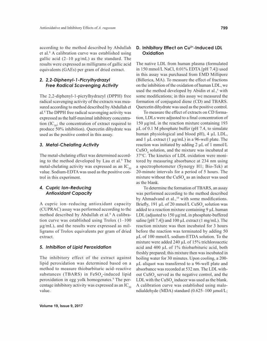
5. Inhibition of Lipid Peroxidation
The inhibitory effect of the extract against lipid peroxidation was determined based on a method to measure thiobarbituric acid–reactive substances (TBARS) in FeSO4-induced lipid peroxidation in egg yolk homogenates.9 The per-centage inhibitory activity was expressed as an IC50 value.
D. Inhibitory Effect on Cu2+-Induced LDL Oxidation
The native LDL from human plasma (formulated in 150 mmol/L NaCl, 0.01% EDTA [pH 7.4]) used in this assay was purchased from EMD Millipore (Billerica, MA). To measure the effect of fractions on the inhibition of the oxidation of human LDL, we used the method developed by Abidin et al.,4 with some modifications; in this assay we measured the formation of conjugated diene (CD) and TBARS. Quercetin dihydrate was used as the positive control.
To measure the effect of extracts on CD forma-tion, LDLs were adjusted to a final concentration of 150 µg/mL in the reaction mixture containing 193 µL of 0.1 M phosphate buffer (pH 7.4, to simulate human physiological and blood pH), 4 µL LDL, and 1 µL extract (1 µg/mL) in a 96-well plate. The reaction was initiated by adding 2 µL of 1 mmol/L CuSO4 solution, and the mixture was incubated at 37°C. The kinetics of LDL oxidation were moni-tored by measuring absorbance at 234 nm using a spectrophotometer (Synergy H1; Bio-Tek) at 20-minute intervals for a period of 5 hours. The mixture without the CuSO4 as an inducer was used as the blank.
To determine the formation of TBARS, an assay was performed according to the method described by Ahmadvand et al.,10 with some modifications. Briefly, 191 µL of 20 mmol/L CuSO4 solution was added to a reaction mixture containing 9 µL human LDL (adjusted to 150 µg/mL in phosphate-buffered saline [pH 7.4]) and 100 µL extract (1 mg/mL). The reaction mixture was then incubated for 3 hours before the reaction was terminated by adding 50 µL of 100 mmol/L sodium-ETDA solution. To the mixture were added 240 µL of 15% trichloroacetic acid and 400 µL of 1% thiobarbituric acid, both freshly prepared; this mixture then was incubated in boiling water for 30 minutes. Upon cooling, a 200-µL aliquot was transferred to a 96-well plate and absorbance was recorded at 532 nm. The LDL with-out CuSO4 served as the negative control, and the LDL with the CuSO4 inducer was used as the blank. A calibration curve was established using malo-ndialdehyde (MDA) standard (0.625–100 μmol/L;

International Journal of Medicinal Mushrooms
Seng, Abdullah, & Aminudin800
Merck, Darmstadt, Germany), assayed under similar conditions, and the result was expressed as nano-moles of MDA equivalents per milligram of LDL protein.
E. Inhibitory Effect on HMG-CoA Reductase Activity
The HMG-CoA reductase assay kit from Sigma-Aldrich was used with the catalytic domain of human enzyme under conditions recommended by the manufacturer. The reference drug pravastatin was used as the positive control. Before the assay, the mushroom extract was hydrolyzed according to the method reported by Yang.11 Briefly, 25 μL of 0.1 N NaOH solution was added to the mush-room extract (10 mg/mL) and incubated at 50°C for 2 hours, followed by neutralization with 25 μL of 0.1 N HCl. To characterize HMG-CoA reduc-tase inhibition, a reaction between 4 µL NADPH and 12 µL HMG-CoA substrate (to obtain a final concentration of 400 µmol/L) in a final volume of 0.2 mL of 100 mmol/L potassium phosphate buffer (pH 7.4) was initiated (time 0) by adding the cata-lytic domain of human HMG-CoA reductase (2 µL). This was incubated in a 96-well plate at 37°C in the presence and absence (control) of 1-µL aliquots of the extracts. The rate of NADPH consumed was monitored every 20 seconds for up to 20 minutes by scanning spectrophotometrically the decrease in absorbance at 340 nm. Results were expressed as the specific activity of the enzyme (micromoles of NADPH oxidized per minute per milligram of HMG-CoA reductase protein).
F. Chromatographic and Mass Spectrometric Analysis
The bioactive fraction was chemically analyzed according to the method described by Lau et al.9 This method uses liquid chromatography–tandem mass spectrometry (LC-MS/MS) (ultrahigh-perfor-mance liquid chromatography; Perkin Elmer, Inc., Waltham, MA) coupled with an AB SCIEX 3200 QTrap hybrid linear ion trap triple-quadruple mass spectrometer equipped with a turbo ion spray source.
Chromatographic separation was performed on a Phenomenex Aqua C18 column (5 µm particle size, 50 mm length × 2 mm internal diameter). Mobile phase A comprised water with 0.1% (v/v) formic acid and 5 mmol/L ammonium formate, whereas mobile phase B consisted of acetonitrile containing 0.1% (v/v) formic acid and 5 mmol/L ammonium formate. Elution was performed by means of a lin-ear gradient from 10−90% mobile phase B (0–8 minutes) held for 3 minutes, returned to 10% phase B in 0.1 minute, and then re-equilibrated for 4 min-utes. Data were analyzed, processed, and interpreted using the AB SCIEX Analyst 1.5 and Advanced Chemistry Development mass spectrometry pro-cessor software (ACD/Labs, Ontario, Canada). MarkerView Software (AB SCIEX, Framingham, MA) was used for principal component analysis. Peaks were identified by comparing our values with published data.
G. Statistical Analysis
All the results were means ± standard deviations of triplicate measurements and were subjected to 1-way analysis of variance. Significance differences were determined with the Duncan test at 95% (P < 0.05) using Statgraphics Plus software version 3.0 for Windows. Correlation and regression were sta-tistically analyzed in Microsoft Excel 2010.
III. RESULTS AND DISCUSSION
A. Antioxidant Capacity of A. rugosum Extracts
Over the past 2 decades, considerable evidence has been gathered in support of the hypothesis that free radical–mediated oxidative processes play key roles in atherosclerosis. Oxidants are constantly generated in vivo, an inevitable feature of aerobic life, leading to oxidative stress. Until recently, the in vitro antiox-idative potential, inhibitory effect of LDL oxidation, and cholesterol lowering via inhibition of HMG-CoA reductase activity using the fruiting bodies of A. rugosum had not been elucidated. Organic solvent extraction followed by liquid-liquid partition served

Volume 19, Issue 9, 2017
Antioxidative and Inhibitory Effects of A. rugosum 801
3 purposes: (1) to separate low–molecular weight compounds based on their chemical nature/structure and polarity, (2) to concentrate bioactive compounds of interest, and (3) to eliminate undesirable or toxic compounds that could mask the potential use of the fraction tested, thereby facilitating further bioassay screenings.
Accordingly, several methods are available to determine antioxidant capacity, such as the estima-tion of total phenolic content, radical scavenging activities, metal-chelating activities, reducing abil-ity, and lipid peroxidation inhibition by mushroom extracts. Our results are summarized in Table 1.
Polyphenols are well known for their antioxi-dant properties; for that reason, the phenolic content of the mushroom extracts was measured using the Folin-Ciocalteu method. In agreement with Chan et al.,7 the EA fraction contained significantly (P < 0.05) larger amounts of total phenols (255.9 ± 0.02 GAEs/g extract) than the other factions: the Hex, DCM, water, and MD extracts contained phenols in
a range of 25.2–30.2 GAEs/g extract. This may be due to the high concentration of semipolar phenolics (e.g., flavonoids and nonflavonoids).
The DPPH assay was used to test free radical scavenging ability. Consistent with its high pheno-lic content, the EA fraction showed the strongest scavenging effects among the 5 extracts tested. The radical scavenging ability of the EA fraction (IC50, 0.34 ± 0.06 mg/mL) was as good as that of the positive control, quercetin dihydrate (IC50, 0.24 ± 0.05 mg/mL). Notably, the EA fraction acts as an excellent radical scavenger, neutralizing the highly energetic radicals and preventing them from further attacking cellular components, which would break chains and cause serious damage.12 According to the “antioxidant hypothesis,” some species that can indirectly induce the generation of reactive radical species are considered oxidants. In this connection, the transition metal ions (Cu2+ and Fe2+) potentially stimulate lipid peroxidation through Fenton reac-tions, producing highly reactive hydroxyl radicals.13
TABLE 1: Antioxidant Capacity of Amauroderma rugosum Extracts
Extracts Total Phenolic Content* (mg
GAEs/g Extract)
DPPH (IC50, mg/mL)
Metal Chelation (IC50 , mg/mL)
CUPRAC†
(mg TEs/g Extract)
Inhibition of Lipid Peroxidation
(IC50, mg/mL)
Hexane 30.2 ± 0.02a 29.6 ± 1.46d 13.5 ± 0.71c 66.1 ± 0.65a 10.5 ± 0.68d
Dichloromethane 25.2 ± 0.01a 7.06 ± 0.72b 2.83 ± 0.75b 270.5 ± 0.43c 3.15 ± 0.11b
Ethyl acetate 255.9 ± 0.02b 0.34 ± 0.06a 1.37 ± 0.15b 1492.9 ± 54.5d 2.83 ± 0.07b
Water 29.8 ± 0.04a 15.0 ± 1.20c 17.2 ± 0.75d 135.2 ± 10.8b 8.08 ± 0.18c
Crude methanol 26.8 ± 0.01a 6.91 ± 1.02b 10.6 ± 0.47c 243.2 ± 5.56c 6.05 ± 0.18c
Positive controls Quercetin
dihydrate 465 ± 0.014c 0.24 ± 0.05a — 1580.9 ± 180.9d 0.29 ± 0.02a
Na2EDTA — — 0.09 ± 0.01a — — α-Tocopherol — — — — 0.87 ± 0.05a
The extracts were dissolved in 50% (v/v) dimethyl sulfoxide in water for the antioxidant assays. Data are means ± standard deviations of triplicate measurements. The different superscript letters (a–e) within a column represent means that are signifi-cantly different (P < 0.05). CURPAC, cupric ion–reducing antioxidant capacity; GAE, gallic acid equivalent; IC50, half-maximal inhibitory concentration; TE, Trolox equivalent.*A 100 µg/mL concentration of extracts was used to estimate total phenolic content, except for the positive control, quercetin dihydrate (10 µg/mL).
†A 1 mg/mL concentration of extracts was used in the CURPAC assay, and Trolox was used as the standard.

International Journal of Medicinal Mushrooms
Seng, Abdullah, & Aminudin802
Both the EA (IC50, 1.37 ± 0.15 mg/mL) and DCM (IC50, 2.83 ± 0.75 mg/mL) fractions exhibited good ferrous ion (Fe2+)–chelating ability compared with the other extracts. Both the EA and DCM fractions showed a low IC50 value in the Fe2+ chelating assay. A low IC50 value is a good indication of metal che-lating ability. Hence, the EA and DCM fractions demonstrated ability as preventative secondary antioxidants, the poisonous metal ion chelators. The CUPRAC method is based on measuring the absorbance of Cu(I)-neocuproine chelate formed as a result of the redox reaction (via electron transfer) of chain-breaking antioxidants with the CUPRAC reagent. The EA fraction showed the most potent reducing power among the 5 extracts—at 1492.9 ± 54.5 mg Trolox equivalents/g extract—showing strength equal to that of the quercetin dihydrate (1580.9 ± 180.9 mg Trolox equivalent/g extract). The EA fraction showed the high value (1492.9 ± 54.5 mg Trolox equivalent/g extract) in the CUPRAC assay. The higher the value of Trolox equivalent/g extract, the better the antioxidant capacity. This value is almost the same as positive control used (quercetin dihydrate). This suggests that EA fraction is as good as quercetin dehydrate used to remove oxidants. Atherosclerosis could be due to the delete-rious actions of oxygen-derived radicals in cellular lipid peroxidation. Damage to cell membrane lipids has been proposed to play a major role in endothelial dysfunction, making them very susceptible to the pathogenesis of atherosclerosis.14 In this study, the inhibitory effect of extracts on lipid peroxidation was assayed using buffered egg yolk as the source of unsaturated fatty acids (oleic, linoleic, palmi-toleic, and linolenic acids) in order to simulate the polyunsaturated fatty acid components of biological membranes, as they share similar lipid peroxidation mechanistic attacks.13 Our results show that the EA and DCM fractions have an inhibitory effect on lipid peroxidation, with IC50 values at 2.83 ± 0.07 and 3.15 ± 0.11 mg/mL, respectively.
A high plasma antioxidant level has been associated with good antiatherosclerotic potential. According to Harris,15 evidence showed that nutri-tional antioxidants play a significant role in the prevention of atherosclerosis in culture experiments
and animal studies. The antioxidants were believed to be able to reduce the susceptibility of LDLs to oxidation. Hence, inhibition of LDL oxidation is a good indication of arrested atherosclerosis. Based on our observations, the EA extract demonstrated the best antioxidant capacity, and thus it is believed to be beneficial in retarding atherosclerotic cardio-vascular disease.
B. Inhibitory Effects of A. rugosum Extracts on LDL Oxidation
The “response to injury” hypothesis of atheroscle-rosis states that endothelial denudation, probably caused by the elevation of ox-LDL in plasma, rep-resents the first step in atherosclerosis.16,17 To explore defense mechanisms against the early stages of ath-erosclerosis, the inhibitory activity of A. rugosum extracts on native human LDLs was studied; the results are presented in Fig. 1. In this context, we used 2 different methods—the formation of CD and of TBARS. In this study, we found that all mushroom extracts showed a gradual increase in absorbance at 234 nm (Fig. 1A). This indicated that LDLs had been oxidized, and the lag phase was detected at 130 minutes for the control (induced by the transi-tion metal, Cu2+). However, the typical lag and log phases were not observed for the mushroom extracts, probably because of delayed LDL-oxidative action or an unknown protective mechanism. Hence, the lag phase was not clearly observed. Regardless, we qual-itatively compared the slope pattern (the change in CD absorbance over time). Absorbance increment as an indication of LDL oxidation showed the strength of the inhibitory effects, which followed the order EA > DCM > MD > water > Hex. To confirm the inhibitory effect, the TBARS method was performed to estimate the MDA product formed. The observed inhibitory pattern was similar to that of CD formation (Fig. 1B). Compared with the control, we found that the EA fraction (64.9%; IC50, 0.77 mg/mL) showed the strongest inhibitory effect against LDL oxida-tion when compared with that of the DCM (51.2%; IC50, 0.92 mg/mL), MD (37.8%), water (30.0%), and Hex (15.6%) fractions. The inhibitory effect of the EA fraction was comparable to that of the positive

Volume 19, Issue 9, 2017
Antioxidative and Inhibitory Effects of A. rugosum 803
control used, quercetin dihydrate (73.0%; IC50, 0.58 mg/mL).
C. Inhibitory Effect on HMG-CoA Reductase Activity
Hypercholesterolemia, or more specifically elevated plasma LDL cholesterol, is an important risk factor for the development and progression of atheroscle-rosis. To investigate whether A. rugosum possesses any cholesterol-lowering property, the quick HMG-CoA reductase assay kit was used.
HMG-CoA reductase is an enzyme that cata-lyzes the 4-electron reduction of HMG-CoA to coenzyme A and mevalonate; this is the rate-limit-ing step in sterol synthesis. Because our mushroom extracts were obtained with an organic solvent and liquid-liquid partition, the compounds present may be primarily preserved in an inactive form, unlike statinlike compounds, which are present in aqueous form (as an active open-ring hydroxy carboxylic acid). Therefore, mushroom extracts needed to be hydrolyzed before this assay to achieve better
measurements. The results were obtained by measur-ing the decrease of NADPH absorbance in the test mixture at 340 nm. The slower change in absorbance indicates a slower rate of NADPH oxidation, thereby suggesting a better inhibitory effect on the enzymatic activity catalyzed by HMG-CoA reductase. In this study the positive control, pravastatin, demonstrated the strongest inhibitory activity. By measuring the initial negative slope of the absorbance change at 340 nm (Fig. 2A), the inhibitory activity of the mush-room extracts can be compared. The results were expressed as micromoles of NADPH oxidized per minute per milligram of protein enzyme (Fig. 2B) using the slope, which was calculated based on a mathematical formula according to the manufac-turer’s instructions. In this regard, consistent with previous antioxidant and LDL oxidation tests, the EA fraction again showed significantly higher and the most potent inhibitory effect on HMG-CoA reductase activity (50.8%), followed by the DCM (45.2%), MD (36.7%), water (36.2%), and Hex (17.5%) fractions, relative to the control. Hence, we can hypothesize that the EA fraction contains the
FIG. 1: Inhibitory effect on low-density lipoprotein (LDL) oxidation by mushroom extracts, evaluated by monitor-ing conjugated diene (CD) and thiobarbituric acid reactive substances (TBARS) formation. Results are expressed as means ± standard deviations of triplicate measurements in 3 independent experiments. (A) In the CD formation assay, a 1 µg/mL concentration of mushroom extracts was used; quercetin dihydrate was used as the positive con-trol. LDL preserved in phosphate-buffered saline (PBS; pH 7.4) without a Cu2+ inducer was used as the blank; an LDL and CuSO4 solution was used as the control. (B) All extracts including quercetin dihydrate (1 mg/mL) were tested in the TBARS assay. LDL and the Cu2+ inducer served as the control, and the test mixture containing only LDL was used as a blank. The different letters (a–f) within a column represent significantly different means (P < 0.05). DCM, dichloromethane; EA, ethyl acetate; Hex, hexane; MD, crude methanol; MDA, malondialdehyde.

International Journal of Medicinal Mushrooms
Seng, Abdullah, & Aminudin804
bioactive compounds to make it possible to interfere with the cholesterol biosynthetic pathway.
D. Chemical Constituents in the EA Fraction per LC-MS/MS Analysis
Based on in vitro antioxidant, LDL oxidation, and HMG-CoA reductase activity assays, the EA fraction demonstrated a potential antiatherosclerotic effect. This fraction was then analyzed using LC-MS/MS to correlate its chemical constituents with its potential/possible functions. The LC-MS/MS chromatogram is shown in Fig. 3, and the interpreted results are
presented in Table 2. Compounds were identified by comparing their forms with published data in the literature. Ten phenolic compounds were identi-fied, including 4 benzoic acid derivatives (vanillic acid, p-hydroxybenzoic acid, protocatechuic acid, and methyl-3,4-dihydroxybenzoate), 3 flavonoids (luteolin, apigenin, and naringenin), cinnamic acid (caffeic acid), dilactone of hexahydroxydiphenic acid (3,3′-di-O-methyl ellagic acid), and a xanthone derivative (jacareubin). The chemical structures are shown in Fig. 4.
To correlate our chemical findings with athero-sclerotic cardiovascular disease, we found that the
FIG. 2: Inhibitory effects on 3-hydroxy-3-methylglutaryl–coenzyme A reductase activity by the hydrolyzed mush-room extracts (5 mg/mL). Graphs show the change in absorbance at 340 nm over time (A) and NADPH oxidation (B). Data in (B) are the means ± standard deviations of triplicate measurements in 3 independent experiments, and the different letters (a–f) represent significantly different means (P < 0.05). Pravastatin from a kit (Sigma) was used as the positive control. DCM, dichloromethane; EA, ethyl acetate; Hex, hexane; MD, crude methanol; MDA, malondialdehyde.
FIG. 3: The chemical constituents present in the ethyl acetate fraction of Amauroderma rugosum fruiting bodies

Volume 19, Issue 9, 2017
Antioxidative and Inhibitory Effects of A. rugosum 805
TABLE 2: Chemical Constituents in the Ethyl Acetate Fraction based on Liquid Chromatography–Mass Spectrometry Analysis
RT(min)
MolecularMass
MolecularFormula
[M-H]− Mass Fragments, MS/MS Suggested Identification
Refs.
4.071 168.0 C8H8O4 167(100), 149 (10), 137(25), 123(80), 109(30), 93(20), 65(30)
Vanillic acid 18, 19
4.468 154.0 C7H6O4 153(32), 109(100), 108(43), 91(10) Protocatechuic acid 18, 205.790 285.1 C15H10O6 284(25), 283(100), 257(10), 163(57) Luteolin 186.056 138.0 C7H6O3 137(100), 136(38), 108(18), 92(10) p-Hydroxybenzoic acid 18, 216.454 180.0 C9H8O4 179(15), 135(100), 137(27), 89(10) Caffeic acid 18–208.050 168.0 C8H8O4 167(100), 135(21), 124(34), 123(18),
111(21), 91(25)Methyl-3,4-dihydroxy-
benzoate22
9.253 271.0 C15H10O5 269(100), 197(38), 167(71), 141(61), 113(15)
Apigenin 20, 22
10.06 272.1 C15H12O5 271(100), 255(63), 239(18), 145(18), 109(46)
Naringenin 20
10.32 331.2 C16H10O8 330(75), 329(100), 229(18), 211(29), 171(79), 139(18)
3,3′-Di-O-methyl ellagic acid
23
11.79 327.2 C18H14O6 236(16), 325(21), 183(100) Jacareubin 24 MS/MS, tandem mass spectrometry; RT, reaction time.
FIG. 4: The structures of phenolic compounds present in the ethyl acetate fraction of Amauroderma rugosum fruit-ing bodies

International Journal of Medicinal Mushrooms
Seng, Abdullah, & Aminudin806
hydroxyphenolic acids (protocatechuic acid, caf-feic acid, vanillic acid, p-hydroxybenzoic acid, and methyl-3,4-dihydroxybenzoate) present in the EA fraction were potent antioxidants, radical scaven-gers, and inhibitors of lipid peroxidation in cellular components. They also have shown promising effects in decreasing serum total cholesterol, very-LDL cholesterol, and LDL cholesterol, and may serve as good hypolipidemic agents.25
IV. CONCLUSIONS
Based on our in vitro studies, the A. rugosum fruiting body is a potential source of in vitro antioxidative and antiatherosclerotic agents. Bioactive compounds are present in the EA fraction, enabling it to exhibit inhibitory effects on human LDL oxidation and HMG-CoA reductase activity, together with a high antioxidant capacity. However, to prove the effec-tiveness of consumption of A. rugosum fruiting bodies in humans, antioxidant and cholesterol-low-ering assays using cellular and in vivo models are necessary. Furthermore, isolation of bioactive com-pounds from the EA extract is encouraged to develop future nutraceutical and pharmaceutical applications.
ACKNOWLEDGMENTS
This study was supported by a high-impact research grant from the Ministry of Higher Education Malaysia (UM-MOHE UM.C/625/1/HIR/MOHE/F00002-21001) and a grant from the University of Malaya (UMRG-RP014D-13AFR). The authors thank the University of Malaya for a Postgraduate Research Grant (PPP Grant No. PG142-2012B). CKS is grateful for a MyPhD Scholarship supported by the Ministry of Higher Education Malaysia. The authors give special thanks to Yap Ken Choy from Advanced Chemistry Solutions for running ultrahigh-performance liquid chromatography, electrospray ionization mass spectrometry, and thin-layer chromatography experiments.
REFERENCES
1. Reinhardt E. Health Watch: the atlas of health disease and stroke. New York (NY): UN Chronicle; 2005.
2. Lowenstein C, Matsushita K. The acute phase response and atherosclerosis. Drug Discov Today Dis Mech. 2004;1: 17–22.
3. Lusis AJ. Atherosclerosis. Nature. 2000;407:233–41.4. Abidin MHZ, Abdullah N, Abidin NZ. Protective effect
of antioxidant extracts from grey oyster mushroom, Pleurotus pulmonarius (Agaricomycetes), against human low-density lipoprotein oxidation and aortic endothelial cell damage. Int J Med Mushrooms. 2016;18(2):109–21.
5. Chang ST, Wasser SP. The role of culinary-medicinal mushrooms on human welfare with a pyramid model for human health. Int J Med Mushrooms. 2012;14(2):95–134.
6. Wasser SP. Medicinal mushroom science: history, current status, future trends, and unsolved problems. Int J Med Mushrooms. 2010;12(1):1–16.
7. Chan PM, Kanagasabapathy G, Tan YS, Sabarathan V, Kuppusamy UR. Amauroderma rugosum (Blume & T. Nees) Torrend: nutritional composition and antioxidant and potential anti-inflammatory properties. Evid Based Complement Alternat Med. 2013;2013:304713.
8. Abdullah N, Ismail SM, Aminudin N, Shuib AS, Lau BF. Evaluation of selected culinary-medicinal mushrooms for antioxidant and ACE inhibitory activities. Evid Based Complement Alternat Med. 2012;2012:464238.
9. Lau BF, Abdullah N, Aminudin N, Lee HB, Yap KC, Sabarathnam V. The potential of mycelium and culture broth of Lignosus rhinocerotis as substitutes for the natu-rally occurring sclerotium with regard to antioxidant capacity, cytotoxicity effect, and low-molecular-weight chemical constituents. PLoS One. 2014;9(7):e102509.
10. Ahmadvand H, Ani M, Moshtaghie AA. Inhibitory effect of Allium Cepa extract on LDL oxidation induced by CuSO4 in vitro compared with Allium sativum and Allium ascalonicom. Iranian J Pharmacol Ther. 2011;10: 61–5.
11. Yang SZ. The divine farmer’s materia medica: a translation of the Shen Nong Ben Cao Jing. 4th ed. Boulder (CO): Blue Poppy Press; 2005.
12. Panda SK. Assay guided comparison for enzymatic and non-enzymatic antioxidant activities with special reference to medicinal plants. In: Antioxidant enzyme. El-Missiry MA, editor. London (UK): InTechOpen; 2012. pp. 382–400.
13. Maxwell SRJ, Lip GYH. Free radicals and antioxi-dants in cardiovascular disease. Br J Clin Pharmacol. 1997;44:307–17.
14. Rikans LE, Hornbrook KR. Lipid peroxidation, anti-oxidant protection and aging. Biochim Biophys Acta. 1997;1362:116–27.
15. Harris WS. The prevention of atherosclerosis with anti-oxidants. Clin Cardiol. 1992;15(9):636–40.
16. Ross R. Atherosclerosis. An inflammatory disease. N Engl J Med. 1999;340:115–26.
17. Steinberg D, Witzum JL. Is the oxidative modification hypothesis relevant to human atherosclerosis? Do the antioxidant trials conducted to date refute the hypothesis? Circulation. 2002;105:2107–11.

Volume 19, Issue 9, 2017
Antioxidative and Inhibitory Effects of A. rugosum 807
18. Hossain M, Rai DK, Brunton NP, Martin-Diana AB, Barry-Ryan C. Characterization of phenolic composition in lamiaceae species by LC-ESI-MS/MS. J Agric Food Chem. 2010;58(19):10576–81.
19. Theerasin S, Baker AT. Analysis and identification of phe-nolic compounds in Diocorea hispida Dennst. Asian J Food Agro Ind. 2009;2(4):547–60.
20. Rabaneda FS, Jauregui O, Casals I, Laceuva CA, Pulido MI, Raventos RML. Liquid chromatographic/electrospray ionisation tandem mass spectrometric study of the phe-nolic composition of cocoa (Theobroma cacao). J Mass Spectrom. 2003;38:35–42.
21. Chen HJ, Inbaraj BS, Chen BH. Determination of phenolic acids and flavonoids in Taraxacum formosanum Kitam by liquid chromatography tandem mass spectrometry coupled with a post-column derivatisations technique. Int J Mol Sci. 2012;13:260–85.
22. Cui XB, Qian XC, Huang P, Zhang YX, Li JS, Yang GM. Simultaneous determination of ten flavonoids of crude and wine-processed Radix scutellariae aqueous extracts in rat plasma by UPLC-ESI-MS/MS and its application to a comparative pharmacokinetics study. Biomed Chromatogr. 2015;29(7):1121–3.
23. Braunberger C, Zehl M, Conrad J, Fisher S, Adhami HR, Beifuss U, Krenn L. LC-NMR, NMR, and LC-MS iden-tification and LC-DAD quantification of flavonoids and ellagic acid derivatives in Drosera peltata. J Chromatogr B Analyt Technol Biomed Life ci. 2013;932:111–6.
24. Wei DJ, Mei WL, Zhong HM, Zeng YB, Wu XD, Dai HF. A new prenylated xanthone from the branches of Calophyllum inophyllum. J Asian Nat Prod Res. 2011;13(3):265–9.
25. Kakkar S, Bais S. A review on protocatechuic acid and its pharmacological potential. ISRN Pharmacol. 2014:1–9.