Embed Size (px)
Citation preview
Antiretroviral drugs induce oxidative stress and neuronaldamage in the central nervous system
Cagla Akay & Michael Cooper & Akinleye Odeleye & Brigid K. Jensen & Michael G. White &
Fair Vassoler & Patrick J. Gannon & Joseph Mankowski & Jamie L. Dorsey & Alison M. Buch &
Stephanie A. Cross & Denise R. Cook & Michelle-Marie Peña & Emily S. Andersen &
Melpo Christofidou-Solomidou & Kathryn A. Lindl & M. Christine Zink & Janice Clements &R. Christopher Pierce & Dennis L. Kolson & Kelly L. Jordan-Sciutto
Received: 24 October 2013 /Revised: 10 December 2013 /Accepted: 13 December 2013 /Published online: 14 January 2014# The Author(s) 2014. This article is published with open access at Springerlink.com
Abstract HIV-associated neurocognitive disorder (HAND),characterized by a wide spectrum of behavioral, cognitive, andmotor dysfunctions, continues to affect approximately 50% ofHIV(+) patients despite the success of combination antiretro-viral drug therapy (cART) in the periphery. Of note, potentialtoxicity of antiretroviral drugs in the central nervous system(CNS) remains remarkably underexplored and may contributeto the persistence of HAND in the cART era. Previous studieshave shown antiretrovirals (ARVs) to be neurotoxic in theperipheral nervous system in vivo and in peripheral neuronsin vitro. Alterations in lipid and protein metabolism, mito-chondrial damage, and oxidative stress all play a role inperipheral ARV neurotoxicity. We hypothesized that ARVsalso induce cellular stresses in the CNS, ultimately leading to
neuronal damage and contributing to the changing clinical andpathological picture seen in HIV-positive patients in the cARTera. In this report, we show that ARVs are neurotoxic in theCNS in both pigtail macaques and rats in vivo. Furthermore,in vitro, ARVs lead to accumulation of reactive oxygen spe-cies (ROS), and ultimately induction of neuronal damage anddeath. Whereas ARVs alone caused some activation of theendogenous antioxidant response in vitro, augmentation ofthis response by a fumaric acid ester, monomethyl fumarate(MMF), blocked ARV-induced ROS generation, and neuronaldamage/death. These findings implicate oxidative stress as acontributor to the underlying mechanisms of ARV-inducedneurotoxicity and will provide an access point for adjunctivetherapies to complement ARV therapy and reduce neurotox-icity in this patient population.
Keywords Antiretroviral . Fumaric acid ester . HIV .
HIV-associated neurocognitive disorder . Macaque .
Oxidative Stress . Reactive oxygen species . SIV
Introduction
Despite introduction of combination antiretroviral therapy(cART), HIV-associated neurocognitive disorder (HAND)continues to affect approximately 50 % of HIV(+) patients(Dore et al. 1999; Heaton et al. 2010). Furthermore, in thecART era, the underlying neuropathology has shifted fromovert subcortical involvement to a more insidious corticaldamage (Gannon et al. 2011). Various factors, such as pooradherence to drug regimen, emergence of resistant virus spe-cies, and residual viral DNA in the central nervous system(CNS), may contribute to these changes (Gannon et al. 2011).However, another likely contributor to HAND in the cARTera
C. Akay :M. Cooper :A. Odeleye : B. K. Jensen :M. G. White :P. J. Gannon :A. M. Buch :M.<M. Peña :K. A. Lindl :K. L. Jordan-Sciutto (*)Department of Pathology, School of Dental Medicine, University ofPennsylvania, 240 S. 40th St, Rm 312 Levy Bldg, Philadelphia,PA 19104-6030, USAe-mail: [email protected]
F. Vassoler :R. C. PierceDepartment of Psychiatry, The Perelman School of Medicine,University of Pennsylvania, Philadelphia, PA, USA
J. Mankowski : J. L. Dorsey :M. C. Zink : J. ClementsDepartment of Molecular and Comparative Pathobiology, JohnsHopkins University School of Medicine, Baltimore, MD, USA
S. A. Cross :D. R. Cook :D. L. KolsonDepartment of Neurology, The Perelman School of Medicine,University of Pennsylvania, Philadelphia, PA, USA
E. S. Andersen :M. Christofidou-SolomidouDepartment of Medicine, The Perelman School of Medicine,University of Pennsylvania, Philadelphia, PA, USA
J. Neurovirol. (2014) 20:39–53DOI 10.1007/s13365-013-0227-1
is the virtually unstudied potential for antiretroviral (ARV)-related toxicity in the CNS. cART has decreased HIV-relatedmorbidity and mortality by limiting T cell loss and controllingopportunistic infections. However, cART regimens are asso-ciated with potentially serious side effects, including dyslip-idemia, lipohypertrophy, and increased risk of atherosclerosis(Vidal et al. 2010). Additionally, cART-associated toxicity inthe peripheral nervous system is well documented, and it islikely that cART would trigger similar responses in the CNS.Pharmacokinetic studies suggest limited ARV penetrance intothe CNS and indicate low cerebrospinal fluid (CSF) andparenchymal drug concentrations (Yilmaz et al. 2004;Yilmaz et al. 2009). However, direct blood–brain barrier(BBB) compromise by viral proteins and neuroinflammationand indirect BBB impairment due to concomitant factors, suchas coexisting infections, can lead to increased CSF and paren-chymal drug concentrations. Thus, the impact of ARVs in theCNS of HIV(+) patients is clinically relevant and must beexamined.
Initial cART usually includes two nucleoside/nucleotidereverse-transcriptase inhibitors (NRTIs) in combination with anon-nucleoside reverse-transcriptase inhibitor (nNRTI) or with aprotease inhibitor (PI) boosted with a low dose of a second PI,Ritonavir. NRTIs and nNRTIs bind to the HIV reverse-transcriptase enzyme and inhibit proviral DNA synthesis; PIsinhibit viral proteases needed for virus maturation and assembly.Despite some crossover, certain ARV classes are more highlyassociated with particular side effects and toxicities than areother classes. PIs alter lipid metabolism and induce the endo-plasmic reticulum stress response in macrophages, linking PIs toincreased risk of atherosclerosis (Touzet and Philips 2010).NRTIs inhibit DNA polymerase γ and lead to decreased mito-chondrial DNA, loss of mitochondrial membrane potential, andoxidative phosphorylation, consequently precipitating oxidativestress (Nolan and Mallal 2004). Previous studies exploringpossible side effects of ARVs in the CNS are scarce and mostlyinvolve cell lines (Cui et al. 1997). Given the mutations andaberrations in immortalized cell lines, these studies may notreflect the ARV toxicity potentially occurring in biological set-tings. Peripheral dorsal root ganglia neurons are the only primarycell type of neural lineage previously studied for ARV toxicity(Werth et al. 1994). In this report, we examined effects of ARVsin primary CNS neurons both in vivo and in vitro. Our findingssuggest that cART induces oxidative stress and neurotoxicity inthe CNS, and that the patients on long-term cART regimenswould benefit from adjuvant therapies that include antioxidantstrategies to overcome deleterious effects of cART in the CNS.
Materials and methods
Chemicals and reagents Chemicals and reagents comprise thefollowing: (1) AIDS Research and Reference Reagent
Program, Division of AIDS, NIAID, NIH, antiretroviral re-agents; (2) Abcam (Cambridge, MA), mouse monoclonalNAD(P)H/quinone oxidoreductase-1 (NQO-1) antibody(A180) and mouse monoclonal synaptophysin antibody(SY38); (3) BioRad (Hercules, CA), Biosafe Coomassie stain,immunoblot polyvinylidene fluoride (PVDF) membrane andprestained broad range molecular weight ladder; (4) CellSignaling Technology (Danvers, MA), rabbit polyclonal anti-body raised against cleaved caspase-3; (5) Citifluor, Ltd.(London, UK), citifluor AF1. Covance (Princeton, NJ), mousemonoclonal microtubule-associated protein 2 (MAP2) anti-body (SMI-52); (6) Dako (Carpinteria, CA), rabbit polyclonalglial fibrillary acidic protein (GFAP) antibody (Z0334); (7)Enzo Life Sciences (Farmingdale, NY), rabbit polyclonal antibodyto heme-oxygenase-1 (HO-1); (8) Frontier Scientific (Logan, UT),Sn(IV) mesophorphyrin IX dichloride (SnMP); (9) JacksonImmunoResearch Labs (West Grove, PA), fluoresceinisothiocyanate-conjugated goat anti-mouse IgG and Cy3-conjugated goat anti-rabbit IgG secondary antibodies; (10)Invitrogen (Carlsbad, CA), Dulbecco's modified Eagle's medium(DMEM), tetramethyl rhodamine methyl ester, goat anti-mousebeta-lactamase TEM-1 conjugate, fluorocillin green substrate,dihydroethidium (DHE), 4′,6-diamidino-2-phenylindole (DAPI),neurobasal media, and B27 supplement; (11) New EnglandBiolabs (Ipswich, MA), tyramide amplification system; (12)Peptide International (Louisville, KY), poly-L-lysine; (13) SigmaAldrich (St. Louis, MO), carbonyl cyanide m-chlorophenylhydrazone, cytosine arabinoside (Ara-C), fetal bovine serum(FBS), monomethylfumarate (MMF), oligomycin, propidium io-dide, and PI cocktail; (14) ScyTek Labs (Logan, UT), normalantibody diluent; (15) Thermo Scientific (Waltham, MA), goatanti-rabbit horse radish peroxidase (HRP) antibody and goat anti-mouse HRP antibody, SuperSignal West Dura extended durationsubstrate; and (16) Tocris Bioscience (Ellisville, MO), thapsigargin.The antibody against calpain-cleaved spectrin was a generous giftfrom Dr. Robert Siman (The Perelman School of Medicine,University of Pennsylvania, Philadelphia, PA).
Primary cortical neuroglial cultures Primary rat cortical neu-roglial, pure neuronal, and pure astrocytic cultures were iso-lated from embryonic day 17 Sprague Dawley rat pups, withmodifications of protocols previously described (Wilcox et al.1994). Briefly, the cortical cell suspensions isolated from ratpups following standard protocols were plated on poly-L-lysine-coated T25 tissue culture flasks (2×10⁶ cells/flask),96-well tissue culture plates (0.5×10⁵ cells/well), or glasscoverslips (0.25×10⁶ cells/well). These cultures were main-tained in neurobasal media with B27 supplement at 37 °Cwith5%CO2 for the generation of neuroglial cultures, as describedpreviously (Wang et al. 2010; Akay et al. 2011b; White et al.2011). Half of the media was replaced with fresh media every7 days and the experiments were performed at 21 days in vitro(DIV), at which time the cultures contain approximately 85–
40 J. Neurovirol. (2014) 20:39–53
90 % neurons and 10–15 % astrocytes/glia. For the prepara-tion of pure neuronal cultures, cultures were treated with10 μMAra-C 48 h after plating and maintained in neurobasalmedia with B27 supplement at 37 °C with 5 % CO2; at 21DIV, the age at which the experiments were conducted, noastrocytes are detectable by staining for GFAP. Pure astrocyticcultures were prepared by initially plating the cortical cellsuspensions in 10 % FBS in DMEM and maintaining at37 °Cwith 5%CO2 for 7–12 days, after which time astrocytesconstitute more than 90 % of the cultures. These pure astro-cytic cultures are then replated in T25 flask or poly-L-lysine-coated glass coverslips in DMEM/10 % FBS and the experi-ments are conducted 3–5 days after replating.
SIV/pigtail macaque model of HIV CNS disease Juvenile pig-tailed macaques (Macaca nemestrina) were inoculated withSIV/DeltaB670 and SIV/17E-Fr, as described previously(Zink et al. 1999). Beginning 12 days after inoculation, ani-mals were treated daily with a four ARV drug combination(cART) until necropsy (range, days 161–175). The treatmentconsisted of the NRTI tenofovir (Gilead) at 30 mg/kg subcu-taneously every day; the PIs saquinavir (Roche) andatazanavir (Bristol- Myers Squibb) at 205 and 270 mg/kgorally twice a day, respectively; and the integrase inhibitorL-870812 (Merck)(Hazuda et al. 2004) at a dose of 10 mg/kgorally twice a day(Zink et al. 2010). The tenofovir dose wasdetermined on the basis of previous studies(Tsai et al. 1997),whereas atazanavir and saquinavir doses were determined bypharmacokinetic experiments conducted in pig-tailed ma-caques, and reflected those that resulted in the same area underthe curve as detected in humans treated with atazanavir andsaquinavir (Zink et al. 2010). The dose of the integrase inhib-itor was based on a previous study conducted in rhesusmacaques(Hazuda et al. 2004).
Rodent model of antiretroviral-induced neurotoxicity All sur-gical procedures were performed with the approval of theInstitutional Animal Care and Use Committee. Adult maleSprague Dawley rats were catheterized via jugular vein, asdescribed previously (Thrivikraman et al. 2002). The drugcocktail composed of AZT (100 mg kg−1 day−1), ritonavir(20 mg kg−1 day−1), and saquinavir (25 mg kg−1 day−1) wasadministered twice daily for 7 days by continuous intravenousinjection. Each drug dose was based on previously publishedstudies in rodents (Shibata et al. 2002; Huisman et al. 2003;Manda et al. 2010; Pistell et al. 2010;Waring et al. 2010; Yanget al. 2010; du Plooy et al. 2011; Fontes et al. 2011; Lledo-Garcia et al. 2011; Wagner et al. 2011; Mak et al. 2013;Reyskens and Essop 2013a, b; Reyskens et al. 2013). Thecatheters were flushed with 0.3 ml heparin (50 IU/ml) in PBSuntil the end of treatments. At time of euthanasia, precedingdecapitation and tissue harvest, catheter patency wasreverified by response to pre-euthanasia sedation (100 mg/kg
ketamine per 10 mg/kg xylazine). The brains were removed,the frontal cortex and the hippocampus were dissected on ice,and the tissue samples were stored at -80 °C until immunoblotanalysis.
Immunofluorescence staining of tissue Tissue slides ofparaffin-embedded tissue sections from hippocampus of maleand female pig-tailed macaques (M. nemestrina) were pre-pared for immunofluorescent staining, as described previously(Akay et al. 2011a) . Briefly, glass slides containing paraffin-embedded sections (10 μm) were heated to 55 °C for at least30 min, deparaffinized in Histoclear, and rehydrated withconsecutive 100, 95, 90, and 70 % ethanol washes.Endogenous peroxidase activity was blocked with 3 % H2O2
in methanol and antigen unmasking was achieved with targetretrieval solution at 95 °C for 1 h. Tissue sections wereblocked with 10 % normal goat serum in phosphate-bufferedsaline solution (PBS). Mouse monoclonal antibodies tosynaptophysin, MAP2, and rabbit polyclonal antibody toGFAP were used at empirically defined dilutions(synaptophysin at 1:500; MAP2 at 1:100; and GFAP at1:80), and DAPI was used to stain nuclei. The tyramideamplification system was used to detect synaptophysin.Slides were mounted in Citifluor AF1, and for each specimen,five to ten randomly selected areas within the CA1–CA3 layerof the hippocampus were scanned along the z-axis to create z-stack images at high magnification (×600) by laser confocalmicroscopy on a Biorad Radiance 2100 equipped with Argon,Green He/Ne, Red Diode, and Blue Diode lasers (Biorad,Hercules, CA). Post-acquisition analysis was conducted usingMetaMorph 6.0 (Universal Imaging, Downingtown, PA).Total intensity for synaptophysin, MAP2 and GFAP weredetermined by the measurement of integrated pixel intensityfor synaptophysin, MAP2 or GFAP per z-stack image, wherethe integrated pixel intensity is defined as total pixel intensityper image times the area of pixels positive for synaptophysin,MAP2 or GFAP. Averages are expressed as mean±SEM. Alldata was analyzed by Prism 5.0 software (GraphPad Software,San Diego, CA).
MAP2 cell-based ELISA A MAP2 cell-based ELISA wasperformed to quantify neuronal damage/death as previouslydescribed (White et al. 2011). Briefly, primary rat corticalneuroglial cultures plated in 96-well plates were fixed for30 min with 4 % paraformaldehyde in 4 % sucrose at theconclusion of treatments. After blocking for 1 h with 5 %normal goat serum in PBS, the plates were incubated withmonoclonal MAP2 antibody overnight at 4 °C, followed bywashes with PBS with 0.1 % Tween-20 (PBS-T). The plateswere then incubated for 30 min with goat anti-mouse second-ary antibody conjugated to beta-lactamase TEM-1 at roomtemperature (RT), washed with PBS-T, and incubated in thedark at RT for 1 h in fluorocillin green substrate. Fluorescence
J. Neurovirol. (2014) 20:39–53 41
intensity was measured using a Fluoroskan Ascent fluorome-ter plate reader (Thermo Electron, Waltham, MA) with exci-tation at 485 nm and emission at 527 nm.
Hand counting of MAP2-positive cells As a complementarymethod to MAP2 ELISA, neuronal survival was verified byhand counting of MAP2-positive neurons; 15 μM propidiumiodide was added to primary cortical neuroglial culturesgrown on coverslips 15 min before the end of the treatments.The coverslips were washed once with PBS and fixed for30 min with 4 % paraformaldehyde in 4 % sucrose, followedby blocking and permeabilization in 0.2 %BSA+0.1% TritonX-100 in PBS for 1 h at RT. The coverslips were then washedtwice with PBS and incubated with anti-MAP2 antibody(1:100) in normal antibody diluent for 2 h at RT. After twowashes with PBS-T, the coverslips were incubated in a fluo-rescein isothiocyanate-conjugated goat anti-mouse IgG sec-ondary antibody (1:200) for 30min at RT. The coverslips weremounted on slides and alive/dead neurons were hand-countedbased on MAP2 and PI-positive staining using a NikonEclipse E400 fluorescent microscope (Nikon Corp, Tokyo,Japan) equipped with Olympus DP70 digital camera(Olympus Corp, Tokyo, Japan). Live cells stain negative forPI, while dead cells retain it in their nuclei. The number ofsurviving MAP2-positive neurons will be positive for MAP2and negative for PI staining. The percentage of MAP2-positive cells ± SEM were calculated from blinded countingof six fields at ×200 in two adjacent vertical columns throughthe center of each coverslip, proceeding from top to bottomand bottom to top. For each condition, three coverslips werecounted from two or more independent experiments.
Quantification of synaptophysin Cultures grown on cover-slips, as described above, were immunofluorescently stainedfor synaptophysin (1:1,000), MAP2 (1:100), and DAPI.Tyramide amplification was used for the detection ofsynaptophysin. For each treatment condition, images of 10randomly selected areas from three coverslips from threeindependent experiments were captured by fluorescence mi-croscopy. Post-acquisition analysis was performed using NIHImageJ program (V 1.36b, Bethesda, MD). Briefly,synaptophysin-positive puncta were detected by backgroundsubtraction and manual thresholding. The number ofsynaptophysin-positive puncta was determined by dividingthe total number of puncta within a dendrite segment by thelength of the segment.
Measurement of reactive oxygen species The superoxide in-dicator dihydroethidium (DHE, Invitrogen) was used to detectthe presence of reactive oxygen species (ROS) in vitro. 3 μMDHEwas added to culturemedia 15min prior to conclusion oftreatments. Cells were then washed with PBS, fixed with 4 %paraformaldehyde in 4 % sucrose at RT for 8 min, and stained
for DAPI. The coverslips were then mounted on slides withCytoSeal and visualized using fluorescent microscopy, asdescribed above. Post-acquisition analysis was performedusing MetaMorph to determine the fluorescence intensity ofDHE normalized to the area of DAPI signal.
Immunoblotting Whole cell extracts of rat tissue sampleswere prepared by homogenization in ice-cold tissue extractionbuffer (50 mM Tris at pH 7.5, 0.5 M NaCl, 1 % NP-40, 1 %SDS, 2 mM EDTA, 2 mM EGTA, 5 mM NaF, 0.4 mMNa3VO4, 1 mM dithiothreitol (DTT), and 1:100 PI cocktail),followed by centrifugation at 12,000×g at 4 °C for 20 min.Whole cell extracts of primary rat cortical cultures were pre-pared with ice-cold cell lysis buffer (50 mM Tris at pH 7.5,120 mM NaCl, 0.5 % NP-40, 10 mM EDTA, 0.4 mMNa3VO4, 100 mM DTT, and 1:100 PI cocktail), followed bycentrifugation at 14,000×g at 4 °C for 10 min. The proteinconcentrations of the collected supernatants were determinedusing the Bradford method and 25–50 μg of protein wasloaded into each lane of a 4–12 % Bis-Tris gradient gel forseparation. A broad range molecular weight ladder was run oneach gel. Subsequent to separation, proteins were transferredonto PVDF membranes, and blocked in Tris-buffered salinewith 0.1 % Tween-20 (TBS-T) and 5% bovine serum albumin(BSA) for 1 h at RT. The membranes were incubated with theprimary antibodies in TBS-Twith 5 % BSA at 4 °C overnight,washed with TBS-T, followed by incubation with correspond-ing HRP-conjugated secondary antibodies. The membraneswere developed using SuperSignal West Dura extended dura-tion substrate. Loading controls were obtained by staining themembranes and the gels with the Biosafe Coomassie Stain for20 min, followed by destaining with deionized water for30 min. For densitometric analysis, autographs were scannedinto Adobe Photoshop (Adobe Systems, San Jose, CA) andregions of interests (ROI) of equal size were determined foreach band. The pixel intensities of ROIs were quantified usingthe NIH ImageJ program (V 1.36b, Bethesda, MD). Targetband intensities were normalized to gel and membranecoomassie stain controls to account for protein degradationand loading discrepancies, and the normalized target bandintensities were used to quantify fold changes over controls.
Quantitative reverse transcription polymerase chainreaction The expression of HO-1 and NQO-1 genes in ratneuroglial cells was quantified by quantitative reverse tran-scription polymerase chain reaction (qRT-PCR). CustomTaqMan® Gene Expression Assays were purchased fromApplied Biosystems for the genes: NQO-1(Rn00566528_m1) and HO-1 (Rn01536933_m1).Approximately 5 ng cDNA was used per reaction.StepOne™ Software v2.0 was used to construct the experi-mental protocol and the qRT-PCR took place in the StepOneReal-Time PCR System (Applied Biosystems, Carlsbad, CA).
42 J. Neurovirol. (2014) 20:39–53
Data was normalized using both β-actin (Rn00667869_m1)and 18S (Hs99999901_s1) and was analyzed according to theΔΔCT method. All samples were run in triplicate from threebiological replicates.
Statistical analysis All data was analyzed by Prism 5.0 soft-ware (GraphPad Software, San Diego, CA). Values areexpressed as mean±SEM, and values of p<0.05 were consid-ered significant for all statistical analyses performed.
Results
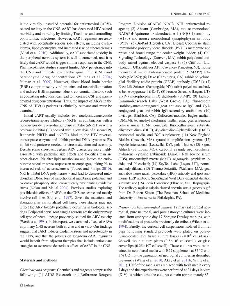
Antiretroviral drugs lead to neuronal damage in vivo
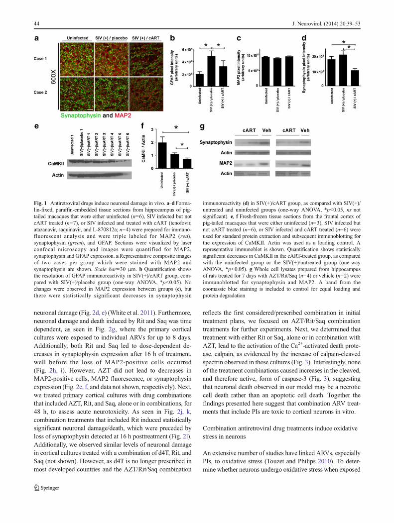
cART-induced peripheral neuropathy is well documented(Power et al. 2009), and it is likely that cART triggers similardamage to neurons in the CNS. To determine the neurotoxicpotential of cART in the CNS, we assessed the effects of anARV regimen on the expression of synaptophysin and MAP2,indicators of synaptic damage and neuronal loss, respectively,utilizing post-mortem tissue from a well-characterized SIV/pigtail macaque model of HIV CNS disease, which was de-signed to address the efficacy of CNS penetrant antiretroviraltherapy in reducing viral load in the CNS (Zink et al. 2010). Inthis study, animals infected with SIVeither received no cARTorreceived early cART treatment that included tenofovir (NRTI),atazanavir (PI), saquinavir (PI), and L-870812a (integrase inhib-itor) 12 days after the virus inoculation. Without cART, 90 % ofanimals develop neurologic disease within 3 months postinocu-lation (p.i.). By contrast, animals receiving cART do not developSIV encephalitis. Rather, they show a rapid reduction in theirplasma and CSF viral load followed by continued suppressionof SIV replication with maintenance of CD4+ Tcell counts untilelective euthanasia around day 160 p.i. Additionally, cART-treated animals do not exhibit any outward signs of neurologicaldeficits. Further, our quantitative immunofluorescent analysis ofhippocampal tissue sections revealed reduced astrogliosis in thehippocampus of SIV-infected, cART-treated animals (SIV(+)/cART), compared with SIV-infected macaques that did notreceive cART ((SIV(+)/placebo)) (Fig. 1b). However, we ob-served statistically significant decreases in synaptophysin ex-pression in the hippocampi of the SIV(+)/cART group, com-pared with that in either the uninfected or the SIV(+)/placebogroup (Fig. 1a, d). In addition, examination of the expression ofa secondmarker of synaptodendritic integrity, calmodulin kinaseII (CaMKII), by immunoblotting showed that CaMKII levelswere significantly lower in the frontal cortex in the SIV(+)/cART macaques than in their SIV(+)/placebo counterparts(Fig. 1e, f). CaMKII is highly expressed in neurons of macaquehippocampus and frontal cortex, whereas its expression in othercell types, including microglia, infiltrating macrophages, and
multinucleated giant cells, is minimal; thus, the differences inCaMKII expression in frontal cortex of the animals from ourexperimental groups are neuron specific (Gupta et al. 2010). Ourresults demonstrate synaptic injury in the presence of cARTdespite effective control of SIV replication in the peripheryand CNS. Interestingly, we did not observe changes in MAP2fluorescence in the hippocampus of infected and/or cART-treated animals compared with untreated/uninfected animals(Fig. 1a, c).
As studies of uninfected, cART-treated macaques have notbeen performed to determine the contribution of cART toneuronal damage independent of SIV infection, we adminis-tered combinations of ARVs intravenously to adult rats. Inpatients, initial cART usually includes two nucleoside/NRTIsin combination with a nNRTI or with a PI boosted with a lowdose of a second PI, ritonavir (Rit). Thus, we used zidovudine(AZT), an NRTI, along with two PIs, saquinavir (Saq) and Rit,at doses based on previously published pharmacokinetic stud-ies of these ARVs (Busidan and Dow-Edwards 1999;Kageyama et al. 2005; Pistell et al. 2010). In agreement withprevious studies (Waring et al. 2010), the animals showed noovert signs of distress during the course of the treatment.However, via immunoblotting, we observed decreases in hip-pocampal synaptophysin expression in cART-treated ratscompared with vehicle-treated rats (Fig. 1g), complementingour findings of synaptic damage in SIV(+)/cART macaquesand further supporting a role for ARV-associated neuronalinjury in the CNS. Of note, we also detected decreases inMAP2 levels in the cART-treated rat hippocampus (Fig. 1g),which likely reflects the acute drug toxicity in this treatmentparadigm, compared to the subtler synaptic injury observed inthe cART-treated macaque brain.
Antiretroviral compounds in therapeutically relevantcombinations are neurotoxic in vitro
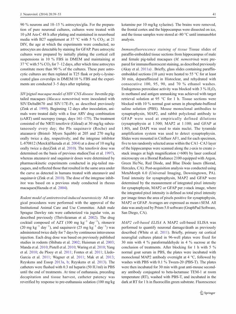
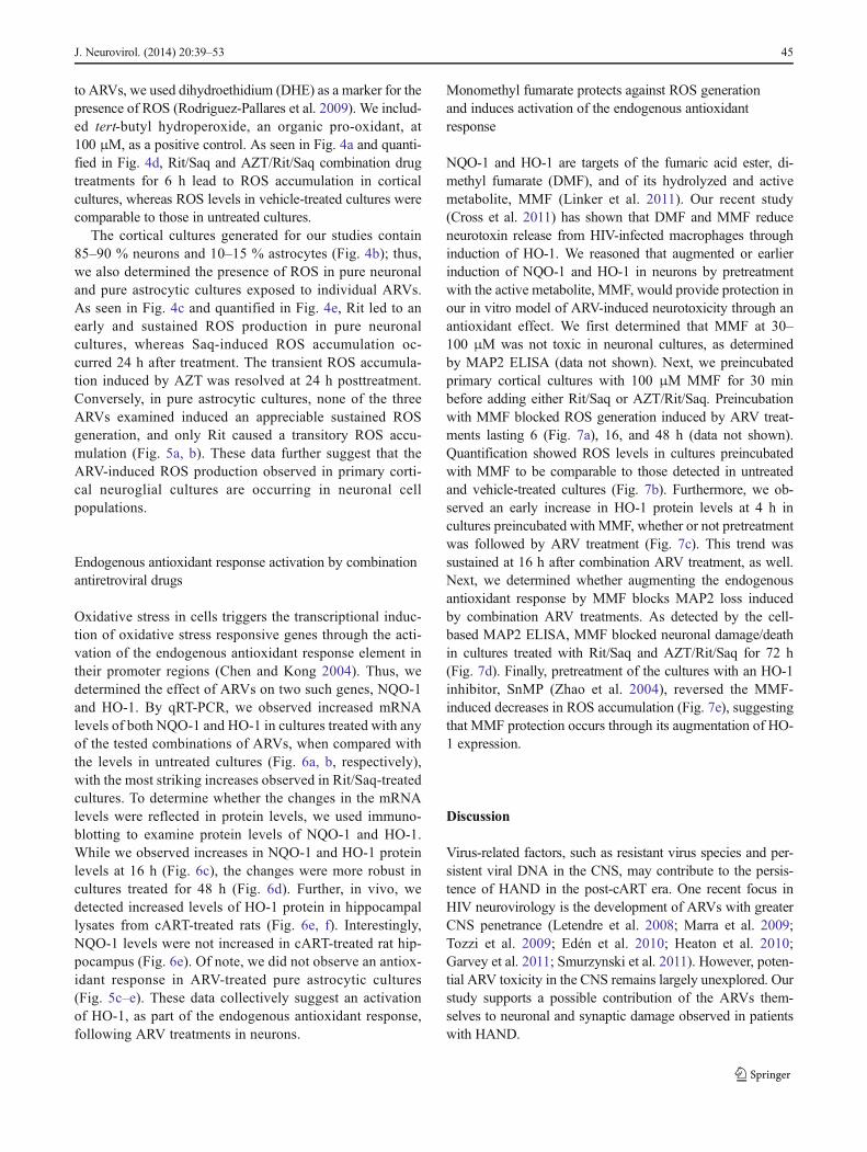
The only primary neural cell-type previously studied for ARVtoxicity was dorsal root ganglia neurons in models of periph-eral neuropathy (Werth et al. 1994). Here, to expand ourstudies on ARV neurotoxicity in the CNS, we used primaryrat cortical neuroglial cultures aged 21 DIV (O'Donnell et al.2006; Wang et al. 2007; White et al. 2011). We first evaluatedthe neuronal viability in response to increasing concentrationsof AZT, Rit, or Saq. We based the range of doses on reportedplasma and CSF levels of ARVs (Wynn et al. 2002;Anthonypillai et al. 2004). Importantly, animal studies predictbrain parenchymal levels to be equal to or greater than CSFlevels (Anthonypillai et al. 2004; Anderson and Rower 2010).At 48 h posttreatment with individual ARVs, Rit and Saq bothled to dose-dependent decreases in MAP2-positive cells, asdetermined via hand counting (Fig. 2a, b, respectively). Theseresults were confirmed with a cell-based MAP2 ELISA,which accurately reflects neuronal numbers, as well as
J. Neurovirol. (2014) 20:39–53 43
neuronal damage (Fig. 2d, e) (White et al. 2011). Furthermore,neuronal damage and death induced by Rit and Saq was timedependent, as seen in Fig. 2g, where the primary corticalcultures were exposed to individual ARVs for up to 8 days.Additionally, both Rit and Saq led to dose-dependent de-creases in synaptophysin expression after 16 h of treatment,well before the loss of MAP2-positive cells occurred(Fig. 2h, i). However, AZT did not lead to decreases inMAP2-positive cells, MAP2 fluorescence, or synaptophysinexpression (Fig. 2c, f, and data not shown, respectively). Next,we treated primary cortical cultures with drug combinationsthat included AZT, Rit, and Saq, alone or in combinations, for48 h, to assess acute neurotoxicity. As seen in Fig. 2j, k,combination treatments that included Rit induced statisticallysignificant neuronal damage/death, which were preceded byloss of synaptophysin detected at 16 h posttreatment (Fig. 2l).Additionally, we observed similar levels of neuronal damagein cortical cultures treated with a combination of d4T, Rit, andSaq (not shown). However, as d4T is no longer prescribed inmost developed countries and the AZT/Rit/Saq combination
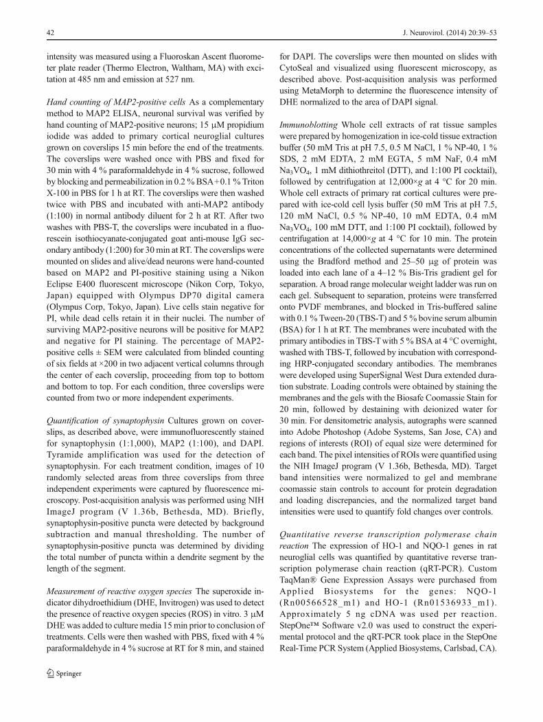
reflects the first considered/prescribed combination in initialtreatment plans, we focused on AZT/Rit/Saq combinationtreatments for further experiments. Next, we determined thattreatment with either Rit or Saq, alone or in combination withAZT, lead to the activation of the Ca2+-activated death prote-ase, calpain, as evidenced by the increase of calpain-cleavedspectrin observed in these cultures (Fig. 3). Interestingly, noneof the treatment combinations caused increases in the cleaved,and therefore active, form of caspase-3 (Fig. 3), suggestingthat neuronal death observed in our model may be a necroticcell death rather than an apoptotic cell death. Together thefindings presented here suggest that combination ARV treat-ments that include PIs are toxic to cortical neurons in vitro.
Combination antiretroviral drug treatments induce oxidativestress in neurons
An extensive number of studies have linked ARVs, especiallyPIs, to oxidative stress (Touzet and Philips 2010). To deter-mine whether neurons undergo oxidative stress when exposed
Fig. 1 Antiretroviral drugs induce neuronal damage in vivo. a–dForma-lin-fixed, paraffin-embedded tissue sections from hippocampus of pig-tailed macaques that were either uninfected (n=6), SIV infected but notcART treated (n=7), or SIV infected and treated with cART (tenofovir,atazanavir, saquinavir, and L-870812a; n=4) were prepared for immuno-fluorescent analysis and were triple labeled for MAP2 (red),synaptophysin (green), and GFAP. Sections were visualized by laserconfocal microscopy and images were quantified for MAP2,synaptophysin and GFAP expression. aRepresentative composite imagesof two cases per group which were stained with MAP2 andsynaptophysin are shown. Scale bar=30 μm. b Quantification showsthe resolution of GFAP immunoreactivity in SIV(+)/cART group, com-pared with SIV(+)/placebo group (one-way ANOVA, *p<0.05). Nochanges were observed in MAP2 expression between groups (c), butthere were statistically significant decreases in synaptophysin
immunoreactivity (d) in SIV(+)/cART group, as compared with SIV(+)/untreated and uninfected groups (one-way ANOVA, *p<0.05, ns notsignificant). e, f Fresh-frozen tissue sections from the frontal cortex ofpig-tailed macaques that were either uninfected (n=3), SIV infected butnot cART treated (n=6), or SIV infected and cART treated (n=6) wereused for standard protein extraction and subsequent immunoblotting forthe expression of CaMKII. Actin was used as a loading control. Arepresentative immunoblot is shown. Quantification shows statisticallysignificant decreases in CaMKII in the cART-treated group, as comparedwith the uninfected group or the SIV(+)/untreated group (one-wayANOVA, *p<0.05). g Whole cell lysates prepared from hippocampusof rats treated for 7 days with AZT/Rit/Saq (n=4) or vehicle (n=2) wereimmunoblotted for synaptophysin and MAP2. A band from thecoomassie blue staining is included to control for equal loading andprotein degradation
44 J. Neurovirol. (2014) 20:39–53
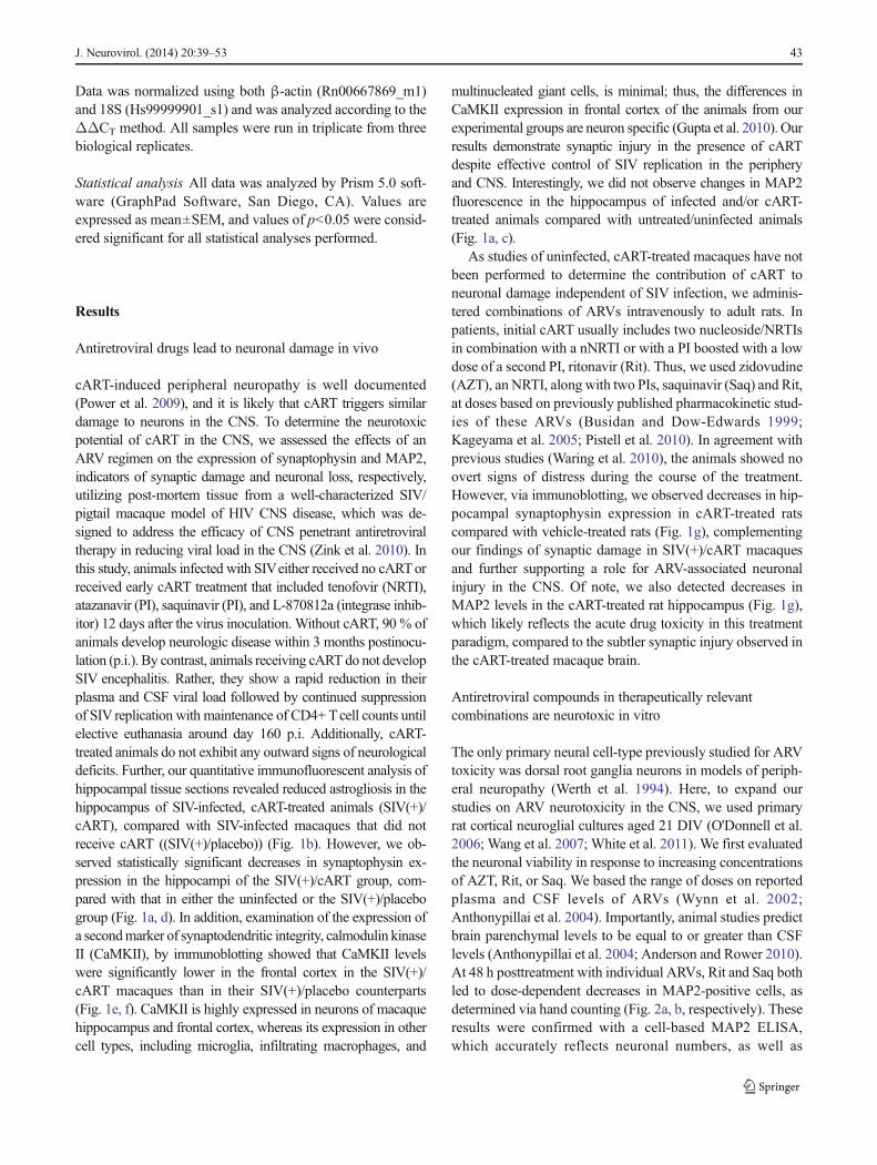
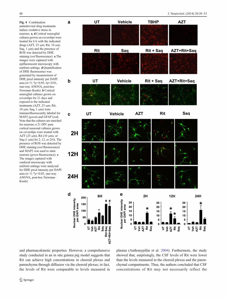
to ARVs, we used dihydroethidium (DHE) as a marker for thepresence of ROS (Rodriguez-Pallares et al. 2009). We includ-ed tert-butyl hydroperoxide, an organic pro-oxidant, at100 μM, as a positive control. As seen in Fig. 4a and quanti-fied in Fig. 4d, Rit/Saq and AZT/Rit/Saq combination drugtreatments for 6 h lead to ROS accumulation in corticalcultures, whereas ROS levels in vehicle-treated cultures werecomparable to those in untreated cultures.
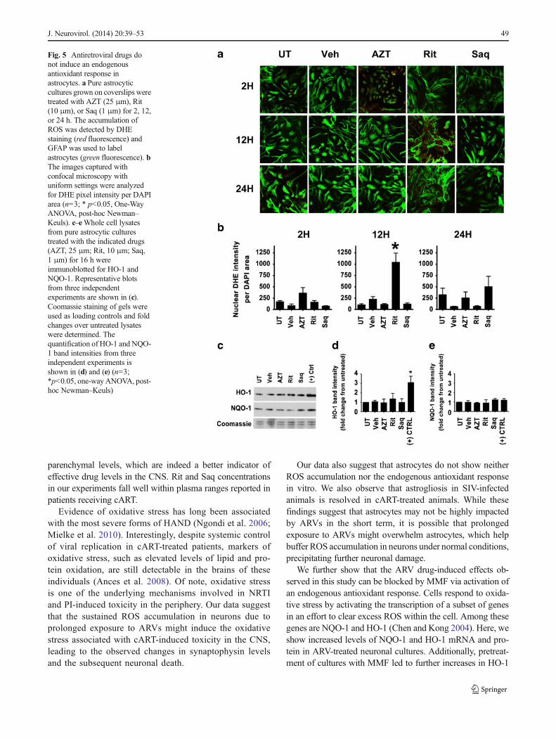
The cortical cultures generated for our studies contain85–90 % neurons and 10–15 % astrocytes (Fig. 4b); thus,we also determined the presence of ROS in pure neuronaland pure astrocytic cultures exposed to individual ARVs.As seen in Fig. 4c and quantified in Fig. 4e, Rit led to anearly and sustained ROS production in pure neuronalcultures, whereas Saq-induced ROS accumulation oc-curred 24 h after treatment. The transient ROS accumula-tion induced by AZT was resolved at 24 h posttreatment.Conversely, in pure astrocytic cultures, none of the threeARVs examined induced an appreciable sustained ROSgeneration, and only Rit caused a transitory ROS accu-mulation (Fig. 5a, b). These data further suggest that theARV-induced ROS production observed in primary corti-cal neuroglial cultures are occurring in neuronal cellpopulations.
Endogenous antioxidant response activation by combinationantiretroviral drugs
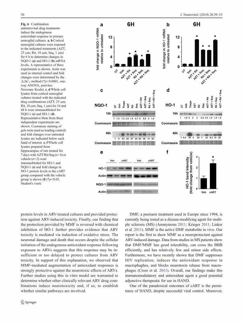
Oxidative stress in cells triggers the transcriptional induc-tion of oxidative stress responsive genes through the acti-vation of the endogenous antioxidant response element intheir promoter regions (Chen and Kong 2004). Thus, wedetermined the effect of ARVs on two such genes, NQO-1and HO-1. By qRT-PCR, we observed increased mRNAlevels of both NQO-1 and HO-1 in cultures treated with anyof the tested combinations of ARVs, when compared withthe levels in untreated cultures (Fig. 6a, b, respectively),with the most striking increases observed in Rit/Saq-treatedcultures. To determine whether the changes in the mRNAlevels were reflected in protein levels, we used immuno-blotting to examine protein levels of NQO-1 and HO-1.While we observed increases in NQO-1 and HO-1 proteinlevels at 16 h (Fig. 6c), the changes were more robust incultures treated for 48 h (Fig. 6d). Further, in vivo, wedetected increased levels of HO-1 protein in hippocampallysates from cART-treated rats (Fig. 6e, f). Interestingly,NQO-1 levels were not increased in cART-treated rat hip-pocampus (Fig. 6e). Of note, we did not observe an antiox-idant response in ARV-treated pure astrocytic cultures(Fig. 5c–e). These data collectively suggest an activationof HO-1, as part of the endogenous antioxidant response,following ARV treatments in neurons.
Monomethyl fumarate protects against ROS generationand induces activation of the endogenous antioxidantresponse
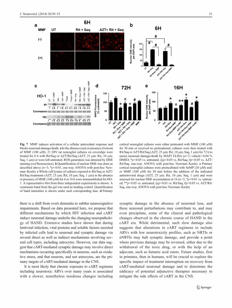
NQO-1 and HO-1 are targets of the fumaric acid ester, di-methyl fumarate (DMF), and of its hydrolyzed and activemetabolite, MMF (Linker et al. 2011). Our recent study(Cross et al. 2011) has shown that DMF and MMF reduceneurotoxin release from HIV-infected macrophages throughinduction of HO-1. We reasoned that augmented or earlierinduction of NQO-1 and HO-1 in neurons by pretreatmentwith the active metabolite, MMF, would provide protection inour in vitro model of ARV-induced neurotoxicity through anantioxidant effect. We first determined that MMF at 30–100 μM was not toxic in neuronal cultures, as determinedby MAP2 ELISA (data not shown). Next, we preincubatedprimary cortical cultures with 100 μM MMF for 30 minbefore adding either Rit/Saq or AZT/Rit/Saq. Preincubationwith MMF blocked ROS generation induced by ARV treat-ments lasting 6 (Fig. 7a), 16, and 48 h (data not shown).Quantification showed ROS levels in cultures preincubatedwith MMF to be comparable to those detected in untreatedand vehicle-treated cultures (Fig. 7b). Furthermore, we ob-served an early increase in HO-1 protein levels at 4 h incultures preincubated with MMF, whether or not pretreatmentwas followed by ARV treatment (Fig. 7c). This trend wassustained at 16 h after combination ARV treatment, as well.Next, we determined whether augmenting the endogenousantioxidant response by MMF blocks MAP2 loss inducedby combination ARV treatments. As detected by the cell-based MAP2 ELISA, MMF blocked neuronal damage/deathin cultures treated with Rit/Saq and AZT/Rit/Saq for 72 h(Fig. 7d). Finally, pretreatment of the cultures with an HO-1inhibitor, SnMP (Zhao et al. 2004), reversed the MMF-induced decreases in ROS accumulation (Fig. 7e), suggestingthat MMF protection occurs through its augmentation of HO-1 expression.
Discussion
Virus-related factors, such as resistant virus species and per-sistent viral DNA in the CNS, may contribute to the persis-tence of HAND in the post-cART era. One recent focus inHIV neurovirology is the development of ARVs with greaterCNS penetrance (Letendre et al. 2008; Marra et al. 2009;Tozzi et al. 2009; Edén et al. 2010; Heaton et al. 2010;Garvey et al. 2011; Smurzynski et al. 2011). However, poten-tial ARV toxicity in the CNS remains largely unexplored. Ourstudy supports a possible contribution of the ARVs them-selves to neuronal and synaptic damage observed in patientswith HAND.
J. Neurovirol. (2014) 20:39–53 45
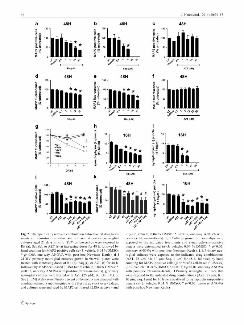
Fig. 2 Therapeutically relevant combination antiretroviral drug treat-ments are neurotoxic in vitro. a–c Primary rat cortical neuroglialcultures aged 21 days in vitro (DIV) on coverslips were exposed toRit (a), Saq (b), or AZT (c) at increasing doses for 48 h, followed byhand counting for MAP2-positive cells (n=3; vehicle, 0.04 % DMSO;* p<0.05, one-way ANOVA with post-hoc Newman–Keuls). d–f21DIV primary neuroglial cultures grown in 96-well plates weretreated with increasing doses of Rit (d), Saq (e), or AZT (f) for 48 h,followed byMAP2 cell-based ELISA (n=2; vehicle, 0.04 %DMSO; *p<0.05, one-way ANOVAwith post-hoc Newman–Keuls). g Primaryneuroglial cultures were treated with AZT (25 μM), Rit (10 μM), orSaq (1 μM) at day zero. Ninety percent of the media was changed withconditioned media supplemented with a fresh drug stock every 2 days,and cultures were analyzed by MAP2 cell-based ELISA at days 4 and
8 (n=2; vehicle, 0.04 % DMSO; * p<0.05, one-way ANOVA withpost-hoc Newman–Keuls). h, i Cultures grown on coverslips wereexposed to the indicated treatments and synaptophysin-positivepuncta were determined (n=3; vehicle, 0.04 % DMSO; * p<0.05,one-way ANOVA with post-hoc Newman–Keuls). j, k Primary neu-roglial cultures were exposed to the indicated drug combinations(AZT, 25 μm; Rit, 10 μm; Saq, 1 μm) for 48 h, followed by handcounting for MAP2-positive cells (j) or MAP2 cell-based ELISA (k)(n=2; vehicle,: 0.04 % DMSO; * p<0.05; # p<0.01, one-way ANOVAwith post-hoc Newman–Keuls). l Primary neuroglial cultures thatwere exposed to the indicated drug combinations (AZT, 25 μm; Rit,10 μm; Saq, 1 μm) for 16 h were analyzed for synaptophysin-positivepuncta (n=2; vehicle, 0.04 % DMSO; * p<0.05, one-way ANOVAwith post-hoc Newman–Keuls)
46 J. Neurovirol. (2014) 20:39–53
In this study, we show cART-induced synaptophysin loss,indicative of synaptic injury in two animal models. In our firstmodel, in an in vivo model of SIV-infected pig-tailed ma-caques, we report decreased synaptophysin and CaMKIIlevels in the SIV(+)/cART group compared with uninfectedor SIV(+)/placebo groups, indicating synaptodendritic dam-age. Interestingly, MAP2 levels did not change significantlyacross groups, which may be because of the relatively shortduration of infection in this retrospective study cohort. Whilethese data demonstrate potential effects of cART drugs in thepresence of viral infection, there are three variables to considerin interpretation of these findings: (1) the time to euthanasiafrom the start of the experiments is different between SIV(+)/placebo and SIV(+)/cART groups, (2) persistent viral DNA inthe CNS of SIV(+)/cART group, and (3) the lack of SIV(-)/cART group. It is not yet known whether brain SIV DNA isreplication competent. As we utilized post-mortem samplesobtained from a cohort of macaques enrolled in a previousstudy addressing the efficacy of CNS penetrant cART inreducing viral loads in the CNS, further experiments thatinclude an additional control group receiving cART but notinoculation with SIV will be instrumental to more clearlydetermine the contribution of viral DNA and cART to synap-tic damage in this model.
In the second in vivo model presented here, adult ratsreceived a therapeutically relevant combination of ARVs(NRTI+PI+Rit boost). In the small number of studies wherepharmacokinetics and effects of ARVs in the CNS wereexamined, pathological read-outs of neuronal damage, suchas MAP2 or synaptophysin loss, were not determined(Huisman et al. 2003; Anthonypillai et al. 2004;Anthonypillai et al. 2006). Synaptic injury is a known indica-tor of neuronal damage and dysfunction in various neurode-generative diseases, including HAND (Gupta et al. 2010) andsynaptodendritic injury persists in HIV-infected individuals in
the post-cART era (Xu and Ikezu 2009). We observed de-creases in synaptophysin and MAP2 protein levels in thehippocampus in response to ARVadministration over 7 days.Thus, our model demonstrates that ARV-associated neurotox-icity warrants consideration in developing therapeutic regi-mens for HIV-infected patients.
We also show that the PIs, Rit and Saq, alone or in combi-nations with the NRTI, AZT, induce oxidative stress, andneuronal damage/death in primary cultures at clinically rele-vant doses. Previous studies examined ARV-induced toxicityin cell lines, and Robertson et al. have provided the firstevidence for ARV-induced neurotoxicity in primary rat neu-rons (Robertson et al. 2012). Here, we provide further evi-dence that PI-induced oxidative stress and neuronal death inprimary neurons can be blocked by the activation of theendogenous antioxidant response. Interestingly, in our exper-imental paradigm, the NRTI, AZT, neither induced neuronaldamage/death by itself, nor augmented PI-induced damage/death when used in combination. We observed similar effectsfrom a combination using another NRTI, stavudine (d4T; notshown). In agreement with our observations, a previous studypresented similar findings, specifically that neither AZT nord4T inhibited cell growth or neurite regeneration in PC-12cells after long-term drug exposure (Cui et al. 1997). It shouldbe noted that NRTIs are unequivocally tied to peripheralneuropathy, where the underlying pathology is mitochondrialtoxicity and oxidative stress. However, NRTIs affect onlycertain cell populations, as they are formulated as pro-drugsand, to become active, need to be phosphorylated by twokinases, thymidine kinase 1 and 2 (TK1 and TK2), and theexpression of the cytoplasmic TK1 is cell cycle dependent(Bazzoli et al. 2010). Thus, in our model utilized to study post-mitotic neurons, AZT is most likely not converted to its activeform, and thus does not contribute to neuronal damage/death.It is of note that our in vitro model of ARV-induced neurotox-icity utilized primary neuroglial cultures rather than cell lines.Primary cells are untransformed, and therefore more accurate-ly reflect and predict ARV-associated effects occurring in thebrains of patients on cART than would immortalized celllines. The molecular pathways we investigated in this studyare highly conserved from yeast to human cells; thus, theresults obtained here in cells of rodent origin are likely con-served in human cells as well.
The drug concentrations used in this study are based on theplasma and CSF levels reported by various in vitro and in vivostudies(Huisman et al. 2003). As reported in such studies,AZT can be detected in the CSF at concentrations that aresimilar to those measured in the plasma (Wynn et al. 2002).Contrarily, as backed by various in vitro and in vivo studies(Wynn et al. 2002), both Rit and Saq are predicted to havelimited CNS penetrance due to the strong tendency of thesedrugs to bind plasma protein because of their lipophilic nature
Fig. 3 Activation of Calpain in antiretroviral drug-treated neurons.Whole cell lysates were prepared from neuroglial cultures treated withthe indicated single or combination drugs (AZT, 25 μm; Rit, 10 μm; Saq,1 μm), or with Thapsigargin (1 μM) as a positive control, for 48 h.Calpain activation was assessed using an antibody to detect the accumu-lation of calpain-cleaved spectrin and an antibody raised against thecleaved and active form of caspase-3 was used for detection of caspaseactivity. A band revealed by coomassie staining of the gel was used as aloading control (n=2; vehicle, 0.04 % DMSO)
J. Neurovirol. (2014) 20:39–53 47
and pharmacokinetic properties. However, a comprehensivestudy conducted in an in situ guinea pig model suggests thatRit can achieve high concentrations in choroid plexus andparenchyma through diffusion via the choroid plexus; in fact,the levels of Rit were comparable to levels measured in
plasma (Anthonypillai et al. 2004). Furthermore, the studyshowed that, surprisingly, the CSF levels of Rit were lowerthan the levels measured in the choroid plexus and the paren-chymal compartments. Thus, the authors concluded that CSFconcentrations of Rit may not necessarily reflect the
Fig. 4 Combinationantiretroviral drug treatmentsinduce oxidative stress inneurons. a, dCortical neuroglialcultures grown on coverslips weretreated for 6 h with the indicateddrugs (AZT, 25 μm; Rit, 10 μm;Saq, 1 μm) and the presence ofROS was detected by DHEstaining (red fluorescence). a Theimages were captured withepifluorescent microscopy withuniform settings. dQuantificationof DHE fluorescence wasgenerated by measurement ofDHE pixel intensity per DAPIarea (n=3; *p<0.05; #p<0.01,one-way ANOVA, post-hocNewman–Keuls). bCorticalneuroglial cultures grown oncoverslips for 21 days andexposed to the indicatedtreatments (AZT, 25 μm; Rit,10 μm; Saq, 1 μm) wereimmunofluorescently labeled forMAP2 (green) and GFAP (red).Note that the cultures are enrichedfor neurons; c 21 DIV purecortical neuronal cultures grownon coverslips were treated withAZT (25 μm), Rit (10 μm), orSaq (1 μm) for 2, 12, or 24 h. Thepresence of ROS was detected byDHE staining (red fluorescence)and MAP2 was used to stainneurons (green fluorescence). eThe images captured withconfocal microscopy withuniform settings were analyzedfor DHE pixel intensity per DAPIarea (n=3; *p<0.05, one-wayANOVA, post-hoc Newman–Keuls)
48 J. Neurovirol. (2014) 20:39–53
parenchymal levels, which are indeed a better indicator ofeffective drug levels in the CNS. Rit and Saq concentrationsin our experiments fall well within plasma ranges reported inpatients receiving cART.
Evidence of oxidative stress has long been associatedwith the most severe forms of HAND (Ngondi et al. 2006;Mielke et al. 2010). Interestingly, despite systemic controlof viral replication in cART-treated patients, markers ofoxidative stress, such as elevated levels of lipid and pro-tein oxidation, are still detectable in the brains of theseindividuals (Ances et al. 2008). Of note, oxidative stressis one of the underlying mechanisms involved in NRTIand PI-induced toxicity in the periphery. Our data suggestthat the sustained ROS accumulation in neurons due toprolonged exposure to ARVs might induce the oxidativestress associated with cART-induced toxicity in the CNS,leading to the observed changes in synaptophysin levelsand the subsequent neuronal death.
Our data also suggest that astrocytes do not show neitherROS accumulation nor the endogenous antioxidant responsein vitro. We also observe that astrogliosis in SIV-infectedanimals is resolved in cART-treated animals. While thesefindings suggest that astrocytes may not be highly impactedby ARVs in the short term, it is possible that prolongedexposure to ARVs might overwhelm astrocytes, which helpbuffer ROS accumulation in neurons under normal conditions,precipitating further neuronal damage.
We further show that the ARV drug-induced effects ob-served in this study can be blocked by MMF via activation ofan endogenous antioxidant response. Cells respond to oxida-tive stress by activating the transcription of a subset of genesin an effort to clear excess ROS within the cell. Among thesegenes are NQO-1 and HO-1 (Chen and Kong 2004). Here, weshow increased levels of NQO-1 and HO-1 mRNA and pro-tein in ARV-treated neuronal cultures. Additionally, pretreat-ment of cultures with MMF led to further increases in HO-1
Fig. 5 Antiretroviral drugs donot induce an endogenousantioxidant response inastrocytes. a Pure astrocyticcultures grown on coverslips weretreated with AZT (25 μm), Rit(10 μm), or Saq (1 μm) for 2, 12,or 24 h. The accumulation ofROS was detected by DHEstaining (red fluorescence) andGFAP was used to labelastrocytes (green fluorescence). bThe images captured withconfocal microscopy withuniform settings were analyzedfor DHE pixel intensity per DAPIarea (n=3; * p<0.05, One-WayANOVA, post-hoc Newman–Keuls). c–eWhole cell lysatesfrom pure astrocytic culturestreated with the indicated drugs(AZT, 25 μm; Rit, 10 μm; Saq,1 μm) for 16 h wereimmunoblotted for HO-1 andNQO-1. Representative blotsfrom three independentexperiments are shown in (c).Coomassie staining of gels wereused as loading controls and foldchanges over untreated lysateswere determined. Thequantification of HO-1 and NQO-1 band intensities from threeindependent experiments isshown in (d) and (e) (n=3;*p<0.05, one-way ANOVA, post-hoc Newman–Keuls)
J. Neurovirol. (2014) 20:39–53 49
protein levels in ARV-treated cultures and provided protec-tion against ARV-induced toxicity. Finally, our finding thatthe protection provided by MMF is reversed with chemicalinhibition of HO-1 further provides evidence that ARVtoxicity is mediated via induction of oxidative stress. Theneuronal damage and death that occurs despite the cellularinitiation of the endogenous antioxidant response followingexposure to ARVs suggests that this response may be in-sufficient or too delayed to protect cultures from ARVtoxicity. In support of this explanation, we observed thatMMF-mediated augmentation of antioxidant responses isstrongly protective against the neurotoxic effects of ARVs.Further studies using this in vitro model are warranted todetermine whether other clinically relevant ARV drug com-binations induce neurotoxicity and, if so, to establishwhether similar pathways are involved.
DMF, a psoriasis treatment used in Europe since 1994, iscurrently being tested as a disease-modifying agent for multi-ple sclerosis (MS) (Anonymous 2011; Krieger 2011; Linkeret al. 2011). MMF is the active DMF metabolite in vivo. Ourreport is the first to show MMF as a neuroprotectant againstARV-induced damage. Data from studies inMS patients showthat DMF/MMF has good tolerability, can cross the BBBefficiently, and has relatively few and minor side effects.Furthermore, we have recently shown that DMF suppressesHIV replication, induces the antioxidant response inmacrophaghes, and blocks neurotoxin release from macro-phages (Cross et al. 2011). Overall, our findings make thisimmunomodulatory and antioxidant agent a good potentialadjunctive therapeutic for use in HAND.
One of the paradoxical outcomes of cART is the persis-tence of HAND, despite successful viral control. Moreover,
Fig. 6 Combinationantiretroviral drug treatmentsinduce the endogenousantioxidant response in primaryneuroglial cultures. a, bCorticalneuroglial cultures were exposedto the indicated treatments (AZT,25 μm; Rit, 10 μm; Saq, 1 μm)for 6 h to determine changes inNQO-1 (a) and HO-1 (b) mRNAlevels. A representative of threeexperiments is shown. Actin wasused as internal control and foldchanges were determined by theΔΔCT method (*p<0.0001, one-way ANOVA, post-hocNewman–Keuls). c, dWhole celllysates from cortical neuroglialcultures treated with the indicateddrug combinations (AZT, 25 μm;Rit, 10 μm; Saq, 1 μm) for 16 and48 h were immunoblotted forNQO-1 (c) and HO-1 (d).Representative blots from threeindependent experiments areshown. Coomassie staining ofgels were used as loading controlsand fold changes over untreatedlysates are indicated below eachband of interest. e, fWhole celllysates prepared fromhippocampus of rats treated for7 days with AZT/Rit/Saq (n=4) orvehicle (n=2) wereimmunoblotted for HO-1 andNQO-1 (e) and fold change inHO-1 protein levels in the cARTgroup compared with the vehiclegroup is shown (f) (*p<0.05,Student's t test)
50 J. Neurovirol. (2014) 20:39–53
there is a shift from overt dementia to subtler neurocognitiveimpairments. Based on data presented here, we propose thatdifferent mechanisms by which HIV infection and cARTinduce neuronal damage underlie the changing neuropatholo-gy of HAND. Extensive studies have shown that duringlentiviral infection, viral proteins and soluble factors secretedby infected cells lead to neuronal and synaptic damage viaseveral direct as well as indirect mechanisms involving sev-eral cell types, including astrocytes. However, our data sug-gest that cART-mediated synaptic damage may involve directmechanisms occurring specifically in neurons, such as oxida-tive stress, and that neurons, and not astrocytes, are the pri-mary targets of cART-mediated damage in the CNS.
It is most likely that chronic exposure to cART regimensincluding neurotoxic ARVs over many years is associatedwith a slower, nonetheless insidious changes including
synaptic damage in the absence of neuronal loss, andthese neuronal perturbations may contribute to, and mayeven precipitate, some of the clinical and pathologicalchanges observed in the chronic course of HAND in thecART era. While detrimental, such slow damage alsosuggests that alterations in cART regimens to includeARVs with low neurotoxicity profiles, such as NRTIs ornNRTIs may halt synaptic damage, and provide a pointwhere previous damage may be reversed, either due to thewithdrawal of the toxic drug, or with the help of anadjuvant, such as fumaric acid esters. Future studies, firstin primates, then in humans, will be crucial to explore thespecific impact of treatment interruption on recovery fromcART-mediated neuronal damage and to determine theeddicacy of potential adjunctive therapies necessary tomitigate the side effects of cART in the CNS.
Fig. 7 MMF induces activation of a cellular antioxidant response andblocks neuronal damage/death. a In the absence (top) or presence (bottom)of MMF (100 μM), 21 DIV rat neuroglial cultures on coverslips weretreated for 6 h with Rit/Saq or AZT/Rit/Saq (AZT, 25 μm; Rit, 10 μm;Saq, 1 μm) or were left untreated. ROS generation was detected by DHEstaining (red fluorescence). bQuantification of nuclear DHE was done asdescribed above (n=3; *p<0.01, one-way ANOVAwith post-hoc New-man–Keuls). cWhole cell lysates of cultures exposed to Rit/Saq or AZT/Rit/Saq treatments (AZT, 25 μm; Rit, 10 μm; Saq, 1 μm) in the absenceor presence ofMMF (100μM) for 4 or 16 h were immunoblotted for HO-1. A representative blot from three independent experiments is shown. Acoomassie band from the gel was used as loading control. Quantificationof band intensities is shown under each corresponding lane. d Primary
cortical neuroglial cultures were either pretreated with MMF (100 μM)for 30 min or received no pretreatment; cultures were then treated withRit/Saq or AZT/Rit/Saq (AZT, 25 μm; Rit, 10 μm; Saq, 1 μm) for 72 h toassess neuronal damage/death by MAP2 ELISA (n=3; vehicle=0.04 %DMSO; *p<0.05 vs. untreated; @p<0.05 vs. Rit/Saq; #p<0.05 vs. AZT/Rit/Saq, one-way ANOVA with post-hoc Newman–Keuls). e Primarycortical neuroglial cultures were preincubated with SnMP (20 μM) and/or MMF (100 μM) for 30 min before the addition of the indicatedantiretroviral drugs (AZT, 25 μm; Rit, 10 μm; Saq, 1 μm) and wereassessed for nuclear DHE accumulation at 16 (n=3; *p<0.01 vs. untreat-ed; **p<0.05 vs. untreated; @p<0.01 vs. Rit/Saq; #p<0.05 vs. AZT/Rit/Saq, one-way ANOVAwith post-hoc Newman–Keuls)
J. Neurovirol. (2014) 20:39–53 51
Acknowledgments We would like to thank Margaret Maronski for herhelp in the preparation of cortical cultures.
Grants This work was supported by the following National Institutesof Health Grants: MH083517 (K.J-S), NS043994 and NS27405 (D.L.K),AG000255 (S.A.C), DA22339 and DA18678 (R.C.P), CA133470(M.C-S), and MH070306 (J.E.C.).
Conflict of interest All authors declare that they have no conflict ofinterest.
Open Access This article is distributed under the terms of the CreativeCommons Attribution License which permits any use, distribution, andreproduction in any medium, provided the original author(s) and thesource are credited.
References
Akay C, Lindl KA, Shyam N, Nabet B, Goenaga-Vazquez Y, RuzbarskyJ, Wang Y, Kolson DL, Jordan-Sciutto KL (2011a) Activation statusof integrated stress response pathways in neurons and astrocytes ofHAND cortex. Neuropathol Appl Neurobiol 118(6):1113–1123
Akay C, Lindl KA, Wang Y, White MG, Isaacman-Beck J, Kolson DL,Jordan-Sciutto KL (2011b) Site-specific hyperphosphorylation ofpRb in HIV-induced neurotoxicity. Mol Cell Neurosci 47:154–165
Ances BM, Roc AC, Korczykowski M, Wolf RL, Kolson DL (2008)Combination antiretroviral therapy modulates the blood oxygenlevel-dependent amplitude in human immunodeficiency virus-seropositive patients. J Neurovirol 14:418–424
Anderson PL, Rower JE (2010) Zidovudine and lamivudine for HIVinfection. Clin Med Rev Ther 2:a2004
Anonymous (2011) Trial watch: phase III success for Biogen’s oralmultiple sclerosis therapy. Nature Reviews Drug Discovery 10:404
Anthonypillai C, Sanderson RN, Gibbs JE, Thomas SA (2004) Thedistribution of the HIV protease inhibitor, ritonavir, to the brain,cerebrospinal fluid, and choroid plexuses of the guinea pig. JPharmacol Exp Ther 308:912–920
Anthonypillai C, Gibbs J, Thomas S (2006) The distribution of the anti-HIV drug, tenofovir (PMPA), into the brain, CSF and choroidplexuses. Cerebrospinal Fluid Res 3:1
Bazzoli C, Jullien V, Le Tiec C, Rey E, Mentré F, Taburet A-M (2010)Intracellular pharmacokinetics of antiretroviral drugs in HIV-infectedpatients, and their correlation with drug action. Clin Pharmacokinet49:17–45. doi:10.2165/11318110-000000000-000000000
Busidan Y, Dow-Edwards DL (1999) Neurobehavioral effects of perina-tal AZT exposure in Sprague–Dawley adult rats. NeurotoxicolTeratol 21:359–363
Chen C, Kong AN (2004) Dietary chemopreventive compounds andARE/EpRE signaling. Free Radic Biol Med 36:1505–1516
Cross SA, Cook DR, Chi AW, Vance PJ, Kolson LL, Wong BJ, Jordan-Sciutto KL, Kolson DL (2011) Dimethyl fumarate, an immunemodulator and inducer of the antioxidant response, suppressesHIV replication and macrophage-mediated neurotoxicity: a novelcandidate for HIV neuroprotection. J Immunol 187:5015–5025
Cui L, Locatelli L, Xie MY, Sommadossi JP (1997) Effect of nucleosideanalogs on neurite regeneration andmitochondrial DNA synthesis inPC-12 cells. J Pharmacol Exp Ther 280:1228–1234
Dore GJ, Correll PK, Li Y, Kaldor JM, Cooper DA, Brew BJ (1999)Changes to AIDS dementia complex in the era of highly activeantiretroviral therapy. AIDS (London, England) 13:1249–1253
du PlooyM, ViljoenM, Rheeders M (2011) Evidence for time-dependentinteractions between ritonavir and lopinavir/ritonavir plasma levels
following P-glycoprotein inhibition in Sprague–Dawley rats. BiolPharm Bull 34:66–70
Edén A, Fuchs D, Hagberg L, Nilsson S, Spudich S, Svennerholm B,Price RW, Gisslén M (2010) HIV-1 viral escape in cerebrospinalfluid of subjects on suppressive antiretroviral treatment. J Infect Dis202:1819–1825
Fontes TM, Nakamura MU, Mattar R, Simoes RS, Wagner A, deCarvalho AM, Espiridiao S, Kulay L Jr (2011) Effects of theassociation zidovudine plus ritonavir on the liver and kidneys ofpregnant rats. Morphological and biochemical aspects. Clin ExpObstet Gynecol 38:126–130
Gannon P, Khan MZ, Kolson DL (2011) Current understanding of HIV-associated neurocognitive disorders pathogenesis. Curr Opin Neurol24:275–283
Garvey L,Winston A,Walsh J, Post F, Porter K, Gazzard B, FisherM, LeenC, Pillay D, Hill T, Johnson M, Gilson R, Anderson J, Easterbrook P,Bansi L, Orkin C, Ainsworth J, Palfreeman A, Gompels M, PhillipsAN, Sabin CA (2011) Antiretroviral therapy CNS penetration andHIV-1-associated CNS disease. Neurology 76:693–700
Gupta RG, Kelly KM, Helke KL, Queen SE, Karper JM, Dorsey JL,Brice AK, Adams RJ, Tarwater PM, Kolson DL, Mankowski JL(2010) HIVand SIV induce alterations in CNS CaMKII expressionand activation: a potential mechanism for cognitive impairment. AmJ Pathol 176:2776–2784
Hazuda DJ, Young SD, Guare JP, Anthony NJ, Gomez RP,Wai JS, VaccaJP, Handt L, Motzel SL, Klein HJ, Dornadula G, Danovich RM,Witmer MV, Wilson KA, Tussey L, Schleif WA, Gabryelski LS, JinL, Miller MD, Casimiro DR, Emini EA, Shiver JW (2004) Integraseinhibitors and cellular immunity suppress retroviral replication inrhesus macaques. Science 305:528–532
Heaton RK, Clifford DB, Franklin DR Jr, Woods SP, Ake C, Vaida F, EllisRJ, Letendre SL, Marcotte TD, Atkinson JH, Rivera-Mindt M, VigilOR, Taylor MJ, Collier AC, Marra CM, Gelman BB, McArthur JC,Morgello S, Simpson DM, McCutchan JA, Abramson I, Gamst A,Fennema-Notestine C, Jernigan TL, Wong J, Grant I (2010) HIV-associated neurocognitive disorders persist in the era of potent antire-troviral therapy: CHARTER Study. Neurology 75:2087–2096
Huisman MT, Smit JW, Wiltshire HR, Beijnen JH, Schinkel AH (2003)Assessing safety and efficacy of directed P-glycoprotein inhibitionto improve the pharmacokinetic properties of saquinavircoadministered with ritonavir. J Pharmacol Exp Ther 304:596–602
Kageyama M, Namiki H, Fukushima H, Terasaka S, Togawa T, TanakaA, Ito Y, Shibata N, Takada K (2005) Effect of chronic administra-tion of ritonavir on function of cytochrome P450 3A and P-glycoprotein in rats. Biol Pharm Bull 28:130–137
Krieger S (2011) Multiple sclerosis therapeutic pipeline: opportunitiesand challenges. Mt Sinai J Med 78:192–206
Letendre S, Marquie-Beck J, Capparelli E, Best B, Clifford D, CollierAC, Gelman BB, McArthur JC, McCutchan JA, Morgello S,Simpson D, Grant I, Ellis RJ (2008) Validation of the CNSPenetration-Effectiveness rank for quantifying antiretroviral pene-tration into the central nervous system. Arch Neurol 65:65–70
Linker RA, Lee DH, Ryan S, van Dam AM, Conrad R, Bista P, Zeng W,Hronowsky X, Buko A, Chollate S, Ellrichmann G, BruckW, DawsonK, Goelz S, Wiese S, Scannevin RH, Lukashev M, Gold R (2011)Fumaric acid esters exert neuroprotective effects in neuroinflammationvia activation of the Nrf2 antioxidant pathway. Brain 134:678–692
Lledo-Garcia R, Nacher A, Casabo VG, Merino-Sanjuan M (2011) Apharmacokinetic model for evaluating the impact of hepatic andintestinal first-pass loss of saquinavir in the rat. Drug MetabDispos 39:294–301
Mak IT, Kramer JH, Chen X, Chmielinska JJ, Spurney CF, Weglicki WB(2013) Mg-supplementation attenuates ritonavir-induced hyperlip-idemia, oxidative stress and cardiac dysfunction in rats. Am JPhysiol Regul Integr Comp Physiol 305(10):R1102–R1111. doi:10.1152/ajpregu.00268.2013
52 J. Neurovirol. (2014) 20:39–53
Manda VK, Mittapalli RK, Bohn KA, Adkins CE, Lockman PR (2010)Nicotine and cotinine increases the brain penetration of saquinavir inrat. J Neurochem 115:1495–1507
Marra CM, Zhao Y, Clifford DB, Letendre S, Evans S, Henry K, Ellis RJ,Rodriguez B, Coombs RW, Schifitto G, McArthur JC, Robertson K(2009) Impact of combination antiretroviral therapy on cerebrospi-nal fluid HIV RNA and neurocognitive performance. AIDS(London, England) 23:1359–1366
Mielke MM, Bandaru VV, McArthur JC, Chu M, Haughey NJ (2010)Disturbance in cerebral spinal fluid sphingolipid content is associ-ated with memory impairment in subjects infected with the humanimmunodeficiency virus. J Neurovirol 16:445–456
Ngondi JL, Oben J, Forkah DM, Etame LH,Mbanya D (2006) The effectof different combination therapies on oxidative stress markers inHIV infected patients in Cameroon. AIDS Res Ther 3:19
Nolan D, Mallal S (2004) Complications associated with NRTI therapy:update on clinical features and possible pathogenic mechanisms.Antivir Ther 9:849–863
O'Donnell LA, Agrawal A, Jordan-Sciutto KL, Dichter MA, Lynch DR,Kolson DL (2006) Human immunodeficiency virus (HIV)-inducedneurotoxicity: roles for the NMDA receptor subtypes. J Neurosci 26:981–990
Pistell PJ, Gupta S, Knight AG, Domingue M, Uranga RM, Ingram DK,Kheterpal I, Ruiz C, Keller JN, Bruce-Keller AJ (2010) Metabolicand neurologic consequences of chronic lopinavir/ritonavir admin-istration to C57BL/6 mice. Antiviral Res 88:334–342
Power C, Boisse L, Rourke S, Gill MJ (2009) NeuroAIDS: an evolvingepidemic. Can J Neurol Sci 36:285–295
Reyskens KM, Essop MF (2013a) HIV protease inhibitors and onset ofcardiovascular diseases: a central role for oxidative stress and dys-regulation of the ubiquitin-proteasome system. Biochim BiophysActa 6:691–700
Reyskens KM, Essop MF (2013b) The maladaptive effects of HIVprotease inhibitors (lopinavir/ritonavir) on the rat heart. Int JCardiol 168:3047–3049
Reyskens KM, Fisher TL, Schisler JC, O'ConnorWG, Rogers AB,WillisMS, Planesse C, Boyer F, Rondeau P, Bourdon E, EssopMF (2013)Cardio-metabolic effectsof HIV protease inhibitors (lopinavir/rito-navir). PLoS One 8:e73347
Robertson K, Liner J, Meeker RB (2012) Antiretroviral neurotoxicity. JNeurovirol 18:388–399
Rodriguez-Pallares J, Parga J, Joglar B, Guerra M, Labandeira-Garcia J(2009) The mitochondrial atp-sensitive potassium channel blocker5-hydroxydecanoate inhibits toxicity of 6-hydroxydopamine on do-paminergic neurons. Neurotox Res 15:82–95
Shibata N, Gao W, Okamoto H, Kishida T, Yoshikawa Y, Takada K(2002) In-vitro and in-vivo pharmacokinetic interactions ofamprenavir, an HIV protease inhibitor, with other current HIVprotease inhibitors in rats. J Pharm Pharmacol 54:221–229
Smurzynski M, Wu K, Letendre S, Robertson K, Bosch RJ, Clifford DB,Evans S, Collier AC, Taylor M, Ellis R (2011) Effects of centralnervous system antiretroviral penetration on cognitive functioningin the ALLRT cohort. AIDS (London, England) 25:357–365,310.1097/QAD.1090b1013e32834171f32834178
Thrivikraman KV, Huot RL, Plotsky PM (2002) Jugular vein catheteri-zation for repeated blood sampling in the unrestrained conscious rat.Brain Res Brain Res Protocol 10:84–94
Touzet O, Philips A (2010) Resveratrol protects against protease inhibitor-induced reactive oxygen species production, reticulum stress and lipidraft perturbation. AIDS (London, England) 24:1437–1447
Tozzi V, Balestra P, Salvatori MF, Vlassi C, Liuzzi G, Giancola ML,Giulianelli M, Narciso P, Antinori A (2009) Changes in cognitionduring antiretroviral therapy: comparison of 2 different rankingsystems to measure antiretroviral drug efficacy on HIV-associatedneurocognitive disorders. JAIDS J Acquir Immune Defic Syndr 52:56–63. doi:10.1097/QAI.1090b1013e3181af1083d1096
Tsai CC, Follis KE, Beck TW, Sabo A, Bischofberger N, Dailey PJ(1997) Effects of (R)-9-(2-phosphonylmethoxypropyl)adeninemonotherapy on chronic SIV infection in macaques. AIDS ResHum Retrovir 13:707–712
Vidal F, Gutierrez F, Gutierrez M, OlonaM, Sanchez V, Mateo G, PeraireJ, Vilades C, Veloso S, Lopez-Dupla M, Domingo P (2010)Pharmacogenetics of adverse effects due to antiretroviral drugs.AIDS Rev 12:15–30
Wagner A, Nakamura MU, Simoes RS, Oliveira-Filho RM, Fontes TM,de Carvalho LP, Espiridiao S, Kulay L Jr (2011) Chronic action ofassociation of zidovudine, lamivudine and ritonavir on pregnantrats. A biologic assay. Clin Exp Obstet Gynecol 38:28–32
Wang Y, White MG, Akay C, Chodroff RA, Robinson J, Lindl KA,Dichter MA, Qian Y, Mao Z, Kolson DL, Jordan-Sciutto KL(2007) Activation of cyclin-dependent kinase 5 by calpains contrib-utes to human immunodeficiency virus-induced neurotoxicity. JNeurochem 103:439–455
Wang Y, ShyamN, Ting JH, Akay C, Lindl KA, Jordan-Sciutto KL (2010)E2F1 localizes predominantly to neuronal cytoplasm and fails toinduce expression of its transcriptional targets in human immunode-ficiency virus-induced neuronal damage. Neurosci Lett 479:97–101
Waring JF, Ciurlionis R, Marsh K, Klein LL, Degoey DA, Randolph JT,Spear B, Kempf DJ (2010) Identification of proteasome gene regu-lation in a rat model for HIV protease inhibitor-induced hyperlipid-emia. Arch Toxicol 84:263–270
Werth JL, Zhou B, Nutter LM, Thayer SA (1994) 2′,3′-Dideoxycytidinealters calcium buffering in cultured dorsal root ganglion neurons.Mol Pharmacol 45:1119–1124
White MG, Wang Y, Akay C, Lindl KA, Kolson DL, Jordan-Sciutto KL(2011) Parallel high throughput neuronal toxicity assays demon-strate uncoupling between loss ofmitochondrial membrane potentialand neuronal damage in a model of HIV-induced neurodegenera-tion. Neurosci Res 70:220–229
WilcoxKS, Buchhalter J, Dichter MA (1994) Properties of inhibitory andexcitatory synapses between hippocampal neurons in very lowdensity cultures. Synapse 18:128–151
Wynn HE, Brundage RC, Fletcher CV (2002) Clinical implications ofCNS penetration of antiretroviral drugs. CNS Drugs 16:595–609
Xu J, Ikezu T (2009) The comorbidity of HIV-associated neurocognitivedisorders and Alzheimer’s disease: a foreseeable medical challengein post-HAART era. J NeuroImmune Pharm 4:200–212
Yang Y, Dahly-Vernon AJ, Blomme EA, Lai-Zhang J, Kempf DJ, MarshKC, Harrington YA, Nye SH, Evans DL, Roman RJ, Jacob HJ,Waring JF (2010) Liver transcriptomic changes associated withritonavir-induced hyperlipidemia in sensitive and resistant strainsof rats. Vet J 185:75–82
Yilmaz A, Ståhle L, Hagberg L, Svennerholm B, Fuchs D, Gisslén M(2004) Cerebrospinal fluid and plasma HIV-1 RNA levels andlopinavir concentrations following lopinavir/ritonavir regimen.Scand J Infect Dis 36:823–828
Yilmaz A, GisslenM, Spudich S, Lee E, Jayewardene A, Aweeka F, PriceRW (2009) Raltegravir cerebrospinal fluid concentrations in HIV-1infection. PLoS One 4:e6877
Zhao S, Zhang Y, Gu Y, Lewis DF, Wang Y (2004) Heme oxygenase-1mediates up-regulation of adhesion molecule expression induced byperoxynitrite in endothelial cells. J Soc Gynecol Investig 11:465–471
ZinkMC, Suryanarayana K,Mankowski JL, ShenA, PiatakM Jr, SpelmanJP, Carter DL, Adams RJ, Lifson JD, Clements JE (1999) High viralload in the cerebrospinal fluid and brain correlates with severity ofsimian immunodeficiency virus encephalitis. J Virol 73:10480–10488
Zink MC, Brice AK, Kelly KM, Queen SE, Gama L, Li M, Adams RJ,Bartizal C, Varrone J, Rabi SA, Graham DR, Tarwater PM,Mankowski JL, Clements JE (2010) Simian immunodeficiencyvirus-infected macaques treated with highly active antiretroviral ther-apy have reduced central nervous system viral replication and inflam-mation but persistence of viral DNA. J Infect Dis 202:161–170
J. Neurovirol. (2014) 20:39–53 53