Embed Size (px)
Citation preview
CLINICAL MICROBIOLOGY REVIEWS,0893-8512/99/$04.0010
Jan. 1999, p. 147–179 Vol. 12, No. 1
Copyright © 1999, American Society for Microbiology. All Rights Reserved.
Antiseptics and Disinfectants: Activity, Action, and ResistanceGERALD MCDONNELL1* AND A. DENVER RUSSELL2
STERIS Corporation, St. Louis Operations, St. Louis, Missouri 63166,1 and Welsh Schoolof Pharmacy, Cardiff University, Cardiff CF1 3XF, United Kingdom2
INTRODUCTION .......................................................................................................................................................148DEFINITIONS ............................................................................................................................................................148MECHANISMS OF ACTION ...................................................................................................................................148
Introduction.............................................................................................................................................................148General Methodology .............................................................................................................................................148Alcohols ....................................................................................................................................................................151Aldehydes .................................................................................................................................................................151
Glutaraldehyde ....................................................................................................................................................151Formaldehyde ......................................................................................................................................................153Formaldehyde-releasing agents.........................................................................................................................153o-Phthalaldehyde.................................................................................................................................................153
Anilides.....................................................................................................................................................................153Biguanides................................................................................................................................................................153
Chlorhexidine ......................................................................................................................................................153Alexidine...............................................................................................................................................................154Polymeric biguanides..........................................................................................................................................154
Diamidines ...............................................................................................................................................................155Halogen-Releasing Agents .....................................................................................................................................155
Chlorine-releasing agents ..................................................................................................................................155Iodine and iodophors .........................................................................................................................................155
Silver Compounds...................................................................................................................................................155Silver nitrate........................................................................................................................................................156Silver sulfadiazine...............................................................................................................................................156
Peroxygens ...............................................................................................................................................................156Hydrogen peroxide..............................................................................................................................................156Peracetic acid ......................................................................................................................................................156
Phenols .....................................................................................................................................................................156Bis-Phenols ..............................................................................................................................................................157
Triclosan ..............................................................................................................................................................157Hexachlorophene.................................................................................................................................................157
Halophenols .............................................................................................................................................................157Quaternary Ammonium Compounds ...................................................................................................................157Vapor-Phase Sterilants ..........................................................................................................................................158
MECHANISMS OF RESISTANCE..........................................................................................................................158Introduction.............................................................................................................................................................158Bacterial Resistance to Antiseptics and Disinfectants ......................................................................................158Intrinsic Bacterial Resistance Mechanisms........................................................................................................158
Intrinsic resistance of bacterial spores............................................................................................................159Intrinsic resistance of mycobacteria ................................................................................................................160Intrinsic resistance of other gram-positive bacteria......................................................................................161Intrinsic resistance of gram-negative bacteria ...............................................................................................161Physiological (phenotypic) adaption as an intrinsic mechanism.................................................................162
Acquired Bacterial Resistance Mechanisms .......................................................................................................164Plasmids and bacterial resistance to antiseptics and disinfectants ............................................................164
(i) Plasmid-mediated antiseptic and disinfectant resistance in gram-negative bacteria......................164(ii) Plasmid-mediated antiseptic and disinfectant resistance in staphylococci .....................................165(iii) Plasmid-mediated antiseptic and disinfectant resistance in other gram-positive bacteria..........166
Mutational resistance to antiseptics and disinfectants.................................................................................166Mechanisms of Fungal Resistance to Antiseptics and Disinfectants ..............................................................167Mechanisms of Viral Resistance to Antiseptics and Disinfectants .................................................................168Mechanisms of Protozoal Resistance to Antiseptics and Disinfectants..........................................................169Mechanisms of Prion Resistance to Disinfectants.............................................................................................169
* Corresponding author. Present address: STERIS Corporation,5960 Heisley Rd., Mentor, OH 44060. Phone: (440) 354-2600. Fax:(440) 354-7038. E-mail: [email protected].
147
on July 11, 2018 by guesthttp://cm
r.asm.org/
Dow
nloaded from
on July 11, 2018 by guesthttp://cm
r.asm.org/
Dow
nloaded from
on July 11, 2018 by guesthttp://cm
r.asm.org/
Dow
nloaded from
CONCLUSIONS .........................................................................................................................................................169REFERENCES ............................................................................................................................................................170
INTRODUCTION
Antiseptics and disinfectants are used extensively in hospi-tals and other health care settings for a variety of topical andhard-surface applications. In particular, they are an essentialpart of infection control practices and aid in the prevention ofnosocomial infections (277, 454). Mounting concerns over thepotential for microbial contamination and infection risks in thefood and general consumer markets have also led to increaseduse of antiseptics and disinfectants by the general public. Awide variety of active chemical agents (or “biocides”) arefound in these products, many of which have been used forhundreds of years for antisepsis, disinfection, and preservation(39). Despite this, less is known about the mode of action ofthese active agents than about antibiotics. In general, biocideshave a broader spectrum of activity than antibiotics, and, whileantibiotics tend to have specific intracellular targets, biocidesmay have multiple targets. The widespread use of antisepticand disinfectant products has prompted some speculation onthe development of microbial resistance, in particular cross-resistance to antibiotics. This review considers what is knownabout the mode of action of, and mechanisms of microbialresistance to, antiseptics and disinfectants and attempts, wher-ever possible, to relate current knowledge to the clinical envi-ronment.
A summary of the various types of biocides used in antisep-tics and disinfectants, their chemical structures, and their clin-ical uses is shown in Table 1. It is important to note that manyof these biocides may be used singly or in combination in avariety of products which vary considerably in activity againstmicroorganisms. Antimicrobial activity can be influenced bymany factors such as formulation effects, presence of an or-ganic load, synergy, temperature, dilution, and test method.These issues are beyond the scope of this review and arediscussed elsewhere (123, 425, 444, 446, 451).
DEFINITIONS
“Biocide” is a general term describing a chemical agent,usually broad spectrum, that inactivates microorganisms. Be-cause biocides range in antimicrobial activity, other terms maybe more specific, including “-static,” referring to agents whichinhibit growth (e.g., bacteriostatic, fungistatic, and sporistatic)and “-cidal,” referring to agents which kill the target organism(e.g., sporicidal, virucidal, and bactericidal). For the purpose ofthis review, antibiotics are defined as naturally occurring orsynthetic organic substances which inhibit or destroy selectivebacteria or other microorganisms, generally at low concentra-tions; antiseptics are biocides or products that destroy or in-hibit the growth of microorganisms in or on living tissue (e.g.health care personnel handwashes and surgical scrubs); anddisinfectants are similar but generally are products or biocidesthat are used on inanimate objects or surfaces. Disinfectantscan be sporostatic but are not necessarily sporicidal.
Sterilization refers to a physical or chemical process thatcompletely destroys or removes all microbial life, includingspores. Preservation is the prevention of multiplication of mi-croorganisms in formulated products, including pharmaceuti-cals and foods. A number of biocides are also used for cleaningpurposes; cleaning in these cases refers to the physical removalof foreign material from a surface (40).
MECHANISMS OF ACTION
Introduction
Considerable progress has been made in understanding themechanisms of the antibacterial action of antiseptics and dis-infectants (215, 428, 437). By contrast, studies on their modesof action against fungi (426, 436), viruses (298, 307), and pro-tozoa (163) have been rather sparse. Furthermore, little isknown about the means whereby these agents inactivate prions(503).
Whatever the type of microbial cell (or entity), it is probablethat there is a common sequence of events. This can be envis-aged as interaction of the antiseptic or disinfectant with the cellsurface followed by penetration into the cell and action at thetarget site(s). The nature and composition of the surface varyfrom one cell type (or entity) to another but can also alter asa result of changes in the environment (57, 59). Interaction atthe cell surface can produce a significant effect on viability (e.g.with glutaraldehyde) (374, 421), but most antimicrobial agentsappear to be active intracellularly (428, 451). The outermostlayers of microbial cells can thus have a significant effect ontheir susceptibility (or insusceptibility) to antiseptics and dis-infectants; it is disappointing how little is known about thepassage of these antimicrobial agents into different types ofmicroorganisms. Potentiation of activity of most biocides maybe achieved by the use of various additives, as shown in laterparts of this review.
In this section, the mechanisms of antimicrobial action of arange of chemical agents that are used as antiseptics or disin-fectants or both are discussed. Different types of microorgan-isms are considered, and similarities or differences in the na-ture of the effect are emphasized. The mechanisms of actionare summarized in Table 2.
General Methodology
A battery of techniques are available for studying the mech-anisms of action of antiseptics and disinfectants on microor-ganisms, especially bacteria (448). These include examinationof uptake (215, 428, 459), lysis and leakage of intracellularconstituents (122), perturbation of cell homeostasis (266,445), effects on model membranes (170), inhibition of en-zymes, electron transport, and oxidative phosphorylation (162,272), interaction with macromolecules (448, 523), effects onmacromolecular biosynthetic processes (133), and microscopicexamination of biocide-exposed cells (35). Additional and use-ful information can be obtained by calculating concentrationexponents (n values [219, 489]) and relating these to mem-brane activity (219). Many of these procedures are valuable fordetecting and evaluating antiseptics or disinfectants used incombination (146, 147, 202, 210).
Similar techniques have been used to study the activity ofantiseptics and disinfectants against fungi, in particular yeasts.Additionally, studies on cell wall porosity (117–119) may pro-vide useful information about intracellular entry of disinfec-tants and antiseptics (204–208).
Mechanisms of antiprotozoal action have not been widelyinvestigated. One reason for this is the difficulty in cultur-ing some protozoa (e.g., Cryptosporidium) under laboratoryconditions. However, the different life stages (trophozoitesand cysts) do provide a fascinating example of the problem
148 MCDONNELL AND RUSSELL CLIN. MICROBIOL. REV.
on July 11, 2018 by guesthttp://cm
r.asm.org/
Dow
nloaded from
of how changes in cytology and physiology can modify re-sponses to antiseptics and disinfectants. Khunkitti et al. (251–255) have explored this aspect by using indices of viability,leakage, uptake, and electron microscopy as experimental tools.
Some of these procedures can also be modified for study-ing effects on viruses and phages (e.g., uptake to whole cellsand viral or phage components, effects on nucleic acids andproteins, and electron microscopy) (401). Viral targets are
TABLE 1. Chemical structures and uses of biocides in antiseptics and disinfectants
Continued on following page
VOL. 12, 1999 ANTISEPTICS AND DISINFECTANTS 149
on July 11, 2018 by guesthttp://cm
r.asm.org/
Dow
nloaded from
predominantly the viral envelope (if present), derived fromthe host cell cytoplasmic or nuclear membrane; the capsid,which is responsible for the shape of virus particles and forthe protection of viral nucleic acid; and the viral genome.Release of an intact viral nucleic acid into the environment
following capsid destruction is of potential concern sincesome nucleic acids are infective when liberated from the cap-sid (317), an aspect that must be considered in viral disin-fection. Important considerations in viral inactivation aredealt with by Klein and Deforest (259) and Prince et al.
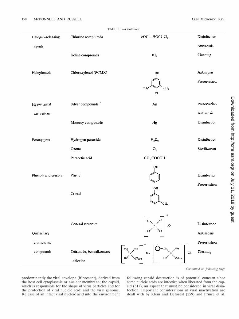
TABLE 1—Continued
Continued on following page
150 MCDONNELL AND RUSSELL CLIN. MICROBIOL. REV.
on July 11, 2018 by guesthttp://cm
r.asm.org/
Dow
nloaded from
(384), while an earlier paper by Grossgebauer is highly rec-ommended (189).
Alcohols
Although several alcohols have been shown to be effectiveantimicrobials, ethyl alcohol (ethanol, alcohol), isopropyl alco-hol (isopropanol, propan-2-ol) and n-propanol (in particular inEurope) are the most widely used (337). Alcohols exhibit rapidbroad-spectrum antimicrobial activity against vegetative bacte-ria (including mycobacteria), viruses, and fungi but are notsporicidal. They are, however, known to inhibit sporulationand spore germination (545), but this effect is reversible (513).Because of the lack of sporicidal activity, alcohols are notrecommended for sterilization but are widely used for bothhard-surface disinfection and skin antisepsis. Lower concen-trations may also be used as preservatives and to potentiate theactivity of other biocides. Many alcohol products include lowlevels of other biocides (in particular chlorhexidine), whichremain on the skin following evaporation of the alcohol, orexcipients (including emollients), which decrease the evapora-tion time of the alcohol and can significantly increase productefficacy (68). In general, isopropyl alcohol is considered slightly
more efficacious against bacteria (95) and ethyl alcohol is morepotent against viruses (259); however, this is dependent on theconcentrations of both the active agent and the test microor-ganism. For example, isopropyl alcohol has greater lipophilicproperties than ethyl alcohol and is less active against hydro-philic viruses (e.g., poliovirus) (259). Generally, the antimicro-bial activity of alcohols is significantly lower at concentrationsbelow 50% and is optimal in the 60 to 90% range.
Little is known about the specific mode of action of alcohols,but based on the increased efficacy in the presence of water, itis generally believed that they cause membrane damage andrapid denaturation of proteins, with subsequent interferencewith metabolism and cell lysis (278, 337). This is supported byspecific reports of denaturation of Escherichia coli dehydroge-nases (499) and an increased lag phase in Enterobacter aero-genes, speculated to be due to inhibition of metabolism re-quired for rapid cell division (101).
Aldehydes
Glutaraldehyde. Glutaraldehyde is an important dialdehydethat has found usage as a disinfectant and sterilant, in partic-ular for low-temperature disinfection and sterilization of en-doscopes and surgical equipment and as a fixative in electron
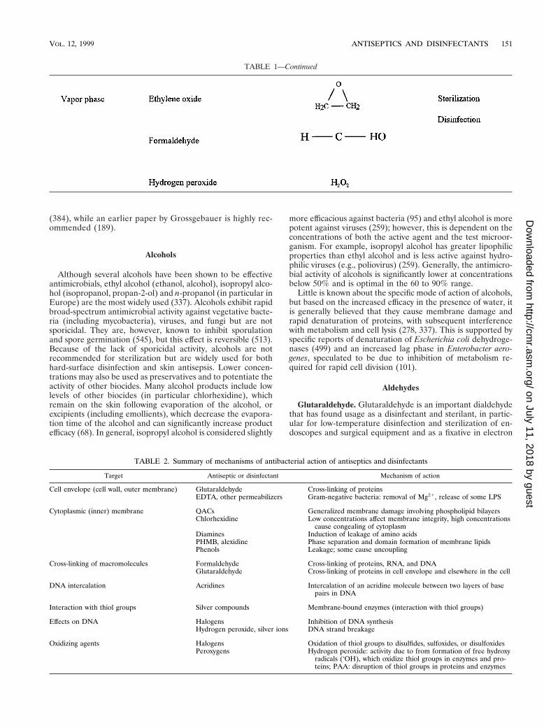
TABLE 1—Continued
TABLE 2. Summary of mechanisms of antibacterial action of antiseptics and disinfectants
Target Antiseptic or disinfectant Mechanism of action
Cell envelope (cell wall, outer membrane) Glutaraldehyde Cross-linking of proteinsEDTA, other permeabilizers Gram-negative bacteria: removal of Mg21, release of some LPS
Cytoplasmic (inner) membrane QACs Generalized membrane damage involving phospholipid bilayersChlorhexidine Low concentrations affect membrane integrity, high concentrations
cause congealing of cytoplasmDiamines Induction of leakage of amino acidsPHMB, alexidine Phase separation and domain formation of membrane lipidsPhenols Leakage; some cause uncoupling
Cross-linking of macromolecules Formaldehyde Cross-linking of proteins, RNA, and DNAGlutaraldehyde Cross-linking of proteins in cell envelope and elsewhere in the cell
DNA intercalation Acridines Intercalation of an acridine molecule between two layers of basepairs in DNA
Interaction with thiol groups Silver compounds Membrane-bound enzymes (interaction with thiol groups)
Effects on DNA Halogens Inhibition of DNA synthesisHydrogen peroxide, silver ions DNA strand breakage
Oxidizing agents Halogens Oxidation of thiol groups to disulfides, sulfoxides, or disulfoxidesPeroxygens Hydrogen peroxide: activity due to from formation of free hydroxy
radicals (zOH), which oxidize thiol groups in enzymes and pro-teins; PAA: disruption of thiol groups in proteins and enzymes
VOL. 12, 1999 ANTISEPTICS AND DISINFECTANTS 151
on July 11, 2018 by guesthttp://cm
r.asm.org/
Dow
nloaded from
icroscopy. Glutaraldehyde has a broad spectrum of activityagainst bacteria and their spores, fungi, and viruses, and aconsiderable amount of information is now available about theways whereby these different organisms are inactivated (Tables2 and 3). Earlier reviews of its mechanisms of action have beenpublished (179, 182, 374, 482).
The first reports in 1964 and 1965 (182) demonstrated thatglutaraldehyde possessed high antimicrobial activity. Subse-quently, research was undertaken to evaluate the nature of itsbactericidal (339–344, 450) and sporicidal (180, 181, 507, 508)action. These bactericidal studies demonstrated (374) a strongbinding of glutaraldehyde to outer layers of organisms such asE. coli and Staphylococcus aureus (179, 212, 339–341, 343, 344),inhibition of transport in gram-negative bacteria (179), inhibi-tion of dehydrogenase activity (343, 344) and of periplasmicenzymes (179), prevention of lysostaphin-induced lysis in S. au-reus (453) and of sodium lauryl sulfate-induced lysis in E. coli(340, 344), inhibition of spheroplast and protoplast lysis inhypotonic media (340, 344), and inhibition of RNA, DNA, andprotein synthesis (320). Strong interaction of glutaraldehydewith lysine and other amino acids has been demonstrated (450).
Clearly, the mechanism of action of glutaraldehyde involvesa strong association with the outer layers of bacterial cells,specifically with unprotonated amines on the cell surface, pos-sibly representing the reactive sites (65). Such an effect couldexplain its inhibitory action on transport and on enzyme sys-tems, where access of substrate to enzyme is prohibited. Partialor entire removal of the cell wall in hypertonic medium, lead-ing to the production of spheroplasts or protoplasts and thesubsequent prevention of lysis by glutaraldehyde when theseforms are diluted in a hypotonic environment, suggests an ad-ditional effect on the inner membrane, a finding substantiatedby the fact that the dialdehyde prevents the selective release ofsome membrane-bound enzymes of Micrococcus lysodeikticus(138). Glutaraldehyde is more active at alkaline than at acidicpHs. As the external pH is altered from acidic to alkaline,more reactive sites will be formed at the cell surface, leading toa more rapid bactericidal effect. The cross-links thus obtainedmean that the cell is then unable to undertake most, if not all,of its essential functions. Glutaraldehyde is also mycobacteri-cidal. Unfortunately, no critical studies have as yet been un-dertaken to evaluate the nature of this action (419).
The bacterial spore presents several sites at which interac-tion with glutaraldehyde is possible, although interaction witha particular site does not necessarily mean that this is associ-ated with spore inactivation. E. coli, S. aureus, and vegetativecells of Bacillus subtilis bind more glutaraldehyde than do rest-
ing spores of B. subtilis (377, 378); uptake of glutaraldehyde isgreater during germination and outgrowth than with maturespores but still lower than with vegetative cells. Low concen-trations of the dialdehyde (0.1%) inhibit germination, whereasmuch higher concentrations (2%) are sporicidal. The alde-hyde, at both acidic and alkaline pHs, interacts strongly withthe outer spore layers (508, 509); this interaction reduces therelease of dipicolinic acid (DPA) from heated spores and thelysis induced by mercaptoethanol (or thioglycolate)-peroxidecombinations. Low concentrations of both acidic and alkalineglutaraldehyde increase the surface hydrophobicity of spores,again indicating an effect at the outermost regions of the cell.It has been observed by various authors (182, 374, 376, 380)that the greater sporicidal activity of glutaraldehyde at alkalinepH is not reflected by differences in uptake; however, uptakeper se reflects binding and not necessarily penetration into thespore. It is conceivable that acidic glutaraldehyde interactswith and remains at the cell surface whereas alkaline glutaral-dehyde penetrates more deeply into the spore. This contentionis at odds with the hypothesis of Bruch (65), who envisaged theacidic form penetrating the coat and reacting with the cortexwhile the alkaline form attacked the coat, thereby destroyingthe ability of the spore to function solely as a result of thissurface phenomenon. There is, as yet, no evidence to supportthis theory. Novel glutaraldehyde formulations based on acidicrather than alkaline glutaraldehyde, which benefit from thegreater inherent stability of the aldehyde at lower pH, havebeen produced. The improved sporicidal activity claimed forthese products may be obtained by agents that potentiate theactivity of the dialdehyde (414, 421).
During sporulation, the cell eventually becomes less suscep-tible to glutaraldehyde (see “Intrinsic resistance of bacterialspores”). By contrast, germinating and outgrowing cells reac-quire sensitivity. Germination may be defined as an irreversibleprocess in which there is a change of an activated spore froma dormant to a metabolically active state within a short period.Glutaraldehyde exerts an early effect on the germination pro-cess. L-Alanine is considered to act by binding to a specificreceptor on the spore coat, and once spores are triggered togerminate, they are committed irreversibly to losing their dor-mant properties (491). Glutaraldehyde at high concentrationsinhibits the uptake of L-[14C]alanine by B. subtilis spores, albeitby an unknown mechanism (379, 414). Glutaraldehyde-treatedspores retain their refractivity, having the same appearanceunder the phase-contrast microscope as normal, untreatedspores even when the spores are subsequently incubated ingermination medium. Glutaraldehyde is normally used as a 2%solution to achieve a sporicidal effect (16, 316); low concen-trations (,0.1%) prevent phase darkening of spores and alsoprevent the decrease in optical density associated with a lateevent in germination. By contrast, higher concentrations (0.1to 1%) significantly reduce the uptake of L-alanine, possibly asa result of a sealing effect of the aldehyde on the cell surface.Mechanisms involved in the revival of glutaraldehyde-treatedspores are discussed below (see “Intrinsic resistance of bacte-rial spores”).
There are no recent studies of the mechanisms of fungicidalaction of glutaraldehyde. Earlier work had suggested that thefungal cell wall was a major target site (179, 182, 352), espe-cially the major wall component, chitin, which is analogous tothe peptidoglycan found in bacterial cell walls.
Glutaraldehyde is a potent virucidal agent (143, 260). Itreduces the activity of hepatitis B surface antigen (HBsAg) andespecially hepatitis B core antigen ([HBcAg] in hepatitis Bvirus [HBV]) (3) and interacts with lysine residues on thesurface of hepatitis A virus (HAV) (362). Low concentrations
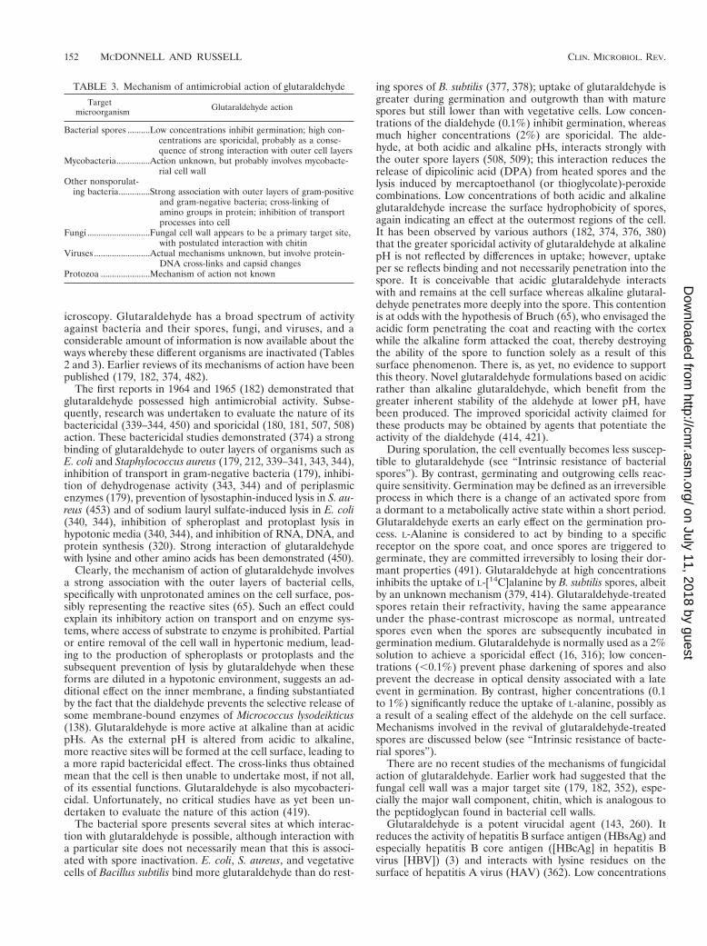
TABLE 3. Mechanism of antimicrobial action of glutaraldehyde
Targetmicroorganism Glutaraldehyde action
Bacterial spores ..........Low concentrations inhibit germination; high con-centrations are sporicidal, probably as a conse-quence of strong interaction with outer cell layers
Mycobacteria...............Action unknown, but probably involves mycobacte-rial cell wall
Other nonsporulat-ing bacteria..............Strong association with outer layers of gram-positive
and gram-negative bacteria; cross-linking ofamino groups in protein; inhibition of transportprocesses into cell
Fungi ............................Fungal cell wall appears to be a primary target site,with postulated interaction with chitin
Viruses .........................Actual mechanisms unknown, but involve protein-DNA cross-links and capsid changes
Protozoa ......................Mechanism of action not known
152 MCDONNELL AND RUSSELL CLIN. MICROBIOL. REV.
on July 11, 2018 by guesthttp://cm
r.asm.org/
Dow
nloaded from
(,0.1%) of alkaline glutaraldehyde are effective against puri-fied poliovirus, whereas poliovirus RNA is highly resistant toaldehyde concentrations up to 1% at pH 7.2 and is only slowlyinactivated at pH 8.3 (21). In other words, the complete po-liovirus particle is much more sensitive than poliovirus RNA.In light of this, it has been inferred that glutaraldehyde-in-duced loss of infectivity is associated with capsid changes (21).Glutaraldehyde at the low concentrations of 0.05 and 0.005%interacts with the capsid proteins of poliovirus and echovirus,respectively; the differences in sensitivity probably reflect ma-jor structural variations in the two viruses (75).
Bacteriophages were recently studied to obtain informationabout mechanisms of virucidal action (298–304, 306, 307). Manyglutaraldehyde-treated P. aeruginosa F116 phage particles hadempty heads, implying that the phage genome had been eject-ed. The aldehyde was possibly bound to F116 double-strandedDNA but without affecting the molecule; glutaraldehyde alsointeracted with phage F116 proteins, which were postulated tobe involved in the ejection of the nucleic acid. Concentrationsof glutaraldehyde greater than 0.1 to 0.25% significantly af-fected the transduction of this phage; the transduction processwas more sensitive to the aldehyde than was the phage itself.Glutaraldehyde and other aldehydes were tested for theirability to form protein-DNA cross-links in simian virus 40(SV40); aldehydes (i.e., glyoxal, furfural, prionaldehyde, acet-aldehyde, and benzylaldehyde) without detectable cross-link-ing ability had no effect on SV40 DNA synthesis, whereasacrolein, glutaraldehyde, and formaldehyde, which formedsuch cross-links (144, 271, 297), inhibited DNA synthesis (369).
Formaldehyde. Formaldehyde (methanal, CH2O) is a mono-aldehyde that exists as a freely water-soluble gas. Formalde-hyde solution (formalin) is an aqueous solution containing ca.34 to 38% (wt/wt) CH2O with methanol to delay polymeriza-tion. Its clinical use is generally as a disinfectant and sterilantin liquid or in combination with low-temperature steam. Form-aldehyde is bactericidal, sporicidal, and virucidal, but it worksmore slowly than glutaraldehyde (374, 482).
Formaldehyde is an extremely reactive chemical (374, 442)that interacts with protein (156, 157), DNA (155), and RNA(155) in vitro. It has long been considered to be sporicidal byvirtue of its ability to penetrate into the interior of bacterialspores (500). The interaction with protein results from a com-bination with the primary amide as well as with the aminogroups, although phenol groups bind little formaldehyde (155).It has been proposed that formaldehyde acts as a mutagenicagent (291) and as an alkylating agent by reaction with car-boxyl, sulfhydryl, and hydroxyl groups (371). Formaldehydealso reacts extensively with nucleic acid (489) (e.g., the DNA ofbacteriophage T2) (190). As pointed out above, it forms pro-tein-DNA cross-links in SV40, thereby inhibiting DNA synthe-sis (369). Low concentrations of formaldehyde are sporostaticand inhibit germination (512). Formaldehyde alters HBsAgand HBcAg of HBV (3).
It isdifficult topinpointaccuratelythemechanism(s)respon-sible for formaldehyde-induced microbial inactivation. Clearly,its interactive, and cross-linking properties must play a consid-erable role in this activity. Most of the other aldehydes (glutar-aldehyde, glyoxyl, succinaldehyde, and o-phthalaldehyde [OPA])that have sporicidal activity are dialdehydes (and of these, gly-oxyl and succinaldehyde are weakly active). The distance be-tween the two aldehyde groups in glutaraldehyde (and possiblyin OPA) may be optimal for interaction of these-CHO groupsin nucleic acids and especially in proteins and enzymes (428).
Formaldehyde-releasing agents. Several formaldehyde-re-leasing agents have been used in the treatment of peritonitis(226, 273). They include noxythiolin (oxymethylenethiourea),
tauroline (a condensate of two molecules of the aminosulponicacid taurine with three molecules of formaldehyde), hexamine(hexamethylenetetramine, methenamine), the resins melamineand urea formaldehydes, and imidazolone derivatives such asdantoin. All of these agents are claimed to be microbicidal onaccount of the release of formaldehyde. However, because theantibacterial activity of taurolin is greater than that of freeformaldehyde, the activity of taurolin is not entirely the resultof formaldehyde action (247).
o-Phthalaldehyde. OPA is a new type of disinfectant that isclaimed to have potent bactericidal and sporicidal activity andhas been suggested as a replacement for glutaraldehyde inendoscope disinfection (7). OPA is an aromatic compoundwith two aldehyde groups. To date, the mechanism of its an-timicrobial action has been little studied, but preliminary evi-dence (526) suggests an action similar to that of glutaralde-hyde. Further investigations are needed to corroborate thisopinion.
AnilidesThe anilides have been investigated primarily for use as
antiseptics, but they are rarely used in the clinic. Triclocarban(TCC; 3,4,49-triclorocarbanilide) is the most extensively stud-ied in this series and is used mostly in consumer soaps anddeodorants. TCC is particularly active against gram-positivebacteria but significantly less active against gram-negative bac-teria and fungi (30) and lacks appreciable substantivity (per-sistency) for the skin (37). The anilides are thought to act byadsorbing to and destroying the semipermeable character ofthe cytoplasmic membrane, leading to cell death (194).
BiguanidesChlorhexidine. Chlorhexidine is probably the most widely
used biocide in antiseptic products, in particular in handwash-ing and oral products but also as a disinfectant and preserva-tive. This is due in particular to its broad-spectrum efficacy,substantivity for the skin, and low irritation. Of note, irritabilityhas been described and in many cases may be product specific(167, 403). Despite the advantages of chlorhexidine, its activityis pH dependent and is greatly reduced in the presence of or-ganic matter (430). A considerable amount of research hasbeen undertaken on the mechanism of the antimicrobial actionof this important bisbiguanide (389) (Tables 2 and 4), althoughmost of the attention has been devoted to the way in which it
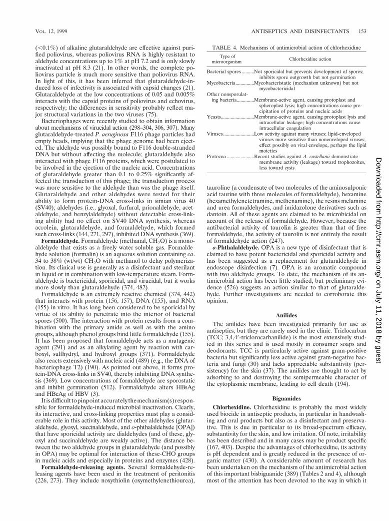
TABLE 4. Mechanisms of antimicrobial action of chlorhexidine
Type ofmicroorganism Chlorhexidine action
Bacterial spores ..........Not sporicidal but prevents development of spores;inhibits spore outgrowth but not germination
Mycobacteria...............Mycobacteristatic (mechanism unknown) but notmycobactericidal
Other nonsporulat-ing bacteria..............Membrane-active agent, causing protoplast and
spheroplast lysis; high concentrations cause pre-cipitation of proteins and nucleic acids
Yeasts...........................Membrane-active agent, causing protoplast lysis andintracellular leakage; high concentrations causeintracellular coagulation
Viruses .........................Low activity against many viruses; lipid-envelopedviruses more sensitive than nonenveloped viruses;effect possibly on viral envelope, perhaps the lipidmoieties
Protozoa ......................Recent studies against A. castellanii demonstratemembrane activity (leakage) toward trophozoites,less toward cysts
VOL. 12, 1999 ANTISEPTICS AND DISINFECTANTS 153
on July 11, 2018 by guesthttp://cm
r.asm.org/
Dow
nloaded from
inactivates nonsporulating bacteria (215, 428, 430, 431, 451).Nevertheless, sufficient data are now available to examine itssporostatic and mycobacteriostatic action, its effects on yeastsand protozoa, and its antiviral activity.
Chlorhexidine is a bactericidal agent (120, 215). Its interac-tion and uptake by bacteria were studied initially by Hugo etal. (222–224), who found that the uptake of chlorhexidine byE. coli and S. aureus was very rapid and depended on thechlorhexidine concentration and pH. More recently, by using[14C]chlorhexidine gluconate, the uptake by bacteria (145) andyeasts (204) was shown to be extremely rapid, with a maximumeffect occurring within 20 s. Damage to the outer cell layerstakes place (139) but is insufficient to induce lysis or cell death.The agent then crosses the cell wall or outer membrane, pre-sumably by passive diffusion, and subsequently attacks the bac-terial cytoplasmic or inner membrane or the yeast plasmamembrane. In yeasts, chlorhexidine “partitions” into the cellwall, plasma membrane, and cytoplasm of cells (205). Damageto the delicate semipermeable membrane is followed by leak-age of intracellular constituents, which can be measured byappropriate techniques. Leakage is not per se responsible forcellular inactivation but is a consequence of cell death (445).High concentrations of chlorhexidine cause coagulation of in-tracellular constituents. As a result, the cytoplasm becomescongealed, with a consequent reduction in leakage (222–224,290), so that there is a biphasic effect on membrane perme-ability. An initial high rate of leakage rises as the concentrationof chlorhexidine increases, but leakage is reduced at higherbiocide concentrations because of the coagulation of the cy-tosol.
Chlorhexidine was claimed by Harold et al. (199) to be aninhibitor of both membrane-bound and soluble ATPase as wellas of net K1 uptake in Enterococcus faecalis. However, onlyhigh biguanide concentrations inhibit membrane-bound ATPase(83), which suggests that the enzyme is not a primary target forchlorhexidine action. Although chlorhexidine collapses the mem-brane potential, it is membrane disruption rather than ATPaseinactivation that is associated with its lethal effects (24, 272).
The effects of chlorhexidine on yeast cells are probably sim-ilar to those previously described for bacteria (204–207). Chlor-hexidine has a biphasic effect on protoplast lysis, with reducedlysis at higher biguanide concentrations. Furthermore, in wholecells, the yeast cell wall may have some effect in limiting theuptake of the biguanide (208). The findings presented here andelsewhere (47, 136, 137, 527) demonstrate an effect on thefungal plasma membrane but with significant actions elsewherein the cell (47). Increasing concentrations of chlorhexidine (upto 25 mg/ml) induce progressive lysis of Saccharomyces cerevi-siae protoplasts, but higher biguanide concentrations result inreduced lysis (205).
Work to date suggests that chlorhexidine has a similar effecton the trophozoites of Acanthameoba castellanii, with the cystsbeing less sensitive (251–255). Furr (163) reviewed the effectsof chlorhexidine and other biocides on Acanthameoba andshowed that membrane damage in these protozoa is a signifi-cant factor in their inactivation.
Mycobacteria are generally highly resistant to chlorhexidine(419). Little is known about the uptake of chlorhexidine (andother antiseptics and disinfectants) by mycobacteria and on thebiochemical changes that occur in the treated cells. Since theMICs for some mycobacteria are on the order of those forchlorhexidine-sensitive, gram-positive cocci (48), the inhibitoryeffects of chlorhexidine on mycobacteria may not be dissimilarto those on susceptible bacteria. Mycobacterium avium-intra-cellulare is considerably more resistant than other mycobacte-ria (48).
Chlorhexidine is not sporicidal (discussed in “Mechanismsof resistance”). Even high concentrations of the bisbiguanidedo not affect the viability of Bacillus spores at ambient tem-peratures (473, 474), although a marked sporicidal effect isachieved at elevated temperatures (475). Presumably, suffi-cient changes occur in the spore structure to permit an in-creased uptake of the biguanide, although this has yet to beshown experimentally. Little is known about the uptake ofchlorhexidine by bacterial spores, although coatless forms takeup more of the compound than do “normal” spores (474).
Chlorhexidine has little effect on the germination of bacte-rial spores (414, 422, 432, 447) but inhibits outgrowth (447).The reason for its lack of effect on the former process but itssignificant activity against the latter is unclear. It could, how-ever, be reflected in the relative uptake of chlorhexidine, sincegerminating cells take up much less of the bisbiguanide than dooutgrowing forms (474). Binding sites could thus be reduced innumber or masked in germinating cells.
The antiviral activity of chlorhexidine is variable. Studieswith different types of bacteriophages have shown that chlor-hexidine has no effect on MS2 or K coliphages (300). Highconcentrations also failed to inactivate Pseudomonas aerugi-nosa phage F116 and had no effect on phage DNA within thecapsid or on phage proteins (301); the transduction processwas more sensitive to chlorhexidine and other biocides thanwas the phage itself. This substantiated an earlier finding (306)that chlorhexidine bound poorly to F116 particles. Chlorhexi-dine is not always considered a particularly effective antiviralagent, and its activity is restricted to the lipid-enveloped viruses(361). Chlorhexidine does not inactivate nonenveloped virusessuch as rotavirus (485), HAV (315), or poliovirus (34). Itsactivity was found by Ranganathan (389) to be restricted to thenucleic acid core or the outer coat, although it is likely that thelatter would be a more important target site.
Alexidine. Alexidine differs chemically from chlorhexidine inpossessing ethylhexyl end groups. Alexidine is more rapidlybactericidal and produces a significantly faster alteration inbactericidal permeability (79, 80). Studies with mixed-lipid andpure phospholipid vesicles demonstrate that, unlike chlorhex-idine, alexidine produces lipid phase separation and domainformation (Table 2). It has been proposed (80) that the natureof the ethylhexyl end group in alexidine, as opposed to thechlorophenol one in chlorhexidine, might influence the abilityof a biguanide to produce lipid domains in the cytoplasmicmembrane.
Polymeric biguanides. Vantocil is a heterodisperse mixtureof polyhexamethylene biguanides (PHMB) with a molecularweight of approximately 3,000. Polymeric biguanides havefound use as general disinfecting agents in the food industryand, very successfully, for the disinfection of swimming pools.Vantocil is active against gram-positive and gram-negative bac-teria, although P. aeruginosa and Proteus vulgaris are less sen-sitive. Vantocil is not sporicidal. PHMB is a membrane-activeagent that also impairs the integrity of the outer membrane ofgram-negative bacteria, although the membrane may also actas a permeability barrier (64, 172). Activity of PHMB increaseson a weight basis with increasing levels of polymerization,which has been linked to enhanced inner membrane perturba-tion (173, 174).
Unlike chlorhexidine but similar to alexidine (Table 2),PHMB causes domain formation of the acidic phospholipids ofthe cytoplasmic membrane (61–64, 172, 173, 227). Permeabilitychanges ensue, and there is believed to be an altered functionof some membrane-associated enzymes. The proposed se-quence of events during its interaction with the cell enve-lope of E. coli is as follows: (i) there is rapid attraction of
154 MCDONNELL AND RUSSELL CLIN. MICROBIOL. REV.
on July 11, 2018 by guesthttp://cm
r.asm.org/
Dow
nloaded from
PHMB toward the negatively charged bacterial cell surface,with strong and specific adsorption to phosphate-containingcompounds; (ii) the integrity of the outer membrane is im-paired, and PHMB is attracted to the inner membrane; (iii)binding of PHMB to phospholipids occurs, with an increase ininner membrane permeability (K1 loss) accompanied by bac-teriostasis; and (iv) complete loss of membrane function fol-lows, with precipitation of intracellular constituents and a bac-tericidal effect.
DiamidinesThe diamidines are characterized chemically as described in
Table 1. The isethionate salts of two compounds, propamidine(4,4-diaminodiphenoxypropane) and dibromopropamidine(2,2-dibromo-4,4-diamidinodiphenoxypropane), have beenused as antibacterial agents. Their antibacterial properties anduses were reviewed by Hugo (213) and Hugo and Russell (226).Clinically, diamidines are used for the topical treatment ofwounds.
The exact mechanism of action of diamidines is unknown,but they have been shown to inhibit oxygen uptake and induceleakage of amino acids (Table 2), as would be expected if theyare considered as cationic surface-active agents. Damage tothe cell surface of P. aeruginosa and Enterobacter cloacae hasbeen described (400).
Halogen-Releasing AgentsChlorine- and iodine-based compounds are the most signif-
icant microbicidal halogens used in the clinic and have beentraditionally used for both antiseptic and disinfectant purposes.
Chlorine-releasing agents. Excellent reviews that deal withthe chemical, physical, and microbiological properties of chlo-rine-releasing agents (CRAs) are available (42, 130). The mostimportant types of CRAs are sodium hypochlorite, chlorinedioxide, and the N-chloro compounds such as sodium di-chloroisocyanurate (NaDCC), with chloramine-T being usedto some extent. Sodium hypochlorite solutions are widely usedfor hard-surface disinfection (household bleach) and can beused for disinfecting spillages of blood containing human im-munodeficiency virus or HBV. NaDCC can also be used forthis purpose and has the advantages of providing a higherconcentration of available chlorine and being less susceptibleto inactivation by organic matter. In water, sodium hypochlo-rite ionizes to produce Na1 and the hypochlorite ion, OCl2,which establishes an equilibrium with hypochlorous acid,HOCl (42). Between pH 4 and 7, chlorine exists predominantlyas HClO, the active moiety, whereas above pH9, OCl2 pre-dominates. Although CRAs have been predominantly used ashard-surface disinfectants, novel acidified sodium chlorite (atwo-component system of sodium chlorite and mandelic acid)has been described as an effective antiseptic (248).
Surprisingly, despite being widely studied, the actual mech-anism of action of CRAs is not fully known (Table 2). CRAsare highly active oxidizing agents and thereby destroy the cel-lular activity of proteins (42); potentiation of oxidation mayoccur at low pH, where the activity of CRAs is maximal,although increased penetration of outer cell layers may beachieved with CRAs in the unionized state. Hypochlorous acidhas long been considered the active moiety responsible forbacterial inactivation by CRAs, the OCl2 ion having a minuteeffect compared to undissolved HOCl (130). This correlateswith the observation that CRA activity is greatest when thepercentage of undissolved HOCl is highest. This concept ap-plies to hypochlorites, NaDCC, and chloramine-T.
Deleterious effects of CRAs on bacterial DNA that involve
the formation of chlorinated derivatives of nucleotide baseshave been described (115, 128, 477). Hypochlorous acid hasalso been found to disrupt oxidative phosphorylation (26) andother membrane-associated activity (70). In a particularly in-teresting paper, McKenna and Davies (321) described the in-hibition of bacterial growth by hypochlorous acid. At 50 mM(2.6 ppm), HOCl completely inhibited the growth of E. coliwithin 5 min, and DNA synthesis was inhibited by 96% butprotein synthesis was inhibited by only 10 to 30%. Becauseconcentrations below 5 mM (260 ppm) did not induce bacterialmembrane disruption or extensive protein degradation, it wasinferred that DNA synthesis was the sensitive target. In con-trast, chlorine dioxide inhibited bacterial protein synthesis (33).
CRAs at higher concentrations are sporicidal (44, 421, 431);this depends on the pH and concentration of available chlorine(408, 412). During treatment, the spores lose refractivity, thespore coat separates from the cortex, and lysis occurs (268). Inaddition, a number of studies have concluded that CRA-treat-ed spores exhibit increased permeability of the spore coat (131,268, 412).
CRAs also possess virucidal activity (34, 46, 116, 315, 394,407, 467, 485, 486). Olivieri et al. (359) showed that chlorineinactivated naked f2 RNA at the same rate as RNA in intactphage, whereas f2 capsid proteins could still adsorb to the host.Taylor and Butler (504) found that the RNA of poliovirus type1 was degraded into fragments by chlorine but that poliovirusinactivation preceded any severe morphological changes. Bycontrast, Floyd et al. (149) and O’Brien and Newman (357)demonstrated that the capsid of poliovirus type 1 was brokendown. Clearly, further studies are needed to explain the anti-viral action of CRAs.
Iodine and iodophors. Although less reactive than chlorine,iodine is rapidly bactericidal, fungicidal, tuberculocidal, viru-cidal, and sporicidal (184). Although aqueous or alcoholic (tinc-ture) solutions of iodine have been used for 150 years as an-tiseptics, they are associated with irritation and excessivestaining. In addition, aqueous solutions are generally unstable;in solution, at least seven iodine species are present in a com-plex equilibrium, with molecular iodine (I2) being primarilyresponsible for antimictrobial efficacy (184). These problemswere overcome by the development of iodophors (“iodine car-riers” or “iodine-releasing agents”); the most widely used arepovidone-iodine and poloxamer-iodine in both antiseptics anddisinfectants. Iodophors are complexes of iodine and a solubi-lizing agent or carrier, which acts as a reservoir of the active“free” iodine (184). Although germicidal activity is maintained,iodophors are considered less active against certain fungi andspores than are tinctures (454).
Similar to chlorine, the antimicrobial action of iodine israpid, even at low concentrations, but the exact mode of actionis unknown. Iodine rapidly penetrates into microorganisms(76) and attacks key groups of proteins (in particular the free-sulfur amino acids cysteine and methionine [184, 267]), nucle-otides, and fatty acids (15, 184), which culminates in cell death(184). Less is known about the antiviral action of iodine, butnonlipid viruses and parvoviruses are less sensitive than lipidenveloped viruses (384). Similarly to bacteria, it is likely thatiodine attacks the surface proteins of enveloped viruses, butthey may also destabilize membrane fatty acids by reacting withunsaturated carbon bonds (486).
Silver Compounds
In one form or another, silver and its compounds have longbeen used as antimicrobial agents (55, 443). The most im-portant silver compound currently in use is silver sulfadiazine
VOL. 12, 1999 ANTISEPTICS AND DISINFECTANTS 155
on July 11, 2018 by guesthttp://cm
r.asm.org/
Dow
nloaded from
(AgSD), although silver metal, silver acetate, silver nitrate, andsilver protein, all of which have antimicrobial properties, arelisted in Martindale, The Extra Pharmacopoeia (312). In recentyears, silver compounds have been used to prevent the infec-tion of burns and some eye infections and to destroy warts.
Silver nitrate. The mechanism of the antimicrobial action ofsilver ions is closely related to their interaction with thiol (sul-fydryl, ™SH) groups (32, 49, 161, 164), although other targetsites remain a possibility (397, 509). Liau et al (287) demon-strated that amino acids such as cysteine and other compoundssuch as sodium thioglycolate containing thiol groups neutral-ized the activity of silver nitrate against P. aeruginosa. By con-trast, amino acids containing disulfide (SS) bonds, non-sulfur-containing amino acids, and sulfur-containing compounds suchas cystathione, cysteic acid, L-methionine, taurine, sodium bi-sulfite, and sodium thiosulfate were all unable to neutralizeAg1 activity. These and other findings imply that interaction ofAg1 with thiol groups in enzymes and proteins plays an essen-tial role in bacterial inactivation, although other cellular com-ponents may be involved. Hydrogen bonding, the effects ofhydrogen bond-breaking agents, and the specificity of Ag1 forthiol groups were discussed in greater detail by Russell andHugo (443) (Table 2). Virucidal properties might also be ex-plained by binding to ™SH groups (510).
Lukens (292) proposed that silver salts and other heavymetals such as copper act by binding to key functional groupsof fungal enzymes. Ag1 causes the release of K1 ions frommicroorganisms; the microbial plasma or cytoplasmic mem-brane, with which is associated many important enzymes, is animportant target site for Ag1 activity (161, 329, 392, 470).
In addition to its effects on enzymes, Ag1 produces otherchanges in microorganisms. Silver nitrate causes marked inhi-bition of growth of Cryptococcus neoformans and is deposit-ed in the vacuole and cell wall as granules (60). Ag1 inhibitscell division and damages the cell envelope and contents ofP. aeruginosa (398). Bacterial cells increase in size, and thecytoplasmic membrane, cytoplasmic contents, and outer celllayers all exhibit structural abnormalities, although without anyblebs (protuberances) (398). Finally, the Ag1 ion interacts withnucleic acids (543); it interacts preferentially with the bases inDNA rather than with the phosphate groups, although thesignificance of this in terms of its lethal action is unclear (231,387, 510, 547).
Silver sulfadiazine. AgSD is essentially a combination of twoantibacterial agents, Ag1 and sulfadiazine (SD). The questionwhether the antibacterial effect of AgSD arises predominantlyfrom only one of the compounds or via a synergistic interac-tion has been posed repeatedly. AgSD has a broad spectrum ofactivity and, unlike silver nitrate, produces surface and mem-brane blebs in susceptible (but not resistant) bacteria (96).AgSD binds to cell components, including DNA (332, 404).Based on a chemical analysis, Fox (153) proposed a polymericstructure of AgSD composed of six silver atoms bonding to sixSD molecules by linkage of the silver atoms to the nitrogens ofthe SD pyrimidine ring. Bacterial inhibition would then pre-sumably be achieved when silver binds to sufficient base pairsin the DNA helix, thereby inhibiting transcription. Similarly, itsantiphage properties have been ascribed to the fact that AgSDbinds to phage DNA (154, 388). Clearly, the precise mecha-nism of action of AgSD has yet to be solved.
Peroxygens
Hydrogen peroxide. Hydrogen peroxide (H2O2) is a widelyused biocide for disinfection, sterilization, and antisepsis. It isa clear, colorless liquid that is commercially available in a va-
riety of concentrations ranging from 3 to 90%. H2O2 is con-sidered environmentally friendly, because it can rapidly de-grade into the innocuous products water and oxygen. Althoughpure solutions are generally stable, most contain stabilizersto prevent decomposition. H2O2 demonstrates broad-spectrumefficacy against viruses, bacteria, yeasts, and bacterial spores(38). In general, greater activity is seen against gram-positivethan gram-negative bacteria; however, the presence of catalaseor other peroxidases in these organisms can increase tolerancein the presence of lower concentrations. Higher concentrationsof H2O2 (10 to 30%) and longer contact times are required forsporicidal activity (416), although this activity is significantlyincreased in the gaseous phase. H2O2 acts as an oxidant byproducing hydroxyl free radicals (•OH) which attack essentialcell components, including lipids, proteins, and DNA. It hasbeen proposed that exposed sulfhydryl groups and doublebonds are particularly targeted (38).
Peracetic acid. Peracetic acid (PAA) (CH3COOOH) is con-sidered a more potent biocide than hydrogen peroxide, beingsporicidal, bactericidal, virucidal, and fungicidal at low concen-trations (,0.3%) (38). PAA also decomposes to safe by-prod-ucts (acetic acid and oxygen) but has the added advantages ofbeing free from decomposition by peroxidases, unlike H2O2,and remaining active in the presence of organic loads (283,308). Its main application is as a low-temperature liquid ster-ilant for medical devices, flexible scopes, and hemodialyzers,but it is also used as an environmental surface sterilant (100,308).
Similar to H2O2, PAA probably denatures proteins and en-zymes and increases cell wall permeability by disrupting sulf-hydryl (™SH) and sulfur (S™S) bonds (22, 38).
Phenols
Phenolic-type antimicrobial agents have long been used fortheir antiseptic, disinfectant, or preservative properties, de-pending on the compound. It has been known for many years(215) that, although they have often been referred to as “gen-eral protoplasmic poisons,” they have membrane-active prop-erties which also contribute to their overall activity (120) (Ta-ble 2).
Phenol induces progressive leakage of intracellular constit-uents, including the release of K1, the first index of membranedamage (273), and of radioactivity from 14C-labeled E. coli(242, 265). Pulvertaft and Lumb (386) demonstrated that lowconcentrations of phenols (0.032%, 320 mg/ml) and other (non-phenolic) agents lysed rapidly growing cultures of E. coli,staphylococci, and streptococci and concluded that autolyticenzymes were not involved. Srivastava and Thompson (487,488) proposed that phenol acts only at the point of separationof pairs of daughter cells, with young bacterial cells being moresensitive than older cells to phenol.
Hugo and Bloomfield (216, 217) showed with the chlori-nated bis-phenol fenticlor that there was a close relationshipbetween bactericidal activity and leakage of 260-nm-absorbingmaterial (leakage being induced only by bactericidal concen-trations). Fentichlor affected the metabolic activities of S. au-reus and E. coli (217) and produced a selective increase inpermeability to protons with a consequent dissipation of theproton motive force (PMF) and an uncoupling of oxidativephosphorylation (41). Chlorocresol has a similar action (124).Coagulation of cytoplasmic constituents at higher phenol con-centrations, which causes irreversible cellular damage, has beendescribed by Hugo (215).
The phenolics possess antifungal and antiviral properties.Their antifungal action probably involves damage to the plas-
156 MCDONNELL AND RUSSELL CLIN. MICROBIOL. REV.
on July 11, 2018 by guesthttp://cm
r.asm.org/
Dow
nloaded from
ma membrane (436), resulting in leakage of intracellular con-stituents. Phenol does not affect the transduction of P. aerugi-nosa PAO by bacteriophage F116 (301), has no effect on phageDNA within the capsid, and has little effect on several of thephage band proteins unless treatments of 20 min or longer areused (303, 304).
Bis-PhenolsThe bis-phenols are hydroxy-halogenated derivatives of two
phenolic groups connected by various bridges (191, 446). Ingeneral, they exhibit broad-spectrum efficacy but have littleactivity against P. aeruginosa and molds and are sporostatic to-ward bacterial spores. Triclosan and hexachlorophane are themost widely used biocides in this group, especially in antisepticsoaps and hand rinses. Both compounds have been shown tohave cumulative and persistent effects on the skin (313).
Triclosan. Triclosan (2,4,49-trichloro-29-hydroxydiphenylether; Irgasan DP 300) exhibits particular activity against gram-positive bacteria (469, 521). Its efficacy against gram-negativebacteria and yeasts can be significantly enhanced by formula-tion effects. For example, triclosan in combination with EDTAcaused increased permeability of the outer membrane (282).Reports have also suggested that in addition to its antibacterialproperties, triclosan may have anti-inflammatory activity (25,522). The specific mode of action of triclosan is unknown, butit has been suggested that the primary effects are on the cyto-plasmic membrane. In studies with E. coli, triclosan at subin-hibitory concentrations inhibited the uptake of essential nutri-ents, while higher, bactericidal concentrations resulted in therapid release of cellular components and cell death (393).Studies with a divalent-ion-dependent E. coli triclosan mutantfor which the triclosan MIC was 10-fold greater than that for awild-type strain showed no significant differences in total en-velope protein profiles but did show significant differences inenvelope fatty acids (370). Specifically, a prominent 14:1 fattyacid was absent in the resistant strain, and there were minordifferences in other fatty acid species. It was proposed thatdivalent ions and fatty acids may adsorb and limit the perme-ability of triclosan to its site of action (370). Minor changes infatty acid profiles were recently found in both E. coli andS. aureus strains for which the triclosan MICs were elevated;however, the MBCs were not affected, suggesting, as for otherphenols, that the cumulative effects on multiple targets con-tribute to the bactericidal activity (318, 319).
Hexachlorophene. Hexachlorophene (hexachlorophane;2,29-dihydroxy-3,5,6,39,59,69-hexachlorodiphenylmethane) isanother bis-phenol whose mode of action has been extensivelystudied. The primary action of hexachlorophene, based onstudies with Bacillus megatherium, is to inhibit the membrane-bound part of the electron transport chain, and the othereffects noted above are secondary ones that occur only at highconcentrations (92, 158, 241, 481). It induces leakage, causesprotoplast lysis, and inhibits respiration. The threshold con-centration for the bactericidal activity of hexachlorphene is 10mg/ml (dry weight), but peak leakage occurs at concentrationshigher than 50 mg/ml and cytological changes occur above 30mg/ml. Furthermore, hexachlorophene is bactericidal at 0°Cdespite causing little leakage at this temperature. Despite thebroad-spectrum efficacy of hexachlorophene, concerns abouttoxicity (256), in particular in neonates, have meant that its usein antiseptic products has been limited.
HalophenolsChloroxylenol (4-chloro-3,5-dimethylphenol; p-chloro-m-xy-
lenol) is the key halophenol used in antiseptic or disinfectant
formulations (66). Chloroxylenol is bactericidal, but P. aerugi-nosa and many molds are highly resistant (66, 432). Surpris-ingly, its mechanism of action has been little studied despite itswidespread use over many years. Because of its phenolic na-ture, it would be expected to have an effect on microbial mem-branes.
Quaternary Ammonium Compounds
Surface-active agents (surfactants) have two regions in theirmolecular structures, one a hydrocarbon, water-repellent (hy-drophobic) group and the other a water-attracting (hydrophilicor polar) group. Depending on the basis of the charge or ab-sence of ionization of the hydrophilic group, surfactants areclassified into cationic, anionic, nonionic, and ampholytic (am-photeric) compounds. Of these, the cationic agents, as exem-plified by quaternary ammonium compounds (QACs), are themost useful antiseptics and disinfectants (160). They are some-times known as cationic detergents. QACs have been used fora variety of clinical purposes (e.g., preoperative disinfection ofunbroken skin, application to mucous membranes, and disin-fection of noncritical surfaces). In addition to having antimi-crobial properties, QACs are also excellent for hard-surfacecleaning and deodorization.
It has been known for many years that QACs are membrane-active agents (221) (Table 2) (i.e., with a target site predomi-nantly at the cytoplasmic (inner) membrane in bacteria or theplasma membrane in yeasts) (215). Salton (460) proposed thefollowing sequence of events with microorganisms exposed tocationic agents: (i) adsorption and penetration of the agentinto the cell wall; (ii) reaction with the cytoplasmic membrane(lipid or protein) followed by membrane disorganization; (iii)leakage of intracellular low-molecular-weight material; (iv)degradation of proteins and nucleic acids; and (v) wall lysiscaused by autolytic enzymes. There is thus a loss of structuralorganization and integrity of the cytoplasmic membrane inbacteria, together with other damaging effects to the bacterialcell (120).
Useful information about the selectivity of membrane actioncan be obtained by studying the effects of biocides on proto-plasts and spheroplasts suspended in various solutes. QACscause lysis of spheroplasts and protoplasts suspended in su-crose (107, 215, 243, 428). The cationic agents react with phos-pholipid components in the cytoplasmic membrane (69), there-by producing membrane distortion and protoplast lysis underosmotic stress. Isolated membranes do not undergo disaggre-gation on exposure to QACs, because the membrane distortionis not sufficiently drastic. The non-QAC agents TCC and tri-chlorosalicylanide have specific effects: TCC induces proto-plast lysis in ammonium chloride by increasing Cl2 permeabil-ity, whereas trichlorosalicylanide induces lysis in ammoniumnitrate by increasing NO3
2 permeability (428). In contrast,QACs (and chlorhexidine) induce lysis of protoplasts or sphe-roplasts suspended in various solutes because they effect gen-eralized, rather than specific, membrane damage.
The bacterial cytoplasmic membrane provides the mecha-nism whereby metabolism is linked to solute transport, flagel-lar movement, and the generation of ATP. Protons are ex-truded to the exterior of the bacterial cell during metabolism.The combined potential (concentration or osmotic effect of theproton and its electropositivity) is the PMF, which drives theseancillary activities (428). The QAC cetrimide was found (121)to have an effect on the PMF in S. aureus. At its bacteriostaticconcentration, cetrimide caused the discharge of the pH com-ponent of the PMF and also produced the maximum amountof 260-nm-absorbing material.
VOL. 12, 1999 ANTISEPTICS AND DISINFECTANTS 157
on July 11, 2018 by guesthttp://cm
r.asm.org/
Dow
nloaded from
QACs are also believed to damage the outer membrane ofgram-negative bacteria, thereby promoting their own uptake.This aspect of QACs is considered below (see “Intrinsic resis-tance of gram-negative bacteria”).
The QAC cetylpyridium chloride (CPC) induces the leakageof K1 and pentose material from the yeast S. cerevisiae andinduces protoplast lysis as well as interacting with crude cellsap (205). Unlike chlorhexidine, however, no biphasic effect onprotoplast lysis was observed. The initial toxic effect of QACson yeast cells is a disorganization of the plasma membranes,with organized lipid structures in the membranes (and in lipidbilayers) being disrupted.
QACs are sporostatic; they inhibit the outgrowth of spores(the development of a vegetative cell from a germinated spore)but not the actual germination processes (development fromdormancy to a metabolically active state), albeit by an unknownmechanism (414). Likewise, the QACs are not mycobacteri-cidal but have a mycobacteriostatic action, although the actualeffects on mycobacteria have been little studied (419).
The QACs have an effect on lipid, enveloped (including hu-man immunodeficiency virus and HBV) but not nonenvelopedviruses (394, 485, 486). QAC-based products induced disinte-gration and morphological changes of human HBV, resultingin loss of infectivity (382). In studies with different phages(298–301, 303–305, 307), CPC significantly inhibited transduc-tion by bacteriophage F116 and inactivated the phage particles.Furthermore, CPC altered the protein bands of F116 but didnot affect the phage DNA within the capsid.
Vapor-Phase Sterilants
Many heat-sensitive medical devices and surgical suppliescan be effectively sterilized by liquid sterilants (in particularglutaraldehyde, PAA, and hydrogen peroxide) or by vapor-phase sterilization systems (Table 1). The most widely usedactive agents in these “cold” systems are ethylene oxide, form-aldehyde and, more recently developed, hydrogen peroxideand PAA. Ethylene oxide and formaldehyde are both broad-spectrum alkylating agents. However, their activity is depen-dent on active concentration, temperature, duration of expo-sure, and relative humidity (87). As alkylating agents, theyattack proteins, nucleic acids, and other organic compounds;both are particularly reactive with sulfhydryl and other en-zyme-reactive groups. Ethylene oxide gas has the disadvan-tages of being mutagenic and explosive but is not generallyharsh on sensitive equipment, and toxic residuals from thesterilization procedure can be routinely eliminated by correctaeration. Formaldehyde gas is similar and has the added ad-vantage of being nonexplosive but is not widely used in healthcare. Vapor-phase hydrogen peroxide and PAA are consideredmore active (as oxidants) at lower concentrations than in theliquid form (334). Both active agents are used in combinationwith gas plasma in low-temperature sterilization systems (314).Their main advantages over other vapor-phase systems includelow toxicity, rapid action, and activity at lower temperature; thedisadvantages include limited penetrability and applications.
MECHANISMS OF RESISTANCE
Introduction
As stated above, different types of microorganisms vary intheir response to antiseptics and disinfectants. This is hardlysurprising in view of their different cellular structure, compo-sition, and physiology. Traditionally, microbial susceptibility toantiseptics and disinfectants has been classified based on these
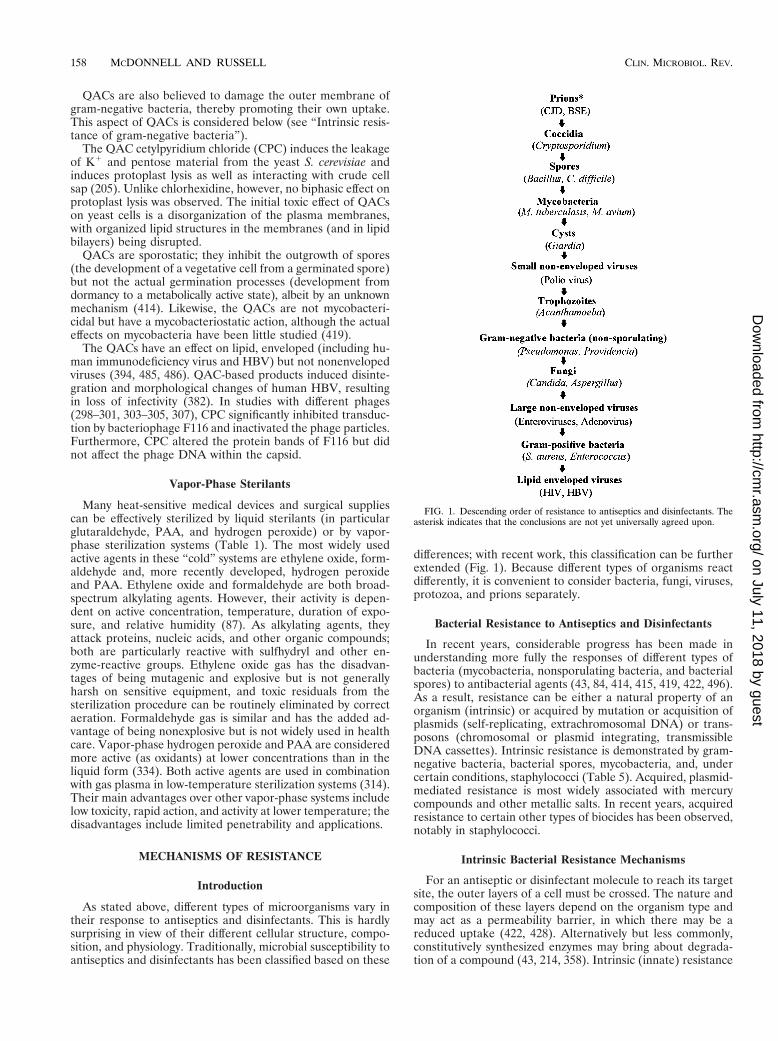
differences; with recent work, this classification can be furtherextended (Fig. 1). Because different types of organisms reactdifferently, it is convenient to consider bacteria, fungi, viruses,protozoa, and prions separately.
Bacterial Resistance to Antiseptics and Disinfectants
In recent years, considerable progress has been made inunderstanding more fully the responses of different types ofbacteria (mycobacteria, nonsporulating bacteria, and bacterialspores) to antibacterial agents (43, 84, 414, 415, 419, 422, 496).As a result, resistance can be either a natural property of anorganism (intrinsic) or acquired by mutation or acquisition ofplasmids (self-replicating, extrachromosomal DNA) or trans-posons (chromosomal or plasmid integrating, transmissibleDNA cassettes). Intrinsic resistance is demonstrated by gram-negative bacteria, bacterial spores, mycobacteria, and, undercertain conditions, staphylococci (Table 5). Acquired, plasmid-mediated resistance is most widely associated with mercurycompounds and other metallic salts. In recent years, acquiredresistance to certain other types of biocides has been observed,notably in staphylococci.
Intrinsic Bacterial Resistance Mechanisms
For an antiseptic or disinfectant molecule to reach its targetsite, the outer layers of a cell must be crossed. The nature andcomposition of these layers depend on the organism type andmay act as a permeability barrier, in which there may be areduced uptake (422, 428). Alternatively but less commonly,constitutively synthesized enzymes may bring about degrada-tion of a compound (43, 214, 358). Intrinsic (innate) resistance
FIG. 1. Descending order of resistance to antiseptics and disinfectants. Theasterisk indicates that the conclusions are not yet universally agreed upon.
158 MCDONNELL AND RUSSELL CLIN. MICROBIOL. REV.
on July 11, 2018 by guesthttp://cm
r.asm.org/
Dow
nloaded from
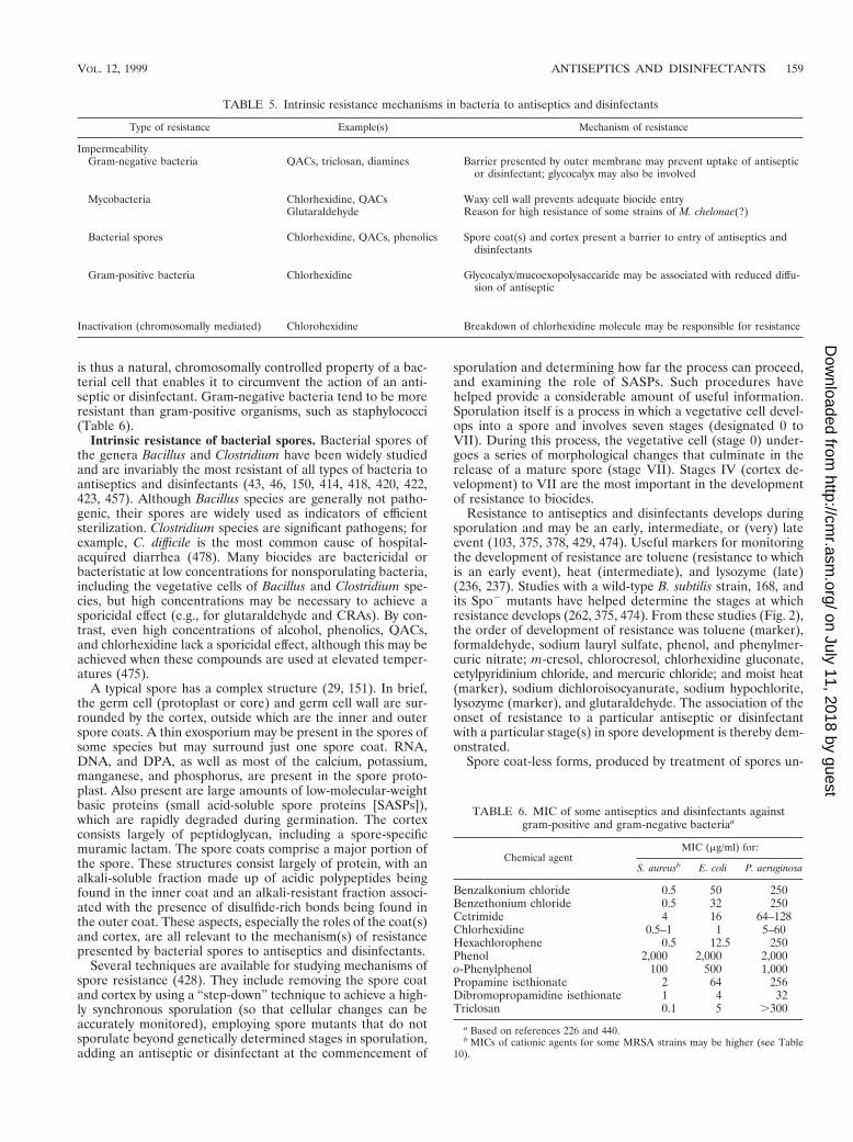
is thus a natural, chromosomally controlled property of a bac-terial cell that enables it to circumvent the action of an anti-septic or disinfectant. Gram-negative bacteria tend to be moreresistant than gram-positive organisms, such as staphylococci(Table 6).
Intrinsic resistance of bacterial spores. Bacterial spores ofthe genera Bacillus and Clostridium have been widely studiedand are invariably the most resistant of all types of bacteria toantiseptics and disinfectants (43, 46, 150, 414, 418, 420, 422,423, 457). Although Bacillus species are generally not patho-genic, their spores are widely used as indicators of efficientsterilization. Clostridium species are significant pathogens; forexample, C. difficile is the most common cause of hospital-acquired diarrhea (478). Many biocides are bactericidal orbacteristatic at low concentrations for nonsporulating bacteria,including the vegetative cells of Bacillus and Clostridium spe-cies, but high concentrations may be necessary to achieve asporicidal effect (e.g., for glutaraldehyde and CRAs). By con-trast, even high concentrations of alcohol, phenolics, QACs,and chlorhexidine lack a sporicidal effect, although this may beachieved when these compounds are used at elevated temper-atures (475).
A typical spore has a complex structure (29, 151). In brief,the germ cell (protoplast or core) and germ cell wall are sur-rounded by the cortex, outside which are the inner and outerspore coats. A thin exosporium may be present in the spores ofsome species but may surround just one spore coat. RNA,DNA, and DPA, as well as most of the calcium, potassium,manganese, and phosphorus, are present in the spore proto-plast. Also present are large amounts of low-molecular-weightbasic proteins (small acid-soluble spore proteins [SASPs]),which are rapidly degraded during germination. The cortexconsists largely of peptidoglycan, including a spore-specificmuramic lactam. The spore coats comprise a major portion ofthe spore. These structures consist largely of protein, with analkali-soluble fraction made up of acidic polypeptides beingfound in the inner coat and an alkali-resistant fraction associ-ated with the presence of disulfide-rich bonds being found inthe outer coat. These aspects, especially the roles of the coat(s)and cortex, are all relevant to the mechanism(s) of resistancepresented by bacterial spores to antiseptics and disinfectants.
Several techniques are available for studying mechanisms ofspore resistance (428). They include removing the spore coatand cortex by using a “step-down” technique to achieve a high-ly synchronous sporulation (so that cellular changes can beaccurately monitored), employing spore mutants that do notsporulate beyond genetically determined stages in sporulation,adding an antiseptic or disinfectant at the commencement of
sporulation and determining how far the process can proceed,and examining the role of SASPs. Such procedures havehelped provide a considerable amount of useful information.Sporulation itself is a process in which a vegetative cell devel-ops into a spore and involves seven stages (designated 0 toVII). During this process, the vegetative cell (stage 0) under-goes a series of morphological changes that culminate in therelease of a mature spore (stage VII). Stages IV (cortex de-velopment) to VII are the most important in the developmentof resistance to biocides.
Resistance to antiseptics and disinfectants develops duringsporulation and may be an early, intermediate, or (very) lateevent (103, 375, 378, 429, 474). Useful markers for monitoringthe development of resistance are toluene (resistance to whichis an early event), heat (intermediate), and lysozyme (late)(236, 237). Studies with a wild-type B. subtilis strain, 168, andits Spo2 mutants have helped determine the stages at whichresistance develops (262, 375, 474). From these studies (Fig. 2),the order of development of resistance was toluene (marker),formaldehyde, sodium lauryl sulfate, phenol, and phenylmer-curic nitrate; m-cresol, chlorocresol, chlorhexidine gluconate,cetylpyridinium chloride, and mercuric chloride; and moist heat(marker), sodium dichloroisocyanurate, sodium hypochlorite,lysozyme (marker), and glutaraldehyde. The association of theonset of resistance to a particular antiseptic or disinfectantwith a particular stage(s) in spore development is thereby dem-onstrated.
Spore coat-less forms, produced by treatment of spores un-
TABLE 6. MIC of some antiseptics and disinfectants againstgram-positive and gram-negative bacteriaa
Chemical agentMIC (mg/ml) for:
S. aureusb E. coli P. aeruginosa
Benzalkonium chloride 0.5 50 250Benzethonium chloride 0.5 32 250Cetrimide 4 16 64–128Chlorhexidine 0.5–1 1 5–60Hexachlorophene 0.5 12.5 250Phenol 2,000 2,000 2,000o-Phenylphenol 100 500 1,000Propamine isethionate 2 64 256Dibromopropamidine isethionate 1 4 32Triclosan 0.1 5 .300
a Based on references 226 and 440.b MICs of cationic agents for some MRSA strains may be higher (see Table
10).
TABLE 5. Intrinsic resistance mechanisms in bacteria to antiseptics and disinfectants
Type of resistance Example(s) Mechanism of resistance
ImpermeabilityGram-negative bacteria QACs, triclosan, diamines Barrier presented by outer membrane may prevent uptake of antiseptic
or disinfectant; glycocalyx may also be involved
Mycobacteria Chlorhexidine, QACs Waxy cell wall prevents adequate biocide entryGlutaraldehyde Reason for high resistance of some strains of M. chelonae(?)
Bacterial spores Chlorhexidine, QACs, phenolics Spore coat(s) and cortex present a barrier to entry of antiseptics anddisinfectants
Gram-positive bacteria Chlorhexidine Glycocalyx/mucoexopolysaccaride may be associated with reduced diffu-sion of antiseptic
Inactivation (chromosomally mediated) Chlorohexidine Breakdown of chlorhexidine molecule may be responsible for resistance
VOL. 12, 1999 ANTISEPTICS AND DISINFECTANTS 159
on July 11, 2018 by guesthttp://cm
r.asm.org/
Dow
nloaded from
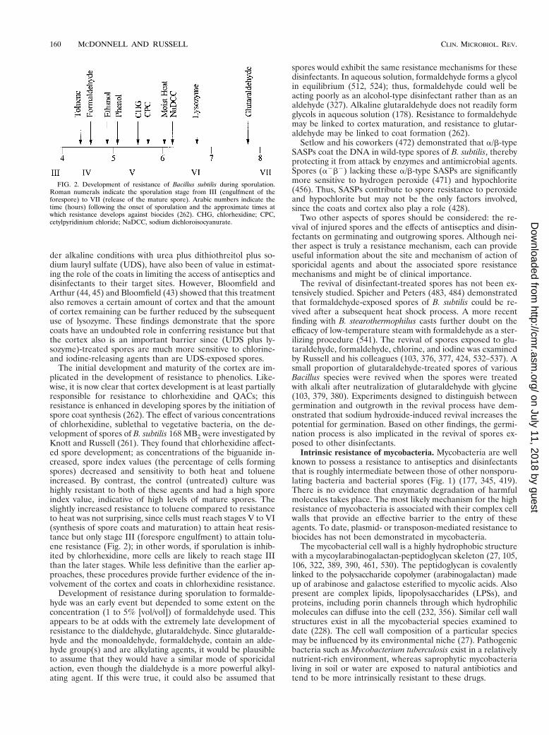
der alkaline conditions with urea plus dithiothreitol plus so-dium lauryl sulfate (UDS), have also been of value in estimat-ing the role of the coats in limiting the access of antiseptics anddisinfectants to their target sites. However, Bloomfield andArthur (44, 45) and Bloomfield (43) showed that this treatmentalso removes a certain amount of cortex and that the amountof cortex remaining can be further reduced by the subsequentuse of lysozyme. These findings demonstrate that the sporecoats have an undoubted role in conferring resistance but thatthe cortex also is an important barrier since (UDS plus ly-sozyme)-treated spores are much more sensitive to chlorine-and iodine-releasing agents than are UDS-exposed spores.
The initial development and maturity of the cortex are im-plicated in the development of resistance to phenolics. Like-wise, it is now clear that cortex development is at least partiallyresponsible for resistance to chlorhexidine and QACs; thisresistance is enhanced in developing spores by the initiation ofspore coat synthesis (262). The effect of various concentrationsof chlorhexidine, sublethal to vegetative bacteria, on the de-velopment of spores of B. subtilis 168 MB2 were investigated byKnott and Russell (261). They found that chlorhexidine affect-ed spore development; as concentrations of the biguanide in-creased, spore index values (the percentage of cells formingspores) decreased and sensitivity to both heat and tolueneincreased. By contrast, the control (untreated) culture washighly resistant to both of these agents and had a high sporeindex value, indicative of high levels of mature spores. Theslightly increased resistance to toluene compared to resistanceto heat was not surprising, since cells must reach stages V to VI(synthesis of spore coats and maturation) to attain heat resis-tance but only stage III (forespore engulfment) to attain tolu-ene resistance (Fig. 2); in other words, if sporulation is inhib-ited by chlorhexidine, more cells are likely to reach stage IIIthan the later stages. While less definitive than the earlier ap-proaches, these procedures provide further evidence of the in-volvement of the cortex and coats in chlorhexidine resistance.
Development of resistance during sporulation to formalde-hyde was an early event but depended to some extent on theconcentration (1 to 5% [vol/vol]) of formaldehyde used. Thisappears to be at odds with the extremely late development ofresistance to the dialdehyde, glutaraldehyde. Since glutaralde-hyde and the monoaldehyde, formaldehyde, contain an alde-hyde group(s) and are alkylating agents, it would be plausibleto assume that they would have a similar mode of sporicidalaction, even though the dialdehyde is a more powerful alkyl-ating agent. If this were true, it could also be assumed that
spores would exhibit the same resistance mechanisms for thesedisinfectants. In aqueous solution, formaldehyde forms a glycolin equilibrium (512, 524); thus, formaldehyde could well beacting poorly as an alcohol-type disinfectant rather than as analdehyde (327). Alkaline glutaraldehyde does not readily formglycols in aqueous solution (178). Resistance to formaldehydemay be linked to cortex maturation, and resistance to glutar-aldehyde may be linked to coat formation (262).
Setlow and his coworkers (472) demonstrated that a/b-typeSASPs coat the DNA in wild-type spores of B. subtilis, therebyprotecting it from attack by enzymes and antimicrobial agents.Spores (a2b2) lacking these a/b-type SASPs are significantlymore sensitive to hydrogen peroxide (471) and hypochlorite(456). Thus, SASPs contribute to spore resistance to peroxideand hypochlorite but may not be the only factors involved,since the coats and cortex also play a role (428).
Two other aspects of spores should be considered: the re-vival of injured spores and the effects of antiseptics and disin-fectants on germinating and outgrowing spores. Although nei-ther aspect is truly a resistance mechanism, each can provideuseful information about the site and mechanism of action ofsporicidal agents and about the associated spore resistancemechanisms and might be of clinical importance.
The revival of disinfectant-treated spores has not been ex-tensively studied. Spicher and Peters (483, 484) demonstratedthat formaldehyde-exposed spores of B. subtilis could be re-vived after a subsequent heat shock process. A more recentfinding with B. stearothermophilus casts further doubt on theefficacy of low-temperature steam with formaldehyde as a ster-ilizing procedure (541). The revival of spores exposed to glu-taraldehyde, formaldehyde, chlorine, and iodine was examinedby Russell and his colleagues (103, 376, 377, 424, 532–537). Asmall proportion of glutaraldehyde-treated spores of variousBacillus species were revived when the spores were treatedwith alkali after neutralization of glutaraldehyde with glycine(103, 379, 380). Experiments designed to distinguish betweengermination and outgrowth in the revival process have dem-onstrated that sodium hydroxide-induced revival increases thepotential for germination. Based on other findings, the germi-nation process is also implicated in the revival of spores ex-posed to other disinfectants.
Intrinsic resistance of mycobacteria. Mycobacteria are wellknown to possess a resistance to antiseptics and disinfectantsthat is roughly intermediate between those of other nonsporu-lating bacteria and bacterial spores (Fig. 1) (177, 345, 419).There is no evidence that enzymatic degradation of harmfulmolecules takes place. The most likely mechanism for the highresistance of mycobacteria is associated with their complex cellwalls that provide an effective barrier to the entry of theseagents. To date, plasmid- or transposon-mediated resistance tobiocides has not been demonstrated in mycobacteria.
The mycobacterial cell wall is a highly hydrophobic structurewith a mycoylarabinogalactan-peptidoglycan skeleton (27, 105,106, 322, 389, 390, 461, 530). The peptidoglycan is covalentlylinked to the polysaccharide copolymer (arabinogalactan) madeup of arabinose and galactose esterified to mycolic acids. Alsopresent are complex lipids, lipopolysaccharides (LPSs), andproteins, including porin channels through which hydrophilicmolecules can diffuse into the cell (232, 356). Similar cell wallstructures exist in all the mycobacterial species examined todate (228). The cell wall composition of a particular speciesmay be influenced by its environmental niche (27). Pathogenicbacteria such as Mycobacterium tuberculosis exist in a relativelynutrient-rich environment, whereas saprophytic mycobacterialiving in soil or water are exposed to natural antibiotics andtend to be more intrinsically resistant to these drugs.
FIG. 2. Development of resistance of Bacillus subtilis during sporulation.Roman numerals indicate the sporulation stage from III (engulfment of theforespore) to VII (release of the mature spore). Arabic numbers indicate thetime (hours) following the onset of sporulation and the approximate times atwhich resistance develops against biocides (262). CHG, chlorhexidine; CPC,cetylpyridinium chloride; NaDCC, sodium dichloroisocyanurate.
160 MCDONNELL AND RUSSELL CLIN. MICROBIOL. REV.
on July 11, 2018 by guesthttp://cm
r.asm.org/
Dow
nloaded from
Antiseptics or disinfectants that exhibit mycobacterial activ-ity are phenol, PAA, hydrogen peroxide, alcohol, and glutar-aldehyde (16, 17, 99, 419, 425, 455). By contrast, other well-known bactericidal agents, such as chlorhexidine and QACs,are mycobacteristatic even when used at high concentrations(51, 52, 419, 425, 455). However, the activity of these can besubstantially increased by formulation effects. Thus, a numberof QAC-based products claim to have mycobacterial activity.For example, a newer formulation (Sactimed-I-Sinald) con-taining a mixture of alkyl polyguanides and alkyl QACs isclaimed to be mycobactericidal (211, 353). However, there issome doubt whether the antibacterial agents had been prop-erly quenched or neutralized to prevent carryover of inhibitoryconcentrations into recovery media.
Many years ago, it was proposed (T. H. Shen, cited in ref-erence 99) that the resistance of mycobacteria to QACs wasrelated to the lipid content of the cell wall. In support of thiscontention, Mycobacterium phlei, which has a low total celllipid content, was more sensitive than M. tuberculosis, whichhas a higher lipid content. It was also noted that the resistanceof various species of mycobacteria was related to the content ofwaxy material in the wall. It is now known that because of thehighly hydrophobic nature of the cell wall, hydrophilic biocidesare generally unable to penetrate the mycobacterial cell wall insufficiently high concentrations to produce a lethal effect. How-ever, low concentrations of antiseptics and disinfectants suchas chlorhexidine must presumably traverse this permeabilitybarrier, because the MICs are of the same order as those con-centrations inhibiting the growth of nonmycobacterial strainssuch as S. aureus, although M. avium-intracellulare may be par-ticularly resistant (51, 52). The component(s) of the mycobac-terial cell wall responsible for the high biocide resistance arecurrently unknown, although some information is available.Inhibitors of cell wall synthesis increase the susceptibility ofM. avium to drugs (391); inhibition of myocide C, arabinoga-lactan, and mycolic acid biosynthesis enhances drug suscepti-bility. Treatment of this organism with m-fluoro-DL-phenylala-nine (m-FL-phe), which inhibits mycocide C synthesis, producessignificant alterations in the outer cell wall layers (106). Eth-ambutol, an inhibitor of arabinogalactan (391, 501) and phos-pholipid (461, 462) synthesis, also disorganizes these layers. Inaddition, ethambutol induces the formation of ghosts withoutthe dissolution of peptidoglycan (391). Methyl-4-(2-octadecyl-cyclopropen-1-yl) butanoate (MOCB) is a structural analogueof a key precursor in mycolic acid synthesis. Thus, the effects ofMOCB on mycolic acid synthesis and m-FL-phe and etham-butol on outer wall biosynthetic processes leading to changesin cell wall architecture appear to be responsible for increas-ing the intracellular concentration of chemotherapeutic drugs.These findings support the concept of the cell wall acting as apermeability barrier to these drugs (425). Fewer studies havebeen made of the mechanisms involved in the resistance ofmycobacteria to antiseptics and disinfectants. However, theactivity of chlorhexidine and of a QAC, cetylpyridinium chlo-ride, against M. avium and M. tuberculosis can be potentiated inthe presence of ethambutol (52). From these data, it may beinferred that arabinogalactan is one cell wall component thatacts as a permeability barrier to chlorhexidine and QACs. It isnot possible, at present, to comment on other components,since these have yet to be investigated. It would be useful tohave information about the uptake into the cells of these an-tiseptic agents in the presence and absence of different cell wallsynthesis inhibitors.
One species of mycobacteria currently causing concern isM. chelonae, since these organisms are sometimes isolated fromendoscope washes and dialysis water. One such strain was not
killed even after a 60-min exposure to alkaline glutaraldehyde;in contrast, a reference strain showed a 5-log-unit reductionafter a contact time of 10 min (519). This glutaraldehyde-re-sistant M. chelonae strain demonstrated an increased toleranceto PAA but not to NaDCC or to a phenolic. Other workershave also observed an above-average resistance of M. chelonaeto glutaraldehyde and formaldehyde (72) but not to PAA (187,294). The reasons for this high glutaraldehyde resistance areunknown. However, M. chelonae is known to adhere strongly tosmooth surfaces, which may render cells within a biofilm lesssusceptible to disinfectants. There is no evidence to date thatuptake of glutaraldehyde by M. chelonae is reduced.
Intrinsic resistance of other gram-positive bacteria. The cellwall of staphylococci is composed essentially of peptidoglycanand teichoic acid. Neither of these appears to act as an effectivebarrier to the entry of antiseptics and disinfectants. Since high-molecular-weight substances can readily traverse the cell wallof staphylococci and vegetative Bacillus spp., this may explainthe sensitivity of these organisms to many antibacterial agentsincluding QACs and chlorhexidine (411, 417, 422, 428, 451).
However, the plasticity of the bacterial cell envelope is awell-known phenomenon (381). Growth rate and any growth-limiting nutrient will affect the physiological state of the cells.Under such circumstances, the thickness and degree of cross-linking of peptidoglycan are likely to be modified and hencethe cellular sensitivity to antiseptics and disinfectants will bealtered. For example, Gilbert and Brown (171) demonstratedthat the sensitivity of Bacillus megaterium cells to chlorhexidineand 2-phenoxyethanol is altered when changes in growth rateand nutrient limitation are made with chemostat-grown cells.However, lysozyme-induced protoplasts of these cells remainedsensitive to, and were lysed by, these membrane-active agents.Therefore, the cell wall in whole cells is responsible for theirmodified response.
In nature, S. aureus may exist as mucoid strains, with thecells surrounded by a slime layer. Nonmucoid strains are killedmore rapidly than mucoid strains by chloroxylenol, cetrimide,and chlorhexidine, but there is little difference in killing byphenols or chlorinated phenols (263); removal of slime bywashing rendered the cells sensitive. Therefore, the slimeplays a protective role, either as a physical barrier to disinfec-tant penetration or as a loose layer interacting with or absorb-ing the biocide molecules.
There is no evidence to date that vancomycin-resistant en-terococci or enterococci with high-level resistance to amino-glycoside antibiotics are more resistant to disinfectants thanare antibiotic-sensitive enterococcal strains (9, 11, 48, 319).However, enterococci are generally less sensitive to biocidesthan are staphylococci, and differences in inhibitory and bac-tericidal concentrations have also been found among entero-coccal species (257).
Intrinsic resistance of gram-negative bacteria. Gram-nega-tive bacteria are generally more resistant to antiseptics anddisinfectants than are nonsporulating, nonmycobacterial gram-positive bacteria (Fig. 2) (428, 440, 441). Examples of MICsagainst gram-positive and -negative organisms are provided inTable 6. Based on these data, there is a marked difference inthe sensitivity of S. aureus and E. coli to QACs (benzalkonium,benzethonium, and cetrimide), hexachlorophene, diamidines,and triclosan but little difference in chlorhexidine susceptibil-ity. P. aeruginosa is considerably more resistant to most ofthese agents, including chlorhexidine, and (not shown) Proteusspp. possess an above-average resistance to cationic agentssuch as chlorhexidine and QACs (311, 440).
The outer membrane of gram-negative bacteria acts as abarrier that limits the entry of many chemically unrelated types
VOL. 12, 1999 ANTISEPTICS AND DISINFECTANTS 161
on July 11, 2018 by guesthttp://cm
r.asm.org/
Dow
nloaded from
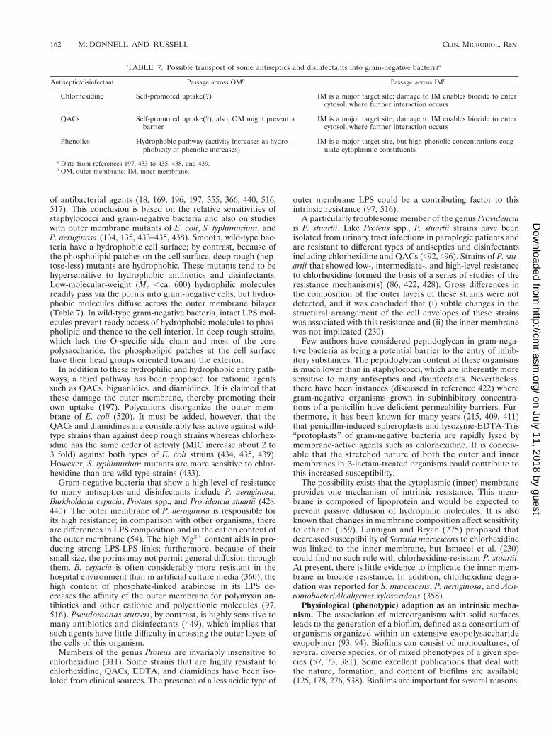
of antibacterial agents (18, 169, 196, 197, 355, 366, 440, 516,517). This conclusion is based on the relative sensitivities ofstaphylococci and gram-negative bacteria and also on studieswith outer membrane mutants of E. coli, S. typhimurium, andP. aeruginosa (134, 135, 433–435, 438). Smooth, wild-type bac-teria have a hydrophobic cell surface; by contrast, because ofthe phospholipid patches on the cell surface, deep rough (hep-tose-less) mutants are hydrophobic. These mutants tend to behypersensitive to hydrophobic antibiotics and disinfectants.Low-molecular-weight (Mr ,ca. 600) hydrophilic moleculesreadily pass via the porins into gram-negative cells, but hydro-phobic molecules diffuse across the outer membrane bilayer(Table 7). In wild-type gram-negative bacteria, intact LPS mol-ecules prevent ready access of hydrophobic molecules to phos-pholipid and thence to the cell interior. In deep rough strains,which lack the O-specific side chain and most of the corepolysaccharide, the phospholipid patches at the cell surfacehave their head groups oriented toward the exterior.
In addition to these hydrophilic and hydrophobic entry path-ways, a third pathway has been proposed for cationic agentssuch as QACs, biguanidies, and diamidines. It is claimed thatthese damage the outer membrane, thereby promoting theirown uptake (197). Polycations disorganize the outer mem-brane of E. coli (520). It must be added, however, that theQACs and diamidines are considerably less active against wild-type strains than against deep rough strains whereas chlorhex-idine has the same order of activity (MIC increase about 2 to3 fold) against both types of E. coli strains (434, 435, 439).However, S. typhimurium mutants are more sensitive to chlor-hexidine than are wild-type strains (433).
Gram-negative bacteria that show a high level of resistanceto many antiseptics and disinfectants include P. aeruginosa,Burkholderia cepacia, Proteus spp., and Providencia stuartii (428,440). The outer membrane of P. aeruginosa is responsible forits high resistance; in comparison with other organisms, thereare differences in LPS composition and in the cation content ofthe outer membrane (54). The high Mg21 content aids in pro-ducing strong LPS-LPS links; furthermore, because of theirsmall size, the porins may not permit general diffusion throughthem. B. cepacia is often considerably more resistant in thehospital environment than in artificial culture media (360); thehigh content of phosphate-linked arabinose in its LPS de-creases the affinity of the outer membrane for polymyxin an-tibiotics and other cationic and polycationic molecules (97,516). Pseudomonas stutzeri, by contrast, is highly sensitive tomany antibiotics and disinfectants (449), which implies thatsuch agents have little difficulty in crossing the outer layers ofthe cells of this organism.
Members of the genus Proteus are invariably insensitive tochlorhexidine (311). Some strains that are highly resistant tochlorhexidine, QACs, EDTA, and diamidines have been iso-lated from clinical sources. The presence of a less acidic type of
outer membrane LPS could be a contributing factor to thisintrinsic resistance (97, 516).
A particularly troublesome member of the genus Providenciais P. stuartii. Like Proteus spp., P. stuartii strains have beenisolated from urinary tract infections in paraplegic patients andare resistant to different types of antiseptics and disinfectantsincluding chlorhexidine and QACs (492, 496). Strains of P. stu-artii that showed low-, intermediate-, and high-level resistanceto chlorhexidine formed the basis of a series of studies of theresistance mechanism(s) (86, 422, 428). Gross differences inthe composition of the outer layers of these strains were notdetected, and it was concluded that (i) subtle changes in thestructural arrangement of the cell envelopes of these strainswas associated with this resistance and (ii) the inner membranewas not implicated (230).
Few authors have considered peptidoglycan in gram-nega-tive bacteria as being a potential barrier to the entry of inhib-itory substances. The peptidoglycan content of these organismsis much lower than in staphylococci, which are inherently moresensitive to many antiseptics and disinfectants. Nevertheless,there have been instances (discussed in reference 422) wheregram-negative organisms grown in subinhibitory concentra-tions of a penicillin have deficient permeability barriers. Fur-thermore, it has been known for many years (215, 409, 411)that penicillin-induced spheroplasts and lysozyme-EDTA-Tris“protoplasts” of gram-negative bacteria are rapidly lysed bymembrane-active agents such as chlorhexidine. It is conceiv-able that the stretched nature of both the outer and innermembranes in b-lactam-treated organisms could contribute tothis increased susceptibility.
The possibility exists that the cytoplasmic (inner) membraneprovides one mechanism of intrinsic resistance. This mem-brane is composed of lipoprotein and would be expected toprevent passive diffusion of hydrophilic molecules. It is alsoknown that changes in membrane composition affect sensitivityto ethanol (159). Lannigan and Bryan (275) proposed thatdecreased susceptibility of Serratia marcescens to chlorhexidinewas linked to the inner membrane, but Ismaeel et al. (230)could find no such role with chlorhexidine-resistant P. stuartii.At present, there is little evidence to implicate the inner mem-brane in biocide resistance. In addition, chlorhexidine degra-dation was reported for S. marcescens, P. aeruginosa, and Ach-romobacter/Alcaligenes xylosoxidans (358).
Physiological (phenotypic) adaption as an intrinsic mecha-nism. The association of microorganisms with solid surfacesleads to the generation of a biofilm, defined as a consortium oforganisms organized within an extensive exopolysaccharideexopolymer (93, 94). Biofilms can consist of monocultures, ofseveral diverse species, or of mixed phenotypes of a given spe-cies (57, 73, 381). Some excellent publications that deal withthe nature, formation, and content of biofilms are available(125, 178, 276, 538). Biofilms are important for several reasons,
TABLE 7. Possible transport of some antiseptics and disinfectants into gram-negative bacteriaa
Antiseptic/disinfectant Passage across OMb Passage across IMb
Chlorhexidine Self-promoted uptake(?) IM is a major target site; damage to IM enables biocide to entercytosol, where further interaction occurs
QACs Self-promoted uptake(?); also, OM might present abarrier
IM is a major target site; damage to IM enables biocide to entercytosol, where further interaction occurs
Phenolics Hydrophobic pathway (activity increases as hydro-phobicity of phenolic increases)
IM is a major target site, but high phenolic concentrations coag-ulate cytoplasmic constituents
a Data from references 197, 433 to 435, 438, and 439.b OM, outer membrane; IM, inner membrane.
162 MCDONNELL AND RUSSELL CLIN. MICROBIOL. REV.
on July 11, 2018 by guesthttp://cm
r.asm.org/
Dow
nloaded from
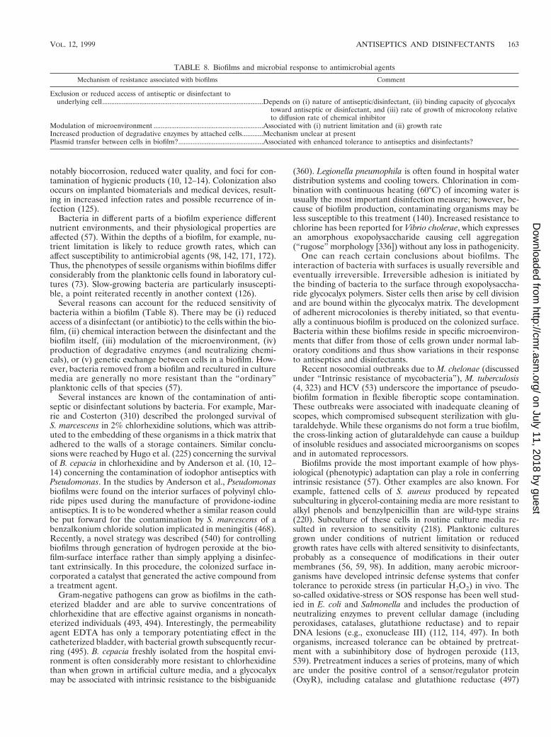
notably biocorrosion, reduced water quality, and foci for con-tamination of hygienic products (10, 12–14). Colonization alsooccurs on implanted biomaterials and medical devices, result-ing in increased infection rates and possible recurrence of in-fection (125).
Bacteria in different parts of a biofilm experience differentnutrient environments, and their physiological properties areaffected (57). Within the depths of a biofilm, for example, nu-trient limitation is likely to reduce growth rates, which canaffect susceptibility to antimicrobial agents (98, 142, 171, 172).Thus, the phenotypes of sessile organisms within biofilms differconsiderably from the planktonic cells found in laboratory cul-tures (73). Slow-growing bacteria are particularly insuscepti-ble, a point reiterated recently in another context (126).
Several reasons can account for the reduced sensitivity ofbacteria within a biofilm (Table 8). There may be (i) reducedaccess of a disinfectant (or antibiotic) to the cells within the bio-film, (ii) chemical interaction between the disinfectant and thebiofilm itself, (iii) modulation of the microenvironment, (iv)production of degradative enzymes (and neutralizing chemi-cals), or (v) genetic exchange between cells in a biofilm. How-ever, bacteria removed from a biofilm and recultured in culturemedia are generally no more resistant than the “ordinary”planktonic cells of that species (57).
Several instances are known of the contamination of anti-septic or disinfectant solutions by bacteria. For example, Mar-rie and Costerton (310) described the prolonged survival ofS. marcescens in 2% chlorhexidine solutions, which was attrib-uted to the embedding of these organisms in a thick matrix thatadhered to the walls of a storage containers. Similar conclu-sions were reached by Hugo et al. (225) concerning the survivalof B. cepacia in chlorhexidine and by Anderson et al. (10, 12–14) concerning the contamination of iodophor antiseptics withPseudomonas. In the studies by Anderson et al., Pseudomonasbiofilms were found on the interior surfaces of polyvinyl chlo-ride pipes used during the manufacture of providone-iodineantiseptics. It is to be wondered whether a similar reason couldbe put forward for the contamination by S. marcescens of abenzalkonium chloride solution implicated in meningitis (468).Recently, a novel strategy was described (540) for controllingbiofilms through generation of hydrogen peroxide at the bio-film-surface interface rather than simply applying a disinfec-tant extrinsically. In this procedure, the colonized surface in-corporated a catalyst that generated the active compound froma treatment agent.
Gram-negative pathogens can grow as biofilms in the cath-eterized bladder and are able to survive concentrations ofchlorhexidine that are effective against organisms in noncath-eterized individuals (493, 494). Interestingly, the permeabilityagent EDTA has only a temporary potentiating effect in thecatheterized bladder, with bacterial growth subsequently recur-ring (495). B. cepacia freshly isolated from the hospital envi-ronment is often considerably more resistant to chlorhexidinethan when grown in artificial culture media, and a glycocalyxmay be associated with intrinsic resistance to the bisbiguanide
(360). Legionella pneumophila is often found in hospital waterdistribution systems and cooling towers. Chlorination in com-bination with continuous heating (60°C) of incoming water isusually the most important disinfection measure; however, be-cause of biofilm production, contaminating organisms may beless susceptible to this treatment (140). Increased resistance tochlorine has been reported for Vibrio cholerae, which expressesan amorphous exopolysaccharide causing cell aggregation(“rugose” morphology [336]) without any loss in pathogenicity.
One can reach certain conclusions about biofilms. Theinteraction of bacteria with surfaces is usually reversible andeventually irreversible. Irreversible adhesion is initiated bythe binding of bacteria to the surface through exopolysaccha-ride glycocalyx polymers. Sister cells then arise by cell divisionand are bound within the glycocalyx matrix. The developmentof adherent microcolonies is thereby initiated, so that eventu-ally a continuous biofilm is produced on the colonized surface.Bacteria within these biofilms reside in specific microenviron-ments that differ from those of cells grown under normal lab-oratory conditions and thus show variations in their responseto antiseptics and disinfectants.
Recent nosocomial outbreaks due to M. chelonae (discussedunder “Intrinsic resistance of mycobacteria”), M. tuberculosis(4, 323) and HCV (53) underscore the importance of pseudo-biofilm formation in flexible fiberoptic scope contamination.These outbreaks were associated with inadequate cleaning ofscopes, which compromised subsequent sterilization with glu-taraldehyde. While these organisms do not form a true biofilm,the cross-linking action of glutaraldehyde can cause a buildupof insoluble residues and associated microorganisms on scopesand in automated reprocessors.
Biofilms provide the most important example of how phys-iological (phenotypic) adaptation can play a role in conferringintrinsic resistance (57). Other examples are also known. Forexample, fattened cells of S. aureus produced by repeatedsubculturing in glycerol-containing media are more resistant toalkyl phenols and benzylpenicillin than are wild-type strains(220). Subculture of these cells in routine culture media re-sulted in reversion to sensitivity (218). Planktonic culturesgrown under conditions of nutrient limitation or reducedgrowth rates have cells with altered sensitivity to disinfectants,probably as a consequence of modifications in their outermembranes (56, 59, 98). In addition, many aerobic microor-ganisms have developed intrinsic defense systems that confertolerance to peroxide stress (in particular H2O2) in vivo. Theso-called oxidative-stress or SOS response has been well stud-ied in E. coli and Salmonella and includes the production ofneutralizing enzymes to prevent cellular damage (includingperoxidases, catalases, glutathione reductase) and to repairDNA lesions (e.g., exonuclease III) (112, 114, 497). In bothorganisms, increased tolerance can be obtained by pretreat-ment with a subinhibitory dose of hydrogen peroxide (113,539). Pretreatment induces a series of proteins, many of whichare under the positive control of a sensor/regulator protein(OxyR), including catalase and glutathione reductase (497)
TABLE 8. Biofilms and microbial response to antimicrobial agents
Mechanism of resistance associated with biofilms Comment
Exclusion or reduced access of antiseptic or disinfectant tounderlying cell...........................................................................................Depends on (i) nature of antiseptic/disinfectant, (ii) binding capacity of glycocalyx
toward antiseptic or disinfectant, and (iii) rate of growth of microcolony relativeto diffusion rate of chemical inhibitor
Modulation of microenvironment ..............................................................Associated with (i) nutrient limitation and (ii) growth rateIncreased production of degradative enzymes by attached cells............Mechanism unclear at presentPlasmid transfer between cells in biofilm?................................................Associated with enhanced tolerance to antiseptics and disinfectants?
VOL. 12, 1999 ANTISEPTICS AND DISINFECTANTS 163
on July 11, 2018 by guesthttp://cm
r.asm.org/
Dow
nloaded from
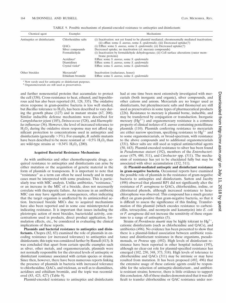
and further nonessential proteins that accumulate to protectthe cell (338). Cross-resistance to heat, ethanol, and hypochlo-rous acid has also been reported (81, 128, 335). The oxidativestress response in gram-positive bacteria is less well studied,but Bacillus tolerance to H2O2 has been described to vary dur-ing the growth phase (127) and in mutant strains (67, 200).Similar inducible defense mechanisms were described forCampylobacter jejuni (185), Deinococcus (528), and Haemophi-lus influenzae (36). However, the level of increased tolerance toH2O2 during the oxidative stress response may not afford sig-nificant protection to concentrations used in antiseptics anddisinfectants (generally .3%). For example, B. subtilis mutantshave been described to be more resistant at ;0.5% H2O2 thanare wild-type strains at ;0.34% H2O2 (200).
Acquired Bacterial Resistance Mechanisms
As with antibiotics and other chemotherapeutic drugs, ac-quired resistance to antiseptics and disinfectants can arise byeither mutation or the acquisition of genetic material in theform of plasmids or transposons. It is important to note that“resistance” as a term can often be used loosely and in manycases must be interpreted with some prudence. This is partic-ularly true with MIC analysis. Unlike antibiotics, “resistance,”or an increase in the MIC of a biocide, does not necessarilycorrelate with therapeutic failure. An increase in an antibioticMIC can may have significant consequences, often indicatingthat the target organism is unaffected by its antimicrobial ac-tion. Increased biocide MICs due to acquired mechanismshave also been reported and in some case misinterpreted asindicating resistance. It is important that issues including thepleiotropic action of most biocides, bactericidal activity, con-centrations used in products, direct product application, for-mulation effects, etc., be considered in evaluating the clinicalimplications of these reports.
Plasmids and bacterial resistance to antiseptics and disin-fectants. Chopra (82, 83) examined the role of plasmids in en-coding resistance (or increased tolerance) to antiseptics anddisinfectants; this topic was considered further by Russell (413). Itwas concluded that apart from certain specific examples suchas silver, other metals, and organomercurials, plasmids werenot normally responsible for the elevated levels of antiseptic ordisinfectant resistance associated with certain species or strains.Since then, however, there have been numerous reports linkingthe presence of plasmids in bacteria with increased toleranceto chlorhexidine, QACs, and triclosan, as well as to diamidines,acridines and ethidium bromide, and the topic was reconsid-ered (83, 423, 427) (Table 9).
Plasmid-encoded resistance to antiseptics and disinfectants
had at one time been most extensively investigated with mer-curials (both inorganic and organic), silver compounds, andother cations and anions. Mercurials are no longer used asdisinfectants, but phenylmercuric salts and thiomersal are stillused as preservatives in some types of pharmaceutical products(226). Resistance to mercury is plasmid borne, inducible, andmay be transferred by conjugation or transduction. Inorganicmercury (Hg21) and organomercury resistance is a commonproperty of clinical isolates of S. aureus containing penicillinaseplasmids (110). Plasmids conferring resistance to mercurialsare either narrow spectrum, specifying resistance to Hg21 andto some organomercurials, or broad-spectrum, with resistanceto the above compounds and to additional organomercurials(331). Silver salts are still used as topical antimicrobial agents(50, 443). Plasmid-encoded resistance to silver has been foundin Pseudomonas stutzeri (192), members of the Enterobacteri-aceae (479, 480, 511), and Citrobacter spp. (511). The mecha-nism of resistance has yet to be elucidated fully but may beassociated with silver accumulation (152, 511).
(i) Plasmid-mediated antiseptic and disinfectant resistancein gram-negative bacteria. Occasional reports have examinedthe possible role of plasmids in the resistance of gram-negativebacteria to antiseptics and disinfectants. Sutton and Jacoby(498) observed that plasmid RP1 did not significantly alter theresistance of P. aeruginosa to QACs, chlorhexidine, iodine, orchlorinated phenols, although increased resistance to hexa-chlorophene was observed. This compound has a much greatereffect on gram-positive than gram-negative bacteria, so that itis difficult to assess the significance of this finding. Transfor-mation of this plasmid (which encodes resistance to carbeni-cillin, tetracycline, and neomycin and kanamycin) into E. colior P. aeruginosa did not increase the sensitivity of these organ-isms to a range of antiseptics (5).
Strains of Providencia stuartii may be highly tolerant to Hg21,cationic disinfectants (such as chlorhexidine and QACs), andantibiotics (496). No evidence has been presented to show thatthere is a plasmid-linked association between antibiotic resis-tance and disinfectant resistance in these organisms, pseudo-monads, or Proteus spp. (492). High levels of disinfectant re-sistance have been reported in other hospital isolates (195),although no clear-cut role for plasmid-specified resistance hasemerged (102, 250, 348, 373, 518). High levels of tolerance tochlorhexidine and QACs (311) may be intrinsic or may haveresulted from mutation. It has been proposed (492, 496) thatthe extensive usage of these cationic agents could be respon-sible for the selection of antiseptic-disinfectant-, and antibiot-ic-resistant strains; however, there is little evidence to supportthis conclusion. All of these studies demonstrated that it was dif-ficult to transfer chlorhexidine or QAC resistance under nor-
TABLE 9. Possible mechanisms of plasmid-encoded resistance to antiseptics and disinfectants
Chemical agent Examples Mechanisms
Antiseptics or disinfectants Chlorhexidine salts (i) Inactivation: not yet found to be plasmid mediated; chromosomally mediated inactivation;(ii) efflux: some S. aureus, some S. epidermidis; (iii) Decreased uptake(?)
QACs (i) Efflux: some S. aureus, some S. epidermidis; (ii) Decreased uptake(?)Silver compounds Decreased uptake; no inactivation (cf. mercury compounds)Formaldehyde (i) Inactivation by formaldehyde dehydrogenase; (ii) Cell surface alterations (outer mem-
brane proteins)Acridinesa Efflux: some S. aureus, some S. epidermidisDiamidines Efflux: some S. aureus, some S. epidermidisCrystal violeta Efflux: some S. aureus, some S. epidermidis
Other biocides Mercurialsb Inactivation (reductases, lyases)Ethidium bromide Efflux: some S. aureus, some S. epidermidis
a Now rarely used for antiseptic or disinfectant purposes.b Organomercurials are still used as preservatives.
164 MCDONNELL AND RUSSELL CLIN. MICROBIOL. REV.
on July 11, 2018 by guesthttp://cm
r.asm.org/
Dow
nloaded from
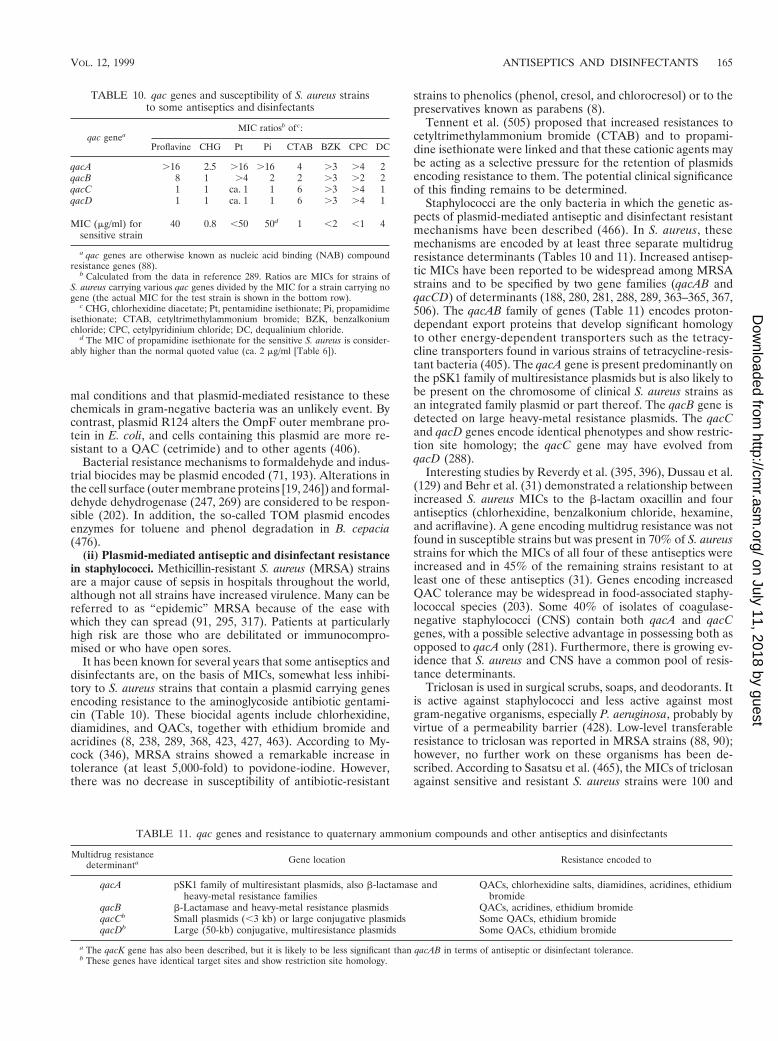
mal conditions and that plasmid-mediated resistance to thesechemicals in gram-negative bacteria was an unlikely event. Bycontrast, plasmid R124 alters the OmpF outer membrane pro-tein in E. coli, and cells containing this plasmid are more re-sistant to a QAC (cetrimide) and to other agents (406).
Bacterial resistance mechanisms to formaldehyde and indus-trial biocides may be plasmid encoded (71, 193). Alterations inthe cell surface (outer membrane proteins [19, 246]) and formal-dehyde dehydrogenase (247, 269) are considered to be respon-sible (202). In addition, the so-called TOM plasmid encodesenzymes for toluene and phenol degradation in B. cepacia(476).
(ii) Plasmid-mediated antiseptic and disinfectant resistancein staphylococci. Methicillin-resistant S. aureus (MRSA) strainsare a major cause of sepsis in hospitals throughout the world,although not all strains have increased virulence. Many can bereferred to as “epidemic” MRSA because of the ease withwhich they can spread (91, 295, 317). Patients at particularlyhigh risk are those who are debilitated or immunocompro-mised or who have open sores.
It has been known for several years that some antiseptics anddisinfectants are, on the basis of MICs, somewhat less inhibi-tory to S. aureus strains that contain a plasmid carrying genesencoding resistance to the aminoglycoside antibiotic gentami-cin (Table 10). These biocidal agents include chlorhexidine,diamidines, and QACs, together with ethidium bromide andacridines (8, 238, 289, 368, 423, 427, 463). According to My-cock (346), MRSA strains showed a remarkable increase intolerance (at least 5,000-fold) to povidone-iodine. However,there was no decrease in susceptibility of antibiotic-resistant
strains to phenolics (phenol, cresol, and chlorocresol) or to thepreservatives known as parabens (8).
Tennent et al. (505) proposed that increased resistances tocetyltrimethylammonium bromide (CTAB) and to propami-dine isethionate were linked and that these cationic agents maybe acting as a selective pressure for the retention of plasmidsencoding resistance to them. The potential clinical significanceof this finding remains to be determined.
Staphylococci are the only bacteria in which the genetic as-pects of plasmid-mediated antiseptic and disinfectant resistantmechanisms have been described (466). In S. aureus, thesemechanisms are encoded by at least three separate multidrugresistance determinants (Tables 10 and 11). Increased antisep-tic MICs have been reported to be widespread among MRSAstrains and to be specified by two gene families (qacAB andqacCD) of determinants (188, 280, 281, 288, 289, 363–365, 367,506). The qacAB family of genes (Table 11) encodes proton-dependant export proteins that develop significant homologyto other energy-dependent transporters such as the tetracy-cline transporters found in various strains of tetracycline-resis-tant bacteria (405). The qacA gene is present predominantly onthe pSK1 family of multiresistance plasmids but is also likely tobe present on the chromosome of clinical S. aureus strains asan integrated family plasmid or part thereof. The qacB gene isdetected on large heavy-metal resistance plasmids. The qacCand qacD genes encode identical phenotypes and show restric-tion site homology; the qacC gene may have evolved fromqacD (288).
Interesting studies by Reverdy et al. (395, 396), Dussau et al.(129) and Behr et al. (31) demonstrated a relationship betweenincreased S. aureus MICs to the b-lactam oxacillin and fourantiseptics (chlorhexidine, benzalkonium chloride, hexamine,and acriflavine). A gene encoding multidrug resistance was notfound in susceptible strains but was present in 70% of S. aureusstrains for which the MICs of all four of these antiseptics wereincreased and in 45% of the remaining strains resistant to atleast one of these antiseptics (31). Genes encoding increasedQAC tolerance may be widespread in food-associated staphy-lococcal species (203). Some 40% of isolates of coagulase-negative staphylococci (CNS) contain both qacA and qacCgenes, with a possible selective advantage in possessing both asopposed to qacA only (281). Furthermore, there is growing ev-idence that S. aureus and CNS have a common pool of resis-tance determinants.
Triclosan is used in surgical scrubs, soaps, and deodorants. Itis active against staphylococci and less active against mostgram-negative organisms, especially P. aeruginosa, probably byvirtue of a permeability barrier (428). Low-level transferableresistance to triclosan was reported in MRSA strains (88, 90);however, no further work on these organisms has been de-scribed. According to Sasatsu et al. (465), the MICs of triclosanagainst sensitive and resistant S. aureus strains were 100 and
TABLE 10. qac genes and susceptibility of S. aureus strainsto some antiseptics and disinfectants
qac geneaMIC ratiosb of c:
Proflavine CHG Pt Pi CTAB BZK CPC DC
qacA .16 2.5 .16 .16 4 .3 .4 2qacB 8 1 .4 2 2 .3 .2 2qacC 1 1 ca. 1 1 6 .3 .4 1qacD 1 1 ca. 1 1 6 .3 .4 1
MIC (mg/ml) forsensitive strain
40 0.8 ,50 50d 1 ,2 ,1 4
a qac genes are otherwise known as nucleic acid binding (NAB) compoundresistance genes (88).
b Calculated from the data in reference 289. Ratios are MICs for strains ofS. aureus carrying various qac genes divided by the MIC for a strain carrying nogene (the actual MIC for the test strain is shown in the bottom row).
c CHG, chlorhexidine diacetate; Pt, pentamidine isethionate; Pi, propamidimeisethionate; CTAB, cetyltrimethylammonium bromide; BZK, benzalkoniumchloride; CPC, cetylpyridinium chloride; DC, dequalinium chloride.
d The MIC of propamidine isethionate for the sensitive S. aureus is consider-ably higher than the normal quoted value (ca. 2 mg/ml [Table 6]).
TABLE 11. qac genes and resistance to quaternary ammonium compounds and other antiseptics and disinfectants
Multidrug resistancedeterminanta Gene location Resistance encoded to
qacA pSK1 family of multiresistant plasmids, also b-lactamase andheavy-metal resistance families
QACs, chlorhexidine salts, diamidines, acridines, ethidiumbromide
qacB b-Lactamase and heavy-metal resistance plasmids QACs, acridines, ethidium bromideqacCb Small plasmids (,3 kb) or large conjugative plasmids Some QACs, ethidium bromideqacDb Large (50-kb) conjugative, multiresistance plasmids Some QACs, ethidium bromide
a The qacK gene has also been described, but it is likely to be less significant than qacAB in terms of antiseptic or disinfectant tolerance.b These genes have identical target sites and show restriction site homology.
VOL. 12, 1999 ANTISEPTICS AND DISINFECTANTS 165
on July 11, 2018 by guesthttp://cm
r.asm.org/
Dow
nloaded from
.6,400 mg/ml, respectively; these results were disputed be-cause these concentrations are well in excess of the solubility oftriclosan (515), which is practically insoluble in water. Sasatsuet al. (464) described a high-level resistant strain of S. aureusfor which the MICs of chlorhexidine, CTAB, and butylparabenwere the same as for a low-level resistant strain. Furthermore,the MIC quoted for methylparaben comfortably exceeds itsaqueous solubility. Most of these studies with sensitive and“resistant” strains involved the use of MIC evaluations (forexample, Table 6). A few investigations examined the bacteri-cidal effects of antiseptics. Cookson et al. (89) pointed out thatcuring of resistance plasmids produced a fall in MICs but nota consistent decrease in the lethal activity of chlorhexidine.There is a poor correlation between MIC and the rate ofbactericidal action of chlorhexidine (88, 89, 319) and triclosan(90, 319). McDonnell et al. (318, 319) have described methi-cillin-susceptible S. aureus (MSSA) and MRSA strains withincreased triclosan MICs (up to 1.6 mg/ml) but showed that theMBCs for these strains were identical; these results were notsurprising, considering that biocides (unlike antibiotics) havemultiple cellular targets. Irizarry et al. (229) compared thesusceptibility of MRSA and MSSA strains to CPC and chlor-hexidine by both MIC and bactericidal testing methods. How-ever, the conclusion of this study that MRSA strains were moreresistant warrants additional comments. On the basis of ratherhigh actual MICs, MRSA strains were some four times moreresistant to chlorhexidine and five times more resistant to aQAC (CPC) than were MSSA strains. At a concentration inbroth of 2 mg of CPC/ml, two MRSA strains grew normallywith a threefold increase in viable numbers over a 4-h testperiod whereas an MSSA strain showed a 97% decrease inviability. From this, the authors concluded that it was reason-able to speculate that the residual amounts of antiseptics anddisinfectants found in the hospital environment could contrib-ute to the selection and maintenance of multiresistant MRSAstrains. Irizarry et al. (229) also concluded that MRSA strainsare less susceptible than MSSA strains to both chronic andacute exposures to antiseptics and disinfectants. However,their results with 4 mg of CPC/ml show no such pattern: at thishigher concentration, the turbidities (and viability) of the twoMRSA and one MSSA strains decreased at very similar rates(if anything, one MRSA strain appeared to be affected to aslightly greater extent that the MSSA strain). Furthermore, theauthors stated that chlorhexidine gave similar results to CPC.It is therefore difficult to see how Irizarry et al. arrived at theirhighly selective conclusions.
Plasmid-mediated efflux pumps are particularly importantmechanisms of resistance to many antibiotics (85), metals (349),and cationic disinfectants and antiseptics such as QACs, chlor-hexidine, diamidines, and acridines, as well as to ethidiumbromide (239, 289, 324–336, 363–368). Recombinant S. aureusplasmids transferred into E. coli are responsible for conferringincreased MICs of cationic agents to the gram-negative organ-ism (505, 544). Midgley (324, 325) demonstrated that a plas-mid-borne, ethidium resistance determinant from S. aureuscloned in E. coli encodes resistance to ethidium bromide andto QACs, which are expelled from the cells. A similar effluxsystem is present in Enterococcus hirae (326).
Sasatsu et al. (463) showed that duplication of ebr is respon-sible for resistance to ethidium bromide and to some antisep-tics. Later, Sasatsu et al. (466) examined the origin of ebr (nowknown to be identical to qacCD) in S. aureus; ebr was found inantibiotic-resistant and -sensitive strains of S. aureus, CNS, andenterococcal strains. The nucleotide sequences of the ampli-fied DNA fragment from sensitive and resistant strains wereidentical, and it was proposed that in antiseptic-resistant cells
there was an increase in the copy number of the ebr (qacCD)gene whose normal function was to remove toxic substancesfrom normal cells of staphylococci and enterococci.
Based on DNA homology, it was proposed that qacA andrelated genes carrying resistance determinants evolved frompreexisting genes responsible for normal cellular transport sys-tems (405) and that the antiseptic resistance genes evolvedbefore the introduction and use of topical antimicrobial prod-ucts and other antiseptics and disinfectants (288, 289, 365, 367,368, 405).
Baquero et al. (23) have pointed out that for antibiotics, thepresence of a specific resistance mechanism frequently contrib-utes to the long-term selection of resistant variants under invivo conditions. Whether low-level resistance to cationic anti-septics, e.g., chlorhexidine, QACs, can likewise provide a selec-tive advantage on staphylococci carrying qac genes remains tobe elucidated. The evidence is currently contentious and in-conclusive.
(iii) Plasmid-mediated antiseptic and disinfectant resistancein other gram-positive bacteria. Antibiotic-resistant coryne-bacteria may be implicated in human infections, especially inthe immunocompromised. ‘Group JK’ coryneforms (Coryne-bacterium jeikeium) were found to be more tolerant than othercoryneforms to the cationic disinfectants ethidium bromideand hexachlorophene, but studies with plasmid-containing andplasmid-cured derivatives produced no evidence of plasmid-associated resistance (285). Enterococcus faecium strains show-ing high level resistance to vancomycin, gentamicin, or both arenot more resistant to chlorhexidine or other nonantibioticagents (9, 11, 20, 319). Furthermore, despite the extensivedental use of chlorhexidine, strains of Streptococcus mutansremain sensitive to it (235). To date, therefore, there is little orno evidence of plasmid-associated resistance of nonstaphylo-coccal gram-positive bacteria to antiseptics and disinfectants.
Mutational resistance to antiseptics and disinfectants.Chromosomal mutation to antibiotics has been recognized fordecades. By contrast, fewer studies have been performed todetermine whether mutation confers resistance to antisepticsand disinfectants. It was, however, demonstrated over 40 yearsago (77, 78) that S. marcescens, normally inhibited by QACs at,100 mg/ml, could adapt to grow in 100,000 mg of a QAC perml. The resistant and sensitive cells had different surface char-acteristics (electrophoretic mobilities), but resistance could belost when the cells were grown on QAC-free media. One prob-lem associated with QACs and chlorhexidine is the turbidityproduced in liquid culture media above a certain concentration(interaction with agar also occurs), which could undoubtedlyinterfere with the determination of growth. This observation isreinforced by the findings presented by Nicoletti et al. (354).
Prince et al. (383) reported that resistance to chlorhexidinecould be induced in some organisms but not in others. Forexample, P. mirabilis and S. marcescens displayed 128- and258-fold increases, respectively, in resistance to chlorhexidine,whereas it was not possible to develop resistance to chlorhex-idine in Salmonella enteritidis. The resistant strains did notshow altered biochemical properties of changed virulence formice, and some strains were resistant to the QAC benzalko-nium chloride. Moreover, resistance to chlorhexidine was sta-ble in S. marcescens but not in P. mirabilis. Despite extensiveexperimentation with a variety of procedures, Fitzgerald et al.(148) were unable to develop stable chlorhexidine resistance inE. coli or S. aureus. Similar observations were made by Cook-son et al. (89), who worked with MRSA and other strains ofS. aureus, and by McDonnell et al. (319), who worked withMRSA and enterococci. Recently, stable chlorhexidine resis-tance was developed in P. stutzeri (502); these strains showed
166 MCDONNELL AND RUSSELL CLIN. MICROBIOL. REV.
on July 11, 2018 by guesthttp://cm
r.asm.org/
Dow
nloaded from
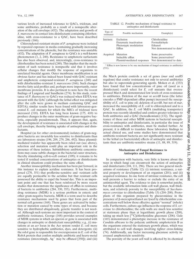
various levels of increased tolerance to QACs, triclosan, andsome antibiotics, probably as a result of a nonspecific alter-ation of the cell envelope (452). The adaptation and growth ofS. marcescens in contact lens disinfectants containing chlorhex-idine, with cross-resistance to a QAC, have been describedpreviously (166).
Chloroxylenol-resistant strains of P. aeruginosa were isolatedby repeated exposure in media containing gradually increasingconcentrations of the phenolic, but the resistance was unstable(432). The adaptation of P. aeruginosa to QACs is a well-knownphenomenon (1, 2, 240). Resistance to amphoteric surfactantshas also been observed, and, interestingly, cross-resistance tochlorhexidine has been noted (240). This implies that the mech-anism of such resistance is nonspecific and that it involvescellular changes that modify the response of organisms tounrelated biocidal agents. Outer membrane modification is anobvious factor and has indeed been found with QAC-resistantand amphoteric compound-resistant P. aeruginosa (240) andwith chlorhexidine-resistant S. marcescens (166). Such changesinvolve fatty acid profiles and, perhaps more importantly, outermembrane proteins. It is also pertinent to note here the recentfindings of Langsrud and Sundheim (274). In this study, resis-tance of P. fluorescens to QACs was reduced when EDTA waspresent with the QAC (although the lethal effect was mitigatedafter the cells were grown in medium containing QAC andEDTA); similar results have been found with laboratory-gen-erated E. coli mutants for which the MICs of triclosan wereincreased (318). EDTA has long been known (175, 410) toproduce changes in the outer membrane of gram-negative bac-teria, especially pseudomonads. Thus, it appears that, again,the development of resistance is associated with changes in thecell envelope, thereby limiting uptake of antiseptics and disin-fectants.
Hospital (as for other environmental) isolates of gram-neg-ative bacteria are invariably less sensitive to disinfectants thanare laboratory strains (196, 209, 279, 286, 492). Since plasmid-mediated transfer has apparently been ruled out (see above),selection and mutation could play an important role in thepresence of these isolates. Subinhibitory antibiotic concentra-tions may cause subtle changes in the bacterial outer structure,thereby stimulating cell-to-cell contact (109); it remains to betested if residual concentrations of antiseptics or disinfectantsin clinical situations could produce the same effect.
Another insusceptibility mechanism has been put forward, inthis instance to explain acridine resistance. It has been pro-posed (270, 351) that proflavine-sensitive and -resistant cellsare equally permeable to the acridine but that resistant cellspossessed the ability to expel the bound dye. This is an impor-tant point and one that has been reinforced by more recentstudies that demonstrate the significance of efflux in resistanceof bacteria to antibiotics (284, 330, 355). Furthermore, multi-drug resistance (MDR) is a serious problem in enteric andother gram-negative bacteria. MDR is a term used to describeresistance mechanisms used by genes that form part of thenormal cell genome (168). These genes are activated by induc-tion or mutation caused by some types of stress, and becausethey are distributed ubiquitously, genetic transfer is not need-ed. Although such systems are most important in the context ofantibiotic resistance, George (168) provides several examplesof MDR systems in which an operon or gene is associated withchanges in antiseptic or disinfectant susceptibility; e.g., (i) mu-tations at an acr locus in the Acr system render E. coli moresensitive to hydrophobic antibiotics, dyes, and detergents; (ii)the robA gene is responsible for overexpression in E. coli of theRobA protein that confers multiple antibiotic and heavy-metalresistance (interestingly, Ag1 may be effluxed [350]); and (iii)
the MarA protein controls a set of genes (mar and soxRSregulons) that confer resistance not only to several antibioticsbut also to superoxide-generating agents. Moken et al. (333)have found that low concentrations of pine oil (used as adisinfectant) could select for E. coli mutants that overex-pressed MarA and demonstrated low levels of cross-resistanceto antibiotics. Deletion of the mar or acrAB locus (the latterencodes a PMF-dependant efflux pump) increased the suscep-tibility of E. coli to pine oil; deletion of acrAB, but not of mar,increased the susceptibility of E. coli to chloroxylenol and to aQAC. In addition, the E. coli MdfA (multidrug transporter)protein was recently identified and confers greater tolerance toboth antibiotics and a QAC (benzalkonium) (132). The signif-icance of these and other MDR systems in bacterial suscepti-bility to antiseptics and disinfectants, in particular the issue ofcross-resistance with antibiotics, must be studied further. Atpresent, it is difficult to translate these laboratory findings toactual clinical use, and some studies have demonstrated thatantibiotic-resistant bacteria are not significantly more resistantto the lethal (or bactericidal) effects of antiseptic and disinfec-tants than are antibiotic-sensitive strains (11, 88, 89, 319).
Mechanisms of Fungal Resistance toAntiseptics and Disinfectants
In comparison with bacteria, very little is known about theways in which fungi can circumvent the action of antisepticsand disinfectants (104, 111, 296). There are two general mech-anisms of resistance (Table 12): (i) intrinsic resistance, a nat-ural property or development of an organism (201); and (ii)acquired resistance. In one form of intrinsic resistance, the cellwall presents a barrier to reduce or exclude the entry of anantimicrobial agent. The evidence to date is somewhat patchy,but the available information links cell wall glucan, wall thick-ness, and relatively porosity to the susceptibility of Saccharo-myces cerevisiae to chlorhexidine (Table 13) (204–208). Proto-plasts of this organism prepared by glucuronidase in thepresence of b-mercaptoethanol are lysed by chlorhexidine con-centrations well below those effective against “normal” (whole)cells. Furthermore, culture age influences the response of S. cer-evisiae to chlorhexidine; the cells walls are much less sensitiveat stationary phase than at logarithmic growth phase (208),taking up much less [14C]chlorhexidine gluconate (206). Gale(165) demonstrated a phenotypic increase in the resistance ofCandida albicans to the polyenic antibiotic amphotericin B asthe organisms entered the stationary growth phase, which wasattributed to cell wall changes involving tighter cross-linking(74). Additionally, any factor increasing glucanase activity in-creased amphotericin sensitivity.
The porosity of the yeast cell wall is affected by its chemical
TABLE 12. Possible mechanisms of fungal resistance toantiseptics and disinfectants
Type ofresistance Possible mechanism Example(s)
Intrinsic Exclusion ChlorhexidineEnzymatic inactivation FormaldehydePhenotypic modulation EthanolEfflux Not demonstrated to datea
Acquired Mutation Some preservativeInducible efflux Some preservativesa
Plasmid-mediated responses Not demonstrated to date
a Efflux is now known to be one mechanism of fungal resistance to antibiotics(531).
VOL. 12, 1999 ANTISEPTICS AND DISINFECTANTS 167
on July 11, 2018 by guesthttp://cm
r.asm.org/
Dow
nloaded from
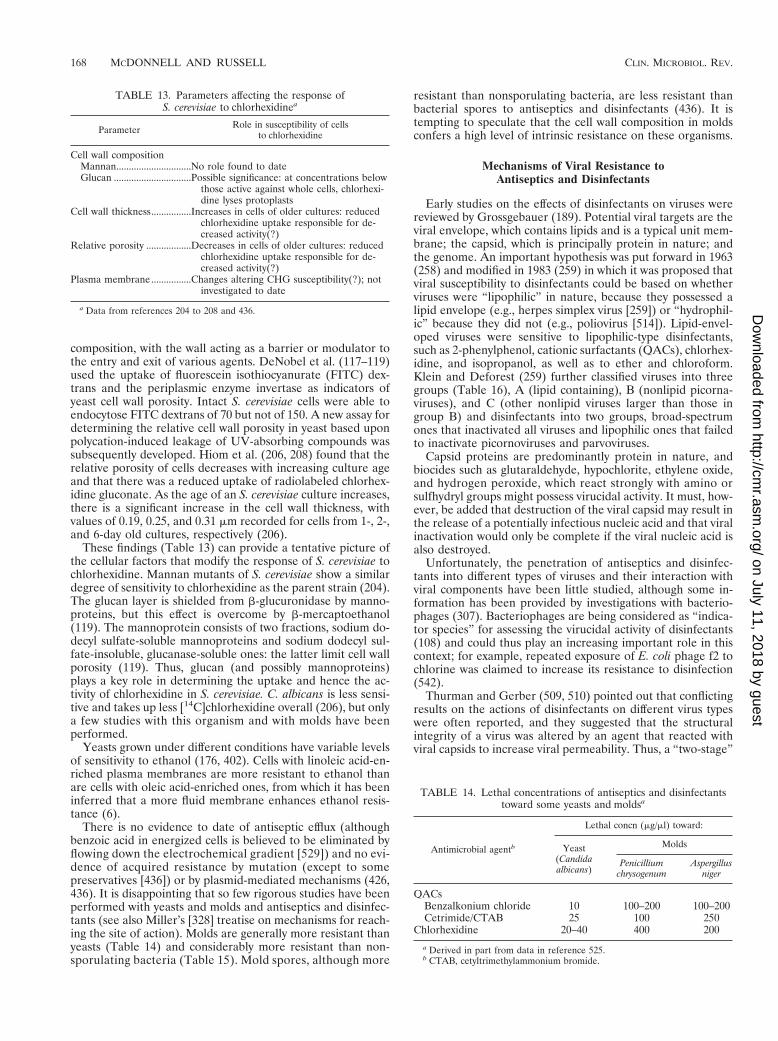
composition, with the wall acting as a barrier or modulator tothe entry and exit of various agents. DeNobel et al. (117–119)used the uptake of fluorescein isothiocyanurate (FITC) dex-trans and the periplasmic enzyme invertase as indicators ofyeast cell wall porosity. Intact S. cerevisiae cells were able toendocytose FITC dextrans of 70 but not of 150. A new assay fordetermining the relative cell wall porosity in yeast based uponpolycation-induced leakage of UV-absorbing compounds wassubsequently developed. Hiom et al. (206, 208) found that therelative porosity of cells decreases with increasing culture ageand that there was a reduced uptake of radiolabeled chlorhex-idine gluconate. As the age of an S. cerevisiae culture increases,there is a significant increase in the cell wall thickness, withvalues of 0.19, 0.25, and 0.31 mm recorded for cells from 1-, 2-,and 6-day old cultures, respectively (206).
These findings (Table 13) can provide a tentative picture ofthe cellular factors that modify the response of S. cerevisiae tochlorhexidine. Mannan mutants of S. cerevisiae show a similardegree of sensitivity to chlorhexidine as the parent strain (204).The glucan layer is shielded from b-glucuronidase by manno-proteins, but this effect is overcome by b-mercaptoethanol(119). The mannoprotein consists of two fractions, sodium do-decyl sulfate-soluble mannoproteins and sodium dodecyl sul-fate-insoluble, glucanase-soluble ones: the latter limit cell wallporosity (119). Thus, glucan (and possibly mannoproteins)plays a key role in determining the uptake and hence the ac-tivity of chlorhexidine in S. cerevisiae. C. albicans is less sensi-tive and takes up less [14C]chlorhexidine overall (206), but onlya few studies with this organism and with molds have beenperformed.
Yeasts grown under different conditions have variable levelsof sensitivity to ethanol (176, 402). Cells with linoleic acid-en-riched plasma membranes are more resistant to ethanol thanare cells with oleic acid-enriched ones, from which it has beeninferred that a more fluid membrane enhances ethanol resis-tance (6).
There is no evidence to date of antiseptic efflux (althoughbenzoic acid in energized cells is believed to be eliminated byflowing down the electrochemical gradient [529]) and no evi-dence of acquired resistance by mutation (except to somepreservatives [436]) or by plasmid-mediated mechanisms (426,436). It is disappointing that so few rigorous studies have beenperformed with yeasts and molds and antiseptics and disinfec-tants (see also Miller’s [328] treatise on mechanisms for reach-ing the site of action). Molds are generally more resistant thanyeasts (Table 14) and considerably more resistant than non-sporulating bacteria (Table 15). Mold spores, although more
resistant than nonsporulating bacteria, are less resistant thanbacterial spores to antiseptics and disinfectants (436). It istempting to speculate that the cell wall composition in moldsconfers a high level of intrinsic resistance on these organisms.
Mechanisms of Viral Resistance toAntiseptics and Disinfectants
Early studies on the effects of disinfectants on viruses werereviewed by Grossgebauer (189). Potential viral targets are theviral envelope, which contains lipids and is a typical unit mem-brane; the capsid, which is principally protein in nature; andthe genome. An important hypothesis was put forward in 1963(258) and modified in 1983 (259) in which it was proposed thatviral susceptibility to disinfectants could be based on whetherviruses were “lipophilic” in nature, because they possessed alipid envelope (e.g., herpes simplex virus [259]) or “hydrophil-ic” because they did not (e.g., poliovirus [514]). Lipid-envel-oped viruses were sensitive to lipophilic-type disinfectants,such as 2-phenylphenol, cationic surfactants (QACs), chlorhex-idine, and isopropanol, as well as to ether and chloroform.Klein and Deforest (259) further classified viruses into threegroups (Table 16), A (lipid containing), B (nonlipid picorna-viruses), and C (other nonlipid viruses larger than those ingroup B) and disinfectants into two groups, broad-spectrumones that inactivated all viruses and lipophilic ones that failedto inactivate picornoviruses and parvoviruses.
Capsid proteins are predominantly protein in nature, andbiocides such as glutaraldehyde, hypochlorite, ethylene oxide,and hydrogen peroxide, which react strongly with amino orsulfhydryl groups might possess virucidal activity. It must, how-ever, be added that destruction of the viral capsid may result inthe release of a potentially infectious nucleic acid and that viralinactivation would only be complete if the viral nucleic acid isalso destroyed.
Unfortunately, the penetration of antiseptics and disinfec-tants into different types of viruses and their interaction withviral components have been little studied, although some in-formation has been provided by investigations with bacterio-phages (307). Bacteriophages are being considered as “indica-tor species” for assessing the virucidal activity of disinfectants(108) and could thus play an increasing important role in thiscontext; for example, repeated exposure of E. coli phage f2 tochlorine was claimed to increase its resistance to disinfection(542).
Thurman and Gerber (509, 510) pointed out that conflictingresults on the actions of disinfectants on different virus typeswere often reported, and they suggested that the structuralintegrity of a virus was altered by an agent that reacted withviral capsids to increase viral permeability. Thus, a “two-stage”
TABLE 13. Parameters affecting the response ofS. cerevisiae to chlorhexidinea
Parameter Role in susceptibility of cellsto chlorhexidine
Cell wall compositionMannan..............................No role found to dateGlucan ...............................Possible significance: at concentrations below
those active against whole cells, chlorhexi-dine lyses protoplasts
Cell wall thickness................Increases in cells of older cultures: reducedchlorhexidine uptake responsible for de-creased activity(?)
Relative porosity ..................Decreases in cells of older cultures: reducedchlorhexidine uptake responsible for de-creased activity(?)
Plasma membrane................Changes altering CHG susceptibility(?); notinvestigated to date
a Data from references 204 to 208 and 436.
TABLE 14. Lethal concentrations of antiseptics and disinfectantstoward some yeasts and moldsa
Antimicrobial agentb
Lethal concn (mg/ml) toward:
Yeast(Candidaalbicans)
Molds
Penicilliumchrysogenum
Aspergillusniger
QACsBenzalkonium chloride 10 100–200 100–200Cetrimide/CTAB 25 100 250
Chlorhexidine 20–40 400 200
a Derived in part from data in reference 525.b CTAB, cetyltrimethylammonium bromide.
168 MCDONNELL AND RUSSELL CLIN. MICROBIOL. REV.
on July 11, 2018 by guesthttp://cm
r.asm.org/
Dow
nloaded from
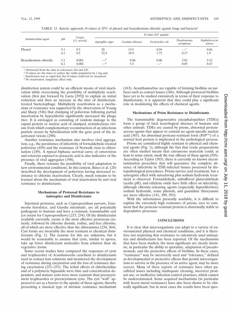
disinfection system could be an efficient means of viral inacti-vation while overcoming the possibility of multiplicity reacti-vation (first put forward by Luria [293]) to explain an initialreduction and then an increase in the titer of disinfectant-treated bacteriophage. Multiplicity reactivation as a mecha-nism of resistance was supported by the observation of Youngand Sharp (546) that clumping of poliovirus following partialinactivation by hypochlorite significantly increased the phagetiter. It is envisaged as consisting of random damage to thecapsid protein or nucleic acid of clumped, noninfectious viri-ons from which complementary reconstruction of an infectiousparticle occurs by hybridization with the gene pool of the in-activated virions (298).
Another resistance mechanism also involves viral aggrega-tion, e.g., the persistence of infectivity of formaldehyde-treatedpoliovirus (458) and the resistance of Norwalk virus to chlori-nation (249). A typical biphasic survival curve of enterovirusand rotavirus exposed to peracetic acid is also indicative of thepresence of viral aggregates (198).
Finally, there remains the possibility of viral adaptation tonew environmental conditions. In this context, Bates et al. (28)described the development of poliovirus having increased re-sistance to chlorine inactivation. Clearly, much remains to belearned about the mechanism of viral inactivation by and viralresistance to disinfectants.
Mechanisms of Protozoal Resistance toAntiseptics and Disinfectants
Intestinal protozoa, such as Cryptosporidium parvum, Enta-moeba histolytica, and Giardia intestinalis, are all potentiallypathogenic to humans and have a resistant, transmissible cyst(or oocyst for Cryptosporidium) (233, 234). Of the disinfectantsavailable currently, ozone is the most effective protozoan cys-ticide, followed by chlorine dioxide, iodine, and free chlorine,all of which are more effective than the chloramines (234, 264).Cyst forms are invariably the most resistant to chemical disin-fectants (Fig. 1). The reasons for this are unknown, but itwould be reasonable to assume that cysts, similar to spores,take up fewer disinfectant molecules from solution than dovegetative forms.
Some recent studies have compared the responses of cystsand trophozoites of Acanthamoeba castellanii to disinfectantsused in contact lens solutions and monitored the developmentof resistance during encystation and the loss of resistance dur-ing excystation (251–255). The lethal effects of chlorhexidineand of a polymeric biguanide were time and concentration de-pendent, and mature cysts were more resistant than preencyst-ment trophozoites or preexcystment cysts. The cyst “wall” ap-peared to act as a barrier to the uptake of these agents, therebypresenting a classical type of intrinsic resistance mechanism
(163). Acanthamoebae are capable of forming biofilms on sur-faces such as contact lenses (186). Although protozoal biofilmshave yet to be studied extensively in terms of their response todisinfectants, it is apparent that they could play a significantrole in modulating the effects of chemical agents.
Mechanisms of Prion Resistance to Disinfectants
The transmissible degenerative encephalopathies (TDEs)form a group of fatal neurological diseases of humans andother animals. TDEs are caused by prions, abnormal protein-aceous agents that appear to contain no agent-specific nucleicacid (385). An abnormal protease-resistant form (PrPres) of anormal host protein is implicated in the pathological process.
Prions are considered highly resistant to physical and chem-ical agents (Fig. 1), although the fact that crude preparationsare often studied means that extraneous materials could, atleast to some extent, mask the true efficacy of these agents (503).According to Taylor (503), there is currently no known decon-tamination procedure that will guarantee the complete ab-sence of infectivity in TDE-infected tissues processed by his-topathological procedures. Prions survive acid treatment, but asynergistic effect with autoclaving plus sodium hydroxide treat-ment is observed. Formaldehyde, unbuffered glutaraldehyde(acidic pH), and ethylene oxide have little effect on infectivity,although chlorine-releasing agents (especially hypochlorites),sodium hydroxide, some phenols, and guanidine thiocyanateare more effective (141, 309, 503).
With the information presently available, it is difficult toexplain the extremely high resistance of prions, save to com-ment that the protease-resistant protein is abnormally stable todegradative processes.
CONCLUSIONS
It is clear that microorganisms can adapt to a variety of en-vironmental physical and chemical conditions, and it is there-fore not surprising that resistance to extensively used antisep-tics and disinfectants has been reported. Of the mechanismsthat have been studied, the most significant are clearly intrin-sic, in particular the ability to sporulate, adaptation of pseudo-monads, and the protective effects of biofilms. In these cases,“resistance” may be incorrectly used and “tolerance,” definedas developmental or protective effects that permit microorgan-isms to survive in the presence of an active agent, may be morecorrect. Many of these reports of resistance have often pa-ralleled issues including inadequate cleaning, incorrect prod-uct use, or ineffective infection control practices, which cannotbe underestimated. Some acquired mechanisms (in particularwith heavy-metal resistance) have also been shown to be clin-ically significant, but in most cases the results have been spec-
TABLE 15. Kinetic approach: D-values at 20°C of phenol and benzalkonium chloride against fungi and bacteriaa
Antimicrobial agent pH Concn(%, wt/vol)
D-value (h)b against:
Aspergillus niger Candida albicans Escherichia coli Pseudomonasaeruginosa
Staphylococcusaureus
Phenol 5.1 0.5 20 13.5 0.94 —c 0.666.1 0.5 32.4 18.9 1.72 0.17 1.9
Benzalkonium chloride 5.1 0.001 —d 9.66 0.06 3.01 3.126.1 0.002 —d 5.5 —c 0.05 0.67
a Abstracted from the data in references 244 and 245.b D-values are the times to reduce the viable population by 1 log unit.c Inactivation was so rapid that the D-values could not be measured.d No inactivation: fungistatic effect only.
VOL. 12, 1999 ANTISEPTICS AND DISINFECTANTS 169
on July 11, 2018 by guesthttp://cm
r.asm.org/
Dow
nloaded from
ulative. Increased MICs have been confirmed, in particular forstaphylococci. However, few reports have further investigatedincreased bactericidal concentrations or actual use dilutions ofproducts, which in many cases contain significantly higher con-centrations of biocides, or formulation attributes, which canincrease product efficacy; in a number of cases, changes in theMICs have actually been shown not to be significant (9, 88, 89,319, 428). Efflux mechanisms are known to be important inantibiotic resistance, but it is questionable if the observed in-creased MICs of biocides could have clinical implications forhard-surface or topical disinfection (423, 428). It has beenspeculated that low-level resistance may aid in the survival ofmicroorganisms at residual levels of antiseptics and disinfec-tants; any possible clinical significance of this remains to betested. With growing concerns about the development of bio-cide resistance and cross-resistance with antibiotics, it is clearthat clinical isolates should be under continual surveillance andpossible mechanisms should be investigated.
It is also clear that antiseptic and disinfectant products canvary significantly, despite containing similar levels of biocides,which underlines the need for close inspection of efficacyclaims and adequate test methodology (183, 423, 428). In ad-dition, a particular antiseptic or disinfectant product may bebetter selected (as part of infection control practices) based onparticular circumstances or nosocomial outbreaks; for exam-ple, certain active agents are clearly more efficacious againstgram-positive than gram-negative bacteria, and C. difficile (de-spite the intrinsic resistance of spores) may be effectively con-trolled physically by adequate cleaning with QAC-based prod-ucts.
In conclusion, a great deal remains to be learned about themode of action of antiseptics and disinfectants. Although sig-nificant progress has been made with bacterial investigations, agreater understanding of these mechanisms is clearly lackingfor other infectious agents. Studies of the mechanisms of ac-tion of and microbial resistance to antiseptics and disinfectantsare thus not merely of academic significance. They are associ-ated with the more efficient use of these agents clinically andwith the potential design of newer, more effective compoundsand products.
REFERENCES
1. Adair, F. W., S. G. Geftic, and J. Gelzer. 1969. Resistance of Pseudomonasto quaternary ammonium compounds. I. Growth in benzalkonium chloridesolution. Appl. Microbiol. 18:299–302.
2. Adair, F. W., S. G. Geftic, and J. Gelzer. 1971. Resistance of Pseudomonasto quaternary ammonium compounds. II. Cross resistance characteristics of
a mutant of Pseudomonas aeruginosa. Appl. Microbiol. 21:1058–1063.3. Adler-Storthz, K., L. M. Sehulster, G. R. Dreesman, F. B. Hollinger, and
J. L. Melnick. 1983. Effect of alkaline glutaraldehyde on hepatitis B virusantigens. Eur. J. Clin. Microbiol. 2:316–320.
4. Agerton, T., S. Valway, B. Gore, C. Pozsik, B. Plikaytis, C. Woodley, and I.Onorato. 1997. Transmission of a highly drug-resistant strain (strain W1) ofMycobacterium tuberculosis. JAMA 278:1073–1077.
5. Ahonkhai, I., and A. D. Russell. 1979. Response RP11 and RP12 strains ofEscherichia coli to antibacterial agents and transfer of resistance to Pseudo-monas aeruginosa. Curr. Microbiol. 3:89–94.
6. Alexandre, H., I. Rousseaux, and C. Charpentier. 1994. Relationship be-tween ethanol tolerance, lipid composition and plasma membrane fluidityin Saccharomyces cerevisiae and Kloeckera apiculata. FEMS Microbiol. Lett.124:17–22.
7. Alfa, M. J., and D. L. Sitter. 1994. In-hospital evaluation of orthophthal-aldehyde as a high level disinfectant for flexible endoscopes. J. Hosp. Infect.26:15–26.
8. Al-Masaudi, S. B., M. J. Day, and A. D. Russell. 1991. Antimicrobialresistance and gene transfer in Staphylococcus aureus. J. Appl. Bacteriol. 70:279–290.
9. Alqurashi, A. M., M. J. Day, and A. D. Russell. 1996. Susceptibility of somestrains of enterococci and streptococci to antibiotics and biocides. J. Anti-microb. Chemother. 38:745.
10. Anderson, R. L. 1989. Iodophor antiseptics: intrinsic microbial contamina-tion with resistant bacteria. Infect. Control Hosp. Epidemiol. 10:443–446.
11. Anderson, R. L., J. H. Carr, W. W. Bond, and M. S. Favero. 1997. Suscep-tibility of vancomycin-resistant enterococci to environmental disinfectants.Infect. Control Hosp. Epidemiol. 18:195–199.
12. Anderson, R. L., B. W. Holland, J. K. Carr, W. W. Bond, and M. S. Favero.1990. Effect of disinfectants on pseudomonads colonized on the interiorsurface of PVC pipes. Am. J. Public Health 80:17–21.
13. Anderson, R. L., R. W. Vess, J. H. Carr, W. W. Bond, A. L. Panlilio, andM. S. Favero. 1991. Investigations of intrinsic Pseudomonas cepacia con-tamination in commercially manufactured povidone-iodine. Infect. ControlHosp. Epidemiol. 12:297–302.
14. Anderson, R. L., R. W. Vess, A. L. Panlilio, and M. S. Favero. 1990.Prolonged survival of Pseudomonas cepacia in commercially manufacturedpovidone-iodine. Appl. Environ. Microbiol. 56:3598–3600.
15. Apostolov, K. 1980. The effects of iodine on the biological activities ofmyxoviruses. J. Hyg. 84:381–388.
16. Ascenzi, J. M. 1996. Glutaraldehyde-based disinfectants, p. 111–132. InJ. M. Ascenzi (ed.), Handbook of disinfectants and antiseptics. MarcelDekker, Inc., New York, N.Y.
17. Ayliffe, G. A. J., D. Coates, and P. N. Hoffman. 1993. Chemical disinfectionin hospitals, 2nd ed. Public Health Laboratory, London, England.
18. Ayres, H., J. R. Furr, and A. D. Russell. 1993. A rapid method of evaluatingpermeabilizing activity against Pseudomonas aeruginosa. Lett. Appl. Micro-biol. 17:149–151.
19. Azachi, M., Y. Henis, R. Shapira, and A. Oren. 1996. The role of the outermembrane in formaldehyde tolerance in Escherichia coli VU3695 andHalomonas sp. MAC. Microbiology 142:1249–1254.
20. Baillie, L. W. J., J. J. Wade, and M. W. Casewell. 1992. Chlorhexidinesensitivity of Enterococcus faecium resistant to vancomycin, high levels ofgentamicin, or both. J. Hosp. Infect. 20:127–128.
21. Bailly, J.-L., M. Chambron, H. Peigue-Lafeuille, H. Laveran, C. deChamps, and D. Beytout. 1991. Activity of glutaraldehyde at low concen-trations (,2%) against poliovirus and its relevance to gastrointestinal en-doscope disinfection procedures. Appl. Environ. Microbiol. 57:1156–1160.
22. Baldry, M. G. C., and J. A. L. Fraser. 1988. Disinfection with peroxygens.Crit. Rep. Appl. Chem. 22:91–116.
23. Baquero, F., C. Patron, R. Canton, and M. M. Ferrer. 1991. Laboratory andin-vitro testing of skin antiseptics: a prediction for in-vitro activity. J. Hosp.Infect. 18(Suppl. B):5–11.
24. Barett-Bee, K., L. Newboult, and S. Edwards. 1994. The membrane desta-bilizing action of the antibacterial agent chlorhexidine. FEMS Microbiol.Lett. 119:249–254.
25. Barkvoll, P., and G. Rolla. 1994. Triclosan protects the skin against der-matitis caused by sodium lauryl sulphate exposure. Clin. Periodontol. 21:717–719.
26. Barrette, W. C., Jr., D. M. Hannum, W. D. Wheeler, and J. K. Hurst. 1989.General mechanism for the bacterial toxicity of hypochlorous acid: aboli-tion of ATP production. Biochemistry 28:9172–9178.
27. Barry, C. E., III, and K. Mdluli. 1996. Drug sensitivity and environmentaladaptation of mycobacterial cell wall components. Trends Microbiol. 4:275–281.
28. Bates, R. C., P. T. B. Schaffer, and S. M. Sutherland. 1977. Development ofpoliovirus having increased resistance to chlorine inactivation. Appl. Envi-ron. Microbiol. 3:849–853.
29. Bayliss, C. E., W. M. Waites, and N. R. King. 1981. Resistance and structureof spores of Bacillus subtilis. J. Appl. Bacteriol. 50:379–390.
30. Beaver, D. J., D. P. Roman, and P. J. Stoffel. 1957. The preparation and
TABLE 16. Viral classification and response to some disinfectantsa
Viralgroup
Lipidenvelopeb Examples of viruses
Effects ofdisinfectantsc
Lipo-philic
Broad-spectrum
A 1 HSV, HIV, Newcastle disease virus,rabies virus, influenza virus
S S
B 2 Non-lipid picornaviruses (poliovirus,Coxsackie virus, echovirus)
R S
C 2 Other larger nonlipid viruses(adenovirus, reovirus)
R S
a Data from reference 259; see also reference 444. For information on theinactivation of poliovirus, see reference 514.
b Present (1) or absent (2).c Lipophilic disinfectants include QACs and chlorhexidine. S, sensitive; R,
resistant.
170 MCDONNELL AND RUSSELL CLIN. MICROBIOL. REV.
on July 11, 2018 by guesthttp://cm
r.asm.org/
Dow
nloaded from
bacteriostatic activity of substituted ureas. J. Am. Chem. Soc. 79:1236–1245.31. Behr, H., M. E. Reverdy, C. Mabilat, J. Freney, and J. Fleurette. 1994.
Relation entre le niveau des concentrations minimales inhibitrices de cinqantiseptiques et la presence du gene qacA chez Staphylococcus aureus.Pathol. Biol. 42:438–444.
32. Belly, R. T., and G. C. Kydd. 1982. Silver resistance in microorganisms. Dev.Ind. Microbiol. 23:567–577.
33. Benarde, M. A., W. B. Snow, V. P. Olivieri, and B. Davidson. 1967. Kineticsand mechanism of bacterial disinfection by chlorine dioxide. Appl. Micro-biol. 15:257–265.
34. Best, M., V. S. Springthorpe, and S. A. Sattar. 1994. Feasibility of a com-bined carrier test for disinfectants: studies with a mixture of five types ofmicro-organisms. Am. J. Infect. Control 22:152–162.
35. Beveridge, E. G., I. Boyd, I. Dew, M. Haswell, and C. W. G. Lowe. 1991.Electron and light microscopy of damaged bacteria. Soc. Appl. Bacteriol.Tech. Ser. 27:135–153.
36. Bishai, W. R., H. O. Smith, and G. J. Barcak. 1994. A peroxide/ascorbate-inducible catalase from Haemophilus influenzae is homologous to the Esch-erichia coli katE gene product. J. Bacteriol. 176:2914–2921.
37. Black, J. G., D. Howes, and T. Rutherford. 1975. Skin deposition andpenetration of trichlorocarbanilide. Toxicology 3:253–264.
38. Block, S. S. 1991. Peroxygen compounds, p. 167–181. In S. S. Block (ed.),Disinfection, sterilization, and preservation, 4th ed. Lea & Febiger, Phila-delphia, Pa.
39. Block, S. S. 1991. Historical review, p. 3–17. In S. S. Block (ed.), Disinfec-tion, sterilization, and preservation, 4th ed. Lea & Febiger, Philadelphia,Pa.
40. Block, S. S. 1991. Definitions of terms, p. 18–125. In S. S. Block (ed.),Disinfection, sterilization, and preservation, 4th ed. Lea & Febiger, Phila-delphia, Pa.
41. Bloomfield, S. F. 1974. The effect of the phenolic antibacterial agent Fen-tichlor on energy coupling in Staphylococcus aureus. J. Appl. Bacteriol. 37:117–131.
42. Bloomfield, S. F. 1996. Chlorine and iodine formulations, p. 133–158. InJ. M. Ascenzi (ed.), Handbook of disinfectants and antiseptics. MarcelDekker, Inc., New York, N.Y.
43. Bloomfield, S. F. Resistance of bacterial spores to chemical agents. In A. D.Russell, W. B. Hugo, and G. A. J. Ayliffe (ed.), Principles and practice ofdisinfection, preservation and sterilization, 3rd ed., in press. Blackwell Sci-ence, Oxford, England.
44. Bloomfield, S. F., and M. Arthur. 1992. Interaction of Bacillus subtilisspores with sodium hypochlorite, sodium dichloroisocyanurate and chlora-mine-T. J. Appl. Bacteriol. 72:166–172.
45. Bloomfield, S. F., and M. Arthur. 1994. Mechanisms of inactivation andresistance of spores to chemical biocides. J. Appl. Bacteriol. Symp. Suppl.76:91S–104S.
46. Bloomfield, S. F., C. A. Smith-Burchnell, and A. G. Dalgleish. 1990. Eval-uation of hypochlorite-releasing disinfectants against the human immuno-deficiency virus (HIV). J. Hosp. Infect. 15:273–278.
47. Bobichon, H., and P. Bouchet. 1987. Action of chlorhexidine on buddingCandida albicans: scanning and transmission electron microscopic study.Mycopathologia 100:27–35.
48. Bradley, C. R., and A. P. Fraise. 1996. Heat and chemical resistance inenterococci. J. Hosp. Infect. 34:191–196.
49. Bragg, P. D., and D. J. Rannie. 1974. The effect of silver ions on therespiratory chain of Escherichia coli. Can. J. Microbiol. 20:883–889.
50. Bridges, K., and E. J. L. Lowbury. 1977. Drug resistance in relation to useof silver sulphadiazine cream in a burn unit. J. Clin. Pathol. 31:160–164.
51. Broadley, S. J., P. A. Jenkins, J. R. Furr, and A. D. Russell. 1991. Antimy-cobacterial activity of biocides. Lett. Appl. Microbiol. 13:118–122.
52. Broadley, S. J., P. A. Jenkins, J. R. Furr, and A. D. Russell. 1995. Poten-tiation of the effects of chlorhexidine diacetate and cetylpyridinium chlorideon mycobacteria by ethambutol. J. Med. Microbiol. 43:458–460.
53. Bronowicki, J. P., V. Venard, C. Botte, N. Monhoven, I. Gastin, L. Chone,H. Hudziak, B. Rhin, C. Delanoe, A. LeFaou, M.-A. Bigard, and P. Gaucher.1997. Patient-to-patient transmission of hepatitis C virus during colonos-copy. N. Engl. J. Med. 337:237–240.
54. Brown, M. R. W. 1975. The role of the cell envelope in resistance, p. 71–99.In M. R. W. Brown (ed.), Resistance of Pseudomonas aeruginosa. JohnWiley & Sons, Ltd., Chichester, England.
55. Brown, M. R. W., and R. A. Anderson. 1968. The bactericidal effect of silverions on Pseudomonas aeruginosa. J. Pharm. Pharmacol. 20(Suppl.):1S–3S.
56. Brown, M. R. W., P. J. Collies, and P. Gilbert. 1990. Influence of growthrate on susceptibility to antimicrobial agents: modification of the cell en-velope and batch and continuous culture studies. Antimicrob. Agents Che-mother. 34:1623–1628.
57. Brown, M. R. W., and P. Gilbert. 1993. Sensitivity of biofilms to antimicro-bial agents. J. Appl. Bacteriol. Symp. Suppl. 74:87S–97S.
58. Brown, M. R. W., and J. Melling. 1969. Loss of sensitivity to EDTA byPseudomonas aeruginosa grown under conditions of Mg limitation. J. Gen.Microbiol. 54:439–444.
59. Brown, M. R. W., and P. Williams. 1985. The influence of environment on
envelope properties affecting survival of bacteria in infections. Annu. Rev.Microbial. 39:527–556.
60. Brown, T. A., and D. G. Smith. 1976. The effects of silver nitrate on thegrowth and ultrastructure of the yeast Cryptococcus albidus. Microbios Lett.3:155–162.
61. Broxton, P., P. M. Woodcock, and P. Gilbert. 1983. A study of the antibac-terial activity of some polyhexamethylene biguanides towards Escherichiacoli ATCC 8739. J. Appl. Bacteriol. 54:345–353.
62. Broxton, P., P. M. Woodcock, and P. Gilbert. 1984. Interaction of somepolyhexamethylene biguanides and membrane phospholipids in Escherichiacoli. J. Appl. Bacteriol. 57:115–124.
63. Broxton, P., P. M. Woodcock, and P. Gilbert. 1984. Injury and recovery ofEscherichia coli ATCC 8739 from treatment with some polyhexamethylenebiguanides. Microbios 40:187–193.
64. Broxton, P., P. M. Woodcock, and P. Gilbert. 1984. Binding of some poly-hexamethylene biguanides to the cell envelope of Escherichia coli ATCC8739. Microbios 41:15–22.
65. Bruck, C. W. 1991. Role of glutaraldehyde and other liquid chemical ster-ilants in the processing of new medical devices, p. 376–396. In R. F. Mor-rissey and Y. I. Prokopenko (ed.), Sterilization of medical products, vol. V.Polyscience Publications, Morin Heights, Canada.
66. Bruch, M. K. 1996. Chloroxylenol: an old-new antimicrobial, p. 265–294. InJ. M. Ascenzi (ed.), Handbook of disinfectants and antiseptics. MarcelDekker, Inc., New York, N.Y.
67. Bsat, N., L. Chen, and J. D. Helmann. 1996. Mutation of the Bacillus subtilisalkyl hydroperoxide reductase (ahpCF) operon reveals compensatory inter-actions among hydrogen peroxide stress genes. J. Bacteriol. 178:6579–6586.
68. Bush, L. E., L. M. Benson, and J. H. White. 1986. Pig skin as a test substratefor evaluating topical antimicrobial activity. J. Clin. Microbiol. 24:343–348.
69. Cabral, J. P. S. 1991. Mode of antibacterial action of dodine (dodecylgua-nidine monoacetate) in Pseudomonas syringae. Can. J. Microbiol. 38:115–123.
70. Camper, A. K., and G. A. McFeters. 1979. Chlorine injury and the enumer-ation of waterborne coliform bacteria. Appl. Environ. Microbiol. 37:633–641.
71. Candal, F. J., and R. G. Eagon. 1984. Evidence for plasmid-mediatedbacterial resistance to industrial biocides. Int. Biodeterior. Biodegrad. 20:221–224.
72. Carson, L. A., N. J. Petersen, M. S. Favero, and S. M. Aguero. 1978. Growthcharacteristics of atypical mycobacteria in water and their comparativeresistance to disinfectants. Appl. Environ. Microbiol. 36:839–846.
73. Caspentier, B., and O. Cerf. 1993. Biofilms and their consequences, withparticular reference to hygiene in the food industry. J. Appl. Bacteriol. 75:499–511.
74. Cassone, A., D. Kerridge, and E. F. Gale. 1979. Ultrastructural changes inthe cell wall of Candida albicans following the cessation of growth and theirpossible relationship to the development of polyene resistance. J. Gen.Microbiol. 110:339–349.
75. Chambon, M., J.-L. Bailly, and H. Peigue-Lafeuille. 1992. Activity of glu-teraldehyde at low concentrations against capsid proteins of poliovirus type1 and echovirus type 25. Appl. Environ. Microbiol. 58:3517–3521.
76. Chang, S. L. 1971. Modern concept of disinfection. J. Sanit. Eng. Div. Proc.ASCE 97:689.
77. Chaplin, C. E. 1951. Observations on quaternary ammonium disinfectants.J. Bot. 29:373–382.
78. Chaplin, C. E. 1952. Bacterial resistance to quaternary ammonium disin-fectants. J. Bacteriol. 63:453–458.
79. Chawner, J. A., and P. Gilbert. 1989. A comparative study of the bacteri-cidal and growth inhibitory activities of the bisbiguanides alexidine andchlorhexidine. J. Appl. Bacteriol. 66:243–252.
80. Chawner, J. A., and P. Gilbert. 1989. Interaction of the bisbiguanideschlorhexidine and alexidine with phospholipid vesicles: evidence for sepa-rate modes of action. J. Appl. Bacteriol. 66:253–258.
81. Chesney, J., J. W. Eaton, and J. R. Mahoney, Jr. 1996. Bacterial glutathi-one: a sacrificial defense against chlorine compounds. J. Bacteriol. 178:2131–2135.
82. Chopra, I. 1982. Plasmids and bacterial resistance, p. 199–206. In A. D.Russell, W. B. Hugo, and G. A. J. Ayliffe (ed.), Principles and practice ofdisinfection, preservation and sterilization. Blackwell Scientific PublicationsLtd., Oxford, England.
83. Chopra, I. 1987. Microbial resistance to veterinary disinfectants and anti-septics, p. 43–65. In A. H. Linton, W. B. Hugo, and A. D. Russell (ed.),Disinfection in veterinary and farm animal practice. Blackwell ScientificPublications Ltd., Oxford, England.
84. Chopra, I. 1991. Bacterial resistance to disinfectants, antiseptics and toxicmetal ions. Soc. Appl. Bacteriol. Tech. Ser. 27:45–64.
85. Chopra, I. 1992. Efflux-based antibiotic resistance mechanisms: the evi-dence for increasing prevalence. J. Antimicrob. Chemother. 30:737–739.
86. Chopra, I., S. C. Johnson, and P. M. Bennett. 1987. Inhibition of Providen-cia stuartii cell envelope enzymes by chlorhexidine. J. Antimicrob. Che-mother. 19:743–751.
87. Christensen, E. A., and H. Kristensen. 1991. Gaseous sterilization, p. 557–
VOL. 12, 1999 ANTISEPTICS AND DISINFECTANTS 171
on July 11, 2018 by guesthttp://cm
r.asm.org/
Dow
nloaded from
572. In A. D. Russell, W. B. Hugo, and G. A. J. Ayliffe (eds.), Principles andpractice of disinfection, preservation and sterilization, 2nd ed. BlackwellScientific Publications Ltd., Oxford, England.
88. Cookson, B. D. 1994. Antiseptic resistance in methicillin-resistant Staphy-lococcus aureus: an emerging problem? p. 227–234. In Proceedings of the7th International Symposium on Staphylococci and Staphlococcal Infec-tions. Gustav Fischer Verlag, Stuttgart, Germany.
89. Cookson, B. D., M. C. Bolton, and J. H. Platt. 1991. Chlorhexidine resis-tance in Staphylococcus aureus or just an elevated MIC? An in vitro and invivo assessment. Antimicrob. Agents Chemother. 35:1997–2002.
90. Cookson, B. D., H. Farrelly, M.-F. Palepou, and R. George. 1992. Trans-ferable resistance to triclosan in MRSA. Lancet 337:1548–1549.
91. Cookson, B. D., and I. Phillips. 1988. Epidemic methicillin-resistant Staph-ylococcus aureus. J. Antimicrob. Chemother. 21(Suppl. C):57–65.
92. Corner, T. R., H. L. Joswick, J. N. Silvernale, and P. Gerhardt. 1971.Antimicrobial actions of hexachlorophane: lysis and fixation of bacterialprotoplasts. J. Bacteriol. 108:501–507.
93. Costerton, J. D., Z. Lewandowski, D. DeBeer, D. Caldwell, D. Korber, andG. James. 1994. Biofilms, the customized niche. J. Bacteriol. 176:2137–2142.
94. Costerton, J. W., K.-J. Cheng, G. G. Geesey, T. I. Ladd, J. C. Nickel,M. Dasgupta, and T. J. Marrie. 1987. Bacterial biofilms in nature anddisease. Annu. Rev. Microbiol. 41:435–464.
95. Coulthard, C. E., and G. Skyes. 1936. Germicidal effect of alcohol. Pharm.J. 137:79–81.
96. Coward, J. S., H. S. Carr, and H. S. Rosenkranz. 1973. Silver sulfadiazineeffect on the ultrastructure of Pseudomonas aeruginosa. Antimicrob. AgentsChemother. 3:621–624.
97. Cox, A. D., and S. G. Wilkinson. 1991. Ionizing groups of lipopolysaccha-rides of Pseudomonas cepacia in relation to antibiotic resistance. Mol.Microbiol. 5:641–646.
98. Cozens, R. M., and M. R. W. Brown. 1983. Effect of nutrient depletion onthe sensitivity of Pseudomonas cepacia to antimicrobial agents. J. Pharm.Sci. 72:1363–1365.
99. Croshaw, B. 1971. The destruction of mycobacteria, p. 420–449. InW. B. Hugo (ed.), Inhibition and destruction of the microbial cell. Aca-demic Press, Ltd., London, England.
100. Crow, S. 1992. Peracetic acid sterilization: a timely development for a busyhealthcare industry. Infect. Control Hosp. Epidemiol. 13:111–113.
101. Dagely, S., E. A. Dawes, and G. A. Morrison. 1950. Inhibition of growth ofAerobacter aerogenes: the mode of action of phenols, alcohols, acetone andethyl acetate. J. Bacteriol. 60:369–378.
102. Dance, D. A. B., A. D. Pearson, D. V. Seal, and J. A. Lowes. 1987. A hospitaloutbreak caused by a chlorhexidine and antibiotic resistant Proteus mirabi-lis. J. Hosp. Infect. 10:10–16.
103. Dancer, B. N., E. G. M. Power, and A. D. Russell. 1989. Alkali-reducedrevival of Bacillus spores after inactivation by glutaraldehyde. FEMS Mi-crobiol. Lett. 57:345–348.
104. D’Arcy, P. F. 1971. Inhibition and destruction of moulds and yeats, p. 613–686. In W. B. Hugo (ed.), Inhibition and destruction of the microbial cell.Academic Press, Ltd., London, England.
105. David, H. L., N. Rastogi, S. Clavel-Seres, F. Clement, and M.-F. Thorel.1987. Structure of the cell envelope of Mycobacterium avium. Zentbl. Bak-teriol. Mikrobiol. Hyg. Ser. A 264:49–66.
106. David, H. L., N. Rastogi, S. Clavel-Seres, F. Clement. 1988. Alterations inthe outer wall architecture caused by the inhibition of mycoside C biosyn-thesis in Mycobacterium avium. Curr. Microbiol. 17:61–68.
107. Davies, A., and B. S. Field. 1969. Action of biguanides, phenol and deter-gents on Escherichia coli and its spheroplasts. J. Appl. Bacteriol. 32:233–243.
108. Davies, J. 1994. Inactivation of antibiotics and the dissemination of resis-tance genes. Science 264:375–382.
109. Davies, J. G., J. R. Babb, C. R. Bradley, and G. A. J. Ayliffe. 1993. Prelim-inary study of test methods to assess the virucidal activity of skin disinfec-tants using poliovirus and bacteriophages. J. Hosp. Infect. 25:125–131.
110. Day, M. J., and A. D. Russell. Antibiotic-resistant cocci. In A. D. Russell,W. B. Hugo, and G. A. J. Ayliffe (ed.), Principles and practice of disinfec-tion, preservation and sterilization, 3rd ed., in press. Blackwell Science,Oxford, England.
111. Dekker, J. 1987. Development of resistance to modern fungicides andstrategies for its avoidance, p. 39–52. In H. Lyr (ed.), Modern slectivefungicides. Longman, Harlow, England.
112. Demple, B. 1991. Regulation of bacterial oxidative stress genes. Annu. Rev.Genet. 25:315–337.
113. Demple, B., and J. Halbrook. 1983. Inducible repair of oxidative damage inE. coli. Nature 304:466–468.
114. Demple, B., and L. Harrison. 1994. Repair of oxidative damage to DNA:enzymology and biology. Annu. Rev. Biochem. 63:915–948.
115. Dennis, W. H., V. P. Olivieri, and C. W. Kruse. 1979. The reaction ofnucleotides with aqueous hypochlorous acid. Water Res. 13:357–362.
116. Dennis, W. H., V. P. Olivieri, and C. W. Kruse. 1979. Mechanism ofdisinfection: incorporation of C1-36 into f2 virus. Water Res. 13:363–369.
117. De Nobel, J. G., C. Dijkers, E. Hooijberg, and F. M. Klis. 1989. Increasedcell wall porosity in Saccharomyces cerevisiae after treatment with dithio-threitol or EDTA. J. Gen. Microbiol. 135:2077–2084.
118. De Nobel, J. G., F. M. Klis, T. Munnik, and H. Van Den Ende. 1990. Anassay of relative cell porosity in Saccharomyces cerevisiae, Kluyveromyceslactis and Schizosaccharomyces pombe. Yeast 6:483–490.
119. De Nobel, J. G., F. M. Klis, J. Priem, T. Munnik, and H. Van Den Ende.1990. The glucanase-soluble mannoproteins limit cell porosity in Saccha-romyces cerevisiae. Yeast 6:491–499.
120. Denyer, S. P. 1995. Mechanisms of action of antibacterial biocides. Int.Biodeterior. Biodegrad. 36:227–245.
121. Denyer, S. P., and W. B. Hugo. 1977. The mode of action of cethyltrimeth-ylammonium bromide (CTAB) on Staphylococcus aureus. J. Pharm. Phar-macol. 29:66P.
122. Denyer, S. P., and W. B. Hugo. 1991. Biocide-induced damage to thecytoplasmic membrane. Soc. Appl. Bacteriol. Tech. Ser. 27:171–187.
123. Denyer, S. P., W. B. Hugo, and V. D. Harding. 1985. Synergy in preservativecombinations. Int. J. Pharm. 25:245–253.
124. Denyer, S. P., W. B. Hugo, and V. D. Harding. 1986. The biochemical basisof synergy between the antibacterial agents chlorocresol and 2-phenyletha-nol. Int. J. Pharm. 29:29–36.
125. Denyer, S. P., S. P. Gorman, and M. Sussman. 1993. Microbial biofilms:formation and control. Soc. Appl. Bacteriol. Tech. Ser. 30.
126. Dodd, C. E. R., R. L. Sharman, S. F. Bloomfield, I. R. Booth, andG. S. A. B. Stewart. 1997. Inimical processes: bacterial self-destruction andsub-lethal injury. Trends. Food Sci. Technol. 8:238–241.
127. Dowds, B. C., P. Murphy, D. J. McConnell, and K. M. Devine. 1987.Relationship among oxidative stress, growth cycle, and sporulation in Ba-cillus subtilis. J. Bacteriol. 169:5771–5775.
128. Dukan, S., and D. Touati. 1996. Hypochlorous acid stress in Escherichiacoli: resistance, DNA damage, and comparison with hydrogen peroxidestress. J. Bacteriol. 178:6145–6150.
129. Dussau, J. Y., J. C. Chapalain, Y. Rouby, M. E. Reverdy, and M. Bartoli.1993. Evaluation par une micromethode de l’activite bactericide de cinqdisinfectancts sur 108 souches hospitalieres. Pathol. Biol. 41:349–357.
130. Dychdala, G. R. 1991. Chlorine and chlorine compounds, p. 131–151. InS. S. Block (ed.), Disinfection, sterilization, and preservation, 4th ed. Lea &Febiger, Philadelphia, Pa.
131. Dye, M., and G. C. Mead. 1972. The effect of chlorine on the viability ofclostridial spores. J. Food Technol. 7:173–181.
132. Edgar, R., and E. Bibi. 1997. MdfA, and Escherichia coli multidrug resis-tance protein with an extraordinarily broad spectrum of drug recognition. J.Bacteriol. 179:2274–2280.
133. Eklund, T., and I. F. Nes. 1991. Effects of biocides on DNA, RNA andprotein synthesis. Soc. Appl. Bacteriol. Tech. Ser. 27:225–234.
134. El-Falaha, B. M. A., A. D. Russell, and J. R. Furr. 1983. Sensitivities ofwild-type and envelope-defective strains of Escherichia coli and Pseudomo-nas aeruginosa to antibacterial agents. Microbios 38:99–105.
135. El-Falaha, B. M. A., A. D. Russell, and J. R. Furr. 1985. Effect of chlor-hexidine diacetate and benzalkonium chloride on the viability of wild-typeand envelope mutants of Escherichia coli and Pseudomonas aeruginosa.Lett. Appl. Microbiol. 1:21–24.
136. Elferink, J. G. R. 1974. The effect of ethylenediamine tetraacetic acid onyeast cell membranes. Protoplasma 80:261–268.
137. Elferink, J. G. R., and H. L. Booij. 1974. Interaction of chlorhexidine withyeast cells. Biochem. Pharmacol. 23:1413–1419.
138. Ellar, D. J., Munoz, and M. R. T. Salton. 1971. The effect of low concen-trations of glutaraldehyde on Micrococcus lysodeikticus membranes. Bio-chim. Biophys. Acta 225:140–150.
139. El-Moug, T., D. T. Rogers, J. R. Furr, B. M. A. El-Falaha, and A. D. Russell.1985. Antiseptic-induced changes in the cell surface of a chlorhexidine-sensitive and a chlorhexidine-resistant strain of Providencia stuartii. J. An-timicrob. Chemother. 16:685–689.
140. Elsmore, R. D. Legionella. In A. D. Russell, W. B. Hugo, and G. A. J.Ayliffe (ed.), Principles and practice of disinfection, preservation and ster-ilization, 3rd ed., in press. Blackwell Science, Oxford, England.
141. Ernst, D. R., and R. E. Race. 1993. Comparative analysis of scrapie agentinactivation methods. J. Virol. Methods 41:193–201.
142. Evans, D. J., D. G. Allison, M. R. W. Brown, and P. Gilbert. 1990. Growthrate and the resistance of Gram-negative biofilms to cetrimide. J. Antimi-crob. Chemother. 26:473–478.
143. Favero, M. S., and W. W. Bond. 1991. Chemical disinfection of medicalsurgical material, p. 617–641. In S. S. Block (ed.), Disinfection, sterilization,and preservation, 4th ed. Lea & Febiger, Philadelphia, Pa.
144. Feron, V. J., H. P. Til, F. de Vrijes, R. A. Wouterson, F. R. Cassee, and P. J.van Bladeren. 1991. Aldehydes: occurrence, carcinogenicity potential,mechanism of action and risk assessment. Mutat. Res. 259:363–385.
145. Fitzgerald, K. A., A. Davies, and A. D. Russell. 1989. Uptake of 14C-chlor-hexidine diacetate to Escherichia coli and Pseudomonas aeruginosa and itsrelease by azolectin. FEMS Microbiol. Lett. 60:327–332.
146. Fitzgerald, K. A., A. Davies, and A. D. Russell. 1992. Effect of chlorhexidineand phenoxyethanol on cell surface hydrophobicity of Gram-positive and
172 MCDONNELL AND RUSSELL CLIN. MICROBIOL. REV.
on July 11, 2018 by guesthttp://cm
r.asm.org/
Dow
nloaded from
Gram-negative bacteria. Lett. Appl. Microbiol. 14:91–95.147. Fitzgerald, K. A., A. Davies, and A. D. Russell. 1992. Mechanism of ation of
chlorhexidine diacetate and phenoxyethanol singly and in combinationagainst Gram-negative bacteria. Microbios 70:215–230.
148. Fitzgerald, K. A., A. Davies, and A. D. Russell. 1992. Sensitivity and resis-tance of Escherichia coli and Staphylococcus aureus to chlorhexidine. Lett.Appl. Microbiol. 14:33–36.
149. Floyd, R. D., G. Sharp, and J. D. Johnson. 1979. Inactivation by chlorine ofsingle poliovirus particles in water. Environ. Sci. Technol. 13:438–442.
150. Foegeding, P. M., and F. F. Busta. 1983. Proposed mechanism for sensiti-zation by hypochlorite treatment of Clostridium botulinum spores. Appl.Environ. Microbiol. 45:1374–1379.
151. Foster, S. J. 1994. The role and regulation of cell wall structural dynamicsduring differentiation of endoscope-forming bacteria. J. Appl. Bacteriol.Symp. Suppl. 76:25S–39S.
152. Foster, T. J. 1983. Plasmid-determined resistance to antimicrobial drugsand toxic metal ions in bacteria. Microbiol. Rev. 47:361–409.
153. Fox, C. L., Jr. 1983. Topical therapy and the development of silver sulfa-diazine. Surg. Gynecol. Obst. 157:82–88.
154. Fox, C. L., Jr., and S. M. Modak. 1974. Mechanism of silver sulfadiazineaction on burn wound infections. Antimicrob. Agents Chemother. 5:582–588.
155. Fraenkel-Conrat, H. 1961. Chemical modification of viral ribonucleic acid(RNA). Alkylating agents. Biochim. Biophys. Acta 49:169–180.
156. Fraenkel-Conrat, H., M. Cooper, and H. S. Olcott. 1945. The reaction offormaldehyde with proteins. J. Am. Chem. Soc. 67:950–954.
157. Fraenkel-Conrat, H., and H. S. Olcott. 1946. Reaction of formaldehydewith proteins. II. Participation of the guanidyl groups and evidence ofcross-linking. J. Am. Chem. Soc. 68:34–37.
158. Frederick, J. F., T. R. Corner, and P. Gerhardt. 1974. Antimicrobial actionsof hexachlorophane: inhibition of respiration in Bacillus megaterium. Anti-microb. Agents Chemother. 6:712–721.
159. Fried, V. A., and A. Novick. 1973. Organic solvents as probes for thestructure and function of the bacterial membrane: effects of ethanol on thewild type and on an ethanol-resistant mutant of Escherichia coli K-12. J.Bacteriol. 114:239–248.
160. Frier, M. 1971. Derivatives of 4-amino-quinaldinium and 8-hydroxyquino-line, p. 107–120. In W. B. Hugo (ed.), Inhibition and destruction of themicrobial cell. Academic Press, Ltd., London, England.
161. Fuhrmann, G. F., and A. Rothstein. 1968. The mechanism of the partialinhibition of fermentation in yeast by nickel ions. Biochim. Biophys. Acta163:331–338.
162. Fuller, S. J. 1991. Biocide-induced enzyme inhibition. Soc. Appl. Bacteriol.Tech. Ser. 27:235–249.
163. Furr, J. R. Sensitivity of protozoa to disinfection B. Acanthamoeba andcontact lens solutions. In A. D. Russell, W. B. Hugo, and G. A. J. Ayliffe(ed.), Principles and practice of disinfection, preservation and sterilization,3rd ed., in press. Blackwell Science, Oxford, England.
164. Furr, J. R., A. D. Russell, T. D. Turner, and A. Andrews. 1994. Antibacterialactivity of Actisorb Plus, Actisorb and silver nitrate. J. Hosp. Infect. 27:201–208.
165. Gale, E. F. 1986. Nature and development of phenotypic resistance toamphotericin B in Candida albicans. Adv. Microb. Physiol. 27:277–320.
166. Gandhi, P. A., A. D. Sawant, L. A. Wilson, and D. G. Ahearn. 1993.Adaptation and growth of Serratia mascescens in contact lens disinfectantsolutions containing chlorhexidine gluconate. Appl. Environ. Microbiol. 59:183–188.
167. Gardner, J. F., and K. G. Gray. 1991. Chlorhexidine, p. 251–270. InS. S. Block (ed.), Disinfection, sterilization, and preservation, 4th ed. Lea &Febiger, Philadelphia, Pa.
168. George, A. M. 1996. Multidrug resistance in enteric and other Gram-neg-ative bacteria. FEMS Microbiol. Lett. 139:1–10.
169. Gilbert, P. 1988. Microbial resistance to preservative systems, p. 171–194. InS. F. Bloomfield, R. Baird, R. E. Leak, and R. Leech (ed.), Microbialquality assurance in pharmaceuticals, cosmetics and toiletries. Ellis Hor-wood, Chichester, England.
170. Gilbert, P., J. Barber, and J. Ford. 1991. Interaction of biocides with modelmembranes and isolated membrane fragments. Soc. Appl. Bacteriol. Tech.Ser. 27:155–170.
171. Gilbert, P., and M. R. W. Brown. 1995. Some perspectives on preservationand disinfection in the present day. Int. Biodeterior. Biodegrad. 36:219–226.
172. Gilbert, P., P. J. Collier, and M. R. W. Brown. 1990. Influence of growthrate on susceptibility to antimicrobial agents: biofilms, cell cycle, dormancyand stringent response. Antimicrob. Agents Chemother. 34:1865–1868.
173. Gilbert, P., D. Pemberton, and D. E. Wilkinson. 1990. Barrier properties ofthe Gram-negative cell envelope towards high molecular weight polyhexa-methylene biguanides. J. Appl. Bacteriol. 69:585–592.
174. Gilbert, P., D. Pemberton, and D. E. Wilkinson. 1990. Synergism withinpolyhexamethylene biguanide biocide formulations. J. Appl. Bacteriol. 69:593–598.
175. Gilleland, H. E., Jr., J. D. Stinnett, and R. G. Eagon. 1974. Ultrastructural
and chemical alteration of the cell envelope of Pseudomonas aeruginosa,associated with resistance to ethylenediamine tetraacetate resulting fromgrowth in a Mg21-deficient medium. J. Bacteriol. 117:302–311.
176. Gomez, R. F., and A. A. Herrero. 1983. Chemical preservation of foods, p.77–116. In A. H. Rose (ed.), Food microbiology, vol. 8. Economic micro-biology. Academic Press, Ltd., London, England.
177. Gordon, S., and P. W. Andrew. 1996. Mycobacterial virulence factors.J. Appl. Bacteriol. Symp. Suppl. 81:10S–22S.
178. Gorman, S. P. 1991. Microbial adherence and biofilm production. Soc.Appl. Bacteriol. Tech. Ser. 27:271–295.
179. Gorman, S. P., and E. M. Scott. 1977. Uptake and media reactivity ofglutaraldehyde solutions related to structure and biocidal activity. Micro-bios Lett. 5:163–169.
180. Gorman, S. P., E. M. Scott, and E. P. Hutchinson. 1984. Interaction of theBacillus subtilis spore protoplast, cortex, ion-exchange and coatless formswith glutaraldehyde. J. Appl. Bacteriol. 56:95–102.
181. Gorman, S. P., E. M. Scott, and E. P. Hutchinson. 1984. Emergence anddevelopment of resistance to antimicrobial chemicals and heat in spores ofBacillus subtilis. J. Appl. Bacteriol. 57:153–163.
182. Gorman, S. P., E. M. Scott, and A. D. Russell. 1980. Antimicrobial activity,uses and mechanism of action of glutaraldehyde. J. Appl. Bacteriol. 48:161–190.
183. Gottardi, W. 1985. The influence of the chemical behavior of iodine on thegermicidal action of disinfectant solutions containing iodine. J. Hosp. In-fect. 6(Suppl. A):1–11.
184. Gottardi, W. 1991. Iodine and iodine compounds, p. 152–166. In S. S. Block(ed.), Disinfection, sterilization, and preservation. 4th ed. Lea & Febiger,Philadelphia, Pa.
185. Grant, K. A., and S. F. Park. 1995. Molecular characterization of katA fromCampylobacter jejuni and generation of a catalase-deficient mutant ofCampylobacter coli by interspecific allelic exchange. Microbiology 141:1369–1376.
186. Gray, T. B., R. T. M. Curson, J. F. Sherwan, and P. R. Rose. 1995. Acan-thamoeba, bacterial and fungal contamination of contact lens storage cases.Br. J. Ophthalmol. 79:601–605.
187. Griffits, P. A., J. R. Babb, C. R. Bradley, and A. P. Fraise. 1997. Glutaral-dehyde-resistant Mycobacterium chelonae from endoscope washer disinfec-tants. J. Appl. Microbiol. 82:519–526.
188. Grinius, L., G. Dreguniene, E. B. Goldberg, C.-H. Liao, and S. J. Projan.1992. A staphylococcal multidrug resistance gene product is a member of anew protein family. Plasmid 27:119–129.
189. Grossgebauer, K. 1970. Virus disinfection, p. 103–148. In M. A. Benarde(ed.), Disinfection. Marcel Dekker, Inc., New York, N.Y.
190. Grossman, L., S. S. Levine, and W. S. Allison. 1961. The reaction offormaldehyde with nucleotides and T2 bacteriophage DNA. J. Mol. Biol. 3:47–60.
191. Gump, W. S. 1977. The bis-phenols, p. 252–281. In S. S. Block (ed.),Disinfection, sterilization, and preservation, 4th ed. Lea & Febiger, Phila-delphia, Pa.
192. Haefeli, C., C. Franklin, and K. Hardy. 1984. Plasmid-determined silverresistance in Pseudomonas stutzeri isolated from a silver mine. J. Bacteriol.158:389–392.
193. Hall, E., and R. G. Eagon. 1985. Evidence for plasmid-mediated resistanceof Pseudomonas putida to hexahydro-1,3,5-triethyl-s-triazine. Curr. Micro-biol. 12:17–22.
194. Hamilton, W. A. 1971. Membrane-active anti-bacterial compounds, p. 77–106. In W. B. Hugo (ed.), Inhibition and destruction of the microbial cell.Academic Press, Ltd., London, England.
195. Hammond, S. A., J. R. Morgan, and A. D. Russell. 1987. Comparativesusceptibility of hospital isolates of Gram-negative bacteria to antisepticsand disinfectants. J. Hosp. Infect. 9:255–264.
196. Hammond, S. M., P. A. Lambert, and A. N. Rycroft. 1984. The bacterial cellsurface. Croom Helm, London, England.
197. Hancock, R. E. W. 1984. Alterations in membrane permeability. Annu. Rev.Microbiol. 38:237–264.
198. Harakeh, S. 1987. Inactivation of enteroviruses, rotaviruses, bacteriophagesby peracetic acid in a municipal sewage effluent. FEMS Microbiol. Lett. 23:27–30.
199. Harold, F. M., J. R. Baarda, C. Baron, and A. Abrams. 1969. Dio 9 andchlorhexidine. Inhibition of membrane bound ATPase and of cation trans-port in Streptococcus faecalis. Biochim. Biophys. Acta 183:129–136.
200. Hartford, O. M., and B. C. Dowds. 1994. Isolation and characterization ofa hydrogen peroxide resistant mutant of Bacillus subtilis. Microbiology 140:297–304.
201. Hector, R. F. 1993. Compounds active against cell walls of medically im-portant fungi. Clin. Microbiol. Rev. 6:1–21.
202. Heinzel, M. 1988. The phenomena of resistance to disinfectants and pre-servatives, p. 52–67. In K. R. Payne (ed.), Industrial biocides. John Wiley &Sons Ltd., Chichester, England.
203. Heir, E., G. Sundheim, and A. L. Holck. 1995. Resistance to quaternaryammonium compounds in Staphylococcus spp. isolated from the food in-
VOL. 12, 1999 ANTISEPTICS AND DISINFECTANTS 173
on July 11, 2018 by guesthttp://cm
r.asm.org/
Dow
nloaded from
dustry and nucleotide sequence of the resistance plasmid pST827. J. Appl.Bacteriol. 79:149–156.
204. Hiom, S. J., J. R. Furr, A. D. Russell, and J. R. Dickinson. 1992. Effects ofchlorhexidine diacetate on Candida albicans, C. glabrata and Saccharomycescerevisiae. J. Appl. Bacteriol. 72:335–340.
205. Hiom, S. J., J. R. Furr, A. D. Russell, and J. R. Dickinson. 1993. Effects ofchlorhexidine diacetate and cetylpyridinium chloride on whole cells andprotoplasts of Saccharomyces cerevisiae. Microbios 74:111–120.
206. Hiom, S. J., J. R. Furr, and A. D. Russell. 1995. Uptake of 14C-chlorohexi-dine diacetate on by Saccharomyces cerevisiae, Candida albicans and Can-dida glabrata. Lett. Appl. Microbiol. 21:20–22.
207. Hiom, S. J., A. C. Hann, J. R. Furr, and A. D. Russell. 1995. X-ray micro-analysis chlorhexidine-treated cells of Saccharomyces cerevisiae. Lett. Appl.Microbiol. 20:353–356.
208. Hiom, S. J., J. R. Furr, A. D. Russell, and A. C. Hann. 1996. The possiblerole of yeast cell walls in modifying cellular response to chlorhexidinediacetate. Cytobios 86:123–135.
209. Hiraishi, A., K. Furunata, A. Matsumoto, K. A. Koike, M. Fukuyama, andK. Tabuchi. 1995. Phenotypic and genetic diversity of chlorine-resistantMethylobacterium strains isolated from various environments. Appl. Envi-ron. Microbiol. 61:2099–2107.
210. Hodges, N. A., and G. W. Hanlon. 1991. Detection and measurement ofcombined biocide action. Soc. Appl. Bacteriol. Tech. Ser. 27:297–310.
211. Holton, J., P. Nye, and V. McDonald. 1994. Efficacy of selected disinfectantsvs. Mycobacteria and Cryptosporidium. J. Hosp. Infect. 27:105–115.
212. Hughes, R. C., and P. F. Thurman. 1970. Cross-linking of bacterial cellwalls with glutaraldehyde. Biochem. J. 119:925–926.
213. Hugo, W. B. 1971. Diamidines, p. 121–136. In W. B. Hugo (ed.), Inhibitionand destruction of the microbial cell. Academic Press, Ltd., London, En-gland.
214. Hugo, W. B. 1991. The degradation of preservatives by micro-organisms.Int. Biodeterior. Biodegrad. 27:185–194.
215. Hugo, W. B. Disinfection mechanisms. In A. D. Russell, W. B. Hugo, andG. A. J. Ayliffe (ed.), Principles and practice of disinfection, preservationand sterilization, 3rd ed., in press. Blackwell Science, Oxford, England.
216. Hugo, W. B., and S. F. Bloomfield. 1971. Studies on the mode of action ofthe phenolic antibacterial agent Fentichlor against Staphylococcus aureusand Escherichia coli. II. The effects of Fentichlor on the bacterial membraneand the cytoplasmic constituents of the cell. J. Appl. Bacteriol. 34:569–578.
217. Hugo, W. B., and S. F. Bloomfield. 1971. Studies on the mode of action ofthe phenolic antibacterial agent Fentichlor against Staphylococcus aureusand Escherichia coli. III. The effect of Fentichlor on the metabolic activitiesof Staphylococcus aureus and Escherichia coli. J. Appl. Bacteriol. 34:579–591.
218. Hugo, W. B., and J. R. Davidson. 1973. Effect of cell lipid depletion inStaphylococcus aureus upon its resistance to antimicrobial agents. II. Acomparison of the response of normal and lipid depleted cells of S. aureusto antibacterial drugs. Microbios 8:63–72.
219. Hugo, W. B., and S. P. Denyer. 1987. The concentration exponent ofdisinfectant and preservatives (biocides). Soc. Appl. Bacteriol. Tech. Ser.22:281–291.
220. Hugo, W. B., and I. Franklin. 1968. Cellular lipid and the antistaphylococcalactivity of phenols. J. Gen. Microbiol. 52:365–373.
221. Hugo, W. B., and M. Frier. 1969. Mode of action of the antibacterialcompound dequalinium acetate. Appl. Microbiol. 17:118–127.
222. Hugo, W. B., and A. R. Longworth. 1964. Some aspects of the mode ofaction of chlorhexidine. J. Pharm. Pharmacol. 16:655–662.
223. Hugo, W. B., and A. R. Longworth. 1965. Cytological aspects of the modeof action of chlorhexidine. J. Pharm. Pharmacol. 17:28–32.
224. Hugo, W. B., and A. R. Longworth. 1966. The effect of chlorhexidine on theelectrophoretic mobility, cytoplasmic content, dehydrogenase activity andcell walls of Escherichia coli and Staphylococcus aureus. J. Pharm. Pharma-col. 18:569–578.
225. Hugo, W. B., L. J. Pallent, D. J. W. Grant, S. P. Denyer, and A. Davies.1986. Factors contributing to the survival of a strain of Pseudomonas cepa-cia in chlorhexidine solutions. Lett. Appl. Microbiol. 2:37–42.
226. Hugo, W. B., and A. D. Russell. Types of antimicrobial agents. In A. D.Russell, W. B. Hugo, and G. A. J. Ayliffe (ed.), Principles and practice ofdisinfection, preservation and sterilization, 3rd ed., in press. Blackwell Sci-ence, Oxford, England.
227. Ikeda, T., S. Tazuke, C. H. Bamford, and A. Ledwith. 1984. Interaction ofa polymeric biguanide with phospholipid membranes. Biochim. Biophys.Acta 769:57–66.
228. Inderlied, C. B., C. A. Kemper, and L. E. M. Bermudez. 1993. The Myco-bacterium avium complex. Clin. Microbiol. Rev. 6:266–310.
229. Irizarry, L., T. Merlin, J. Rupp, and J. Griffith. 1996. Reduced suscepti-bility of methicillin-resistant Staphylococcus aureus to cetylpyridinium chlo-ride and chlorhexidine. Chemotherapy 42:248–252.
230. Ismaeel, N., T. El-Moug, J. R. Furr, and A. D. Russell. 1986. Resistance ofProvidencia stuartii to chlorhexidine: a consideration of the role of the innermembrane. J. Appl. Bacteriol. 60:361–367.
231. Izatt, R. M., J. J. Christensen, and J. H. Rytting. 1971. Sites and thermo-
dynamic quantities associated with proton and metal interaction with ribo-nucleic acid, deoxyribonucleic acid and their constituent bases, nucleosidesand nucleotides. Chem. Rev. 71:439–471.
232. Jarlier, V., and H. Nikaido. 1990. Permeability barrier to hydrophilic sol-utes in Mycobacterium chelonei. J. Bacteriol. 172:1418–1423.
233. Jarroll, E. L. 1988. Effect of disinfectant on Giarda cysts. Crit. Rev. Envi-ron. Control 18:1–28.
234. Jarroll, E. L. Sensitivity of protozoa to disinfection. A. Intestinal protozoa.In A. D. Russell, W. B. Hugo, and G. A. J. Ayliffe (ed.), Principles andpractice of disinfection, preservation and sterilization, 3rd ed., in press.Blackwell Science, Oxford, England.
235. Jarvinen, H. J. Temovuo, and P. Huovinen. 1993. In vitro susceptibility ofStreptococcus mutans to chlorhexidine and six other antimicrobial agents.Antimicrob. Agents Chemother. 37:1158–1159.
236. Jenkinson, H. F. 1981. Germination and resistance defects in spores of aBacillus subtilis mutant lacking a coat polypeptide. J. Gen. Microbiol. 127:81–91.
237. Jenkinson, H. F., D. Kay, and J. Mandelstam. 1980. Temporal dissociationof late events in Bacillus subtilis sporulation from expression of genes thatdetermine them. J. Bacteriol. 141:793–805.
238. Joly, B. 1995. La resistance microbienne a l’action des antiseptiques etdesinfectants, p. 52–65. In J. Fleurette, J. Freney, and M.-E. Reverdy (ed.),Antisepsie et desinfection. Editions ESKA, Paris, France.
239. Jones, I. G., and M. Midgley. 1985. Expression of a plasmid-borne ethidiumresistance determinant from Staphylococcus aureus in Escherichia coli: ev-idence for an efflux system. FEMS Microbiol. Lett. 28:355–358.
240. Jones, M. V., T. M. Herd, and H. J. Christie. 1989. Resistance of Pseudo-monas aeruginosa to amphoteric and quaternary ammonium biocides. Mi-crobios 58:49–61.
241. Joswick, H. L., T. R. Corner, J. N. Silvernale, and P. Gerhardt. 1971.Antimicrobial actions of hexachlorophane: release of cytoplasmic materials.J. Bacteriol. 108:492–500.
242. Judis, J. 1962. Studies on the mode of action of phenolic disinfectants. I.Release of radioactivity from carbon-14-labelled Escherichia coli. J. Pharm.Sci. 51:261–265.
243. Kanazawa, A., T. Ikeda, and T. Endo. 1995. A novel approach to mode ofaction of cationic biocides: morphological effect on antibacterial activity.J. Appl. Bacteriol. 78:55–60.
244. Karabit, M. S., O. T. Juneskans, and P. Ludngren. 1985. Studies on theevaluation of preservative efficacy. I. The determination of antimicrobialcharacteristics of phenol. Acta Pharm. Suec. 22:281–290.
245. Karabit, M. S., O. T. Juneskans, and P. Ludngren. 1988. Studies on theevaluation of preservative efficacy. III. The determination of antimicrobialcharacteristics of benzalkonium chloride. Int. J. Pharm. 46:141–147.
246. Kaulfers, P.-M., H. Karch, and R. Laufs. 1987. Plasmid-mediated formal-dehyde resistance in Serratia marcescens and Escherichia coli: alterations inthe cell surface. Zentbl. Bakteriol. Parasitol. Infektionskr. Hyg. I Abt. Orig.Reihe A 226:239–248.
247. Kaulfers, P.-M., and A. Masquardt. 1991. Demonstration of formalde-hyde dehydrogenase activity in formaldehyde-resistant Enterobacteria-ceae. FEMS Microbiol. Lett. 65:335–338.
248. Kemp, G. K. (Alcide Corporation). 1998. Personal communication.249. Keswick, B. H., T. K. Satterwhite, P. C. Johnson, H. L. DuPont, S. L. Secor,
J. A. Bitsura, G. W. Gary, and J. C. Hoff. 1985. Inactivation of Norwalkvirus in drinking water by chlorine. Appl. Environ. Microbiol. 50:261–264.
250. Khor, S. Y., and M. Jegathesan. 1983. Heavy metal and disinfectant resis-tance in clinical isolates of Gram-negative rods. Southeast Asian J. Trop.Med. Public Health 14:199–203.
251. Khunkitti, W., S. V. Avery, D. Lloyd, J. R. Furr, and A. D. Russell. 1997.Effects of biocides on Acanthamoeba castellanii as measured by flow cytom-etry and plaque assay. J. Antimicrob. Chemother. 40:227–223.
252. Khunkitti, W., A. C. Hann, D. Lloyd, J. R. Furr, and A. D. Russell. Bigu-anide-induced changes in Acanthamoeba castellanii: an electron micro-scopic study. J. Appl. Microbiol., in press.
253. Khunkitti, W., D. Lloyd, J. R. Furr, and A. D. Russell. 1996. The lethaleffects of biguanides on cysts and trophozoites of Acanthamoeba castellanii.J. Appl. Microbiol. 81:73–77.
254. Khunkitti, W., D. Lloyd, J. R. Furr, and A. D. Russell. 1997. Aspects of themechanisms of action of biguanides on cysts and trophozoites of Acan-thamoeba castellanii. J. Appl. Microbiol. 82:107–114.
255. Khunkitti, W., D. Lloyd, J. R. Furr, and A. D. Russell. Acanthamoebacastellanii: growth, encystment, excystment and biocide susceptibility. J. In-fect., in press.
256. Kimbrough, R. D. 1973. Review of the toxicity of hexachlorophene, includ-ing its neurotoxicity. J. Clin. Pharmacol. 13:439–451.
257. Klein, D., and G. McDonnell. 1998. Unpublished results.258. Klein, M., and A. Deforest. 1963. Antiviral action of germicides. Soap
Chem. Spec. 39:70–72.259. Klein, M., and A. Deforest. 1983. Principles of viral inactivation, p. 422–434.
In S. S. Block (ed.), Disinfection, sterilization and preservation, 3rd ed. Lea& Febiger, Philadelphia, Pa.
260. Kobayashi, H., M. Tsuzuki, K. Koshimizu, H. Toyama, N. Yoshihara, T.
174 MCDONNELL AND RUSSELL CLIN. MICROBIOL. REV.
on July 11, 2018 by guesthttp://cm
r.asm.org/
Dow
nloaded from
Shikata, K. Abe, K. Mizuno, N. Otomo, and T. Oda. 1984. Susceptibility ofhepatitis B virus to disinfectants or heat. J. Clin. Microbiol. 20:214–216.
261. Knott, A. G., and A. D. Russell. 1995. Effects of chlorhexidine gluconate onthe development of spores of Bacillus subtilis. Lett. Appl. Microbiol. 21:117–120.
262. Knott, A. G., A. D. Russell, and B. N. Dancer. 1995. Development ofresistance to biocides during sporulation of Bacillus subtilis. J. Appl. Bac-teriol. 79:492–498.
263. Kolawole, D. O. 1984. Resistance mechanisms of mucoid-grown Staphylo-coccus aureus to the antibacterial action of some disinfectants and antisep-tics. FEMS Microbiol. Lett. 25:205–209.
264. Korich, D. G., J. R. Mead, M. S. Madore, N. A. Sinclair, and C. R. Sterling.1990. Effects of ozone, chlorine dioxide, chlorine and monochloramine onCryptosporidium parvum oocyst viability. Appl. Environ. Microbiol. 56:1423–1428.
265. Kroll, R. G., and G. D. Anagnostopoulos. 1981. Potassium leakage as alethality index of phenol and the effect of solute and water activity. J. Appl.Bacteriol. 50:139–147.
266. Kroll, R. G., and R. A. Patchett. 1991. Biocide-induced perturbations of cellhomeostrasis: intracellular pH, membrane potential and solute transport.Soc. Appl. Bacteriol. Tech. Ser. 27:189–202.
267. Kruse, W. C. 1970. Halogen action on bacteria, viruses and protozoa, p.113–137. In Proceedings of the National Special Conference on Disinfec-tion. ASCE, Amherst, Mass.
268. Kulikovsky, A., H. S. Pankratz, and H. L. Sadoff. 1975. Ultrastructural andchemical changes in spores of Bacillus cereus after action of disinfectants.J. Appl. Bacteriol. 38:39–46.
269. Kummerle, N., H. H. Feucht, and P. M. Kaulfers. 1996. Plasmid-mediatedformaldehyde resistance in Escherichia coli: characterization of resistancegene. Antimicrob. Agents Chemother. 40:2276–2279.
270. Kushner, D. J., and S. R. Khan. 1968. Proflavine uptake and release insensitive and resistant Escherichia coli. J. Bacteriol. 96:1103–1114.
271. Kuykendall, J. R., and M. S. Bogdanffy. 1992. Efficiency of DNA-histonecrosslinking induced by saturated and unsaturated aldehydes in vitro. Mu-tat. Res. 283:131–136.
272. Kuyyakanond, T., and L. B. Quesnel. 1992. The mechanism of action ofchlorhexidine. FEMS Microbiol. Lett. 100:211–216.
273. Lambert, P. A., and S. M. Hammond. 1973. Potassium fluxes. First indica-tions of membrane damage in microorganisms. Biochem. Biophys. Res.Commun. 54:796–799.
274. Langsrud, S., and G. Sundheim. 1997. Factors contributing to the survivalof poultry associated Pseudomonas spp. exposed to a quaternary ammo-nium compound. J. Appl. Microbiol. 82:705–712.
275. Lannigan, R., and L. E. Bryan. 1985. Decreased susceptibility of Serratiamascescens to chlorhexidine related to the inner membrane. J. Antimicrob.Chemother. 15:559–565.
276. Lappin-Scott, H. M., and J. W. Costerton. 1995. Microbial biofilms. Cam-bridge University Press, Cambridge, England.
277. Larson, E. L. 1996. Antiseptics, p. 19-1–19-7, G1–G17. In R. N. Olmstad(ed.), APIC infection control & applied epidemiology: principles & prac-tices. Mosby-Year Book, Inc., St. Louis, Mo.
278. Larson, E. L., and H. E. Morton. 1991. Alcohols, p. 191–203. In S. S. Block(ed.), Disinfection, sterilization, and preservation, 4th ed. Lea & Febiger,Philadelphia, Pa.
279. LeChevalier, M. W., C. C. Cawthorn, and R. G. Lee. 1988. Mechanisms ofbacterial survival in chlorinated water supplies. Appl. Environ. Microbiol.54:2492–2499.
280. Leelaporn, A., N. Firth, I. T. Paulsen, and R. A. Skurray. 1996. IS257-mediated cointegration in the evolution of a family of staphylococcal tri-methoprim resistance plasmids. J. Bacteriol. 178:6070–6073.
281. Leelaporn, A., I. T. Paulsen, J. M. Tennent, T. G. Littlejohn, and R. A.Skurray. 1994. Multidrug resistance to antiseptics and disinfectants in co-agulase-negative staphylococci. J. Med. Microbiol. 40:214–220.
282. Leive, L. 1974. The barrier function of the Gram-negative envelope. Ann.N. Y. Acad Sci. 235:109–129.
283. Lensing, H. H., and H. L. Oei. 1984. Study of the efficiency of disinfectantsagainst antrax spores. Tijdschr. Diergeneeskd. 109:557–563.
284. Levy, S. B. 1992. Active efflux mechanisms for antimicrobial resistance.Antimicrob. Agents Chemother. 36:695–703.
285. Lewis, R. 1988. Antiseptic resistance in JK and other coryneforms. J. Hosp.Infect. 11:150–154.
286. Leyval, C., C. Arz, J. C. Block, and M. Rizet. 1984. Escherichia coli resis-tance to chlorine after successive chlorinations. Environ. Technol. Lett. 5:359–364.
287. Liau, S. Y., D. C. Read, W. J. Pugh, J. R. Furr, and A. D. Russell. 1997.Interaction of silver nitrate with readily identifiable groups: relationship tothe antibacterial action of silver ions. Lett. Appl. Microbiol. 25:279–283.
288. Littlejohn, T. G., D. DiBeradino, L. J. Messerotti, S. J. Spiers, and R. A.Skurray. 1990. Structure and evolution of a family of genes encodingantiseptic and disinfectant resistance in Staphylococcus aureus. Gene 101:59–66.
289. Littlejohn, T. G., I. T. Paulsen, M. T. Gillespie, J. M. Tennent, M. Midgley,
I. G. Jones, A. S. Purewal, and R. A. Skurray. 1992. Substrate specificity andenergetics of antiseptic and disinfectant resistance in Staphylococcus aureus.FEMS Microbiol. Lett. 95:259–266.
290. Longworth, A. R. 1971. Chlorhexidine, p. 95–106. In W. B. Hugo (ed.),Inhibition and destruction of the microbial cell. Academic Press, Ltd.,London, England.
291. Loveless, A. 1951. Quality aspects of the chemistry and biology of radiom-imetic (mutagenic) substances. Nature (London) 167:338–342.
292. Lukens, R. J. 1983. Chemistry of fungicidal action. Mol. Biol. Biochem.Biophys. 10.
293. Luria, S. E. 1947. Reactivation of irradiated bacteriophage by transfer ofself-reproducing units. Proc. Natl. Acad. Sci. USA 33:253.
294. Lynam, P. A., J. R. Babb, and A. P. Fraise. 1995. Comparison of themycobactericidal activity of 2% alkaline glutaraldehyde and ’Nu-Cidex‘(0.35% peracetic acid). J. Hosp. Infect. 30:237–239.
295. Lyon, B. R., and R. A. Skurray. 1987. Antimicrobial resistance of Staphy-lococcus aureus: genetic basis. Microbiol. Rev. 51:88–134.
296. Lyr, H. 1987. Selectivity in modern fungicides and its basis, p. 31–58. In H.Lyr (ed.), Modern selective fungicides. Longman, Harlow, England.
297. Ma, T.-H., and M. M. Harris. 1988. Review of the genotoxicity of formal-dehyde. Mutat. Res. 196:37–59.
298. Maillard, J.-Y. 1998. Mechanisms of viricidal action. In A. D. Russell,W. B. Hugo, and G. A. J. Ayliffe (ed.), Principles and practice of disinfec-tion, preservation and sterilization, 3rd ed., in press. Blackwell Science,Oxford, England.
299. Maillard, J.-Y., T. S. Beggs, M. J. Day, R. A. Hudson, and A. D. Russell.1993. Effect of biocides on Pseudomonas aeruginosa phage F116. Lett. Appl.Microbiol. 17:167–170.
300. Maillard, J.-Y., T. S. Beggs, M. J. Day, R. A. Hudson, and A. D. Russell.1994. Effect of biocides on MS2 and K coliphages. Appl. Environ. Micro-biol. 60:2205–2206.
301. Maillard, J.-Y., T. S. Beggs, M. J. Day, R. A. Hudson, and A. D. Russell.1995. Effects of biocides on the transduction of Pseudomonas aeruginosaPAO by F116 bacteriophage. Lett. Appl. Microbiol. 21:215–218.
302. Maillard, J.-Y., T. S. Beggs, M. J. Day, R. Hudson, and A. D. Russell. 1995.Electronmicroscopic investigation of the effects of biocides on Pseudomo-nas aeruginosa PAO bacteriophage F116. J. Med. Microbiol. 42:415–420.
303. Maillard, J.-Y., T. S. Beggs, M. J. Day, R. A. Hudson, and A. D. Russell.1996. Damage to Pseudomonas aeruginosa PAO1 bacteriophage F116 DNAby biocides. J. Appl. Bacteriol. 80:540–554.
304. Maillard, J.-Y., T. S. Beggs, M. J. Day, R. A. Hudson, and A. D. Russell.1996. The effect of biocides on proteins of Pseudomonas aeruginosa PAObacteriophage F116. J. Appl. Bacteriol. 80:291–295.
305. Maillard, J.-Y., T. S. Beggs, M. J. Day, R. A. Hudson, and A. D. Russell.1996. The use of an automated assay to assess phage survival after a biocidaltreatment. J. Appl. Bacteriol. 80:605–610.
306. Maillard, J.-Y., A. C. Hann, T. S. Beggs, M. J. Day, R. A. Hudson, and A. D.Russell. 1995. Energy dispersive analysis of x-rays study of the distributionof chlorhexidine diacetate and cetylpyridinium chloride on the Pseudomo-nas aeruginosa bacteriophage F116. Lett. Appl. Microbiol. 20:357–360.
307. Maillard, J.-Y., and A. D. Russell. 1997. Viricidal activity and mechanismsof action of biocides. Sci. Progr. 80:287–315.
308. Malchesky, P. S. 1993. Peracetic acid and its application to medical instru-ment sterilization. Artif. Organs 17:147–152.
309. Manuelidis, L. 1997. Decontamination of Creutzfeldt-Jakob disease andother transmissible agents. J. Neurovirol. 3:62–65.
310. Marrie, T. J., and J. W. Costerton. 1981. Prolonged survival of Serratiamarcescens in chlorhexidine. Appl. Environ. Microbiol. 42:1093–1102.
311. Martin, T. D. M. 1969. Sensitivity of the genus Proteus to chlorhexidine.J. Med. Microbiol. 2:101–108.
312. Martindale Extra Pharmacopoeia. 1993. Silver nitrate, p. 1412; silver sul-fadiazine, p. 201. Pharmaceutical Press, London, England.
313. Marzulli, F. N., and M. Bruch. 1981. Antimicrobial soaps: benefits versusrisks, p. 125–134. In H. Maibach and R. Aly (ed.), Skin microbiology:relevance to clinical infection. Springer-Verlag, New York, N.Y.
314. Mayworm, D. 1998. Low temperature sterilization revisited. Infect. ControlSteril. Tech. 4:18–35.
315. Mbithi, J. N., V. S. Springthorpe, and S. A. Sattar. 1990. Chemical disin-fection of hepatitis: a virus on environmental surfaces. Appl. Environ.Microbiol. 56:3601–3604.
316. Mbithi, J. N., V. S. Springthorpe, S. A. Sattar, and M. Pacquette. 1993.Bactericidal, virucidal, and mycobactericidal activities of reused alkalinegluteraldehyde in an endoscopy unit. J. Clin. Microbiol. 31:2988–2995.
317. McClure, A. R., and J. Gordon. 1992. In vitro evaluation of povidone-iodineand chlorhexidine against methicillin-resistant Staphylococcus aureus. J.Hosp. Infect. 21:291–299.
318. McDonnell, G. 1998. Unpublished results.319. McDonnell, G., K. Kornberger, and D. Pretzer. 1997. Antiseptic resistance:
a survey of Staphylococcus and Enterococcus. In The healthcare continuummodel: topical antimicrobial wash products in healthcare settings, the foodindustry and the home, June 1997. SDA/CTFA, Washington, D.C.
320. McGucken, P. V., and W. Woodside. 1973. Studies on the mode of action of
VOL. 12, 1999 ANTISEPTICS AND DISINFECTANTS 175
on July 11, 2018 by guesthttp://cm
r.asm.org/
Dow
nloaded from
glutaraldehyde on Escherichia coli. J. Appl. Bacteriol. 36:419–426.321. McKenna, S. M., and K. J. A. Davies. 1988. The inhibition of bacterial
growth by hypochlorous acid. Biochem. J. 254:685–692.322. McNeil, M. R., and P. J. Brennan. 1991. Structure, function and biogenesis
of the cell envelope of mycobacteria in relation to bacterial physiology,pathogenesis and drug resistance: some thoughts and possibilities arisingfrom recent structural information. Res. Microbiol. 142:451–463.
323. Michele, T. M., W. A. Cronin, N. M. H. Graham, D. M. Dwyer, D. S. Pope,S. Harrington, R. E. Chaisson, and W. R. Bishai. 1997. Transmission ofMycobacterium tuberculosis by a fiberoptic bronchoscope. JAMA 278:1093–1095.
324. Midgley, M. 1986. The phosphonium ion ion efflux system of Escherichiacoli: a relationship to the ethidium efflux system and energetic studies.J. Gen. Microbiol. 132:3187–3193.
325. Midgley, M. 1987. An efflux system for cationic dyes and related compoundsin Escherichia coli. Microbiol. Sci. 14:125–127.
326. Midgley, M. 1994. Characteristics of an ethidium efflux system in Entero-coccus hirae. FEMS Microbiol. Lett. 120:119–124.
327. Milhaud, P., and G. Balassa. 1973. Biochemical genetics of bacterial sporu-lation. IV. Sequential development of resistance to chemical and physicalagents during sporulation of Bacillus subtilis. Mol. Gen. Genet. 125:241–250.
328. Miller, L. P. 1969. Mechanisms for reaching the site of actin, p. 1–58. InD. C. Torgeson (ed.), Fungicides: an advanced treatise, vol. 2. AcademicPress, Inc., New York, N.Y.
329. Miller, L. P., and S. E. A. McCallan. 1957. Toxic action of metal ions tofungus spores. Agric. Food Chem. 5:116–122.
330. Miller, P. F., and M. C. Sulavik. 1996. Overlaps and parallels in theregulation of intrinsic multiple-antibiotic resistance in Escherichia coli. Mol.Microbiol. 21:441–448.
331. Misra, T. K. 1992. Bacterial resistance to organic mercury salts and orga-nomercurials. Plasmid 27:17–28.
332. Modak, S. M., and C. L. Fox, Jr. 1973. Binding of silver sulfadiazine to thecellular components of Pseudomonas aeruginosa. Biochem. Pharmacol. 22:2391–2404.
333. Moken, M. C., L. M. McMurry, and S. B. Levy. 1997. Selection of multiple-antibiotic-resistant (Mar) mutants of Escherichia coli by using the disinfec-tant pine oil: roles of the mar and acrAB loci. Antimicrob. Agents Che-mother. 41:2770–2772.
334. Moore, F. C., and L. R. Perkinson. 1979. U.S. patent 4,169,123.335. Morgan, R. W., M. F. Christman, F. S. Jacobson, G. Storz, and B. N. Ames.
1986. Hydrogen peroxide-inducible proteins in Salmonella typhimuriumoverlap with heat shock and other stress proteins. Proc. Natl. Acad. Sci.USA 83:8059–8063.
336. Morris, J. G., Jr., M. B. Sztein, E. W. Rice, J. P. Nataro, G. A. Losonsky,P. Panigrahi, C. O. Tacket, and J. A. Johnson. 1996. Vibrio cholerae O1 canassume a chlorine-resistant rugose survival form that is virulent for humans.J. Infect. Dis. 174:1364–1368.
337. Morton, H. E. 1983. Alcohols, p. 225–239. In S. S. Bloch (ed.), Disinfection,sterilization, and preservation, 3rd ed. Lea & Febiger, Philadelphia, Pa.
338. Mukhopadhyay, S., and H. E. Schellhorn. 1997. Identification and charac-terization of hydrogen peroxide-sensitive mutants of Escherichia coli: genesthat require OxyR for expression. J. Bacteriol. 179:330–338.
339. Munton, T. J., and A. D. Russell. 1970. Aspects of the action of glutaral-dehyde on Escherichia coli. J. Appl. Bacteriol. 33:410–419.
340. Munton, T. J., and A. D. Russell. 1970. Effect of glutaraldehyde on proto-plasts of Bacillus megaterium. J. Gen. Microbiol. 63:367–370.
341. Munton, T. J., and A. D. Russell. 1971. Interaction of glutaraldehyde withsome micro-organisms. Experientia 27:109–110.
342. Munton, T. J., and A. D. Russell. 1972. Effect of glutaraldehyde on theouter layers of Escherichia coli. J. Appl. Bacteriol. 35:193–199.
343. Munton, T. J., and A. D. Russell. 1973. Effect of glutaraldehyde on cellviability, triphenyltetrazolium reduction, oxygen uptake and b-galactosi-dase activity in Escherichia coli. Appl. Microbiol. 26:508–511.
344. Munton, T. J., and A. D. Russell. 1973. Interaction of glutaraldehyde withspheroplasts of Escherichia coli. J. Appl. Bacteriol. 36:211–217.
345. Musser, J. M. 1995. Antimicrobial agent resistance in mycobacteria: mo-lecular genetic insights. Clin. Microbiol. Rev. 8:496–514.
346. Mycock, G. 1985. Methicillin/antiseptic-resistant Staphylococcus aureus.Lancet ii:949–950.
347. Myers, J. A., M. C. Allwood, M. J. Gidley, and J. K. M. Sanders. 1980. Therelationship between structure and activity of taurolin. J. Appl. Bacteriol.48:89–96.
348. Nagai, I., and H. Ogase. 1990. Absence of role for plasmids in resistance tomultiple disinfectants in three strains of bacteria. J. Hosp. Infect. 15:149–155.
349. Nies, D. H., and S. Silver. 1995. Ion efflux systems involved in bacterialmetal resistances. J. Ind. Microbiol. 14:186–199.
350. Nakajima, H., K. Kobayashi, M. Kobayashi, H. Asako, and R. Aono. 1995.Overexpression of the robA gene increases organic solvent tolerance andmultiple antibiotic and heavy metal ion resistance in Escherichia coli. Appl.Environ. Microbiol. 61:2302–2307.
351. Nakamura, H. 1966. Acriflavine-binding capacity of Escherichia coli inrelation to acriflavine sensitivity and metabolic activity. J. Bacteriol. 92:1447–1452.
352. Navarro, J. M., and P. Monsan. 1976. Etude du mecanisme d’interaction duglutaraldehyde avec les microorganismes. Ann. Microbiol. (Inst. Pasteur)127B:295–307.
353. Nicholson, G., R. A. Hudson, M. V. Chadwick, and H. Gaya. 1995. Theefficacy of the disinfection of bronchoscopes contaminated in vitro withMycobacterium tuberculosis and Mycobacterium avium intracellulose in spu-tum: a comparison of Sactimed-I-Sinald and glutaraldehyde. J. Hosp. In-fect. 29:257–264.
354. Nicoletti, G., V. Boghossian, F. Gureviteh, R. Borland, and P. Morgenroth.1993. The antimicrobial activity in vitro of chlorhexidine, a mixture ofisothiazolinones (’Kathon‘ CG) and cetyltrimethylammonium bromide(CTAB). J. Hosp. Infect. 23:87–111.
355. Nikaido, H. 1994. Prevention of drug access to bacterial targets: permeabil-ity barriers and active efflux. Science 264:382–388.
356. Nikaido, H., S.-H. Kim, and E. Y. Rosenberg. 1993. Physical organization oflipids in the cell wall of Mycobacterium chelonae. Mol. Microbiol. 8:1025–1030.
357. O’Brien, R. T., and J. Newman. 1979. Structural and compositional changesassociated with chlorine inactivation of polioviruses. Appl. Environ. Micro-biol. 38:1034–1039.
358. Ogase, H., I. Nigai, K. Kameda, S. Kume, and S. Ono. 1992. Identificationand quantitative analysis of degradation products of chlorhexidine withchlorhexidine-resistant bacteria with three-dimensional high performanceliquid chromatography. J. Appl. Bacteriol. 73:71–78.
359. Olivieri, V. P., C. W. Kruse, Y. C. Hsu, A. C. Griffiths, and K. Kawata. 1975.The comparative mode of action of chlorine, bromine, and iodine of f2bacterial virus, p. 145–162. In J. D. Johnson (ed.), Disinfection-water andwastewater. Ann Arbor Science, Ann Arbor, Mich.
360. Pallent, L. J., W. B. Hugo, D. J. W. Grant, and A. Davies. 1983. Pseudo-monas cepacia and infections. J. Hosp. Infect. 4:9–13.
361. Park, J. B., and N. H. Park. 1989. Effect of chlorhexidine on the in vitro andin vivo herpes simplex virus infection. Oral Surg. 67:149–153.
362. Passagot, J., J. M. Crance, E. Biziagos, H. Laveran, F. Agbalika, and R.Deloince. 1987. Effect of glutaraldehyde on the antigenicity and infectivityof hepatitis A virus. J. Virol. Methods 16:21–28.
363. Paulsen, I. T., M. H. Brown, S. J. Dunstan, and R. A. Skurray. 1995.Molecular characterization of the staphylococcal multidrug resistance ex-port protein QacC. J. Bacteriol. 177:2827–2833.
364. Paulsen, I. T., M. H. Brown, T. G. Littlejohn, B. A. Mitchell, and R. A.Skurray. 1996. Multidrug resistance proteins QacA and QacB from Staph-ylococcus aureus: membrane topology and identification of residues in-volved in substrate specificity. Proc. Natl. Acad. Sci. USA 93:3630–3635.
365. Paulsen, I. T., T. G. Littlejohn, P. Radstrom, L. Sundstrom, O. Skold, G.Swedberg, and R. A. Skurray. 1993. The 31 conserved segment of integronscontain a gene associated with multidrug resistance to antiseptics and dis-infectants. Antimicrob. Agents Chemother. 34:761–768.
366. Paulsen, I. T., J. H. Park, P. S. Choi, and M. H. Saier. 1997. A family ofgram-negative outer membrane factors that function in the export of pro-teins, carbohydrates, drugs and heavy metals from gram-negative bacteria.FEMS Microbiol. Lett. 156:1–8.
367. Paulsen, I. T., R. A. Skurray, R. Tam, M. H. Saier, R. J. Turner, J. H.Weiner, E. B. Goldberg, and L. L. Grinius. 1996. The SMR family: a novelfamily of multidrug efflux proteins involved with the efflux of lipophilicdrugs. Mol. Microbiol. 19:1167–1175.
368. Paulsen, I. T., and R. A. Skurray. 1993. Topology, structure and evolutionof two families of proteins involved in antibiotic and antiseptic resistance ineukaryotes and prokaryotes—an analysis. Gene 124:1–11.
369. Permana, P. A., and R. M. Snapka. 1994. Aldehyde-induced protein-DNAcrosslinks disrupt specific stages of SV40 DNA replication. Carcinogenesis15:1031–1036.
370. Persino, R., and D. L. Lynch. 1982. Divalent cation dependent resistance inE. coli LMR-26 to the broad spectrum antimicrobial agent Irgasan. Micro-bios 34:41–58.
371. Phillips, C. R. 1952. Relative resistance of bacterial spores and vegetativebacteria to disinfectants. Bacteriol. Rev. 16:135–138.
372. Phillips-Jones, M. K., and M. E. Rhodes-Roberts. 1991. Studies of inhibi-tors of respiratory electron transport and oxidative phosphorylation. Soc.Appl. Bacteriol. Tech. Ser. 27:203–224.
373. Pitt, T. L., M. Gaston, and P. N. Hoffman. 1983. In vitro susceptibility ofhospital isolates in various bacterial genera to chlorhexidine. J. Hosp. In-fect. 4:173–176.
374. Power, E. G. M. 1995. Aldehydes as biocides. Prog. Med. Chem. 34:149–201.
375. Power, E. G. M., B. N. Dancer, and A. D. Russell. 1988. Emergence ofresistance to glutaraldehyde in spores of Bacillus subtilis 168. FEMS Mi-crobiol. Lett. 50:223–226.
376. Power, E. G. M., B. N. Dancer, and A. D. Russell. 1989. Possible mecha-nisms for the revival of glutaraldehyde-treated spores of Bacillus subtilisNCTC 8236. J. Appl. Bacteriol. 67:91–98.
176 MCDONNELL AND RUSSELL CLIN. MICROBIOL. REV.
on July 11, 2018 by guesthttp://cm
r.asm.org/
Dow
nloaded from
377. Power, E. G. M., B. N. Dancer, and A. D. Russell. 1990. Effect of sodiumhydroxide and two proteases on the revival of aldehyde-treated spores ofBacillus subtilis. Lett. Appl. Microbiol. 10:9–13.
378. Power, E. G. M., and A. D. Russell. 1989. Glutaraldehyde: its uptake by spor-ing and non-sporing bacteria, rubber, plastic and an endoscope. J. Appl. Bac-teriol. 67:329–342.
379. Power, E. G. M., and A. D. Russell. 1989. Uptake of L-(14C)-alanine toglutaraldehyde-treated and untreated spores of Bacillus subtilis. FEMS Mi-crobiol. Lett. 66:271–276.
380. Power, E. G. M., and A. D. Russell. 1990. Sporicidal action of alkalineglutaraldehyde: factors influencing activity and a comparison with otheraldehydes. J. Appl. Bacteriol. 69:261–268.
381. Poxton, I. R. 1993. Prokaryote envelope diversity. J. Appl. Bacteriol. Symp.Suppl. 70:1S–11S.
382. Prince, D. L., H. N. Prince, O. Thraenhart, E. Muchmore, E. Bonder, andJ. Pugh. 1993. Methodological approaches to disinfection of human hepa-titis B virus. J. Clin. Microbiol. 31:3296–3304.
383. Prince, H. N., W. S. Nonemaker, R. C. Norgard, and D. L. Prince. 1978.Drug resistance with topical antiseptics. J. Pharm. Sci. 67:1629–1631.
384. Prince, H. N., D. L. Prince, and R. N. Prince. 1991. Principles of viralcontrol and transmission, p. 411–444. In S. S. Block (ed.), Disinfection,sterilization, and preservation, 4th ed. Lea & Febiger, Philadelphia, Pa.
385. Prusiner, S. B. 1982. Novel proteinaceous infectious particles cause scrapie.Science 216:136–144.
386. Pulvertaft, R. J. V., and G. D. Lumb. 1948. Bacterial lysis and antiseptics.J. Hyg. 46:62–64.
387. Rahn, R. O., and L. C. Landry. 1973. Ultraviolet irradiation of nucleic acidscomplexed with heavy atoms. II. Phosphorescence and photodimerizationof DNA complexed with Ag1. Photochem. Photobiol. 18:29–38.
388. Rahn, R. O., J. K. Setlow, and L. C. Landry. 1973. Ultraviolet irradiation ofnucleic acids complexed with heavy atoms. III. Influence of Ag1 and Hg1
on the sensitivity of phage and of transforming DNA to ultraviolet radia-tion. Photochem. Photobiol. 18:39–41.
389. Ranganthan, N. S. 1996. Chlorhexidine, p. 235–264. In J. M. Ascenzi (ed.),Handbook of disinfectants and antiseptics. Marcel Dekker, Inc., New York,N.Y.
390. Rastogi, N. S., C. Frehel, A. Ryter, H. Ohayon, M. Lesowd, and H. L. David.1981. Multiple drug resistance in Mycobacterium avium: is the wall archi-tecture responsible for the exclusion of antimicrobial agents? Antimicrob.Agents Chemother. 20:666–677.
391. Rastogi, N. S., K. S. Goh, and H. L. David. 1990. Enhancement of drugsusceptibility of Mycobacterium avium by inhibitors of cell envelope synthe-sis. Antimicrob. Agents Chemother. 34:759–764.
392. Rayman, M. K., T. C. Y. Lo, and B. D. Sanwal. 1972. Transport of succinatein Escherichia coli. J. Biol. Chem. 247:6332–6339.
393. Regos, J., and H. R. Hitz. 1974. Investigations on the mode of action oftriclosan, a broad spectrum antimicrobial agent. Zentbl. Bakteriol. Mikro-biol. Hyg. I Abt Orig. 226:390–401.
394. Resnick, L., K. Varen, S. Z. Salahuddin, S. Tondreau, and P. D. Markham.1986. Stability and inactivation of HTLV-III/LAV under clinical and labo-ratory environments. JAMA 255:1887–1891.
395. Reverdy, M. E., M. Bes, Y. Brun, and J. Fleurette. 1993. Evolution de laresistance aux antibiotiques et aux antiseptiques de souche hospitalieres deStaphylococcus aureus isolees de 1980 a 1991. Pathol. Biol. 41:897–904.
396. Reverdy, M.-E., M. Bes, C. Nervi, A. Martra, and J. Fleurette. 1992. Activityof four antiseptics (acriflavine, benzalkonium chloride, chlorhexidine diglu-conate and hexamidine di-isethionate) and of ethidium bromide on 392strains representing 26 Staphylococcus species. Med. Microbiol. Lett. 1:56–63.
397. Richards, R. M. E. 1981. Antimicrobial action of silver nitrate. Microbios31:83–91.
398. Richards, R. M. E., H. A. Odelola, and B. Anderson. 1984. Effect of silveron whole cells and spheroplasts of a silver resistant Pseudomonas aerugi-nosa. Microbios 39:151–158.
399. Richards, R. M. E., R. B. Taylor, and D. K. L. Xing. 1991. An evaluation ofthe antibacterial activities of sulfonamides, trimethoprim, dibromopropa-midine, and silver nitrate compared with their uptakes by selected bacteria.J. Pharm. Sci. 80:861–867.
400. Richards, R. M. E., J. Z. Xing, D. W. Gregory, and D. Marshall. 1993.Investigation of cell envelope damage to Pseudomonas aeruginosa and En-terobacter cloacae by dibromopropamidine isethionate. J. Pharm. Sci. 82:975–977.
401. Rogers, F. G., P. Hufton, E. Kurzawska, C. Molloy, and S. Morgan. 1985.Morphological response of human rotavirus to ultraviolet radiation, heatand disinfectants. J. Med. Microbiol. 20:123–130.
402. Rose, A. H. 1987. Responses to the chemical environment, p. 5–40. In A. H.Rose and J. S. Harrison (ed.), The yeasts, 2nd ed., vol. 2. Yeasts and theenvironment. Academic Press, Ltd., London, England.
403. Rosenberg, A., S. D. Alatary, and A. F. Peterson. 1976. Safety and efficacyof the antiseptic chlorhexidine gluconate. Surg. Gynecol. Obstet. 143:789–792.
404. Rosenkranz, H. S., and S. Rosenkranz. 1972. Silver sulfadiazine: interaction
with isolated deoxyribonucleic acid. Antimicrob. Agents Chemother. 2:373–383.
405. Rouche, D. A., D. S. Cram, D. Di Berardino, T. G. Littlejohn, and R. A.Skurray. 1990. Efflux-mediated antiseptic gene qacA from Staphylococcusaureus: common ancestry with tetracycline and sugar-transport proteins.Mol. Microbiol. 4:2051–2062.
406. Roussow, F. T., and R. J. Rowbury. 1984. Effects of the resistance plasmidR124 on the level of the OmpF outer membrane protein and on theresponse of Escherichia coli to environmental agents. J. Appl. Bacteriol. 56:73–79.
407. Rubin, J. 1991. Human immunodeficiency virus (HIV) disinfection andcontrol, p. 472–481. In S. S. Block (ed.), Disinfection, sterilization andpreservation, 4th ed. Lea & Febiger, Philadelphia, Pa.
408. Rudolf, A. S., and M. Levine. 1941. Iowa Engineering and ExperimentalStation bulletin 150. Iowa Engineering and Experimental Station, Iowa.
409. Russell, A. D. 1968. Use of protoplasts, spheroplasts, L-forms and pleuro-pneumonia-like organisms in disinfection studies. Lab. Pract. 17:804–808.
410. Russell, A. D. 1971. Ethylenediamine tetraacetic acid, p. 209–224. InW. B. Hugo (ed.), Inhibition and destruction of the microbial cell. Aca-demic Press, Ltd., London, England.
411. Russell, A. D. 1981. Modification of the bacterial cell envelope and en-hancement of antibiotic susceptibility, p. 119–165. In C. H. Stuart-Harrisand D. M. Harris (ed.), The control of antibiotic-resistant bacteria. Aca-demic Press, Ltd., London, England.
412. Russell, A. D. 1982. The destruction of bacterial spores, p. 169–231. Aca-demic Press, Ltd., London, England.
413. Russell, A. D. 1985. The role of plasmids in bacterial resistance to antisep-tics, disinfectants and preservatives. J. Hosp. Infect. 6:9–19.
414. Russell, A. D. 1990. Bacterial spores and chemical sporicidal agents. Clin.Microbiol. Rev. 3:99–119.
415. Russell, A. D. 1990. Mechanisms of bacterial resistance to non-antibiotics:food additives and food and pharmaceutical preservatives. J. Appl. Bacte-riol. 71:191–201.
416. Russell, A. D. 1991. Chemical sporicidal and sporostatic agents, p. 365–376.In S. S. Block (ed.), Disinfection, sterilization, and preservation, 4th ed. Lea& Febiger, Philadelphia, Pa.
417. Russell, A. D. 1991. Mechanisms of bacterial resistance to non-antibiotics:food additives and food and pharmaceutical preservatives. J. Appl. Bacte-riol. 71:191–201.
418. Russell, A. D. 1992. Effect of liquid phase antibacterial agents, p. 169–231.In A. D. Russell, The destruction of bacterial spores. Academic Press, Ltd.,London, England.
419. Russell, A. D. 1996. Activity of biocides against mycobacteria. J. Appl.Bacteriol, Symp. Suppl. 81:87S–101S.
420. Russell, A. D. 1993. Microbial cell walls and resistance of bacteria toantibiotics and biocides. J. Infect. Dis. 168:1339–1340.
421. Russell, A. D. 1994. Glutaraldehyde: current status and uses. Infect. ControlHosp. Epidemiol. 15:724–733.
422. Russell, A. D. 1995. Mechanisms of bacterial resistance to biocides. Int.Biodeterior. Biodegrad. 36:247–265.
423. Russell, A. D. 1997. Plasmids and bacterial resistance to biocides. J. Appl.Microbiol. 82:155–165.
424. Russell, A. D. Assessment of sporicidal activity. Int Biodeterior. Biodegrad.,in press.
425. Russell, A. D. Mechanisms of bacterial resistance to antibiotics and bio-cides. Progr. Med. Chem., in press.
426. Russell, A. D. Antifungal activity of biocides. In A. D. Russell, W. B. Hugo,and G. A. J. Ayliffe (ed.), Principles and practice of disinfection, preserva-tion and sterilization, 3rd ed., in press. Blackwell Science, Oxford, England.
427. Russell, A. D. Plasmids and bacterial resistance. In A. D. Russell,W. B. Hugo, and G. A. J. Ayliffe (ed.), Principles and practice of disinfec-tion, preservation and sterilization, 3rd ed., in press. Blackwell Science,Oxford, England.
428. Russell, A. D., and I. Chopra. 1996. Understanding antibacterial action andresistance, 2nd ed. Ellis Horwood, Chichester, England.
429. Russell, A. D., B. N. Dancer, and E. G. M. Power. 1991. Effects of chemicalagents on bacterial sporulation, germination and outgrowth. Soc. Appl.Bacteriol. Tech. Ser. 27:23–44.
430. Russell, A. D., and M. J. Day. 1993. Antibacterial activity of chlorhexidine.J. Hosp. Infect. 25:229–238.
431. Russell, A. D., and M. J. Day. 1996. Antibiotic and biocide resistance inbacteria. Microbios 85:45–65.
432. Russell, A. D., and J. R. Furr. 1977. The antibacterial activity of a newchloroxylenol formulation containing ethylenediamine tetraacetic acid.J. Appl. Bacteriol. 43:253–260.
433. Russell, A. D., and J. R. Furr. 1986. The effects of antiseptics, disinfectantsand preservatives on smooth, rough and deep rough strains of Salmonellatyphimurium. Int. J. Pharm. 34:115–123.
434. Russell, A. D., and J. R. Furr. 1986. Susceptibility of some porin- andlipopolysaccharide-deficient strains of Escherichia coli to some antisepticsand disinfectants. J. Hosp. Infect. 8:47–56.
435. Russell, A. D., and J. R. Furr. 1987. Comparative sensitivity of smooth,
VOL. 12, 1999 ANTISEPTICS AND DISINFECTANTS 177
on July 11, 2018 by guesthttp://cm
r.asm.org/
Dow
nloaded from
rough and deep rough strains of Escherichia coli to chlorhexidine, quater-nary ammonium compounds and dibromopropamidine isethionate. Int.J. Pharm. 36:191–197.
436. Russell, A. D., and J. R. Furr. 1996. Biocides: mechanisms of antifungalaction and fungal resistance. Sci. Prog. 79:27–48.
437. Russell, A. D., J. R. Furr, and J.-Y. Maillard. 1997. Microbial susceptibilityand resistance to biocides. ASM News 63:481–487.
438. Russell, A. D., J. R. Furr, and W. J. Pugh. 1985. Susceptibility of porin- andlipopolysaccharide-deficient mutants of Escherichia coli to a homologousseries of esters of p-hydroxybenzoic acid. Int. J. Pharm. 27:163–173.
439. Russell, A. D., J. R. Furr, and W. J. Pugh. 1987. Sequential loss of outermembrane lipopolysaccharide and sensitivity of Escherichia coli to antibac-terial agents. Int. J. Pharm. 35:227–233.
440. Russell, A. D., and G. W. Gould. 1988. Resistance of Enterobacteriaceae topreservatives and disinfectants. J. Appl. Bacteriol. Symp. Suppl. 65:167S–195S.
441. Russell, A. D., and H. Haque. 1975. Inhibition of EDTA-lysozyme lysis ofPseudomonas aeruginosa by glutaraldehyde. Microbios 13:151–153.
442. Russell, A. D., and D. Hopwood. 1976. The biological uses and importanceof glutaraldehyde. Prog. Med. Chem. 13:271–301.
443. Russell, A. D., and W. B. Hugo. 1994. Antimicrobial activity and action ofsilver. Prog. Med. Chem. 31:351–371.
444. Russell, A. D., and W. B. Hugo. 1987. Chemical disinfectants, p. 12–42. InA. H. Linton, W. B. Hugo, and A. D. Russell (ed.), Disinfection in veter-inary and farm animal practice. Blackwell Scientific Publications, Oxford,England.
445. Russell, A. D., and W. B. Hugo. 1988. Perturbation of homeostatic mech-anisms in bacteria by pharmaceuticals, p. 206–219. In R. Whittenbury,G. W. Gould, J. G. Banks, and R. G. Board (ed.), Homeostatic mechanismsin microorganisms. Bath University Press, Bath, England.
446. Russell, A. D., W. B. Hugo, and G. A. J. Ayliffe (ed.). 1992. Principle andpractices of disinfection, preservation and sterilization, 2nd ed. BlackwellScientific Publications Ltd., Oxford, England.
447. Russell, A. D., B. D. Jones, and P. Milburn. 1985. Reversal of the inhibitionof bacterial spore germination and outgrowth by antibacterial agents. Int.J. Pharm. 25:105–112.
448. Russell, A. D., A. Morris, and M. C. Allwood. 1973. Methods for assessingdamage to bacteria induced by chemical and physical agents. MethodsMicrobiol. 8:95–182.
449. Russell, A. D., and A. P. Mills. 1974. Comparative sensitivity and resistanceof some strains of Pseudomonas aeruginosa and Pseudomonas stutzeri toantibacterial agents. J. Clin. Pathol. 27:463–466.
450. Russell, A. D., and T. J. Munton. 1974. Bactericidal and bacteriostaticactivity of glutaraldehyde and its interaction with lysine and proteins. Mi-crobios 11:147–152.
451. Russell, A. D., and N. J. Russell. 1995. Biocides: activity, action and resis-tance. Symp. Soc. Gen. Microbiol. 53:327–365.
452. Russell, A. D., U. Tattawajaet, J.-Y. Maillard, and A. D. Russell. Possiblelinked bacterial resistance to antibiotics and biocides. Antimicrob. AgentsChemother, in press.
453. Russell, A. D., and G. N. Vernon. 1975. Inhibition by glutaraldehyde oflysostaphin-induced lysis of Staphylococcus aureus. Microbios 13:147–149.
454. Rutala, W. A. 1995. APIC guidelines for selection and use of disinfectants.Am. J. Infect. Control 23:313–342.
455. Rutala, W. A., E. C. Cole, M. S. Wannamaker, and D. J. Weber. 1991.Inactivation of Mycobacterium tuberculosis and Mycobacterium bovis by 14hospital disinfectants. Am. J. Med. 91(Suppl. B):267S–271S.
456. Sabli, M. Z. H., P. Setlow, and W. M. Waites. 1996. The effect of hypo-chlorite on spores of Bacillus subtilis lacking small acid-soluble proteins.Lett. Appl. Microbiol. 22:405–507.
457. Sagripanti, J.-L., and A. Bonifacino. 1996. Comparative sporicidal effects ofliquid chemical agents. Appl. Environ. Microbiol. 62:545–551.
458. Salk, J. E., and J. B. Gori. 1960. A review of theoretical, experimental andpractical considerations in the use of formaldehyde for inactivation ofpoliovirus. Ann. N. Y. Acad. Sci. 83:609–637.
459. Salt, W. G., and D. Wiseman. 1991. Biocide uptake by bacteria. Soc. Appl.Bacteriol. Tech. Ser. 27:65–86.
460. Salton, M. R. J. 1968. Lytic agents, cell permeability and monolayer pen-etrability. J. Gen. Physiol. 52:277S–252S.
461. Sareen, M., and G. K. Khuller. 1988. Phospholipids of ethambutol-suscep-tible and resistant strains of Mycobacterium smegmatis. J. Biosci. 13:243–248.
462. Sareen, M., and G. K. Khuller. 1990. Cell wall composition of ethambutol-susceptible and resistant strains of Mycobacterium smegmatis A7CC607.Lett. Appl. Microbiol. 11:7–10.
463. Sasatsu, M., Y. Shibata, N. Noguchi, and M. Kono. 1992. High-level resis-tance to ethidium bromide and antiseptics in Staphylococcus aureus. FEMSMicrobiol. Lett. 93:109–114.
464. Sasatsu, M., Y. Shidata, N. Noguchi, and M. Kono. 1994. Substrates aninhibitors of antiseptic resistance in Staphylococcus aureus. Biol. Pharm.Bull. 17:163–165.
465. Sasatsu, M., K. Shimuzu, N. Noguchi, and M. Kono. 1993. Triclosan-
resistant Staphylococcus aureus. Lancet 341:756.466. Sasatsu, M., Y. Shirai, M. Hase, N. Noguchi, M. Kono, H. Behr, J. Freney,
and T. Arai. 1995. The origin of the antiseptic-resistance gene ebr in Staph-ylococcus aureus. Microbios 84:161–169.
467. Sattar, S. A., V. S. Springthorpe, B. Conway, and Y. Xu. 1994. Inactivationof the human immunodeficiency virus: an update. Rev. Med. Microbiol. 5:139–150.
468. Sautter, R. L., L. H. Mattman, and R. C. Legaspi. 1984. Serratia marcescensmeningitis associated with a contaminated benzalkonium chloride solution.Infect. Control 5:223–225.
469. Savage, C. A. 1971. A new bacteriostat for skin care products. Drug Cosmet.Ind. 109:36–39, 161–163.
470. Schreurs, W. J. A., and H. Rosenburgh. 1982. Effect of silver ions ontransport and retention of phosphate by Escherichia coli. J. Bacteriol.152:7–13.
471. Setlow, B., and P. Setlow. 1993. Binding of small, acid-soluble spore pro-teins to DNA plays a significant role in the resistance of Bacillus subtilisspores to hydrogen peroxide. Appl. Environ. Microbiol. 59:3418–3423.
472. Setlow, P. 1994. Mechanisms which contribute to the long-term survival ofspores of Bacillus species. J. Appl. Bacteriol. Symp. Suppl. 76:49S–60S.
473. Shaker, L. A., B. N. Dancer, A. D. Russell, and J. R. Furr. 1988. Emergenceand development of chlorhexidine resistance during sporulation of Bacillussubtilis 168. FEMS Microbiol. Lett. 51:73–76.
474. Shaker, L. A., J. R. Furr, and A. D. Russell. 1988. Mechanism of resistanceof Bacillus subtilis spores to chlorhexidine. J. Appl. Bacteriol. 64:531–539.
475. Shaker, L. A., A. D. Russell, and J. R. Furr. 1986. Aspects of the action ofchlorhexidine on bacterial spores. Int. J. Pharm. 34:51–56.
476. Shields, M. S., M. J. Reagin, R. R. Gerger, R. Campbell, and C. Somerville.1995. TOM a new aromatic degradative plasmid from Burkholderia(Pseudomonas) cepacia G4. Appl. Environ. Microbiol. 61:1352–1356.
477. Shih, K. L., and J. Lederberg. 1976. Effects of chloramine on Bacillussubtilis deoxyribonucleic acid. J. Bacteriol. 125:934–945.
478. Silva, J., Jr. 1994. Clostridium difficile nosocomial infections-still lethal andpersistent. Infect. Control Hosp. Epidemiol. 15:368–370.
479. Silver, S., and S. Misra. 1988. Plasmid-mediated heavy metal resistances.Annu. Rev. Microbiol. 42:711–743.
480. Silver, S., G. Nucifora, L. Chu, and T. K. Misra. 1989. Bacterial ATPases:primary pumps for exporting toxic cations and anions. Trends Biochem. Sci.14:76–80.
481. Silvernale, J. N., H. L. Joswick, T. R. Corner, and P. Gerhardt. 1971.Antimicrobial actions of hexachlorophane: cytological manifestations.J. Bacteriol. 108:482–491.
482. Scott, E. M., and S. P. Gorman. 1991. Glutaraldehyde, p. 596–614. InS. S. Block (ed.), Disinfection, sterilization and preservation, 4th ed. Lea &Febiger, Philadelphia, Pa.
483. Spicher, G., and J. Peters. 1976. Microbial resistance to formaldehyde. I.Comparative quantitative studies in some selected species of vegetativebacteria, bacterial spores, fungi, bacteriophages and viruses. Zentbl. Bak-teriol. Parasitenkd. Infektionskr. Hyg. Abt 1 Orig. Reihe B 163:486–508.
484. Spicher, G., and J. Peters. 1981. Heat activation of bacterial spores afterinactivation by formaldehyde. Dependence on heat activation on temper-ature and duration of action. Zentbl. Bakteriol. Parasitenkd. Infektionskr.Hyg. Abt 1 Orig. Reihe B 173:188–196.
485. Springthorpe, V. S., J. L. Grenier, N. Lloyd-Evans, and S. A. Sattar. 1986.Chemical disinfection of human rotaviruses: efficacy of commercially-avail-able products in suspension tests. J. Hyg. 97:139–161.
486. Springthorpe, V. S., and S. A. Satter. 1990. Chemical disinfection of virus-contaminated surfaces. Crit. Rev. Environ. Control 20:169–229.
487. Srivastava, R. B., and R. E. M. Thompson. 1965. Influence of bacterial cellage on phenol action. Nature (London) 206:216.
488. Srivastava, R. B., and R. E. M. Thompson. 1966. Studies in the mechanismof action of phenol on Escherichia coli cells. Br. J. Exp. Pathol. 67:315–323.
489. Stewart, G. S. A. B., S. A. A. Jassim, and S. P. Denyer. 1991. Mechanismsof action and rapid biocide testing. Soc. Appl. Bacteriol. Tech. Ser. 27:319–329.
490. Stewart, G. S. A. B., K. Johnstone, E. Hagelberg, and D. J. Ellar. 1981.Commitment of bacterial spores to germinate. A measure of the triggerreaction. Biochem. J. 198:101–106.
491. Stewart, M. H., and B. H. Olson. 1992. Physiological studies of chloramineresistance developed by Klebsiella pneumoniae under low-nutrient growthconditions. Appl. Environ. Microbiol. 58:2918–2927.
492. Stickler, D. J., B. Thomas, J. C. Clayton, and J. A. Chawla. 1983. Studies onthe genetic basis of chlorhexidine resistance. Br. J. Clin. Pract. Symp. Suppl.25:23–28.
493. Stickler, D. J., J. Dolman, S. Rolfe, and J. Chawla. 1989. Activity of someantiseptics against urinary Escherichia coli growing as biofilms on siliconesurfaces. Eur. J. Clin. Microbiol. Infect. Dis. 8:974–978.
494. Stickler, D. J., J. Dolman, S. Rolfe, and J. Chawla. 1991. Activity of anti-septics against urinary tract pathogens growing as biofilms on silicone sur-faces. Eur. J. Clin. Microbiol. Infect. Dis. 10:410–415.
495. Stickler, D. J., and P. Hewett. 1991. Activity of antiseptics against biofilms
178 MCDONNELL AND RUSSELL CLIN. MICROBIOL. REV.
on July 11, 2018 by guesthttp://cm
r.asm.org/
Dow
nloaded from
of mixed bacterial species growing on silicone surfaces. Eur. J. Clin. Mi-crobiol. Infect. Dis. 10:416–421.
496. Stickler, D. J., and B. J. King. Intrinsic resistance. In A. D. Russell,W. B. Hugo, and G. A. J. Ayliffe (ed.), Principles and practice of disinfec-tion, preservation and sterilization, 3rd ed., in press. Blackwell Science,Oxford, England.
497. Storz, G., and S. Altuvia. 1994. OxyR regulon. Methods Enzymol. 234:217–223.
498. Sutton, L., and G. A. Jacoby. 1978. Plasmid-determined resistance to hexa-chlorophene in Pseudomonas aeruginosa. Antimicrob. Agents Chemother.13:634–636.
499. Sykes, G. 1939. The influence of germicides on the dehydrogenases of Bact.coli. 1. The succinic acid dehydrogenase of Bact. coli. J. Hyg. 39:463–469.
500. Sykes, G. 1970. The sporicidal properties of chemical disinfectants. J. Appl.Bacteriol. 33:147–156.
501. Takayama, K., and J. O. Kilburn. 1989. Inhibition of synthesis of arabino-galactran by ethambutol in Mycobacterium smegmatis. Antimicrob. AgentsChemother. 33:1493–1499.
502. Tattawasart, U., J.-Y. Maillard, J. R. Furr, A. C. Hann, and A. D. Russell.1997. Basis of the resistance of Pseudomonas stutzeri to antibiotics andbiocides. Poster presented at Society for Applied Microbiology, AutumnMeeting.
503. Taylor, D. M. Inactivation of unconventional agents of the transmissibledegenerative encephalopathies. In A. D. Russell, W. B. Hugo, and G. A. J.Ayliffe (ed.), Principles and practice of disinfection, preservation and ster-ilization, 3rd ed., in press. Blackwell Science, Oxford, England.
504. Taylor, G. R., and M. Butler. 1982. A comparison of the virucidal propertiesof chlorine, chlorine dioxide, bromine chloride and iodine. J. Hyg. 89:321–328.
505. Tennent, J. M., B. R. Lyon, M. T. Gillespie, J. W. May, and R. A. Skurray.1985. Cloning and expression of Staphylococcus aureus plasmid-mediatedquaternary ammonium resistance in Escherichia coli. Antimicrob. AgentsChemother. 27:79–83.
506. Tennent, J. M., B. R. Lyon, M. Midgley, J. G. Jones, A. S. Purewal, andR. A. Skurray. 1989. Physical and chemical characterization of the qacAgene encoding antiseptic and disinfectant resistance in Staphylococcus au-reus. J. Gen. Microbiol. 135:1–10.
507. Thomas, S., and A. D. Russell. 1974. Studies on the mechanism of thesporicidal action of glutaraldehyde. J. Appl. Bacteriol. 37:83–92.
508. Thomas, S., and A. D. Russell. 1974. Temperature-induced changes in thesporicidal activity and chemical properties of glutaraldehyde. Appl. Micro-biol. 28:331–335.
509. Thurmann, R. B., and C. P. Gerba. 1988. Molecules mechanisms of viralinactivation by water disinfectants. Adv. Appl. Microbiol. 33:75–105.
510. Thurmann, R. B., and C. P. Gerba. 1989. The molecules mechanisms ofcopper and silver ion disinfection of bacteria and viruses. Crit. Rev. Envi-ron. Control 18:295–315.
511. Trevor, J. T. 1987. Silver resistance and accumulation in bacteria. EnzymeMicrob. Technol. 9:331–333.
512. Trujillo, P., and T. J. David. 1972. Sporistatic and sporicidal properties ofaqueous formaldehyde. Appl. Microbiol. 23:618–622.
513. Trujillo, R., and N. Laible. 1970. Reversible inhibition of spore germinationby alcohols. Appl. Microbiol. 20:620–623.
514. Tyler, R., G. A. J. Ayliffe, and C. Bradley. 1990. Viricidal activity of disin-fectants: studies with the poliovirus. J. Hosp. Infect. 15:339–345.
515. Uhl, S. 1993. Triclosan-resistant Staphylococcus aureus. Lancet 342:248.516. Vaara, M. 1992. Agents that increase the permeability of the outer mem-
brane. Microbiol. Rev. 56:395–411.517. Vaara, M., and J. Jakkola. 1989. Sodium hexametaphosphate sensitizes
Pseudomonas aeruginosa, several other species of Pseudomonas, and Esch-erichia coli to hydrophobic drugs. Antimicrob. Agents Chemother. 33:1741–1747.
518. Van Cuyck-Gandre, H., G. Molin, and Y. Cenatiempo. 1985. Etude de laresistance plasmidique aux antiseptiques. Mise au point de methodes.Pathol. Biol. 33:623–627.
519. Van Klingeren, B., and W. Pullen. 1993. Glutaraldehyde-resistant myco-bacteria from endoscope washers. J. Hosp. Infect. 25:147–149.
520. Viljanen, P. 1987. Polycations which disorganize the outer membrane in-hibit conjugation in Escherichia coli. J. Antibiot. 40:882–886.
521. Vischer, W. A., and J. Regos. 1973. Antimicrobial spectrum of Triclosan, abroad-spectrum antimicrobial agent for topical application. Zentbl. Bakte-riol. Mikrobiol. Hyg. Abt Orig. A 226:376–389.
522. Waaler, S. M., G. Rolla, K. K. Skjorland, and B. Ogaard. 1993. Effects oforal rinsing with triclosan and sodium lauryl sulfate on dental plaque for-mation: pilot study. Scand. J. Dent. Res. 101:192–195.
523. Waites, W. M., and C. E. Bayliss. 1979. The effect of changes in spore coaton the destruction of Bacillus cereus spores by heat and chemical treatment.J. Appl. Biochem. 1:71–76.
524. Walker, J. F. 1964. Formaldehyde. ACS Monogr. Ser. 3. Reinhold Publish-ing, New York, N.Y.
525. Wallhausser, K. 1984. Antimicrobial preservatives used by the cosmeticsindustry, p. 605–745. In J. J. Kabara (ed.), Cosmetic and drug preservation.Principles and practice. Marcel Dekker, Inc., New York, N.Y.
526. Walsh, S., J.-Y. Maillard, and A. D. Russell. 1997. Effects of testing methodon activity of high level antibacterial disinfectants. Poster presented atSociety for Applied Microbiology Autumn Meeting.
527. Walters, T. H., J. R. Furr, and A. D. Russell. 1983. Antifungal action ofchlorhexidine. Microbios 38:195–204.
528. Wang, P., and H. E. Schellhorn. 1995. Induction of resistance to hydrogenperoxide and radiation in Deionococcus radiodurnans. Can. J. Microbiol. 41:170–176.
529. Warth, A. D. 1988. Effect of benzoic acid on growth yield of years differingin their resistance to preservatives. Appl. Environ. Microbiol. 54:2091–2095.
530. Wheeler, P. R., G. S. Besra, D. E. Minnikin, and C. Ratledge. 1993. Inhi-bition of mycolic acid biosynthesis in a cell-wall preparation from Myco-bacterium smegmatis by methyl 4-(2-octadedylcyclopropen-1-yl)butanoate,a structural analogue of a key precursor. Lett. Appl. Microbiol. 17:33–36.
531. White, D. C. 1997. Antifungal drug resistance in Candida albicans. ASMNews 63:427–433.
532. Williams, N. D., and A. D. Russell. 1992. The nature and site of biocide-induced sublethal injury in Bacillus subtilis spores. FEMS Microbiol. Lett.99:277–280.
533. Williams, N. D., and A. D. Russell. 1992. Increased susceptibility of injuredspores of Bacillus subtilis to cationic and other stressing agents. Lett. Appl.Microbiol. 15:253–255.
534. Williams, N. D., and A. D. Russell. 1993. Injury and repair in biocide-treated spores of Bacillus subtilis. FEMS Microbiol. Lett. 106:183–186.
535. Williams, N. D., and A. D. Russell. 1993. Revival of biocide-treated sporesof Bacillus subtilis. J. Appl. Bacteriol. 75:69–75.
536. Williams, N. D., and A. D. Russell. 1993. Revival of Bacillus subtilis sporesfrom biocide-induced injury in germination processes. J. Appl. Bacteriol.75:76–81.
537. Williams, N. D., and A. D. Russell. 1993. Conditions suitable for the re-covery of biocide-treated spores of Bacillus subtilis. Microbios 74:121–129.
538. Wimpenny, J., W. Nichols, D. Stickler, and H. Lappin-Scott. 1994. Bacterialbiofilms and their control in medicine and industry. BioLine, Cardiff,Wales.
539. Winquist, L., U. Rannug, A. Rannug, and C. Ramel. 1984. Protection fromtoxic and mutagenic effects of hydrogen peroxide by catalase induction inSalmonella typhimurium. Mutat. Res. 141:145–147.
540. Wood, P., M. Jones, M. Bhakoo, and P. Gilbert. 1996. A novel strategy forcontrol of microbial biofilms through generation of biocide at the biofilm-surface interface. Appl. Environ. Microbiol. 62:2598–2602.
541. Wright, A. M., E. V. Hoxey, C. J. Soper, and D. J. G. Davies. 1997. Biolog-ical indicators for low temperature steam and formaldehyde sterilization:effect of variations in recovery conditions on the response of spores ofBacillus stearothermophilus NCIMB 8224 to low temperature steam andformaldehyde. J. Appl. Microbiol. 82:552–556.
542. Wu, Z. C., and X. J. Jiang. 1990. The effects of chlorine disinfection on theresistance of bacteriophage f2 in water. Chung Hua Yu Fang I Hsueh TsaChih 24:196–198.
543. Yakabe, Y., T. Sano, H. Ushio, and T. Yasunaga. 1980. Kinetic studies of theinteraction between silver ion and deoxyribonucleic acid. Chem. Lett. 4:373–376.
544. Yamamoto, T., Y. Tamura, and T. Yokota. 1988. Antiseptic and antibioticresistance plasmid in Staphylococcus aureus that possesses ability to conferchlorhexidine and acrinol resistance. Antimicrob. Agents Chemother. 32:932–935.
545. Yasuda-Yasuki, Y., S. Namiki-Kanie, and Y. Hachisaka. 1978. Inhibition ofgermination of Bacillus subtilis spores by alcohols, p. 113–116. In G. Cham-bliss and J. C. Vary (eds.), Spores VII. American Society for Microbiology,Washington, D.C.
546. Young, D. C., and D. C. Sharp. 1985. Virion conformational forms and thecomplex inactivation kinetics of echovirus by chlorine in water. Appl. En-viron. Microbiol. 49:359–364.
547. Zavriev, S. K., L. E. Minchenkova, M. Vorlickova, A. M. Kolchinsky, M. V.Volkenstein, and V. I. Ivanov. 1979. Circular dichroism anisotrope of DNAwith different modifications at N7 of guanine. Biochim. Biophys. Acta 564:212–224.
VOL. 12, 1999 ANTISEPTICS AND DISINFECTANTS 179
on July 11, 2018 by guesthttp://cm
r.asm.org/
Dow
nloaded from
ERRATUM
Antiseptics and Disinfectants: Activity, Action, and ResistanceGERALD MCDONNELL AND A. DENVER RUSSELL
STERIS Corporation, St. Louis Operations, St. Louis, Missouri 63166, and Welsh School of Pharmacy,Cardiff University, Cardiff CF1 3XF, United Kingdom
Volume 12, no. 1, p. 147–179, 1999. Page 168, Table 14, spanner: “Lethal concn (mg/ml)” should read “Lethal concn (mg/ml).”
227


![Preoperative bathing or showering with skin antiseptics to ... › app › download › 5783312138 › ... · [Intervention Review] Preoperative bathing or showering with skin antiseptics](https://img.pdfslide.net/doc/110x75/5ed54d7eac702a729d4a4064/preoperative-bathing-or-showering-with-skin-antiseptics-to-a-app-a-download.jpg)