Embed Size (px)
Citation preview
Anwar versus Melles Deep AnteriorLamellar Keratoplasty for KeratoconusA Prospective Randomized Clinical Trial
Alireza Baradaran-Rafii, MD,1 Medi Eslani, MD,1,2 Mohammad-Mehdi Sadoughi, MD,1
Hamed Esfandiari, MD,1 Farid Karimian, MD1
Purpose: To compare the outcomes of 2 techniques (Anwar vs. Melles) of deep anterior lamellar kerato-plasty (DALK) in patients with keratoconus.
Design: Randomized, double-blind clinical trial.Participants: Fifty-seven eyes of 57 patients 20 to 35 years of age were enrolled.Methods: Patients with clinical diagnosis of keratoconus who were contact lens intolerant and whose
corrected distance visual acuity (CDVA) was less than 20/80 were enrolled. Eligible eyes were allocated randomlyinto 2 groups: the Anwar technique (23 eyes) or the Melles technique (25 eyes).
Main Outcome Measures: The primary outcome measure was CDVA. Secondary outcomes were sphericalequivalent, contrast sensitivity, corneal aberrations, corneal biomechanical properties, endothelial cell count, andcentral corneal thickness. All outcomes were compared 15 months after surgery.
Results: The CDVA was 0.17�0.09 logarithm of the minimum angle of resolution (logMAR) units and0.18�0.11 logMAR units in the Anwar and Melles groups, respectively (P � 0.803). Spherical equivalent was�1.82�2.7 diopters (D) and �2.69�3.94 D in the Anwar and Melles groups, respectively (P � 0.155). Overall, thedifference in photopic and mesopic contrast sensitivity function between the 2 groups was statistically significant(P�0.05). There was no significant difference between 2 groups in total and higher-order aberrations up to thefifth order (P�0.05 for all parameters). Corneal hysteresis was not significantly different between the 2 groups(9.9�0.8 vs. 9.9�0.6; P � 0.606). The corneal resistance factor was 10.02�0.8 and 10.13�0.76 (P � 0.509).There was no significant difference in percentage of endothelial cell loss between the 2 groups (1�2% vs. 1�3%in the Anwar and Melles groups, respectively; P � 0.869). Mean central corneal thickness was 525.56�47.87 �mversus 504.64�54.20 �m in the Anwar and Melles groups, respectively (P � 0.155).
Conclusions: The Anwar and Melles techniques of DALK have comparable visual acuity and refractiveoutcomes, aberrometric profiles, biomechanical properties, corneal thicknesses, and endothelial cell densities.However, patients who underwent the Anwar technique showed better contrast sensitivity.
Financial Disclosure(s): The author(s) have no proprietary or commercial interest in any materials discussedin this article. Ophthalmology 2012;xx:xxx © 2012 by the American Academy of Ophthalmology.
oaoninfa
P
TdO
Penetrating keratoplasty (PKP) has been the treatment ofchoice for advanced cases of keratoconus for a long time.1–3
During the past decade, however, because of advancementin surgical techniques, deep anterior lamellar keratoplasty(DALK) has gained popularity in the treatment of kerato-conus.4,5 The advantages of DALK over PKP surgery in-clude the following: immune rejection of the corneal endo-thelium cannot occur in DALK, DALK is extraocular andnot intraocular, topical corticosteroids usually can be dis-continued earlier with DALK, there is minor loss of endo-thelial cells with DALK, DALK may have superior resis-tance to rupture of the globe after blunt trauma, and suturescan be removed earlier after DALK.6,7 With the advent ofnewer techniques and instrumentations, visual and refrac-tive outcomes after DALK have been reported to be com-
parable with those after PKP.5,8–10 D© 2012 by the American Academy of OphthalmologyPublished by Elsevier Inc.
Different techniques for DALK have been introduced3;f them, the Anwar (or big-bubble) and Melles techniquesre the 2 most popular. Two studies have compared theutcomes of these techniques for the treatment of keratoco-us.11,12 To the best of our knowledge, there is no random-zed clinical trial comparing the outcome of these 2 tech-iques. The purpose of this study was to compare differenteatures of Anwar versus Melles techniques in patients withdvanced keratoconus.
atients and Methods
his prospective, randomized, double-blind clinical trial was con-ucted at Labafinejad Medical Center from June 2009 throughctober 2011. The study protocol was based on the tenets of the
eclaration of Helsinki. It was approved by the institutional re-1ISSN 0161-6420/12/$–see front matterhttp://dx.doi.org/10.1016/j.ophtha.2012.07.090
ticeaeftt
MAaorAamMpitdbTwtdmrmah
PPspfdmrass((tGRNos
OTwct
CC
Ophthalmology Volume xx, Number x, Month 2012
view board and ethics committee of the Ophthalmic ResearchCenter, Shahid Beheshti University of Medical Sciences. All pos-sible risks and benefits were explained clearly to the patientsbefore enrollment, and informed consent was obtained from all ofthem. The protocol of this trial has been registered and is availablepublicly at clinicaltrials.gov (identifier number NCT00850148).
Participants
Fifty-seven eyes of 57 patients 20 to 35 years of age with a clinicaldiagnosis of keratoconus who were contact lens intolerant andwhose corrected distance visual acuity (CDVA) was less than20/80 were enrolled. Clinical diagnosis was based on history,typical slit-lamp biomicroscopic findings (Vogt’s striae, cornealthinning, and protrusion), keratometry, refraction, and topographicpattern. All patients were fitted in multiple sessions with multiplerigid gas permeable lenses by an experienced contact lens fitter.The best-fitted contact lens was administered for the patients.Contact lens intolerability was defined as constant foreign bodysensation and eye irritation that obliged the patient to remove thecontact lens. Exclusion criteria were prior intraocular surgery,deep central corneal opacities, previous history of hydrops, com-plicated postoperative course (rejection episodes, corneal ulcer-ation, cataract development, and raised intraocular pressure), his-tory of glaucoma or ocular hypertension, pregnancy, any type ofallergic ocular diseases, and systemic disorders.
Intervention
A complete eye examination including visual acuity measurement,biomicroscopic examination, intraocular pressure measurement,and funduscopy was performed. Eligible eyes were allocated ran-domly into 2 groups: those undergoing the Anwar technique andthose undergoing the Melles technique.
Surgical Technique
All participants were operated by 1 surgeon (A.B.R.). All surgerieswere performed under general anesthesia. The trephine (Hessburg-Barron vacuum trephine; Katena Products, Denville, NJ) diameterwas 3 mm less than the vertical corneal diameter. A 0.25-mmoversize donor was used for a vitreous length of 16.0 mm or more,and a 0.50-mm oversize donor was used for a vitreous length ofless than 16.0 mm.13 All grafts were secured using 16 interrupted10-0 nylon sutures. Suture removal was based on the amount ofcorneal astigmatism, guided by topographic pattern. All patientsunderwent elevation topography (Orbscan II; Bausch & Lomb,Rochester, NY) to obtain thorough topographic and pachymetricmaps of the cornea.
Anwar (Big-Bubble) Technique
The recipient cornea was trephined for approximately 60% to 80%of its thickness, considering the thinnest point of the cornea in thearea of trephination. A 27- or 30-gauge, 60- to 75-degree bentneedle attached to a 5-ml air-filled syringe was inserted beveldown deep into the corneal stroma and was advanced for 2 to 4mm, aiming posteriorly toward Descemet’s membrane in a para-central position at an angle almost parallel to the cornea. Air thenwas injected forcefully into the deep stroma, reaching a plane andcausing a separation of the Descemet’s membrane from the over-lying stroma up to 0.5 to 1 mm away from the trephination edge.A partial-thickness anterior keratectomy then was performed usinga crescent knife. A 15-degree knife was used to make an incision
in the most elevated anterior wall of the big bubble. Viscoelastic s2
hen was injected in the collapsed space. A blunt spatula wasntroduced in the space to make sure that the cleavage plane wasomplete. The stromal layers were divided in 4 quadrants and werexcised with blunt-tipped microscissors. Descemet’s membranend endothelium of the donor eye were stained with trypan blue tonable identification and then were removed with a dry swab ororceps. After removal, the full-thickness corneal donor button wasrephined with a Hessburg-Barron trephine and was sutured intohe bed (Video 1, available at http://aaojournal.org).
elles Techniqueself-sealing side-port incision was made at the limbus to release
queous and to fill the anterior chamber with air. This created anptical air–endothelium interface, which acted as a convex mirror,eflecting back the depth of an instrument in the deep stroma.fterward, the conjunctiva was opened at the 12-o’clock position
nd a scleral incision 5-mm long and 350-�m deep was made 1m posterior to the limbus using a micrometer diamond knife. Aelles dissector was used for corneal separation up to the corneal
eriphery. A black band was visualized in front of the dissectingnstrument, which represented twice the residual posterior stromalhickness. The correct depth was obtained when the black bandisappeared and wrinkles became apparent in Descemet’s mem-rane. The scleral incision was fixed with 2 10-0 nylon sutures.he air then was exchanged for balanced salt solution, viscoelasticas injected into the interface, and a Hessburg-Barron suction
rephine was centered over the cornea and the blade was turnedown until the anterior corneal lamella was perforated. The re-aining tissue was removed using keratoplasty scissors, and the
ecipient bed was irrigated to remove overlying viscoelastic. Re-oval of Descemet’s membrane and endothelium as well as ker-
toplasty were performed as indicated above (Video 2, available atttp://aaojournal.org).
ostoperative Managementatients received topical chloramphenicol 0.5% and dexametha-one 0.1% eye drops. The antibiotic was discontinued after com-lete epithelialization, whereas the steroid was tapered over theollowing 2 to 3 months. Patients were examined on postoperativeays 1, 3, 7, 14, and 28; then biweekly until 3 months; thenonthly until 1 year; and quarterly thereafter. All sutures were
emoved up to 12 months after surgery. At month 15 (3 monthsfter final suture removal; primary end point), refraction, contrastensitivity function (Mono ELC contrast sensitivity vision monitorystem; Metrovision CS, Pérenchies, France), aberrometric profileZywave; Bausch & Lomb, Rochester, NY), endothelial cell countConfoscan 3.4; Nidek Technology, Padova, Italy), central cornealhickness (ultrasonic pachymeter, Nidek UP 1000; Nidek Co.,amagori, Japan), and corneal biomechanical properties (Ocularesponse Analyzer; Reichert Ophthalmic Instruments, Buffalo,Y) were compared. All clinical examinations were carried out byne of the authors (M.M.S.), who was masked by the type ofurgery.
utcome Measureshe primary outcome measure was CDVA. Secondary outcomesere spherical equivalent, contrast sensitivity, corneal aberrations,
orneal biomechanical properties, endothelial cell count, and cen-ral corneal thickness.
ontrast Sensitivity Testingontrast sensitivity was assessed using a Mono ELC contrast
ensitivity vision monitoring system with best spectacle correction
rttZ5
S
Toltaw
R
RtTssmipspt
S
S(cabwue
R
FwcfeA1t
Baradaran-Rafii et al � Anwar vs. Melles DALK for Keratoconus
in place. The monitor system was positioned 3.5 m from thesubject. The examination room had a luminance of 85 cd/mm2 inphotopic conditions and 5 cd/mm2 in mesopic conditions. The testwas performed using vertical sinusoidal gratings. Contrast thresh-olds were measured with an ascending limit technique for 5 spatialfrequencies (0.8, 1.6, 3.2, 6.4, 12.8, and 25.6 cycles/degree) underphotopic and mesopic illuminations. The results were presented inunits of decibels (contrast [dB] � �10�log contrast).
Wavefront Aberration Measurement
The corneal wavefront aberrations were measured in natural sco-topic conditions after 5 minutes of dark adaptation. The second- tofourth-order aberrations were analyzed. The wavefront aberrationswere presented as root mean square values. Three measurementswere obtained from each eye after adjustment of the machine for
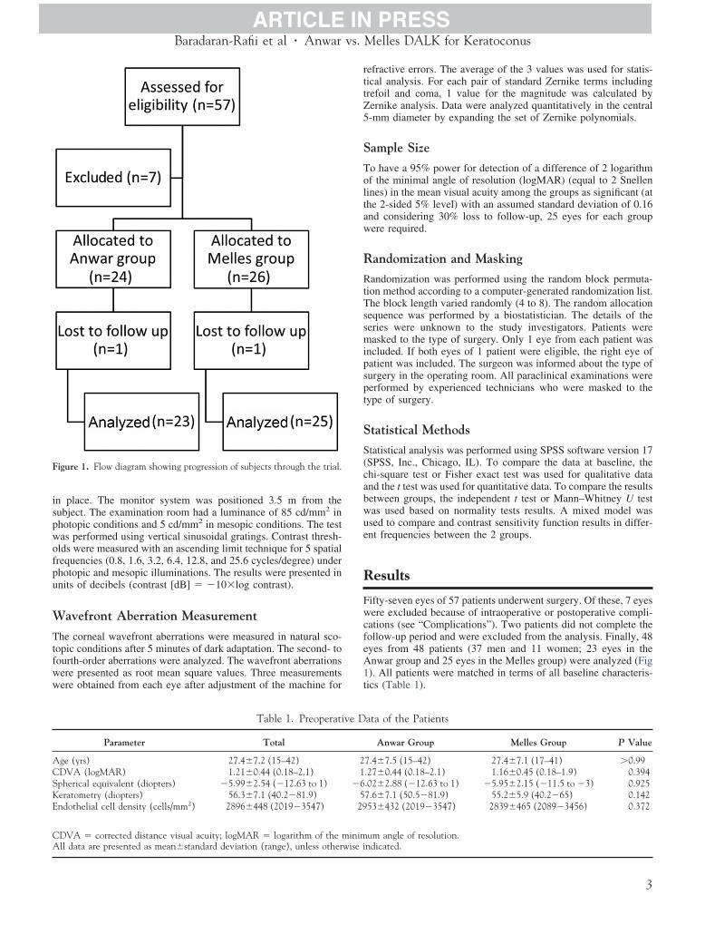
Figure 1. Flow diagram showing progression of subjects through the trial.
Table 1. Preoperati
Parameter Total
Age (yrs) 27.4�7.2 (15–42)CDVA (logMAR) 1.21�0.44 (0.18–2.1)Spherical equivalent (diopters) �5.99�2.54 (�12.63 to 1)Keratometry (diopters) 56.3�7.1 (40.2�81.9)Endothelial cell density (cells/mm2) 2896�448 (2019�3547)
CDVA � corrected distance visual acuity; logMAR � logarithm of the m
All data are presented as mean�standard deviation (range), unless otherwise iefractive errors. The average of the 3 values was used for statis-ical analysis. For each pair of standard Zernike terms includingrefoil and coma, 1 value for the magnitude was calculated byernike analysis. Data were analyzed quantitatively in the central-mm diameter by expanding the set of Zernike polynomials.
ample Size
o have a 95% power for detection of a difference of 2 logarithmf the minimal angle of resolution (logMAR) (equal to 2 Snellenines) in the mean visual acuity among the groups as significant (athe 2-sided 5% level) with an assumed standard deviation of 0.16nd considering 30% loss to follow-up, 25 eyes for each groupere required.
andomization and Masking
andomization was performed using the random block permuta-ion method according to a computer-generated randomization list.he block length varied randomly (4 to 8). The random allocationequence was performed by a biostatistician. The details of theeries were unknown to the study investigators. Patients wereasked to the type of surgery. Only 1 eye from each patient was
ncluded. If both eyes of 1 patient were eligible, the right eye ofatient was included. The surgeon was informed about the type ofurgery in the operating room. All paraclinical examinations wereerformed by experienced technicians who were masked to theype of surgery.
tatistical Methods
tatistical analysis was performed using SPSS software version 17SPSS, Inc., Chicago, IL). To compare the data at baseline, thehi-square test or Fisher exact test was used for qualitative datand the t test was used for quantitative data. To compare the resultsetween groups, the independent t test or Mann–Whitney U testas used based on normality tests results. A mixed model wassed to compare and contrast sensitivity function results in differ-nt frequencies between the 2 groups.
esults
ifty-seven eyes of 57 patients underwent surgery. Of these, 7 eyesere excluded because of intraoperative or postoperative compli-
ations (see “Complications”). Two patients did not complete theollow-up period and were excluded from the analysis. Finally, 48yes from 48 patients (37 men and 11 women; 23 eyes in thenwar group and 25 eyes in the Melles group) were analyzed (Fig). All patients were matched in terms of all baseline characteris-ics (Table 1).
ata of the Patients
Anwar Group Melles Group P Value
7.4�7.5 (15–42) 27.4�7.1 (17–41) �0.99.27�0.44 (0.18–2.1) 1.16�0.45 (0.18–1.9) 0.394.02�2.88 (�12.63 to 1) �5.95�2.15 (�11.5 to �3) 0.9257.6�7.1 (50.5�81.9) 55.2�5.9 (40.2�65) 0.14253�432 (2019�3547) 2839�465 (2089�3456) 0.372
um angle of resolution.
ve D
21
�65
29
inim
ndicated.3
A8�1tg
EFw2ai2r5�r
CIpcstf
D
TD
Ophthalmology Volume xx, Number x, Month 2012
Visual and Refractive Outcomes
Fifteen months after surgery, the mean CDVA was 0.17�0.09logMAR (range, 0�0.3 logMAR) in the Anwar group and0.18�0.11 logMAR (range, 0�0.4 logMAR) in the Melles group(95% confidence interval [CI], �0.07 to 0.05; P � 0.803). Themean spherical equivalent was �1.82�2.7 diopters (D; range,�11 to 3.25 D) and �2.69�3.94 D (range, �10.75 to 3.63 D) inthe Anwar and Melles groups, respectively (95% CI, �1.06 to 2.8;P � 0.155). Keratometric astigmatism was not significantly dif-ferent between 2 groups. The mean was 3.16�1.82 D (range,0.5�7.5 D) and 3.24�2.53 D (range, 0�10 D) in the Anwar andMelles groups, respectively (95% CI, �1.34 to 1.19; P � 0.384).
Contrast Sensitivity
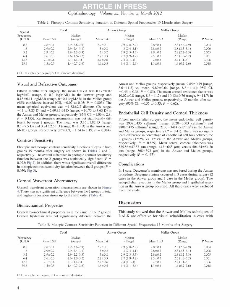
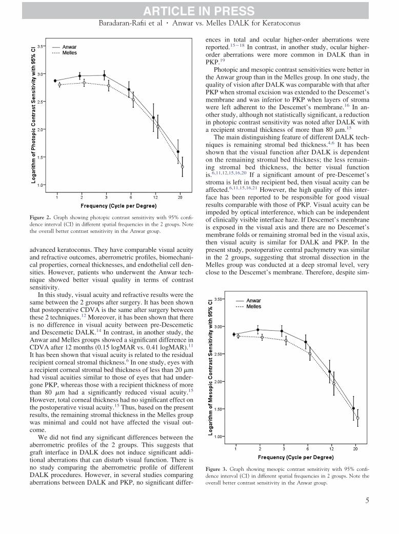
Photopic and mesopic contrast sensitivity functions of eyes in bothgroups 15 months after surgery are shown in Tables 2 and 3,respectively. The overall difference in photopic contrast sensitivityfunction between the 2 groups was statistically significant (P �0.023; Fig 2). In addition, there was a significant overall differencein mesopic contrast sensitivity function between the 2 groups (P �0.030; Fig 3).
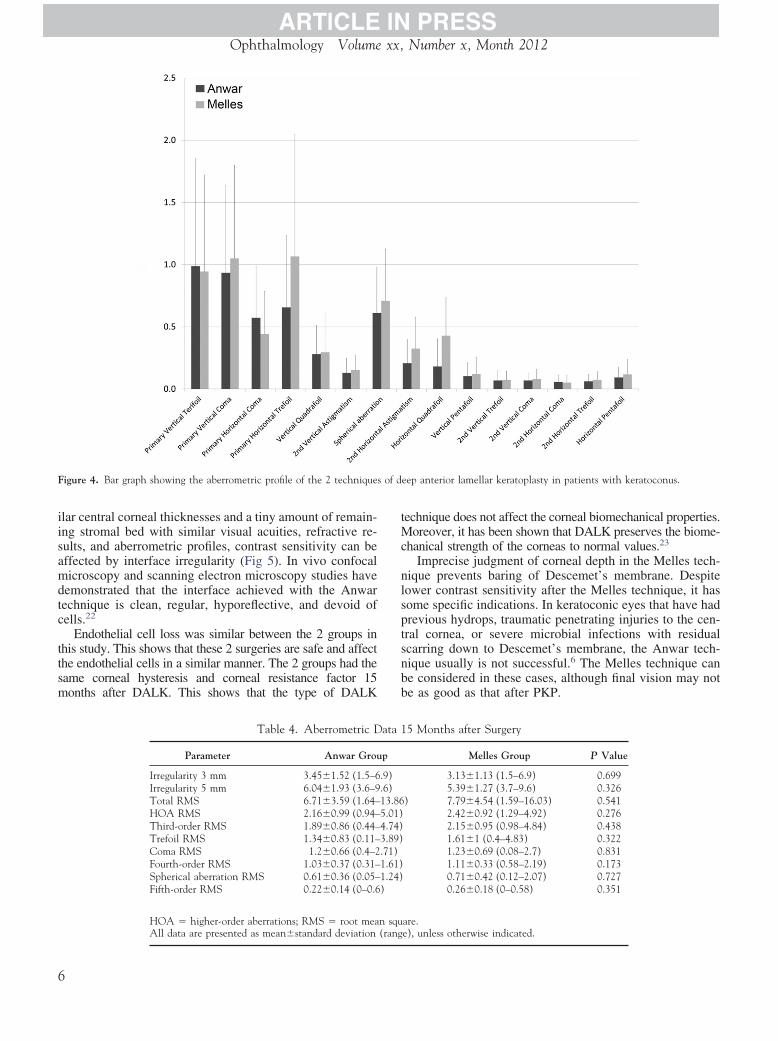
Corneal Wavefront Aberrometry
Corneal wavefront aberration measurements are shown in Figure4. There was no significant difference between the 2 groups in totaland higher-order aberrations up to the fifth order (Table 4).
Biomechanical Properties
Corneal biomechanical properties were the same in the 2 groups.Corneal hysteresis was not significantly different between the
Table 2. Photopic Contrast Sensitivity Function in
SpatialFrequency
(CPD)
Total Anw
Mean�SDMedian(Range) Mean�SD
0.8 2.8�0.1 2.9 (2.6–2.9) 2.9�0.11.6 2.9�0.2 2.9 (2.4–3.1) 3�0.23.2 2.9�0.2 2.9 (2.2–3.3) 3�0.26.4 2.6�0.3 2.6 (1.8–3.2) 2.7�0.3
12.8 2.1�0.6 2.3 (1.1–3) 2.2�0.625.6 1.5�0.5 1.4 (0.7–2.6) 1.6�0.5
CPD � cycles per degree; SD � standard deviation.
Table 3. Mesopic Contrast Sensitivity Function in
Frequency(CPD)
Total Anw
Mean�SDMedian(Range) Mean�SD
0.8 2.8�0.1 2.9 (2.6–2.9) 2.9�0.11.6 2.9�0.2 2.9 (2.4–3.1) 3�0.23.2 2.9�0.2 2.9 (2.2–3.3) 3�0.26.4 2.6�0.3 2.6 (1.8–3.2) 2.7�0.3
12.8 2.1�0.6 2.3 (1.1–3) 2.2�0.625.6 1.5�0.5 1.4 (0.7–2.6) 1.6�0.5
CPD � cycle per degree; SD � standard deviation.
4
nwar and Melles groups, respectively (mean, 9.85�0.79 [range,.6�11.3] vs. mean, 9.89�0.64 [range, 8.8�11.4]; 95% CI,0.45 to 0.36; P � 0.83). The mean corneal resistance factor was
0.02�0.8 (range, 8.6�11.7) and 10.13�0.76 (range, 9�11.7) inhe Anwar and Melles groups, respectively, 15 months after sur-ery (95% CI, �0.55 to 0.33; P � 0.62).
ndothelial Cell Density and Corneal Thicknessifteen months after surgery, the mean endothelial cell densityas 2939�435 cell/mm2 (range, 2020�3503 cell/mm2) and808�435 cell/mm2 (range, 2130�3414 cell/mm2) in the Anwarnd Melles groups, respectively (P � 0.41). There was no signif-cant difference in percentage of endothelial cell loss between the
groups (1�2% vs. 1�3% in the Anwar and Melles groups,espectively; P � 0.869). Mean central corneal thickness was25.56�47.87 �m (range, 442�668 �m) versus 504.64�54.20m (range, 368�593 �m) in the Anwar and Melles groups,
espectively (P � 0.155).
omplicationsn 1 case, Descemet’s membrane was not bared during the Anwarrocedure. Descemet rupture occurred in 3 cases during surgery (2ases in the Anwar group and 1 case in the Melles group). Twoubepithelial rejections in the Melles group and 1 epithelial rejec-ion in the Anwar group occurred. All these cases were excludedrom the study.
iscussion
his study showed that the Anwar and Melles techniques ofALK are effective for visual rehabilitation in eyes with
erent Spatial Frequencies 15 Months after Surgery
roup Melles Group
P ValueMedian(Range) Mean�SD
Median(Range)
2.9 (2.8–2.9) 2.8�0.1 2.8 (2.6–2.9) 0.0043 (2.4–3.1) 2.8�0.2 2.8 (2.5–3.1) 0.006
2.9 (2.3–3.3) 2.8�0.2 2.8 (2.2–3.3) 0.0052.7 (1.9–3.2) 2.5�0.3 2.6 (1.8–3.2) 0.0612.4 (1.1–3) 2�0.5 2.2 (1.1–3) 0.3061.4 (1.1–2.6) 1.3�0.4 1.4 (0.7–2.6) 0.048
Diff
ar G
Diff
ar G
eroP
tqPmwoia
nsoiisafrioimtpiMc
Fd
Baradaran-Rafii et al � Anwar vs. Melles DALK for Keratoconus
advanced keratoconus. They have comparable visual acuityand refractive outcomes, aberrometric profiles, biomechani-cal properties, corneal thicknesses, and endothelial cell den-sities. However, patients who underwent the Anwar tech-nique showed better visual quality in terms of contrastsensitivity.
In this study, visual acuity and refractive results were thesame between the 2 groups after surgery. It has been shownthat postoperative CDVA is the same after surgery betweenthese 2 techniques.12 Moreover, it has been shown that thereis no difference in visual acuity between pre-Descemeticand Descemetic DALK.14 In contrast, in another study, theAnwar and Melles groups showed a significant difference inCDVA after 12 months (0.15 logMAR vs. 0.41 logMAR).11
It has been shown that visual acuity is related to the residualrecipient corneal stromal thickness.6 In one study, eyes witha recipient corneal stromal bed thickness of less than 20 �mhad visual acuities similar to those of eyes that had under-gone PKP, whereas those with a recipient thickness of morethan 80 �m had a significantly reduced visual acuity.15
However, total corneal thickness had no significant effect onthe postoperative visual acuity.15 Thus, based on the presentresults, the remaining stromal thickness in the Melles groupwas minimal and could not have affected the visual out-come.
We did not find any significant differences between theaberrometric profiles of the 2 groups. This suggests thatgraft interface in DALK does not induce significant addi-tional aberrations that can disturb visual function. There isno study comparing the aberrometric profile of differentDALK procedures. However, in several studies comparing
Figure 2. Graph showing photopic contrast sensitivity with 95% confi-dence interval (CI) in different spatial frequencies in the 2 groups. Notethe overall better contrast sensitivity in the Anwar group.
aberrations between DALK and PKP, no significant differ- o
nces in total and ocular higher-order aberrations wereeported.15�18 In contrast, in another study, ocular higher-rder aberrations were more common in DALK than inKP.19
Photopic and mesopic contrast sensitivities were better inhe Anwar group than in the Melles group. In one study, theuality of vision after DALK was comparable with that afterKP when stromal excision was extended to the Descemet’sembrane and was inferior to PKP when layers of stromaere left adherent to the Descemet’s membrane.16 In an-ther study, although not statistically significant, a reductionn photopic contrast sensitivity was noted after DALK withrecipient stromal thickness of more than 80 �m.15
The main distinguishing feature of different DALK tech-iques is remaining stromal bed thickness.4,6 It has beenhown that the visual function after DALK is dependentn the remaining stromal bed thickness; the less remain-ng stromal bed thickness, the better visual functions.6,11,12,15,16,20 If a significant amount of pre-Descemet’stroma is left in the recipient bed, then visual acuity can beffected.6,11,15,16,21 However, the high quality of this inter-ace has been reported to be responsible for good visualesults comparable with those of PKP. Visual acuity can bempeded by optical interference, which can be independentf clinically visible interface haze. If Descemet’s membranes exposed in the visual axis and there are no Descemet’sembrane folds or remaining stromal bed in the visual axis,
hen visual acuity is similar for DALK and PKP. In theresent study, postoperative central pachymetry was similarn the 2 groups, suggesting that stromal dissection in the
elles group was conducted at a deep stromal level, verylose to the Descemet’s membrane. Therefore, despite sim-
igure 3. Graph showing mesopic contrast sensitivity with 95% confi-ence interval (CI) in different spatial frequencies in 2 groups. Note the
verall better contrast sensitivity in the Anwar group.5
tMc
nlsptsnbb
of d
Ophthalmology Volume xx, Number x, Month 2012
ilar central corneal thicknesses and a tiny amount of remain-ing stromal bed with similar visual acuities, refractive re-sults, and aberrometric profiles, contrast sensitivity can beaffected by interface irregularity (Fig 5). In vivo confocalmicroscopy and scanning electron microscopy studies havedemonstrated that the interface achieved with the Anwartechnique is clean, regular, hyporeflective, and devoid ofcells.22
Endothelial cell loss was similar between the 2 groups inthis study. This shows that these 2 surgeries are safe and affectthe endothelial cells in a similar manner. The 2 groups had thesame corneal hysteresis and corneal resistance factor 15months after DALK. This shows that the type of DALK
Figure 4. Bar graph showing the aberrometric profile of the 2 techniques
Table 4. Aberrometric D
Parameter Anwar Grou
Irregularity 3 mm 3.45�1.52 (1.5–6Irregularity 5 mm 6.04�1.93 (3.6–9Total RMS 6.71�3.59 (1.64–HOA RMS 2.16�0.99 (0.94–Third-order RMS 1.89�0.86 (0.44–Trefoil RMS 1.34�0.83 (0.11–Coma RMS 1.2�0.66 (0.4–2Fourth-order RMS 1.03�0.37 (0.31–Spherical aberration RMS 0.61�0.36 (0.05–Fifth-order RMS 0.22�0.14 (0–0.6
HOA � higher-order aberrations; RMS � root mea
All data are presented as mean�standard deviation (rang6
echnique does not affect the corneal biomechanical properties.oreover, it has been shown that DALK preserves the biome-
hanical strength of the corneas to normal values.23
Imprecise judgment of corneal depth in the Melles tech-ique prevents baring of Descemet’s membrane. Despiteower contrast sensitivity after the Melles technique, it hasome specific indications. In keratoconic eyes that have hadrevious hydrops, traumatic penetrating injuries to the cen-ral cornea, or severe microbial infections with residualcarring down to Descemet’s membrane, the Anwar tech-ique usually is not successful.6 The Melles technique cane considered in these cases, although final vision may note as good as that after PKP.
eep anterior lamellar keratoplasty in patients with keratoconus.
5 Months after Surgery
Melles Group P Value
3.13�1.13 (1.5–6.9) 0.6995.39�1.27 (3.7–9.6) 0.326
) 7.79�4.54 (1.59–16.03) 0.5412.42�0.92 (1.29–4.92) 0.2762.15�0.95 (0.98–4.84) 0.4381.61�1 (0.4–4.83) 0.3221.23�0.69 (0.08–2.7) 0.8311.11�0.33 (0.58–2.19) 0.1730.71�0.42 (0.12–2.07) 0.7270.26�0.18 (0–0.58) 0.351
are.
ata 1
p
.9)
.6)13.865.01)4.74)3.89).71)1.61)1.24))
n squ
e), unless otherwise indicated.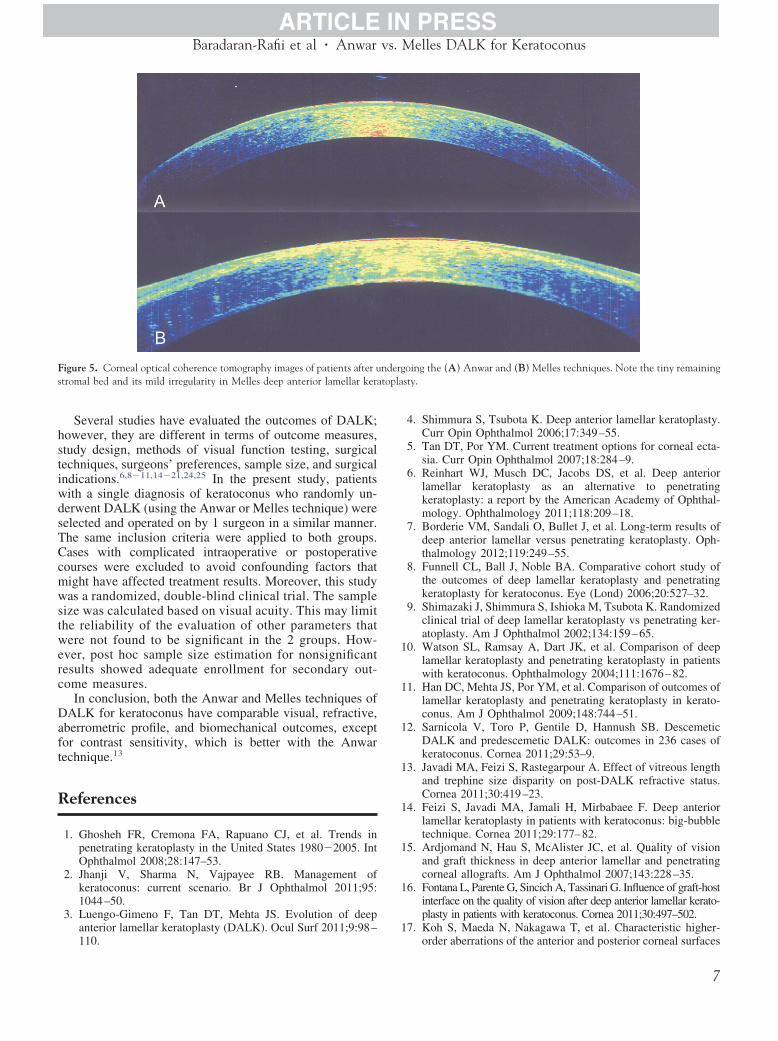
1
1
1
1
1
1
1
1
topla
Baradaran-Rafii et al � Anwar vs. Melles DALK for Keratoconus
Several studies have evaluated the outcomes of DALK;however, they are different in terms of outcome measures,study design, methods of visual function testing, surgicaltechniques, surgeons’ preferences, sample size, and surgicalindications.6,8�11,14�21,24,25 In the present study, patientswith a single diagnosis of keratoconus who randomly un-derwent DALK (using the Anwar or Melles technique) wereselected and operated on by 1 surgeon in a similar manner.The same inclusion criteria were applied to both groups.Cases with complicated intraoperative or postoperativecourses were excluded to avoid confounding factors thatmight have affected treatment results. Moreover, this studywas a randomized, double-blind clinical trial. The samplesize was calculated based on visual acuity. This may limitthe reliability of the evaluation of other parameters thatwere not found to be significant in the 2 groups. How-ever, post hoc sample size estimation for nonsignificantresults showed adequate enrollment for secondary out-come measures.
In conclusion, both the Anwar and Melles techniques ofDALK for keratoconus have comparable visual, refractive,aberrometric profile, and biomechanical outcomes, exceptfor contrast sensitivity, which is better with the Anwartechnique.13
References
1. Ghosheh FR, Cremona FA, Rapuano CJ, et al. Trends inpenetrating keratoplasty in the United States 1980�2005. IntOphthalmol 2008;28:147–53.
2. Jhanji V, Sharma N, Vajpayee RB. Management ofkeratoconus: current scenario. Br J Ophthalmol 2011;95:1044–50.
3. Luengo-Gimeno F, Tan DT, Mehta JS. Evolution of deepanterior lamellar keratoplasty (DALK). Ocul Surf 2011;9:98–
Figure 5. Corneal optical coherence tomography images of patients after ustromal bed and its mild irregularity in Melles deep anterior lamellar kera
110.
4. Shimmura S, Tsubota K. Deep anterior lamellar keratoplasty.Curr Opin Ophthalmol 2006;17:349–55.
5. Tan DT, Por YM. Current treatment options for corneal ecta-sia. Curr Opin Ophthalmol 2007;18:284–9.
6. Reinhart WJ, Musch DC, Jacobs DS, et al. Deep anteriorlamellar keratoplasty as an alternative to penetratingkeratoplasty: a report by the American Academy of Ophthal-mology. Ophthalmology 2011;118:209–18.
7. Borderie VM, Sandali O, Bullet J, et al. Long-term results ofdeep anterior lamellar versus penetrating keratoplasty. Oph-thalmology 2012;119:249–55.
8. Funnell CL, Ball J, Noble BA. Comparative cohort study ofthe outcomes of deep lamellar keratoplasty and penetratingkeratoplasty for keratoconus. Eye (Lond) 2006;20:527–32.
9. Shimazaki J, Shimmura S, Ishioka M, Tsubota K. Randomizedclinical trial of deep lamellar keratoplasty vs penetrating ker-atoplasty. Am J Ophthalmol 2002;134:159–65.
0. Watson SL, Ramsay A, Dart JK, et al. Comparison of deeplamellar keratoplasty and penetrating keratoplasty in patientswith keratoconus. Ophthalmology 2004;111:1676–82.
1. Han DC, Mehta JS, Por YM, et al. Comparison of outcomes oflamellar keratoplasty and penetrating keratoplasty in kerato-conus. Am J Ophthalmol 2009;148:744–51.
2. Sarnicola V, Toro P, Gentile D, Hannush SB. DescemeticDALK and predescemetic DALK: outcomes in 236 cases ofkeratoconus. Cornea 2011;29:53–9.
3. Javadi MA, Feizi S, Rastegarpour A. Effect of vitreous lengthand trephine size disparity on post-DALK refractive status.Cornea 2011;30:419–23.
4. Feizi S, Javadi MA, Jamali H, Mirbabaee F. Deep anteriorlamellar keratoplasty in patients with keratoconus: big-bubbletechnique. Cornea 2011;29:177–82.
5. Ardjomand N, Hau S, McAlister JC, et al. Quality of visionand graft thickness in deep anterior lamellar and penetratingcorneal allografts. Am J Ophthalmol 2007;143:228–35.
6. Fontana L, Parente G, Sincich A, Tassinari G. Influence of graft-hostinterface on the quality of vision after deep anterior lamellar kerato-plasty in patients with keratoconus. Cornea 2011;30:497–502.
7. Koh S, Maeda N, Nakagawa T, et al. Characteristic higher-
oing the (A) Anwar and (B) Melles techniques. Note the tiny remainingsty.
nderg
order aberrations of the anterior and posterior corneal surfaces
7
2
2
2
2
Ophthalmology Volume xx, Number x, Month 2012
in 3 corneal transplantation techniques. Am J Ophthalmol2012;153:284–90.
18. Javadi MA, Feizi S, Yazdani S, Mirbabaee F. Deep anteriorlamellar keratoplasty versus penetrating keratoplasty forkeratoconus: a clinical trial. Cornea 2011;29:365–71.
19. Bahar I, Kaiserman I, Srinivasan S, et al. Comparison of threedifferent techniques of corneal transplantation for keratoco-nus. Am J Ophthalmol 2008;146:905–12.
20. Feizi S, Javadi MA, Rastegarpour A. Visual acuity and refrac-tion after deep anterior lamellar keratoplasty with and withoutsuccessful big-bubble formation. Cornea 2011;29:1252–5.
21. Alio JL. Visual improvement after late debridement of residual
stroma after anterior lamellar keratoplasty. Cornea 2008;27:871–3.Footnotes and Financial Disclosures
Iran.
FTd
SUd
CMd
8
2. Feizi S, Javadi MA, Kanavi MR. Cellular changes of donorcorneal tissue after deep anterior lamellar keratoplasty versuspenetrating keratoplasty in eyes with keratoconus: a confocalstudy. Cornea 2011;29:866–70.
3. Hosny M, Hassaballa MA, Shalaby A. Changes in cornealbiomechanics following different keratoplasty techniques.Clin Ophthalmol 2011;5:767–70.
4. Borderie VM, Werthel AL, Touzeau O, et al. Comparison oftechniques used for removing the recipient stroma in anteriorlamellar keratoplasty. Arch Ophthalmol 2008;126:31–7.
5. Panda A, Bageshwar LM, Ray M, et al. Deep lamellar kera-toplasty versus penetrating keratoplasty for corneal lesions.
Cornea 1999;18:172–5.Originally received: January 22, 2012.Final revision: July 30, 2012.Accepted: July 31, 2012.Available online: ●●● Manuscript no. 2012-93.
1 Ophthalmic Research Center, Labbafinejad Medical Center, Shahid Be-heshti University of Medical Sciences, Tehran, Iran.
2 School of Medicine, Tehran University of Medical Sciences, Tehran,
inancial Disclosure(s):he author(s) have no proprietary or commercial interest in any materialsiscussed in this article.
upported by a grant from Ophthalmic Research Center, Shahid Beheshtiniversity of Medical Sciences, Tehran, Iran, which had no role in theesign or conduct of this research.
orrespondence:edi Eslani, MD, Labbafinejad Medical Center, Boostan 9 Street, Pas-
aran Avenue, Tehran 16666, Iran. E-mail: [email protected].