Embed Size (px)
Citation preview
“Bound” residues from biomass and CO2 in soils – formation, fate
and stability during biotic incubation
Der Fakultät für Mathematik, Informatik und Naturwissenschaften der Rheinisch- Westfälischen Technischen Hochschule Aachen vorgelegte Dissertation zur Erlangung des
akademischen Grades einer Doktorin der Naturwissenschaften
von Diplom-Ingenieurin im Umweltschutz
Karolina Malgorzata Nowak aus Olsztyn, Polen

Parts of this thesis are published or submitted for publication in
scientific journals:
NOWAK, K.M.; MILTNER, A.; GEHRE, M.; SCHÄFFER, A.; KÄSTNER, M. Formation and fate of
bound residues from microbial biomass during 2,4-D degradation in soil, Environ. Sci.
Technol., in press;
NOWAK, K.M.; GIRARDI, C.; MILTNER, A.; GEHRE, M.; SCHÄFFER, A.; KÄSTNER, M.
Formation and fate of non-extractable residues during microbial degradation of 13C6-ibuprofen in soil (in preparation);
GIRARDI, C.; NOWAK, K.M.; LEWKOW, B.; MILTNER, A.; GEHRE, M.; KÄSTNER, M.
Comparison of microbial degradation of the C-isotope-labelled pharmaceutical ibuprofen and
the herbicide 2,4-D in water and soil, submitted to Environ. Pollut.
This thesis is dedicated to the memory of my father,
Prof. Dr. habil. Grzegorz Nowak (1946 – 2002),
who inspired my fascination in environmental science

LIST OF CONTENTS IV
LIST OF CONTENTS ...........................................................................................................VI
LIST OF ABBREVIATIONS.............................................................................................. VII
SUMMARY............................................................................................................................ ..X
ZUSAMMENFASSUNG ................................................................................................... ..XII
1 INTRODUCTION ............................................................................................................... 1
2 STATE OF THE ART......................................................................................................... 3
2.1 Complexity of the soil system ...................................................................................... 3
2.2 Microbial activity in the soil ........................................................................................ 6
2.2.1 Natural organic compounds biodegradation in soil........................................................ 9
2.2.2 Anthropogenic organic compounds biodegradation in soil.......................................... 10
2.3 Bioavailability of organic contaminants in soil........................................................ 11
2.4 Definition of the non-extractable residues (NER) ................................................... 14
2.5 Determination of NER in soil .................................................................................... 15
2.6 Formation of NER in soil ........................................................................................... 16
2.6.1 Parent compounds and metabolites ........................................................................................ 17
2.6.2 Components of microbial biomass (Fatty acids and amino acids) ................................ 20
2.7 Stability of NER in soil............................................................................................... 23
2.8 Risk assessment of NER............................................................................................. 25
3 AIMS OF THE STUDY .................................................................................................... 29
4 MODEL COMPOUNDS................................................................................................... 31
4.1 2,4-Dichlorophenoxyacetic acid (2,4-D).................................................................... 31
4.2 Ibuprofen (Ibu) ........................................................................................................... 34

LIST OF CONTENTS V
5 MATERIAL AND METHODS........................................................................................ 37
5.1 Chemicals and materials............................................................................................ 37
5.2 Liquid culture experiments ....................................................................................... 38
5.2.1 Strain ............................................................................................................................................... 38
5.2.2 Medium........................................................................................................................................... 39
5.2.3 Preculture incubation.................................................................................................................. 39
5.2.4 Incubation experiment with 13C6-2,4-D ................................................................................ 40
5.2.5 Incubation experiment under 13CO2 atmosphere ................................................................ 41
5.3 Soil experiments.......................................................................................................... 42
5.3.1 Soil ................................................................................................................................................... 43
5.3.2 Incubation experiment................................................................................................................ 43
5.3.3 Soil incubation experiment with 13C6-2,4-D........................................................................ 44
5.3.4 Soil incubation experiment under 13CO2 atmosphere ....................................................... 44
5.3.5 Soil incubation experiment with 13C6-ibu............................................................................. 45
5.4 Chemical analyses ..................................................................................................................... 45
5.4.1 CO2 measurement ........................................................................................................................ 46
5.4.2 Parent compound and metabolites measurement ............................................................... 46
5.4.3 Total biomass and NER measurement .................................................................................. 47
5.4.4 Fatty acids (FA) analyses .......................................................................................................... 48
5.4.5 Amino acids (AA) analyses...................................................................................................... 50
5.5 Data analyses............................................................................................................................... 52
6 RESULTS........................................................................................................................... 53
6.1 Liquid culture experiments ....................................................................................... 53
6.1.1 Incubation experiment with 13C6-2,4-D ................................................................................ 53
6.1.1.1 Mass balance of 13C6-2,4-D in the system ............................................................................ 54
6.1.1.2 Incorporation of 13C into PLFA of C. necator JMP 134 grown on 13C6-2,4-D.......... 56
6.1.1.3 Incorporation of 13C into AA of C. necator JMP 134 grown on 13C6-2,4-D .............. 56
6.1.2 Incubation experiment under 13CO2 atmosphere ................................................................ 57
6.1.2.1 Incorporation of 13C into biomass of C. necator JMP 134 grown under 13CO2......... 58
6.1.2.2 Incorporation of 13C into PLFA of C. necator JMP 134 grown under 13CO2............. 59
6.1.2.3 Incorporation of 13C into AA of C. necator JMP 134 grown under 13CO2................. 60

LIST OF CONTENTS VI
6.2 Soil experiments.......................................................................................................... 60
6.2.1 Soil incubation experiment with 13C6-2,4-D........................................................................ 61
6.2.1.1 Mass balance of 13C6-2,4-D in the soil .................................................................................. 61
6.2.1.2 Formation of FA and their fate in soil incubated with 13C6-2,4-D ................................ 62
6.2.1.3 Formation of AA and their fate in soil incubated with 13C6-2,4-D ............................... 66
6.2.1.4 Biogenic residues in soil incubated with 13C6-2,4-D......................................................... 68
6.2.2 Soil incubation experiment under 13CO2 atmosphere ....................................................... 68
6.2.2.1 Formation of FA and their fate in soil incubated under 13CO2 atmosphere................ 68
6.2.3 Soil incubation experiment 13C6-ibu ...................................................................................... 71
6.2.3.1 Mass balance of 13C6-ibu in the soil ....................................................................................... 71
6.2.3.2 Formation of FA and their fate in soil incubated with 13C6-ibu..................................... 72
6.2.3.3 Formation of AA and their fate in soil incubated with 13C6-ibu .................................... 76
6.2.3.4 Biogenic residues in soil incubated with 13C6-ibu.............................................................. 78
7 DISCUSSION..................................................................................................................... 79
7.1 Liquid culture experiments ....................................................................................... 79
Incorporation of 13C into biomass of C. necator JMP 134 grown on 2,4-D medium
7.2 Soil experiments.......................................................................................................... 83
7.2.1 Soil incubation experiment with 13C6-2,4-D........................................................................ 83
7.2.1.1 Biogenic residues as NER in soil incubated with 13C6-2,4-D......................................... 83
7.2.1.2 Fate and stability of FA in soil incubated with 13C6-2,4-D ............................................. 86
7.2.1.3 Fate and stability of AA in soil incubated with 13C6-2,4-D............................................. 87
7.2.2 Soil incubation experiment with 13C6-ibu............................................................................. 89
7.2.2.1 Biogenic residues as NER in soil incubated with 13C6-ibu.............................................. 89
7.2.2.2 Fate and stability of FA in soil incubated with 13C6-ibu .................................................. 91
7.2.2.3 Fate and stability of AA in soil incubated with 13C6-ibu.................................................. 92
8 CONCLUSIONS................................................................................................................ 94
LITERATURE .......................................................................................................................97
ACKNOWLEDGEMENTS................................................................................................. 113

LIST OF ABBREVIATIONS VII
LIST OF ABBREVIATIONS
13C Labelled (stable isotope) 14C Labelled (radioactive isotope)
2,4-D 2,4-Dichlorophenoxyacetic acid
2,4-DCP 2,4-Dichlorophenol
2-OH-ibu Hydroxyibuprofen
a (for FA) Anteiso
AA Amino Acids
AC Acetylchloride
ACN Acetone
Ala Alanine
ASE Accelerated Solvent Extraction
Asp/Asg Aspartate/Asparagine
BAME Bacterial Acid Methyl Esters
bioAA Biomass Amino Acids
br (for FA) Branched
Corg Organic Carbon
cy (for FA) cyclopropyl
DCM Dichloromethane
Dept Department
DT50 Half -life in soil
DW Dry Weight
EA-C-irMS Elemental Analyser-Combustion-isotope ratio Mass Spectrometry
EDTA Ethylenediaminetetraacetic acid
FA Fatty Acids
FAME Fatty Acid Methyl Esther
FAME 21:0 Heneicosanoate methyl ester
GC-C-irMS Gas Chromatography Combustion-isotope ratio Mass Spectrometry
GC-MS Gas Chromatography Mass Spectrometry
Glu/Gln Glutamate/Glutamine
Gly Glycine
i (for FA) Iso
Ibu Ibuprofen
LIST OF ABBREVIATIONS VIII
Ile Isoleucine
IP Isopropanol
IUPAC The International Union of Pure and Applied Chemistry
Koc, logKoc Soil-sorption coefficient
Kow, logKow Octanol/water partition coefficient
Leu Leucine
Lys Lysine
M Molar concentration
Me (for FA) Mid-chain branched
MeOH Methanol
MM Minimum Medium
Na2CO3 Sodium bicarbonate
n.d. not detectable
NER Non-Extractable Residues
NH4OH Ammonium Hydroxide
nm Nanometre
NTotal Total Nitrogen
OD Optical Density
OECD Organization for Economic Co-operation and Development
OM Organic Matter
PAH Polycyclic Aromatic Hydrocarbons
PB Phosphate Buffer
PEG 600 Polyethylenglycol 600
Phe Phenylalanine
PLFA Phospholipid Fatty Acids
Pro Proline
Ser Serine
SOM Soil Organic Matter
SPE Solid Phase Extraction
tAA Total Amino Acids
TCC Tricarboxylic acid Cycle
tFA Total Fatty Acids
TFAA Trifluroacetic Acid Anhydride
Threo Threonine

LIST OF ABBREVIATIONS IX
TM Tryptone Medium
TMCS Trimethylchlorosilane
TNT Trinitrotoluene
TOC Total Organic Carbon
UFZ Centre for Environmental Research
USA United States of America
UV/VIS Ultraviolet-Visible Spectrophotometry
Val Valine
WHC Water Holding Capacity
WWTP Wastewater treatment plant

SUMMARY X
SUMMARY
Biodegradation of organic pollutants in soil generally results in the formation of metabolites,
microbial biomass, mineralisation products and bound or non-extractable residues (NER). It is
speculated that NER can pose a risk for humans due to the remobilisation and further
distribution of active parent compounds or their metabolites over the food web
(BARRACLOUGH ET AL., 2005). Although there are many studies on NER formation available,
their chemical structures are still unknown. However, without this knowledge, a proper risk
assessment for the pollutant and the related NER during its transformation in soil is
impossible. Part of the NER may be biogenic, since the pollutant-derived C and CO2 released
from its mineralisation are assimilated by microorganisms into their cellular components [e.g.
fatty acids (FA) and amino acids (AA)], which are subsequently incorporated into soil organic
matter (SOM) after cell death
In order to study the microbial biomass contribution to NER formation, soil was
incubated with either 13C-labelled 2,4-Dichlorophenoxy acetic acid (2,4-D) or ibuprofen (Ibu)
for 64 and 90 days, respectively. At different sampling dates, the soil was analysed for the
presence of the 13C label in FA and AA. The 13C label assimilated in biomolecules was
determined in both the living and the non-living SOM fractions. Moreover, for investigating
the relevance of heterotrophic CO2 fixation for the incorporation of 13C into biogenic NER in
soil and to distinguish between formations of NER directly from 2,4-D and via CO2, an
experiment with unlabelled 2,4-D and labelled CO2 was conducted. In addition, to understand
both the processes of the biogenic NER formation and the incorporation of 13C label into FA
and AA from either 13C6-2,4-D or 13CO2 in the complex soil environment, a simple biological
model system with the known 2,4-D degrader (Cupriavidus necator JMP 134), was studied.
After 64 days of 13C6-2,4-D incubation in soil, the total contents of 13C detected in AA
in SOM indicated that 44% of the initially applied 13C6-2,4-D equivalents had been converted
to microbial biomass and finally to biogenic residues. The intermediate maximum of 13C-FA
in SOM indicated a 20% conversion of 13C6-2,4-D to biomass. However, 13C-FA in the non-
living SOM fraction decreased to 50% indicating their metabolisation and their further
distribution within the food web. Contrary to the 13C-FA, 13C-AA in non-living SOM pool
were surprisingly stable.
The soil experiment with 13CO2 showed that after 16 days of incubation, the
heterotrophic CO2 fixation was relevant in the assimilation of 13C label into biomass
components, in particular FA. In addition, the liquid culture experiment with C. necator JMP
134 demonstrated the high importance of CO2 fixation in the incorporation of 13C label into

SUMMARY XI
the biomass components in the later phase of incubation. In this study about 4% of 13C
derived from 13C6-2,4-D was assimilated in the biomass of this bacterial strain via CO2
fixation.
A total content of biogenic NER of 54% of 13C6-ibu equivalents at the end of the 13C6-ibu soil experiment was indicated by the amount of 13C found in AA in SOM. From the
maximum content of 13C-FA detected in this experiment, at least 24% (of 13C6-ibu
equivalents) biomass must have been formed. Contrary to the 13C6-2,4-D soil incubation
experiment, the 13C-FA remained stable until the end. However, the formation of NER in the
soil incubated with 13C6-ibu started much later than that in the 13C6-2,4-D study, due to the
longer lag phase and the interruption of the incubation before the destabilisation of biogenic
NER. The 13C-AA in SOM remained also stable as found in 13C6-2,4-D experiment.
The results from these soil biodegradation experiments provide the first evidence that
nearly all NER both from 2,4-D and Ibu are biogenic, containing only natural microbial
biomass components stabilised in SOM. However, biogenic residues are formed if the
respective organic contaminant is readily degraded by microorganisms under significant
formation of CO2. Depending on the yield coefficients of C conversion into biomass, we
expect ratios of biomass plus biogenic residues to CO2 of about 0.2 to 1. In the 13C6-2,4-D
study, the ratio was ~ 0.8 and in the 13C6-ibu was 1.2.
Biogenic residues are clearly excluded from the definition of NER according to IUPAC. Due
to biogenic residue formation, the potential risk of NER from the readily metabolised organic
contaminants in soils is thus highly overestimated in many cases. Therefore, the formation of
biogenic residues must be taken into consideration, when determining the mass balances of
contaminants during their microbial degradation in soil.

ZUSAMMENFASSUNG XII
ZUSAMMENFASSUNG
Biologischer Abbau von organischen Schadstoffen im Boden führt generell zur
Mineralisierung und zur Bildung von Metaboliten und gebundenen oder nicht-extrahierbaren
Rückständen (NER). Es wird spekuliert, dass NER ein Risiko für den Menschen darstellen
können, da sie möglicherweise remobilisiert werden können und die aktiven
Ausgangsverbindungen oder deren Metabolite weiter in der Nahrungskette verbreitet werden
können (BARRACLOUGH AT AL., 2005). Trotz vieler Studien über die NER-Bildung sind ihre
genauen chemischen Strukturen noch nicht bekannt. Allerdings ist ohne dieses Wissen eine
korrekte Risikobewertung für den Schadstoff und die damit verbundenen NER während der
Transformation im Boden unmöglich. Ein Teil der NER könnten biogen sein, weil der
schadstoffbürtige Kohlenstoff und das durch Mineralisierung freigesetzte CO2 von
Mikroorganismen in ihre zellulären Bestandteile eingebaut werden, z.B. Fettsäuren (FS) und
Aminosäuren (AS), die nach dem Zelltod in der organischen Bodensubstanz (OBS) festgelegt
werden können.
Um den Beitrag der mikrobiellen Biomasse zur NER-Bildung zu untersuchen, wurde
ein Boden mit 13C-markierter 2,4-Dichlorphenoxyessigsäure (2,4-D) bzw. Ibuprofen (Ibu) für
64 bzw. 90 Tagen inkubiert. Die Bodenproben wurden zu verschiedenen
Probenahmezeitpunkten auf die 13C-Anreicherung in FS und AS analysiert. Die in den
Biomolekülen gebundene 13C-Markierung wurde sowohl in der lebenden als auch in der toten
OBS Fraktionen bestimmt. Außerdem wurden Experimente mit unmarkiertem 2,4-D und
markiertem CO2 durchgeführt, um den Beitrag der heterotrophen CO2-Fixierung zum Einbau
von 13C in biogene NER in Böden zu untersuchen. Damit kann zwischen direkter NER-
Bildung aus 2,4-D und indirekter über CO2 unterschieden werden. Zudem wurde ein
einfaches biologisches Modellsystem mit dem bekannten 2,4-D Abbauer Cupriavidus necator
JMP 134 untersucht, um sowohl die an der Bildung von biogenen NER beteiligten Prozesse
als auch den Einbau von 13C aus 13C6-2,4-D bzw. 13CO2 in FS und AS in der komplexen
Bodenumwelt besser zu verstehen.
Die Menge der gesamten 13C-AS in SOM nach 64-tägiger Inkubation mit 13C6-2,4-D im
Boden deutet darauf hin, dass 44% der anfänglichen 13C6-2,4-D-Äquivalente in mikrobielle
Biomasse und schließlich in biogene Rückstände umgesetzt wurden. Das zwischenzeitliche
Maximum der 13C-FS in der organischen Bodensubstanz (SOM) zeigte, dass 20% des 13C6-
2,4-D in mikrobielle Biomasse umgesetzt wurden. Allerdings sanken die 13C-FS in der toten
SOM Fraktion wegen ihrer Metabolisierung und ihrer weiteren Verbreitung im Nahrungsnetz

ZUSAMMENFASSUNG XIII
auf 50% ab. Im Gegensatz zu den 13C-FS waren 13C-AS im toten SOM Pool überraschend
stabil.
Das Bodenexperiment mit 13CO2 zeigte nach 16-tägiger Inkubation die Relevanz der
heterotrophen CO2-Fixierung bei der Assimilation von 13C in die Bestandteile der
mikrobiellen Biomasse, insbesondere in die FA. Zudem wies das Flüssigkultur Experiment
mit C. necator JMP 134 auf die hohe Bedeutung der CO2-Fixierung für den Einbau der 13C-
Markierung in die Biomasse-Bestandteile während der späteren Phase der Inkubation hin. In
dieser Studie wurde etwa 4% des 2,4-D-bürtigen Kohlenstoffs über CO2-Fixierung in die
Biomasse dieses Bakterienstamms assimiliert.
Am Ende des Bodenexperiments mit 13C6-Ibu wies die Menge der 13C-AS in der SOM
auf einen gesamten biogenen NER-Gehalt von 54% der 13C6-Ibu-Äquivalente hin. Auf der
Basis des maximalen 13C-FA Gehalts, der in diesem Experiment gefunden wurde, müssen
mindestens 24% (der 13C6-Ibu-Äquivalente) Biomasse gebildet worden sein. Im Gegensatz
zur Inkubation des Bodens mit 13C6-2,4-D blieben die 13C-FA im Falle von 13C6-Ibu bis zum
Ende des Experiments stabil. Allerdings begann die NER-Bildung in dem Boden, der mit 13C6-Ibu inkubiert wurde, viel später als in der 13C6-2,4-D-Studie, was auf der längeren Lag-
Phase und dem Abbruch der Inkubation vor der Destabilisierung der biogenen NER beruht.
Die 13C-AA in der SOM blieben ebenfalls stabil, wie bereits im 13C6-2,4-D Experiment.
Die Ergebnisse dieser Abbauexperimente im Boden liefern den ersten Beweis dafür,
dass sowohl im Falle des 2,4-D als auch des Ibu fast die gesamten NER biogen sind. Sie
enthalten deshalb nur natürliche Bestandteile der mikrobiellen Biomasse, die in der SOM
stabilisiert sind. Die Bildung biogener Rückstände ist vor allem dann von Bedeutung, wenn
der jeweilige organische Schadstoff durch Mikroorganismen leicht abbaubar ist und dabei
erhebliche Mengen CO2 gebildet werden. Abhängig vom Ertragskoeffizienten der C
Umwandlung in Biomasse, erwarten wir Verhältnisse von Biomasse plus biogene Rückstände
zu CO2 von etwa 0,2 bis 1. In der 13C6-2,4-D-Studie war dieses Verhältnis ~ 0,8 und in der 13C6-ibu betrug es 1,2.
Biogene Rückstände sind eindeutig von der NER-Definition nach IUPAC ausgeschlossen. Da
sie aber analytisch in der Regel nicht von direkt schadstoffbürtigen NER abgetrennt werden,
wird das potenzielle Risiko von NER aufgrund der biogenen Rückstandsbildung aus leicht
abbaubaren organischen Schadstoffen in Böden oft weit überschätzt Deswegen sollte die
biogene Rückstandsbildung bei der Erstellung von Massenbilanzen für Schadstoffe während
ihres mikrobiellen Abbaus im Boden berücksichtigt werden.

INTRODUCTION 1
1 INTRODUCTION
Soil as a very complex medium with a large number of interaction sites is a major sink for
xenobiotics (KÄSTNER, 2000), which are released in huge amounts due to human activities.
Organic contaminants, which enter the soil environment harbouring an enormous number and
high diversity of bacteria, are generally subject to microbial degradation. Microbial
degradation of these contaminants is often the main pathway of their disappearance from soils
(WALDMAN AND SHEVAH, 1993; EDGEHILL AND FIN, 1983).
Microbial degradation of organic pollutants in soil generally results in the formation of
metabolites, microbial biomass, mineralisation products (CO2 and H2O) and bound or Non-
Extractable Residues (NER; KÄSTNER, 2000). The process of the NER formation from
organic contaminants as well as their stability in soils over time has gained strong interest as a
subject of research in the recent years. It is generally believed that NER are formed as a result
of the various physicochemical interactions between parent compound or its metabolites and
soil organic matter [(SOM); BOLLAG ET AL., 1992; SENESI, 1992; VERSTRAETE AND
DEVLIEGHER, 1996; FÜHR, 1998; ALEXANDER, 2000; GEVAO ET AL., 2000; LOISEAU AND
BARRIUSO, 2002; MORDAUNT ET AL., 2005]. The formation of these residues in soil decreases
the bioavailability of contaminants and thereby reduces their toxicity (FÜHR, 1998;
NORTHCOTT AND JONES 2000). Enhanced organic contaminants transformation into NER is
thus actually suggested as highly desirable process for the natural elimination of the
contaminants from soils (BOLLAG, 1992; BERRY AND BOYD, 1985; VERSTRAETE AND
DEVLIEGHER, 1996). On the other hand, recent studies on the stability of these residues have
revealed that they are not always irreversibly bound to SOM and can be remobilised
(BURAUEL AND FÜHR, 2000; BOIVIN ET AL., 2005; GEVAO ET AL., 2005; LERCH ET AL., 2009B).
Thus toxic acive compounds and their derivatives of unknown structure can be spread to other
compartments of the environment, such as surface- and ground waters, which may finally
result in their distribution over the food web (BARRACLOUGH ET AL., 2005). In terms of the
public health safety these potential risks related to NER formation in soils make this natural
detoxification process one of the hot topics of the world-wide scientific debate.
Most of the available studies on the formation and the fate of NER are performed with
radioactive tracer compounds. However, this labelling technique only allows the quantitative
distribution analyses and does not provide any information of the chemical nature of tested
compounds (RICHNOW ET AL., 1999). Therefore it is not clear, how NER formed during
biodegradation of contaminants in soils are composed and if they really pose a risk for the
environment and humans. Stable isotopes allow tracing the flux of pollutant-derived C into

INTRODUCTION 2
different chemical structures using more sophisticated analytical techniques such as gas-
chromatography-mass spectrometry (RICHNOW ET AL., 1999). Therefore, detailed elucidation
of the formation and chemical nature of NER during the biodegradation of organic pollutants
in soil is possible using these isotopes as a tracer.
Some part of NER formed during the microbial degradation of organic contaminants
may be biogenic. It is generally known that a wide variety of microorganisms utilise the
pollutant-derived C during its biodegradation in soils for their growth and biomass formation
(KÄSTNER AND RICHNOW, 2001). The biomass components are incorporated into the non-
living SOM fraction after their death and cell lysis (KINDLER ET AL., 2006, 2009), and thus
may contribute to the formation of “biogenic” form of NER.
Contrary to the xenobiotic-derived NER, biogenic residues are composed only of non-
toxic microbial components stabilised in the SOM pool, which do not pose any risks for the
environment. However, soil is a very complex system, which is not fully understood, thus it is
difficult to analyse the NER in SOM and the mechanisms of their formation are not elucidated
yet. In addition, all available studies on NER formation during biodegradation of organic
contaminants are limited to quantitative analyses. Therefore, it is essential to study the NER
structure in detail in order to assess properly the risks related with the NER formation during
biodegradation of contaminants in soil.
The overall objective of the present study was to trace the NER formation during
biodegradation of organic contaminants in soil, and 2,4-Dichlorophenoxyacetic acid (2,4-D)
and Ibuprofen (Ibu) were selected as the model compounds. In order to investigate in detail
the fate of these contaminants in the complex soil system, stable isotope tracers (13C) were
used for the proper quantitative analyses and for tracing the flux of the pollutant-derived C.
The transformation of 13C label from 13C6-2,4-D or 13C6-ibu into CO2, biomass components,
metabolites, biogenic NER and non-biogenic NER was determined. For assessing the risks
related to the NER formation from 13C6-2,4-D or 13C6-ibu, the estimated biogenic NER
contents were compared with the non-biogenic NER amounts.
STATE OF THE ART 3
2 STATE OF THE ART
2.1 Complexity of the soil system
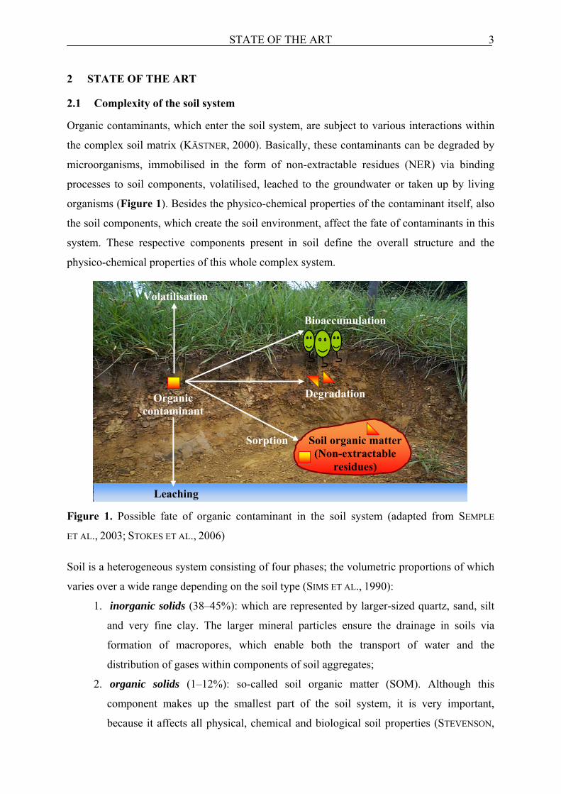
Organic contaminants, which enter the soil system, are subject to various interactions within
the complex soil matrix (KÄSTNER, 2000). Basically, these contaminants can be degraded by
microorganisms, immobilised in the form of non-extractable residues (NER) via binding
processes to soil components, volatilised, leached to the groundwater or taken up by living
organisms (Figure 1). Besides the physico-chemical properties of the contaminant itself, also
the soil components, which create the soil environment, affect the fate of contaminants in this
system. These respective components present in soil define the overall structure and the
physico-chemical properties of this whole complex system.
Figure 1. Possible fate of organic contaminant in the soil system (adapted from SEMPLE
ET AL., 2003; STOKES ET AL., 2006) Soil is a heterogeneous system consisting of four phases; the volumetric proportions of which
varies over a wide range depending on the soil type (SIMS ET AL., 1990):
1. inorganic solids (38–45%): which are represented by larger-sized quartz, sand, silt
and very fine clay. The larger mineral particles ensure the drainage in soils via
formation of macropores, which enable both the transport of water and the
distribution of gases within components of soil aggregates;
2. organic solids (1–12%): so-called soil organic matter (SOM). Although this
component makes up the smallest part of the soil system, it is very important,
because it affects all physical, chemical and biological soil properties (STEVENSON,
Volatilisation
DegradationOrganic contaminant
Leaching
Sorption
Bioaccumulation
Soil organic matter (Non-extractable
residues)

STATE OF THE ART 4
1994). Furthermore, it plays a crucial role in the protection of soil from degradation
and erosion (PICCOLO, 1996);
3. soil water (15–35%): is present in pore and capillary spaces. Water in pore spaces is
a solvent for nutrients and salts, which support microbial activity in soil (HAIDER
AND SCHÄFFER, 2009). Therefore, microbial biomass tends to concentrate along
water flow paths in the soil (VINTHER ET AL., 1999; BUNDT ET AL., 2003). Moreover,
it affects the soil aeration status, the soil water osmotic pressure and the pH of soil
solution (PAUL AND CLARK; 1989);
4. soil gases (15–35%): are distributed within pore spaces and are necessary for
microbial and plant root respiration.
The distribution and type of solid particles in the soil system defines its overall structure and
thus the active surface area affecting the fate of organic compounds (GAVRILESCU, 2005). The
interaction of SOM with clay in presence of polyvalent cations (e.g. Fe3+, Ca2+, Al3+) results
in the formation of stable clay-organic complexes and thus microaggregates (BRONICK AND
LAL, 2005; HAIDER AND SCHÄFFER, 2009). Microaggregates are joined with larger soil
particles (e.g. quartz and sand) forming macroaggregates (TISDALL, 1996). This aggregation
process plays an important role in shaping the physical, chemical and biological properties of
the soil system. Adhesive exopolymers excreted by living microorganisms on soil particles
and fungal hyphae on roots stabilise macroaggregates in the form of “sticky string bags”
(KÄSTNER, 2000, see Figure 2).
Figure 2. Aggregation of soil constituents (KÄSTNER 2000, ATLAS AND BARTHA 1997)
The water content, the presence of soil biota and the composition of solid components (in
particular clays and SOM) affect the aggregate size (BRONICK AND LAL, 2005). Pore sizes and
Quartz
Air
H2O
Sand
Silt
SOMMicroorganisms within the water film
Clay domaine
Particles coated by humic molecules and
microbial exopolymers
Microcolony of bacteria
Fungal hyphae
STATE OF THE ART 5
relative proportions of water and air in the soil system have an impact on the mobility of
contaminants (SIMS ET AL., 1990).
The main actors in the interactions with organic contaminants in soils are small-sized
soil particles clays and soil organic matter (CALDERBANK, 1989; SIMS ET AL., 1990;
STEVENSON, 1994). Clay particles have a large reactive charged surface, thus they are
believed to be involved in physico-chemical interactions with organic contaminants
(SIMS ET AL., 1990). Interactions with pesticides and other organic chemicals have also been
reported (WHITE, 1976; THENG, 1982; MORTLAND, 1986; ZIELKE ET AL., 1989).
SOM possessing many highly reactive functional groups (SIMS ET AL., 1990) was
proposed as the dominant factor affecting the organic contaminants interactions within soil
systems (BRUSSEAU ET AL., 1991; CORNELISSEN ET AL., 1998). This soil fraction, called “non-
living” SOM is a complex mixture of organic materials derived from fragments and
decomposed products of plant residues (e.g. proteins, starch, polysaccharides, lignins, lipids),
microbial cell components (e.g. lipids, carbohydrates, proteins) and humic substances
(KÖGEL-KNABNER, 2002; STEVENSON, 1994; SOLLINS ET AL., 1996; ZECH ET AL., 1997).
Because of their small size, microorganisms cannot be separated from SOM and thus form a
living fraction of it. The living fraction (1–5% of total SOM) containing a wide variety of
microorganisms, higher animals and plant roots (KLEBER ET AL., 1998; OADES, 1995),
constitutes a primary source for SOM formation (KELLEHER ET AL., 2006).
Humic substances are considered to be dominant components of SOM (50–60%, PAUL
AND CLARK, 1996, SCHNITZER, 1978; STEVENSON, 1994) and show molecular weights ranging
from a few hundred to several hundred thousand Daltons (HUANG ET AL., 2003). They are
believed to form from decaying plant biomass in a process called “humification” (HAIDER,
1998). The main components of the dead plant biomass, high-molecular-weight
polysaccharides and lignin are supposed to affect the size of humic substances (HAIDER,
1998). Humic substances are commonly classified depending on their solubility in alkaline or
acidic solutions into humins (insoluble in both solvents), fulvic acids (soluble in acid and
alkali) and humic acids [(soluble in alkali and insoluble in acid); NORTHCOTT AND JONES,
2000; STEVENSON, 1994]. This classification into these three fractions is only operational and
does not indicate any chemical behaviour or structure of humic acids (HAYES ET AL., 1989;
STEVENSON, 1994). Due to the fact that humic substances are very complex heterogenous
organic compounds, their overall chemical structure has not been clarified yet (ZIECHMANN,
1994). PAULI (1967) suggested a model, in which humic substances are present as complex
soil colloids with micellar structure. In this model, hydrophobic aromatic and aliphatic

STATE OF THE ART 6
building blocks, which are linked by covalent bonding, carry reactive functional groups with a
hydrophilic character. Both hydrophilic and hydrophobic sites of humic substances are
commonly considered to be involved in various interactions with organic contaminants
(KÄSTNER, 2000). Other authors have recently described humic substances as supramolecular
associations of low-molecular-mass organic biomolecules (SUTTON AND SPOSITO, 2005),
which have been excluded from traditional definitions of humic substances (STEVENSON,
1994; HAIDER, 1998). These low-molecular-mass organic molecules include for example
branched and linear alkanes, alkenes, fatty acids, dicarboxylic acids, and long chain
alcohols/ethers, ketones/aldehydes, amino acids and esters (FABBRI ET AL., 1996; SCHULTEN,
1999; SCHNITZER, 2000; KRAMER ET AL., 2001; CHEFETZ ET AL., 2002; GRASSET ET AL., 2002;
STENSON ET AL., 2002; MUGO AND BOTTARO, 2004). In addition, the hydrophobic properties
of humic substances, which have been suggested in the proposed models of humic substances,
have not yet been identified (SUTTON AND SPOSITO, 2005). Therefore, SUTTON AND SPOSITO
(2005) proposed to reevaluate the biogeochemical pathways of their formation and to redefine
the concept of “humification”.
KÄSTNER (2000) proposed an other definition of “humification”, which is in reality a
prolonged stabilisation process of organic components from non-living components of SOM
leading to the formation of refractory SOM. This refractory SOM is formed by complex
processes, in which metabolites and fragments of microbial and plant biomass metabolised by
microorganisms rearrange to form macromolecular aggregates. The continuous process of
biomolecules incorporation results in increasing molecular weight and thus in the formation
of macromolecules, which are characterised commonly as humins, humic acids and fulvic
acids. These macromolecules rearrange with each other, fragments of biomass and clay
minerals and finally form larger aggregates. In cultivated soils, 50–75% of SOM is associated
with clay-sized organo-mineral particles (CHRISTENSEN, 2001). Due to the fact that SOM is
tightly bound to clays in the form of clay-organic complexes (HAIDER AND SCHÄFFER, 2009),
it is difficult to distinguish between the contributions of clay and SOM to the interactions with
organic contaminants (KÄSTNER, 2000).
2.2 Microbial activity in the soil
Microorganisms are known to play a crucial role in biogeochemical cycles and in sustainable
development of the biosphere (VAN HAMME, 2004; ADRIANO AND BOLLAG, 1999). Soil
microbes need to cover their energy expenditure by uptake of the products of enzymatic
breakdown of organic substrate in order to survive in an active stage (EKSCHMITT ET AL.,
2005). The microbial transformation of organic compounds into various metabolites, which

STATE OF THE ART 7
become integral parts of the soil after stabilisation processes, can ultimately define the overall
structure and thus the physico-chemical properties of this system (YOUNG AND CRAWFORD,
2004).
Microorganisms, which might be detected in the soil system, are eubacteria,
actinomycetes, archaea, fungi, algae, protozoa and viruses (KÄSTNER, 2000). Scientists have
estimated that in one gram of fertile soil between 5000–7000 different bacterial species can
exist and that their populations can often exceed one hundred million individuals
(GAVRILESCU, 2005). The soil systems are heterogeneous habitats, therefore it is difficult to
characterise their microbial community in detail and only 1–10% of the microscopically
visible soil bacteria were estimated to be actually culturable by artificial media (PICKUP, 1991;
LEADBETTER, 1997).
Due to the high abundance and diversity of microorganisms present in soils, most of the
natural and anthropogenic organic compounds are subject to microbial attack (KÄSTNER,
2000). Owing to the presence of small-sized microorganisms (few µm) at hardly accessible
places within the soil system, organic contaminant, even if occluded within pore spaces, can
be degraded. Therefore, soil microbes are the major agents in the disappearance of organic
contaminants from soil in the so-called biodegradation process. This decontamination
mechanism is considered to be environmentally friendly (GOLOVLEVA ET AL., 1990; SINGH
AND WALKER, 2006; MÜLLER ET AL., 2007), because they are capable of degrading of a vast
diversity of compounds without hazardous by products. Among the microbial communities,
bacteria, fungi, archaea and actinomycetes are the main organic compound degraders
(HÄGGBLOM, 1992; MÜLLER ET AL., 2007).
The microbial degradation (biodegradation) of organic compound in soil is dependent
on many factors (BOLLAG AND LIU, 1990; SIMS AT AL., 1990; GAVRILESCU, 2005):
• Soil conditions: temperature, aeration, pH, moisture and SOM content, that influence
the activity of soil microorganisms, their size and diversity. In addition, the organic
compound availability for microorganisms is closely linked to the soil conditions (e.g.
decrease by sorption to SOM or lower pH);
• Organic compound characteristics, which include solubility in water, tendency to
adsorb to the soil matrix and persistence in the soil (half-life);
• Frequency of organic compound application: repeated application of a compound
may result in the development of a microbial community capable of the organic
compound degradation;
STATE OF THE ART 8
• Concentration of organic compound: very low concentrations may not provide
maintenance energy levels sufficient for microorganisms. Very high concentrations
may either be toxic and thus inhibit the microbial activity or, as an easily available
substrate, stimulate their growth.
The organic compound characteristics are important in determining its transformation, fate
and persistence in the soil system. Among others these characteristics include (AGA AND
THURMAN, 2001; ANDREU AND PICÓ, 2004; GAVRILESCU, 2005):
• water solubility, which strongly depends on temperature and pH of the solution. It
controls the mobility of organic compound in soil;
• soil-adsorption coefficient (Koc, log Koc), which describes the tendency of a compound
to be adsorbed to soil particles. The higher the Koc value, the more strongly the
contaminant is sorbed, which leads to reduced availability and mobility;
• octanol/water partition coefficient (Kow, log Kow), which is related to water solubility
and soil/sediment sorption coefficients (Koc), for instance: log Kow < 2.5 indicates low
sorption potential, values log Kow between 2.5 and 4.0 indicate intermediate sorption
potential, whereas logKow > 4.0 indicates high sorption potential (JONES-LEPP AND
STEVENS, 2007). In addition, Kow, log Kow is widely accepted to describe
bioconcentration in aquatic organisms, which is indicative of the tendency for
bioaccumulation of a contaminant in organisms;
• half-life in soil (DT50), is the amount of time necessary for disappearance of 50 % of
parent compound from soil.
Depending on the chemical nature of a compound, the degradation rate can be different.
Easily degradable compounds are usually degraded immediately accompanied by the
formation of biomass and mineralisation products (H2O and CO2), whereas readily and hardly
degradable xenobiotics are degraded at slow rates from the beginning or at higher rates, but
after only prolonged phases of adaptation (KÄSTNER, 2000). During degradation, the C
derived from the pollutant is used as energy and C source by microorganisms to form their
biomass components (MÜLLER ET AL., 2007). However, many organic contaminants are also
degraded cometabolically (MÜLLER ET AL., 2007). In this cometabolic process, the organic
contaminants do not serve as C or energy source and are metabolised together with another
substrate used for growth (SIMS AT AL., 1990; HÄGGBLOM, 1992). This type of transformation
is frequently based on metabolic reactions catalysed by extracellular enzymes present in soils
(MÜLLER ET AL., 2007; SIMS ET AL., 1990).

STATE OF THE ART 9
2.2.1 Natural organic compounds biodegradation in soil
The high-molecular-weight organic compounds derived from plant and microbial biomass can
be mineralised to CO2 and H2O with the formation of microbial biomass (see Figure 3). After
the death of microorganisms, low-molecular-weight microbial compounds are incorporated
into SOM and form so-called biogenic residues. Thereafter, these residues are stabilised in
SOM, which leads to the formation of refractory SOM (humic substances in “humification”).
Biogenic residues, their metabolites and biomass components are also probably parent
materials for renewed degradation and conversion reactions (KÄSTNER AND RICHNOW, 2001).
Figure 3. Scheme of C flow during microbial degradation of natural organic compounds in
soil (adapted from KÄSTNER AND RICHNOW, 2001)
Formation of biogenic residues (up to 35%) in soil was observed during the biodegradation of
plant biomass residues (STOTT ET AL., 1983). In addition, even after a one-year incubation of
soil with 14C-glucose, residues derived from this easily degradable compound were still
detected in SOM (10%; BALDOCK ET AL. 1989).
Plant residues are suggested to be the primary source for refractory SOM formation
(STEVENSON, 1994; SCHOLES ET AL., 1997; HAIDER, 1998; KÖGEL-KNABNER, 2002). This is
caused by the relative high inputs to soil and the fact that plant components, in particular
high-molecular-weight compounds (e.g. lignin) are believed to be highly resistant to
microbial degradation (STEVENSON, 1994; HAIDER, 1998; KÖGEL-KNABNER, 2002). However,
recent findings show that lignin is less persistent than the average of bulk SOM (VON LÜTZOW
ET AL., 2008; MARSCHNER ET AL., 2008). The importance of microbial biomass C in the
formation of SOM is considered to be minor (SCHOLES AND SCHOLES, 1995), because of both
its small pool size and fast turnover (JENKINSON AND LADD, 1981; COLEMAN ET AL., 1983).
Incorporation
CO2 + H2O
Biomass (+ exopolymers)
Mineralisation
SOM Biogenic residues
Parent compound + metabolites
Sorption Desorption ……
.………

STATE OF THE ART 10
However, studies on the molecular composition of SOM proved that plant-derived organic
matter (OM) was not stored in arable soils, but was transformed to microbial residues, i.e.
carbohydrates and proteins, which were kept in soil by organo-mineral interactions (BOL ET
AL., 2009). Several recent studies on SOM showed that the contribution of microbial biomass
to SOM formation in the generally accepted range of 1-5% is highly underestimated
(SIMPSON ET AL., 2007). For instance, soil incubation with 13C-labelled Escherichia coli
indicated that microbial biomass-derived C significantly contributed to the formation of
refractory SOM (LÜDERS ET AL., 2006; KINDLER ET AL., 2006, 2009; MILTNER ET AL., 2009).
In this experiment about 56% of the bulk C of E. coli was mineralised, the residual 44% was
stabilised in the soil after 224 days (KINDLER ET AL., 2006). Further research reported that
SOM was predominantly of microbial origin (> 50% of extractable humic acids; SIMPSON ET
AL., 2007). The high content of E. coli biomass-derived C stabilised in SOM after 224 days
thus indicates that microorganisms may form significant amounts of biogenic residues in soil
and finally the refractory SOM during biodegradation of organic compounds.
2.2.2 Anthropogenic organic compounds biodegradation in soil
The microbial degradation of organic contaminants in soil is generally understood as their
transformation into mineralisation products, metabolites, microbial biomass and non-
extractable residues (NER; see mass balance in Figure 4).
Figure 4. Conventional model of C flow during microbial degradation of organic
contaminants in soil (mass balance)
The organic contaminant or products of its partial biodegradation are believed to sorb to SOM
via various physico-chemical mechanisms leading to the NER formation (ALEXANDER, 2000;
GEVAO ET AL., 2000; GAVRILESCU, 2005; LOISEAU AND BARRIUSO, 2002; MORDAUNT ET AL.,
CO2 + H2O
Mineralisation
Humic substances NER
Parent compound + metabolites
Sorption Desorption …?
Biomass (+ exopolymers)

STATE OF THE ART 11
2005). The sorption of contaminants to SOM is considered as the major factor preventing
their complete biodegradation in soil (BÜYÜKSÖNMEZ ET AL., 1999). The mass balance of
organic contaminant within soil fractions during its biodegradation is usually determined
using radioactive tracers (14C-labelled compounds; GERST AND KLIGER, 1990; KUBIAK ET AL.,
1990; RICHNOW, 1999). The rate of microbial transformation of an organic compound is
estimated only by the determination of the residual concentrations in soil (FOGARTY AND
TUOVINEN, 1991). The analyses of parent compounds and their metabolites remaining in soil
solution are usually accomplished using different extraction methods such as batch solvent
shaking, Soxhlet extraction, supercritical fluid extraction or accelerated solvent extraction
(HAWTHORNE ET AL., 2000; NORTHCOTT AND JONES, 2000) with aqueous or organic solvents.
However, these conventional and enhanced solvent extraction techniques do not extract
contaminants that are strongly bound or sequestered into the components of soil matrix like
SOM (NORTHCOTT AND JONES, 2000).
Therefore, the total amount of unextracted contaminant residues is mostly quantified as 14CO2 released from combustion of soil samples (WAIS, 1998; BARRIUSO ET AL., 2008). Using
this destructive approach, it is not possible to check if NER are intact contaminants, their
metabolites or 14C accumulated in microbial biomass or in humic substances (BARRIUSO ET
AL., 2008). Alternatively, the distribution of the 14C in humic acids, fulvic acids and the
insoluble humins of soil containing non-extractable contaminant residues is often analysed
after the extraction of both fulvic and humic acids (NORTHCOTT AND JONES, 2000). A major
obstacle for deeper analysis of these residues is that no proper method for the identification of
the chemical nature of the transformation products bound to complex soil matrix using
radiotracers is available. Therefore, due to the problems with NER analyses, the detailed
biodegradation pathways of many organic contaminants in these complex soil systems have
not yet been elucidated (FOGARTY AND TUOVINEN, 1991).
2.3 Bioavailability of organic contaminants in soil
Bioavailability of contaminants is the key factor, which controls their overall fate in soil, in
particular their biodegradability and toxicity for biota (SEMPLE ET AL., 2007). Bioavailability
is affected by many factors such as: the properties of contaminant and soil, aging time in the
soil, climate and organisms of concern (KATAYAMA ET AL., 2010). On the whole, the
assessment of the bioavailability of contaminants in soil is necessary both for understanding
the risks, which may be posed by these contaminants and for the proper choice of the method
for soil remediation (SEMPLE ET AL., 2003).
STATE OF THE ART 12
The term bioavailability has been used in many different scientific fields, thus there are many
definitions (SEMPLE ET AL., 2007), which generally consider the interactions between an
organism and a chemical (SEMPLE ET AL., 2004). Toxicologists term bioavailability as the
fraction of chemical absorbed and able to reach systemic circulation in an organism (SEMPLE
ET AL., 2004). For instance, the National Research Council reports:
“Bioavailability may represent the fraction of a chemical accessible to an organism for
absorption, the rate at which a substance is absorbed into a living system or a measure
of the potential to cause a toxic effect” (NRC, 2002).
EHLERS AND LUTHY (2003) have stated that:
“Bioavailability refers to the extent to which humans and ecological receptors are
exposed to contaminants in soil or sediment”.
Environmental scientists consider bioavailability as the accessibility of soil-bound chemicals
for assimilation and possible toxicity (ALEXANDER, 2000). SEMPLE ET AL. (2004) identified
the lack of clarity of the term “bioavailability” among environmental scientists, and thus
proposed the distinction of two terms bioavailability and bioaccessibility:
“Bioavailability represents, at a given time, the fraction of the chemical that is freely
available to cross an organism’s membrane from the medium which the organism
inhabits.” Bioaccessibility was defined as: “that which is available to cross an
organism’s (cellular) membrane from the environment it inhabits, if the organism had
access to it; however, it may be either physically removed from the organism or only
bioavailable after a period of time. Bioaccessibility encompasses what is actually
bioavailable now and what is potentially bioavailable” (SEMPLE ET AL., 2004).
Not only bioavailable compounds present in the water-soluble fraction of soil are available,
but also these actually desorbed from soil during the time when a target organism is in direct
contact with the soil (HARMSEN, 2007). Bioaccessibility includes both the readily available
contaminants present in the water-soluble fraction of soil and the contaminants which can
become available after desorption from the soil matrix (Figure 5). This includes also the
contaminants, which may be released after longer timescale (slowly reversible). The
accessibility depends on the desorption conditions (e.g. shaking, temperature) and desorption
time (REICHENBERG AND MAYER, 2006). Both bioavailability and bioaccessibility of
contaminants decrease with increasing contact time of contaminants with soil matrix [(so-
called “aging”); REID ET AL., 2000].

STATE OF THE ART 13
Figure 5. Bioavailability and bioaccessibility of organic contaminants in soil system (adapted
from SEMPLE ET AL., 2004); ( ) parent compound; ( ) metabolites
A chemical immobilised in SOM or present in the soil solution can become bioaccessible.
This involves a number of consecutive steps until eventually the compound is absorbed into
an organism (see bioavailability processes A-D in Figure 6; EHLERS AND LUTHY, 2003;
SEMPLE ET AL., 2004).
Figure 6. Bioavailability processes in soil system (EHLERS AND LUTHY, 2003; SEMPLE ET AL.,
2004)
Process “A” encompasses the possible behaviour of the contaminant within the soil matrix
(sorption or desorption). When environmental conditions are changed (e.g. changes in water
saturation, pH or temperature) immobilised contaminant in SOM (NER), may be released or
transformed into more stable associations over time (aging) and become non-accessible. The
release of immobilised contaminant from SOM is the key step toward the assessment of its
bioaccessibility and thus the toxicity for living organisms (HARMSEN, 2007). Therefore, the
Organism Soil matrix
SOM NER
Site of biological response
Absorbed contaminant in organism
Sorption Desorption A
C
B
D
E
Released compound
Non-bioaccessible
Water-soluble fraction
Biovailable
BioaccessibleSilt
particle
Sand particle
SOM NER

STATE OF THE ART 14
formation of NER during microbial degradation of organic contaminants in soils is actually
discussed in terms of the probability of the risks which they can pose to the environment
(BARRACLOUGH ET AL., 2005). Processes B and C include the transport of the contaminant,
which is present in the soil solution (B) or bound to soil components (C). D shows the barrier
between external environment (soil matrix) and an organism and represents the uptake of the
contaminant through the cell membrane into the target organism. E refers to paths taken by
contaminant after uptake into the cells (e.g. metabolic transformation or exerting toxic effects
within cells) and addresses toxicological bioavailability (EHLERS AND LUTHY, 2003).
Various chemical or biological measurements are employed to assess the bioavailability
of an organic contaminant for living organisms. Chemical measurements involve various
extraction methods of the contaminant from soil, whereas biological ones are based on
monitoring the toxic effects of a contaminant taken up by the target living organism
(HARMSEN, 2007).
2.4 Definition of the non-extractable residues (NER)
The term NER has been defined in different ways over the years (MORDAUNT ET AL., 2005).
In the past, NER were considered to be pesticide residues remaining in the soil after
application until the next growing season or the planting of the following crop (CRAVEN,
2000). Pesticides were and are still of particular interest in this regard, since they have
selective activity and are deliberately manufactured for application in terrestrial systems
(GEVAO ET AL., 2000).
The first official definition of the term NER was provided in 1975 by the American
Institute of Biological Sciences – Environmental Task Group (NORTHCOTT AND JONES, 2000),
which stated:
“bound pesticide residues in the soil are unextractable and chemically unidentifiable
pesticide residues remaining in the fulvic, humic acids and humin fractions after
exhaustive sequential extraction with nonpolar and polar solvents”.
The most accepted and widely used NER definition, proposed by the Applied Chemistry
Division, Commission on Pesticide Chemistry of the International Union of Pure and Applied
Chemistry (IUPAC), is the following:
“bound residues (also referred as “non-extractable” residues or “non-extracted”
residues) in plants and soils as chemical species originating from pesticides, used
according to good agricultural practice, that are unextracted by methods which do not
significantly change the chemical nature of these residues. These residues are

STATE OF THE ART 15
considered to exclude fragments recycled through metabolic pathways leading to
natural products” (ROBERTS, 1984).
Biogenic residues formed during biodegradation of organic compounds are thus excluded
from the IUPAC NER definition, because they are composed only of natural compounds
derived from microbial biomass stabilised in SOM. However, with the use of radiotracers for
estimating of mass balance of the tested contaminant in soil, these natural compounds cannot
be excluded from the quantitative analysis of NER.
The IUPAC NER definition was later slightly modified but not substantially changed
(NORTHCOTT AND JONES, 2000) and was extended to include that “the NER formation reduces
the bioaccessibility and bioavailability significantly” (CALDERBANK, 1989; FÜHR ET AL.,
1998).
2.5 Determination of NER in soil
The determination of NER is based on quantitative analyses accomplished after the extraction
of residual parent compound and its metabolites from soil as mentioned in section 2.2.2. The
distinction between extractable and non-extractable residues depends on extraction methods
and conditions employed (KHAN 1991; NORTHCOTT AND JONES, 2000; MORDAUNT ET AL.,
2005). Numerous “exhaustive” (harsh) and “non-exhaustive” (mild) extraction methods
employed for the assessment of bioavailability of organic contaminants for living organisms
show different quantitative recovery of a target compound (NORTHCOTT AND JONES, 2000;
SEMPLE ET AL., 2007). The main aim of harsh extraction methods measuring the total
concentration of contaminant in soil is to recover all or as much as possible of the
contaminant from environmental samples (HATZINGER AND ALEXANDER, 1995; NOORDKAMP
ET AL., 1997; ALEXANDER, 2000; REID ET AL., 2000; STOKES ET AL., 2006). These harsh
methods involving mostly heated organic solvents include soxhlet extraction, microwave
extraction, supercritical fluid extraction, ultrasonication and accelerated solvent extraction
[(ASE); NORTHCOTT AND JONES, 2000]. It is generally assumed that the amount of compound
recovered by exhaustive soil extraction is 100% available when assessing its potential risk for
environment (ALEXANDER, 2000). However, the measurement of total concentration besides
the accessible fraction of contaminant may also include parts or the entire recalcitrant fraction
of contaminants (e.g. aged or sorbed strongly to SOM). In addition, the soil matrix by these
extraction techniques is altered thus no longer represents a real soil (MORDAUNT ET AL.,
2005). This occurs, because used organic solvents remove H2O and SOM (mostly humic
acids) from the soil matrix (MORDAUNT ET AL., 2003). Therefore, these harsh methods were
considered to be improper for the assessment of the toxicity and thus the risk of soil

STATE OF THE ART 16
associated organic contaminants for the environment (KELSEY ET AL., 1997; GEVAO ET AL.,
2003). Mild extractions methods that remove only the “labile” pool of the contaminant
(water-soluble and loosely adsorbed contaminants on surface of soil particles) are more useful
for the measurement of bioaccessibility of contaminants in soils and their potential risks
(KELSEY ET AL., 1997; REID ET AL., 2000; SEMPLE ET AL., 2007). These mild extraction
methods involving natural extractants (e.g. ionic solution of CaCl2) mimic solutions likely to
be present in the soil (MORDAUNT ET AL., 2003). These methods are aqueous-based
extractions including solid-phase extraction (e.g. Tenax TA) and the cyclodextrin technique
(REID ET AL., 2000; LISTE AND ALEXANDER, 2002; SEMPLE ET AL., 2007).
Besides the physico-chemical properties of compound and soil, the amount of NER also
strongly depends on the extraction method (MORDAUNT ET AL., 2005). Therefore, the methods
used for the extraction of contaminants from soil affects also the quantitative analyses of
NER. For instance, harsh extraction methods result in lower estimates of NER, whereas the
mild ones give higher estimates for NER in soils. Hence, the numerous presented data on the
total amounts of NER formed during the microbial degradation of the same model organic
contaminant in soils show high variations. Summing up the above, the NER term is defined
operationally by the extraction method employed; hence it is important to clarify how the
method is developed from this definition and what information this method will provide
(MORDAUNT ET AL., 2005).
2.6 Formation of NER in soil
It is believed that formation of NER during biodegradation of a contaminant is based on the
various physico-chemical interactions between parent compounds or its metabolites and SOM
(BOLLAG ET AL., 1992; SENESI, 1992; VERSTRAETE AND DEVLIEGHER, 1996; FÜHR, 1998;
ALEXANDER, 2000; GEVAO ET AL., 2000; LOISEAU AND BARRIUSO, 2002; MORDAUNT ET AL.,
2005). However, biodegradation of natural organic compounds in soil results in the formation
of biogenic residues (as shown in Figure 3 in section 2.2.1). Organic contaminants similar to
the natural organic compounds are also subject to microbial degradation, which can lead to
the formation of organic contaminant-derived biogenic residues. These contaminant-derived
biogenic residues are only quantified as NER and no detailed information about their
chemical structure is available (RICHNOW ET AL., 2000; KÄSTNER AND RICHNOW, 2001;
BARRIUSO, 2008). Therefore, it is necessary to consider the formation of biogenic residues
during biodegradation of organic contaminants and to distinguish between formations of NER
via components of microbial biomass and via parent compound or its metabolites in soils.
STATE OF THE ART 17
The amounts of NER formed during the biodegradation of organic contaminants depend on
the previously mentioned physico-chemical properties of both the contaminant and the soil
system. In addition, the position of the label in the molecule of the tested contaminant has an
impact on the results on the fate of this contaminant in general including the formation of
NER (BARRIUSO ET AL., 2008). For instance, if the label is positioned in a labile molecular
fragment of contaminant (which is easily evolved as 14CO2), the mineralisation will be
overestimated, whereas the detected amounts of NER tend to be low (BARRIUSO ET AL.,
2008).
2.6.1 Parent compounds and metabolites
Humic acids as major components of SOM were previously regarded as the main agents
leading to the formation of NER in soil (SENESI, 1992; WAIS, 1998). This is caused by the fact
that it is believed that humic acids are large aromatic polymers with many binding sites for
organic contaminants (hydrophilic and hydrophobic) in their molecular structure (SENESI,
1992).
Investigations of the interactions between organic contaminants and SOM involved
various extraction techniques basing mostly on the isolation and fractionation of soil humic
substances. To characterise NER, several attempts to cleave bonds of the contaminant with
SOM were used. For instance, alkaline hydrolysis was used to cleave organic compounds
bound to SOM via ester bonds (RICHNOW ET AL., 1998), and acid hydrolysis to release
compounds linked to SOM by ether bonds (RICHNOW ET AL., 1997). However, in most cases,
the physico-chemical interactions of contaminants with humic substances were studied in
simple systems with solutions containing only humic acids (mainly synthetic) and the
contaminant (BOLLAG, 1991; BOLLAG 1992; HATCHER ET AL., 1993; PICCOLO ET AL., 2001).
These approaches follow only a specific single reaction or binding mechanism of the
respective contaminant to humic acids (NORTHCOTT AND JONES, 2000). The physico-chemical
interactions were thus described mainly theoretically. Therefore, these data on the
mechanisms of NER formation cannot necessarily be extrapolated to the realistic processes,
which take place in complex soil systems, especially when considering the fact that the
molecular structure of humic acids is not yet clear.
Organic contaminants are believed to bind to humic acids via various mechanisms of
adsorption, covalent bonds and sequestration (BOLLAG ET AL., 1992; WAIS, 1998; GEVAO
ET AL., 2000).

STATE OF THE ART 18
Adsorption
Adsorption is the binding of the organic compound to the surface of solid particles through
weak and reversible bonds (BÜYÜKSÖNMEZ ET AL., 1999). Minerals, in particular clays and
SOM can take part in the sorption of the organic contaminants. The clay minerals are
responsible for adsorption of polar and hydrophilic compounds, whereas SOM has both
hydrophilic and hydrophobic sites and therefore can interact with polar, charged as well as
apolar and lipophilic contaminants (VERSTRAETE AND DEVLIEGHER, 1996). This type of
association is reversible and leads to the formation of unstable NER subject to microbial
degradation (VERSTRAETE AND DEVLIEGHER, 1996). This can be caused by the fact that
microorganisms, which colonise surfaces of soil particles, might have direct access to
adsorbed chemicals and degrade them.
The extent of adsorption depends on the properties of the soil (e.g. SOM content) and
the nature of contaminant e.g. if it is acidic or alkaline (GEVAO ET AL., 2000). Adsorption
occurs via several mechanisms like ionic and hydrogen bonding, charge-transfer, ligand
exchange, van der Waals forces and hydrophobic bonding (KHAN, 1978; PIGNATELLO, 1989;
GEVAO ET AL., 2000).
Ionic binding: this type of binding occurs with those contaminants and their metabolites that
exist in the cationic form or can become cationic upon protonation (SENESI, 1992; WAIS,
1998; GEVAO ET AL., 2000). These contaminants react with ionised or easily ionisable
carboxylic and phenolic hydroxyl groups of humic acids or hydroxyl groups of minerals
(SENESI, 1992).
Hydrogen binding: nonionic contaminants can interact with oxygen- and nitrogen-containing
functional groups at the surfaces of humic substances or oxygen- and nitrogen-containing
functional groups of the contaminant can react with hydrogen containing groups of SOM
(WAIS, 1998).
Charge-transfer complexes: also called donor-acceptor complexes are formed when
molecules with a high electron density (e.g. π-electrons in aromatic systems) react with
electron-deficient molecules (e.g. quinones; WAIS, 1998).
Ligand exchange: in this type metal ions complexed by humic acids are usually associated
with water molecules, which can be replaced by functional groups (e.g. carboxylic or amino
groups) of organic contaminants (WAIS, 1998). However, this bonding is rare and weak and
thus plays minor role.

STATE OF THE ART 19
Van der Waals forces: are relatively weak short-range dipolar or induced-dipolar attractions
that exist in presence of stronger binding forces, in all adsorbent-adsorbate interactions
(GEVAO ET AL., 2000).
Hydrophobic bonding: is the interaction mechanism between hydrophobic groups of humic
substances and non-polar contaminants (WAIS, 1998). Active partners are aliphatic side
chains, fat constituents or lignin components of humic substances (SENESI, 1993).
Covalent bonding
Covalent bonding is a chemical interaction between a contaminant or its metabolites and
humic substances driven by strong bonding forces (> 300kJ/mol; BLASCHETTE, 1974). This
type of bonding is generally accepted to be an irreversible and very stable association
(SENESI, 1992; WAIS, 1998), which results in the formation of persistent contaminant NER in
soil. These interactions can be mediated by enzymatic, chemical or photochemical catalysts
(SENESI, 1992; GEVAO ET AL, 2000) and result in ester, ether or carbon-carbon linkages
(KÄSTNER AND RICHNOW, 2001). The organic contaminants, which are bound covalently to
soil humic acids, are integral components of humic substances (BOLLAG ET AL., 1992; SENESI,
1992). Thereafter, they are subject to all further transformation processes involved in
humification (WAIS, 1998). The residues bound chemically to soil humic acids are considered
to be toxicologically inactive (CALDERBANK, 1989), because they are not bioavailable.
However, the exact chemical nature and structure of this type of NER in SOM have not yet
been elucidated for all xenobiotics (KATAYAMA ET AL., 2010). The studies on NER formation
by covalent bonding are limited to simple humic acids-contaminant systems (BOLLAG, 1991,
1992; HATCHER ET AL., 1993).
Sequestration (Aging)
Sequestration (aging) contrary to the adsorption mechanisms does not include reactions that
alter the structure of an organic contaminant (HATZINGER AND ALEXANDER, 1995).
Sequestration is also referred to a slow sorption/diffusion (PIGNATELLO AND XING, 1996; DEC
AND BOLLAG, 1997) of non-polar and hydrophobic compounds, which in comparison to
adsorption is a very long-term process (GEVAO ET AL., 2000). During aging, the molecule
becomes progressively more tightly bound or entrapped in SOM and correspondingly less
bioavailable (BARRACLOUGH ET AL., 2005). The size distribution of micropores is a factor in
aging: the smaller the micropore, the slower aging proceeds, but the effect is stronger
(ALEXANDER, 2000; KATAYAMA ET AL., 2010). Aging may also result from covalent bonding
of contaminant with soil humic acids after sorption inside or within micropores in soil
aggregates (ALEXANDER, 2000; KATAYAMA ET AL., 2010).

STATE OF THE ART 20
2.6.2 Components of microbial biomass
Several studies showed that the formation of NER from degradation of most pesticides is
often related to soil biological activity and to the amount of SOM present in the soil
(KAUFMANN AND BLAKE, 1973; ABDELHAFID ET AL., 2000A, B). For instance, soil microbial
activity, which is generally higher in topsoil layers, induces the formation of NER in higher
amounts than in deeper soil horizons (SCHIAVON, 1988; BALUCH ET AL., 1993; STOLPE AND
SHEA, 1995; RICE ET AL., 2002). In addition, soil amendments with organic materials such as
manure and straw enhanced the formation of NER and the dissipation of pesticides (DOYLE ET
AL., 1978; PRINTZ ET AL., 1995). For example, during the incubation of composted non-sterile
straws with 2,4-dichlorophenoxyacetic acid (2,4-D), 2,4-dichlorophenol (2,4-DCP) or
4-chlorophenol, the contents of NER were high, whereas in sterile ones these values were
negligible (BENOIT AND BARRIUSO, 1997). The addition of glucose, which usually induces the
microbial activity, increased the rate of NER formation in soil amended with atrazine in
comparison to a soil without glucose application (ABDELHAFID ET AL., 2000A, B).
Also for Polycyclic Aromatic Hydrocarbons (PAH) adsorption and covalent bonding were
proposed as possible mechanisms of NER formation in soils. However, the molecular
analyses of SOM by pyrolysis gas chromatography-mass spectrometry after alkaline
hydrolysis of ester bonds revealed that the contents of both parent compounds and known
metabolites of PAH were very low (0.5% of the initial amount of PAH; RICHNOW, ET AL.,
1994). Other study on the 15N-labelled simazine-derived NER in 15N-depleted plant compost
indicated that these NER contained no parent compound and were composed of degradation
products resulting from N-dealkylation and triazine ring destruction (BERNS ET AL., 2005).
KÄSTNER ET AL. (1999) suggested for the first time that NER formed during biodegradation of
PAH (9-[14C]-anthracene) could be of “biogenic” origin. In their experiments, addition of
compost led to higher mineralisation rate of anthracene and lower amounts of NER in
comparison with the native soil (see Table 1). The amount of NER in the experiment with 9-
[14C]-anthracene was relatively high, in spite of the fact that the C at the labelled position is
subject to release as CO2. In addition, an experiment with 14CO2 (in the amount corresponding
to the final mineralisation of 9-[14C]-anthracene) showed that the label was found mostly in
NER. This indicates that they were not directly formed from anthracene, but via CO2 fixation
and microbial biomass. In addition, the partitioning of 14C in humines, humic and fulvic acids
in the 14CO2 experiment was similar to the one in the 9-[14C]-anthracene experiment. The
amounts of 14C originating from 14CO2 in NER of soil inhibited with CHCl3 were
significantly lower than in soil without CHCl3 treatment.

STATE OF THE ART 21
Table 1. Distribution of 14C after microbial degradation of 9-[14C]-anthracene and in native
soil and soil-compost-mixture (after 176 days) and after incubation under 14CO2-atmosphere
[(after 90 days); from KÄSTNER ET AL. 1999]
Applied radioactivity [%] 9-[14C]-anthracene
14CO2
Mass balance Native soil Soil-compost a Soil-compost a,b +CHCl3 a,b,c
Total recovery CO2 Total soil Extraction+alk. hydrolysis “bound” residues
98.7 43.8 54.9 9.5 45.4
91.9 67.2 24.6 3.9
20.7 d
99.8 4.6 e 95.2 f 9.4 85.8
98.7 11.3 e 87.4 44.2 43.2
a soil-compost-mixture (80% : 20% dry weight); b 90d; c microflora inhibited by fumigation with CHCl3 d amount represents 25.5 Bq/g of soil; e recovered 14CO2 (initially applied amount: 24.8 Bq/g of soil)
f 31% bound in acid labile carbonates, 67% bound to SOM, < 2% bound to clays and silicates The above-mentioned studies clearly indicate that in many cases microorganisms mediate
NER formation in soils. As already mentioned, the C derived from organic contaminants can
be used for the formation of microbial biomass during their biodegradation in soil (MÜLLER
ET AL., 2007). The CO2 evolved during mineralisation of organic contaminants can be utilised
as an additional C source by soil microorganisms, since even heterotrophic microorganisms
need CO2 for their growth (KREBS, 1941). PEREZ AND MATIN (1982) showed that during
heterotrophic growth about 10% of the cell C originated from CO2. Another experiment with
labelled CO2 in the atmosphere proved that soil microorganisms assimilated C and
incorporated the label into SOM (MILTNER ET AL. 2004, 2005). Therefore, NER formed
during biodegradation of organic contaminants in soil, can in reality be biogenic residues
(KÄSTNER AND RICHNOW, 2001; Figure 7), the formation of which is also observed during
biodegradation of natural organic compounds (Figure 3 in section 2.2.1; KÄSTNER, 2000).
Figure 7. Biogenic residues formation during microbial degradation of organic contaminants
in soil In addition, when labelled contaminants are used for soil biodegradation studies, a certain
amount of this label incorporated into microbial biomass components, which thereafter are
SOM Biogenic residues
CO2
Parent compound
Metabolites
Fixation
Incorporation
Mineralisation
Microbial biomass

STATE OF THE ART 22
stabilised in SOM, will be detected as NER. Biogenic residues can be formed via direct
incorporation of C from pollutant or indirectly via fixation of CO2 released during
mineralisation of the contaminant (see Figure 7).
Fatty acids and amino acids as representatives for biogenic residues
Fatty acids (FA) and amino acids (AA) that are known microbial biomarkers (BOSCHKER AND
MIDDELBURG, 2002) can be incorporated into SOM and thus form biogenic residues. Free and
bound lipids, amino acids and carbohydrates form the major fraction of analytically
recognisable compounds in SOM of various origin (ALLARD, 2006). In addition, the
extraction of known microbial biomarkers (e.g. FA and AA) from soil after addition of C
isotope tracer allows an estimation of microbial activity in the biogeochemical cycling of C
(PELZ ET AL., 1998). Biomarker analysis is thus also useful in studying the microbial
transformation of labelled organic contaminants in soil (BOSCHKER AND MIDDELBURG, 2002).
Therefore, the analysis of C isotope label distributions in FA and AA within the living and
non-living SOM fraction enables understanding the formation and the fate of biogenic
residues during biodegradation of a labelled organic contaminant in soil.
FA are major constituents of the soil lipids (PAUL AND CLARK, 1996) and may originate
from both plant residues and soil organisms (STEVENSON, 1994). This biomarker is used for
identification of microbial populations involved in specific geochemical transformations, e.g.
organic contaminant degradation in soils after deliberately added tracers (BOSCHKER AND
MIDDELBURG, 2002). Short-chain FA (C:4-C:20) are typical for microorganisms (SCHNITZER
ET AL., 1986). On average, FA represent about of 5% of the dry weight (DW) of the microbial
biomass (BAS ET AL., 2003). Phospholipid fatty acids (PLFA) are characteristic for the lipids
of living cells (ZELLES, 1999). PLFA are known to be stable only in intact cells and are
hydrolysed within weeks after their death (VESTAL AND WHITE ET AL., 1989). They therefore
can be used as biomarkers for representative groups of the living microorganisms in the
environment (GREEN AND SCOW, 2000; KAUR ET AL., 2005; ZELLES, 1999). The characteristic
chain lengths of ester-linked saturated FA of PLFA range from C14:0 to C18:0 for bacteria and
from C14:0-C24:0 for fungi (ZELLES, 1997). Several authors (GREEN AND SCOW, 2000; KAUR ET
AL., 2005; ZELLES, 1999) proposed that the following five classes of PLFA are typical for
specific groups of microorganisms: (1) saturated straight chain PLFA for all microorganisms,
(2) saturated methyl branched PLFA for Gram-positive bacteria, (3) monounsaturated PLFA
for Gram-negative bacteria, (4) polyunsaturated PLFA for fungi and (5) saturated cyclopropyl
PLFA for starving Gram-negative bacteria.

STATE OF THE ART 23
AA, which account for 10–20% of total C and 30–50% of total N in SOM, are mainly present
as polymers e.g. proteins, protein-humic complexes and peptides (STEVENSON, 1982). AA are
also the most abundant components (55% of dry weight) of bacterial cells (MADIGAN AND
MARTINKO, 2006). Total free AA concentrations in soil are very low representing
< 0.05% of total soil N (JONES AT AL., 2005) and have very short turnover times (JONES,
1999), because they are easily available and degraded immediately by soil microorganisms.
Two biodegradation studies with labelled organic contaminants showed that the label
was incorporated into AA. In the first biodegradation experiment with tar oil contaminated
soil spiked with 1-[13C]-phenanthrene significant amounts of the label were found in
hydrolysable AA, which represented 11% of total NER (RICHNOW ET AL., 2000). The second
experiment studying the fungal degradation of [15N]-Trinitrotoluene (15N-TNT) indicated that
1.7% of the 15N label was detected as “biogenic” AA in the wheat straw containing the fungus
(WEIß ET AL., 2004). In contrast to the wheat straw with fungus, no incorporation of the label
from 15N-TNT into AA was observed in the soil layer without fungus. A study on the fate of 13C-labelled E. coli demonstrated high stability of 13C-amino acids, whose contents remained
constant even after 224 days of incubation (MILTNER ET AL., 2009). This is caused by the fact
that proteins are amphiphilic molecules with a strong tendency to bind to surfaces (HLADY
AND BUIJS, 1996), especially to mineral surfaces, over long periods of time (KLEBER ET AL.,
2007).
2.7 Stability of NER in soil
The stability of NER formed during microbial degradation of organic contaminants in soils
has been investigated since the late 1960s (HATZINGER AND ALEXANDER, 1995). Since then, it
has been observed that aged organic compounds in NER are not always immobilised
irreversibly in the soil. Therefore, many scientists focused on the possible release of NER
from soils under certain conditions (BURAUEL AND FÜHR, 2000; BOIVIN ET AL., 2005; GEVAO
ET AL., 2005; LERCH ET AL., 2009) and the long-term environmental impact of these residues
(GEVAO ET AL., 2001).
The release of organic contaminant or its various metabolites immobilised in SOM is
related closely to SOM decomposition as a result of either biochemical processes or physico-
chemical mechanisms (GEVAO ET AL., 2000; BARRACLOUGH ET AL., 2005). However, the
activity of microorganisms is considered as the primary factor responsible for the release of
NER (GEVAO ET AL., 2000). To explore the possible scenarios of long-term behaviour of
contaminant NER in soils during SOM turnover, several biodegradation studies were
conducted. Their overall aim was to simulate enhanced SOM decay by stimulation of

STATE OF THE ART 24
microorganisms. Microbial activity was induced by modification of the physicochemical
properties of soil like pH or by an increase of SOM content. The addition of fresh soil to soil
containing [U-14C]3,4-dichloroaniline-derived NER induced a slight mineralisation of these
NER to 14CO2 (HSU AND BARTHA, 1974). A similar effect was also observed in a soil
experiment with [14C]prometryn, in which the addition of the fresh soil inoculum remobilised
27% of the initial amount of [14C]prometryn-derived NER, which could be extracted with
methanol (KHAN AND IVARSON, 1981). No release of [14C]prometryn-derived NER in the
sterile control experiment highlighted the importance of microorganisms in the destabilisation
of NER derived from this contaminant (KHAN AND IVARSON, 1981). Another experiments
with prometryn, in which the effect of pH was studied, revealed that an increase of pH from 4
to 8 caused a release of up to 25% of the initial amount of prometryn NER from soil (YEE ET
AL., 1985). Addition of either glucose or cow manure, both known to induce the microbial
activity, resulted in higher release of [14C]parathion NER from soil compared to controls
(RACKE AND LICHTENSTEIN, 1985). In an inverse experiment, the microbial activity in soil
was supressed by either the bactericide chloramphenicol or the fungicide captafol. Both
treatments led to the significant decrease of the mineralisation rate of [14C]parathion NER in
soil (RACKE AND LICHTENSTEIN, 1985).
ESCHENBACH ET AL. (1998) and WEISS ET AL. (2004) investigated the stability of NER formed
during microbial degradation of 9-[14C]-anthracene or 14C-TNT by simulation of extreme
physical, chemical or biological situations. They used the following treatments:
1. Physical treatment = simulation of climatic effects by changing the soil texture via
grinding, freezing and thawing of soil;
2. Chemical treatment = extraction of soil with the metal complexing agent EDTA, in
order to estimate the effects of bivalent cations on the aggregation of
macromolecular organic compounds and their potential release from soil. The
extraction of soil with acidified H2O was used to simulate the acid rain impact on the
release of 14C-TNT NER;
3. Biological treatment = simulation of increased turnover of SOM by addition of the
compost and incubation of the soil containing NER with ligninolytic fungi. The
ligninolytic fungi are capable of depolymerising lignin and humic substances
(KÄSTNER AND HOFRICHTER, 2001). Additionally, the impact of the plants on the
uptake or mobilisation of NER was studied.
Neither physical nor biological treatments caused a mobilisation of “bound”
9-[14C]-anthracene and 14C-TNT residues in soils. In contrast, a low (< 15% of initial amounts

STATE OF THE ART 25
in soil) release of NER from soil containing residues from microbial degradation of either
9-[14C]-anthracene or 14C-TNT was observed after addition of the complexing agent EDTA.
Simulation of acid rain also mobilised 14C-TNT residues in soil. Neither 9-[14C]-anthracene
nor 14C-TNT was released from SOM after EDTA or the acid rain treatment.
However, the NER released by these mobilisation treatments were analysed only for the
presence of the parent compounds and their known main transformation products, which are
supposed to be responsible for their formation. A detailed characterisation of the chemical
nature of other compounds, which may form NER, is still lacking. However, the absence of
the relevant toxic components clearly indicates that they were further transformed into
different kind of compounds during residue formation. On the whole, the NER formed during
microbial degradation of 14C-TNT or 9-[14C]-anthracene were very stable (ESCHENBACH
ET AL., 1998; WEISS ET AL., 2004). The stability of NER is strongly dependant on their “age”,
as it was shown in soil experiments with 15 years-old NER and 90 years-old NER derived
from 2,4-D (LERCH ET AL., 2009). The addition of fresh soil to younger NER induced their
mineralisation, whereas the older ones were stable and turned over at similar rates as SOM
(LERCH ET AL., 2009).
2.8 Risk assessment of NER
Independent of the binding mechanism, the formation of NER leads to the limitation of
bioavailability of contaminants (NORTHCOTT AND JONES 2000). In addition, the binding of
contaminants to humic substances was reported to reduce the amounts which may interact
with the biota and thus their complexed products should be less toxic than the free parent
compounds (BOLLAG ET AL., 1992). Therefore, the enhanced transformation of organic
contaminants into NER has been proposed as an alternative remediation method for polluted
soils (BERRY AND BOYD, 1985; BOLLAG, 1992; VERSTRAETE AND DEVLIEGHER, 1996). On the
other hand, under certain conditions the release of NER was observed; therefore, it is
speculated that the compounds of unknown structure released from SOM can become
bioavailable and thus toxic for the living organisms (BARRACLOUGH ET AL., 2005). Due to the
difficulties in the identification of NER, the complications of multiple applications of a range
of compounds and the uncertainties around their bioavailability it has been suggested to
include them in the “conventional” environmental risk assessments (BARRACLOUGH ET AL.,
2008).
The “conventional” risk assessment comprises the followings steps (ISO/DIS, 2006):
1. The hazard identification: recognition of the potential of the compound, which may
cause harm to human health or the environment.
STATE OF THE ART 26
2. The exposure assessment: estimation whether and how much exposure will occur
between a receptor and a contaminated source.
3. The dose-response assessment: characterisation of the relationship between the dose
of a chemical and the anticipated incidence of health and environmental effect in an
exposed population.
4. The risk characterisation: description of nature and magnitude of health or
environmental risk.
In step 1, to identify the hazard, respective samples containing the contaminant are taken from
the investigation site and their total concentrations are measured. For that purpose, different
soil extraction methods are employed. These concentrations are compared with threshold
values from dose-response relationships (step 3). Crossing the threshold is considered to be a
risk. This procedure leads to overestimation of the risks (HARMSEN, 2007). In step 2, both the
bioavailability of the contaminant and the exposure of a target receptor to the contaminant are
analysed. In the assessment of the environmental risk, which may be posed by the
contaminant, the receptor or protection goal is a leading principle (HARMSEN, 2007). These
receptors can be human and higher animals or soil-living organisms. Furthermore, soils and
sediments act as buffers, since they may absorb contaminants and prevent their transport to
groundwater, surface water and the terrestrial food chain (HARMSEN, 2007).
Using this model for the assessment of environmental risk associated with NER is more
complex than for human health risk assessment (EHLERS AND LUTHY, 2003). Therefore,
EHLERS AND LUTHY (2003) proposed to redefine “bioavailability” into “bioavailability
processes” in order to allow the accurate assessment of the environmental risk posed by a
chemical in soil (as shown in Figure 6). Up to date, however, there are still many factors,
which may impede the proper risk assessment of a target compound in soils, for example:
1. The complex and heterogeneous soil matrix, makes the direct analysis of the
chemical nature of NER without application of any specific techniques and
separation methods impossible;
2. Measurement of bioavailability of organic contaminants in soils is often
inappropriate. The prediction of the potential risk of chemicals in soils is conducted
by using various solvent extraction methods (GEVAO ET AL., 2003) for the assessment
of “bioavailability”. However, these chemical methods used for extraction of the
target compound from soil show still large uncertainties in the estimated NER
content (EHLERS AND LUTHY, 2003; total concentration or bioaccessibility are
measured). Furthermore, apart from parent compounds and their metabolites

STATE OF THE ART 27
extracted from soils, other unidentified small molecular fragments may be present in
extracts, in forms that do not pose ecological risks (KELSEY ET AL., 1997; CHUNG
AND ALEXANDER, 1998; ROBERTSON AND ALEXANDER, 1998);
3. Proper knowledge about possible mechanisms of NER formation during
microbial degradation of organic contaminants in the soil system is missing.
Available studies on the NER formation during biodegradation of organic contaminants in
soil are mostly limited to quantitative analyses using radioactive tracers (GERST AND KLIGER,
1990; KUBIAK ET AL., 1990; RICHNOW, 1999). It is suggested that NER are formed by sorption
or sequestration of parent compound or metabolites within SOM components and their
possible release is considered as a potential risk for both environment and humans
(ALEXANDER, 2000; BARRACLOUGH ET AL, 2005), since their chemical nature is still not clear
(see conventional model in Figure 8).
Figure 8. Proposed concept and the conventional model of the risk assessment related to NER
formation during the microbial degradation of organic contaminants in soil
In case of readily biodegradable contaminants, mainly the misunderstanding of the processes
of NER formation in soil is thus the key problem affecting the proper assessment of the risk
which they can pose. It should be kept in mind that NER formed during microbial degradation
of organic contaminants can be also biogenic residues (Figure 8). Biogenic residues are
composed only of natural compounds derived from microbial biomass, such as FA and AA,
which are incorporated into the SOM pool. Over time these residues are more and more
irreversibly bound to SOM by the stabilisation process. Residues from biomass, however, do
not present any toxicological hazard and thus would lead to an overestimation of the risk
CO2
Fixation
SOM Biogenic residues
Microbial biomass Fatty acids
Amino acids
Organic contaminant + metabolites
Sorption, sequestration
Bioaccumulation
Humic substances NER
RISK OF NER
Mineralisation
Conventional model
STATE OF THE ART 28
related to NER in soil. Although biogenic residues are clearly excluded by the IUPAC
definition (ROBERTS ET AL. 1984), they are generally not analysed and considered in studies
on the fate and in the risk assessment concepts.
Therefore, the detailed study on the formation of biogenic residues during the microbial
degradation of organic contaminants in soil is needed to establish both their realistic
degradation rates and a proper assessment of their potential risk.

AIMS OF THE STUDY 29
3 AIMS OF THE STUDY
Studies on the formation of NER during microbial degradation of various organic
contaminants in soils presented in the literature are mostly limited to quantitative analyses.
Therefore, the chemical nature of NER is not clear yet. However, the soil environment is a
very complex system, thus the detailed analysis of NER structure needs a thorough
understanding of the mechanisms of their formation and specific extraction methods. In the
present study, it has been assumed that some part of NER formed during biodegradation of
organic contaminants in soils is composed of microbial components. This is based on the fact
that microorganisms utilise C derived from organic compounds or from CO2 for biomass
synthesis and after death their cell constituents are incorporated into SOM and form biogenic
residues. This mechanism occurs for both natural organic compounds and readily degradable
contaminants in soil.
The overall aim of this study was to elucidate the pathways of biogenic residue
formation originating from microbial degradation of organic contaminants in soil and to study
their stability in time course. The formation and fate of two representatives for biogenic
residues, FA and AA during biodegradation of either 13C6-2,4-Dichlorophenoxyacetic acid
(2,4-D) or 13C6-Ibuprofen (Ibu) in soil were investigated. In addition, an incubation
experiment with 13CO2 and unlabelled 2,4-D was carried out in order to study the
incorporation of the labelled C into FA and AA via heterotrophic CO2 fixation. A simple
model experiment with a well-known 2,4-D degrader in the pure culture was performed to
obtain a better view on the pathways of biogenic residue formation in complex soil systems.
FA and AA were extracted from soil and analysed for their concentration and isotopic
composition.
The specific objectives of this study were:
1. To quantify the extent and the kinetics of biogenic residue formation during
microbial degradation of two model organic contaminants. Up to date, there is no
report on the contribution of microorganisms to biogenic residue formation. This is
caused by the fact that it is generally accepted that the population of microorganisms
colonising soil particles is negligible in comparison with the large surfaces of both
clays and SOM with many binding sites. The main questions adressing this point are:
• Are biogenic residues formed during microbial degradation of organic
contaminants in soil?

AIMS OF THE STUDY 30
• If yes, how much biogenic residues are formed and what is their contribution to
the total NER content in soil? Are these values really important for the
environment?
2. To evaluate if specific biomass components are preferentially incorporated into
NER. Are FA or AA more relevant for biogenic residues formation in soil?
3. To assess if heterotrophic CO2 fixation plays any role for the incorporation of C into
biogenic residues in soil.
4. To check the stability of biogenic residues in soil and their possible remobilisation in
time course. Several studies have revealed that some part of the NER was released as
CO2 even after the target compound was completely depleted. Therefore in this
study, besides the quantification of the total content of biogenic residues at
respective sampling dates, we also focused on the monitoring of the labelled C
distribution within the living and the non-living SOM fraction. The non-living
fraction provides information about the stabilisation process of biogenic residues in
the SOM pool.

MODEL COMPOUNDS 31
4 MODEL COMPOUNDS
2,4-Dichlorophenoxyacetic acid (2,4-D) and Ibuprofen (Ibu) were selected as model
compounds for the biodegradation studies. These compounds are aromatic and acidic
molecules, and are used extensively world-wide. In addition, both 2,4-D and Ibu are reported
to degrade readily in soil and to form NER, which potentially may pose a risk for the
environment. Detailed description and current state of the art of these compounds are
presented in two next sections.
4.1 2,4-Dichlorophenoxyacetic acid (2,4-D)
2,4-Dichlorophenoxyacetic acid (2,4-D) as a member of the phenoxy group compounds is
among the most widely used herbicids worldwide to control broad leaf weeds in cereal and
grass seed crops (MCGHEE AND BURNS, 1995; VOOS AND GROFFMAN, 1997; BOIVIN ET AL.,
2005) with an annual production of more than 100,000 tons (MERINI ET AL., 2008). This
herbicide is a weak by acidic molecule (Figure 9) with a relatively low molecular mass of
221 g/mol and a high solubility of 0.6 mg/L in H2O (VILLAVERDE AT AL., 2008). The
octanol/water partition coefficient (log Kow) 2.83 indicates a moderate sorption potential of
2,4-D (BOIVIN AT AL., 2005). The persistence of 2,4-D is considered low to moderate (DT50
5-59 days; VILLAVERDE AT AL., 2008).
Figure 9. The molecular structure of 2,4-Dichlorophenoxyacetic acid
The microbial degradation of 2,4-D is well described (FULTHORPE ET AL., 1996; KAMAGATA
ET AL., 1997; VOOS AND GROFFMAN, 1997B; CRESPÍN ET AL., 2001) and this compound is
classified as readily biodegradable. 2,4-D is used as C and energy source (SANDMANN ET AL.,
1988; SMITH AND LAFOND, 1990 LERCH ET AL., 2009A) by a wide range of microorganisms
even in pristine soils (FULTHORPE ET AL., 1996; KAMAGATA ET AL., 1997). Bacteria genera,
which have been reported to degrade 2,4-D include: Pseudomonas (KILPI ET AL., 1980),
O
COOH
Cl
Cl

MODEL COMPOUNDS 32
Alcaligenes (PIEPER ET AL., 1988), Arthrobacter (BEADLE AND SMITH, 1982) and
Flavobacterium (CHAUDHRY AND HUANG, 1988). Microbial breakdown of 2,4-D in soils is
initiated by the removal of the carboxyl side chain or the cleavage of the ether bond (FORSTER
AND MCKERCHER, 1973; ROBERTS ET AL., 1998) leading to the formation of 2,4-
dichlorophenol (2,4-DCP) and other phenolic metabolites that are further degraded by
cleavage of the phenyl ring (SMITH AND AUBIN, 1991; ROBERTS ET AL., 1998). The final
products of 2,4-D degradation are succinyl-CoA and Acetyl-CoA, which enter the
tricarboxylic acid cycle [(TCC); MANZANO ET AL., 2007; see Figure 10). Besides 2,4-DCP,
the removal of the two-C side chain from 2,4-D also results in the formation of glyoxylate
(AMY ET AL., 1985), which joins the TCC.
Figure 10. Scheme of 2,4-D degradation pathway (adapted from E. Young; source: map http://umbbd.msi.umn.edu/2,4-d/2,4-d_map.html)
TCC
Glyoxylate
Succinate
Oxaloacetate
Malate
Citrate
Isocitrate
2-oxoglutarate
O
COOH C
Cl
Cl
OH
Cl
Cl
OH
Cl
Cl OH
COO¯
O
COO¯COSCoA
O
COO¯
trans-2-Chloro dienelactone COO¯ O
Cl
O
COO¯
O
Cl
O COO¯Cl
O
COO¯COO¯
O
COO¯
2,4-D
2,4-DCP
3,5-Dichlorocatechol
2,4-Dichloro-cis,cis-muconate
3-Oxoadipate 2-Chloromaleyacetate cis-2-Chloro dienelactone Maleyacetate
Acetyl-CoA Succinyl-CoA
3-Oxoadipyl-CoA
O
SCoAO
OO
SCoA COO¯
Cl Cl ¯OOC
Alcaligenes eutrophus, Ralstonia eutropha Flavobacterium sp., Arthrobacter sp.

MODEL COMPOUNDS 33
Most laboratory studies on the fate of 2,4-D in soils are limited to radiotracer experiment with
[ring-U-14C] 2,4-D (BENOIT AND BARRIUSO, 1997; BOIVIN ET AL., 2005; GAULTIER ET AL.,
2008). Under non-sterile conditions the mineralisation of 2,4-D in soil reached about 50–60%
of the initially applied compound (BENOIT AND BARRIUSO, 1997; BOIVIN ET AL., 2005). The
degradation rate of 2,4-D in soils with higher total organic C (TOC) content was significantly
higher than in soils with lower amount of TOC (GETENGA ET AL., 2004). It is generally known
that the TOC content of a soil affects the microbial activity, which is higher in soils
containing more TOC (GETENGA ET AL., 2004). However, it is speculated that 2,4-D is only
partially degraded in soil, since a portion of 2,4-D and its metabolites are “bound” to SOM
forming NER, which become stable over time (BARRIUSO ET AL., 1997; BENOIT AND
BARRIUSO, 1997; BOIVIN ET AL., 2005). The amounts of NER formed during microbial
degradation of 2,4-D in soil were reported to be relatively high, in a range of 20–56% of the
initially added pollutant (BARRIUSO ET AL, 1997; BENOIT AND BARRIUSO, 1997; XIE ET AL.,
1997).
A very reactive metabolite of 2,4-D degradation 2,4-DCP, is considered to be a major
precursor for NER formation by binding to SOM (BARRIUSO ET AL., 1997; BENOIT AND
BARRIUSO, 1997). Binding of 2,4-DCP to SOM occurs mainly by covalent bonding, which is
catalysed by oxidoreductive enzymes, such as laccases and peroxidases (BOLLAG, 1991;
BOLLAG 1992; HATCHER ET AL., 1993; DEC AND BOLLAG, 2000; XU AND BHANDARI, 2003A,
B). The complexes resulting from covalent binding of 2,4-DCP to humic material were
reported to be highly persistent (SAXENA AND BARTHA, 1983; BOLLAG, 1991, 1992).
KLIBANOV ET AL. (1983) thus proposed using horseradish peroxidase for an in-situ
stabilisation of phenolic compounds at contaminated sites. Horseradish peroxidase affected
the sorption potential of 2,4-DCP in soil slurries; more 2,4-DCP was bound to SOM in
presence of horseradish peroxidase than in soil slurries without this enzyme (PALOMO AND
BHANDARI, 2005, 2006). However, horseradish peroxidase is a very reactive enzyme, which
may react differently than peroxidases of microbial origin. In addition, soils used in these
experiments were autoclaved several times; both the soil structure and physicochemical
properties are changed during autoclaving. SHAW ET AL. (1999) reported that autoclaving soil
increased dissolved organic C concentration 20-fold and reduced soil pH. This resulted in
higher 2,4-DCP adsorption to autoclaved soil than to non-autoclaved one in their experiments.
They also suggested using both re-inoculated sterile and sterile soils for a proper investigation
of sorption-biodegradation interactions. Other study also demonstrated that autoclaving
MODEL COMPOUNDS 34
changes also the aggregation state of the soil, which may result in a higher surface area of
soils and thus may lead to an increased sorption potential (BERNS ET AL., 2008).
The humification of straw was reported to increase the sorption potential of pollutants by
increasing the proportion of constituents with sorption capacities relative to cellulosic
constituents with low sorption capacities (BENOIT ET AL., 1996). However, even the presence
of highly humified sterile straw did not induce a sorption of [U-ring-14C] 2,4-D and
[U-ring-14C] 2,4-DCP to SOM (BENOIT AND BARRIUSO, 1997). XIE ET AL. (1997) fractionated
[U-ring-14C] 2,4-D residues bound to humin after previous removal of soluble fulvic and
humic acids from soil. They showed that most of the label was associated with the aqueous
bound-humic acid solution and a very low amount was bound to the mineral fractions. Most
recent studies have revealed that aged NER formed during 2,4-D biodegradation in soils are
still available to microorganisms (BOIVIN, 2005; LERCH ET AL., 2009B) and that the stability
was dependent on their age (LERCH ET AL., 2009B).
All studies on NER formation by sorption of either 2,4-D or its main metabolite
2,4-DCP are limited to simple humic acids-contaminant models (BOLLAG, 1991; BOLLAG
1992; HATCHER ET AL., 1993; PICCOLO ET AL., 2001) or to autoclaved soil slurries (PALOMO
AND BHANDARI, 2005; 2006). In addition, NER formed during 2,4-D biodegradation in the
complex soil systems were only quantified, thus their chemical structure is still unknown. It is
difficult to compare the processes of NER formation occurring in the simple systems with the
ones which may take place in the complex soil environment. Moreover, the comparison of
biotic experiment with abiotic one is perhaps not appropriate, since these soils have different
soil physico-chemical properties and a contaminant may thus behave differently. To conclude,
in spite of many studies on NER formation during microbial degradation of 2,4-D in soil, a
satisfying explanation on the mechanism of their formation is still missing.
4.2 Ibuprofen (Ibu)
Ibuprofen (Ibu) represents a novel environmentally relevant compound; is an important anti-
inflammatory and analgesic non-prescription drug (BUSER ET AL., 1999), with a global annual
production of several kilotons and a high daily dose of 600–1200 mg (ZWIENER ET AL., 2002).
Ibu is derived from propionic acid (Figure 11), has a relatively low molecular mass of
206.28 g/mol and a moderate solubility of 21 mg/L in H2O (YALKOWSKY AND
DANNENFELSER, 1992). The log Kow of Ibu is 3.5 (STUER-LAURIDSEN ET AL., 2000), indicating
its moderate sorption potential. However, a study on the environmental fate of ibuprofen
[carboxyl-14C] in water/sediment systems pointed to a low affinity for sorption onto sediment

MODEL COMPOUNDS 35
(LÖFFLER ET AL., 2005). Ibuprofen displayed a low persistence in soils with DT50 between
0.91 and 6.09 days (XU ET AL., 2009).
Figure 11. The molecular structure of ibuprofen
After normal therapeutic use, large amounts of Ibu are excreted as the parent compound or its
various metabolites, e.g. hydroxyibuprofen, carboxyibuprofen and carboxyhydratropic acid to
the wastewater treatment plant (WWTP; ZWIENER ET AL., 2002). The main mechanisms
involved in the removal of pharmaceuticals in WWTP are biodegradation, sorption on sludge
or particulate matter, filtration and chemical oxidation (MIÈGE ET AL., 2009). Ibu is eliminated
in WWTP efficiently (between 75% to more than 98% [CLARA ET AL., 2005; JOSS ET AL.,
2005; CASTIGLIONI ET AL., 2006; GÓMEZ ET AL., 2007; JONES ET AL., 2007; KOSJEK ET AL.,
2007; MATAMOROS ET AL., 2008]). However, presence of this pharmaceutical was analysed
only in the water phase of effluents, thus its exact removal mechanism cannot be fully
elucidated. After chemical step of WWTP using FeCl3 only a small decrease (25%) of Ibu
concentration was reported (ZORITA ET AL., 2009). The last step of WWTP sand filtration
eliminated up to 20% of the initial concentration of Ibu (ZORITA ET AL., 2009). Despite the
fact that Ibu is mostly removed during biological step of WWTP (JONES ET AL., 2007; ZORITA
ET AL., 2009), this pharmaceutical is still detected at high concentration in biosolids (246–750
ng/g of dry weight of dewatered municipal biosolids [EDWARDS ET AL., 2009; MCCLELLAN
AND HALDEN, 2010]). Application of biosolids in agriculture as a fertiliser may introduce Ibu
into soils (EDWARDS ET AL., 2009).
In comparison with 2,4-D, up to date, there are only few studies on the fate of Ibu in
soils after sewage sludge application. Ibu was easily degraded in the water/sediment system
(77% was evolved as CO2 after 100 days) and its metabolite 2-hydroxyibuprofen (2-OH-ibu)
COOH

MODEL COMPOUNDS 36
dissipated already after 28 days of incubation (LÖFFLER ET AL., 2005). The degradation of Ibu
was also quick in four agricultural soils with different OM contents; its dissipation kinetics
depended on OM content and was much slower in soils containing more OM (XU ET AL.,
2009). The degradation of this pharmaceutical in non-sterile soils was 34.5-fold faster than in
sterile ones (XU ET AL. 2009) clearly indicating the contribution of microorganisms to its
transformation. Another study focussing on the leaching potential of Ibu from soil
demonstrated the absence of this compound in the leachate already after 20 hours (LAPEN
ET AL., 2008), suggesting its microbial degradation or sorption to soil components. The first
study on the label distribution within the soil system during 14C3-ibu biodegradation was
presented by RICHTER ET AL. (2007). In their studies, after 100 days, ~ 35 % of 14C3-ibu
equivalents was mineralised, ~ 50% of label was found in 14C3-ibu-derived NER and 11%
was found as unidentified extractable 14C3-ibu residues. The formation of 14C3-ibu-derived
NER was quick, reaching a maximum on day 4, until when the extractable 14C3-ibu residues
were detectable in soil. Thereafter, their contents decreased at a very slow rate (~ 5% of 14C3-ibu equivalents after 100 days).
Microbial degradation of Ibu and NER formation in soil, up to date, are not studied in
detail. In addition, neither data on the chemical structure of NER formed by Ibu nor
information about the mechanisms of their formation in soil is available.

MATERIALS AND METHODS 37
5 MATERIAL AND METHODS
5.1 Chemicals and materials
All chemicals and materials, except where otherwise specified (see Table 2 and Table 3)
were purchased from VWR/Merck (Darmstadt, Germany) at the highest quality commercially
available.
Table 2. Chemicals
Chemical Company 13C6-2,4-D
Alsachim, Illkirch, France; (isotopic purity: 99 at%;
chemical purity: ~ 98%; 13C label in 6 positions)
Na213CO3
Chemotrade, Leipzig, Germany; (isotopic purity:
99 at%; chemical purity: ~ 98%) 13C6-ibu
2-OH-ibu
Alsachim, Illkirch, France; (isotopic purity: 99 at%;
chemical purity: ~ 98%; 13C label in 6 positions)
LGC GmbH (Luckenwalde, Germany); chemical
purity 99.8%)
Bacterial Acid Methyl Esters (BAME) Supelco, München, Germany
Heneicosanoate methyl ester (FAME
21:0)
Sigma-Aldrich, Munich, Germany
Sodium deoxycholate Sigma-Steinheim, Germany
Polyethylenglycol (PEG 6000) Carl Roth, Karlsruhe, Germany
Table 3. Materials
Material Company
CHROMABOND® EASY, 3ml
200 mg (for 2,4-D + metabolites)
Macherey-Nagel, Düren, Germany
Silicagel (for PLFA-analysis) Unisil, Clarkson Chromatography Products, South
Wiliamsport, USA
Silicagel (for tFA-analysis) Mallinckrodt Baker, Griesheim, Germany
glass fiber membrane filters
(for AA-analysis)
0.7 μm, Puradisc 25 GF/F, Whatman Inc.,
Cliffton, USA
Cupriavidus necator JMP 134 Environmental Microbiology Department (UFZ,
Leipzig); DMSZ 4058, Braunschweig, Germany
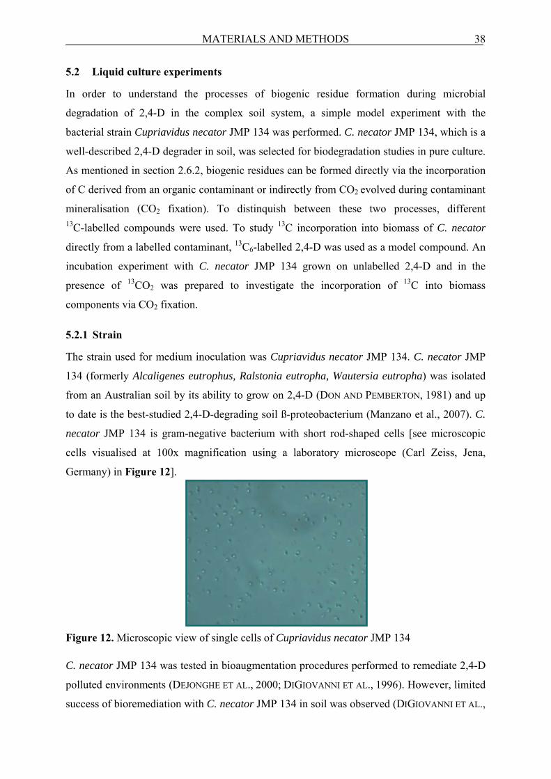
MATERIALS AND METHODS 38
5.2 Liquid culture experiments
In order to understand the processes of biogenic residue formation during microbial
degradation of 2,4-D in the complex soil system, a simple model experiment with the
bacterial strain Cupriavidus necator JMP 134 was performed. C. necator JMP 134, which is a
well-described 2,4-D degrader in soil, was selected for biodegradation studies in pure culture.
As mentioned in section 2.6.2, biogenic residues can be formed directly via the incorporation
of C derived from an organic contaminant or indirectly from CO2 evolved during contaminant
mineralisation (CO2 fixation). To distinquish between these two processes, different 13C-labelled compounds were used. To study 13C incorporation into biomass of C. necator
directly from a labelled contaminant, 13C6-labelled 2,4-D was used as a model compound. An
incubation experiment with C. necator JMP 134 grown on unlabelled 2,4-D and in the
presence of 13CO2 was prepared to investigate the incorporation of 13C into biomass
components via CO2 fixation.
5.2.1 Strain
The strain used for medium inoculation was Cupriavidus necator JMP 134. C. necator JMP
134 (formerly Alcaligenes eutrophus, Ralstonia eutropha, Wautersia eutropha) was isolated
from an Australian soil by its ability to grow on 2,4-D (DON AND PEMBERTON, 1981) and up
to date is the best-studied 2,4-D-degrading soil ß-proteobacterium (Manzano et al., 2007). C.
necator JMP 134 is gram-negative bacterium with short rod-shaped cells [see microscopic
cells visualised at 100x magnification using a laboratory microscope (Carl Zeiss, Jena,
Germany) in Figure 12].
Figure 12. Microscopic view of single cells of Cupriavidus necator JMP 134 C. necator JMP 134 was tested in bioaugmentation procedures performed to remediate 2,4-D
polluted environments (DEJONGHE ET AL., 2000; DIGIOVANNI ET AL., 1996). However, limited
success of bioremediation with C. necator JMP 134 in soil was observed (DIGIOVANNI ET AL.,
MATERIALS AND METHODS 39
1996). This is caused by the fact that various biotic (e.g. presence of eukaryotic microbiota)
and abiotic factors in the complex soil environment may affect both the survival and catabolic
performance of this strain in bioremediation processes (MANZANO ET AL., 2007).
5.2.2 Medium
The cultures were grown in a Minimal Medium (MM) prepared according to LERCH ET AL.
(2007). 2,4-D was added as the sole C source at a total concentration of 73 mg/L
(1978.3 µmol/L). MM components are presented in Table 4. MM was autoclaved (Systec
Autoclave, Wettenberg, Germany) one time at 121°C for 20 min before use for the incubation
experiment.
Table 4. MM components for cultivation of C. necator JMP 134 (LERCH ET AL., 2007)
Component Concentration K2HPO4 1.5 g/L KH2PO4 0.5 g/L (NH4)2SO4 1 g/L MgSO4 (H2O)7 207 mg/L ZnSO4 (H2O)7 200 µg/L MgCl2 (H2O)4 10 µg/L H3BO3 5 µg/L CoCl2 (H2O)6 25 µg/L CuSO4 100 µg/L NiCl2 (H2O)6 5 µg/L FeSO4 (H2O)7 250 µg/L EDTA 125 µg/L
5.2.3 Preculture incubation
Prior to the incubation experiment, to increase the biomass content and to adapt C. necator
JMP 134 for 2,4-D degradation, this strain was pre-cultivated on tryptone medium (TM) as
described previously by LERCH ET AL. (2007), which consisted of 5 g/L tryptone, 3 g/L yeast
extract and 50 mg/L 2,4-D. Unlabelled 2,4-D dissolved in acetone (ACN) was placed in 500
mL Erlenmeyer flasks. In order to prevent killing the bacteria, the solvent was evaporated
under the gentle air stream in a sterile bench. Both the tryptone and the yeast extract were
dissolved in de-ionised water, autoclaved and transferred into the Erlenmeyer flasks after
removal of ACN. After inoculation of the TM with cells of C. necator JMP 134, the batch
preculture was incubated on a rotary shaker (~ 70 min-1; Infors HT, Bottmingen, Switzerland)
for 48 hours, in the dark and at 30°C. After 48 hours of incubation, an aliquot of 10 mL of the
culture was centrifuged (11,000 min-1 for 15 min at 4°C; Jouan KR25i, DBJ Labcare Ltd,
Buckinghamshire, UK). After centrifugation, the biomass pellets were rinsed once with
MATERIALS AND METHODS 40
Phosphate Buffer (PB) prior to the inoculation of the MM to avoid carryover of C from TM to
MM.
5.2.4 Incubation experiment with 13C6-2,4-D
Three different incubation experiments were prepared, in which MM (for details see section
5.2.2) was spiked with:
1. 13C6-2,4-D (271 µmol/L) and 12C6-2,4-D (1707.3 µmol/L). The 13C-labelled
2,4-D was mixed with the unlabelled one in order to increase the 2,4-D concentration
for formation of the minimum biomass necessary for analysis (13C-labelled
compounds are expensive, therefore 13C6-2,4-D was diluted with unlabelled 2,4-D);
2. 12C6-2,4-D (1978.3 µmol/L; control);
3. abiotic (sterile) containing 13C6-2,4-D (271 µmol/L) and 12C6-2,4-D (1707.3
µmol/L).
Control samples provided the information on the natural abundance of 13C in the medium
after addition of unlabelled 2,4-D. The abiotic system was performed to prove that the
incorporation of 13C-label into the biomass during 13C6-2,4-D degradation is a result of biotic
processes. 13C labelled or unlabelled 2,4-D were dissolved separately in ACN and then placed
in 500 mL of Duran glass bottles. The ACN was evaporated and 250 mL (final volume of
culture) of MM was placed in the glass bottles and the content of the bottle was mixed for 10
min. The medium in each bottle was inoculated with cells of C. necator JMP 134
(~ 10 mg/bottle) from the preculture on TM (see previous section 5.2.3). The abiotic systems
were then autoclaved once. Duran glass bottles were sealed with Teflon-lined caps and
incubated on the rotary shaker for 14 days under controlled laboratory conditions (in the dark
and constant temperature 30 ± 2°C). To ensure a sufficient amount of O2 for bacterial
respiration, the systems were flushed periodically with humidified (bottle with de-ionised
H2O) and CO2-free air. During aeration of systems, CO2 evolved during 2,4-D mineralisation
was trapped in two vials containing 2 M NaOH (see Figure 13).
The 13C label incorporation into biomass components such as fatty acids (FA) and
amino acids (AA) and into total biomass of C. necator during 13C6-2,4-D biodegradation were
determined after 1, 2, 3, 7 and 14 days of incubation. In addition, to set up a detailed mass
balance of 13C in the system, the 13C in the CO2 produced during mineralisation of 2,4-D in
the gas space of the bottle and in the medium was also monitored. At each sampling date, the
optical density (OD) indicating bacterial biomass growth was determined using a Lambda 2S
spectrophotometer UV/VIS (λ = 560 nm; Perkin Elmer, Massachutes, USA).

MATERIALS AND METHODS 41
Figure 13. The liquid culture incubation experiment with 13C6-2,4-D in intermittent aeration
systems
5.2.5 Incubation experiment under 13CO2 atmosphere
To study the incorporation of C into biogenic residues via CO2 released during 2,4-D
mineralisation (CO2 fixation), MM in Duran glass bottles spiked previously with unlabelled
2,4-D at a concentration of 1978.3 µmol/L was incubated as three different incubation
experiments under:
1. 13CO2 (33.93 µmol/bottle);
2. 12CO2 (33.93 µmol/bottle; control);
3. 13CO2 (33.93 µmol/bottle) in an abiotic system (sterile).
Small tubes with either labelled Na213CO3 or unlabelled Na2CO3 dissolved in de-ionised H2O,
were placed in each Duran glass bottle (Figure 14). To ensure conditions of 13CO2 fixation
experiment as close as possible to those in the 13C6-2,4-D incubation experiment, the same
concentration of unlabelled 2,4-D was used. The initial amount of CO2 in each bottle was
adjusted to 33.93 µmol and corresponded to an assumed 50% mineralisation of the initial
amount of 2,4-D in the 13C6-2,4-D experiment (67.75 µmol/bottle). After closing the Duran
bottle, CO2 was released from Na2CO3 by adding 6M HCl drop wise into this tube using a
syringe. Thereafter, the bottle was incubated in the same way as for the 13C6-2,4-D incubation
experiment. The bottles were destructively sampled after 2, 3, 7 and 14 days and the 13C label
was analysed in FA, AA and in the total biomass of C. necator. However, no samples were
taken after 1 day of incubation as it was done for 13C6-2,4-D incubation experiment. This is
H2O Cl Cl O
C O OH
C
NaOH traps Safety trap
pump

MATERIALS AND METHODS 42
caused by the fact that CO2 fixation plays a secondary role at the initial phase of incubation.
One triplicate set of liquid culture systems with 13CO2 (33.93 µmol/bottle) and without 2,4-D
for 7 days was incubated to check if CO2 fixation takes place in the absence of the main C
source. At each sampling date, the OD of the bacterial biomass suspension was measured.
Figure 14. The preparation of 13CO2 fixation liquid culture incubation experiment treated
with unlabelled 2,4-D
5.3 Soil experiments
The formation of biogenic residues in soil systems during microbial degradation of two most
widely used organic contaminants 2,4-D and Ibu was investigated. The herbicide 2,4-D was
selected as a first model compound, due to numerous studies on its microbial degradation and
reported high NER content in the soil. In spite of many studies on NER formation during
2,4-D biodegradation, their chemical structure is still not known and there is no report on the
contribution of microorganisms to biogenic residues formation. Therefore, to trace biogenic
residues formation directly from 2,4-D, 13C6-2,4-D incubation experiments were prepared. In
addition, also the indirect incorporation of C into biogenic residues via CO2 fixation process
during 2,4-D biodegradation was studied in the soils amended with unlabelled 2,4-D and
incubated in presence of 13CO2. The second model compound Ibu is a emerging contaminant,
which both microbial degradation and the processes of NER formation are not studied in
detail compared to 2,4-D. In order to investigate biogenic residues formation via the direct
incorporation of C from Ibu, soil systems were incubated with 13C6-ibu.
CO2
CO2
CO2
HCL
Na2CO3

MATERIALS AND METHODS 43
Summing up the above, the mechanisms of NER formation and thus the risks for an
environment related to these two extensively used organic contaminants are not elucidated
yet. Therefore, the knowledge on biogenic residues formation is necessary for a proper risk
assessment and a realistic degradation rate of these contaminants in the soil.
5.3.1 Soil
The soil was collected from A horizon of the agricultural long-term experiment “Statischer
Düngungversuch” located in Bad Lauchstädt, Germany (BLAIR ET AL., 2006). It is a Haplic
Chernozem, which has been cultivated continuously with a crop rotation (sugar beet, summer
barley, potatoes and winter wheat) and fertilised every second year with a farmyard manure
(30 t/ha) since 1902. Detailed soil characteristics are presented in Table 5. Table 5. Soil characteristics used for an incubation experiment (KÖRSCHENS ET AL., 2000)
Parameter Content Clay 21% Silt 68% Sand 11% Ntotal 0.17% Corg 2.1% pH 6.6 WHC 37.5%
5.3.2 Incubation experiment
Three different biotic soil experiments with 13C6-2,4-D, 13C6-2,4-ibu or 13CO2 were prepared.
In order to distinguish between the abiotic and the biotic NER formations, one or two
triplicate sets of abiotic systems for each experiment were also incubated. All soil incubation
experiments were prepared according to the OECD guideline 307 addressing the aerobic and
anaerobic transformation of chemicals in the soil (OECD, 2002). The experimental setup of
the soil systems (Figure 15) was the same as for the liquid culture experiments (Figure 13;
section 5.2.4).
To prevent killing all microorganisms, first only a small amount (10% of the total
amount used for an experiment) of soil (13C6-2,4-D experiment) or soil-sludge mixture (13C6-
ibu experiment) was spiked. Either 13C-labelled or unlabelled model compound was dissolved
in MeOH. This initially spiked portion was then mixed with the rest of the soil to yield a final
compound concentration of 20 mg/kg. The water content of the soil was adjusted to 60% of
its maximum water holding capacity (WHC). Finally 40 gram of this soil was weighed using
laboratory balance (Sartorius AG, Göttingen, Germany) and placed in 1000 mL Duran glass
bottles sealed with Teflon-lined caps.

MATERIALS AND METHODS 44
Figure 15. Incubation experiment of soil treated with organic contaminant (according to
OECD 307) The soil systems were incubated in modified intermittent aeration systems under controlled
laboratory conditions in the darkness and a constant temperature of 20°C (± 2°C). Similar to
the liquid culture systems, soil systems were aerated periodically and the CO2 was trapped in
2 M NaOH solutions.
5.3.3 Soil incubation experiment with 13C6-2,4-D
Three different soil incubations were prepared:
1. soil spiked with 13C6-2,4-D,
2. soil spiked with 12C6-2,4-D (control),
3. non-amended (blank, without 2,4-D).
Blank and control samples allow the correction for the natural abundance of 13C in soil. The
final concentration of spiked 2,4-D in the soil was 20 mg/kg (542.5 μmol 13C/kg) of soil. The
soil was incubated for 64 days. After 2, 4, 8, 16, 32 and 64 days of incubation, the bottles
were sampled destructively and the soil was analysed for the amount and the isotopic
composition (13C) of FA and AA as representatives for biogenic residues. In addition, two
triplicate sets of abiotic soil systems with 13C6-2,4-D were also incubated and sampled
destructively after 32 and 64 days.
5.3.4 Soil incubation experiment under 13CO2 atmosphere
In order to investigate CO2 fixation (from mineralisation of 2,4-D residues) by soil
microorganisms and its incorporation into biogenic residues, three soil incubations were
prepared:
1. with unlabelled 2,4-D (20 mg/kg) and 13CO2 (12.6 μmol/bottle);
2. with unlabelled 2,4-D (20 mg/kg) and unlabelled CO2 (12.6 μmol/bottle);
MATERIALS AND METHODS 45
3. with neither 2,4-D nor additional CO2.
The preparation of an incubation experiment under CO2 atmosphere was already presented in
detail in section 5.2.5 (Figure 14). The initial amount of CO2 in each bottle was adjusted to
the final concentration of 13CO2 evolved during mineralisation of 13C6-2,4-D. To estimate the
incorporation of 13C-label into FA, the soil was extracted and analysed after 8, 16, 32 and 64
days of incubation. No samples were taken on days 2 and 4 due to a minor role of CO2
fixation in the initial phase of incubation. In addition, because of a low level of the isotopic
enrichment (in a range of the natural isotopic abundance) in the extracted AA and tFA from
soils, they were not further quantified. One triplicate set of soil systems with 13CO2
(12.6 μmol/bottle) without 2,4-D and abiotic systems with 13CO2 and unlabelled 2,4-D were
also incubated for 64 and 32 days, respectively. The soil systems without 2,4-D were prepared
to check if the presence of this readily available organic substrate stimulates CO2 fixation.
5.3.5 Soil incubation experiment with 13C6-ibu
Similar to the soil incubation experiment with 13C6-2,4-D, also three types of soil systems
were prepared:
1. soil spiked with 13C6-ibu;
2. soil spiked with 12C6-ibu (control);
3. non-amended (blank, without Ibu).
Contrary to 13C6-2,4-D soil incubation experiment, the soil was amended additionally with
stabilised sewage sludge (Klärwerk Rosental, Leipzig, Germany), because Ibu can only be
introduced into the soil by sewage sludge application (EDWARDS ET AL., 2009). Soil and
sludge were mixed at a 9:1 (DW/DW) ratio. The initial Ibu concentration was 20 mg/kg
(581.5 μmol/kg) of soil. The soil systems were incubated for 90 days and sampled
destructively after 2, 7, 14, 28, 59 and 90 days of incubation and analysed for the amount and
the isotopic composition of FA and AA. Additionally, two triplicate sets of abiotic soil
systems with 13C6-2,4-ibu were also incubated and sampled destructively after 28 and 90 days
of incubation.
5.4 Chemical analyses
The main focus of all experiments was to study the 13C incorporation into two representatives
for biogenic residues, fatty acids (FA) and amino acids (AA). FA and AA are known
microbial biomarkers (BOSCHKER AND MIDDELBURG, 2002), which represent the major
fraction of the analytically recognisable compounds in SOM (ALLARD, 2006). Owing to FA

MATERIALS AND METHODS 46
and AA extraction and their amount and isotopic composition analyses, we can outline the
formation and the fate of biogenic residues in the complex soil system.
However, for a better understanding of biogenic residue formation, additional
information on the 13C label distribution within a system (mass balance) is necessary. In order
to set up a detailed mass balance of 13C in a system, the amount and the isotopic composition
of the following components should be considered: CO2, solvent-extractable parent
compound and its metabolites and finally either 13C incorporation into total biomass (liquid
culture experiment) or total NER (soil experiments).
5.4.1 CO2 measurement
The 13CO2 trapped in 2 M NaOH solution was quantified by measurement of the total
inorganic C using a Total Organic Carbon Analyser (Schimadzu TOC-5050, Duisburg,
Germany). The isotopic composition (atom %) of CO2 was determined using a GC-
Combustion-isotope ratio Mass Spectrometry (GC-C-irMS; Finnigan MAT 252, Thermo
Electron, Bremen, Germany, coupled to Hewlett Packard 6890 GC, Agilent Technologies,
Germany), equipped with Porabondt Q-HT Plot FS column (50 m × 0.32 m × 5 μm;
Chrompack, Middburg, Netherlands). In order to measure the isotopic composition, 2 ml of
the NaOH solution was transferred to 15 ml crimp cap vials and acidified with 400 µl of
phosphoric acid (85%). Headspace samples of 100-250 µl were then injected into the GC-C-
irMS. CO2 was separated isothermally at a temperature of 40°C. The temperature of the
oxidation oven was 940°C and the one of the reduction oven was 640°C. Both the C content
and isotopic composition of CO2 trapped in MM after removal of C. necator biomass by
centrifugation was determined in the same way as CO2 trapped in the NaOH solutions.
5.4.2 Parent compound and metabolites measurement 13C6-2,4-D or 13C6-ibu residues were extracted from soil using an Accelerated Solvent
Extraction (ASE) 200 system (Dionex, Sunnyvale, CA) equipped with 11 mL stainless steel
extraction cells according to RADJENOVIĆ ET AL. (2009) with few modifications. 5 gram of
soil was placed in the extraction cell and mixed with diatomaceous earth (Hydromatrix™,
Varian Associates, Inc., Hansen Way, Palo Alto, USA) and 20 µg of the internal standard
2-methyl-4-chlorophenoxyacetic acid (MCPA) used for quantitative analyses. The soil
samples were extracted three times with methanol-water (1:1, v/v) at the following operating
conditions: extraction temperature, 100ºC; pressure 100 bar; preheating period, 5 min; static
extraction period, 10 min; solvent flush, 150% of the cell volume; and nitrogen purge, 150 s.

MATERIALS AND METHODS 47
The biomass of C. necator was separated from MM by cetrifugation (11,000 min-1 for 15 min
at 4°C, Jouan KR52i). The supernatant containing 13C6-2,4-D and 13C6-2,4-D-derived
metabolites was then purified.
MM after centrifugation and soil extracts were acidified to pH 2 prior to purification by
Solid Phase Extraction (SPE, Macherey Nagel, Düren, Germany). The SPE cartridges
(CHROMABOND® EASY) were conditioned with 3 mL of MeOH and 3 mL of de-ionised
H2O. After the conditioning step, the acidified sample was passed slowly through the column
applying a slight vacuum. The column then was washed with 10 mL of de-ionised H2O and
dried under vacuum using a vacuum pump (KNF Neuberger, Frankfurt, Germany) for 15 min.
The elution of 2,4-D and its metabolites from column was performed 2 times with 5 mL of
methanol (MeOH)-ACN mixture (1:1, v/v). A mixture of MeOH-tetrahydrofuran (1:1, v/v; 5
mL; 2 times) was used for the elution of ibu and ibu-derived metabolites. The eluates were
then evaporated under a gentle stream of N2. Dried sample was resuspended in the mixture of
acetonitrile/trimethylsilyltrifluoroacetamide (1:2, v/v) for silylation. This mixture was then
incubated for 10 min at 60 ºC, next cooled down at room temperature and finally transferred
into a GC vial.
Parent compounds and their metabolites were quantified and identified using Gas
Chromatography-Mass Spectrometry (GC-MS, Hewlett Packard 6890 GC, Agilent
Technologies, Germany) equipped with a BPX-5 column (30 m × 0.32 m × 0.25 μm, SGE,
Darmstadt, Germany) with the following temperature program: initial temperature 60°C
(1.5 min), heat to 120°C (0 min) at 20°C/min, to 160°C (0 min) at 4°C/min and finally to
260°C (5 min) at 16°C/min (ZWIENER AT EL., 2002). The injector was set at 260°C and the He
flow was 1.5 mL/min. The identification of 2,4-D, 2,4-DCP, Ibu and 2-OH-ibu in sample was
done by comparison of their retention times with those of a standard.
5.4.3 Total biomass and NER measurement
At each sampling date, bacterial cells of C. necator were harvested from MM by
centrifugation (11,000 min-1 for 15 min at 4°C; Jouan KR25i) and then freeze-dried (2 h;
Christ RVC 2-25, Osterode am Harz, Germany). About of 2-5 mg of either air-dried soil
sample (after removal of solvent-extractable parent compound and its metabolites) or freeze-
dried biomass was weighed. Both the amount of 13C and isotopic composition in either total
biomass or total NER were determined using Elemental Analyser-Combustion-isotope ratio
Mass Spectrometry (EA-C-irMS; Finnigan MAT 253, Thermo Electron, Bremen, Germany)
coupled to Euro EA 3000, Eurovector, Milano, Italy). The temperature of the oxidation oven

MATERIALS AND METHODS 48
was 1020°C and the one of the reduction oven was 650°C. In addition, bulk extracts from soil
before purification by SPE were also analysed for 13C content and the isotopic composition.
5.4.4 Fatty acids (FA) analyses
We analysed the 13C label incorporation into FA of living biomass of C. necator and into FA
of living soil microbial biomass. To distinguish between 13C distributions in FA within the
living (PLFA) and the non-living SOM fraction, total fatty acids (tFA) in the soil were also
determined. The tFA contains FA both from the living and the dead biomass stabilised in
SOM (non-living SOM; DRENOVSKY ET AL., 2004).
However, the PLFA extraction procedure from the soil microbial biomass is different
than from the biomass of C. necator and includes two purification steps. Therefore, PLFA
extractions from C. necator and the soil are presented below separately.
Biomass of C. necator was harvested from MM by centrifugation (3300 min-1 for
15 min at 4°C; Jouan KR25i) prior to PLFA extraction. The PLFA were extracted with a
mixture of PB/MeOH/chloroform from the C. necator (0.5 mL/1 mL/2 mL) according to
BLIGH AND DYER (1959). The PB/MeOH/chloroform mixture containing cells of C. necator
was votexed for 3 min (Vibromix®, Sartorius Stedim Biotech, Aubagne Cedex, France).
After addition of H2O (0.5 mL), the mixture was shaken again for 1 min and finally separated
into two phases by centrifugation for 10 min at 1000 min-1 (Jouan KR25i). The bottom phase
(chloroform) containing the PLFA was evaporated under a gentle stream of N2 prior to
derivatisation
The PLFA were extracted from a soil using also the PB/MeOH/chloroform mixture (2
mL/5 mL/2.5 mL; BLIGH AND DYER, 1959). The soil suspended in the PB/MeOH/chloroform
mixture was shaken for 2 h on a rotary shaker (Infors HT, Bottmingen, Switzerland). After
shaking, 2.5 mL of each H2O and chloroform was added to the mixture in order to separate
into two phases (overnight). The bottom chloroform phase was dried using sodium sulfate and
evaporated under N2. This phase was then fractionated into neutral lipids, glycolipids and
PLFA according to MILTNER ET AL. (2005) by chromatography over silica columns (Unisil)
impregnated previously with ammonium acetate (0.02 M). To elute neutral lipids, glycolipids
and PLFA chloroform (5 mL), ACN (5 mL) and MeOH (15 mL) were used, respectively. The
MeOH was evaporated under N2 and the PLFA were then derivatised.
The dried samples containing PLFA extracted from either C. necator or the soil were
resuspended in the mixture of MeOH/Trimethylchlorosilane [(TMCS); 9:1; v:v] and
methylated at 60°C in a oven (Memmert, Schwabach, Germany) for 2 h as described by
THIEL ET AL., (2001). After cooling to room temperature, the excess of derivatisation reagent

MATERIALS AND METHODS 49
was removed in the N2-gasstream. Finally, the dried sample was redissolved in 50 µL of
hexane prior to further gas chromatographic analysis.
The tFA in the soil were analysed by methylation with the same derivatisation reagent
as for PLFA, (MeOH/TMCS; 1.8 mL/0.2 mL) added directly to the dry soil (THIEL ET AL.,
2001). After derivatisaton, methylated tFA were extracted from soil according to MILTNER, ET
AL. (2005) with diethyl ether and purified over silica gel columns (Baker). The eluate was
then evaporated under N2 and dried sample was finally dissolved in 100 µL of hexane prior to
GC/MS and GC-C-irMS analyses.
The Fatty Acid Methyl Esters (FAME) in PLFA and tFA fractions were quantified and
identified by GC/MS equipped with BPX-5 column as described in section 5.4.2. FA are
designated as A:BωC where A is the number of C atoms, B is the number of double bonds
and C is the position of the double bond relative to the aliphatic end of the molecule. The
prefix “cy” assigns a cyclopropyl fatty acid, and branched fatty acids are indicated by i-, a- or
10Me.
For quantification, heneicosanoate methyl ester (FAME 21:0) was added to each sample
as an internal standard prior to injection into the GC/MS. The FAME were separated using
the following temperature program: initial temperature 50°C (hold for 1 min), heat to 250°C
(0 min) at 4°C/min and finally to 300°C (10 min) at 2°C/min. The injector was set at 280°C,
the He flow was 2.5 mL/min. FAME were identified by comparison of the retention times and
the mass spectra with those of a standard mixture BAME (Bacterial Acid Methyl Esters). The
isotopic composition of the FAME was determined GC-C-irMS (Finnigan MAT 253 coupled
to Trace GC, Thermo Electron, Bremen, Germany) equipped with BPX-5 column (50 m ×
0.32 m × 0.5 μm, SGE International, Darmstadt, Germany). The FAME were separated with
the following temperature program for GC-C-irMS: initial temperature 70°C (1 min), heat to
130°C (0 min) at 20°C/min, to 150°C (5 min) at 2°C/min, to 165°C (5 min) at 2°C/min, to
230°C (0 min) at 2°C/min and finally to 300°C (5 min) at 20°C/min. The injector was set at
250°C and the He flow was 2 mL/min.
With the methylation, one additional C is introduced resulting in a shift of the isotopic
composition. This shift was corrected for the original isotopic composition of the respective
FA and calculated according to BOSCHKER (2004) as follows:
ICcorr = (nFAME + nMeOH ) × ICmeas−ICMeOH
nFAME
where:
nFAME – number of C in respective FAME without the methyl group

MATERIALS AND METHODS 50
nMeOH – number of C of MeOH
ICcorr – corrected isotopic composition of FAME without the methyl group
ICmeas – measured isotopic composition of FAME with the methyl group
ICMeOH – C isotopic composition of MeOH
5.4.5 Amino acids (AA) analyses 13C-amino acids (AA) hydrolysed from proteins were analysed in soil in two fractions: in the
total AA fraction (tAA) and in the living microbial biomass AA fraction (bioAA). As
considered for the fatty acids, the difference between the total and the biomass AA fractions
represents the refractory AA in the non-living SOM. AA in C. necator were hydrolysed
directly from the biomass similarly as the tAA from the total soil, whereas bioAA in the soil
were determined in the extracted living microbial biomass.
Prior to hydrolysis, bacterial cells were harvested from MM by centrifugation (3300
min-1 for 15 min at 4°C; Jouan KR25i). AA in the biomass of C. necator and in the total soil
(tAA) were hydrolysed with 6 M HCl for 22 hours at 110°C as described by MACKO AND
UHLE (1997) and SILFER ET AL. (1991). After hydrolysis, biomass pellets and soil particles
were discarded by filtering through glass-fibre membrane filters. The HCl was then
evaporated in an N2-gasstream and AA from biomass of C. necator were further derivatised,
whereas the soil hydrolysate was purified.
After evaporation of the HCl excess, soil sample was resuspended in 4 mL of 0.1 M
HCl and purified over cation exchange resin (DOWEX 50W-X8) according to AMELUNG AND
ZHANG (2001). Cation exchange resin was prepared according to BOAS ET AL. (1953), by pre-
rinsing with 2 M NaOH, conditioning with 2 M HCl, and washing with de-ionized H2O until
the pH of the eluate was neutral. The sample containing the AA was pipetted on top of the
column. The column then was washed with oxalic acid (25 mL, pH 1.6-1.8) followed by 0.01
M HCl (5 mL) and de-ionised H2O (5 mL). Finally the AA were eluted with 2.5 M NH4OH
(30 mL) and NH4OH was evaporated. The dried samples were re-dissolved in 4 mL 0.1 M
HCl and centrifuged for 15 min at 4200 min-1 (Jouan KR25i). After centrifugation, the 0.1 M
HCl excess was evaporated under N2 prior to derivatisation.
The carboxyl groups of AA in dried sample were esterified with a mixture of
Isopropanol/Acetylchloride (IP/AC; 1 mL/250 μL) at 110°C for 1 h and the amino groups
trifluoroacetylated with a mixture of Dichloromethane/Trifluroacetic Acid Anhydride
(DCM/TFAA; 500 µL/500 µL) at 60°C for 1 h as described previously by MILTNER ET AL.
(2005). After derivatisation, the samples were dried under N2 and purified using a mixture of
PB (1 mL) and chloroform (0.5 mL) as described previously by UEDA ET AL. (1989). The

MATERIALS AND METHODS 51
mixture was shaken, centrifuged (Centrifuge 5415R eppendorf; Eppendorf, Hamburg,
Germany) at 600 min-1 for 10 min and the chloroform phase was finally evaporated. After
drying, the samples containing the derivatised AA were re-dissolved in 100 μL DCM for
injection into the GC-MS.
For the determination of the AA in the living biomass (bioAA), the biomass was first
extracted from soil as described by JACOBSEN AND RASMUSSEN (1992). The efficiency of the
bacterial biomass extraction has been reported to be in the range of 40% by independent
studies (JACOBSEN AND RASMUSSEN, 1992; MILTNER ET AL., 2009). Hence, bioAA are
underestimated but the relative trends of bio-AA are correct because of the relatively constant
recoveries even in different soils. The cells were extracted from soil by shaking using an end-
over-end shaker (2h; GFL 3040, Fachhandel für Labor- und Medizintechnik GmbH, Kothen,
Germany) according to MILTNER ET AL. (2009) with 1 g Chelex 100 (chelating cation
exchange resin) and 10 mL of sodium deoxycholate/Polyethylenglycol 600 solution
(0.1%/2.5%). After shaking, soil particles were removed using low speed centrifugation
(960 min-1, 15 min; Jouan KR25i) and finally the biomass in supernatant was harvested using
high-speed centrifugation (11,000 min-1, 1h, 4°C; Jouan KR25i). Biomass pellets containing
AA were further hydrolysed, purified and derivatised in the same way as tAA.
The identity, quantity and isotopic composition of the AA were determined by GC/MS
and GC-C-irMS. The temperature program of GC/MS was as follows: 70°C (5 min) to 100°C
(30 min) at 30°C/min, to 175°C (5 min) at 10°C/min, to 250°C (5 min) at 10°C/min, to 340°C
(15 min) at 30°C/min. The injector was set at 280°C; the He flow was 1.7 mL/min. For
quantification and identification of respective AA in samples, an external standard containing
all detectable AA in the samples [alanine (Ala), glycine (Gly), threonine (Thr), serine (Ser),
valine (Val), leucine (Leu), isoleucine (Ile), proline (Pro), aspartate/asparagine (Asp),
glutamate/glutamine (Glu), phenylalanine (Phe), and lysine (Lys)] was used. Asparagine and
glutamine are desaminated during the acid hydrolysis, resulting in aspartate and glutamate,
respectively. Therefore they are shown as Asp and Glu. The internal standard L-norleucine
was added to each sample before hydrolysis to estimate the losses of AA in soil during the
two purification steps. The isotopic composition of each AA was determined using
GC-C-irMS with the same column as for the FAME and the following temperature program:
50°C (0 min) to 100°C (10 min) at 15°C/min, to 130°C (0 min) at 2°C/min, to 220°C (0 min)
at 10°C/min, to 250°C (10 min) at 30°C/min. The injector was set at 250°C; the He flow was
2 mL/min.

MATERIALS AND METHODS 52
The original isotopic enrichment of respective AA in samples, after shifting their isotopic
composition during two derivatisation steps, was corrected using the following equation
(SILFER ET AL., 1991):
ICcorr =ICmeas × (nAA + nTFAA + nIP ) − ICTFAA × nTFAA − ICIP × nIP
nAA
where:
nAA– number of C in respective AA
nTFAA - number of C of TFAA
nIP - number of C of IP
ICcorr – corrected isotopic composition of AA
ICmeas – measured isotopic composition of AA
ICTFAA – C isotopic composition of TFAA
ICIP – C isotopic composition of IP
5.5 Data analyses
A stable isotope tracer (13C) was used to investigate the formation and the fate of biogenic
residues during biodegradation of a target contaminant in a system. The application of 13C-labelled compounds in biodegradation studies allows the analysis of their structural
assignments using more sophisticated analytical techniques such as GC-MS (RICHNOW ET AL.,
1999). By extraction of known 13C-labelled biogenic residues representatives (FA and AA)
formed during 13C-labelled compound biodegradation and inspection of their chemical
structures using GC-MS, we can thus prove the microbial origin of NER in the soil.
However, the soil is also naturally abundant in 13C (~ 1%), therefore blank (without
compound application) and control (12C-labelled compound) samples were used for the
correction of 13C abundance in a soil amended with the tested 13C-labelled compound.
Similarly to the soil, 13C abundance in the liquid culture experiments was also corrected
accordingly due to addition of 12C-2,4-D to 13C-2,4-D incubation experiments .
The contents of 13C-FA, 13C-AA, evolved 13CO2 during mineralisation, 13C-parent
compound and its 13C-metabolites and 13C in either total biomass or NER were presented as a
percentage of the initially applied 13C-labelled compound to an experiment. In addition, a
mass balance of 13C in the system was taken into consideration when interpreting the data on
the 13C label incorporation into biogenic residues.
All incubation experiments were prepared in triplicates and results are shown as
averages. The error bars represent the standard deviation of these triplicates.

RESULTS 53
6 RESULTS
The incorporation of 13C label into biogenic residue representatives (FA and AA) during
microbial degradation of 13C-labelled compounds was studied in two different systems:
simple liquid pure culture experiments and complex soil systems.
The main objective of liquid culture experiments was to trace the fate of 13C label
derived from 13C6-2,4-D in the simple biological system with a particular focus on 13C-incorporation into the biomass of the 2,4-D degrader. Due to simplicity, these
experiments were also designed to estimate the ratio of 13C-FA and 13C-AA to the total
biomass 13C content, which are necessary for a proper quantification of the total amount of
biogenic residues in the soil systems.
The formation and the fate of biogenic residues during biodegradation of two different
model compounds (13C6-2,4-D and 13C6-ibu) were investigated in complex soil experiments.
The general objective was to estimate the extent of biogenic residue formation and their
amount in relation to total NER and finally to assess the risk for the environment related to
NER formation from readily degradable organic contaminants in soil.
6.1 Liquid culture experiments
Cupriavidus necator JMP 134 was selected for the biodegradation studies, because it is a
well-described 2,4-D soil degrader. C. necator grows on 2,4-D as C source. The 13C label
incorporation into FA and AA of C. necator during microbial degradation of 2,4-D was
investigated directly from 13C6-2,4-D and indirectly via CO2 fixation from 13CO2
The detailed description of PLFA classes typical for specific groups of microorganisms,
their designation and AA abbreviations used for data interpretation have already been
presented in the sections 2.6.2.1, 5.4.4 and 5.4.5, respectively.
6.1.1 Incubation experiment with 13C6-2,4-D
The 13C incorporation into the biomass of C. necator during biodegradation of 13C6-2,4-D was
studied over 14 days. The 13C content in FA, AA, in total biomass and the 13C mass balance in
the system were analysed after 1, 2, 3, 7 and 14 days of incubation. The abiotic experiment
with C. necator and 13C6-2,4-D showed neither 13C6-2,4-D biodegradation nor 13C
incorporation into FA and AA of C. necator after 14 days, indicating the importance of
bacterial activity in the degradation of this compound and in the formation of 13C-labelled FA
and AA.

RESULTS 54
6.1.1.1 Mass balance of 13C6-2,4-D in the system
The Duran glass bottle (500 mL) was aerated with humidified air prior to CO2 measurement
in order to trap the evolved CO2 in a headspace into NaOH solution. However, due to
reported high overpressure in the small headspace volume of about 250 mL and the need to
connect the bottle to a pump, some CO2 was lost before aeration. The experiment with
C. necator JMP134 grown on unlabelled 2,4-D and under 13CO2 atmosphere, in which the
initial 13CO2 concentration was known, revealed that at each sampling date ~ 70% of the
initial 13CO2 was lost (data not shown). Therefore, the data on the mineralisation kinetics
presented in Figure 16 are highly underestimated.
Figure 16. Distribution of 13C label in an incubation experiment with A C. necator JMP 134
grown on 13C6-2,4-D medium. Mineralisation ( ) and biomass ( ). Extractable 13C6-2,4-D
and metabolites not detectable since day 1 After the first day of incubation, 15% of 13C derived from 13C6-2,4-D was already mineralised
(50% of 13C6-2,4-D equivalents after correction for 70% loss). At that time, neither 13C6-2,4-D
nor its known 13C-aromatic ring metabolites were detected, indicating their complete
utilisation. After day 1, the content of 13CO2 evolved during 2,4-D mineralisation increased
slowly and finally on the day 7 reached a plateau (~ 83% of 13C6-2,4-D equivalents after
correction for 70% loss). The incorporation of 13C from 13C6-2,4-D into the biomass started
immediately (after 1 day) and their contents remained constant until day 3 (~ 10% of 13C6-2,4-D equivalents; see Table 6). Thereafter, the amount of 13C found in the total biomass
increased slowly reaching finally 17.4% of the added 13C6-2,4-D equivalents. However, based
on the OD measurement of a culture suspension, the biomass growth of C. necator JMP134
0
5
10
15
20
25
30
0 2 4 6 8 10 12 14
incubation time (days)
% o
f ini
tial 13
C6-
2,4-
D (%
)
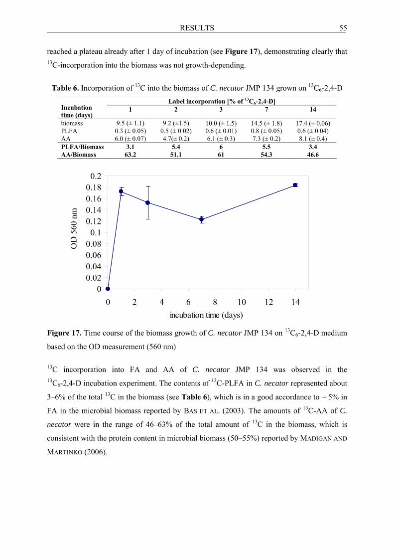
RESULTS 55
reached a plateau already after 1 day of incubation (see Figure 17), demonstrating clearly that 13C-incorporation into the biomass was not growth-depending.
Table 6. Incorporation of 13C into the biomass of C. necator JMP 134 grown on 13C6-2,4-D
Label incorporation [% of 13C6-2,4-D] Incubation time (days)
1 2 3 7 14
biomass 9.5 (± 1.1) 9.2 (±1.5) 10.0 (± 1.5) 14.5 (± 1.8) 17.4 (± 0.06) PLFA 0.3 (± 0.05) 0.5 (± 0.02) 0.6 (± 0.01) 0.8 (± 0.05) 0.6 (± 0.04) AA 6.0 (± 0.07) 4.7(± 0.2) 6.1 (± 0.3) 7.3 (± 0.2) 8.1 (± 0.4) PLFA/Biomass 3.1 5.4 6 5.5 3.4 AA/Biomass 63.2 51.1 61 54.3 46.6
Figure 17. Time course of the biomass growth of C. necator JMP 134 on 13C6-2,4-D medium
based on the OD measurement (560 nm)
13C incorporation into FA and AA of C. necator JMP 134 was observed in the 13C6-2,4-D incubation experiment. The contents of 13C-PLFA in C. necator represented about
3–6% of the total 13C in the biomass (see Table 6), which is in a good accordance to ~ 5% in
FA in the microbial biomass reported by BAS ET AL. (2003). The amounts of 13C-AA of C.
necator were in the range of 46–63% of the total amount of 13C in the biomass, which is
consistent with the protein content in microbial biomass (50–55%) reported by MADIGAN AND
MARTINKO (2006).
00.020.040.060.08
0.10.120.140.160.18
0.2
0 2 4 6 8 10 12 14incubation time (days)
OD
560
nm
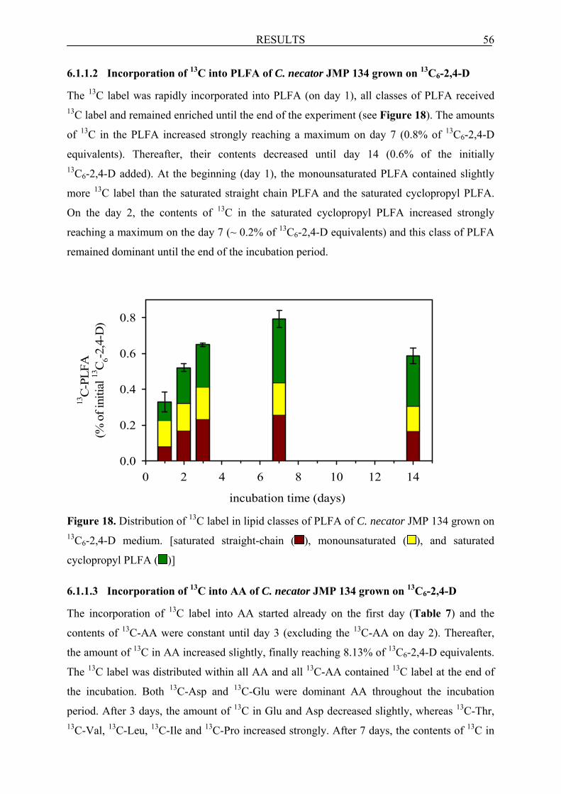
RESULTS 56
6.1.1.2 Incorporation of 13C into PLFA of C. necator JMP 134 grown on 13C6-2,4-D
The 13C label was rapidly incorporated into PLFA (on day 1), all classes of PLFA received 13C label and remained enriched until the end of the experiment (see Figure 18). The amounts
of 13C in the PLFA increased strongly reaching a maximum on day 7 (0.8% of 13C6-2,4-D
equivalents). Thereafter, their contents decreased until day 14 (0.6% of the initially 13C6-2,4-D added). At the beginning (day 1), the monounsaturated PLFA contained slightly
more 13C label than the saturated straight chain PLFA and the saturated cyclopropyl PLFA.
On the day 2, the contents of 13C in the saturated cyclopropyl PLFA increased strongly
reaching a maximum on the day 7 (~ 0.2% of 13C6-2,4-D equivalents) and this class of PLFA
remained dominant until the end of the incubation period.
Figure 18. Distribution of 13C label in lipid classes of PLFA of C. necator JMP 134 grown on 13C6-2,4-D medium. [saturated straight-chain ( ), monounsaturated ( ), and saturated
cyclopropyl PLFA ( )]
6.1.1.3 Incorporation of 13C into AA of C. necator JMP 134 grown on 13C6-2,4-D
The incorporation of 13C label into AA started already on the first day (Table 7) and the
contents of 13C-AA were constant until day 3 (excluding the 13C-AA on day 2). Thereafter,
the amount of 13C in AA increased slightly, finally reaching 8.13% of 13C6-2,4-D equivalents.
The 13C label was distributed within all AA and all 13C-AA contained 13C label at the end of
the incubation. Both 13C-Asp and 13C-Glu were dominant AA throughout the incubation
period. After 3 days, the amount of 13C in Glu and Asp decreased slightly, whereas 13C-Thr, 13C-Val, 13C-Leu, 13C-Ile and 13C-Pro increased strongly. After 7 days, the contents of 13C in
incubation time (days)
0 2 4 6 8 10 12 14
13C
-PLF
A(%
of i
nitia
l 13C
6-2,
4-D
)
0.0
0.2
0.4
0.6
0.8

RESULTS 57
Threo and Val increased again, while 13C-Phe decreased. At the end, 13C-Thr and 13C-Gly
decreased (~ 2 times and 4 times, respectively), while the amount of 13C in Leu, Ile, Pro, Asp,
Glu and Phe increased. At the same time, a very low content of 13C label was also detected in
Ser. Table 7. Distribution of the 13C label in various 13C-AA of C. necator JMP 134 grown on 13C6-2,4-D medium
13C-AA [% of 13C6-2,4-D] Incubation time (days)
1 2 3 7 14
Alanine n.d. n.d. 0.43 (± 0.6) 0.46 (± 0.07) 0.28 (± 0.08) Glycine 0.07 (± 0.01) 0.16 (± 0.02) n.d. 0.53 (± 0.04) 0.13↓ (± 0.02) Threonine 0.25 (± 0.004) 0.24 (± 0.11) 0.65↑ (± 0.08) 0.86↑ (± 0.002) 0.46↓ (± 0.05) Valine 0.09 (± 0.09) 0.12 (± 0.06) 0.27↑ (± 0.03) 0.6↑ (± 0.004) 0.56 (± 0.09) Serine n.d. n.d. n.d. n.d. 0.003 (± 0.11) Leucine 0.61 (± 0.003) 0.62 (± 0.04) 0.94↑ (± 0.02) 0.82 (± 0.1) 1.2↑ (± 0.04) Isoleucine 0.14 (± 0.02) 0.13 (± 0.01) 0.30↑ (± 0.009) 0.32 ( ± 0.01) 0.38↑ (± 0.01) Proline 0.55 (± 0.02) 0.39 (± 0.03) 0.51↑ (± 0.07) 0.59 (± 0.02) 0.73↑ (± 0.03) Aspartatea 1.19 (± 0.03) 1.17 (± 0.24) 1.01↓ (± 0.07) 1.07 (± 0.02) 1.56↑ (± 0.32) Glutamateb 1.52 (± 0.08) 0.98↓ (± 0.06) 0.89↓ (± 0.18) 1.07 (± 0.004) 1.53↑ (± 0.33) Phenylanine 0.77 (± 0.02) 0.59 (± 0.04) 0.76 (± 0.14) 0.58↓ (± 0.03) 0.77↑ (± 0.04) Lysine 0.85 (± 0.005) 0.25 (± 0.02) 0.35 (± 0.17) 0.44 (± 0.12) 0.49 (± 0.01) TOTAL 6.06 (± 0.07) 4.67 (± 0.17) 6.13 (± 0.34) 7.34 (± 0.19) 8.13 (± 0.41) a incl. asparagine; b incl. glutamine; n.d. - not detectable; values are shown as averages ± standard deviation values printed in bold represent characteristic values, arrows visualise increase or decrease of the respective
AA compared to the preceeding sampling time
6.1.2 Incubation experiment under 13CO2 atmosphere
The incorporation of 13C label from 13CO2 into the biomass of C. necator JMP 134 grown on
unlabelled 2,4-D, but in the presence of 13CO2 was studied over 14 days. The content and the
isotopic composition of 13C in PLFA, AA and in total biomass were analysed after 2, 3, 7 and
14 days of incubation. The main objective of this experiment was to study the relevance of
heterotrophic CO2 fixation in 13C label incorporation into the biomass of C. necator during
2,4-D biodegradation. However, the first sampling date (day 1) considered in the incubation
experiment with 13C6-2,4-D was excluded and not analysed, because CO2 fixation plays
usually a minor role in the initial phase of biodegradation experiment. One triplicate set of the
biotic incubation experiment with 13CO2 and without 2,4-D showed no incorporation of 13C
into the biomass of C. necator, demonstrating strong dependence of 13CO2 fixation on the
presence 2,4-D in the medium. The abiotic incubation experiment also revealed no 13C
incorporation into the PLFA and AA of C. necator after 7 days, indicating the importance of
biological activity for CO2 fixation.

RESULTS 58
6.1.2.1 Incorporation of 13C into the biomass of C. necator JMP 134 grown under 13CO2
A very low 13C incorporation into the biomass of C. necator JMP 134 from 13CO2 was
observed already after 2 days of incubation (0.07% of the initially 13CO2 added; see Table 8).
Thereafter, the amount of 13C in the total biomass was increasing progressively throughout
the incubation time, finally reaching 4.2% of the added 13CO2 equivalents. The biomass grew
rapidly from day 0 until day 3 (see Figure 19), indicating the high importance of CO2 fixation
in the later phase of the incubation experiment. Thereafter, the biomass growth reached a
plateau.
0
0.05
0.1
0.15
0.2
0.25
0 2 4 6 8 10 12 14incubation time (days)
OD
560
nm
Figure 19. Time course of the biomass growth of C. necator JMP 134 on unlabelled 2,4-D
medium and under 13CO2 based on the OD measurement (560 nm)
The 13C label was incorporated into PLFA and AA of C. necator JMP 134 in the 13CO2
fixation experiment. The amount of 13C-PLFA in C. necator JMP 134 was in the range of
3–10% of the total 13C in the biomass (excluding the day 2 where the amount of PLFA was
very low; see Table 8), what is in good agreement with the data of BAS ET AL. (2003) and
with those of the 13C6-2,4-D experiment with C. necator. 13C-AA in C. necator JMP 134
represented 42–62% of the total 13C in the biomass, which is close to the percentage of
proteins in biomass reported by MADIGAN AND MARTINKO (2006) and to the 13C6-2,4-D
experiment with C. necator.

RESULTS 59
Table 8. Incorporation of 13C into biomass of C. necator JMP 134 grown on unlabelled
2,4-D medium and under 13CO2
13C-AA [% of 13CO2] Incubation time (days)
2 3 7 14
biomass 0.07 (± 0.5) 1.6 (±0.4) 3.5 (± 0.2) 4.2 (± 0.2) PLFA 0.01 (± 0.004) 0.17 (± 0.02) 0.15 (± 0.008) 0.12 (± 0.003) AA 0.02 (± 0.07) 1.0 (± 0.4) 1.5 (± 0.3) 1.8 (± 0.08) PLFA/Biomass 14.3 10.5 4.3 2.9 AA/Biomass 28.6 62.5 42.9 42.9
6.1.2.2 Incorporation of 13C into PLFA of C. necator JMP 134 grown under 13CO2
The 13C derived from 13CO2 was incorporated into the PLFA immediately on day 2, only the
monounsaturated PLFA and the saturated straight chain PLFA carried 13C label (see
Figure 20).
Figure 20. Distribution of 13C-label in lipid classes of PLFA of C. necator JMP 134 grown on
unlabelled 2,4-D medium and under 13CO2.
[saturated straight-chain ( ), monounsaturated ( ), and saturated cyclopropyl PLFA ( )]
The contents of 13C-PLFA increased rapidly, reaching their maximum on the day 3 (0.17% of
the initially 13CO2 added) and remained constant until day 7. At the end, the 13C-PLFA
decreased to 0.12% of the initially 13CO2 added. From day 3 onwards, the 13C label was
distributed within all classes of PLFA and on both day 3 and 7, the monounsaturated PLFA
dominated over other classes of PLFA. Similarly as observed in the 13C6-2,4-D experiment,
incubation time (days)
0 2 4 6 8 10 12 14
13C
-PLF
A(%
of i
nitia
l 13C
O2)
0.00
0.05
0.10
0.15

RESULTS 60
the amount of 13C in the cyclopropyl PLFA increased strongly at the end of experiment,
indicating the onset of starvation (KAUR ET AL., 2005). At the same time, the content of 13C-monounsaturated PLFA decreased to about half of the value at day 3.
6.1.2.3 Incorporation of 13C into AA of C. necator JMP 134 grown under 13CO2
The incorporation of 13C started on the second day of incubation and this label was detected
only in Asp (see Table 9). After 2 days, a continuous increase of 13C in AA until the end of
incubation was observed and finally on day 14, 13C-AA reached 1.85% of the initially added
13CO2. After 3 days, Thr contained most 13C and dominated over all other AA until the end,
whereas 13C-Glu and 13C-Asp were also significantly enriched. After 7 days, the amounts of 13C in Thr, Asp, Ile increased, but the 13C in Glu and Phe decreased. At the end, an increase of 13C in Val and Leu was observed, while the amount of thelabel in Thr decreased.
Table 9. Distribution of the 13C label in various 13C-AA of C. necator JMP 134 grown on
unlabelled 2,4-D medium and under 13CO2 13C-AA [% of 13C6-2,4-D]
Incubation time (days)
2 3 7 14
Alanine n.d. n.d. n.d. 0.07 (± 0.04) Glycine n.d. 0.02 (± 0.01) 0.04 (± 0.04) n.d. Threonine n.d. 0.32↑ (± 0.13) 0.66↑ (± 0.03) 0.46↓ (± 0.03) Valine n.d. n.d. 0.03 (± 0.01) 0.26↑ (± 0.01) Serine n.d. n.d. n.d. n.d. Leucine n.d. 0.07 (± 0.02) 0.08 (± 0.06) 0.16↑ (± 0.004) Isoleucine n.d. 0.01 (± 0.002) 0.12↑ (± 0.01) 0.12 ( ± 0.01) Proline n.d. 0.05 (± 0.02) 0.05 (± 0.009) n.d. Aspartatea 0.02 (± 0.07) 0.19↑ (± 0.05) 0.29↑ (± 0.005) 0.26 (± 0.02) Glutamateb n.d. 0.19 (± 0.02) 0.14↓ (± 0.02) 0.13 (± 0.0008) Phenylanine n.d. 0.13 (± 0.09) 0.07↓ (± 0.002) 0.11 (± 0.007) Lysine n.d. n.d. n.d. 0.25 (± 0.04) TOTAL 0.02 (± 0.07) 0.97 (± 0.42) 1.49 (± 0.29) 1.85 (± 0.08)
a incl. asparagine; b incl. glutamine; n.d. - not detectable; values are shown as averages ± standard deviation values printed in bold represent characteristic values, arrows visualise increase or decrease of the respective AA
compared to the preceeding sampling time
6.2 Soil experiments
The formation and the fate of biogenic residues in soil systems were investigated with two
various organic contaminants, 13C6-2,4-D and 13C6-ibu. Soil samples were analysed for the
amounts and isotopic compositions of FA and AA. The supplemental data on the 13C mass
balance in the soil, which includes mineralisation, solvent-extractable 13C6-2,4-D or 13C6-ibu
and their metabolites, and total NER were kindly provided by Cristobal Girardi (Dept.
Environmental Biotechnology, UFZ, Leipzig; for details see also GIRARDI ET AL., submitted).

RESULTS 61
The labelled biomarkers were extracted from the living part of SOM as 13C-PLFA or as 13C-bioAA, whereas the total fractions were directly analysed from soil (13C-tFA and 13C-tAA). The data presented in this section as FA or AA in the non-living SOM fraction
were calculated by subtracting the amounts of the living fractions (PLFA or bioAA) from the
amounts of total fraction (tFA or tAA). The detailed description of FA classes typical for
specific groups of microorganisms, their designations and AA abbreviations used for the data
interpretation have already been presented in the sections 2.6.2.1, 5.4.4 and 5.4.5,
respectively.
6.2.1 Soil incubation experiment with 13C6-2,4-D
The formation and the fate of FA and AA during microbial degradation of 2,4-D in the soil
system were studied in two different soil experiments. In the first experiment, which focused
on tracing 13C label incorporation into FA and AA directly from the labelled herbicide, the
soil was amended with 13C6-2,4-D. In the second experiment, the soil was treated with
unlabelled 2,4-D and incubated under 13CO2 in order to study the indirect 13C incorporation
into PLFA via heterotrophic CO2 fixation.
The formation and fate of FA and AA in the soil experiment with 13C6-2,4-D was
studied over 64 days. These microbial biomarkers were analysed for their 13C content and
isotopic composition after 2, 4, 8, 16, 32 and 64 days of incubation. No incorporation of 13C
into FA and AA after 32 and 64 days was observed in sterile soil experiments incubated with 13C6-2,4-D, indicating their biotic formation. The results on the formation of FA and AA and
their fate in the soil incubated with 13C6-2,4-D were published in NOWAK ET AL. (in press).
6.2.1.1 Mass balance of 13C6-2,4-D in the soil
The kinetic of 13C distribution within the soil fractions during microbial degradation of 13C6-2,4-D is presented in Figure 21 (GIRARDI ET AL., submitted). The degradation of 13C6-2,4-D in soil started immediately without an apparent lag phase, and the readily available
2,4-D was present at high concentrations until day 4 (30.4% of the 13C6-2,4-D added). From
day 8 onwards, only residual amounts of 13C6-2,4-D were detected in soil (0.5–2.3% of the
initially 13C6-2,4-D added). At the end of incubation, ~ 58% of the initial 13C label in the soil
was evolved as 13CO2, ~ 36% was measured as NER and only 0.5% constituted the solvent
extractable 13C6-2,4-D (after purification by SPE). The total amounts of unidentified solvent
extractable (before purification) were relatively high until day 8 (40–90% of 13C6-2,4-D
equivalents) and decreased to ~ 8% of 13C6-2,4-D equivalents on the day 64. The solvent
extractable 2,4-D residues were extracted from soil samples with a MeOH/de-ionised H2O

RESULTS 62
mixture (1:1, v/v) using ASE, which is a relatively harsh extraction method (NORTHCOTT AND
JONES, 2000). Therefore, the high solvent extractable 2,4-D residues could be assigned to
easily extractable biogenic residues before their stabilisation in SOM. The total recovery of 13C from 13C6-2,4-D on day 64 was ~ 102.5%.
0
20
40
60
80
100
120
0 10 20 30 40 50 60 70
incubation time (days)
13C
labe
l in
syste
m (%
of 13
C6-
2,4-
D)
Figure 21. Distribution of the initial 13C label from 13C6-2,4-D (%) in the system within 64
days of incubation experiment (GIRARDI ET AL., submitted). Mineralisation ( ), extractable
amount before purification ( ), extractable amount after purification ( ), NER ( ) and
recovery ( ) No mineralisation was found in the abiotic soil experiments after 64 days, the solvent
extractable 13C6-2,4-D and 13C6-2,4-DCP accounted for about of 60% of 13C6-2,4-D
equivalents (GIRARDI ET AL., submitted). At the same time, NER amounted to about 19% of
the 13C6-2,4-D equivalents, indicating for their formation even in absence of living
microrganisms.
6.2.1.2 Formation of FA and their fate in soil incubated with 13C6-2,4-D
The 13C label was found only in short chain FA (C14-C20), which are typical for bacteria
(CRANWELL, 1974). These organisms were thus involved in 2,4-D degradation in soil. The
incorporation of 13C label into the PLFA during the degradation of 13C6-2,4-D started already
after 2 days of incubation and the PLFA remained enriched in 13C until the end of the
incubation experiment (Figure 22A).

RESULTS 63
incubation time (days)
0 10 20 30 40 50 60
13C
-non
-livi
ng F
A(%
of i
nitia
l 13C
6-2,
4-D
)
0.0
0.1
0.2
0.3
0.4
0.5
0.6
13C
-PLF
A(%
of i
nitia
l 13C
6-2,
4-D
)
0.0
0.1
0.2
0.3
0.4
0.5
0.6
C
B
13C
-labe
l dis
tribu
tion
in so
il (%
)
0.0
0.2
0.4
0.6
0.8
1.0
1.2A
Figure 22. Time course of 13C label incorporation during 13C6-2,4-D microbial degradation in
soil (A) within tFA ( ) and PLFA ( ). Distribution of 13C-label in lipid classes of PLFA (B)
and t-FA in non-living SOM (C).
[saturated straight-chain ( ), saturated methyl-branched ( ), monounsaturated ( ),
polyunsaturated ( ) and saturated cyclopropyl FA ( )]

RESULTS 64
The maximum content of 13C in PLFA (~ 0.7% of the initially added 13C6-2,4-D equivalents)
was detected already on day 4. Thereafter, the amounts of 13C-labelled PLFA from
membranes of the living microbial biomass decreased continuously until day 32 (~ 0.2% of 13C6-2,4-D equivalents). After 4 days, a simultaneous flux of 13C from the living biomass to
the non-living fraction of SOM was observed. On days 8 and 16, the contents of 13C-FA in the
non-living fraction of SOM reached a maximum (~ 0.5% of the initial 13C6-2,4-D amount in
soil). The 13C-FA in the non-living SOM fraction started to decline after 16 days. After day
32, already about of 70% of the label found in the 13C-tFA could be assigned to the non-living
SOM. At the same time, the contents of both 13C-PLFA and 13C-FA in the non-living SOM
fraction remained nearly constant until the end of incubation. At the end of the experiment,
the 13C-FA in the non-living fraction declined to 50% and the 13C-PLFA to 20% of their
maximum, indicating clearly the degradation of biomass-derived residues in SOM.
During the initial phase of 13C6-2,4-D degradation, the 13C label was found only in the
saturated straight chain and in the monounsaturated PLFA, indicating that Gram-negative
bacteria were the initial degraders of this herbicide (Figure 22B; see also Table 10). On day
4, when the content of 13C-PLFA was highest, the monounsaturated PLFA contained most of
the label in comparison to other classes of PLFA, and 16:1ω7c PLFA was the dominant
PLFA. On day 4, when a continuous decrease of 13C-PLFA in soil was observed, the
incorporation of 13C label into the saturated cyclopropyl PLFA started. After 8 days, when the
amounts of 13C-monounsaturated PLFA (e.g. 16:1ω7c, 16:1ω5c, 18:1ω7c and 18:1ω5c)
declined strongly, the 13C-cy 17:0 carried most of the label (except for 16:0) and dominated
until the end of incubation period. At the same time, the labelled saturated straight-chain
PLFA, saturated methyl-branched PLFA and the monounsaturated PLFA from the living
biomass were already partly incorporated into the non-living SOM (Figure 22C). However,
the 13C cyclopropyl PLFA were not incorporated into the non-living SOM (apart from day
16), what might be explained by the fact that this class of FA became labelled later than other
FA.

Table 10. The classification and composition of 13C label in respective PLFA during microbial degradation of 13C6-2,4-D in soil 13C-PLFA [10-4; % of 13C6-2,4-D] Incubation
time (days) 2 4 8 16 32 64 Saturated straight chain 14:0 23.6 (± 2.4) 25.0 (± 25.3) n.d. n.d. n.d. n.d. 15:0 n.d.a n.d. 3.6 (± 2.7) n.d. n.d. n.d. 16:0 232 (± 1.7) 1615 (± 19.6) 1185.3 (± 161.5) 552.6 (± 41.4) 403.6 (± 23.9) 220.4 (± 11.7) 18:0 n.d. 57 (± 31.9) 25.9 (± 42.5) 32.2 (± 6.5) 31.6 (± 29.7) n.d. 20:0 n.d. 6.8 (± 10.5) n.d. 6.6 (± 9.3) 19.7 (± 9.3) n.d. Saturated methyl branched i b 14:0 n.d. n.d. n.d. n.d. n.d. n.d. i 15:0 n.d. 227.8 (± 58.6) 42.7 (± 42.6) 25.6 (± 10.5) 23.7 (± 37.5) 51.7 (± 5.1) a c 15:0 n.d. 142.1 (± 46.7) 12.2 (± 38.6) 43.4 (± 3.3) 7.8 (± 22.9) 28.7 (± 5.7) i 16:0 n.d. 64.8 (± 12.7) 4.9 (± 49) n.d. n.d. n.d. i 17:0 n.d. 99.6 (± 5.2) 20.2 (± 4.2) 22.3 (± 0.13) 7.2 (± 15.6) 26.2 (± 9.6) a 17:0 n.d. 33.9 (± 11.1) 13.6 (± 0.8) n.d. n.d. 26.3 (± 29.4) 10 Me d 16:0 n.d. n.d. 158.8 (± 2) n.d. 19.4 (± 30.6) n.d. 10 Me 17:0 n.d. n.d. 28.5 (± 6) 32.9 (± 10.4) 2.5 (± 0.2) n.d. 10 Me 18:0 n.d. 77.4 (± 18) 32.4 (± 28.9) 39.7 (± 3.5) 2.5 (± 6.8) n.d. 18:0 br f n.d 71.6 (± 0.07) 25.6 (± 3.2) 674.2 (± 531.4) 199.9 (± 24.9) n.d. Monounsaturated 16:1ω7cc 139.3 (± 38.4) 2100.6 (± 72) 671.9 ↓ (± 70.4) 215.9 (± 9.0) 115 (± 22) 136.3 (± 7.7) 16:1ω5c 56.9 (± 43) 292.2 (± 5.1) 73.1 ↓ (± 15.4) n.d. n.d. 30.5 (± 2.2) 18:1ω9c 59.7 (± 10.2) 201.9 (± 39.6) 42.4 (± 4.3) 143 (± 29.7) n.d. n.d. 18:1ω7c 252.6 (± 2) 988.7 (± 18.9) 779.1 ↓ (± 89.8) 589.5 (± 30.2) 173.2 (± 70.2) 232.8 (± 23.6) 18:1ω5c 20.1 (± 5) 63.2 (± 9.3) 9.1 ↓ (± 3.5) n.d. n.d. n.d. Monounsaturated branched 15:1 br n.d. 27.7 (± 11.5) 16.2 (± 6.8) 10.4 (± 8.2) 0.6 (± 5.4) 4.4 (± 2.1) 17:1 br n.d. 51.0 (± 5.5) 36.0 (± 2.7) 28.1 (± 10) n.d. 10.7 (± 18) Polyunsaturated 18:2 n.d. 1.5 (± 12) 1.6 (± 0.02) 7.1 (± 0.8) n.d. n.d. 18:2ω6,9 n.d. 10.3 (± 12.5) n.d. 15.7 (± 23) 39.9 (± 12.9) 35.2 (± 73.7) Saturated cyclopropyl cy 17:0 n.d. 239 (± 59.2) 1292 ↑ (± 80) 893.8 (± 49.2) 457.8 (± 11.4) 335.7 (± 6.4) cy 19:0 n.d. 94.2 (± 24.8) 118.8 (± 27.5) 78.7 (± 4.0) 30.2 (± 26.9) 94.7 (± 33.2) TOTAL 784 (± 102.8) 6486 (± 511) 4677 (± 683.7) 3412 (± 775.2) 1535 (± 350.3) 1234 (± 228.6)
aa- anteiso; bbr – branched; cc - cis isomer; di – iso; e n.d. - not detectable; fMe - methyl; values in brackets (±) represent the standard deviation of the average of triplicate; values printed in bold represent characteristic values, arrows visualise increase or decrease of the respective FA compared to the preceeding sampling time
65

RESULTS 66
6.2.1.3 Formation of AA and their fate in soil incubated with 13C6-2,4-D
The incorporation of the 13C label into the living biomass AA fraction (bioAA) during
microbial degradation of 2,4-D in soil started 2 days later than into the PLFA (Figure 23).
Figure 23. Time course of 13C label incorporation within tAA ( ), bioAA ( ) and bioAA ( )
recalculated based on 40% extraction efficiency (for details see section 5.4.5) during
microbial degradation of 13C6-2,4-D in soil
The amounts of 13C-bioAA were highest on days 4 and 8 (2.2% and 2.5%, respectively, of the
initially 13C6-2,4-D equivalents added). From day 16, a decay of 13C-bioAA was observed and
at that time the majority (~ 70%) of the 13C-tAA was already detected in the non-living part of
SOM, although it clearly derived from the living biomass. A 13C flux of AA from the living to
the non-living fraction of SOM started after 4 days as it was also observed for PLFA. After 32
days, the contents of both 13C-bioAA and 13C-AA in the non-living SOM did not change
significantly until the end of the experiment. At the end of the incubation (day 64), the
contents of 13C-AA in the living biomass fraction of SOM were 64% lower (0.9% of 13C6-2,4-D equivalents) than their maximum detected in this experiment. Finally, 90% of the 13C-tAA in soil was stabilised in the non-living SOM pool, reaching 22% of the initial content
of 13C6-2,4-D equivalents added to soil. On day 4, when the incorporation of 13C label into
bioAA fraction was initiated, Glu carried most of this label (1.9% of the initial amount 13C6-2,4-D in soil; see Table 11A) among the 13C-AA. At the same time, much lower
amounts of 13C were found in Asp and Val (each ~ 0.2% of the initially 13C6-2,4-D added).
incubation time (days)
0 10 20 30 40 50 60
13C
-labe
l dis
tribu
tion
in so
il (%
)
0
5
10
15
20
25

RESULTS 67
Table 11. Distribution of the 13C label in various 13C-bioAA (A) and 13C-AA in the non-
living SOM (B) during microbial degradation of 13C6-2,4-D in soil
A 13C-AA [% of 13C6-2,4-D]
Incubation time (days)
2 4 8 16 32 64
Alanine n.d. n.d. n.d. 0.10 (± 0.05) 0.04 (± 0.10) 0.05 (± 0.03) Glycine n.d. n.d. n.d. 0.19 (± 0.09) 0.02 (± 0.14) n.d. Threonine n.d. n.d. n.d. n.d. n.d. n.d. Valine n.d. 0.16 (± 0.04) 0.13 (± 0.04) 0.34 (± 0) 0.10 (± 0.11) 0.04 (± 0.01) ß-alanine n.d. n.d. n.d. n.d. 0.002 (± 0.01) 0.004 (± 0.007) Leucine n.d. n.d. 0.38 (± 0.03) 0.15 (± 0.03) 0.10 (± 0.93) 0.15 (± 0.01) Isoleucine n.d. 0.02 (± 0.05) 0.08 (± 0.04) 0.04 (± 0) 0.12 (± 0.44) 0.07 (± 0.03) Proline n.d. n.d. 0.35 (± 0.03) 0.26 (± 0.1) 0.21 (± 0.66) 0.18 (± 0.05) Aspartatea n.d. 0.14 (± 0.14) 0.60 ↑ (± 0.17) 0.27 (± 0.47) n.d. n.d. Glutamateb n.d. 1.90 (± 0.50) 0.23 ↓ (± 0.06) 0.25 (± 0.58) 0.11 (± 0.17) n.d. Phenylanine n.d. n.d. 0.31 (± 0.04) 0.16 (± 0.17) 0.15 (± 0.50) 0.34 (± 0.09) Lysine n.d. n.d. 0.42 (± 0.01) 0.30 (± 0.14) 0.31 (± 0.29) 0.08 (± 0.03) TOTAL n.d. 2.22 (± 0.37) 2.50 (± 0.07) 2.06 (± 0.33) 1.16 (± 0.48) 0.91 (± 0.12)
B 13C-AA [% of 13C6-2,4-D]
Incubation time (days)
2 4 8 16 32 64
Alanine n.d. 0.5 (± 4.4) 3.9 (± 2.7) 1.0 (± 7.4) 1.0 (± 1.3) 1.6 (± 4.0) Glycine n.d. n.d. n.d. n.d. n.d. n.d. Threonine n.d. n.d. n.d. n.d. 0.01 (± 0.01) 0.3 (± 0.1) Valine n.d. 2.6 (± 1.8) n.d. 0.7 (± 2.1) n.d. 0.9 (± 0.7) ß-alanine n.d. n.d. n.d. 0.3 (± 2.0) 0.2 (± 0.9) 0.3 (± 0.2) Leucine n.d. n.d. n.d. 3.4 (± 0.4) 1.8 (± 5.5) 3.7 (± 0.5) Isoleucine n.d. 2.3 (± 1.5) n.d. 0.5 ( ± 1.7) 0.6 (± 1.7) 1.8 (± 0.3) Proline n.d. n.d. n.d. n.d. 3.8 (± 5.1) 2.5 (± 0.02) Aspartatea n.d. 1.6 (± 3.7) 0.5 (± 0.8) 1.3 (± 0.1) 1.6 (± 0.7) 1.3 (± 0,5) Glutamateb n.d. n.d. n.d. 3.6 (± 0.03) 2.7 (± 1.1) 1.4 (± 0.2) Phenylanine n.d. n.d. 0.2 (± 0.6) 0.5 (± 0.2) 5.4 (± 1.6) 4.8 (± 0.2) Lysine n.d. n.d. 1.5 (± 2.3) 0.02 (± 0.01) 1.5 (± 2.9) 1.2 (± 0.3) TOTAL n.d. 7.0 (± 0.3) 6.1 (± 1.9) 11.3 (± 4.1) 18.6 (± 1.8) 19.8 (± 0.6)
a incl. asparagine; b incl. glutamine; n.d. - not detectable; values are shown as averages ± standard deviation values printed in bold represent characteristic values, arrows visualise increase or decrease of the respective AA
compared to the preceeding sampling time After 8 days, when the content of 13C in Glu decreased strongly, the 13C-Asp increased
rapidly and this bioAA was dominant over the other 13C-AA, indicating heterotrophic CO2
fixation (MILTNER ET AL., 2005). In addition, on day 8, the 13C label was also distributed to
lesser extent within Val, Leu, Ile, Pro, Phe and Lys. On day 16, when the 13C-bioAA
decreased, the incorporation of 13C into two additional AA Ala and Gly, was observed. The 13C-AA from the living biomass fraction were also transferred into the non-living SOM pool
(see Table 11B). After 32 days of incubation, when the contents of both 13C-bioAA and 13C-AA in the non-living SOM did not change significantly, the distribution of the 13C label
in the individual AA in the non-living and living SOM fraction remained nearly constant until
the end of the experiment.

RESULTS 68
6.2.1.4 Biogenic residues in soil incubated with 13C6-2,4-D
The FA represent about 5% of the microbial biomass (BAS ET AL., 2003); this was also proved
in the liquid experiments with C. necator JMP 134 grown on 2,4-D, which clearly showed
that 13C-labelled FA represented ~ 5% of the total 13C in the biomass (see sections 6.1.1.2 and
6.1.2.1). Therefore, considering this content of FA in bacterial cells, we can conclude from
the highest amounts of the 13C-tFA (about 1%) determined on day 8 that at least 20% biomass
in 13C6-2,4-D experiment had been formed. The proteins containing AA are the most
abundant components (50–55% of the microbial biomass) of bacterial cells (MADIGAN AND
MARTINKO, 2006), which is also consistent with the results obtained in the experiment with C.
necator (sections 6.1.1.1 and 6.1.2.1). Therefore, taking into consideration this conversion
factor of 2, we can conclude from the amounts of 22% found in the 13C-tAA at the end of soil
incubation, that there were 44% biogenic residues in this experiment.
6.2.2 Soil incubation experiment under 13CO2 atmosphere
The 13CO2 evolved during mineralisation of 13C6-2,4-D in the soil may be used as an
additional C source by microorganisms for the synthesis of their cellular components (KREBS,
1941), which are also finally incorporated into SOM. Therefore, the objective of this soil
experiment was to study the contribution of heterotrophic CO2 fixation to the formation of
biogenic residues during 2,4-D biodegradation and to distinguish from 13C incorporation into
these residues directly from the labelled 2,4-D.
The formation and the fate of 13C-PLFA in soil spiked with non-labelled 2,4-D and
incubated under 13CO2 was studied over 64 days. The labelled CO2 was added in an amount
equivalent to the final release of CO2 evolved during biodegradation of labelled 2,4-D in the
soil. One triplicate set of abiotic incubation experiments with 13CO2 and with unlabelled
2,4-D showed no incorporation of the label into the PLFA, indicating the relevance of
microorganisms in the CO2 fixation process in the soil.
6.2.2.1 Formation of PLFA and their fate in soil incubated under 13CO2 atmosphere
GIRARDI ET AL. (submitted; see also section 6.2.1.1) reported that the readily available 13C6-2,4-D was at high concentrations in soils until day 4; thereafter, only traces of this
herbicide were detected. The CO2 fixation usually plays a minor role during first days of
incubation, when the readily available C source is still present at high concentrations.
Therefore, two sampling dates (days 2 and 4), considered in the investigation of the formation
and fate of PLFA in soils incubated with 13C6-2,4-D were excluded and not analysed in the 13CO2 fixation experiments. The incorporation of 13C label into PLFA during heterotrophic

RESULTS 69
CO2 fixation was compared with the amounts of 13C-PLFA found in the 13C6-2,4-D
experiment. The distribution of this label derived from 13C6-2,4-D or 13CO2 during 2,4-D
degradation in these two experiments is presented in Figure 24A.
Figure 24. Comparison of the distribution of the 13C label within PLFA (A) from
biodegradation of 13C6-2,4-D ( ) and from 13CO2 fixation ( ) and within classes of PLFA
after 13CO2 fixation (B).
[saturated straight-chain ( ), saturated methyl-branched ( ), monounsaturated ( ),
polyunsaturated ( ) and saturated cyclopropyl FA ( )]
After 8 days of incubation, the incorporation of 13C into the PLFA from 13C6-2,4-D was still
three times higher (~ 0.5% of the initial 13C6-2,4-D equivalents added) than that from 13CO2
fixation (0.15% of the initial 13CO2 in system). The highest incorporation of 13C into the
incubation time (days)
0 10 20 30 40 50 60
13C
-PLF
A(%
of i
nitia
l 13C
O2)
0.0
0.1
0.2
0.3
0.4B
13C
-labe
l dis
tribu
tion
in so
il (%
)
0.0
0.1
0.2
0.3
0.4
0.5
0.6A

RESULTS 70
PLFA via heterotrophic CO2 fixation was observed on day 16 (~ 0.4% of the initial 13CO2)
and was in the same range as the 13C incorporation into PLFA from 13C6-2,4-D at that time.
On day 16, a decrease of both 13C-PLFA and 13C-FA in the non-living SOM fraction was
observed in the experiment with 13C6-2,4-D. This indicates that in the absence of the readily
degradable 13C6-2,4-D in soil, the 13C derived from these FA was recycled via heterotrophic
CO2 fixation, which reached a maximum on that day. From day 32, the CO2 fixation
decreased slowly until the end of the incubation period.
The 13C label derived from 13CO2 was incorporated to all classes of PLFA
(Figure 24B). After 8 days, the saturated methyl-branched PLFA (e.g. i:15:0, a:15:0, i:17:0
and 10Me16:0; see Table 12) contained more 13C label than the monounsaturated PLFA
(except for 18:1ω7c), in particular in comparison to 16:1ω7c, which was the dominant PLFA
detected in the soil incubated with 13C6-2,4-D (Table 10). After 16 days of incubation, the
amounts of 13C in the monounsaturated PLFA and saturated methyl-branched PLFA were
nearly similar, indicating the contribution of both Gram-negative and Gram-positive bacteria
to CO2 fixation. At that time, however, more 13C from 13CO2 was incorporated into the
monounsaturated 18:1ω7c and 18:1ω9c and into the polyunsaturated 18:2ω6,9 in comparison
to the other 13C-PLFA. From day 32, the methyl-branched PLFA contained slightly more 13C
from 13CO2 than the monounsaturated PLFA until the end of experiment.
The presence of a readily available organic substrate, e.g. acetic acid, stimulates CO2
fixation (MILTNER ET AL., 2005). In order to estimate this effect on the fixation process, we
also incubated one triplicate set of soil systems with 13CO2 without the herbicide for 64 days.
After 64 days of incubation, the incorporation of 13C into the PLFA from CO2 in the absence
of 2,4-D was 10 times lower (0.013% of the initial 13CO2) than the amounts fixed by soil
microorganisms grown in the presence of this readily degradable compound (0.13% of the
initial 13C6-2,4-D).

RESULTS 71
Table 12. The classification and composition of 13C label in respective PLFA during 13CO2 fixation in soil.
13C-PLFA [10-4; % of 13CO2] Incubation time (days) 8 16 32 64 Saturated straight chain 14:0 n.d.e n.d. 37.0 (± 35.5) 25.3 (± 7) 15:0 n.d. 62.8 (± 45.9) n.d. n.d. 16:0 n.d. 459.6 (± 209.1) 348.5 (± 14.6) 209.1 (±19.3) 18:0 n.d. 200.9 (± 99.3) 10.1 (± 15.2) 122.1 (±22.3) 20:0 8.5 (± 1.4) 149.8 (± 84.2) 64 (± 12.4) 55.9 (±18.6) Saturated methyl branched di 14:0 n.d. n.d. n.d. n.d. i 15:0 231.9 (± 31.7) 132.8 (± 146.2) 167 (± 33.3) 33.3 (± 5.5) aa 15:0 136.2 (± 19.4) 78.8 (± 200.1) 90.3 (± 20.5) 41.1 (± 2.2) i 16:0 n.d. 110.2 (± 64.8) 190.7 (± 34.1) 8.5 (± 7) i 17:0 107.6 (± 23.9) n.d. 97 (± 4.8) 9.4 (± 1.4) a 17:0 68.3 (± 15.6) 189.8 (± 121.9) n.d. 1.5 (± 23.9) 10 Mef 16:0 105.3 (± 33.8) 217.5 (± 89.6) 247.2 (± 61.7) 39.7 (± 6) 10 Me 17:0 28.6 (± 26.3) 34.0 (± 4.9) 48.9 (± 0.9) 56.1 (± 0.8) 10 Me 18:0 65.3 (± 3.9) 70.1 (± 99.8) 54 (± 3.8) 101.9 (± 29.2) 18:0 brb 36.1 (± 25.9) 84.2 (± 69.5) 64.2 (± 0.9) 45.1 (± 13.3) Monounsaturated 16:1ω7cc 56.6 (± 25.1) 52.4 (± 104.7) 75 (± 25.7) 46.7 (± 39.8) 16:1ω5c 41.4 (± 19.7) 63.2 (± 72.7) 69.2 (± 0.6) 27.9 (± 12.3) 18:1ω9c n.d. 326.3 (± 164.3) 113.3 (± 65.4) n.d. 18:1ω7c 261.2 (± 35.2) 347.5 (± 70.2) 237.1 (± 118.2) 87.7 (± 138.9) 18:1ω5c n.d. 22.31 (± 18.8) 109.6 (± 0.5) 62.9 (± 13.2) Monounsaturated branched 15:1br 12.8 (± 8.1) 80.4 (± 16.6) 1.6 (± 4.8) 6.0 (± 9.7) 17:1br 38.9 (± 14.1) 77.8 (± 55.7) 66.4 (± 13.4) 10.1 (± 3.9) Polyunsaturated 18:2 18.3 (± 19.9) n.d. 118.3 (± 13.3) 19.3 (± 17.0) 18:2ω6,9 45 (± 48.8) 462.4 (± 92.9) n.d. n.d. Saturated cyclopropyl cy 17:0 91.5 (± 13.4) 121.6 (± 18.8) 1.6 (± 14.6) 159.7 (± 18.5) cy 19:0 107.8 (± 50.9) 179 (± 202.6) 25.8 (± 7.5) n.d. TOTAL 1462 (± 29.7) 3524 (± 501.4) 2237 (± 175.3) 1169 (± 143.1)
aa- anteiso; bbr – branched; cc - cis isomer; di – iso; e n.d. - not detectable; fMe - methyl; ; values in brackets (±) represent the standard deviation of the average of triplicates; values printed in bold represent characteristic
values
6.2.3 Soil incubation experiment 13C6-ibu
The formation and fate of biogenic residues during microbial degradation of 13C6-ibu in the
soil were studied over 90 days. The content and the isotopic composition of FA and AA were
determined after 2, 7, 14, 28, 59 and 90 days of incubation. The supporting data on the mass
balance of 13C derived from 13C6-ibu in the soil system were provided by Cristobal Girardi
(see also GIRARDI ET AL., submitted). Similar to the 13C6-2,4-D experiment, no 13C
incorporation into FA and AA was observed in the abiotic soil experiments with 13C6-ibu.
6.2.3.1 Mass balance of 13C6-ibu in the soil
The distribution of the 13C in the soil system during microbial degradation of 13C6-ibu is
shown in Figure 25 (GIRARDI ET AL., submitted).

RESULTS 72
Figure 25. Distribution of the initial 13C label from 13C6-ibu (%) in the system over 90 days
of incubation experiment (GIRARDI ET AL., submitted). Mineralisation ( ), extractable amount
before purification ( ), extractable amount after purification ( ), NER ( ) and recovery ( ) The degradation of 13C6-ibu started later than 13C6-2,4-D degradation. The solvent extractable 13C6-ibu and its metabolites were detected until day 59 (~ 1% of 13C6-ibu equivalents). At the
end, 45% of the initial 13C label in soil was mineralised, 30% was found as NER and only
0.5% was identified as 13C6-ibu (after purification by SPE). The content of solvent bulk
extractable 13C (before purification) was high until day 59 (48% of 13C6-ibu equivalents) and
its decrease was observed on day 90 (13% of 13C6-ibu equivalents). The solvent extractable
Ibu residues were extracted from soil using the same method as used for 13C6-2,4-D. The total
recovery of 13C at the end was much lower (88% of the initial 13C in the system) than in the 13C6-2,4-D soil experiment.
No mineralisation of 13C6-ibu after 90 days was seen in the abiotic soil experiments, the
solvent extractable fraction accounted for 65% of the initially added,13C6-ibu and ~ 16% of 13C6-ibu equivalents was found as NER (GIRARDI ET AL., submitted).
6.2.3.2 Formation of FA and their fate in soil incubated with 13C6-ibu
The incorporation of 13C label into PLFA started immediately and they were enriched until
the end of the incubation time. During the first 28 days of incubation, the 13C label was
detected only in the membranes of living biomass (PLFA, see Figure 26A).
0
20
40
60
80
100
120
0 10 20 30 40 50 60 70 80 90 100
incubation time (days)
13 C
labe
l in
syste
m (%
of 13
C6-
ibup
rofe
n)
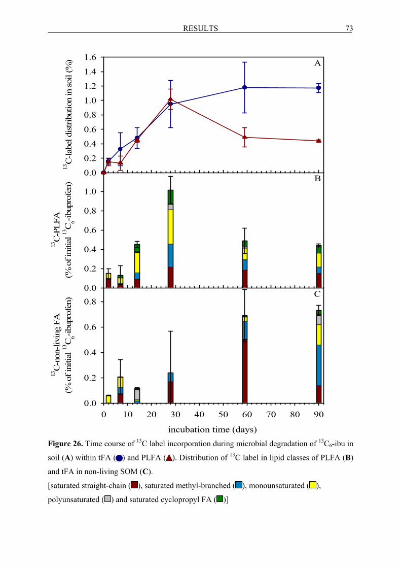
RESULTS 73
incubation time (days)
0 10 20 30 40 50 60 70 80 90
13C
-non
-livi
ng F
A(%
of i
nitia
l 13C
6-ib
upro
fen)
0.0
0.2
0.4
0.6
0.8
13C
-PLF
A(%
of i
nitia
l 13C
6-ib
upro
fen)
0.0
0.2
0.4
0.6
0.8
1.0
C
B
13C
-labe
l dis
tribu
tion
in so
il (%
)
0.0
0.2
0.4
0.6
0.8
1.0
1.2
1.4
1.6A
Figure 26. Time course of 13C label incorporation during microbial degradation of 13C6-ibu in
soil (A) within tFA ( ) and PLFA ( ). Distribution of 13C label in lipid classes of PLFA (B)
and tFA in non-living SOM (C).
[saturated straight-chain ( ), saturated methyl-branched ( ), monounsaturated ( ),
polyunsaturated ( ) and saturated cyclopropyl FA ( )]

RESULTS 74
On day 28, the 13C-PLFA reached their maximum (1% of 13C6-ibu equivalents). After 28
days, the 13C-PLFA decreased significantly and a 13C flux from the living part to the non-
living fraction of SOM was observed. After 59 days, the content of 13C-PLFA decreased to
50% of their maximum (~ 0.5% of 13C6-ibu equivalents), whereas the 13C-FA in the non-
living fraction were highest and reached 0.7% of the initial 13C6-ibu amounts in soil. After day
59, about of 60% of the label in the t-FA could be already assigned to the non-living SOM.
From this time, the amounts of both 13C-PLFA and 13C-FA in the non-living SOM were
nearly constant until the end of the experiment.
At the beginning of the incubation, the 13C label was found in the saturated straight
chain and in the monounsaturated PLFA (16:1ω7c, 16:1ω5c, 18:1ω9c and 18:1ω7c, Figure
26B, see also Table 13). From day 7 until day 28, the monounsaturated PLFA carried more 13C label than the other PLFA classes. During that time the amounts of 13C-monounsaturated
(in particular 16:1ω7c 18:1ω9c and 18:1ω7c) and also the 13C-saturated cyclopropyl (cy 17:0)
increased rapidly. However, incorporation of 13C into the saturated methyl branched PLFA
started already on day 7 and their contents increased strongly until day 28. On day 28, when
the amount of 13C-PLFA reached its maximum, the highest amount (except for 16:0) of the 13C label was found in the monounsaturated 16:1ω7c and 18:1ω7c and the cyclopropyl
cy 17:0. In addition, a high incorporation of 13C into the polyunsaturated 18:2ω6,9 and the
saturated methyl branched i:16:0 was also observed. After 59 days, the label was distributed
nearly equally within the monounsaturated PLFA and the saturated methyl branched PLFA,
whereas at the end of experiment, the monounsaturated PLFA contained more 13C than
saturated branched PLFA.
On day 59, since when 13C-FA were detected in the non-living SOM, saturated strain
chain FA carried most of 13C label (Figure 26C). From that time onwards, the saturated
methyl branched contained more 13C than monounsaturated FA. Finally, 13C-cyclopropyl FA
and 13C-polyunsaturated FA were also incorporated into the non-living SOM.

Table 13. The classification and composition of 13C label in respective PLFA during microbial degradation of 13C6-ibu in soil 13C-PLFA [10-4; % of 13C6-ibu] Incubation
time (days) 2 7 14 28 59 90 Saturated straight chain 14:0 n.d. 29.6 (± 98.6) 32.3 (± 8.4) 301.4 (± 12.5) 81.7 (± 94.7) 4.7 (± 0.8) 15:0 24.7 (± 12.4) 3,2 (± 24.2) 38.8 (± 4.3) 51.2 (± 30.6) 104.2 (± 85.3) n.d. 16:0 74.1 (± 138.1) 377.6 (± 655.4) 728.9 (± 82.5) 1361.3 (± 130) 1499 (± 719.4) 934.8 (± 171.2) 18:0 141.8 (± 345.1) n.d. 135.2 (± 47.7) 470.6 (± 14.9) 116.9 (± 158.7) 560.6 (± 126.6) 20:0 n.d. n.d. n.d. n.d. 70.2 (± 33.5) 22.6 (± 6.5) Saturated methyl branched i b 14:0 n.d.e n.d. 7.4 (± 0.8) 8,3 (± 0) 21.7 (± 13.0) 5.4 (± 2) i 15:0 n.d. n.d. 61.1 (± 5.4) 387 (± 64) 287.5 (± 188.3) 41 (± 136.4) a c 15:0 n.d. 2.8 (± 109.9) 160.8 (± 8.3) 190 (± 51.2) 15.1 (± 100.2) 12 (± 84.7) i 16:0 n.d. n.d. 74.5 (± 9.2) 523.9 (± 48) 57.9 (± 98.6) 48.8 (± 55.6) i 17:0 n.d. 20.4 (± 125.7) 41.7 (± 19.4) 148.2 (± 27.7) 99.4 (± 96.8) 61.5 (± 52) a 17:0 3.2 (± 3.4) 31.1 (± 116) 19.2 (± 30.9) 110.5 (± 30.4) 253.4 (± 249.6) 141 (± 16.7) 10 Me d 16:0 n.d. n.d. 120.5 (± 11.8) 382.3 (± 48.6) 191.1 (± 164.5) 278.5 (± 54.5) 10 Me 17:0 n.d. n.d. n.d. n.d. n.d. 16.4 (± 4) 10 Me 18:0 n.d. 54.7 (± 98.9) 76.8 (± 0.9) 231.6 (± 26) 95.5 (± 57.6) n.d. 18:0 br f n.d. 18.5 (± 140.7) 72.5 (± 33.9) 193.8 (± 8) 62 (± 46.4) 55.6 (± 24.4) Monounsaturated 16:1ω7cc 359 (± 28.4) 196.7 (± 10.1) 855.7 ↑ (± 41.9) 1041 ↑ (± 98.7) 232.5 (± 162.2) 49 (± 83.6) 16:1ω5c 66.2 (± 9.7) n.d. 25.1 (± 13.8) 242.3 (± 63.3) n.d. 39.3 (± 57.4) 18:1ω9c 89.5 (± 181.5) 136 (± 371.8) 189.3 ↑ (± 92.5) 539.6 ↑ (± 76.9) 28.1 (± 219) 473.8 (± 104.1) 18:1ω7c 67.6 (± 243.3) 179.4 (± 358.9) 971.8 ↑ (± 47) 1679 ↑ (± 377.3) 872.2 (± 378) 861 (± 64.8) 18:1ω5c n.d. n.d. 57.7 (± 0) 68.5 (± 45) n.d. 18.7 (± 3.9) Monounsaturated branched 17:1 br n.d. n.d. 29.7 (± 13.1) 217.7 (± 13.1) n.d. 10.9 (± 72.7) Polyunsaturated 18:2 n.d. 4.4 (± 209.4) n.d. n.d. n.d. n.d. 18:2ω6,9 n.d. 18 (± 481.9) 78.3 (± 79.1) 525.1 (± 3.7) 188.2 (± 204.3) n.d. Saturated cyclopropyl cy 17:0 n.d. 153.5 (± 169.4) 649 ↑ (± 36.4) 944.5 ↑ (± 238.8) 317.3 (± 153) 616.7 (± 107.5) cy 19:0 n.d. 96.4 (± 249.9) 89 (± 45.8) 550.7 (± 75) 294.1 (± 245.7) 145.2 (± 75.8) TOTAL 1498.2 (± 509) 1325 (± 980.1) 4515.7 (± 312.4) 10168 (± 1416.6) 4887.5 (± 1322.2) 4397.6 (± 168.7)
aa- anteiso; bbr – branched; cc - cis isomer; di – iso; e n.d. - not detectable; fMe - methyl; values in brackets (±) represent the standard deviation of the average of triplicates; values printed in bold represent characteristic values, arrows visualise increase or decrease of the respective FA compared to the preceeding sampling time
75

RESULTS 76
6.2.3.3 Formation of AA and their fate in soil incubated with 13C6-ibu
The incorporation of 13C into the living biomass AA fraction (bioAA) started later (day 7, see
Figure 27) than into PLFA (day 2). A flux of 13C-bioAA to the non-living SOM fraction was
already detected on day 7 and continued throughout the incubation. The amounts of 13C-bioAA reached a maximum on day 28 (~ 3.2% of 13C6-ibu equivalents) and then declined
gradually until the end of the experiment, whereas the contents of 13C-AA in the non-living
fraction remained stable already after 58 days. The content of 13C-bioAA decreased to 23% of
its maximum, reaching on day 90 about 0.7% of the initially added 13C6-ibu. At the end (day
90), 93% of the total 13C-AA in soil was stabilised in the non-living SOM pool, reaching
finally 27% of 13C6-ibu equivalents.
Figure 27. Time course of 13C label incorporation within tAA ( ), bioAA ( ) and bioAA ( )
recalculated based on 40% extraction efficiency during microbial degradation of 13C6-ibu in
soil On day 7, only Asp carried 13C (see Table 14A). After 14 days, the 13C-Asp dominated (0.4%
of 13C6-ibu equivalents) over other bioAA. However, Gly and Lys (each ~ 0.2% of 13C6-ibu
equivalents) also contained high amount of 13C. Only traces of 13C were detected in Thr and
Ile. When the amounts of 13C-bioAA were highest (day 28), both 13C-Asp (0.9% of 13C6-ibu
equivalents) and 13C-Glu (0.8% of 13C6-ibu equivalents) dominated, but the label was also
incorporated to a lesser extent into other AA (e.g. Gly, Thr, Leu, Ile, Pro, Glu, Phe and Lys).
Interestingly, at that time, a very low amount of 13C in the nonprotein amino acid ß-Ala
(0.008% of 13C6-ibu equivalents) was found. On day 59, when the contents of two
incubation time (days)
0 20 40 60 80
13C
-labe
l dis
tribu
tion
in so
il (%
)
0
5
10
15
20
25
30
35
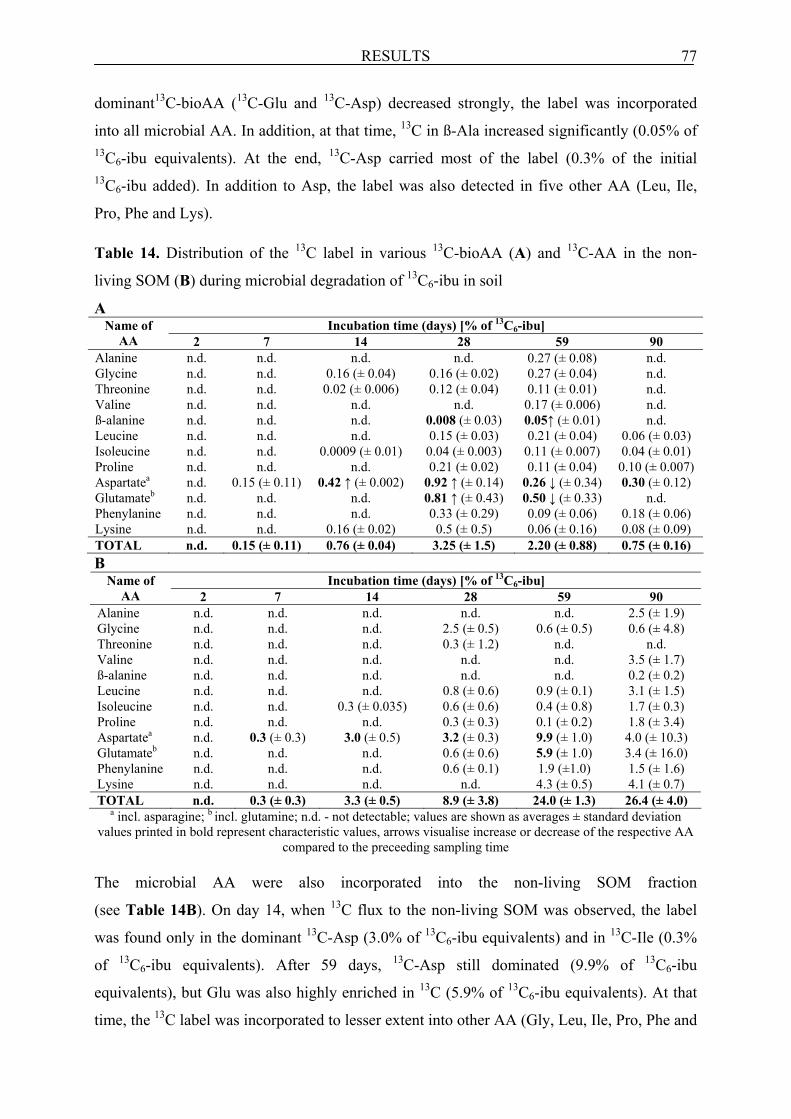
RESULTS 77
dominant13C-bioAA (13C-Glu and 13C-Asp) decreased strongly, the label was incorporated
into all microbial AA. In addition, at that time, 13C in ß-Ala increased significantly (0.05% of 13C6-ibu equivalents). At the end, 13C-Asp carried most of the label (0.3% of the initial 13C6-ibu added). In addition to Asp, the label was also detected in five other AA (Leu, Ile,
Pro, Phe and Lys). Table 14. Distribution of the 13C label in various 13C-bioAA (A) and 13C-AA in the non-
living SOM (B) during microbial degradation of 13C6-ibu in soil
A Incubation time (days) [% of 13C6-ibu] Name of
AA 2 7 14 28 59 90 Alanine n.d. n.d. n.d. n.d. 0.27 (± 0.08) n.d. Glycine n.d. n.d. 0.16 (± 0.04) 0.16 (± 0.02) 0.27 (± 0.04) n.d. Threonine n.d. n.d. 0.02 (± 0.006) 0.12 (± 0.04) 0.11 (± 0.01) n.d. Valine n.d. n.d. n.d. n.d. 0.17 (± 0.006) n.d. ß-alanine n.d. n.d. n.d. 0.008 (± 0.03) 0.05↑ (± 0.01) n.d. Leucine n.d. n.d. n.d. 0.15 (± 0.03) 0.21 (± 0.04) 0.06 (± 0.03) Isoleucine n.d. n.d. 0.0009 (± 0.01) 0.04 (± 0.003) 0.11 (± 0.007) 0.04 (± 0.01) Proline n.d. n.d. n.d. 0.21 (± 0.02) 0.11 (± 0.04) 0.10 (± 0.007) Aspartatea n.d. 0.15 (± 0.11) 0.42 ↑ (± 0.002) 0.92 ↑ (± 0.14) 0.26 ↓ (± 0.34) 0.30 (± 0.12) Glutamateb n.d. n.d. n.d. 0.81 ↑ (± 0.43) 0.50 ↓ (± 0.33) n.d. Phenylanine n.d. n.d. n.d. 0.33 (± 0.29) 0.09 (± 0.06) 0.18 (± 0.06) Lysine n.d. n.d. 0.16 (± 0.02) 0.5 (± 0.5) 0.06 (± 0.16) 0.08 (± 0.09) TOTAL n.d. 0.15 (± 0.11) 0.76 (± 0.04) 3.25 (± 1.5) 2.20 (± 0.88) 0.75 (± 0.16) B
Incubation time (days) [% of 13C6-ibu] Name of AA 2 7 14 28 59 90
Alanine n.d. n.d. n.d. n.d. n.d. 2.5 (± 1.9) Glycine n.d. n.d. n.d. 2.5 (± 0.5) 0.6 (± 0.5) 0.6 (± 4.8) Threonine n.d. n.d. n.d. 0.3 (± 1.2) n.d. n.d. Valine n.d. n.d. n.d. n.d. n.d. 3.5 (± 1.7) ß-alanine n.d. n.d. n.d. n.d. n.d. 0.2 (± 0.2) Leucine n.d. n.d. n.d. 0.8 (± 0.6) 0.9 (± 0.1) 3.1 (± 1.5) Isoleucine n.d. n.d. 0.3 (± 0.035) 0.6 (± 0.6) 0.4 (± 0.8) 1.7 (± 0.3) Proline n.d. n.d. n.d. 0.3 (± 0.3) 0.1 (± 0.2) 1.8 (± 3.4) Aspartatea n.d. 0.3 (± 0.3) 3.0 (± 0.5) 3.2 (± 0.3) 9.9 (± 1.0) 4.0 (± 10.3) Glutamateb n.d. n.d. n.d. 0.6 (± 0.6) 5.9 (± 1.0) 3.4 (± 16.0) Phenylanine n.d. n.d. n.d. 0.6 (± 0.1) 1.9 (±1.0) 1.5 (± 1.6) Lysine n.d. n.d. n.d. n.d. 4.3 (± 0.5) 4.1 (± 0.7) TOTAL n.d. 0.3 (± 0.3) 3.3 (± 0.5) 8.9 (± 3.8) 24.0 (± 1.3) 26.4 (± 4.0)
a incl. asparagine; b incl. glutamine; n.d. - not detectable; values are shown as averages ± standard deviation values printed in bold represent characteristic values, arrows visualise increase or decrease of the respective AA
compared to the preceeding sampling time The microbial AA were also incorporated into the non-living SOM fraction
(see Table 14B). On day 14, when 13C flux to the non-living SOM was observed, the label
was found only in the dominant 13C-Asp (3.0% of 13C6-ibu equivalents) and in 13C-Ile (0.3%
of 13C6-ibu equivalents). After 59 days, 13C-Asp still dominated (9.9% of 13C6-ibu
equivalents), but Glu was also highly enriched in 13C (5.9% of 13C6-ibu equivalents). At that
time, the 13C label was incorporated to lesser extent into other AA (Gly, Leu, Ile, Pro, Phe and

RESULTS 78
Lys). At the end, all 13C-microbial AA except for Thr were partly stabilised in the non-living
part of SOM and the label was distributed within these AA nearly equally.
6.2.3.4 Biogenic residues in soil incubated with 13C6-ibu
The content of 13C-tFA reached 1.2% of 13C6-ibu equivalents at the end of the experiment;
considering a conversion factor of 20 we can conclude that at least 24% of 13C label from 13C6-ibu went through the microbial biomass. From the amount of 13C-tAA of 27% (of 13C6-ibu equivalents) at the last day, we can estimate that there were 54% biogenic residues in
this experiment (using the conversion factor of 2).

DISCUSSION 79
7 DISCUSSION
The soil biodegradation studies with 13C6-2,4-D and 13C6-ibu proved that microorganisms
used C directly from these contaminants or indirectly via CO2 fixation to form their biomass
components. After the death of microorganisms, their biomass components were incorporated
into SOM and finally formed biogenic residues, which lead to the overestimation of the risk
related to NER formed during biodegradation of these contaminants in soils.
The simple liquid culture experiment with C. necator JMP 134 provided a general view
on the processes of 13C-FA and 13C-AA formation during 2,4-D biodegradation. In addition,
this experiment showed the relative abundance of FA and AA in the total biomass of this
strain, necessary for an estimation of the total biogenic residues content in the soil
experiments.
7.1 Liquid culture experiments
Two liquid culture experiments with 13C6-2,4-D and with 13CO2 showed that the bacterial
strain C. necator JMP 134 used the 13C derived either from this herbicide or from 13CO2 for
its PLFA and AA synthesis. In addition, the abiotic systems clearly showed that 13C label
incorporation from labelled 2.4-D or from CO2 were relevant only for biotic incubation
experiments. 13C6-2,4-D was degraded rapidly and dissipated already after the first day of
incubation. Therefore, 13C label incorporation into PLFA and AA directly from the labelled
2,4-D was observed only until day 1. In the later phase of experiment, when neither 2,4-D nor
its known aromatic metabolites were detected in the medium, 13C was incorporated into
PLFA and AA only via CO2 fixation.
Incorporation of 13C into biomass of C. necator JMP 134 grown on 2,4-D medium
The 13C label incorporation into the biomass of C. necator JMP 134 in the 13C6-2,4-D
experiment started immediately and remained constant until day 3. Thereafter, the 13C content
in the biomass still increased, although already after the first day of incubation 13C6-2,4-D
was completely depleted and no biomass growth was observed (see results in section 5.1.1.1
and 5.1.2.1). This is contrary to the results in the 13C6-2,4-D experiment presented by LERCH
ET AL. (2007), which indicated a low content of 13C in the biomass of C. necator JMP 134 at
the beginning and its strong increase in the later phase of incubation. This biomass growth
was also consistent with the OD measurement of the culture (LERCH ET AL., 2007). However,
in their experiment the initial concentration of 2,4-D was much higher (250 mg/L) than in the
present study (77 mg/L) and this herbicide was still detectable after the first day of incubation.

DISCUSSION 80
Although the amounts of 13C label in PLFA increased continuously until day 7, this however,
was not related to growth. At the beginning of incubation, the 13C-monounsaturated PLFA
were dominant over other PLFA, their amounts then decreased strongly, whereas the content
of 13C in saturated cyclopropyl PLFA increased rapidly throughout the incubation time. The
cyclopropyl PLFA are known starvation biomarkers, formed by the methylation of the double
bond in monounsaturated PLFA (KAUR ET AL., 2005). This modification makes these FA
more stable and minimizes the membrane lipid losses under stress conditions (KAUR ET AL.,
2005). Therefore, this mechanism was employed by C. necator JMP 134 in the absence of
2,4-D in the medium. The continuous increase of the 13CO2 evolution observed until day 7 in
the 13C6-2,4-D study and of the 13C incorporation into PLFA in the 13CO2 fixation experiment
until day 3 clearly indicates the continuous metabolisation of 13C incorporated into biomass
residues via CO2 fixation. The presence of CO2 is reported to be essential for the growth of
many microorganisms (KREBS, 1941), since CO2 plays a key role in the synthesis of the
tricarboxylic acid cycle (TCC) intermediates malate and oxalacetate from pyruvate or
phosphoenolpyruvate (MILTNER ET AL., 2004; FEISTHAUER ET AL., 2008; see Figure 28). At
the end of the incubation, the contents of 13C in PLFA decreased strongly in both experiments
and the 13C-saturated cyclopropyl PLFA were dominant over other PLFA (in particular in the 13CO2 experiment), indicating the decreasing contribution of CO2 fixation.
At the beginning of the 13C6-2,4-D experiment 13C was highest in Glu. This AA is
synthesised from 2-oxoglutarate formed in the TCC (see Figure 28). At that time, Asp also
contained significant amounts of 13C in comparison to other 13C-AA. Asp is derived from
oxaloacetate, which is the acceptor molecule for acetate in the TCC (MICHAL, 1999). When
oxaloacetate is removed from the TCC for Asp synthesis, it has to be replenished by various
anaplerotic reactions, which include the carboxylation of phosphoenolpyruvate; thus Asp is a
direct product of heterotrophic CO2 fixation (FEISTHAUER ET AL., 2008; MILTNER ET AL.,
2004). This is in good accordance to the 13CO2 experiment, in which the 13C label was found
only in Asp at the beginning (day 2), when the CO2 fixation started. Both Glu and Asp formed
within the TCC are important precursors for other AA, Pro and Thr, Ile, Lys, respectively
(arginine and methionine were not detected in experiments). Therefore, these 13C-labelled AA
were found in the later phase of incubation period. Interestingly, the contents of 13C in Thr
increased strongly from day 3 until day 7 in both experiments and then decreased rapidly at
the end. The simultaneous decline of 13C in both Thr and PLFA at the end thus indicates the
decrease of CO2 fixation contribution to 13C incorporation into the biomass of C. necator.
Other AA, such as Ala, Val and Leu formed from pyruvate and Phe from

DISCUSSION 81
phosphoenolpyruvate were also enriched in the later phase of incubation period. However, the
trends of both decline and increase of their 13C contents could be assigned to the different
food demands of this bacterial strain throughout the incubation time. The low incorporation of 13C into Ser found only at the end of 13C6-2,4-D experiment demonstrates the formation of
this AA from 13C-Gly. The 13C-Gly was presumably converted directly from glyoxylate,
which is a cleavage product of isocitrate (MICHAL, 1999) or of 2,4-D metabolite (see Figure
10 in section 4.1).
At the end of the 13C6-2,4-D incubation experiment, ~ 17% of initially added 13C6-2,4-D
was detected in the total biomass of C. necator JMP 134, 8% was found in AA and 0.6% was
incorporated into the PLFA. However, also a relatively high amount of 13C was incorporated
into the biomass of C. necator via CO2 fixation (~ 4.2% of the inital 13CO2 amount).
Therefore, it should be kept in mind that CO2 fixation can contribute significantly to the
incorporation of C into biomass components and thus to the NER formation in soils. In
addition, the results from both 13C6-2,4-D and 13CO2 incubation experiments showed that 13C-labelled AA represented ~ 50% of the total 13C in the biomass, demonstrating their
relatively high abundance in microbial cells. Therefore, this microbial biomarker can be
helpful in the quantification of biogenic residues in complex soil systems.

DISCUSSION 82
Figure 28. Scheme of the relevant pathways for AA and FA synthesis in TCC and glycolysis;
and the anaplerotic reactions ( ) replenishing the TCC (adapted from MICHAL, 1999;
FEISTHAUER ET AL., 2008)
Phenylalanine
TCC
Glyoxylate
Succinate
Oxaloacetate
Malate
Citrate
Isocitrate
2-oxoglutarate
Glutamate Glutamine
Proline Arginine
Aspartate Asparagine Threonine Isoleucine
Lysine Methionine
Alanine Valine
Leucine
Pyruvate
Acetyl-CoA
Glycine
Serine
Even numbered FA
Odd numbered FA
Propionyl-CoA
Phosphoenolpyruvate
3-phosphoglycerate
Glucose
Gly
coly
sis
CO2 fixation

DISCUSSION 83
7.2 Soil experiments
Two soil degradation experiments with 13C6-2,4-D and 13C6-2,4-ibu prove for the first time
that biogenic residues formation can be a relevant process during microbial degradation of
many organic contaminants in soil. In case of the readily biodegradable 2,4-D and Ibu, the
majority of NER are formed from biomass components. However, the kinetics of biogenic
residue formation in the biodegradation experiments with these various organic contaminants
was different and depended strictly on the degradation kinetics in soil.
7.2.1 Soil incubation experiment with 13C6-2,4-D
The results from the soil incubation experiment with 13C6-2,4-D provide detailed insight into
the C flux from the 13C-labelled pollutant 2,4-D or from labelled CO2 (evolved during 2,4-D
mineralisation) via microbial biomass to non-living SOM. It has been clearly shown that
microorganisms use the readily available C derived from 2,4-D for their biomass synthesis,
which was proven by identifying the 13C label within the biomarkers FA and AA. After cell
lysis and the decay of microbial biomass, both 13C-labelled PLFA and bioAA were distributed
within the microbial food web and incorporated into the non-living fraction of SOM, which
was finally stabilised. These results are also in good agreement with recent studies on the fate
of 13C labelled E. coli in soil (KINDLER ET AL., 2006, 2009; MILTNER ET AL., 2009), which
showed that biomass-derived C was distributed within the microbial food web and finally
contributed to the formation of refractory non-living SOM (KINDLER ET AL., 2006, 2009;
LÜDERS ET AL., 2006). The majority of NER formed during microbial degradation of 2,4-D in
soil was composed of biomass-derived components.
7.2.1.1 Biogenic residues as NER in soil incubated with 13C6-2,4-D
At the end of the incubation experiment, the amount of 13C found in tAA (non-living SOM +
bioAA fraction) was high and reached 22% of the initially added 13C6-2,4-D equivalents
corresponding to 61% of the NER based on isotope mass balance. It should be kept in mind
that these AA do not represent free AA, which are reported to be degraded very rapidly in soil
(JONES, 1999; VINOLAS ET AL., 2001), but they were hydrolysed from peptides and proteins.
However, it is impossible to extract all biomolecules from soil microbial biomass, therefore
only part of them could be analysed. By the extraction of some known microbial
biomolecules e.g. FA and AA, we can estimate the actual amounts of biogenic residues in
soil. The particular focus however, has been laid on AA from proteins and peptides due to
their high abundance in microbial biomass. Two experiments with C. necator JMP 134 grown
on 2,4-D medium showed that 13C-labelled AA represented ~ 50% of the total 13C in the

DISCUSSION 84
biomass (see results in sections 6.1.1.1 and 6.1.2.1). In addition, the ratio of 13C-AA to total 13C in biomass was relatively stable at the different sampling dates; thus AA are relevant for
estimating the NER content in soil. Therefore, both the results from this experiment and a
conservative conversion factor of 2 (MADIGAN AND MARTINKO, 2006) was considered for
quantification of the actual incorporation of C derived from 2,4-D into the biomass, which
resulted in a total amount of 44% (of 13C6-2,4-D equivalents) for biogenic residues. The
amount of these residues is close to the total NER amount of ~ 36% (of 13C6-2,4-D
equivalents) in soil measured by GIRARDI ET AL. (submitted; see also results in section
6.2.1.1). This small discrepancy in the NER contents could be related to the different methods
used for NER determination. NER are generally quantified as CO2 released from combusted
soil after the pollutant residues extraction (BARRIUSO ET AL., 2008; WAIS, 1998). This
analytical approach was also implemented by GIRARDI ET AL. (submitted). 2,4-D residues
were extracted from soil by ASE, which is a harsh extraction method (NORTHCOTT AND
JONES, 2000). At the end of experiment, about 8% of the initial 13C6-2,4-D equivalents was
extracted from soil by ASE, but from that amount only 0.5% was identified as 2,4-D.
Therefore, a small part of biogenic residues stabilised in SOM could be still extracted from
soil and thus affected the lower total NER content measured by GIRARDI ET AL. (submitted). It
should be noted that the hydrolysis of AA from proteins is a very efficient extraction method,
which enables also the extraction of AA stabilised in SOM; this could thus affect the higher
biogenic NER content than the total NER content. However, the estimated amounts of
biogenic residues indicate that the biomass compounds were finally converted into the
biogenic residues already after 32 days and represented the majority of 2,4-D-derived NER in
the soil (see Figure 29).
Figure 29. Scheme of the 13C conversion over microbial degradation of 13C6-2,4-D in soil.
A: conventional mass balance and B: new mass balance considering biogenic residues
formation. *The biogenic residues were estimated from AA using a conversion factor of 2
(for details see text).
0%
10%
20%
30%
40%
50%
60%
70%
80%
90%
100%
2 4 8 16 32 640%
10%
20%
30%
40%
50%
60%
70%
80%
90%
100%
2 4 8 16 32 64
Mineralisation Mineralisation
Biogenic residues*
Proteins
Non-biogenic NER
Extractable Extractable
NER
incubation time (days) incubation time (days)
B A
rela
tive
amou
nts o
f app
lied
2,4-
D (%
)

DISCUSSION 85
Studies with anthracene (KÄSTNER ET AL., 1999), phenanthrene (RICHNOW ET AL., 2000) and
TNT (WEISS ET AL., 2004) proved that C or N from these contaminants was incorporated into
soil microbial biomass, which contributed to the NER formation in soils. The NER amounts
formed during degradation of [ring-14C-U]-labelled 2,4-D or 2,4-DCP in non-sterile
composted straws were much higher than in sterile ones, where the parent compounds
remained unchanged (BENOIT AND BARRIUSO, 1997). The high NER content found in the
present study, where the 13C6-2,4-D was nearly mineralised already after 16 days (GIRARDI ET
AL., submitted) in comparison with the negligible mineralisation in sterile soils is in a good
accordance to those in non-sterile straws (BENOIT AND BARRIUSO, 1997). Therefore, high
contents of NER in non-sterile composted straw or soil underlines the key role of soil
microorganisms in the formation of residues from 2,4-D in native soil. Another soil
biodegradation experiment with [ring-14C-U] 2,4-D showed that the amount of 14C label from
this herbicide detected in the microbial biomass was similar to that observed for [1-14C]
glucose, suggesting the utilisation of 2,4-D as a C source (ROBERTSON AND ALEXANDER,
1994). In addition, the well-known 2,4-D soil degrader C. necator JMP 134 also used C from
2,4-D for their biomass components synthesis in the liquid culture experiments (see
discussion in section 7.1.1).
There is also evidence for abiotic residue formation, because 18.6% of 13C6-2,4-D
equivalents were recovered as NER in sterile soils at the end of incubation. However, in the
sterile soils, the biogenic residue representatives 13C-FA and 13C-AA were not formed,
indicating that these NER were exclusively formed by the reported abiotic sequestration
mechanisms (PIGNATELLO AND XING, 1996; ALEXANDER, 2000; GEVAO ET AL., 2000;
KATAYAMA ET AL., 2010). It was reported that autoclaving changes the soil physico-chemical
properties (SHAW ET AL., 1999; BERNS ET AL., 2008), therefore the contaminant behaviour in
sterile soils can be different than in native soils. In addition, the rapid mineralisation of 13C6-
2,4-D in the biotic soil experiment, will reduce the extent of abiotic NER formation because
rapid 2,4-D degradation competes with abiotic residue formation. The fact that the non-living
AA and FA can explain most of the NER formation strongly suggests a high contribution of
biotic NER formation. Several studies demonstrated that microorganisms can also mediate the
enzymatic covalent oxidative coupling of 2,4-DCP to humic substances (BOLLAG, 1991;
PALOMO AND BHANDARI, 2005, 2006). However, it should be noted that these studies were
limited to simple (BOLLAG, 1991; HATCHER ET AL., 1993), or abiotic systems (PALOMO AND
BHANDARI, 2005; 2006) and covalently bound 2,4-DCP in SOM has never been detected.

DISCUSSION 86
In addition, all available studies on NER formation during microbial degradation of 2,4-D in
soil are limited to quantitative analyses with radioactive tracers (BENOIT AND BARRIUSO,
1997; BOIVIN ET AL., 2005). In contrast to these available results, where no information on the
chemical nature of NER in SOM was presented, the present study shows that nearly all NER
formed during biodegradation of 13C6-2,4-D in soil were composed from microbial
components stabilised in the SOM pool during the later phase of experiment (aging process).
Finally, the high content of 13C-AA detected in the present study contradicts the
generally accepted view that the parent compounds or their primary metabolites ) are the main
precursors of phenoxyacetic acid herbicides derived NER, in particular via sorption processes
(BOLLAG ET AL., 1991; BOLLAG ET AL., 1992; PALOMO AND BHANDARI, 2005, 2006).
7.2.1.2 Fate and stability of FA in soil incubated with 13C6-2,4-D
The formation of 13C-PLFA during microbial degradation of 13C6-2,4-D in soil was rapid
during the first four days of incubation, since the amount of the readily available 13C6-2,4-D
in soil was still high (GIRARDI ET AL., submitted). Taking into consideration a relatively rapid
turnover of FA in bacterial cells and a total FA content of ~ 5% (see results in section
6.2.1.4), for the maximum of about 1% of 13C6-2,4-D equivalents detected in the tFA fraction
on the day 8, it can be concluded that more than 20% of the 13C initially added must have
passed through the microbial biomass. The high amount of 13C found in the monounsaturated
PLFA indicates that Gram-negative bacteria were initially involved in 2,4-D transformation.
This is in good accordance with the previous study reported by LERCH ET AL. (2009A), in
which the content of 13C in monounsaturated tFA fraction (including the PLFA) was highest.
The high incorporation of 13C label into even-numbered PLFA (such as: 16:1ω7c; 16:0;
18:1ω7c; 18:1ω9c; 16:1ω5c) in the initial phase of incubation experiment clearly indicates for
their direct synthesis from Acetyl-CoA (see Figure 28 in section 7.6.1), which is the end
product of 2,4-D degradation (see Figure 10 in section 4.1). In addition, a lower
incorporation of 13C within the saturated methyl-branched PLFA, which are typical for Gram-
positive bacteria, was also observed in this experiment. This indicates that this group of
bacteria took indirectly part in the transformation of 2,4-D via fixation of the CO2 evolved
during mineralization of this pollutant or by uptake of metabolites formed by Gram-negative
bacteria. When the amounts of 13C in monounsaturated PLFA started to decrease after 8 days,
the amount of 13C in saturated cyclopropyl PLFA was highest. This high of 13C content in the
cyclopropyl PLFA indicates that bacteria employed starvation mechanism (KAUR ET AL.,
2005), which agrees with the low residual concetrations of 2,4-D at that time (GIRARDI ET AL.,
submitted). In the later phase of the experiment, when the microbial biomass started to decay,

DISCUSSION 87
a flux of 13C from living cells to non-living SOM was observed. After 16 days, besides 13C-
PLFA of the decaying biomass, the 13C-FA of the non-living SOM were also continuously
metabolised at a low level and distributed within the food web. Therefore, when 13C6-2,4-D
was nearly depleted, the 13C released from these 13C-FA was thus rapidly recycled via CO2
fixation by Gram-negative and Gram-positive bacteria and by fungi.
Two independent studies showed that the NER contents formed during 2,4-D
biodegradation in soil (BOIVIN ET AL., 2005) and 15 years-old NER after fresh soil
amendment (LERCH ET AL., 2009B) decreased slightly over time. BOIVIN ET AL. (2005)
suggested that this decline could be related to the temporal immobilisation of 14C from 2,4-D
in the protoplasm of some soil microorganisms. The decrease of NER in these above-
mentioned studies thus could be assigned to the release of FA from both the decaying biomass
and biomass residues in the non-living SOM, as it was observed in the present study.
At the end of the 13C6-2,4-D soil incubation experiment, the amount of 13C-PLFA
decreased to 20% of its maximum value, indicating the metabolisation of cell components of
the initial 2,4-D degraders via the microbial food web. However, the 13C-FA in the non-living
SOM decreased to 50% of their maximum value, demonstrating that the compounds in non-
living fraction were partly stabilised in SOM against microbial attack. KINDLER ET AL. (2009)
also reported the decline of 13C-tFA derived from E. coli to 24% (of the initial amount of 13C-E. coli) in soil after eight months of incubation. In spite of the long incubation time, still
about 50% of microbial biomass C remained in soil (KINDLER ET AL., 2006), demonstrating
the high stability of microbial biomass residues in soil.
7.2.1.3 Fate and stability of AA in soil incubated with 13C6-2,4-D
Compared to PLFA, the incorporation of 13C into the bioAA fraction started 2 days later,
indicating that PLFA as components of microbial cell membranes are turned over faster and
thus received the 13C label earlier than the AA. However, the flux of 13C from the decaying
biomass AA fraction into the non-living SOM was higher than that observed for the PLFA. At
the beginning of the incubation, when the readily degradable 13C6-2,4-D was still present at
high amounts (GIRARDI ET AL., submitted), Glu carried highest label of all 13C-AA detected in
this experiment. However, on day 8, the content of 13C in Asp was highest compared to the
other 13C-AA. Glu is formed from 2-oxoglutarate derived from the TCC; see Figure 28 in
section 7.1.1. The dominant incorporation of 13C into Glu thus clearly indicates for a different
route of the label into AA in first days of incubation, which was followed by CO2 fixation
from mineralised CO2 and from decaying microbial biomass, as it was shown by the high
amount of 13C-Asp. Asp has been reported to be a direct product of heterotrophic CO2
DISCUSSION 88
fixation in soil (MILTNER ET AL., 2005), which is also in good accordance to the pure culture
experiment with C. necator JMP 134 grown in presence of 13CO2 atmosphere (see section
6.1.1). In addition, CO2 fixation as a relevant process for biogenic residue formation in the
later phase of 2,4-D degradation experiment was also proven by the detection of 13C label in
the PLFA of the 13CO2 soil experiment. Overall, both Asp and Glu are important precursors
for other AA, e.g. Glu for Pro and Asp for Lys and Ile (see Figure 28 in section 7.1.1).
Therefore, lower amounts of 13C-Lys, 13C-Ile and 13C-Pro were also detected later in this
experiment. On day 16, when only traces of readily available 2,4-D residues were left and 13C-bioAA started to decline, the 13C label also was distributed within Ala, Gly and Phe,
which would point to their biosynthesis via different intermediates of glycolysis (MADIGAN
AND MARTINKO, 2006; MICHAL, 1999).
At the end of the incubation, the content of the 13C-bioAA fraction decreased to 36% of
its maximum found in the experiment, indicating the decrease of the contribution of the living
biomass fraction to SOM pool over the incubation time. Surprisingly, the amounts of 13C-AA
in the non-living SOM from day 32 onwards remained stable and finally reached 19.8% of the
initial amount of 13C6-2,4-D in soil, demonstrating the stabilisation of proteins from the
decaying microbial biomass. MILTNER ET AL. (2009) also demonstrated the high stability of 13C-labelled bacterial proteins in the non-living part of SOM; their contents decreased only
marginally even over a period of several months. Proteins have been reported to be stabilised
in SOM by various sorption processes (KLEBER ET AL., 2007), which protect them from the
microbial degradation (RILLIG ET AL., 2007) and affect their stability for a very long period of
time (KNICKER ET AL., 1993; KÖGEL-KNABNER, 2002). These results thus underline the high
importance of AA in NER formation from 2,4-D due to their high abundance in the microbial
biomass and the stability in contrast to FA, which were turned over relatively fast.
In a study on the fractionation of [U-ring-14C] 2,4-D residues XIE ET AL. (1997) showed
that most of the radioactivity was associated with humic acids. It should be noted that proteins
can bind to humic acids by hydrophobic and ionic interactions, which protect them from
chemical and microbial attack (ZANG ET AL., 2000). Therefore, the dominance of 14C label in
the bound-humic acid fraction could be assigned to AA, as observed in the present study.
Moreover, in the soil experiments with 13C6-2,4-D, the amount of labelled biogenic residues
did not change after 32 days of incubation, which was caused by their stabilisation in SOM.
This could also explain the constant mineralisation rate of 90-years-old NER after addition of
the fresh soil reported by LERCH ET AL. (2009B), when taking into consideration the fact that
these NER are mostly biogenic.

DISCUSSION 89
7.2.2 Soil incubation experiment with 13C6-ibu
This part of the study provides insight into the C flux from 13C6-ibu during its biodegradation
via microbial biomass components to the non-living part of SOM. The microorganisms used
the C derived from this contaminant to synthesise their biomass, as it was shown by 13C
incorporation into the microbial biomarkers FA and AA. After cell death, biomass
constituents such as 13C-PLFA and 13C-bioAA from the decaying cells were distributed into
the food web and finally incorporated into the non-living SOM fraction. These results are in
good agreement with the studies on the fate of both labelled E. coli in soil (KINDLER ET AL.,
2006, 2009; MILTNER ET AL., 2009) and 13C-biogenic residue in the soil incubated with 13C6-2,4-D (NOWAK ET AL., in press). However, the kinetics of biogenic residues formation in
soil incubated with 13C6-ibu was initially slower than that observed in the 13C6-2,4-D
experiment. Nevertheless, biomass-derived compounds such as FA and AA also contributed
significantly to NER formation from13C6-ibu in soil. Biomass residues represented the
majority of 13C6-ibu-derived NER, which is also consistent with the 13C6-2,4-D experiment.
7.2.2.1 Biogenic residues as NER in soil incubated with 13C6-ibu
The content of 13C-tAA detected at the end of the 13C6-ibu experiment was high, reaching
27% of the initial 13C6-ibu equivalents and corresponding to 90% of the NER based on
isotope mass balance. Considering a conservative conversion factor of 2 (for details on the
conversion factors see section 6.1.1.1), these 27% in 13C-tAA corresponds to a total amount
of 54% (of 13C6-ibu equivalents) for biogenic residues. However, the total 13C-NER content
measured by GIRARDI ET AL (submitted; see also section 6.2.3.1) was much lower (30% of 13C6-ibu equivalents) than the estimated amount of biogenic residues. It should be noted that
the total NER content was determined by EA-C-irMS after the removal of Ibu residues using
a harsh extraction method (ASE). The estimated amount of NER strongly depends on the
extraction method used for the bioavailability measurement of a target compound (KHAN
1991; NORTHCOTT AND JONES, 2000; MORDAUNT ET AL., 2005). GIRARDI ET AL. (submitted;
see also results in section 6.2.3.1) reported that the solvent bulk extractable amount prior to
purification by SPE accounted for ~ 14% of 13C6-ibu equivalents; and from that only 0.5%
was identified as Ibu. In addition, the contents of the bulk solvent extractable varied over
time. At the beginning of biogenic residue formation, a high content of the solvent bulk
extractable (~ 50% of 13C6-ibu equivalents on the day 28) was observed, which decreased
strongly at the end; this could be assigned to the aging processes of NER. Therefore, the
~ 14% extracted at the end from soil prior to quantitative NER analyses could still contain a
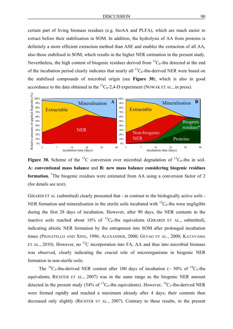
DISCUSSION 90
certain part of living biomass residues (e.g. bioAA and PLFA), which are much easier to
extract before their stabilisation in SOM. In addition, the hydrolysis of AA from proteins is
definitely a more efficient extraction method than ASE and enables the extraction of all AA,
also those stabilised in SOM, which results in the higher NER estimation in the present study.
Nevertheless, the high content of biogenic residues derived from 13C6-ibu detected at the end
of the incubation period clearly indicates that nearly all 13C6-ibu-derived NER were based on
the stabilised compounds of microbial origin (see Figure 30), which is also in good
accordance to the data obtained in the 13C6-2,4-D experiment (NOWAK ET AL., in press).
Figure 30. Scheme of the 13C conversion over microbial degradation of 13C6-ibu in soil.
A: conventional mass balance and B: new mass balance considering biogenic residues
formation. *The biogenic residues were estimated from AA using a conversion factor of 2
(for details see text). GIRARDI ET AL (submitted) clearly presented that - in contrast to the biologically active soils -
NER formation and mineralisation in the sterile soils incubated with 13C6-ibu were negligible
during the first 28 days of incubation. However, after 90 days, the NER contents in the
inactive soils reached about 16% of 13C6-ibu equivalents (GIRARDI ET AL., submitted),
indicating abiotic NER formation by ibu entrapment into SOM after prolonged incubation
times (PIGNATELLO AND XING, 1996; ALEXANDER, 2000; GEVAO ET AL., 2000; KATAYAMA
ET AL., 2010). However, no 13C incorporation into FA, AA and thus into microbial biomass
was observed, clearly indicating the crucial role of microorganisms in biogenic NER
formation in non-sterile soils.
The 14C3-ibu-derived NER content after 100 days of incubation (~ 50% of 14C3-ibu
equivalents; RICHTER ET AL., 2007) was in the same range as the biogenic NER amount
detected in the present study (54% of 13C6-ibu equivalents). However, 14C3-ibu-derived NER
were formed rapidly and reached a maximum already after 4 days; their contents then
decreased only slightly (RICHTER ET AL., 2007). Contrary to these results, in the present
incubation time (days)
0%
10%
20%
30%
40%
50%60%
70%
80%
90%
100%
2 7 14 28 59 90
Extractable
NER
Mineralisation A
incubation time (days)
0%
10%
20%
30%
40%
50%
60%
70%
80%
90%
100%
2 7 14 28 59 90
Mineralisation Extractable
Biogenic residues*
Proteins Non-biogenic NER
B
Rel
ativ
e am
ount
s of a
pplie
d ib
upro
fen
(%)

DISCUSSION 91
13C6-ibu experiment, the NER reached a maximum after 59 days and their contents remained
stable until the end of the incubation period. This is related to the different labelling positions
of the compound used in these two studies. In the present study, the compound was labelled
in the aromatic ring, which is cleaved much later during biodegradation than the methyl group
labelled in the 14C3-ibu experiment is removed (RICHTER ET AL., 2007), which is reflected by
different kinetics of NER formation. NER in the 13C6-2,4-D experiment were also formed
very quickly (NOWAK ET AL., in press; see also section 7.2.1.1), but this contaminant was
degraded much faster than 13C6-ibu (GIRARDI ET AL., submitted). This was caused by the
ability of the present soil microorganisms to degrade 2,4-D after previous 2-methyl-4-chloro
phenoxyacetic acid (MCPA) application to the soil (INES MERBACH, personal
communication), which also belongs to the group of phenoxy compounds as 2,4-D.
7.2.2.2 Fate and stability of FA in soil incubated with 13C6-ibu
Incorporation of the 13C-label into PLFA started immediately and increased rapidly until day
28, when the readily available 13C6-ibu was still present at relatively high concentrations in
the soil (GIRARDI ET AL., submitted; see also section 6.2.3.1). The monounsaturated PLFA
carried most of the 13C label and dominated over other 13C-labelled PLFA until day 28,
indicating that Gram-negative bacteria were important initial Ibu degraders in soil. This is in
good accordance to the study on NER formation from 13C6-2,4-D in soil (NOWAK ET AL., in
press; see section 7.2.1.2). As the same soil was used in these two experiments, the same
groups of microorganisms might have been involved in the degradation of both acidic
aromatic molecules 13C6-ibu and 13C6-2,4-D. In the later phase of incubation period, also the
saturated methyl branched PLFA were highly enriched in 13C and their importance increased
over time, in particular when only traces of 13C6-ibu were left. This class of PLFA is typical
for Gram-positive bacteria, which indirectly take part in the degradation of 13C6-ibu, as it was
also demonstrated previously in the 13C6-2,4-D soil experiment. The observed continuous
increase of 13C incorporation into the cyclopropyl PLFA throughout the incubation time
indicates starvation of the microorganisms (KAUR ET AL., 2005). In addition, the continuous
decline of 13C-PLFA in the living biomass, the distribution of 13C within all classes of PLFA
and the contaminant depletion in soil over the incubation period indicated that the Ibu-derived
C was distributed within the food web, as found in the 13C6-2,4-D study. In addition to
bacteria, fungi also took part in the turnover of 13C-labelled PLFA from dead cells, as proven
by the 13C found in the fungal biomarker PLFA 18:2ω6,9. From the content of 13C-tFA of
1.2% (of 13C6-ibu equivalents) detected at the end of experiment and considering a conversion
factor of 20 (for details see 6.1.1.1) we can conclude that at least 24% of biomass must have

DISCUSSION 92
been formed. This is close to the amount found in the 2,4-D soil incubation experiment (22%
of 13C6-2,4-D equivalents).
At the end of the experiment, 13C-PLFA decreased to 50% of their maximum, whereas 13C-FA in the non-living SOM fraction remained unchanged. This is contradictive to the
study with 13C6-2,4-D, where apart from the observed decline of 13C-labelled PLFA to
20% (of 13C6-2,4-D equivalents), the contents of 13C-FA in the non-living fraction also
decreased (50% of 13C6-2,4-D equivalents; see section 6.2.1.2). However, 13C6-ibu dissipated
slower than 13C6-2,4-D. The PLFA thus received the 13C label much later. Therefore, both
their decline and 13C flux to the non-living fraction were also observed later than in the 13C6-2,4-D experiment. Additionally, both the lower decrease of 13C-PLFA and the stability of 13C-FA in the non-living SOM until the end of 13C6-ibu experiment may be related to the
duration of the incubation, which might have been stopped before destabilisation of part of
Ibu residues started, as it was seen in the 2,4-D study. In addition, RICHTER ET AL. (2007) also
reported a slight release of rapidly formed 14C-NER derived from 14C3-ibu over the incubation
time (~ 5% of 14C3-ibu equivalents after 100 days).
7.2.2.3 Fate and stability of AA in soil incubated with 13C6-ibu
AA in biomass-living fraction received 13C label later than PLFA, which is in good agreement
with the data obtained in the experiment with 13C6-2,4-D (NOWAK ET AL., in press; see section
7.2.1.3). However, 13C-bioAA derived from 13C6-ibu started to decline at the same time as 13C-PLFA, what is different from the soil experiment with 13C6-2,4-D, where the decrease of 13C-bioAA content was observed later than that of 13C-PLFA. This discrepancy in the decline
of bioAA is difficult to explain and could be related to the different dissipation kinetics of the
used contaminants and also to timing of the sampling events relative to proceses in soil in
these two soil incubation experiments. After 58 days, the contents of 13C-AA in the non-living
fraction derived from 13C6-ibu did not change and remained stable until the end, whereas the 13C-bioAA decreased to 23% of their maximum after 90 days (of 13C6-ibu equivalents). This
is in good accordance to the 13C6-2,4-D soil experiment, where 13C-AA in the non-living
SOM were also constant, indicating their high importance in the NER formation.
In the initial phase of 13C6-ibu degradation, the label was incorporated only into Asp.
Thereafter, the content of 13C-Asp increased significantly and this AA was dominant until day
28, when 13C6-ibu was present at a low concentration in soil (GIRARDI ET AL., submitted; see
also 6.2.3.1). The fast appearance of the label in Asp and its later dominance over the other 13C-bioAA suggest that ibu-derived 13C is incorporated into the bioAA via heterotrophic CO2
fixation (MILTNER ET AL., 2004; FEISTHAUER ET AL., 2008). After 14 days, a lower amount of

DISCUSSION 93
13C was found also in Lys and Ile, for which Asp is an important precursor. This however is,
in contrast to the study with 13C6-2,4-D, where the label was first incorporated into Glu, at the
time when the amount of readily available 13C6-2,4-D in soil was still high. Although the
molecular structures of 13C6-ibu and 13C6-2,4-D are similar, these compounds are degraded by
soil microorganisms via different pathways. In the soil incubated with 13C6-ibu, 13C-Glu was
also formed, but later, when the 13C-bioAA started to decline and 13C6-ibu was nearly
depleted. Interestingly, on days 28 and 58, the non-protein AA 13C-ß-Ala was found. The
ratio of Asp to its decomposition product ß-Ala informs about the intensity of SOM
decomposition (DAUWE AND MIDDELBURG, 1998). The ratio of Asp/ß-Ala on day 28 was 121,
whereas on day 58 it decreased to 5.4, which clearly shows that the 13C-AA in the microbial
biomass were decaying. The high contents of 13C-Glu found in the later phase of the
experiment would thus point to a different degradation pathway of ibu-derived 13C.
Furthermore, similar to the 13C6-2,4-D study, in the later phase of 13C6-ibu-amended soil
incubation, a lower label incorporation into other AA (Pro, Ala, Gly, Thr, Leu, Phe, and Val)
was also observed.

CONCLUSIONS 94
8 CONCLUSIONS
In the present study, the incorporation of 13C into biomass constituents (FA and AA) and their
contribution to NER formation in soil during microbial degradation of 13C6-2,4-D and 13C6-ibu were studied and discussed. Both experiments were carried out with stable isotope
labelled compounds, which enabled a detailed analysis of the chemical structure of the
extracts containing either AA or FA. The detection of the 13C label in the biomass
components (PLFA and bioAA) and the comparison of their amounts to the contents of 13C in
the non-living SOM part (tFA and tAA) in soil clearly showed that these biomass
components, which were stabilised in SOM pool, contributed to NER formation in soil.
Special attention was given to the AA due to their high abundance (~ 50%) in microbial cells,
as it was proven in the simple pure culture system with C. necator JMP 34.
Two soil experiments with 13C6-2,4-D and 13C6-ibu proved for the first time that
biogenic residue formation contributes to a major extent to the formation of NER from these
contaminants. For both 2,4-D and Ibu, nearly all of the NER derived from microbial biomass.
The incorporation of the label into the biomass components was very fast in the 2,4-D
experiment and slower at the beginning in the case of Ibu study due to a prolonged phase of
microorganisms adaptation. However, in both soil experiments in the later phase of
incubation, the biomolecules derived from the decaying biomass, in particular AA, were
stabilised in SOM for longer periods.
In general, major contributions of biogenic residues in NER formation are to be
expected if the respective organic contaminant is readily degraded by microorganisms under
significant formation of CO2. The exact pathways of biogenic residue formation during
microbial degradation of organic contaminants in soil are summarised in Figure 31.
Depending on the yield coefficients of C conversion into biomass, we expect ratios of
biomass plus biogenic residues to CO2 of about 0.2 to 1. In the 13C6-2,4-D study, the ratio was
~ 0.8 and in the 13C6-ibu was 1.2. However, in case of highly reduced C-containing organic
compounds, this ratio may be higher as it was seen in the 13C6-ibu biodegradation experiment.
However, the position of the label in the parent molecule requires consideration in terms of
incorporation into biomass. For instance, due to the oxidation state of the C atoms in the
triazine ring of atrazine they will not be incorporated into microbial biomass (STRUTHERS
ET AL., 1998) but will be released as CO2, which then might be assimilated by
microorganisms. Similar effects are observed for the C at the 9 position of anthracene
(KÄSTNER ET AL., 1999) or phenanthrene (RICHNOW ET AL., 2000). CO2 fixation has also been
reported to be a relevant process in soils (MILTNER ET AL., 2004), which also contributes to

CONCLUSIONS 95
Degradation Organic
contaminant
Mineralisation
Incorporation
CO2
Starvation Biomass residues
CO2
Fixation
Stabilisation
SOM NER
SOM Biogenic residues
Sorption, Sequestration
RISK OF NER
Abi
otic
pro
cess
es
Living biomass
NER formation, as it was shown in two experiments, in the 13CO2 experiment with unlabelled
2,4-D and C. necator JMP 134 experiment, where ~ 4% of 13C assimilated in the biomass was
derived from 13CO2.
Figure 31. Scheme of the abiotic and the biotic NER formation during microbial degradation
of organic contaminants in soil
The results from the two soil experiments indicate that biogenic residues formation can be
relevant for many biodegradable pesticides and chemicals of environmental concern. These
residues in soil contain only microbial components, which are natural products and explicitly
excluded from the IUPAC definition of NER (ROBERTS, 1984). This is contrary to the
generally accepted view that NER originating from the microbial degradation of organic
contaminants consist mainly of the parent compounds or their metabolites, which are sorbed
or sequestered within SOM components (BOLLAG ET AL., 1992; ALEXANDER, 2000;
BARRACLOUGH, ET AL., 2005; BARRIUSO ET AL., 2008; see also abiotic processes in Figure
31). However, abiotic NER formation and biogenic residues formation are the competitive
processes and do not occur together in a similar extent, because in the biotic treatment, the
rapid mineralisation of an organic compound, reduces the extent of abiotic NER formation.
The physico-chemical interactions of a contaminant with the SOM were reported mainly in
sterile soils (PIGNATELLO AND XING, 1996; ALEXANDER, 2000; PALOMO AND BHANDARI,
2005; 2006), and were also observed in the abiotic treatments with either 13C6-2,4-D or 13C6-ibu (GIRARDI ET AL., submitted). In addition, this abiotic NER formation can also occur,
when a target compound is toxic for soil microorganisms, and thus inhibits the biotic NER
formation.

CONCLUSIONS 96
The difficulties in proper identification of NER in soils are caused by the limitations of
radiotracers, which only enable proper quantitative analyses. In addition, other factors may
have led to the fact that the formation of biogenic residues has not been observed for decades.
Firstly, AA in soil are generally bound in peptides or proteins, which are difficult to extract
using organic solvents or water (even when harsh methods like ASE are used), in particular if
they are stabilised in SOM. Secondly, native proteins in soil are surprisingly resistant to
microbial degradation (RILLIG ET AL., 2007) and remain there for long period of time
(KNICKER ET AL., 1993; KÖGEL-KNABNER, 2002), because they are stabilised in SOM by
sorption to both organic and mineral surfaces (KLEBER ET AL., 2007).
Therefore, it is essential to distinguish the formation of xenobiotic-derived bound
residues via various abiotic processes according to IUPAC from its non-toxic biogenic
counterpart when assessing the risks of organic contaminants in soil. This differentiation is of
utmost importance for assessing the risk of easily biodegradable active compounds, as shown
by the results on the degradation of 2,4-D and Ibu in soil.

LITERATURE 97
LITERATURE
ABDELHAFID, R.H., HOUOT, S., BARRIUSO, E. 2000A. Dependence of atrazine degradation on C and N availability in adapted and non-adapted soils. Soil Biol. Biochem. 32, 389–401.
ABDELHAFID, R.H., HOUOT, S., BARRIUSO, E. 2000B. How increasing availabilities of carbon and nitrogen affect atrazine behaviour in soils. Biol. Fert. Soils 30, 333–340.
ADRIANO, D.C.; BOLLAG, J.-M.; FRANKENBERGER, J.R.; SIMS, R.C. 1999. Bioremeditation of contaminated soils, Agronomy monograph 37, American Society of Agronomy. eds. (1999).
AGA, D.S.; THURMAN, E. M. 2001. Formation and transport of the sulfonic acid metabolites of alachlor and metolachlor in soil. Environ. Sci. Technol. 35, 2455–2460.
ALEXANDER, M. 2000. Aging, bioavailability, and overestimation of risk from environmental pollutants. Environ. Sci. Technol. 34, 4259–4265.
ALLARD, B. 2006. A comparative study on the chemical composition of humic acids from forest soil, agricultural soil and lignite deposit, bound lipid, carbohydrate and amino acids distributions. Geoderma 130, 77–96.
AMELUNG, W.; ZHANG, X. 2001. Determination of amino acids enantiomers in soil. Soil Biol. Biochem. 33, 553–562.
AMY, P.S.; SCHULKE, J.W.; FRAZIER, L.M.; SEIDLER, R.J. 1985. Characterization of aquatic bacteria and cloning of genes specifying partial degradation of 2,4-dichlorophenoxyacetic acid. Appl. Environ. Microbiol. 49, 1237–1245.
ANDREU, V.; PICÓ. Y. 2004. Determination of pesticides and their degradation products in soil: critical review and comparison of methods. Trends in Anal. Chem. 23, 772–789
ATLAS, R.M.; BARTHA, R. 1997. Microbial Ecology, Fundamentals and Applications 4th Edn., The Benjamin/Cummings Publishing Company, Redwood City, CA.
BALDOCK, J.A.; OADES, J. M.; VASSALLO, A. M.; WILSON, M. A. 1989. Incorporation of uniformly labelled 13C-glucose carbon into the organic fraction of a soil. Carbon balance and CP/MAS 13C NMR measurements. Soil Biol. Biochem. 27, 725–746.
BALUCH, H.U.; SOMASUNDARAM, L.; KANWAR, R.S.; COATS, J.R. 1993. Fate of major degradation products of atrazine in Iowa soils. J Environ Sci. Health, Part B28, 127–149.
BARRACLOUGH, D.; KEARNEY, T.; CROXFORD, A. 2005. Bound residues: environmental solution or future problem? Environ. Pollut. 133, 85–90.
BARRIUSO, E.; BENOIT, P.; DUBUS, I.G. 2008. Formation of pesticide nonextractable (bound) residues in soil: magnitude, controlling factors and reversibility. Environ. Sci. Technol. 42, 1845–1854.
BARRIUSO, E.; HOUOT, S.; SERRA-WITTLING, C. 1997. Influence of compost addition to soil on the behaviour of herbicides. Pest. Sci. 49, 65–75.
BAS, P.; ARCHIMEDE, H.; ROUZEAU, A.; SAUVANT, D. 2003. Fatty acid composition of mixed-rumen bacteria: effect of concentration and type of forage. J Dairy Sci. 86, 2940–2948.
BEADLE, C.A.; SMITH, A.R.W. 1982. The purification and properties of 2,4-dichlorophenol hydroxylase from a strain Acinetobacter species. Eur. J Biochem. 123, 323–332.

LITERATURE 98
BENOIT, P.; BARRIUSO, E. 1997. Fate of 14C-ring labeled 2,4-D, 2,4-dichlorophenol and 4-chlorophenol during straw composting. Biol. Fert. Soils 25, 53-59.
BENOIT, P.; BARRIUSO, E.; HOUOT, S.; CALVET, R. 1996. Influence of the nature of soil organic matter on the sorption-desorption of 4-chlorophenol, 2,4-dichlorophenol and the herbicide 2,4-dichlorophenoxyacetic acid (2,4-D). Eur. J Soil Sci. 47, 567–578.
BERNS, A.E.; PHILIPP, H.; NARRES, H.-D.; BURAUEL, P.; VEREECKEN, H.; TAPPE, W. 2008. Effect of gamma-sterilization and autoclaving on soil organic matter structure as studied by solid state NMR, UV and fluorescence spectroscopy. Eur. J. Soil Sci. 59, 540–550.
BERNS, A.E.; VINKEN, R.; BERTMER, M.; BREITSCHWERDT, A.; SCHÄFFER, A. 2005. Use of 15N-depleted compost in bound residue studies. Chemosphere 59, 649–658.
BERRY, D.F., BOYD, S.A. 1985. Decontamination of soil through enhanced formation of bound residues. Environ. Sci. Technol. 19, 1132–1133.
BLAIR, N.; FAULKNER, R.D.; TILL, A.R.; KORSCHEN, M.; SCHULZ, E. 2006. Long-term management impacts on soil C, N and physical fertility Part II: Bad Lauchstadt static and extreme FYM experiments. Soil Till. Res. 91, 39–47.
BLASCHETTE, A., 1974. Allgemeine Chemie, Band I: Atome, Molekule, Kristalle Akademische Verlagsgesellschaft, Frankfurt/Main.
BLIGH, E.G.; DYER, W.J. 1959. A rapid method of total lipid extraction and purification. Can J Biochem. Physiol. 37, 911–917.
BOAS, N., 1953. Method for the determination of hexosamines in tissues. J Biol Chem 204, 553-563.
BOIVIN, A.; AMELLAL, S.; SCHIAVON, M.; VAN GENUCHTEN, M.T. 2005. 2,4-Dichlorophenoxyacetic acid (2,4-D) sorption and degradation dynamics in three agricultural soils. Environ. Pollut. 138, 92–99.
BOL, R.; POIRIER, N.; BALESDENT, J.; GLEIXNER, G. 2009. Molecular turnover time of soil organic matter in particle-size fractions of an arable soil. Rapid Com. in Mass Spectr. 23, 2551–2558.
BOLLAG, J.-M. (ED.). 1991. Enzymatic binding of pesticide degradation products to soil organic matter and their possible release. ACS, Washington.
BOLLAG, J.M.; LIU, S.Y. 1990. Biological transformation processes of pesticides, in: Cheng, H.H. (Ed.), Pesticides in the Soil Environment: Processes, Impacts and Modeling. SSSA, Masison WI, pp. 169–211.
BOLLAG, J.M.; MYERS, C.J.; MINARD, R.D. 1992. Biological and chemical interactions of pesticides with soil organic matter. Sci. Total Environ. 123-124, 205–217.
BOSCHKER, H.T.S; MIDDELBURG, J.J. 2002. Stable isotopes and biomarkers in microbial ecology. FEMS Microbiol. Ecol. 40, 85–95.
BOSCHKER, H.T.S. 2004. Linking microbial community structure and functioning: stable isotope (13C) labeling in combination with PLFA analysis, in: Kowalchuk, G.A.B.; F. J. D.; Head, I. M.; Akkermans, A. D. L.; Elsas, J. D. V. (Ed.), Molecular Microbial Ecology Manual, Second Edition. Kluwer Academic Publisher, pp. 1673–1688.
BRONICK, C.J.; LAL, R. 2005. Soil structure and management: a review. Geoderma 124, 3–22

LITERATURE 99
BRUSSEAU, M.L.; JESSUP, R.E.; RAO, S.C. 1991. Nonequilibrium sorption of organic chemicals: elucidation of rate-limiting processes. Environ. Sci. Technol. 25, 134–142.
BUNDT, M.,;WIDMER, F.; PESARO, M.; ZEYER, J.; BLASER, P. 1995. Preferential flow paths: biological ‘hot spots’ in soils Soil Biol. Biochem. 33, 729–738.
BURAUEL, P.; FÜHR, F., 2000. Formation and long-term fate of non-extractable residues in outdoor lysimeter studies. Environ. Pollut. 108, 45–52.
BUSER, H.-R.; POIGER, T.; MÜLLER, M.D. 1999. Occurrence and environmental behavior of the chiral pharmaceutical drug ibuprofen in surface waters and in wastewater. Environ. Sci. Technol. 33, 2529–2535.
BÜYÜKSÖNMEZ , F.; RINK, R.; HESS, T.; BECHINSKI, E. 1999. Occurrence, degradation and fate of pesticides during composting, Part I, Composting, pesticides, and pesticides degradation. Compost Sci. Util. 7, 66–82.
CALDERBANK, A. 1989. The occurrence and significance of bound pesticide residues in soil. Rev. Environ. Contam. Toxicol. 108, 71–102.
CASTIGLIONI, S.; BAGNATI, R.; FANELLI, R.; POMATI, F.; CALAMARI, D.; ZUCCATO, E. 2006. Removal of pharmaceuticals in sewage treatment plants in Italy. Environ. Sci. Technol. 40, 357–363.
CHAUDHRY, G.R.; HUANG, G.H. 1988. Isolation and characterisation of a new plasmid from a Flavobacterium sp. which carries the genes for degradation of 2,4-dichlorophenoxyacetic acid. J Bacteriol. 170, 3897–3902.
CHEFETZ, B.T.; TARCHITZKY, J.; DESHMUKH, A. P.; HATCHER, P. G.; CHEN, Y. 2002. Structural characterization of soil organic matter and humic acids in particle-size fractions of an agricultural soil. Soil Sci. Soc. Am. J. 66, 129–141.
CHRISTENSEN, B.T. 2001. Physical fractionation of soil and structural and functional complexity in organic matter turnover. Eur J Soil Sci. 52, 345–353.
CHUNG, N.; ALEXANDER, M. 1998. Differences in sequestration and bioavailability of organic compounds aged in dissimilar soils. Environ. Sci. Technol. 32, 855–860.
CLARA, M.; KREUZINGER, N.; STRENN, B.; GANS, O.; KROISS, H. 2005. The solids retention time-a suitable design parameter to evaluate the capacity of wastewater treatment plants to remove micropollutants. Water Res. 39, 97–106.
COLEMAN, D.C.; REID, C.P.P.; COLE, C.V. 1983. Biological strategies of nurient cycling in soil systems. Adv. in Ecol. Res. 13, 1–55.
CORNELISSEN, G.; RIGTERINK, H.; FERDINANDY, M.M.A; VAN NOORT, P.C.M. 1998. Rapidly desorbing fractions of PAHs in contaminated sediments as a predictor of the extent of bioremediation. Environ. Sci. Technol. 32, 966–970.
CRANWELL, P.A. 1974. Monocarboxylic acids in lake sediments: indicators derived from terrestrial and aquatic biota of paleoenvironmental trophic levels. Chem. Geol. 14, 1–14.
CRAVEN, A. 2000. Bound residues of organic compounds in the soil: the significance of pesticide persistence in soil and water: a European regulatory view. Environ. Pollut. 108, 15–18.
CRESPÍN, M.; GALLEGO, M.; VALCÁRCEL 2001. Study of the degradation of the herbicides 2,4-D and MCPA at different depths in contaminated agricultural soil. Environ. Sci. Technol. 35, 4265–4270.

LITERATURE 100
DAUWE, B.; MIDDELBURG, J.J. 1998. Amino acids and hexosamines as indicators of organic matter degradation state in North Sea sediments. Limnol. Oceanogr. 43, 782–798
DEC, J.; BOLLAG, J.-M. 1997. Determination of covalent and non- covalent binding interactions between xenobiotic chemicals and soil. Soil Sci. 162, 858–874.
DEJONGHE, W.; GORIS, J.; EL FANTROUSSI, S.; HOFTE, M.; DE VOS, P.; VERSTRAETE, W.; TOP, E.M. 2000. Effect of dissemination of 2,4-D degradation and on bacterial community structure in two different soil horizons. Appl. Environ. Microbiol. 66, 3297–3304.
DIGIOVANNI, G.D.;NEILSON, J.W.; PEPPER, I.L.; SINCLAIR, N.A. 1996. Gene transfer of Alcaligenes eutrophus JMP 134 plasmid pJP4 to indigenous recipients. Appl. Environ. Microbiol. 62, 2521–2526.
DON, R.H.; PEMBERTON, J.M. 1981. Related properties of six pesticide degradation plasmids isolated from Alcaligenes paradoxus and Alcaligenes eutrophus. J Bacteriol. 145, 681–686.
DOYLE, R.C.; KAUFMANN, D.D.; BURT, G.W. 1978. Effect of dairy manure and sewage on 14C-pesticide degradation in soil. J Agric. Food Chem. 26, 987–989.
DRENOVSKY, R. E.; ELLIOTT, G. N.; GRAHAM, K. J.; SCOW, K. M. 2004. Comparison of phospholipid fatty acid (PLFA) and total soil fatty acid methyl esters (TSFAME) for characterizing soil microbial communities. Soil Biol. Biochem., 36 ,1793–1800.
EDGEHILL, R.U; FIN, R.K. 1983. Microbial treatment of soil to remove pentachlorophenol. Appl. Environ. Microbiol. 45, 1122–1125.
EDWARDS, M.; TOPP, E.; METCALFE, C.D.; LI, H.; GOTTSCHALL, N.; BOLTON, P.; CURNOE, W.; PAYNE, M.; BECK, A.; KLEYWEGT, S.; LAPEN, D.R. 2009. Pharmaceutical and personal care products in tile drainage following surface spreading and injection of dewatered municipal biosolids to an agricultural field. Sci. Total Environ. 407, 4220–4230.
EHLERS, L.J.; LUTHY, R.G. 2003. Contaminant bioavailability in soil and sediment. Environ. Sci. Technolnol. 37, 295A–302A.
EKSCHMITT, K.; LIU, M.; VETTER, S.; FOX, O.; WOLTERS, V. 2005. Strategies used by soil biota to overcome soil organic matter stability-why is dead organic matter left over in the soil? . Geoderma 128, 167–176.
ESCHENBACH, A.; WIENBERG, R.; MAHRO, B. 1998. Fate and stability of non-extractable residues of 14C-PAH in contaminated soils under environmental stress conditions. Environ. Sci. Technol. 32, 2585–2590.
FABBRI, D.C.; CHIAVARI, G.; GALLETTI, G. C. 1996. Characterization of soil humin by pyrolysis(methylation)-gas chromatography/mass spectrometry: Structural relationships with humic acids. J. Anal. Appl. Pyrolysis 37, 161–172.
FEISTHAUER, S.; WICK, L.Y.; KÄSTNER, M.; KASCHABEK, S.R.; SCHLOMANN, M.; RICHNOW, H.-H. 2008. Differences of heterotrophic 13CO2 assimilation by Pseudomonas knackmussii strain B13 and Rhodococcus opacus 1CP and potential impact on biomarker stable isotope probing. Environ. Microbiol. 10, 1641–1651.
FOGARTHY, A.; TUOVINEN, O.H. 1991. Microbiological degradation of pesticides in yard waste composting. Microbiol. Mol. Biol. Rev. 55, 225–233.

LITERATURE 101
FORSTER, R.K.; MCKERCHER, R.B. 1973. Laboratory incubation studies of chlorophenoxyacetic acids in chernozemic soils. Soil Biol. Biochem. 5, 333–337.
FÜHR, F.; OPHOFF, H.; BURAUEL, P.; WANNER, U.; HAIDER, K. 1998. Modification of definition of bound residues, in: Fuhr, F., Ophoff (Ed.), Pesticide Bound Residues in Soil. Wiley-VCH, Weinheim, pp. 175–176.
FULTHORPE R.R.; RHODES, A.N.; TIEDJE J.M. 1996. Pristine soils mineralize 3-chlorobenzoate and 2,4-dichlorophenoxyacetic acid via different microbial populations. Appl. Environ. Microbiol. 62, 1159–1166.
GAULTIER, J.; FARENHORST, A.; CATHCART, J.; GODDARDB, T. 2008. Degradation of [carboxyl-14C] 2,4-D and [ring-U-14C] 2,4-D in 114 agricultural soils as affected by soil organic carbon content Soil Biol. Biochem. 40, 217–227.
GAVRILESCU, M. 2005. Fate of pesticides in the environment and its bioremediation. Eng. Life Sci. 5, 497–526.
GERSTL, Z.; KLIGER, L. 1990. Fractionation of the organic matter in soils and sediments and their contribution to the sorption of pesticides. J Environ. Sci. Health 25, 729–741
GETENGA, Z.M.; MADADI, V.; WANDIGA, S.O. 2004. Studies on biodegradation of 2,4-D and metribuzin in soil under controlled conditions. Bull. Environ. Contam. Toxicol. 72, 504–513.
GEVAO, B.; SEMPLE, K.T.; JONES, K.C. 2000. Bound pesticide residues in soils: a review. Environ. Pollut. 108, 3–14.
GEVAO, B.; JONES, K.C.; SEMPLE, K.T.; CRAVEN, A.; BURAUEL, P. 2003. Nonextractable pesticide residues in soil. Environ. Sci. Technol. 37, 138A–144A.
GEVAO, B.; JONES, K.C.; SEMPLE, K.T. 2005. Formation and release of non-extractable 14C-Dicamba residues in soil under sterile and non-sterile regimes. Environ. Pollut. 133, 17–24.
GOMEZ, M.J.; MARTINEZ BUENO, M.J.; LACORTE, S.; FERNANDEZ-ALBA, A.R.; AGUERA, A. 2007. Pilot survey monitoring pharmaceuticals and related compounds in a sewage treatment plant located on the Mediterranean coast. Chemosphere 66, 993–1002.
GOVLOVEVA, L.A.; AHARONSON, N.; GREENHALGH, R.; SETHUNATHAN, N.; VONK, J.W. 1990. The role and limitations of microorganisms, in the conversion of xenobiotics. Pure Appl. Chem. 62, 351–364.
GRASSET, L.G.; GUIGNARD, C.; AMBLES, A. 2002. Free and esterified aliphatic carboxylic acids in humin and humic acids from a peat sample as revealed by pyrolysis with tetramethylammonium hydroxide or tetraethylammonium acetate. Org. Geochem. 33, 181–188.
GREEN, C.T.; SCOW, K.M. 2000. Analysis of phospholipid fatty acids (PLFA) to characterize microbial communities in aquifers. Hydrogeol. J 8, 126–141.
HÄGGBLOM, M. 1992. Microbial breakdown of halogenated aromatic pesticides and related compounds. FEMS Microbiol. Rev. 103, 29–71.
HAIDER, K. 1998. Von der toten organischen Substanz zum Humus. Pflanzernährung Bodenkunde 162, 363–317.
HAIDER, K.; SCHÄFFER, A. 2009. Soil biochemistry. science Publishers, Enfield, Jersey, Plymouth.

LITERATURE 102
HARMSEN, J. 2007. Measuring bioavailability: from a scientific approach to standard methods. J Environ. Qual. 36, 1420–1428.
HATCHER, P.G.; BORTIATYNSKI, J.M; MINARD, R.D.; DEC, J.; BOLLAG, J.-M. 1993. Use of high-resolution 13C-NMR to examine the enzymatic covalent binding of 13C-labeled 2,4-dichlorophenol to humic substances. Environ. Sci. Technol., 27, 2098–2103.
HATZINGER, P.B.; ALEXANDER, M. 1995. Effect of aging of chemicals in soil on their biodegradability and extractability. Environ. Sci. Technol. 29, 537–545.
HAWTHORNE, S.B.; GRABANSKI, C.B.; MARTIN, E.; MILLER, D.J. 2000. Comparisons of soxhlet extraction, pressurized liquid extraction, supercritical fluid extraction and subcritical water extraction for environmental solids: recovery, selectivity and effects on sample matrix. J Chromatogr. A 892, 421–433.
HAYES, M.H.B.; MACCARTHY, P.; MALCOLM, R.L.; SWIFT, R.S. 1989. Humic Substances II, Search of Structure. Chichester John Wiley&Sons.
HLADY, V.; BUIJS, J. 1996. Protein adsorption on solid surfaces. Cur. Opin. Biotechnol. 7, 72–77.
HSU, T.-S.; BARTHA, R. 1974. Interaction of pesticide-derived chloroaniline residues with soil organic matter. Soil Sci. 116, 444–452.
HUANG, W.; PENG, P.; YU, Z.; FU, J. 2003. Effects of organic matter heterogeneity on sorption and desorption of organic contaminants by soils and sediments. Appl. Geochem. 18, 955–972.
ISO/DIS, 2006. Soil quality. Guidance for the selection and application of methods for the assessment of bioavailability of contaminants in soil and soil materials. ISO, Geneva, Switzerland.
JACOBSEN, C.S.; RASMUSSEN, O.F. 1992. Development and application of a new method to extract bacterial DNA from soil based on separation of bacteria from soil with cation-exchange resin. Appl. Environ. Microbiol. 58, 2458–2462.
JENKINSON, D.S.; LADD, J.N. 1981. Microbial biomass in soil: measurement and turnover, in: Paul, E.A., Ladd, J.N. (Ed.), Soil Biochemistry. Marcel Dekker, New York, pp. 415–471.
JONES, D.L. 1999. Amino acid biodegradation and its potential effects on organic nitrogen capture by plants Soil Biol. Biochem. 31, 613–622 .
JONES, D.L.; HEALEY, J.R.; WILLET, V.B.; FARRAR, J.F.; HODGE, A. 2005. Dissolved organic nitrogen uptake by plants - an important N uptake pathway? Soil Biol. Biochem. 37, 413–423.
JONES, O.A.H.; VOULVOULIS, N.; LESTER, J.N. 2007. The occurrence and removal of selected pharmaceutical compounds in a sewage treatment works utilising activated sludge treatment. Environ. Pollut .145, 738–744.
JONES-LEPP, T.L.; STEVENS R. 2007. Pharmaceuticals and personal care products in biosolids/sewage sludge - the interface between analytical chemistry and regulation. Anal. Bioanal. Chem. 387, 1173–1183.
JOSS, A.; KELLER, E.; ALDER, A.C.; GOBEL, A.; MCARDELL, C.S.; TERNES, T.; SIEGRIST, H. 2005. Removal of pharmaceuticals and fragrances in biological wastewater treatment. Water. Res. 39, 3139–3152.

LITERATURE 103
KÄSTNER, M.; STREIBICH, S.; BEYRER, M.; RICHNOW, H.-H.; FRITSCHE, W. 1999. Formation of bound residues during microbial degradation of [14C]anthracene in soil. Appl. Environ. Microbiol. 65, 1834–1842.
KÄSTNER, M. 2000. "Humification" Process or formation of refractory soil organic matter, in: Rehm, H.J.; Reed, G.; Pühler, A.; Stadler, P. (Ed.), Biotechnology, 2nd Edition. Wiley-VCH, Weinheim, pp. 89–125.
KÄSTNER, M.; HOFRICHTER, M. 2001. Biodegradation of humic substances, in: Steinbüchel, A.; Hofrichter, M. (Ed.), Biopolymers Vol. 1 - Lignin, humic substances and coal. Wiley-VCH, Weinheim, pp. 350–378.
KÄSTNER, M.; RICHNOW, H.-H. 2001. Formation of residues of organic pollutants within the soil matrix – mechanisms and stability, in: Stegmann, R.; Brunner, G., Calmano; W., Matz, G. (Ed.), Treatment of Contaminated Soil. Springer, Berlin, Heidelberg, New York, pp. 219–251.
KAMAGATA, Y.; FULTHORPE, R.R.; TAMURA, K.; TAKAMI, H.; FORNEY, L.J.; TIEDJE, J.M. 1997. Pristine environments harbor a new group of oligotrophic 2,4-dichlorophenoxyacetic acid-degrading bacteria. Appl. Environ. Microbiol. 63, 2266–2272.
KATAYAMA A., B.R.; BURNS G.R.; CARAZO E.; FELSOT A.; HAMILTON D.; HARRIS C.; KIM Y.H.; KLETER G.; KÖDEL W.; LINDERS J.; PEIJNENBURG J.G.; SABLJIC A.; STEPHENSON R.G.; RACKE D.K.; RUBIN B.; TANAKA K.; UNSWORTH J.; WAUCHOPE R.D. 2010. Bioavailability of xenobiotics in the soil environment., in: Whitacre, D.M. (Ed.), Rev Environ Contam Toxicol. Springer, New York , Dordrecht, Heidelberg, London, pp. 1–86.
KAUFMANN, D.D.; BLAKE, J. 1973. Microbial degradation of several acetamide, acylanilide, carbetamide, toluidine and urea pesticides. Soil Microbiol. Biochem. 5, 297–308.
KAUR, A.; CHAUDHARY, A.; KAUR, A.; CHOUDHARY, R.; KAUSHIK, R. 2005. Phospholipid fatty acid – A bioindicator of environment monitoring and assessment in soil ecosystem. Cur. Sci. 89, 1103–1112.
KELLEHER, B.R.; SIMPSON, A.J. 2006. Humic substances in soils: are they really chemically distinct?. Environ. Sci. Technol. 40, 4605–4611.
KELSEY, J.W.; KOTTLER, B.D.; ALEXANDER, M. 1997. Selective chemical extractants to predict bioavailability of soil-aged organic chemicals. Environ. Sci. Technol. 31, 214–217.
KHAN, S.U.; IVARSON, K.C. 1981. Microbiological release of unextracted (bound) residues from an organic soil treated with prometryn. J Agric. Food Chem. 29, 1301–1303.
KHAN, S.U.; DUPONT, S. 1987. Bound pesticide residues and their bioavailability, in: Greenhalgh, R., Roberts, T.R. (Ed.), Pesticide Science and Technology. Blackwell Scientific Publications, Oxford, pp. 417–420.
KHAN , S.U. 1991. Bound residues. In: Grover, R., Cessna, A.J. Eds. Environmental Chemistry of herbicides, Vol. 2., CRC, Boca Raton, FL, pp. 265–279.
KILPI, S.; BACKSTROM, V.; KORHOLA, M. 1980. Degradation of 2-methyl-4-chlorophenoxyacetic acid (MCPA), 2,4-dichlorophenoxyacetic acid (2,4-D), benzoic acid and salicylic acid by Pseudomonas sp. HV3. FEMS Microbiol. Lett. 8, 177–182.
KINDLER, R.; MILTNER, A.; RICHNOW, H.-H.; KÄSTNER, M. 2006. Fate of gram-negative bacterial biomass in soil-mineralization and contribution to SOM. Soil Biol. Biochem. 38, 2860–2870.

LITERATURE 104
KINDLER, R.; MILTNER, A.; THULLNER, M.; RICHNOW, H.-H.; KÄSTNER, M. 2009. Fate of bacterial biomass derived fatty acids in soil and their contribution to soil organic matter. Org. Geochem. 40, 29–37.
KLEBER, M.; SOLLINS, P.; SUTTON, R. 2007. A conceptual model of organo-mineral interactions in soils: self-assembly of organic molecular fragments into zonal structures on mineral surfaces. Biogeochem. 85, 9–24.
KLEBER, M.; HONG, J.-P.; K. STAHR. 1998. Microbial biomass C- and N-dynamics in grassland soils amended with liquid manure. Zeitschrift für Pflanzenernaehrung und Bodenkunde 161, 87–92.
KLIBANOV, A.M.; TU, T.-M.; SCOTT, K.P. 1983. Peroxidase-catalyzed removal of phenols from coal-conversion wastewater. Sci 21, 259–260.
KNICKER, H.; FRÜND, R.; LÜDEMANN, H.-D. 1993. The chemical nature of nitrogen in native soil organic matter. Naturwissenschaften 80, 219–221.
KÖGEL-KNABNER, I. 2002. The macromolecular organic composition of plant and microbial residues as inputs to soil organic matter. Soil Biol. Biochem. 34, 139–162.
KÖRSCHENS, M; MERBACH, I.; SCHULZ, E. 2000. 100 Jahre Statischer Düngungsversuch Bad Lauchstädt. Herausgegeben anlässlich des Internationalen Symposiums vom. 5. bis 7. Juni 2000.
KOSJEK, T.; HEATH, E.; KOMPARE, B. 2007. Removal of pharmaceutical residues in a pilot wastewater treatment plant. Anal. Bioanal. Chem. 387, 1379–1387.
KRAMER, R.W.K.; E. B.; ZANG, X.; GREEN-CHURCH, K. B.; JONES, R. B.; FREITAS, M. A.; HATCHER, P. G. 2001. Studies of the structure of humic substances by electrospray ionization coupled to a quadrupole-time of flight (Qq-TOF) mass spectrometer, in: Ghabbour, E.A., Davies, G. (Ed.), Humic Substances: Structures, Models and Functions. Royal Society of Chemistry, Cambridge, UK, pp. 95–107.
KREBS, H.A. 1941. Carbon dioxide assimilation in heterotrophic organisms. Nature 147, 560–563.
KUBIAK, R.; FÜHR, F.; MITTELSTAEDT, W. 1990. Comparative studies on the formation of bound residues in soil in outdoor and laboratory experiments. International J Environ. Anal. Chem. 39, 47–57.
LAPEN, D.R.; TOPP, E.; METCALFE, C.D.; LI, H.; EDWARDS, M.; GOTTSCHALL, N.; BOLTON, P.; CURNOE, W.; PAYNE, M.; BECK, A. 2008. Pharmaceutical and personal care products in tile drainage following land application of municipal biosolids. Sci. Total Environ. 399, 50–65.
LEADBETTER, E.R. 1997. Procaryotic diversity: form, ecophysiology, and habitat, in: Hurst, C.J., Knudsen, G.R., McInerney, M.J., Stetzenbach, L.D., Walter, M.V. (Ed.), Manual of Environmental Microbiology. Washington, DC: ASM Press, pp. 14–24.
LERCH, T.Z.; DIGNAC, M.-F.; NUNAN, N.; BARDOUX, G.; BARRIUSO, E.; MARIOTTI, A. 2009A. Dynamics of soil microbial populations involved in 2,4-D biodegradation revealed by FAME-based Stable Isotope Probing. Soil Biol. Biochem. 41, 77–85.
LERCH, T.Z.; DIGNAC, M.F.; NUNAN, N.; BARRIUSO, E.; MARIOTTI, A. 2009B. Ageing processes and soil microbial community effects on the biodegradation of soil 13C-2,4-D nonextractable residues. Environ. Pollut.157, 2985–2993.
LITERATURE 105
LERCH, T.Z.; DIGNAC, M.F.; BARRIUSO, E.; BARDOUX, G.; MARIOTTI, A. 2007. Tracing 2,4-D metabolism in Cupriavidus necator JMP134 with 13C-labelling technique and fatty acid profiling. J Microbiol. Methods 71, 162–174.
LISTE, H.-H.; ALEXANDER, M. 2002. Butanol extraction to predict bioavailability of PAHs in soil. Chemosphere 46, 1011–1017.
LOFFLER, D.; ROMBKE, J.; MELLER, M.; TERNES, T.A. 2005. Environmental fate of pharmaceuticals in water/sediment systems. Environ. Sci. Technol. 39, 5209–5218.
LOISEAU, L.,;BARRIUSO, E. 2002. Characterization of the Atrazine's Bound (Nonextractable) Residues Using Fractionation Techniques for Soil Organic Matter. Environ. Sci. Technol. 36, 683–689.
LÜDERS, T.; KINDLER, R.; MILTNER, A.; FRIEDRICH, M.W.; KÄSTNER, M. 2006. Identification of bacterial micropredators distinctively active in a soil microbial food web. Appl. Environ. Microbiol. 72, 5342–5348.
MACKO, S.A.; UHLE, M.E. 1997. Stable nitrogen isotope analysis of amino acid enantiomers by gas chromatography/combustion/isotope ratio mass spectrometry. Anal. Chem. 69, 926–929.
MADIGAN, M.; MARTINKO, J. 2006. Brock Biology of Microorganisms. Pearson Prentice Hall.
MANZANO, M.; MORÁN, A.C.;TESSER, B.; GONZÁLEZ, B. 2007. Role of eukaryotic microbiota in soil survival and catabolic performance of the 2,4-D herbicide degrading bacteria Cupriavidus necator JMP134. Antonie van Leeuwenhoek 91, 115–126.
MARSCHNER, B.; BRODOWSKI, S.; DREVES, A.; GLEIXNER, G.; GUDE, A.; GROOTES, P.M.; HAMER, U.; HEIM, A.; JANDL, G.; JI, R.; KAISER, K.; KALBITZ, K.; KRAMER, C.; LEINWEBER, P.; RETHEMEYER, J.; SCHÄFFER, A.; SCHMIDT, M.W.I.; SCHWARK, L.; WIESENBERG, G.L.B. How relevant is recalcitrance for the stabilization of organic matter in soils? J. Plant Nutr. Soil Sci. 171, 91–110.
MATAMOROS, V.; GARCIA, J.; BAYONA, J.M. 2008. Organic micropollutant removal in a full-scale surface flow constructed wetland fed with secondary effluent. Water Res. 42, 653–660.
MCCLELLAN, K.; HALDEN, R.U. 2010. Pharmaceuticals and personal care products in archived U.S. biosolids from the 2001 EPA National Sewage Sludge Survey. Water Res. 44, 658–668.
MCGHEE, I.; BURNS, R.G. 1995. Biodegradation of 2,4-dichlorophenoxyacetic acid (2,4-D) and 2-methyl-4-chlorophenoxyacetic acid (MCPA) in contaminated soil. Appl. Soil Ecol. 2, 143–154.
MERINI, L.J.; CUADRADO, V.; GIULIETTI, A.M. 2008. Spiking solvent, humidity and their impact on 2,4-D and 2,4-DCP extractability from high humic matter content soils. Chemosphere 71, 2168–2172.
MICHAL, G. 1999. Biochemical Pathways. Spektrum Akademischer Verlag GmbH, Heidelberg.
MIEGE, C.; CHOUBERT, J.M.; RIBEIRO, L.; EUSEBE, M.; COQUERY, M. 2009. Fate of pharmaceuticals and personal care products in wastewater treatment plants-conception of a database and first results. Environ. Pollut. 157, 1721–1726.
MILTNER, A.; RICHNOW, H.-H.; KOPINKE, F.-D.; KÄSTNER, M. 2004. Assimilation of CO2 by soil microorganisms and transformation into soil organic matter. Org. Geochem. 35, 1015–1024.
MILTNER, A.K.; F.-D.; KINDLER, R.; SELESI, D.; HARTMANN, A.; KÄSTNER, M. 2005. Non-phototrophic CO2 fixation by soil microorganisms. Plant Soil 269, 193–203.

LITERATURE 106
MILTNER, A.; KINDLER, R.; KNICKER, H.; RICHNOW, H.-H.; KÄSTNER, M. 2009. Fate of microbial biomass-derived amino acids in soil and their contribution to soil organic matter. Org. Geochem. 40, 978–985.
MORDAUNT, C.J.; GEVAO, B.; JONES, K.C.; SEMPLE, K.T. 2005. Formation of non-extractable pesticide residues: observations on compound differences, measurement and regulatory issues. Environ. Pollut. 133, 25–34.
MORTLAND, M.M. 1986. Mechanisms of adsorption of non-humic organic species by clays, in: Huang, W., Schnitzer, M. (Ed.), Interaction of Soil Minerals with Natural Organics and Microbes. SSSA Special Publication 17, pp. 59–75.
MUGO, S.M.B., BOTTARO, C. S. 2004. Characterization of humic substances by matrix-assisted laser desorption/ionization time-of-flight mass spectrometry. Rapid Comm. Mass Spec. 18, 2375–2382.
MÜLLER, K.; MAGESAN, G.N.; BOLAN, N.S. 2007. A critical review of the influence of effluent irrigation on the fate of pesticides in soil. Agric. Ecos. Environ. 120, 93–116.
NOORKAMP, E.R.J.; GROTENHUIS, T.C.; RULKENS, W.H. 1997. Selection of an efficient extraction method for the determination of polycyclic aromatic hydrocarbons in contaminated soil and sediment. Chemosphere 35, 1907–1917
NORTHCOTT, G.L.; JONES, K.C. 2000. Experimental approaches and analytical techniques for determining organic compound bound residues in soil and sediment. Environ. Pollut. 108, 19–43.
NRC (NATIONAL RESEARCH COUNCIL) 2002. Bioavailability of Contaminants in Soils and Sedminents: Processes, Tools and Applications, National Academies Press, Washington DC.
OADES, J.M. (ED.) 1995. An Overview of Processes Affecting the Cycling of Organic Carbon in Soils. Wiley, New York.
OECD, 2002. Guideline For Testing of Chemicals. Aerobic and Anaerobic Transformation in Soil. 307.
PALOMO, M.; BHANDARI, A. 2005. Time-Dependent Sorption−Desorption Behavior of 2,4-Dichlorophenol and Its Polymerization Products in Surface Soils. Environ. Sci. Technol. 39, 2143–2151.
PALOMO, M.; BHANDARI, A. 2006. Impact of aging on the formation of bound residues after peroxidase-mediated treatment of 2,4-DCP contaminated soils. Environ. Sci. Technol. 40, 3402–3408.
PAUL, E.A.; CLARK, F.E. 1989. Soil Microbiology and Biochemistry. Academic Press, Inc., San Diego, CA.
PAUL, E.A., CLARK, F.E., 1996. Soil Microbiology and Biochemistry. Academic Press, San Diego, CA.
PAULI, F.W. 1967. Soil Fertility. London Adam Hilger.
PELZ, O.; CIFUENTES, L.A.; HAMMER, B.T.; KELLEY, C.A.; COFFIN, R.B. 1998. Tracing the assimilation of organic compopounds using 13C analysis of unique amino acids in the bacterial peptidoglycan cell wall. FEMS Microbiol. Ecol. 25, 229–240.
PEREZ, R.C.; MATIN, A. 1982. Carbon dioxide assimilation by Thiobacillus Novellus under nutrient-limited mixotrophic conditions J. Bacteriol. 150, 46–51.

LITERATURE 107
PICCOLO, A. 1996. Humic Substances In Terrestrial Ecosystems, in: Piccolo, A. (Ed.), Humus and soil conservation. Elsevier, Amsterdam, pp. 225–264.
PICCOLO, A.; CONTE, P.; COZZOLINO, A.; PACI, M. 2001. Combined effects of an oxidative enzyme and dissolved humic substances on 13C-labelled 2,4-D herbicide as revealed by high-resolution 13C NMR spectroscopy. J Ind. Microbiol. Biotechnol. 26, 70–76.
PICKUP, R.W. 1991. Development of molecular methods for the detection of specific bacteriain the environment. J. Gen. Microbiol. 137, 1009–1019.
PIEPER, D.M.; REINEKE, W.; ENGESSER, K.-H.; KNACKMUSS, H.-J. 1988. Metabolism of 2,4-dichlorophenoxyacetic acid, 4-chloro-2-methylphenoxyacetic acid and 2-methylphenoxyacetic acid by Alcaligenes eutrophus JMP 134. Arch. Microbiol. 150, 95–102.
PIGNATELLO, J.J. 1989. Sorption dynamics of organic compounds in soils and sediments, in: Sawhney, B.L., Brown, B.K. (Ed.), Reactions and Movements of Organic Chemicals in Soil. SSSA and ASA Publisher, Madison USA, pp. 45–79.
PIGNATELLO, J.J.; XING, B. 1996. Mechanisms of slow sorption of organic chemicals to natural particles. Environ. Sci. Technol. 31, 1–11.
PRINTZ, H.; BURAUEL, P.; FÜHR, F. 1995. Effect of organic amendment on degradation and formation of bound residues of Methabenthiazuron in soil under constant climatic conditions. J Environ. Sci. Health B30, 435–456.
RACKE, K.D.; LICHTENSTEIN, E.P. 1985. Effects of soil microorganisms in the release of bound 14C residues from soils previously treated with [14C]Parathion. J Agric. Food Chem. 33, 938–943.
RADJENOVIĆ, J.; JELIĆ, A.; PETROVIĆ, M.; BARCELÓ, D. 2009. Determination of pharmaceuticals in sewage sludge by pressurized liquid extraction (PLE) coupled to liquid chromatography-tandem mass spectrometry (LC-MS/MS). Anal. Bioanal. Chem. 393, 1685–1695.
REICHENBERG, F.; MAYER, P. 2006. Two complementary sides of bioavailability: accessibility and chemical activity of organic contaminants in sediments and soils. Environ. Toxicol. Chem. 25, 1239–1245.
REID, B.J.; JONES, K.C.; SEMPLE K.T. 2000. Bioavailability of persistent organic pollutants in soils and sediments--a perspective on mechanisms, consequences and assessment. Environ. Pollut.108, 103–112.
RICE, P.J.; ANDERSON, T.A.; COATS, J.R. 2002. Degradation and persistence of metolachlor in soil: effects of concentration, soil moistrure, soil depth, and sterilization. Environ. Toxicol. Chem. 21, 2640–2648.
RICHNOW, H.-H.; SEIFERT, R.; HEFTER, J.; LINK, M.; FRANCKE, W.; SCHÄFER, G.; MICHAELIS, W. 1997. Organic pollutants associated with macromolecular soil organic matter: Mode of binding. Org. Geochem. 26, 745–758.
RICHNOW H.-H; SEIFERT, R.; HEFTER, J.; KÄSTNER, M.; MAHRO, B.; MICHAELIS, W. 1994. Metabolites of xenobiotica and mineral oil constituents linked to macromolecular organic matter in polluted environments. Org. Geochem. 22, 671–681.
RICHNOW, H.-H.; ANNWEILER, E.; KONING, M.; LUTH, J.C.; STEGMANN, R.; GARMS, C.; FRANCKE, W.; MICHAELIS, W. 2000. Tracing the transformation of labelled [1-13C]phenanthrene in a soil bioreactor. Environ. Pollut .108, 91–101.
LITERATURE 108
RICHNOW, H.-H.; ESCHENBACH, A.; MAHRO, B.; KÄSTNER, M.; ANNWEILER, E.; SEIFERT, R.; MICHAELIS, W. 1999. Formation of nonextractable soil residues: a stable isotope approach. Environ. Sci. Technol. 33, 3761–3767.
RICHNOW, H.-H.; ESCHENBACH, A.; MAHRO, B.; SEIFERT, R.; WEHRUNG, P.; ALBRECHT, P.; MICHAELIS, P. 1998. The use of 13C-labelled polycyclic aromatic hydrocarbons for the analysis of their transformation in soil. Chemosphere 36, 1477–1483.
RICHTER, O.; KULLMER, C.; KREUZIG, R. 2007. Metabolic fate modeling of selected human pharmaceuticals in soils. Clean 35, 495–503.
RILLIG, M.C.; CALDWELL, B.A.; WOESTEN, H.A.B.; SOLLINS, P. 2007. Role of proteins in soil carbon and nitrogen storage: controls on persistence. Biogeochem. 85, 25–44.
ROBERTS, T.R. 1984. Non-extractable pesticide residues in soils and plants. Pure Appl. Chem. 56, 945–995.
ROBERTS, T.R.; HUTSON, D.H.; LEE, P.W.; NICHOLLS, P.H.; PLIMMER, J.R. (Ed.), 1998. Metabolic Pathways of Agrochemical Part 1. The Royal Society of Chemistry, Cambridge, UK, pp. 66–74.
ROBERTSON, B.K.; ALEXANDER, M. 1998. Sequestration of DDT and dieldrin in soil: Disappearance of acute toxicity but not the compounds. Environ. Toxicol. Chem. 17, 1034–1038.
SANDMANN, E.R.I.C.; LOOS, M.A.; VAN DYK, L.P. 1988. The microbial degradation of 2,4-dichlorophenoxyacetic acid in soil. Rev. Environ. Contam. Toxicol. 101, 1–51.
SAXENA, A., BARTHA, R., 1983. Microbial mineralization of humic acid-3,4-dichloroaniline complexes. Soil Biol Biochem 15, 59–62.
SHAW, L.J.; BEATON, Y.; GLOVER, L.A.; KILLHAM, K.; MEHARG, A.A. 1999. Re-inoculation of autoclaved soil as a non-sterile treatment for xenobiotic sorption and biodegradation studies. Appl. Soil Ecol., 11, 217–226.
SCHIAVON, M. 1988. Studies of the movement and the formation of bound residues of atrazine, of its chlorinated derivatives, and of hydroxyatrazine in soil using 14C-ring-labelled compounds under outdoor conditions. Ecotoxicol. Environ. Saf. 10, 347–354.
SCHNITZER, M. 1978. Humic Substances: Chemistry and Reactions., in: Schnitzer, M., Khan, S.U. (Ed.), Soil Organic Matter. Elsevier, Amsterdam.
SCHNITZER, M.; HINDLE, C.A.; MEGLIC, M. 1986. Supercritical gas extraction of alkanes and alkanoic acids from soil and humic material. Soil Sci. Soc. Am. J 50, 913–919.
SCHNITZER, M.A. 2000. Lifetime perspective on the chemistry of soil organic matter, in: Sparks, D.L. (Ed.), Adv Agron. Academic Press San Diego, CA, pp. 1–58.
SCHOLES, R.J.; SCHOLES, M.C. 1995. The effect of land use on nonliving organic matter in the soil, in: Zepp, R.G., Sonntag, C. (Ed.), Role of Nonliving Organic Matter in the Earth's Carbon Cycle. John Wiley&Sons, Chichester, pp. 209–226.
SCHULTEN, H.-R. 1999. Analytical pyrolysis and computational chemistry of aquatic humic substances and dissolved organic matter. J. Anal. Appl. Pyrolysis 49, 385–415.
SEMPLE, K.T.; DOICK, K.J.; JONES, K.C.; BURAUEL, P.; CRAVEN, A.; HARMS, H. 2004. Defining bioavailability and bioaccessibility of contaminated soil and sediment is complicated. Environ. Sci. Technol. 38, 228A–231A.

LITERATURE 109
SEMPLE, K.T.; DOICK, K.J.; WICK, L.Y.; HARMS, H. 2007. Microbial interactions with organic contaminants in soil: definitions, processes and measurement. Environ. Pollut. 150, 166–176.
SEMPLE, K.T.; MORRISS, A.W.J.; PATON, G.I. 2003. Bioavailability of hydrophobic organic contaminants in soils: fundamental concepts and techniques for analysis. European J Soil Sci. 54, 809–818.
SENESI, N. 1992. Binding mechanisms of pesticides to soil humic substances. Sci. Total Environ. 123-124, 63–76.
SILFER, J.A.; ENGEL, M.H.; MACKO, S.A.; JUMEAU, E.J. 1991. Stable carbon isotope analysis of amino acid enantiomers by conventional isotope ratio mass spectrometry and combined gas chromatography/isotope ratio mass spectrometry. Anal. Chem. 63, 370–374.
SIMS, J.L.; SIMS, R.C.; MATTHEWS, J.E. 1990. Approach to bioremediation of contaminated soil. Hazardous Waste Hazardous Materials 7, 117–149.
SIMPSON, A.; SIMPSON, M.; SMITH, E.; KELLEHER, B. 2007. Microbially derived inputs to soil organic matter: are current estimates too low? Environ. Sci. Technol. 41, 8070–8076.
SINGH, B.K.; WALKER, A. 2006. Microbial degradation of organophosphorus compounds. FEMS Microbiol. Rev. 30, 428–471.
SMITH, A.E.; AUBIN, A.J. 1991. Metabolites of [14C]-2,4-dichlorophenoxyacetic acid in Saskatchewan soils. J Agric. Food Chem. 39, 2019–2021.
SMITH, A.E.; LAFOND, G.P. 1990. Effect of long-term phenoxyacetic acid field applications on the rate of microbial degradation. ACS Symp Ser, pp. 14–22.
SOLLINS, P.; HOMANN, P.; CALDWELL, B.A. 1996. Stabilization and destabilization of soil organic matter: mechanisms and controls. Geoderma 74, 65–105
STENSON, A.C.; LANDING, W. M.; MARSHALL, A. G.; COOPER, W. T. 2002. Ionization and fragmentation of humic substances in electrospray ionization Fourier transform-ion cyclotron resonance mass spectrometry. Anal. Chem. 74, 4397–4409.
STEVENSON, F.J. 1994. Humus Chemistry: Genesis, Composition, Reaction. John Viley&Sons, New York.
STOKES, J.D.; PATON, G.I.; SEMPLE, K.T. 2006. Behaviour and assessment of bioavailability of organic contaminants in soil: relevance for risk assessment and remediation. Soil Use Managem. 21, 475–486.
STOLPE, N.B.; SHEA, P.J. 1995. Alachlor and atrazine degradation in a Nebraska soil and underlying sediments. Soil Sci. 160, 359–370.
STOTT, D.E.; KASSIM, G.; JARREL, W.M.; MARTIN, J.P.; HAIDER, K., 1983. Stabilisation and incorporation into biomass of specific plant carbons during biodegradation in soil. Plant Soil 70, 15–26.
STRUTHERS, J.K.; JAYACHANDRAN, K.; MOORMAN, T.B. 1998. Biodegradation of atrazine by Agrobacterium radiobacter J14a and use of this strain in bioremediation of contaminated soil. Appl. Environ. Microbiol. 64, 3368–3375.

LITERATURE 110
STUER-LAURIDSEN, F.; BIRKVED, M.; HANSEN, L.P.; LUTZHOFT, H.C.; HALLING-SORENSEN, B. 2000. Environmental risk assessment of human pharmaceuticals in Denmark after normal therapeutic use. Chemosphere 40, 783–793.
SUTTON, R.; SPOSITO, G. 2005. Molecular structure in soil humic substances: the new view. Environ. Sci. Technol. 39, 9009–9015.
THENG, B.K.G. 1982. Clay activated organic interactions. Dev. Sedimentol 35, 197–238.
THIEL, V.; PECKMANNB, J.; RICHNOW, H.H.; LUTH, U.; REITNER, J.; MICHAELIS, W. 2001. Molecular signals for anaerobic methane oxidation in Black Sea seep carbonates and a microbial mat. Mar. Chem. 73, 97–112.
TISDALL, J.M.,1996. Formation of soil aggregates and accumulation of soil organic matter. In. Carter, M.R., Stewart, B.A. (Eds.), Structure and organic matter storage in agricultural soils. CRC Press, Boca Raton, FL, pp. 57–96
UEDA, K.; MORGAN, S.L.; FOX, A.; GILBART, J.; SONESSON, A.; LARSSON, L.; ODHAM, G. 1989. D-Alanine as a chemical marker for the determination of streptococcal cell wall levels in mammalian tissues by gas chromatography/negative ion chemical ionization mass spectrometry. Anal. Chem. 61, 265–270.
VAN HAMME, J.D. 2004. Bioavailability and biodegradation of organic pollutants - A microbial perspective, in: Soil Biology, Vol. 2: Biodegradation and Bioremendiation, Eds. Singh, A.; Ward, O.P., Springer Verlag, Berlin, Heidelberg, pp. 37–56.
VERSTRAETE, W.; DEVLIEGHER, W. 1996. Formation of non-bioavailable organic residues in soil: Perspectives for site remediation Biodegradation 7, 471–485.
VILLAVERDE, J.; KAH, M.; BROWN, C.D. 2008. Adsorption and degradation of four acidic herbicides in soils from southern Spain. Pest. Manag. Sci. 64, 703–710.
VESTAL, J.R.; WHITE, D.C. 1989. Lipid Analysis in Microbial Ecology. Biosci. 39, 535–541.
VINOLAS, L.C.; HEALEY, J.R.; JONES, D.L. 2001. Kinetics of soil microbial uptake of free amino acids Biol. Fert. Soils 33, 67–74.
VINTHER, F.P.; EILAND, F.; LAIND, A.-M.; ELSGAARD, L. 1999. Microbial biomass and numbers of denitrifiers related to macropore channels in agricultural and forest soils Soil Biol. Biochem. 31, 603–611.
VON LÜTZOW, M.; KÖGEL-KNABNER, I.; LUDWIG, B.; MATZNER, E.; FLESSA, H.; EKSCHMITT, K.; GUGGENBERGER, G.; MARSCHNER, B.; KALBITZ, K. 2008. Stabilization mechanisms of organic matter in four temperate soils: Development and application of a conceptual model. J. Plant Nutr. Soil Sci. 171, 111–124.
VOOS, G.; GROFFMAN, P.M. 1997A. Dissipation of 2,4-D and dicamba in a heterogeneous landscape. Appl. Soil Ecol. 5, 181–187.
VOOS, G.; GROFFMAN, P.M. 1997B. Relationships between microbial biomass and dissipation of 2,4-D and dicamba in soil. Biol. Fert. Soils 24, 106–110.
WAIS, A. 1998. Non-extractable Residues of Organic Xenobiotics in Soils - A Review, in: Fuhr, F., Ophoff, H. (Ed.), Pesticide Bound Residues in Soil. Wiley-VCH, Weinheim, pp. 5–17.

LITERATURE 111
WALDMAN, M.; SHEVAH, Y. 1993. Biodegradation and leaching of pollutants: monitoring aspects. Pure Appl. Chem. 65, 1595–1603.
WEISS, M.; GEYER, R.; RUSSOW, R.; RICHNOW, H.H.; KÄSTNER, M. 2004. Fate and metabolism of [15N]2,4,6-trinitrotoluene in soil. Environ. Toxicol. Chem. 23, 1852–1860.
WEISS, M.; GEYER, R.; GÜNTHER, T.; KÄSTNER, M. 2004. Fate and stability of 14C-labeled 2,4,6-trinitrotoluene in contaminated soil following microbial bioremediation processes. Environ. Toxicol. Chem. 23, 2049–2060.
WHITE, J.L. 1976. Clay-pesticide interaction, in: Kaufmann, D.D., Still, G.G, Paulson, G.D., Bandal, S.K. (Ed.), Bound and Conjugated Pesticide Residues. ACS Symposium Series 29, pp. 208–218.
XIE, H.; GUETZLOFF, T.F.; RICE, J.A. 1997. Fractionation of pesticide residues bound to humin. Soil Sci. 162, 421–429.
XU, F.; BHANDARI, A. 2003A. Retention and distribution of 1-naphthol and naphthol polymerization products on surface soils. J Environ. Eng. 129, 1041–1050.
XU, F.; BHANDARI, A. 2003B. Retention and extractability of phenol, cresol, and dichlorophenol exposed to two surfaces soil in the presence of horseradish peroxidase. J Agric. Food Chem. 51, 183–188.
XU, J.; WU, L.; CHANG, A.C. 2009. Degradation and adsorption of selected pharmaceuticals and personal care products (PPCPs) in agricultural soils. Chemosphere 77, 1299–1305.
YALKOWSKY, S.H.; DANNENFELSER, R.M. 1992. Aquasol database of aqueous solubility. College of Pharmacy, University of Arizona, Tuscon, AZ.
YEE, D.; WEINBERGER, P.; KHAN, S.U. 1985. Release of of soil-bound prometryne residues under different soil pH and nitrogen fertilizer regimes. Weed Sci. 33, 882–887.
YOUNG, E. http://umbbd.msi.umn.edu/2,4-d/2,4-d_map.html.
YOUNG, J.M.; CRAWFORD, J.W. 2004. Interactions and self-organization in the soil-microbe complex. Sci. 304, 1634–1637.
ZANG, X.; VAN HEEMST, J.D.H; DRIA, K.J.; HATCHER, P.G. 2000. Encapsulation of protein in humic acid from a histosol as an explanation for the occurrence of organic nitrogen in soil and sediment Org.Geochem. 31, 679–695.
ZECH, W.; SENESI, N.; GUGGENBERGER, G.; KAISER, K.; LEHMANN, J.; MIANO, T.M.; MILTNER, A.; SCHROTH, G. 1997. Factors controlling humification and mineralization of soil organic matter in the tropics. Geoderma 79, 117–161.
ZELLES, L. 1997. Phospholipid fatty acid profiles in selected members of soil microbial communities. Chemosphere 35, 275–294.
ZELLES, L. 1999. Fatty acid patterns of phospholipids and lipopolysaccharides in the characterisation of microbial communities in soil: a review. Biol. Fert. Soils 29, 111–129.
ZIECHMANN, W. 1994. Humic Substances. BI Wiss Verlag, Mannheim, Wien, Zürich.
ZIELKE, R.C.; PINNAVAIA, T.J.; MORTLAND, M.M. 1989. Adsorption reactions of selected organic molecules on clay mineral surfaces, in: Sawhney, B.L., Brown, B.K. (Ed.), Reactions and Movement of Organic Chemicals in Soils. SSSA Special Publication 22, pp. 81–97.
LITERATURE 112
ZORITA S; MÅRTENSSON, L.; MATHIASSON L. 2009. Occurrence and removal of pharmaceuticals in a municipal sewage treatment system in the south of Sweden. Sci. Total Environ. 407, 2760–2770.
ZWIENER, C.; SEEGER, S.; GLAUNER, T.; FRIMMEL, F.H. 2002. Metabolites from the biodegradation of pharmaceutical residues of ibuprofen in biofilm reactors and batch experiments. Anal. Bioanal. Chem. 372, 569–575.

ACKNOWLEDGEMENTS 113
ACKNOWLEDGEMENTS
This work has been carried out at the Department of Environmental Biotechnology of
Helmholtz Centre for Environmental Research – UFZ, Leipzig, in the frame of RAISEBIO
Marie Curie Early Stage Research Training. The European Commission is acknowledged for
funding of the RAISEBIO Project (Contract: MEST-CT-2005-020984) under the Human
Resources and Mobility Activity within the 6th Framework Programme.
My sincerest gratitude and greatest thanks to my supervisor Prof. Dr. Matthias Kästner for
giving me the chance to carry out the research work within RAISEBIO project.at his Institute.
I would like to thank Prof. Dr. Andreas Schäffer for his willingness to be supervisor at
RWTH Aachen University and for his strong interest in disussions concerning my PhD work.
Special thanks to Prof. Dr. Matthias Kästner and Dr. Anja Miltner for their encouragement,
supervision and scientific support from the preliminary to the concluding level of my PhD
project.
Many thanks to Dr. Herman Heipieper for his valuable scientific advices, Dr. Matthias Gehre
and Ursula Günther for their great help in support in GC-C-irMS measurements and all
instructions.
A special thank to Annette Schmidt, the manager of RAISEBIO project for her unconditional
help on any social problems during the the project time and her willingness for discussion.
Thanks also to my colleague Cristobal Girardi for the cooperation in mass balances of organic
contaminants experiments.
I would like to thank also all my colleagues from RAISEBIO project, from UBT and ISOBIO
departments for very nice and warm atmosphere in workingplace, which is very important,
especially during the bad moments in our life.
I would to express my gratitude to Prof. Hans Harms and Annemarie Harms, without them
my scientific carrer would not have been started.
Finally, I would like to thank my family, in particular my mother, for her strong belief in me
and her efforts in enabling me to live in the world of sounds. I thank also my sister and her
husband for their sharing my time abroad.
114
CURRICULUM VITAE
PERSONAL DATA
Family name: Nowak First name: Karolina Małgorzata Date of birth: 12 February 1980 Place of birth: Olsztyn, Poland Nationality: Polish EDUCATION since 05.07 PhD student at the Department of Environmental Biotechnology of
Helmholtz Centre for Environmental Research – UFZ, Leipzig, Germany
07–12.2006 Short-term Fellowship in AQUAbase project (Marie-Curie-Training site) at
Technical University in Aachen, Germany
2004–2006 Collaboration at the Department of Agricultural Chemistry and Environmental Protection, Faculty of Environmental Management and Agriculture of University of Warmia and Mazury in Olsztyn, Poland
2002–2004 Dipl-Ing Degree in Environmental Management (Management of
Environmental Resources in Rural Areas) at the Faculty of Environmental Management and Agriculture of University of Warmia and Mazury in Olsztyn, Poland
1999–2002 Ing in Environmental Protection at the Faculty of Environmental
Management and Agriculture of University of Warmia and Mazury in Olsztyn, Poland
1995–1999 Secondary School in Olsztyn (English-oriented class), Poland 1987–1995 Primary School in Olsztyn, Poland

115
Publications: Journals: NOWAK, K.M.; KOULOUMBOS, V.N.; SCHÄFFER, A.; CORVINI, P.F.-X. (2008). Effect of sludge treatment on the bioaccumulation of nonylphenol in grass grown on sludge amended soil. Environ. Chem. Lett. 6, 53-58; NOWAK, K.M.; MILTNER, A.; GEHRE, M.; SCHÄFFER, A; KÄSTNER, M. Formation and fate of bound residues from microbial biomass during 2,4-D degradation in soil. Environ. Sci. Technol. in press; NOWAK, K.M.; GIRARDI, C.; MILTNER, A.; GEHRE, M.; SCHÄFFER, A.; KÄSTNER, M. Formation and fate of biogenic non-extractable residues during biodegradation of 13C6-ibuprofen in soil”, in preparation; GIRARDI, C.; NOWAK, K.M.; LEWKOW, B.; MILTNER, A.; GEHRE, M.; KÄSTNER, M. Comparison of microbial degradation of C-isotope-labelled pharmaceutical ibuprofen and the herbicide 2,4-D in water and soil, submitted to Environ. Pollut.
Conferences & Proceedings:
NOWAK, K.M.; MILTNER, A.; SCHÄFFER, A.; KÄSTNER, M. Formation and fate of “non-extractable” residues from biomass and CO2 during the biotic degradation of pollutant in soil. Oral presentation at SETAC Europe 20th Annual Meeting, Seville, Spain, 23–27.05.2010; NOWAK, K.M.; MILTNER, A.; SCHÄFFER, A.; KÄSTNER, M. Formation and fate of “non-extractable” residues from biomass and CO2 during the biotic degradation of pollutant in soil. Oral presentation at International Symposium Microbial contaminant degradation at biogeochemical interfaces, Leipzig, 2–4.03.2010; NOWAK, K.M.; KÄSTNER, M.; MILTNER, A. Formation of “bound” residues from biomass during the biotic degradation of an herbicide in soil. Oral presentation at EGU, General Assembly, Vienna, Austria, 19–24.04.2009; NOWAK, K.M.; KÄSTNER, M.; MILTNER, A. “Bound” residues from biomass and CO2 in soils – formation, fate and stability during biotic incubation. Poster presentation at SETAC Europe 18th Annual Meeting, Warsaw, Poland, 25–29.05.2007;
NOWAK, K.M.; KOULOUMBOS, V.N.; SCHÄFFER, A.; CORVINI, P.F.-X. Fate of organic pollutants in the system soil – grass in sewage sludge amended soils. Oral presentation, Micropol & Ecohazard, 5th IWA Specialized Conference on Assessment and Control of Micropollutants/Hazardous Substances in Water, Frankfurt 17–20.06.2007.