Embed Size (px)
Citation preview

Medicina (Ribeirão Preto) 2012;45(4): 428-35
ARTIGO ORIGINAL
CorrespondingDanilo Candido de Almeida
Street: Rua Pedro de Toledo, 669, 10th floor, front, LICELaboratory/UNIFESP - Vila Clementino, City: São Paulo, State:
São Paulo, Country: Brazil, Zip Code: 04039-003;Phone: +55 (11) 9694-8929; 4-)Email: [email protected].
Artigo recebido em 26/04/2012Aprovado para publicação em 17/08/2012
ExExExExExequibility ofequibility ofequibility ofequibility ofequibility of dif dif dif dif difffffferererererential gential gential gential gential geneeneeneeneeneeeeeexprxprxprxprxpression analession analession analession analession analysis bysis bysis bysis bysis by DDRy DDRy DDRy DDRy DDRTTTTT-PCR-PCR-PCR-PCR-PCRin min min min min murine bone marurine bone marurine bone marurine bone marurine bone marrrrrrooooow cellsw cellsw cellsw cellsw cells
Exequibilidade do DDRT-PCR para análise da expressão gênicadiferencial em células de medula óssea murina
Danilo C. de Almeida1, Rodrigo da S. Santos2, João T. Ribeiro-Paes3
ABSTRACT:
Model of study: Experimental study. Introduction: Recently, stem cell research has generated greatinterest due to its applicability in regenerative medicine. Bone marrow is considered the most importantsource of adult stem cells and the establishment of new methods towards gene expression analysisregarding stem cells has become necessary. Thus Differential Display Reverse Transcription Polymer-ase Chain Reaction (DDRT-PCR) may be an accessible tool to investigate small differences in the geneexpression of different stem cells in distinct situations.Aim: In the present study, we investigated the exequibility of DDRT-PCR to identify differences in globalgene expression of mice bone marrow cells under two conditions.Methods: First, bone marrow cells were isolated fresh and a part was cultivated during one week withoutmedium replacement. Afterwards, both bone marrow cells (fresh and cultivated) were submitted to geneexpression analyses by DDRT-PCR.Results: Initially, it was possible to observe in one week-cultured bone marrow cells, changes in mor-phology (oval cells to fibroblastic-like cells) and protein profile, which was seen through differences inband distribution in SDS-Page gels. Finally through gene expression analysis, we detected three bands(1300, 1000 and 225 bp) exclusively expressed in the fresh bone marrow group and two bands (400 and300 bp) expressed specifically in the cultivated bone marrow cell group.Conclusions: In summary, the DDRT-PCR method was proved efficient towards the identification ofsmall differences in gene expression of bone marrow cells in two defined conditions. Thus, we expectthat DDRT-PCR can be fast and efficiently designed to analyze differential gene expression in severalstem cell types under distinct conditions.
Key-words: DDRT-PCR. Gene Expression. Bone Marrow Cells.
1- Mestre em Ciências Médicas - Universidade Federal de SãoPaulo, Departamento de Medicina - (UNIFESP/EPM) - São Pau-lo, Brasil.
2- Mestre em Biologia Celular e Molecular - Universidade de SãoPaulo - Faculdade de Medicina de Ribeirão Preto, Departamen-to de Genética - (USP/FMRP) - Ribeirão Preto-SP, Brasil.
3- Professor assistente doutor, Universidade Estadual Paulista"Júlio de Mesquita Filho", Departamento de Ciências Biológi-cas - (UNESP) - Assis-SP, Brasil.
Medicina (Ribeirão Preto) 2012;45(4): 428-35http://revista.fmrp.usp.br
429
Almeida DC, Santos RS, Ribeiro-Paes J. Differentialgene expression in bone marrow cells by DDRT-PCR
IntroductionIntroductionIntroductionIntroductionIntroduction
In the recent past, stem cell research has re-ceived great interest in medicine principally due to thefact that stem cells possess the ability to differentiateinto several cells and participate in the regenerativeprocess of damaged tissues.1
Bone marrow is considered the major sourceof adult stem cells and the establishment of new meth-ods in basic research involving these cells has becomeextremely necessary. The application of moleculartechniques towards the study of gene expression inmodels of cell culture and co-culture, presents itselfas a promising tool for the analysis of potential genepathways related with cell proliferation and differen-tiation. The Differential Display Reverse Transcrip-tion Polymerase Chain Reaction (DDRT-PCR), origi-nally developed by Liang & Pardee2, presents itselfas a useful tool for the detection and understanding ofgene expression.2
This procedure allows the identification of dif-ferentially expressed genes by comparative analysisbetween two populations of RNA transcripts in dif-ferent tissues or cell types or, in different situationsthat received individual environmental influences. Dueto these aspects, DDRT-PCR can also be considereduseful as an efficient method for cloning and construc-tion of cDNA libraries.3
Since its development, variations of theDDRT-PCR method have been observed in many dif-ferent biological systems, such as to identify newmarkers important for neuronal development inmammals and investigate gene expression in lung tis-sues, especially those expressed during organ devel-opment and pathophysiological processes, includinginjury and repair, tumorigenesis and response to par-ticular treatment.3,4 This technique also played an im-portant role in the search for genes involved in regu-lating embryonic development in rats and in their re-sponses to inflammatory processes.5,6 In addition,DDRT-PCR also was efficient to identify genes dif-ferentially expressed between metastatic tumor andnormal prostate cells.7
Thus, in the present work, we investigated theexequibility of DDRT-PCR in identifying differencesin gene expression of bone marrow cells under differ-ent conditions (freshly isolated and one week culti-vated bone marrow cells). Through DDRT-PCR, itwas possible to verify some small differences in thegene expression profile of fresh and cultivated bone
marrow cells. Three bands, identified with 1300, 1000and 200 bp, were exclusively present in the fresh groupand two bands, with 400 and 300 bp, were exclusivelypresent in the cultivated group. All together these re-sults can demonstrate that DDRT-PCR may be anefficient technique , being accessible to identify dif-ferences in the gene expression profile for differentcells under distinct conditions.
Material and MethodsMaterial and MethodsMaterial and MethodsMaterial and MethodsMaterial and Methods
Animals
Swiss mice with 8 to 10 weeks were kept inpolypropylene boxes with controlled lighting (12/12hours) and temperature (22 °C). The animals in theexperimental procedure were given solid diet and waterad libitum. All animal handling protocols were approvedby the local Animal Care Committee.
Cell Isolation and Culture
In order to collect and culture bone marrow cells,the methodology described by Minghell and co-authorswas used.8 Male Swiss mice were used to obtain adultbone marrow cells. The femurs and tibias of eachanimal were extracted to obtain bone marrow cells byflushing.9 After, the cells were washed by centrifuga-tion at 400g for 10 minutes and cell viability was as-sessed with a Neubauer chamber. Subsequently thecells were suspended in fresh medium at a final con-centration of approximately 13 x 106 cells/µL. Thesewere then transferred to culture flasks of 25 cm2 con-taining 5.5 mL DMEM medium supplemented with10% fetal bovine serum (GIBCO-BRL). Cultures weremaintained in an incubator with a humid atmospherewith 5% CO2, at 37oC for 1 week.
Protein Evaluation
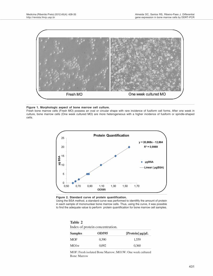
Total protein of all samples was extracted us-ing Ripa buffer (25 mM Tris-HCl pH 7.6, 150 mMNaCl, 1% NP-40, 1% sodium deoxycholate, 0.1%SDS). Sample protein was quantified using a BSAcurve (Figure 2). Approximately 50 µg of each sam-ple was loaded onto a SDS-PAGE gel 12,5 % (30%acrylamide, 1.5 M Tris, pH 8.8, 10% SDS, 10% Am-monium persulfate, 30 µL of TEMED, SIGMA) withsample buffer (0,125 mM Tris-HCl pH 6.8, 4% SDS,20% Glicerol, 0,02% bromofenol blue). It was utilizedtwo pool of tree samples per experiments for eachconditions evaluated (n=2).

430
Medicina (Ribeirão Preto) 2012;45(4): 428-35http://revista.fmrp.usp.br
Almeida DC, Santos RS, Ribeiro-Paes J. Differentialgene expression in bone marrow cells by DDRT-PCR
mRNA Isolation
Total mRNA was extracted from cell pellet us-ing TRIZOL reagent protocol, according to the manu-facture's recommendations (GIBCO-BRL). In orderto verify the quantity and the purity and quality of totalmRNA, processed samples were then analyzed usinga spectrophotometer at wavelengths of 260 nm and280 nm. mRNA purity was determined by the absorb-ance ratio (A260/A280 nm) and visualization of bandsin 1.5% denaturant agarose gel (Table 1 and Figure4). mRNA from both cell types was transcribed intocDNA by using the Thermo Script System kit, accord-ing to the manufacturer's specifications (GIBCO-BRL). It was utilized two pool of tree samples perexperiments for each conditions evaluated (n=2).
DDRT-PCR
Gene expression profile of fresh and cultivatedbone marrow cells was performed by DDRT-PCR,using a single random primer with 10 bp (`5`-CCTTGACGCA-`3).10 cDNA of each cell type wassubjected to amplification using the primer describedabove. Each reaction consisted in the use of µ2 L ofcDNA, 70 pmol of primer, 100 µM of each dNTP, 1.5mM MgCl2, 1X PCR buffer (200mM Tris-HCl pH8.4, 500 mM KCl) and 2 U Taq DNA polymerase(INVITROGEN). Reactions with bone marrow cellswere performed using the following cycle: Cycle 1(1x) incubated at 95°C for 30 seconds, 30°C for 2minutes and 72°C for 1 minute. The next step wasperformed for over 33 cycles under the following con-ditions: 95°C for 30 seconds, 35°C for 2 minutes and72°C for 1 minute. The annealing temperature waschanged to 60°C and extension time increased for 5minutes in the final cycle. Later, the samples werestored at -20°C. An amplification without cDNA wasperformed under the same conditions as a negativecontrol for possible contamination with exogenousDNA. As an endogenous control, amplification with ahousekeeping gene (β-actin) was performed for eachsample as mentioned above.11. It was utilized two poolof tree samples per experiments for each conditionsevaluated (n=2).
Electrophoresis in 8% polyacrylamide gel andsilver staining
For electrophoresis assays, the protocol de-scribed by Dias Neto and collaborators was adopted.12
Each amplification product was added to sample abuffer (0.125% bromophenol blue, 0.125% xylene-
cyanol, and 15% glycerol) and subsequently loadedonto a 8% polyacrylamide gel (acrylamide-bisacrila-mide 29:1) with TBE buffer (90 mm Tris-borate, 1mM EDTA, pH 8.0).13 Next, the gel was subjected toelectrophoresis in a vertical cube at 60V, for 6 h andafter, the gel was fixed in a solution of 10% ethanoland 0.5% acetic acid (v/v), stained with 0.2% silvernitrate and washed with deionized water. Finally, inorder to visualize the bands, the gel was incubatedwith a solution containing 0.75 M NaOH and 0.1 Mformaldehyde.13
ResultsResultsResultsResultsResults
Bone marrow cell culture in distinct conditi-ons presented differences in cell morphology
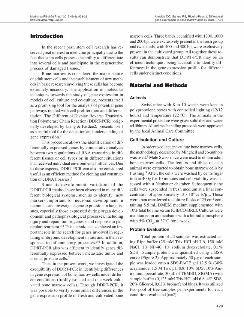
We observed the general morphology of bonemarrow cells under different conditions, analyzed freshor after one week in culture. It was demonstrated thatfresh bone marrow cells represented in their majorityoval cells while cultivated bone marrow cells showed,at least, two distinct morphologies, representing ovaland fibroblastic-like cells (Figure 1).
Protein profile of fresh and cultivated bonemarrow cells
In order to evaluate if SDS-PAGE gel can bedesigned to visualize the protein profile of fresh bonemarrow cells and one-week cultured cells, we performeda 12,5% acrylamide gel. First, both sample types ofbone marrow were submitted to protein quantificationusing the BSA method (Figure 2 and Table 2). After,the samples were visualized in 12,5% polyacrylamidegel and a distinct distribution was identified on theirband profile. Fresh bone marrow cells presented ex-clusively two bands (one with ≈170 and another with≈90 kDa) while cultured bone marrow cells did notshow any difference in their profile (Figure 3).
Table 1Measurement of mRNA index.
Samples 260 nm 280 nm 260/280 [RNA] µg/µL
MOF 0,390 0,233 1,873 1,559
MO1w 0,092 0,076 1,910 0,360
MOF: Freshly isolated Bone Marrow; MO1W: One week cul-tured Bone Marrow
Medicina (Ribeirão Preto) 2012;45(4): 428-35http://revista.fmrp.usp.br
431
Almeida DC, Santos RS, Ribeiro-Paes J. Differentialgene expression in bone marrow cells by DDRT-PCR
Table 2Index of protein concentration.
Samples OD595 [Protein] µg/µL
MOF 0,390 1,559
MO1w 0,092 0,360
MOF: Fresh isolated Bone Marrow; MO1W: One week culturedBone Marrow
Figure 2. Standard curve of protein quantification.Using the BSA method, a standard curve was performed to identifify the amount of proteinin each sample of mononuclear bone marrow cells. Thus, using the curve, it was possibleto find the adequate value to perform protein quantification for bone marrow cell samples.
Figure 1. Morphologic aspect of bone marrow cell culture.Fresh bone marrow cells (Fresh MO) possess an oval or circular shape with rare incidence of fusiform cell forms. After one week inculture, bone marrow cells (One week cultured MO) are more heterogeneous with a higher incidence of fusiform or spindle-shapedcells.

432
Medicina (Ribeirão Preto) 2012;45(4): 428-35http://revista.fmrp.usp.br
Almeida DC, Santos RS, Ribeiro-Paes J. Differentialgene expression in bone marrow cells by DDRT-PCR
Differences in gene expression analysisof bone marrow cells can be detected byDDRT-PCR
To investigate if DDRT-PCR can detect differ-ences in the gene expression profile of bone marrowcells, we carried out an experiment with two condi-tions: freshly isolated cells and cells after one weekbeing cultivated. The purity of both the quantities ofRNA in each sample is shown in Table 2 and Figure 4.The RNA extraction process produced ideal values(1.9-2.1 for), indicating low contamination of DNA,
Figure 3. Profile of protein banding using SDS PAGE gel.
(A) Standard molecular weight refers to 220 KDa (Myosin), 170KDa (á-2-Macroglobulin), 116 KDa (â-Galactosidade), 76 KDa(Transferrin) and 53 KDa (Glutamate Dehydrogenase). (B) Refersto fresh bone marrow cells and (C) indicate bone marrow cellscultured during one week. After the SDS page process, onedifferential profile was observed among the two samples (A andB). The fresh bone marrow cells presented two bands (170 and90 kDa), that were not identified in cultured bone marrow cells.
Figure 4. Visualization of subunits of ribosomal RNA inagarose gel.It is possible to observe the presence of two band referring to the28s and 18s subunits of ribosomal RNA.
proteins, polypeptides and phenol.13 During electro-phoresis in a 1.5% agarose gel, there is a regular qual-ity of samples, and two bands can be observed refer-ring to the 18S and 28S ribosomal RNA subunits (Fig-ure 4). In sequence, by DDRT-PCR, it was possibleto identify in a 8% polyacrylamide gel a distinct pro-file in gene expression for both samples. Fresh bonemarrow cells presented three specific bands with ap-proximately 1300, 1000 and 225 bp while cultivatedbone marrow cells showed only two bands differen-tially expressed one with ≈400 bp and another with≈300 bp (Figure 5).
Discussion and ConclusionsDiscussion and ConclusionsDiscussion and ConclusionsDiscussion and ConclusionsDiscussion and Conclusions
The application of molecular techniques to evalu-ate differences in gene expression is very important instem cell research. In this sense, recent literature hasdescribed several methodologies to perform gene ex-pression analyses. Philips and co-authors, for exam-ple, using a similar technique elegantly described sev-eral gene products differentially expressed in hemat-opoietic stem cells and in fetal liver stem cells.14 Later,Ramalho-Santos and colleagues, using subtractive hy-bridization and microarray analysis, performed a com-parison of the transcription profile of three strains ofstem cells (Embryonic, Neural and Hematopoietic).15
These authors observed the presence of specific genespresent in each population of stem cells, and demon-strated a cell-specific gene expression profile.15 Inaddition, in another study, through hybridization tech-niques and advanced bioinformatics, an analysis of thegene expression in three distinct types of hematopoi-etic stem cells within a homogeneous population wasperformed.16
Alternatively, here we described DDRT-PCRas a quick, easy and efficient method to detect differ-ential gene expression in bone marrow cells. We chosetwo conditions to analyze gene expression: fresh cellsand one week cultivated cells. Initially, changes wereobserved in cell morphology and in protein profile us-ing SDS-PAGE gel. To demonstrate the applicabilityof our methods, DDRT-PCR was performed in thissame condition. Confirming the findings observed inthe SDS-PAGE gel, DDRT-PCR also showed differ-ences among the groups studied (fresh cells and culti-vated cells) but more details may be observed.
The success of this method is related with thechoice of primer designated as L5. In this work L5 wasdefined from previous work from the Jing and Li group,

Medicina (Ribeirão Preto) 2012;45(4): 428-35http://revista.fmrp.usp.br
433
Almeida DC, Santos RS, Ribeiro-Paes J. Differentialgene expression in bone marrow cells by DDRT-PCR
in which good results were obtained.17 These authors,using this primer and others, found 299 fragments thatdetermined genetic similarity in three families ofAnguilla.17 Promising results were also obtained by Baeand co-authors, using this same primer.18 In Bae`s work,such result, by the RAPD method, made it possible toidentify several random primers including L5, relatedwith the identification of new retro-transposons frag-ments in Clonorchis sinensis.18 The use of two timepoints for evaluation, also was established from goodresults in previous work from Xu and collaborators.19
These researchers achieved encouraging results us-ing 0h-4h-8h and 12h for the verification of differentialgene expression during the regeneration of hepatocytesin rat liver.19 Following the same methodology, Milesand co-authors also used 1h and 24h to demonstratedgreat differences in gene expression of rat bone cellsbefore and after an osteogenic stimulus.20
In the present manuscript, the analysis throughDDRT-PCR showed a clear difference in gene ex-pression profiles between the cell types used. Bothsamples were amplified using the L5 primer and thedetermination and confirmation of potential polymor-phic markers between the cell types in the study weremade from the analysis of electrophoretic profiles andband analysis in polyacrylamide gel (Figure 5). Thepresence of three bands was verified in fresh bonemarrow cells (1300, 1000, 225 bp) and two bands werepresent in the cultivated group (400 and 300 bp).
The bands found in the first group may be re-lated to specific genes restricted to progenitor stemcell pools (hematopoietic, endothelial and mesenchy-mal cells), besides lymphocytes, monocytes and othermyeloid cells present within the bone marrow envi-ronment. On the other hand, the two bands observedfor the cultivated cells may be related with the proc-ess of cellular differentiation in vitro, and enrichmentof mesenchymal cells due to culture conditions(DEMEN 10% SBF).21,22,23 However, other studiessuggest that the loss of constitutive expression forsome genes by in vitro culture methods, which wouldbe normally expressed in vivo, may be occurring.20
In this work, the authors showed in their results thatthe gene for myeloperoxidase (bactericidal enzymefound primarily in neutrophils and monocytes) is ex-pressed in bone in vivo, but its expression was notdetected in in vitro culture of osteoblasts.20 In addi-tion, we could not exclude the possibility that thesetwo bands found for cultured cells could also be re-lated to a differential expression of bone marrow cells
Figure 5. Differential display layout of gene expressionanalysis of bone marrow cells.
Through the DDRT-PCR method it is possible to observe a distinctprofile in gene expression of fresh bone marrow cells (MO1) andbone marrow cells cultured during one week (MO2). In fresh bonemarrow cells, there are three fragments (1300, 1000 and 200 bp,respectively) differentially expressed. On the other hand, forcultured bone marrow cells, two exclusive bands (400 and 300bp) were identified. This result may indicated that during one weekin culture, bone marrow cells may shift their gene expressionprofile due to the proliferation of different cells types, a naturaldifferentiation process or a response to metabolic events such asstress. MW refers to standard molecular weight with 1.800, 1.300,1.000, 900, 800, 700. 600. 500. 400. 300 and 200 bp.

434
Medicina (Ribeirão Preto) 2012;45(4): 428-35http://revista.fmrp.usp.br
Almeida DC, Santos RS, Ribeiro-Paes J. Differentialgene expression in bone marrow cells by DDRT-PCR
in response to the change of microenvironment byactivating on an alternative metabolic pathway, princi-pally due to "stress" provided by new conditions suchas acidophilic medium, lack of nutrients and imbalanceof electrolytes.24
Regardless of these questions, DDRT-PCR wasreliable, accessible and efficient to detect small dif-ferences in gene expression on two populations of bonemarrow cells. In conclusion, together these resultsdemonstrated that DDRT-PCR is potentially feasible
for application in studies specifically formulated toevaluate large and small differences in the gene ex-pression of distinct cells or the same cells in severalconditions. This study represents a work that servesas a basis for the generation of future studies withstem cells in different approaches. We believe thatDDRT-PCR is a simple and quick method to evaluategene expression and can be used for other stem typesfor differential gene expression analyses in the areaof cell-based therapy.
RESUMO
Modelo do estudo: Estudo Experimental. Introdução: Atualmente a pesquisa com células-tronco temgerado grande interesse devido a sua aplicabilidade no campo na medicina regenerativa. A medulaóssea é considerada a maior fonte de células-tronco adultas e o estabelecimento de novos métodospara a análise da expressão gênica torna-se estritamente necessário. Desse modo, o "DifferentialDisplay Reverse Transcription Polymerase Chain Reaction (DDRT-PCR)", pode ser uma ferramentaacessível para a investigação de pequenas diferenças no nível de expressão gênica em diferentes tiposcelulares, sob distintas condições.Objetivo: Neste presente trabalho nós investigamos a exequibilidade do DDRT-PCR na identificação dediferenças no nível de expressão gênica global em células da medula óssea de camundongos sobduas condições. Métodos: Primeiramente, a medula óssea foi isolada frescamente e uma secundaparte foi cultivada por uma semana sem troca de meio. Posteriormente, as células da medula (fresca ecultivada) foram submetidas a análise da expressão gênica, seguindo a metodologia de DDRT-PCR.Resultados: Inicialmente, foi possível identificar em células da medula óssea com uma semana decultivo, pequenas alterações morfológicas (células ovais para fibroblastóides) e no perfil de proteínas,por meio da visualização de bandas em SDS-Page gel. Finalmente, a análise da expressão gênica porDDRT-PCR, mostrou uma expressão diferencial com a presença de três bandas (1300, 1000 and 225pb) exclusivamente expressas na medula óssea fresca e mais duas bandas (400 and 300 pb) presen-tes somente nas células de medula cultivadas.Conclusões: Em suma, a metodologia de DDRT-PCR mostrou-se eficiente para a identificação depequenas diferenças no nível de expressão gênica em células da medula óssea sob duas definidascondições. Portanto, nós acreditamos que o DDRT-PCR possa ser designado de forma rápida e eficien-te para a análise diferencial de expressão gênica em diferentes tipos de células-tronco, sob diferentescondições.
Palavras-chave: DDRT-PCR. Expressão Gênica. Células da Medula Óssea.
ReferencesReferencesReferencesReferencesReferences
1. Covas DT. Terapia Celular. In: Bordin JO, Langhi Júnior, DM,Covas DT, Editors. Hemoterapia: Fundamentos e Prática. 1 ed.São Paulo: Atheneu, 2007, v. 1, p. 601-11.
2. Liang P, Pardee AB. Differential display of eukaryotic messen-ger RNA by means of the polymerase chain reaction. Sci-ence. 1992; 257: 967-71.
3. Sunday ME. Differential Display RT-PCR for identifying novelgene expression in lung. Am J Physiol, Lung Cell Mol Physiol.1995 269: L273-84.
4. Watson JB and Margulies JE. Differential cDNA screening strat-egies to identify novel stage-specific proteins in the develop-ing mammalian brain. Dev Neurosci. 1993; 15: 77-86.
5. Gupta R, Thomas P, Beddington RSP, Rigby PWJ. Isolation ofdevelopmentally regulated genes by differential displayscreening of cDNA libraries. Nucleic Acids Res. 1998; 26:4538-9.
6. Silva AM, Pires EG, Abrantes EF, Ferreira LRP, Gazzinelli RT,Reis LFL. Application of the differential display RT-PCR strat-egy for the identification of inflammation-related mouse genes.Braz J Med Biol Res. 1999; 32: 845-52.
7. Pienta KJ, Schwab ED. Modified differential display techniquethat eliminates radioactivity and decreases screening time.Biotechniques. 2000; 28: 272-7.
8. Minguell JJ, Erices A, Conget P. Mesenchymal Stem Cells. ExpBiol Med (Maywood). 2001; 226: 507-20.
9. Fontes AM, Orellana MD, Prata KL. Células-Tronco e seusMétodos de Estudo. In: Zago MA, Covas DT, Editors. Células-

Medicina (Ribeirão Preto) 2012;45(4): 428-35http://revista.fmrp.usp.br
435
Almeida DC, Santos RS, Ribeiro-Paes J. Differentialgene expression in bone marrow cells by DDRT-PCR
Tronco, A Nova Fronteira da Medicina. 1ª ed. São Paulo:Atheneu, 2006, v.,p. 93-106.
10. Saito S, Sawai K, Ugai H, Moriyasu S, Minamihashi A,Yamamoto Y, et al. Generation of cloned calves andtransgenic chimeric embryos from bovine embryonic stem-like cells. Biochem Biophys Res Commun. 2003; 309:104-13.
11. Terskikh A, Lukyanov S, Diatchenko L, Weissaman. HuntingHematopoietic Stem Cell-Specific Genes Using PCR-SelectcDNA Subtraction. Clontechniques [serial on line] 2000; Jan-Mar. [cited 2005 May, 13]; 1:(1). Available from: http://www.clontech.com.
12. Dias Neto E, Steindel M, Passos LK, de Souza CP, Rollinson D.The use of RAPDs for the study of the genetic diversity ofSchistosoma mansoni and Trypanosoma cruzi. Exs 1993;67: 339-45.
13. Sambrook J, Fritsch EF and Manitis T. Editors. Molecular Clon-ing: A Laboratory Manual. 2nd ed. Plainview, (NY), USA.Cold Spring Habor Laboratory Press, 1989.
14. Phillips RL, Ernst RE, Brunk B, Ivanova N, Mahan MA,Deanehan JK, et al.. The Genetic Program of HematopoieticStem Cells. Science. 2000; 288: 1635-40.
15. Ramalho-Santos MR, Yoon S, Matsuzaki Y, Mulligan RC,Melton DA. "Stemness": Transcriptional Profiling of Embry-onic and Adult Stem Cells. Science. 2002; 298: 597-600.
16. Park IK, He Y, Lin F, Laerum OD, Tian Q, Bumgarner R, et al.Differential gene expression profiling of adult murine hemat-opoietic stem cells. Blood 2002; 99: 488-98.
17. Jing QJ and Li YP. Random amplified polymorphic DNA analy-sis of eel genome. Cell Research 1999; 9: 217-23.
18. Bae YA, Moon SY, Kong Y, Cho SY, Rhyu MG. CsRn1, A NovelActive Retrotransposon in a Parasitic Trematode, Clonor-chis sinensis, Discloses a New Phylogenetic Clade of Ty3/gypsy-like LTR Retrotransposons. Mol Biol Evol. 2001;18:1474-83.
19. Xu CS, Yuan JY, Li WQ, Han HP, Yang KJ, Chang CF, Zhao L-F, Li YC, Zhang HY, Rahman S, Zhang JB. Identification ofexpressed genes in regenerating rat liver in 0-4-8-12 h shortinterval successive partial hepatectomy. Worl J Gastro 2005;21;11: 2296-305.
20. Miles RR, Turner CH, Santerre R, Tu Y, Mcclelland P, Argot J,et al. Analysis of differential gene expression in rat tibiaafter an osteogenic stimulus in vivo: mechanical loading regu-lates osteopontin and myeloperoxidase. J Cell Biochem.1998;68:355-65.
21. Dieudonné SC, Kerr JM, Xu T, Sommer B, Derubeis AR,Kznetsov SA, et al. Differential display of human marrowstromal cells reveals unique mRNA expression patterns inresponse to dexamethasone. J Cell Biochem. 1999;76:231-43.
22. Silva-Meirelles LS, Nardi NB. Murine marrow-derived mesen-chymal stem cell: isolation, in vitro expansion, and charac-terization. Br J Haematol. 2003;123:702-11.
23. Covas DT. Células-Tronco Mesenquimais. In: Zago MA, CovasDT, Editors. Células-Tronco, A Nova Fronteira da Medicina.1ª ed. São Paulo: Atheneu, 2006, v. 1, p. 35-48.
24. Cheng L, Hammond H, Ye Z, Zhan X, Dravid G. Human adultmarrow cells support prolonged expansion of human em-bryonic stem cells in culture. Stem Cells. 2003;21:131-42.