Embed Size (px)
Citation preview

Aortic Dissection and Aneurysms
Presented by Dr. Daniel Kranitz
Prepared by Mary Edwards
September 27, 2005
Tintanalli Chapter 58, Pages 404-409
Abdominal Aortic Aneurysms (AAA)
Risk factors Elderly (>60) Familial trend (18% with 1° relative) Connective Tissue D/O (Marfan’s) Other aneurysms Atherosclerosis (HTN, Lipids, smoking, DM)
AAA
Pathogenesis Intima infiltrated by atherosclerosis and thinned
media. Possible intraluminal thrombus and adventitia
infiltrated by inflammatory cells
AAA
Average rate of growth 0.25-0.5 cm per year.
Larger aneurysms extend more rapidly than smaller ones. (LaPlace law)
AAA
Clinical Features Syncope (10-12%) Back and/or Abdominal Pain –severe and abrupt,
ripping or tearing sensation (50%) Shock –intraperitoneal rupture, massive blood
loss Sudden death
AAA
Physical Exam Pain on palpation or not Retroperitoneal hematoma
Cullen sign (periumbilical ecchymosis)Grey-Turner sign (flank ecchymosis)Scrotal hematoma or inguinal mass (blood dissecting
to these areas) Iliopsoas signFemoral nerve neuropathy
AAA
Found aneurysms refer to follow up >5cm diameter –increased chance of rupture <5cm –decreased chance of rupture Symptomatic aneurysms of any size =
Emergency!!
AAA
Diagnosis Includes differential diagnoses of syncope, abd
pain, CP, back pain and shock. If with combo of two or more think aortic dz.
AAA
Radiologic Evaluation Should not delay operative treatment!!
Plain abd film (calcified bulging) US (bedside, up to 100% sensitive, not reliable
to detect rupture) CT (with IV contrast only if stable) MRI
AAA
ED Treatment Urgent surgical consult Make diagnosis & assist rapid transfer to OR 2 large bore IVs Cardiac Monitor O2 ? Blood transfusion IV fluid resuscitation –controversial amount b/c too much can
be harmful RADIOGRAPHIC STUDIES ONLY IF UNLIKELY
TO HAVE RUPTURED AAA!!!
AAA
½ of patients with ruptured AAA who reach the OR die!
A Bit About Thoracic Aortic Aneursym
Presenting symptoms include esophageal, tracheal, bronchial, or even neurologic disorders.
If it erodes to adjacent structures it is immediately fatal!!
Aortic Dissection
Pathogenesis Prominent cause of sudden death Presents with severe abd., chest, and back pain Violation of intima that allows blood to enter
media and dissect b/w intimal and adventitial layers
Common site is ascending aorta at ligamentum arteriosum
Aortic Dissection
Common presenting groups >50 yoa with HTN 2/3 male Marfan’s syndrome Congenital heart disease Pregnancy
Aortic Dissection
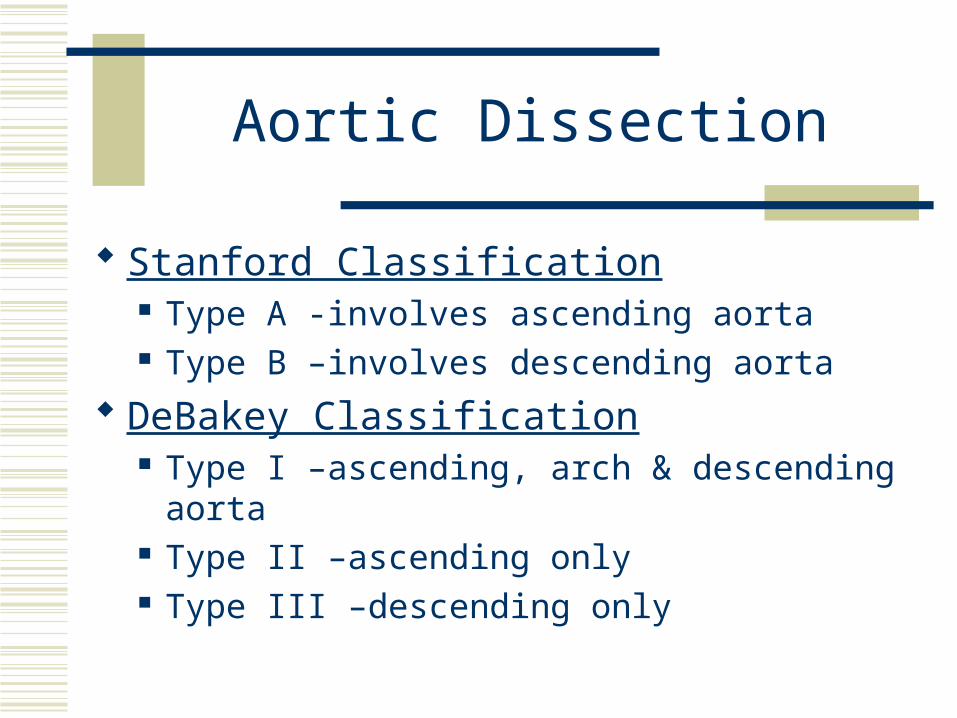
Stanford Classification Type A -involves ascending aorta Type B –involves descending aorta
DeBakey Classification Type I –ascending, arch & descending aorta Type II –ascending only Type III –descending only
Aortic Dissection
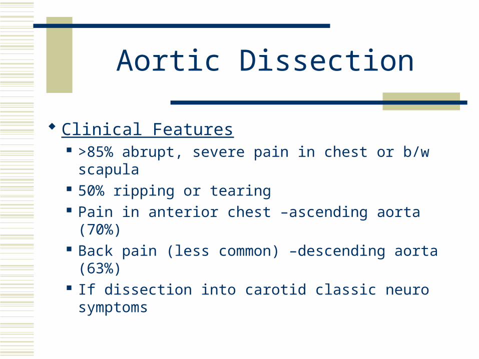
Clinical Features >85% abrupt, severe pain in chest or b/w scapula 50% ripping or tearing Pain in anterior chest –ascending aorta (70%) Back pain (less common) –descending aorta
(63%) If dissection into carotid classic neuro symptoms
Aortic Dissection
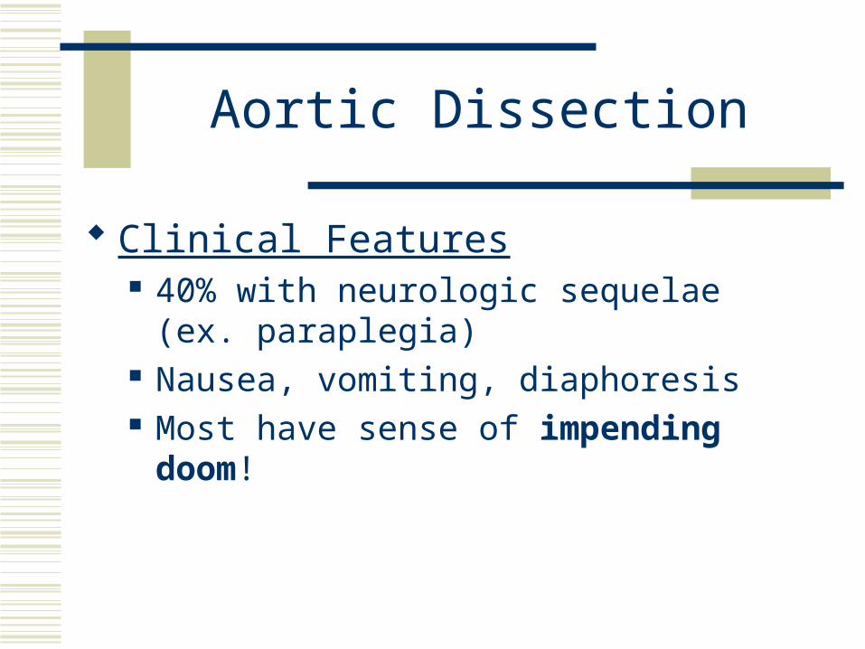
Clinical Features 40% with neurologic sequelae (ex. paraplegia) Nausea, vomiting, diaphoresis Most have sense of impending doom!
Aortic Dissection
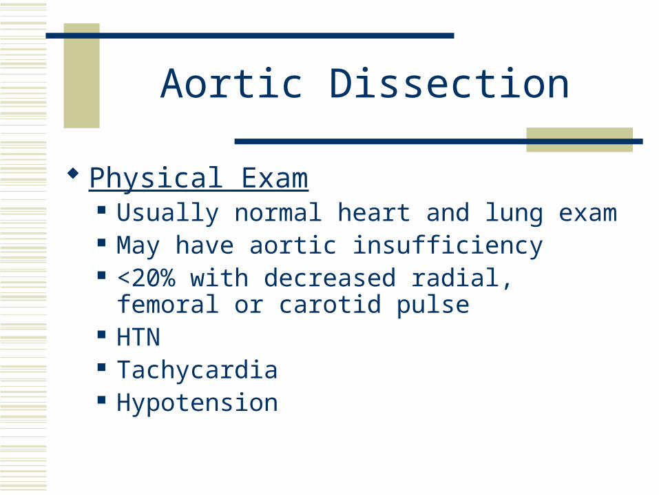
Physical Exam Usually normal heart and lung exam May have aortic insufficiency <20% with decreased radial, femoral or carotid
pulse HTN Tachycardia Hypotension
Aortic Dissection
Physical Exam Pericardial tamponade (muffled heart tones,
JVD, pulsus paradoxus) Hoarseness (compression of recurrent laryngeal
nerve) Horner’s Syndrome (compression of superior
cervical sympathetic ganglion)
Aortic Dissection
Diagnosis Ischemic end-organ manifestation such as MI,
pericardial dz, pulmonary d/o, stroke, SCI, musculoskeletal dz of extremities, intraabdominal ischemia.
Can change location with time as dissects.
Aortic Dissection
Thoracic Dissection 90% have abnormal CXR
Widened mediastinumAbnormal aortic contourPleural effusionDeviation of trachea, mainstem bronchi, or esophagus Intimal calcium visable & distant from edge (calcium
sign)
Aortic Dissection
Diagnosis CT
83-100% sensitive 87-100% specificUse spiral CT with IV contrastWill not give anatomic details of arterial branches or
aortic valve competence.Modality of choice in unstable patient
Aortic Dissection
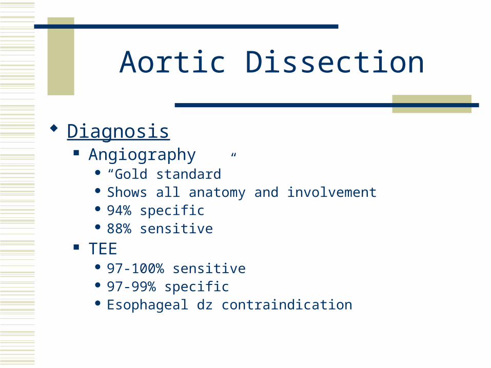
Diagnosis Angiography
“Gold standard” Shows all anatomy and involvement 94% specific 88% sensitive
TEE 97-100% sensitive 97-99% specific Esophageal dz contraindication
Aortic Dissection
In contrast to ruptured AAA, SUSPECTED DISSECTIONS MUST BE CONFIRMED RADIOLOGICALLY PRIOR TO SENDING TO OR!!!
Aortic Dissection
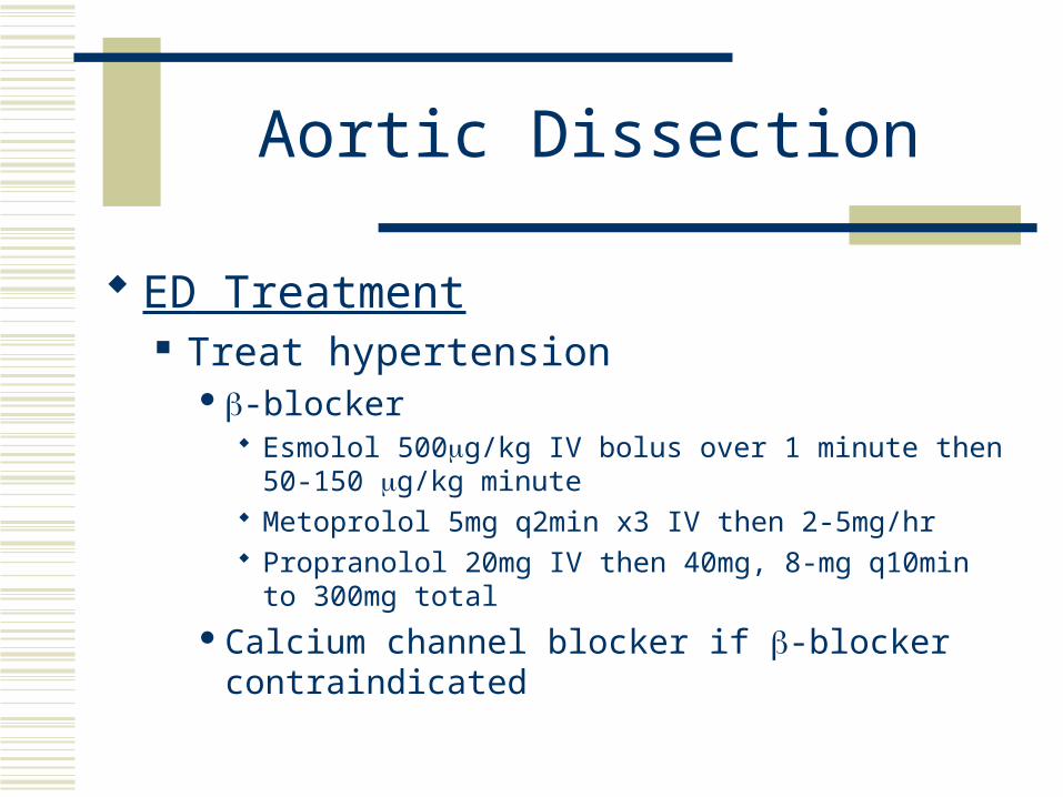
ED Treatment Treat hypertension
-blocker Esmolol 500g/kg IV bolus over 1 minute then 50-150 g/kg
minute Metoprolol 5mg q2min x3 IV then 2-5mg/hr Propranolol 20mg IV then 40mg, 8-mg q10min to 300mg
total
Calcium channel blocker if -blocker contraindicated
Aortic Dissection
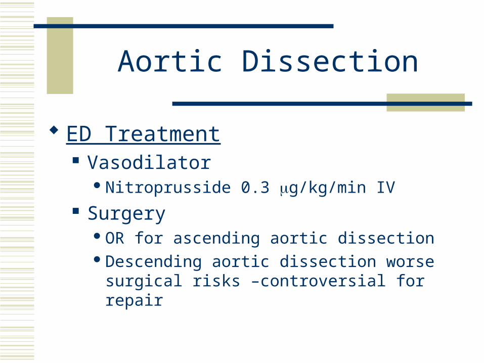
ED Treatment Vasodilator
Nitroprusside 0.3 g/kg/min IV Surgery
OR for ascending aortic dissectionDescending aortic dissection worse surgical risks –
controversial for repair

Any Questions????
Questions
1. A patient with a suspected aortic dissection should be immediately tranferred to OR without radiographic studies.
A. True B. False
2. Females are more likely than males to develop aortic dissection. A. True B. False
3. Dissection of the ascending aorta only is DeBakey classification A. Type I B. Type II C. Type III D. Type A E. Type B
4. Patients with a ruptured AAA can present with all of the following symptoms except A. Shock B. Syncope C. Sudden death D. Nausea and vomiting E. Headache
5. Which of the following radiologic modalities is considered the “gold standard” for diagnosing an aortic dissection? A. CT B. MRI C. TEE D. Angiography E. CXR
Answers
1. B 2. B 3. B 4. E 5. D