Embed Size (px)
Citation preview
Aortic Regurgitation: Etiology and Echo Quantification
Martin G. Keane, MD, FASEProfessor of MedicineLewis Katz School of Medicine
at Temple University
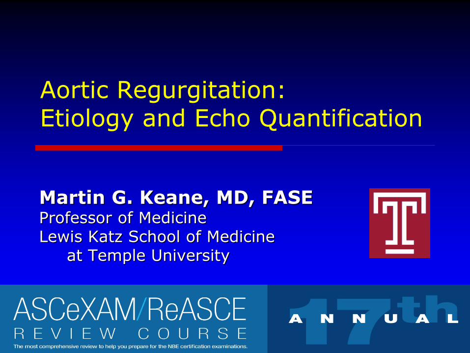
Review Question #1
Possible reasons that thiscolor Doppler evaluation mayoverestimate AR include:
A. Low color Doppler gain setting
B. Use of too small a color Doppler sector
C. Low Nyquist velocity setting
D. High pulse repetition frequency (PRF)
Review Question #2
Which condition may lead to over-estimation of AR severity by deceleration slope (or P1/2) of diastolic Continuous Wave (CW) Doppler:
A. Advanced restrictive myocardial disease
B. Severe mitral valve stenosis
C. Severe aortic valve stenosis
D. Low Nyquist limit setting

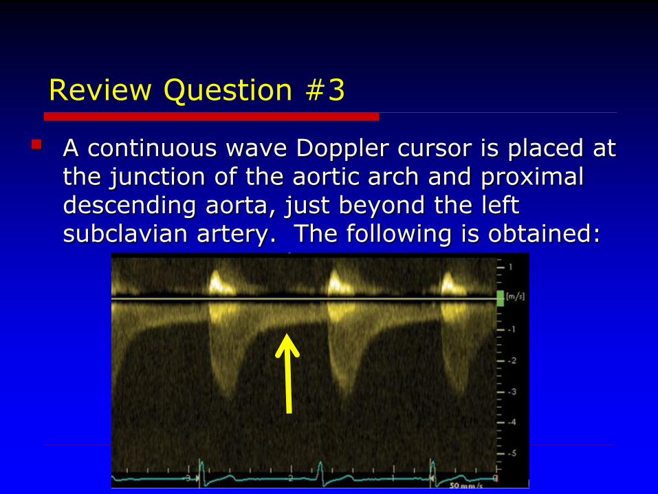
Review Question #3
A continuous wave Doppler cursor is placed at the junction of the aortic arch and proximaldescending aorta, just beyond the left subclavian artery. The following is obtained:

Review Question #3
The etiology of the diastolic Doppler flow indicated by the arrow is:
A. Stenosis of the left subclavian artery
B. Severe aortic regurgitation
C. Moderate aortic regurgitation
D. Severe coarctation of the aorta
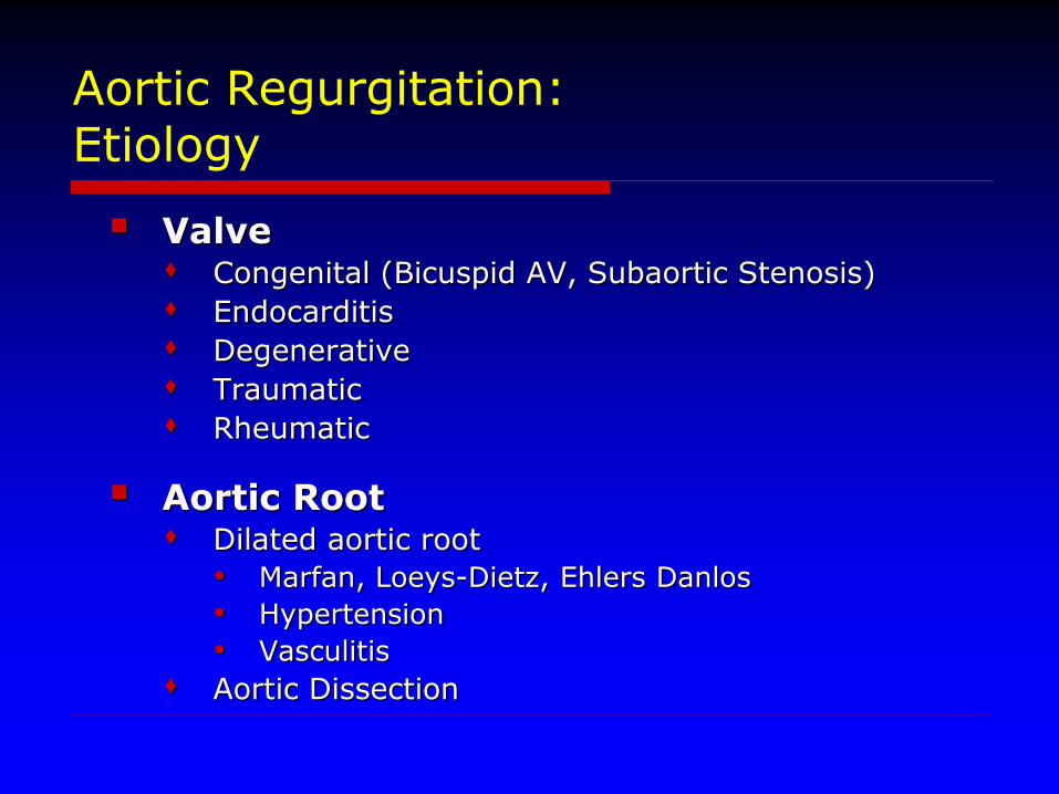
Aortic Regurgitation:Etiology
Valve Congenital (Bicuspid AV, Subaortic Stenosis)
Endocarditis
Degenerative
Traumatic
Rheumatic
Aortic Root Dilated aortic root
• Marfan, Loeys-Dietz, Ehlers Danlos
• Hypertension
• Vasculitis
Aortic Dissection
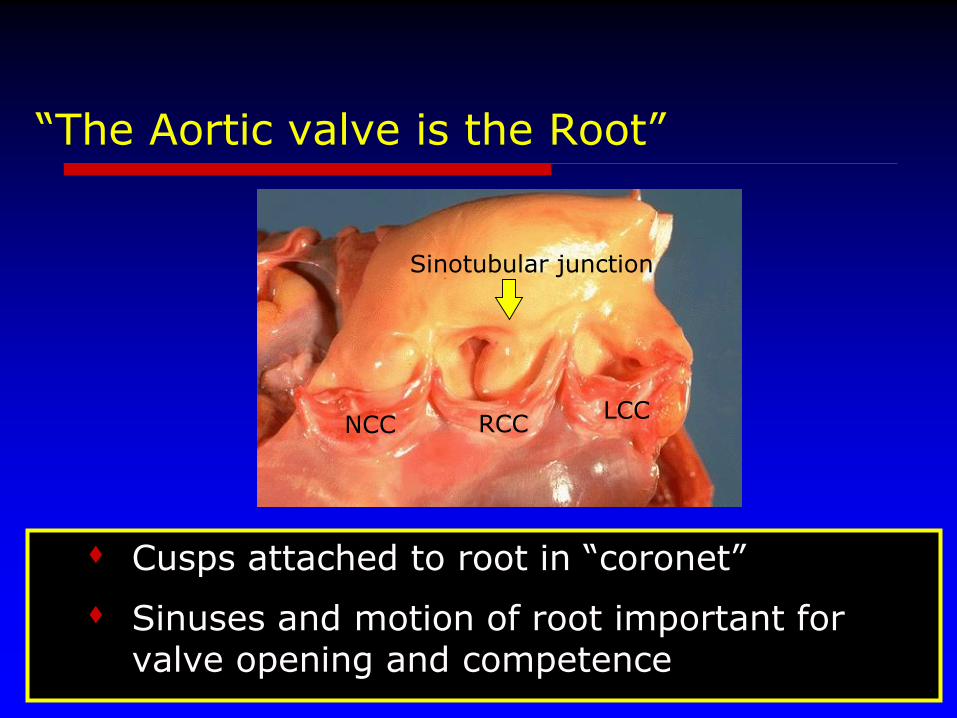
“The Aortic valve is the Root”
Cusps attached to root in “coronet”
Sinuses and motion of root important for valve opening and competence
NCC RCCLCC
Sinotubular junction
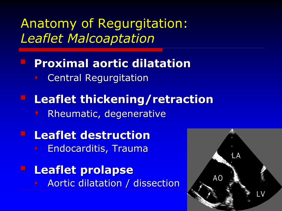
Anatomy of Regurgitation:Leaflet Malcoaptation
Proximal aortic dilatation
Central Regurgitation
Leaflet thickening/retraction
Rheumatic, degenerative
Leaflet destruction Endocarditis, Trauma
Leaflet prolapse Aortic dilatation / dissection
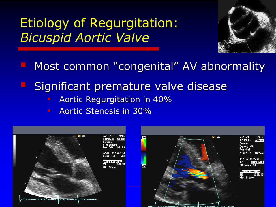
Etiology of Regurgitation:Bicuspid Aortic Valve
Most common “congenital” AV abnormality
Significant premature valve disease• Aortic Regurgitation in 40%
• Aortic Stenosis in 30%
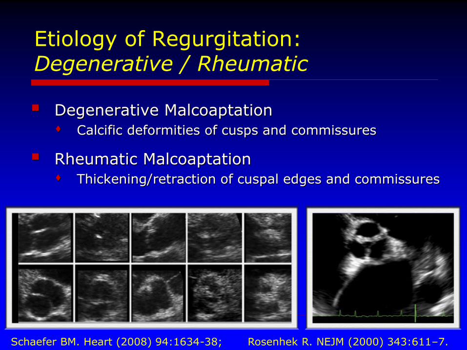
Etiology of Regurgitation:Degenerative / Rheumatic
Degenerative Malcoaptation
Calcific deformities of cusps and commissures
Rheumatic Malcoaptation
Thickening/retraction of cuspal edges and commissures
Schaefer BM. Heart (2008) 94:1634-38; Rosenhek R. NEJM (2000) 343:611–7.
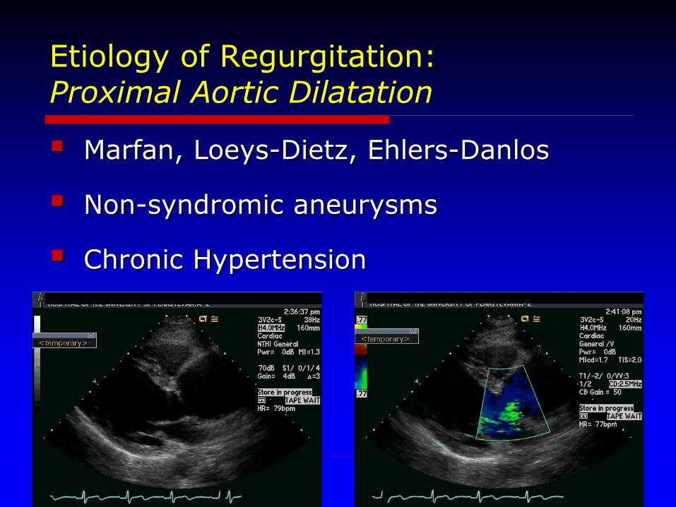
Etiology of Regurgitation:Proximal Aortic Dilatation
Marfan, Loeys-Dietz, Ehlers-Danlos
Non-syndromic aneurysms
Chronic Hypertension
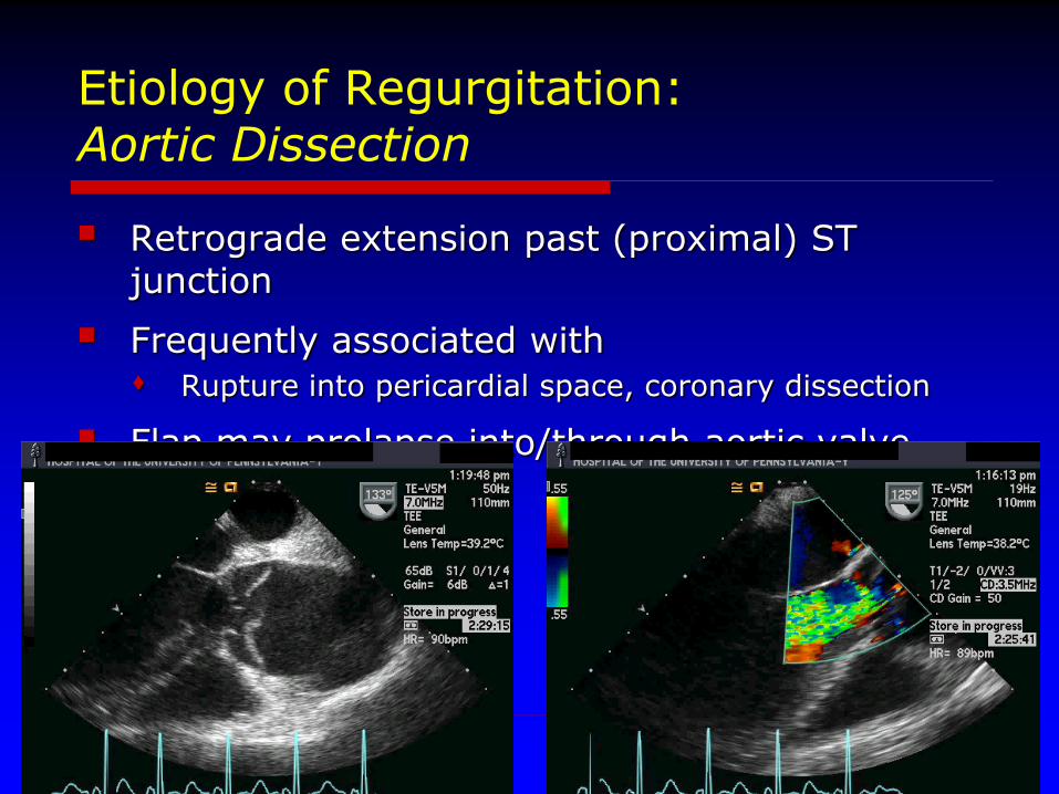
Etiology of Regurgitation:Aortic Dissection
Retrograde extension past (proximal) ST junction
Frequently associated with
Rupture into pericardial space, coronary dissection
Flap may prolapse into/through aortic valve
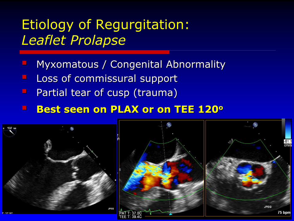
Etiology of Regurgitation:Leaflet Prolapse
Myxomatous / Congenital Abnormality
Loss of commissural support
Partial tear of cusp (trauma)
Best seen on PLAX or on TEE 120o
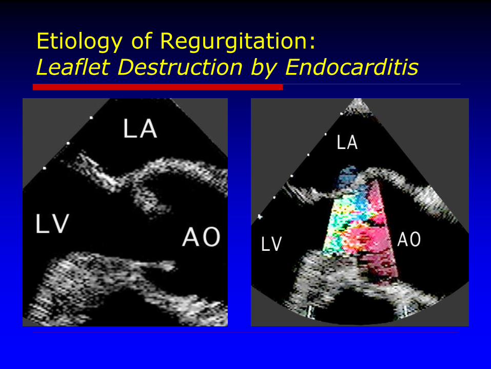
Etiology of Regurgitation:Leaflet Destruction by Endocarditis
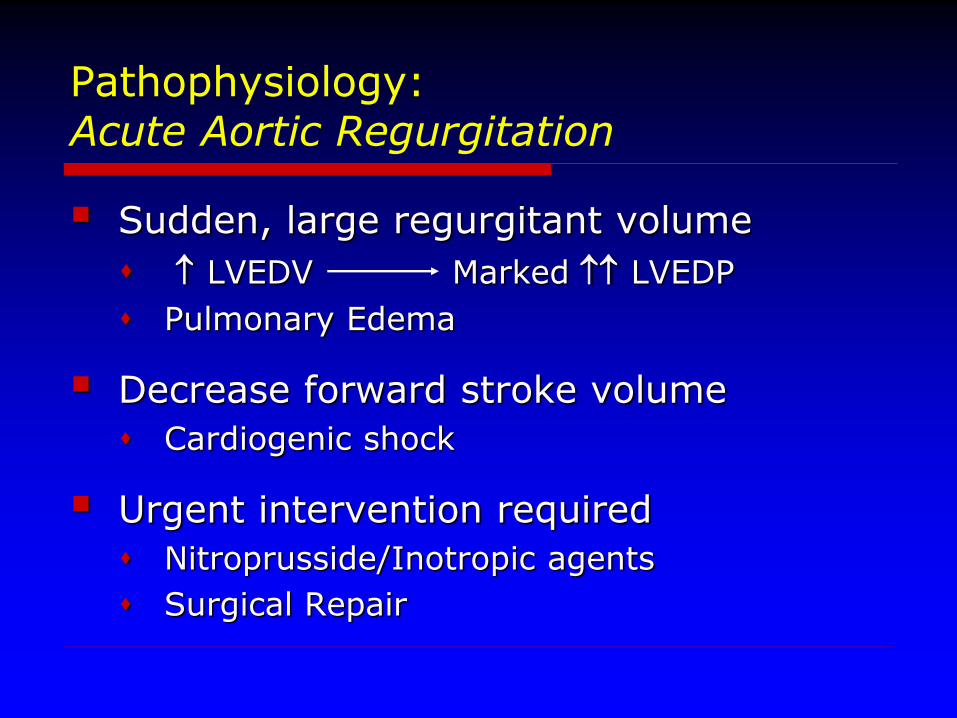
Pathophysiology:Acute Aortic Regurgitation
Sudden, large regurgitant volume
LVEDV Marked LVEDP
Pulmonary Edema
Decrease forward stroke volume
Cardiogenic shock
Urgent intervention required
Nitroprusside/Inotropic agents
Surgical Repair
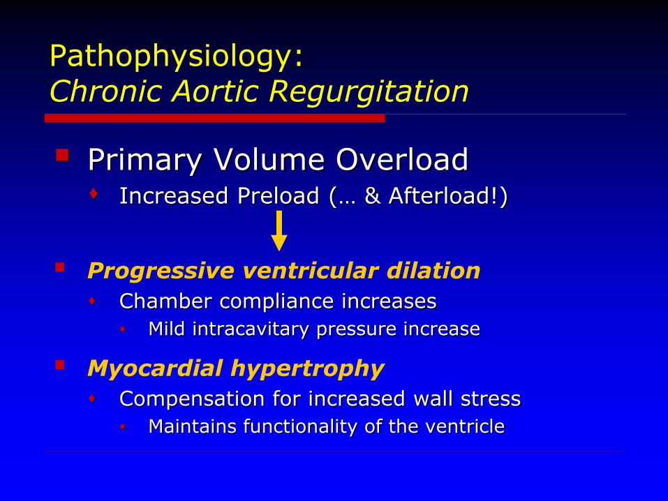
Pathophysiology:Chronic Aortic Regurgitation
Primary Volume Overload Increased Preload (… & Afterload!)
Progressive ventricular dilation
Chamber compliance increases
• Mild intracavitary pressure increase
Myocardial hypertrophy
Compensation for increased wall stress
• Maintains functionality of the ventricle
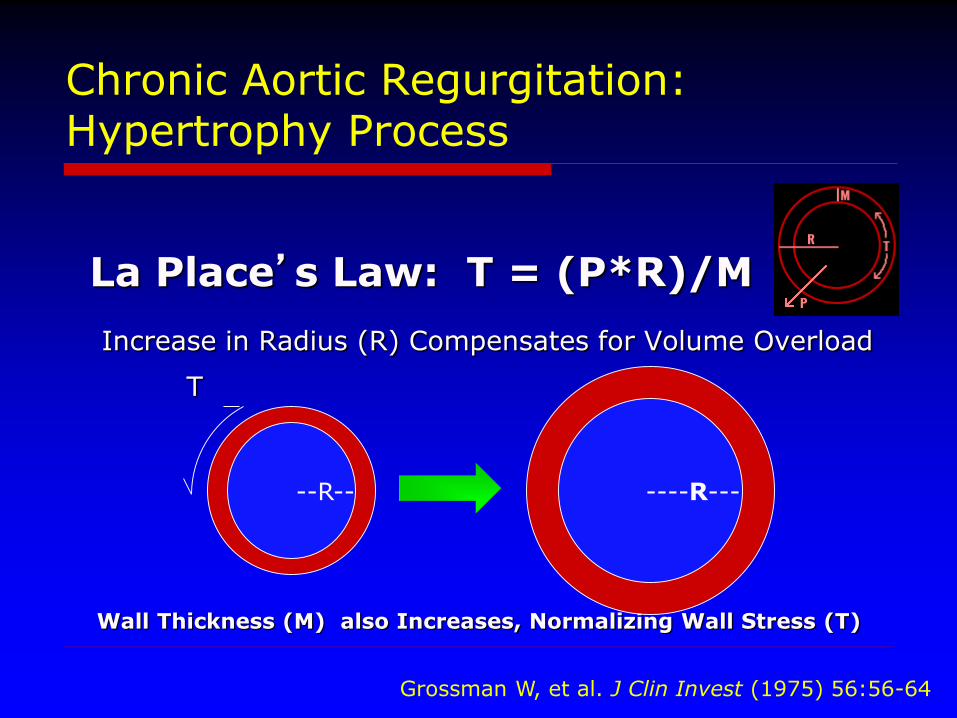
La Place’s Law: T = (P*R)/M
Increase in Radius (R) Compensates for Volume Overload
T
M M
Wall Thickness (M) also Increases, Normalizing Wall Stress (T)
Chronic Aortic Regurgitation:Hypertrophy Process
--R-- ----R---
Grossman W, et al. J Clin Invest (1975) 56:56-64
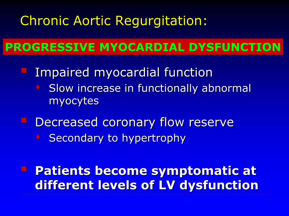
Chronic Aortic Regurgitation:
Impaired myocardial function
Slow increase in functionally abnormal myocytes
Decreased coronary flow reserve
Secondary to hypertrophy
Patients become symptomatic at different levels of LV dysfunction
PROGRESSIVE MYOCARDIAL DYSFUNCTION
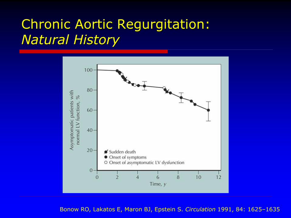
Chronic Aortic Regurgitation:Natural History
Bonow RO, Lakatos E, Maron BJ, Epstein S. Circulation 1991, 84: 1625–1635
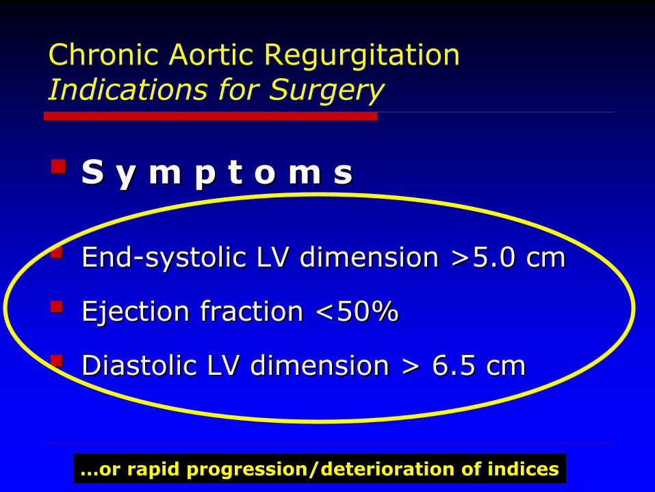
Chronic Aortic RegurgitationIndications for Surgery
S y m p t o m s
End-systolic LV dimension >5.0 cm
Ejection fraction <50%
Diastolic LV dimension > 6.5 cm
…or rapid progression/deterioration of indices
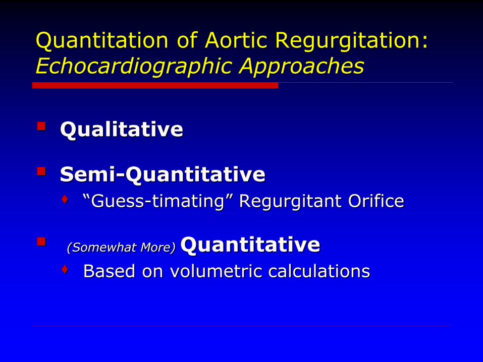
Quantitation of Aortic Regurgitation:Echocardiographic Approaches
Qualitative
Semi-Quantitative
“Guess-timating” Regurgitant Orifice
(Somewhat More) Quantitative
Based on volumetric calculations
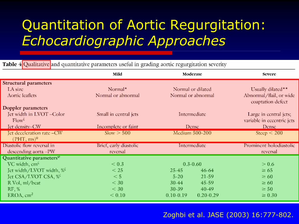
Quantitation of Aortic Regurgitation:Echocardiographic Approaches
Zoghbi et al. JASE (2003) 16:777-802.
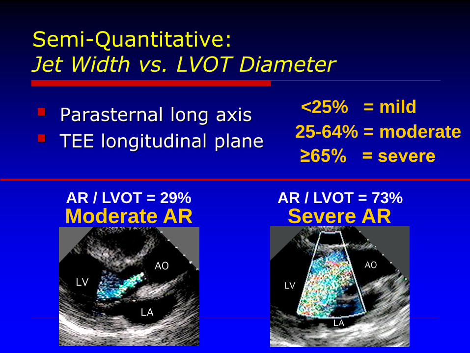
Semi-Quantitative:Jet Width vs. LVOT Diameter
Parasternal long axis
TEE longitudinal plane
<25% = mild
25-64% = moderate
≥65% = severe
AR / LVOT = 29%
Moderate ARAR / LVOT = 73%
Severe AR
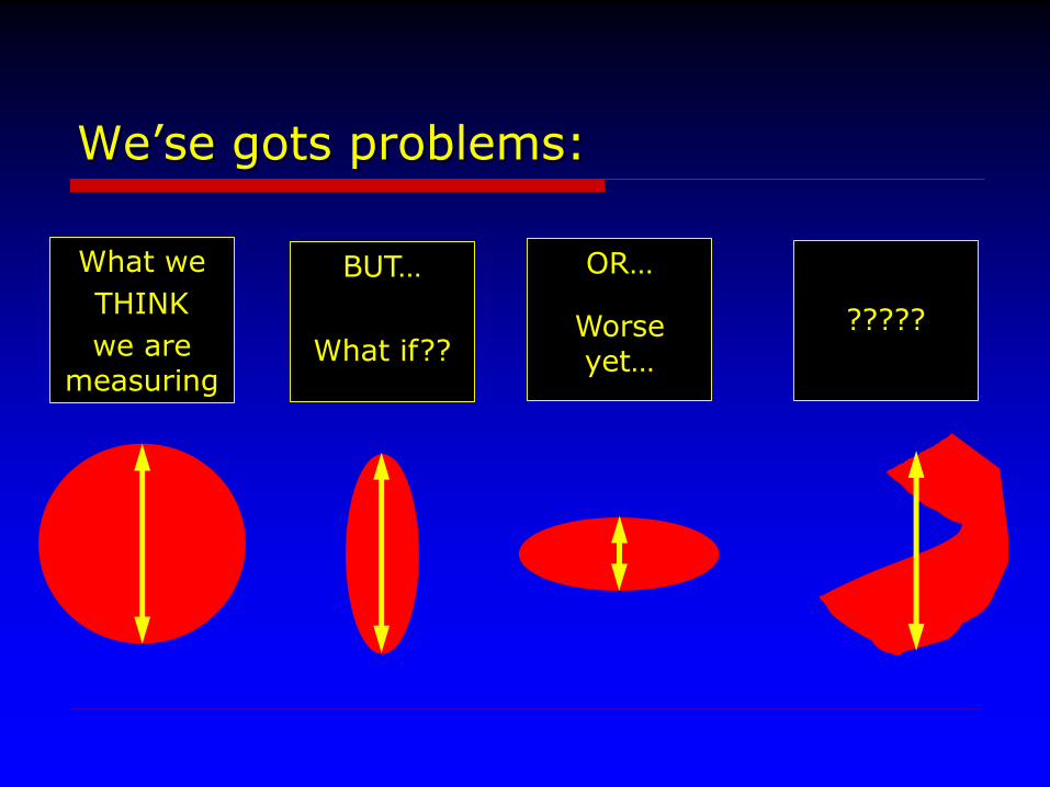
BUT…
What if??
We’se gots problems:
What we
THINK
we are measuring
OR…
Worse yet…
?????
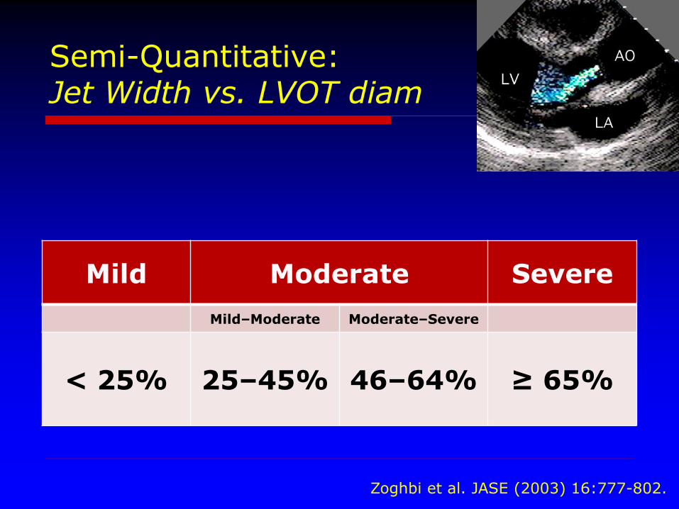
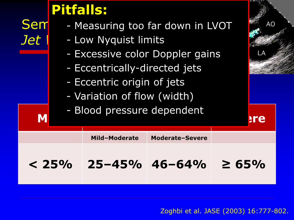
Semi-Quantitative:Jet Width vs. LVOT diam
Mild Moderate Severe
Mild–Moderate Moderate–Severe
< 25% 25–45% 46–64% ≥ 65%
Zoghbi et al. JASE (2003) 16:777-802.
Semi-Quantitative:Jet Width vs. LVOT diam
Mild Moderate Severe
Mild–Moderate Moderate–Severe
< 25% 25–45% 46–64% ≥ 65%
Zoghbi et al. JASE (2003) 16:777-802.
Pitfalls:- Measuring too far down in LVOT
- Low Nyquist limits
- Excessive color Doppler gains
- Eccentrically-directed jets
- Eccentric origin of jets
- Variation of flow (width)
- Blood pressure dependent
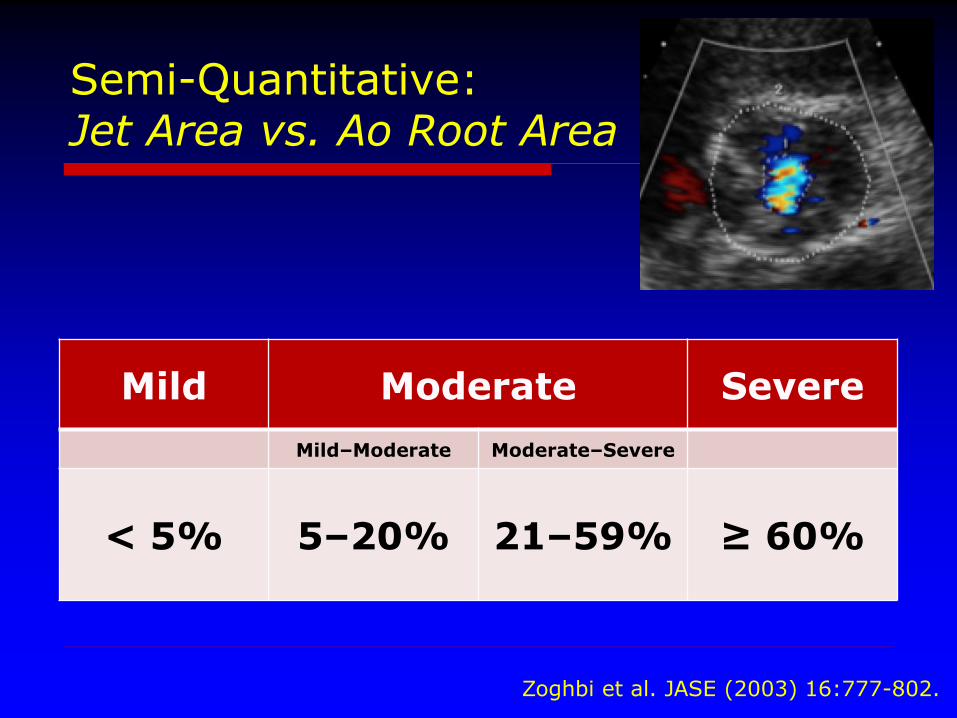
Semi-Quantitative:Jet Area vs. Ao Root Area
Mild Moderate Severe
Mild–Moderate Moderate–Severe
< 5% 5–20% 21–59% ≥ 60%
Zoghbi et al. JASE (2003) 16:777-802.

Semi-Quantitative:Jet Area vs. Ao Root Area
Mild Moderate Severe
Mild–Moderate Moderate–Severe
< 5% 5–20% 21–59% ≥ 60%
Zoghbi et al. JASE (2003) 16:777-802.
Pitfalls:- Measuring below the valve
- Low Nyquist limits
- Excessive color Doppler gains
- Multiple jets
- Variation of flow (width)
- Blood pressure dependent
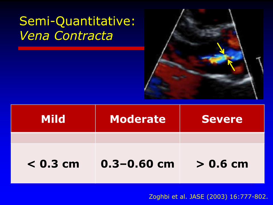
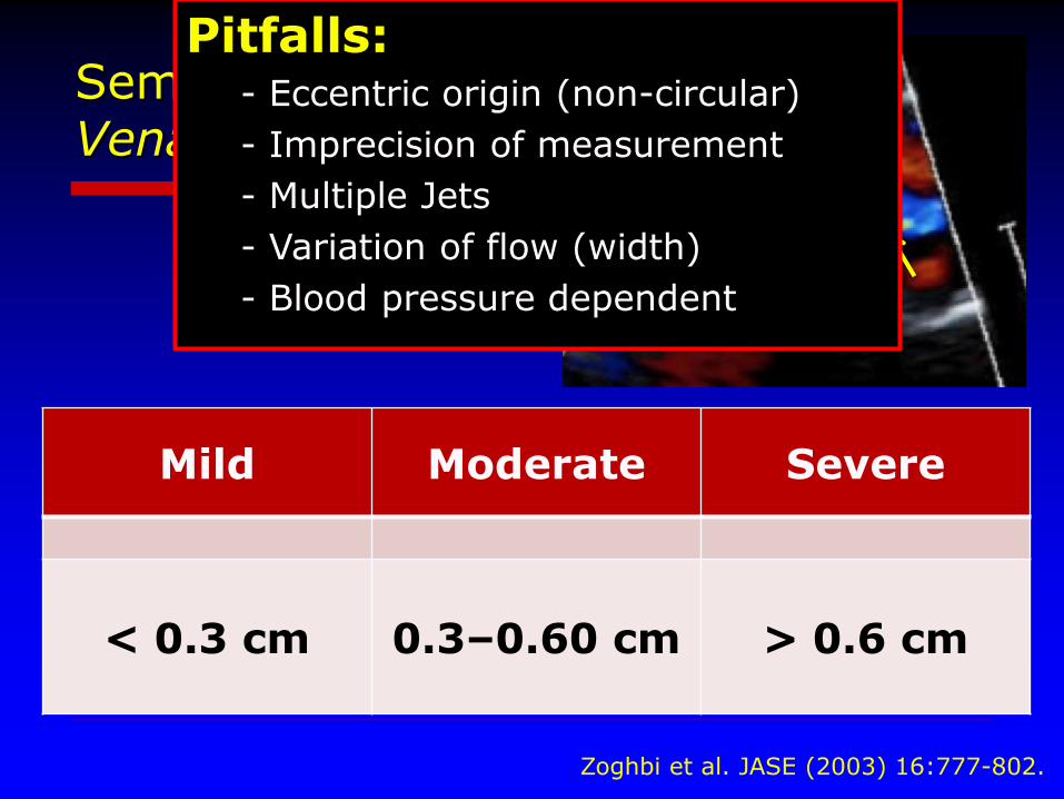
Semi-Quantitative:Vena Contracta
Mild Moderate Severe
< 0.3 cm 0.3–0.60 cm > 0.6 cm
Zoghbi et al. JASE (2003) 16:777-802.
Semi-Quantitative:Vena Contracta
Mild Moderate Severe
< 0.3 cm 0.3–0.60 cm > 0.6 cm
Zoghbi et al. JASE (2003) 16:777-802.
Pitfalls:- Eccentric origin (non-circular)
- Imprecision of measurement
- Multiple Jets
- Variation of flow (width)
- Blood pressure dependent
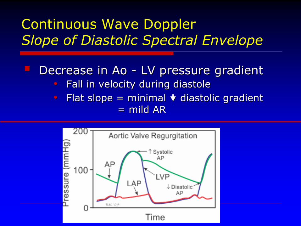
Continuous Wave DopplerSlope of Diastolic Spectral Envelope
Decrease in Ao - LV pressure gradient• Fall in velocity during diastole
• Flat slope = minimal diastolic gradient = mild AR
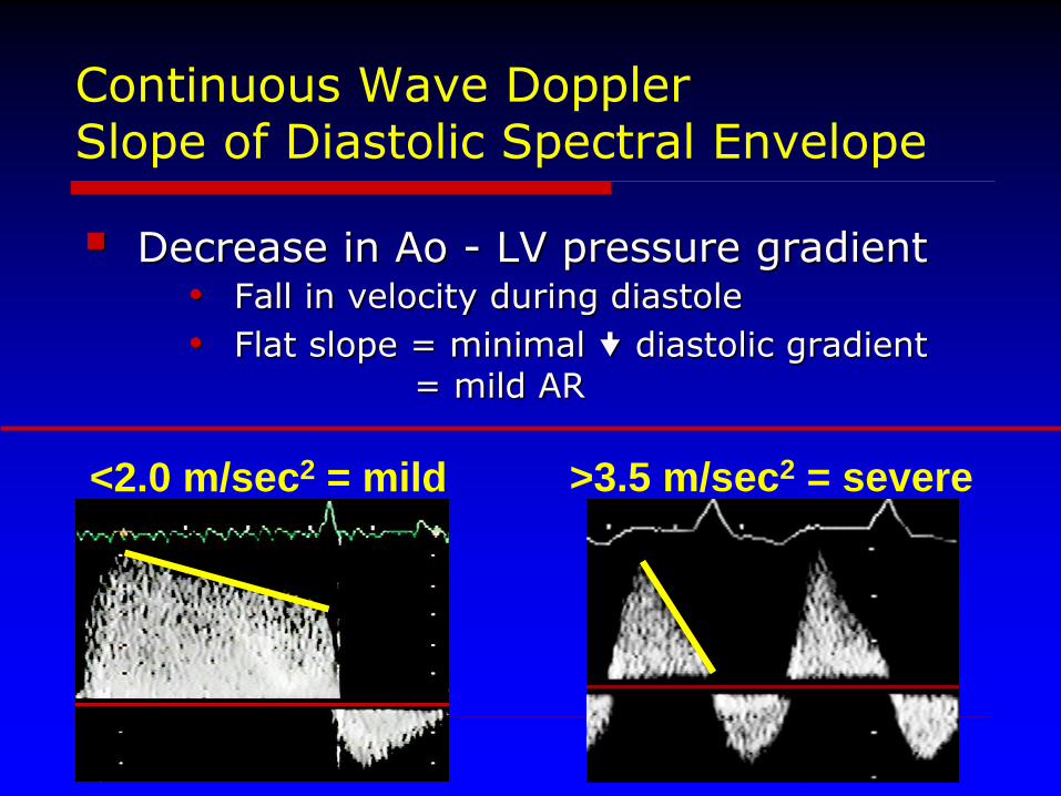
Continuous Wave DopplerSlope of Diastolic Spectral Envelope
Decrease in Ao - LV pressure gradient• Fall in velocity during diastole
• Flat slope = minimal diastolic gradient = mild AR
<2.0 m/sec2 = mild >3.5 m/sec2 = severe
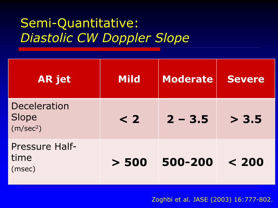
Semi-Quantitative:Diastolic CW Doppler Slope
AR jet Mild Moderate Severe
Deceleration Slope(m/sec2)
< 2 2 – 3.5 > 3.5
Pressure Half-time(msec)
> 500 500-200 < 200
Zoghbi et al. JASE (2003) 16:777-802.
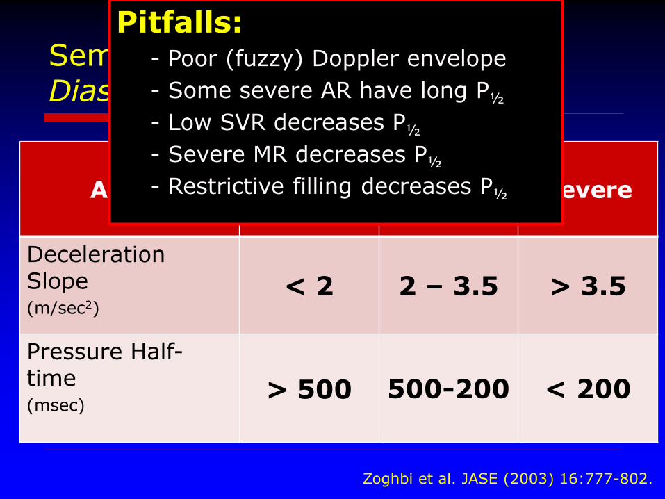
Semi-Quantitative:Diastolic CW Doppler Slope
AR jet Mild Moderate Severe
Deceleration Slope(m/sec2)
< 2 2 – 3.5 > 3.5
Pressure Half-time(msec)
> 500 500-200 < 200
Zoghbi et al. JASE (2003) 16:777-802.
Pitfalls:- Poor (fuzzy) Doppler envelope
- Some severe AR have long P½
- Low SVR decreases P½
- Severe MR decreases P½
- Restrictive filling decreases P½
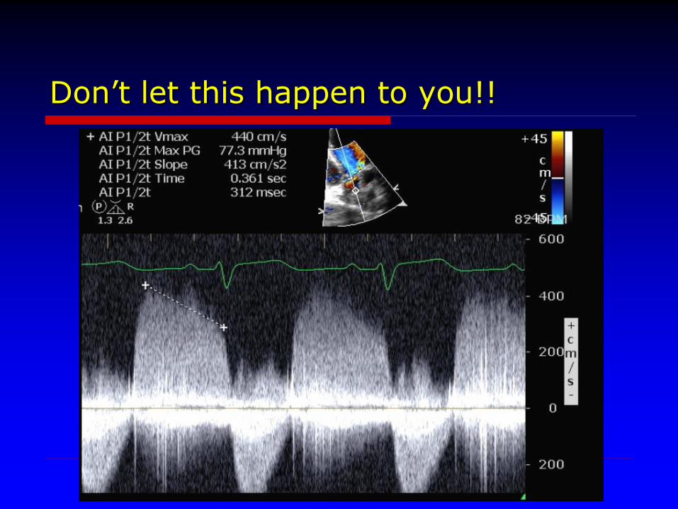
Don’t let this happen to you!!
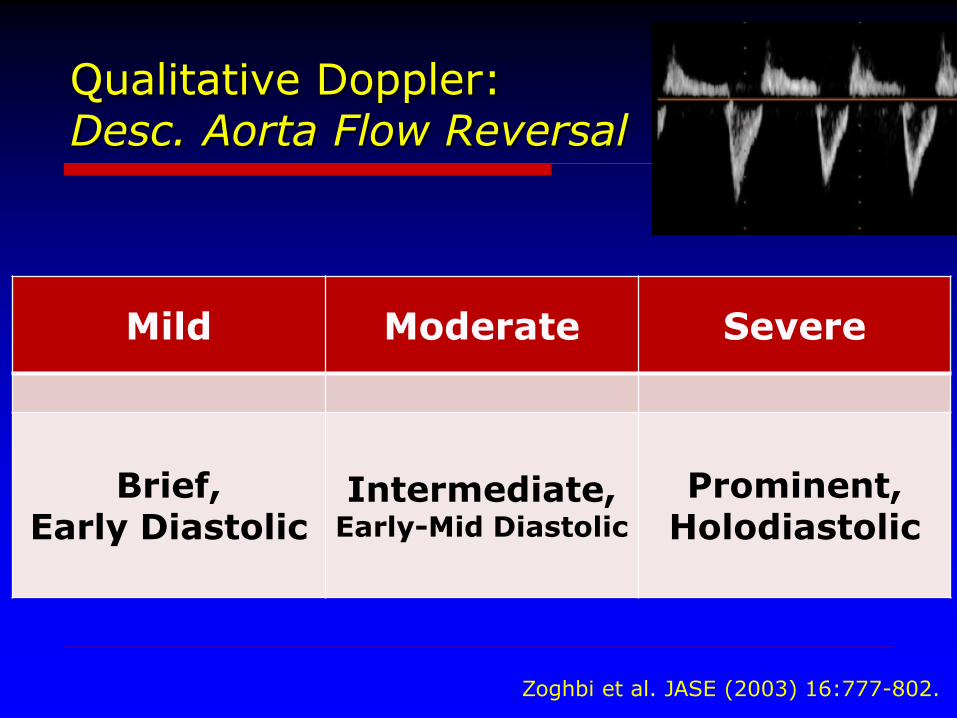
Qualitative Doppler:Desc. Aorta Flow Reversal
Mild Moderate Severe
Brief,Early Diastolic
Intermediate,Early-Mid Diastolic
Prominent,Holodiastolic
Zoghbi et al. JASE (2003) 16:777-802.
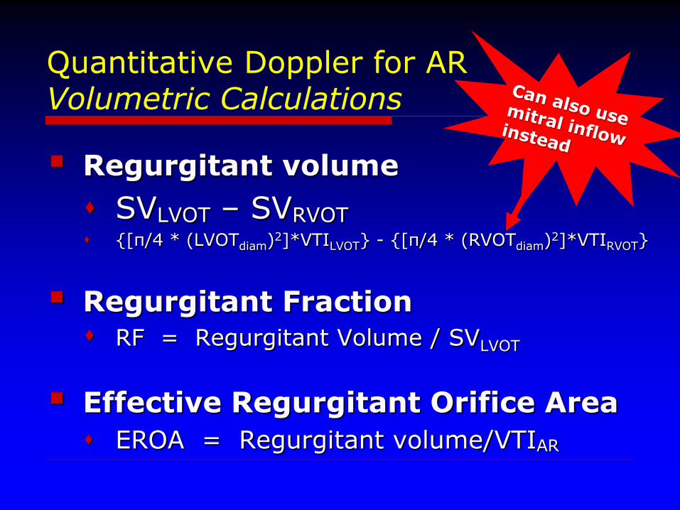
Quantitative Doppler for ARVolumetric Calculations
Regurgitant volume
SVLVOT – SVRVOT {[π/4 * (LVOTdiam)2]*VTILVOT} - {[π/4 * (RVOTdiam)2]*VTIRVOT}
Regurgitant Fraction RF = Regurgitant Volume / SVLVOT
Effective Regurgitant Orifice Area
EROA = Regurgitant volume/VTIAR
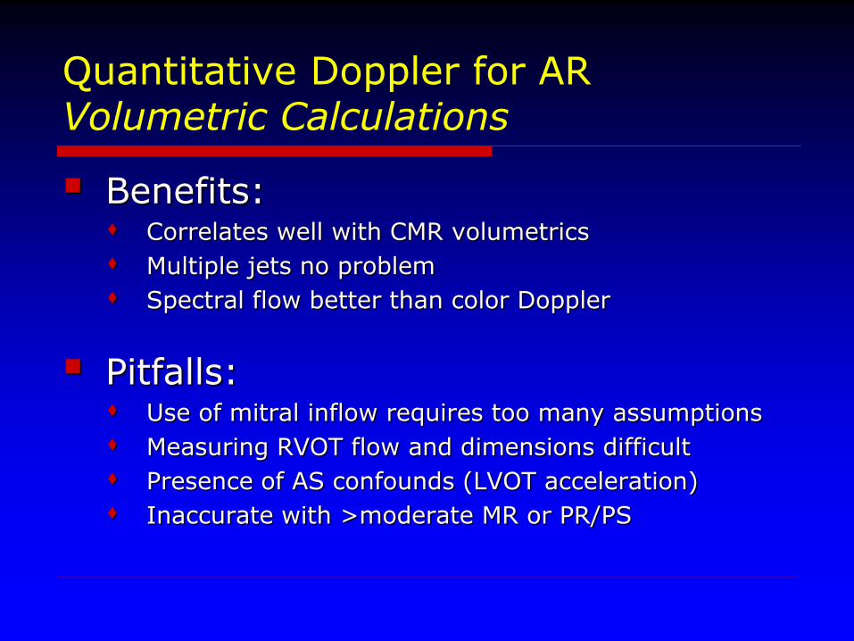
Quantitative Doppler for ARVolumetric Calculations
Benefits: Correlates well with CMR volumetrics
Multiple jets no problem
Spectral flow better than color Doppler
Pitfalls: Use of mitral inflow requires too many assumptions
Measuring RVOT flow and dimensions difficult
Presence of AS confounds (LVOT acceleration)
Inaccurate with >moderate MR or PR/PS
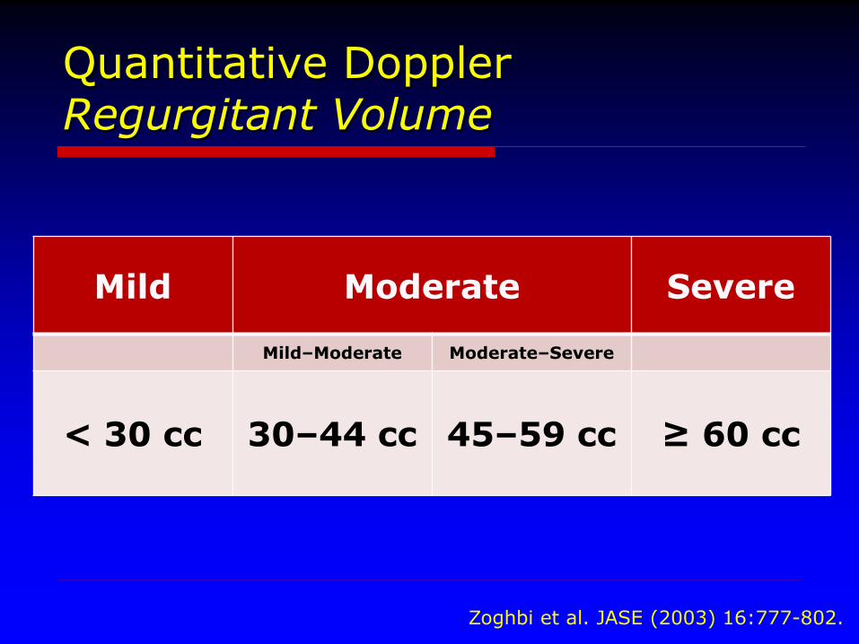
Quantitative DopplerRegurgitant Volume
Zoghbi et al. JASE (2003) 16:777-802.
Mild Moderate Severe
Mild–Moderate Moderate–Severe
< 30 cc 30–44 cc 45–59 cc ≥ 60 cc
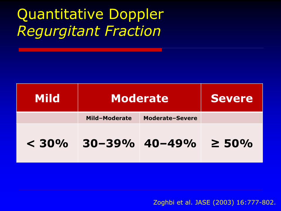
Quantitative DopplerRegurgitant Fraction
Mild Moderate Severe
Mild–Moderate Moderate–Severe
< 30% 30–39% 40–49% ≥ 50%
Zoghbi et al. JASE (2003) 16:777-802.
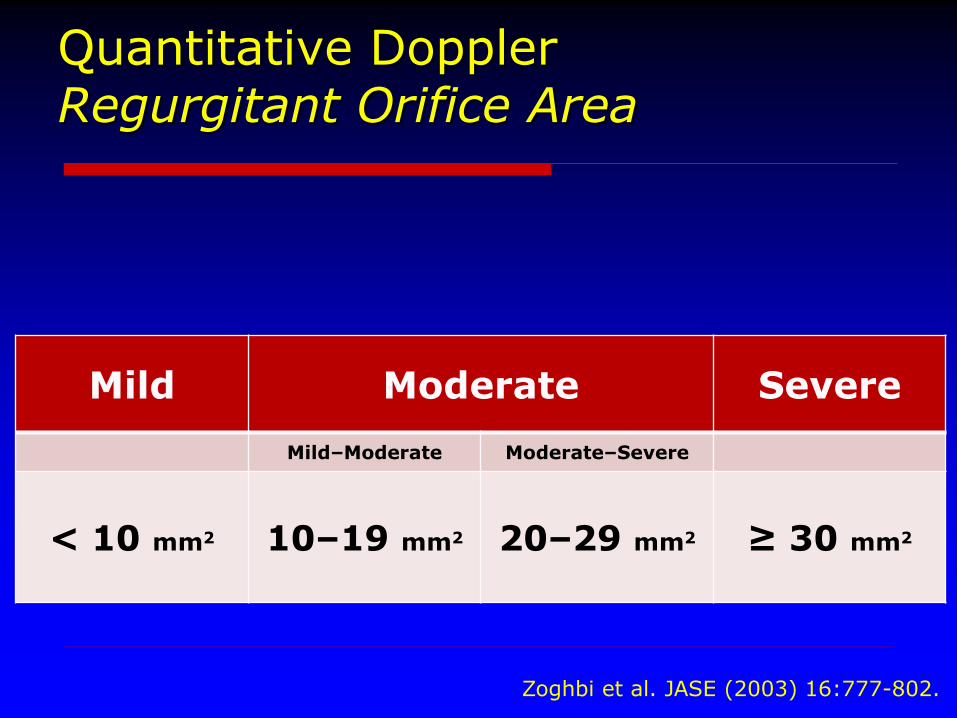
Quantitative DopplerRegurgitant Orifice Area
Mild Moderate Severe
Mild–Moderate Moderate–Severe
< 10 mm2 10–19 mm2 20–29 mm2 ≥ 30 mm2
Zoghbi et al. JASE (2003) 16:777-802.
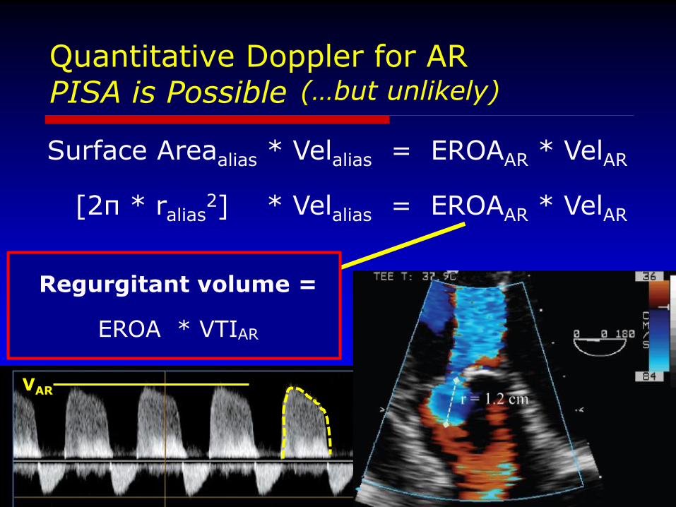
Quantitative Doppler for ARPISA is Possible
Surface Areaalias * Velalias = EROAAR * VelAR
[2π * ralias2] * Velalias = EROAAR * VelAR
[2π * ralias2] * Velalias
VelAR
VAR
(…but unlikely)
Regurgitant volume =
EROA * VTIAR

WHY DO WE CARE ABOUT THIS??
Timing of Surgical Intervention
Appropriate Patient Follow Up
Passing Echo Board Exams
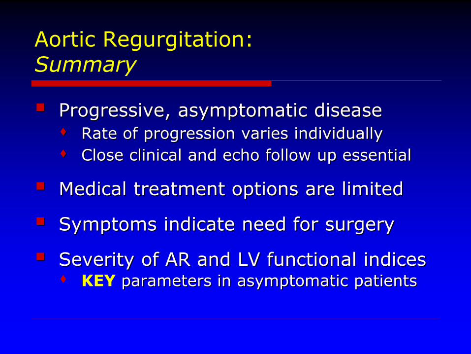
Aortic Regurgitation:Summary
Progressive, asymptomatic disease Rate of progression varies individually
Close clinical and echo follow up essential
Medical treatment options are limited
Symptoms indicate need for surgery
Severity of AR and LV functional indices KEY parameters in asymptomatic patients
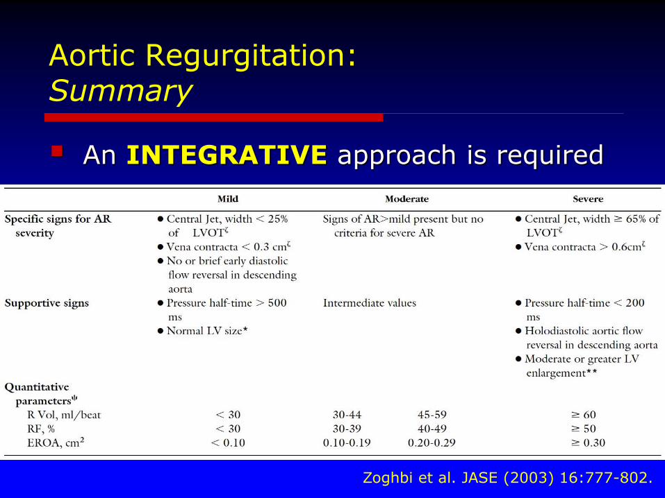
Aortic Regurgitation:Summary
An INTEGRATIVE approach is required
Zoghbi et al. JASE (2003) 16:777-802.

Review Question #1
Possible reasons that thiscolor Doppler evaluation mayoverestimate AR include:
A. Low color Doppler gain setting
B. Use of too small a color Doppler sector
C. Low Nyquist velocity setting
D. High pulse repetition frequency (PRF)
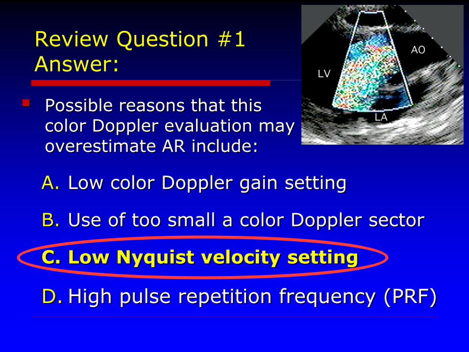
Review Question #1Answer:
Possible reasons that thiscolor Doppler evaluation mayoverestimate AR include:
A. Low color Doppler gain setting
B. Use of too small a color Doppler sector
C. Low Nyquist velocity setting
D. High pulse repetition frequency (PRF)

Review Question #2
Which condition may lead to over-estimation of AR severity by deceleration slope (or P1/2) of diastolic Continuous Wave (CW) Doppler:
A. Advanced restrictive myocardial disease
B. Severe mitral valve stenosis
C. Severe aortic valve stenosis
D. Low Nyquist limit setting
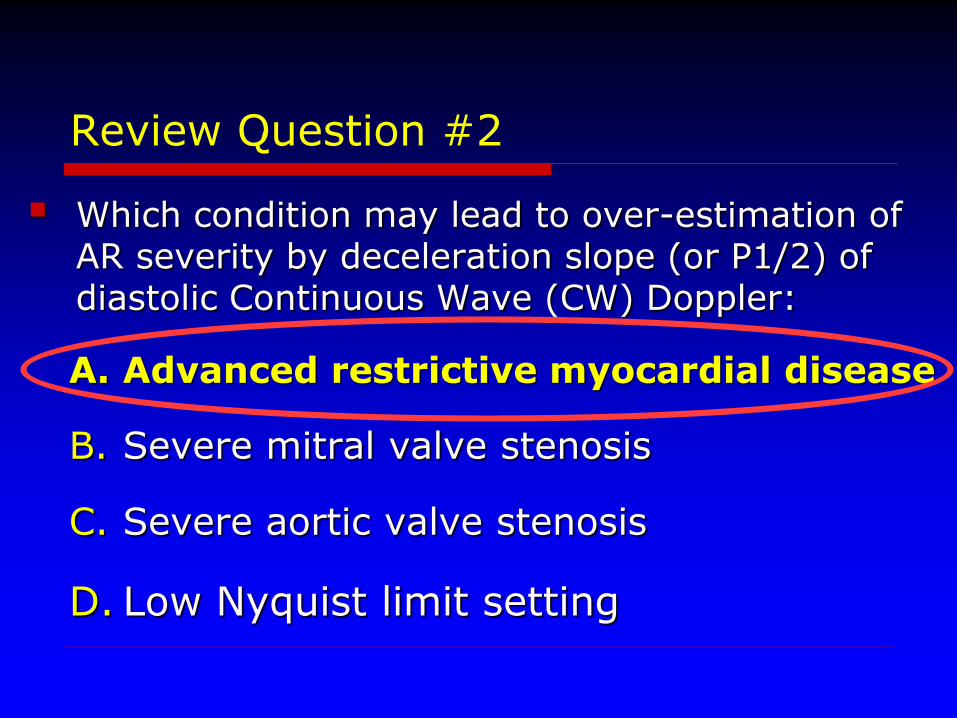
Review Question #2
Which condition may lead to over-estimation of AR severity by deceleration slope (or P1/2) of diastolic Continuous Wave (CW) Doppler:
A. Advanced restrictive myocardial disease
B. Severe mitral valve stenosis
C. Severe aortic valve stenosis
D. Low Nyquist limit setting
Review Question #3
A continuous wave Doppler cursor is placed at the junction of the aortic arch and proximaldescending aorta, just beyond the left subclavian artery. The following is obtained:

Review Question #3
The etiology of the diastolic Doppler flow indicated by the arrow is:
A. Stenosis of the left subclavian artery
B. Severe aortic regurgitation
C. Moderate aortic regurgitation
D. Severe coarctation of the aorta
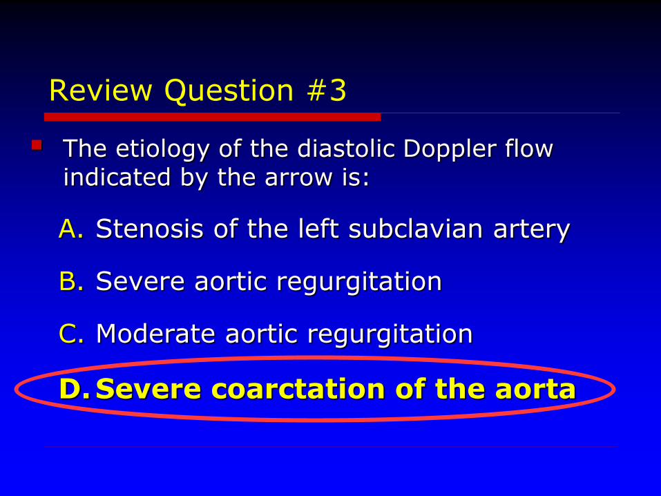
Review Question #3
The etiology of the diastolic Doppler flow indicated by the arrow is:
A. Stenosis of the left subclavian artery
B. Severe aortic regurgitation
C. Moderate aortic regurgitation
D.Severe coarctation of the aorta

Thank You!