Embed Size (px)
Citation preview
2006-2007AP Biology
Muscles & Motor Locomotion
Why Do We Need All
That ATP?
AP Biology
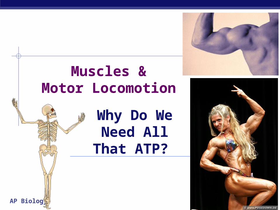
Animal LocomotionWhat are the advantages of locomotion?
motilesessile

AP Biology
Lots of ways to get around…

AP Biology
Lots of ways to get around…
mollusk mammalbird reptile

AP Biology
Lots of ways to get around…
bird arthropodmammal bird
AP Biology
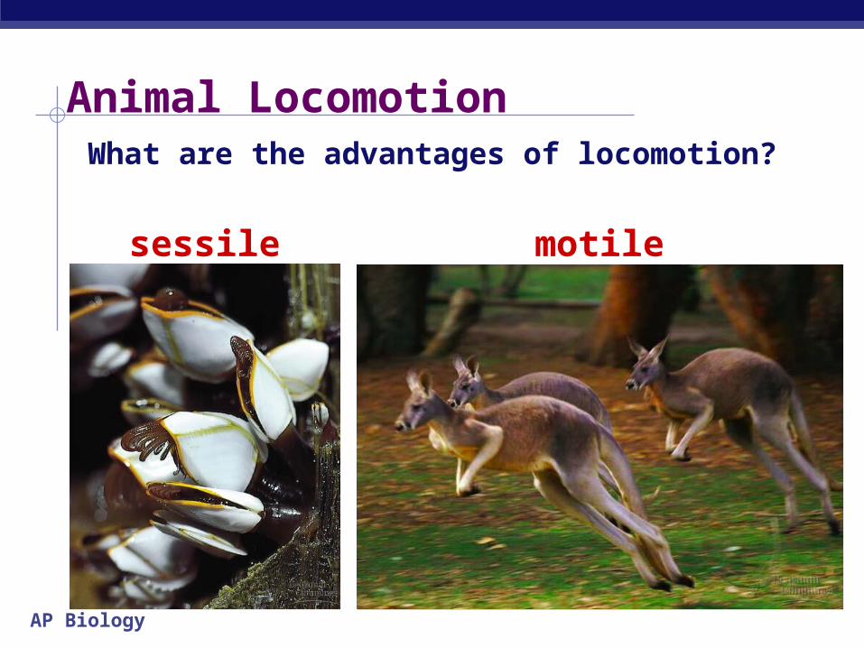
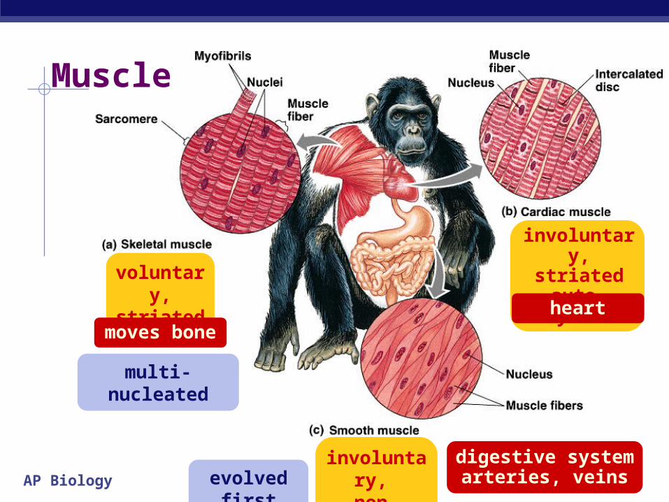
Muscle
voluntary, striated
involuntary, striated
auto-rhythmic
involuntary,
non-striated
evolved first
multi-nucleated
digestive systemarteries, veins
heartmoves bone
AP Biology
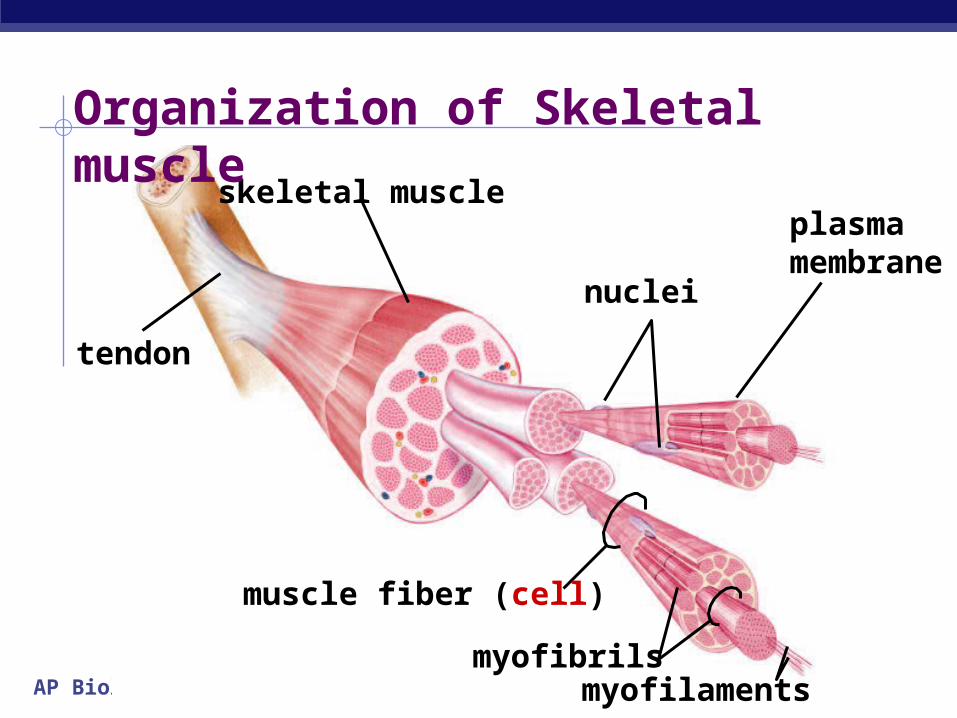
tendon
skeletal muscle
muscle fiber (cell)
myofilamentsmyofibrils
plasma membrane
nuclei
Organization of Skeletal muscle
AP Biology
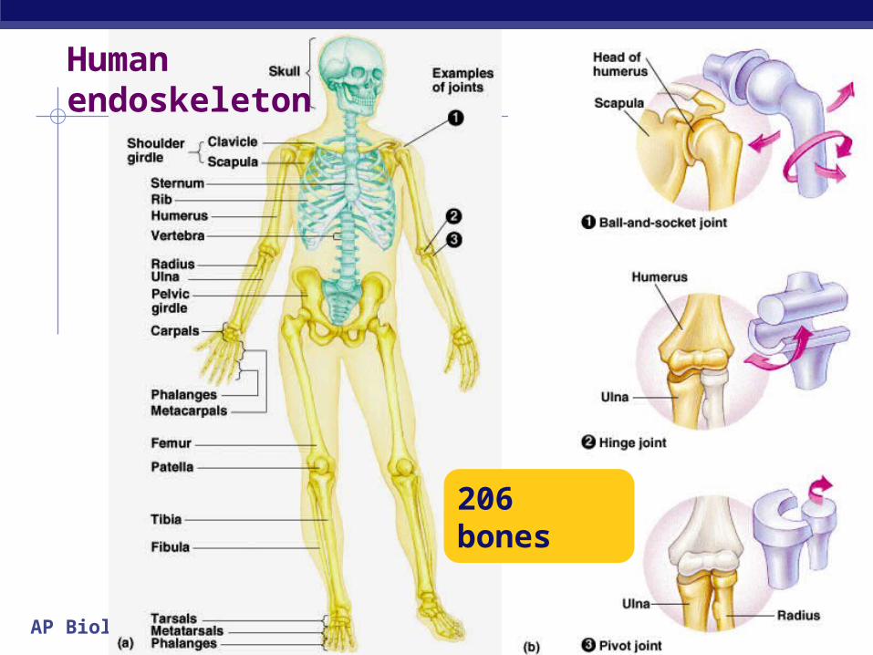
Human endoskeleton
206 bones
AP Biology
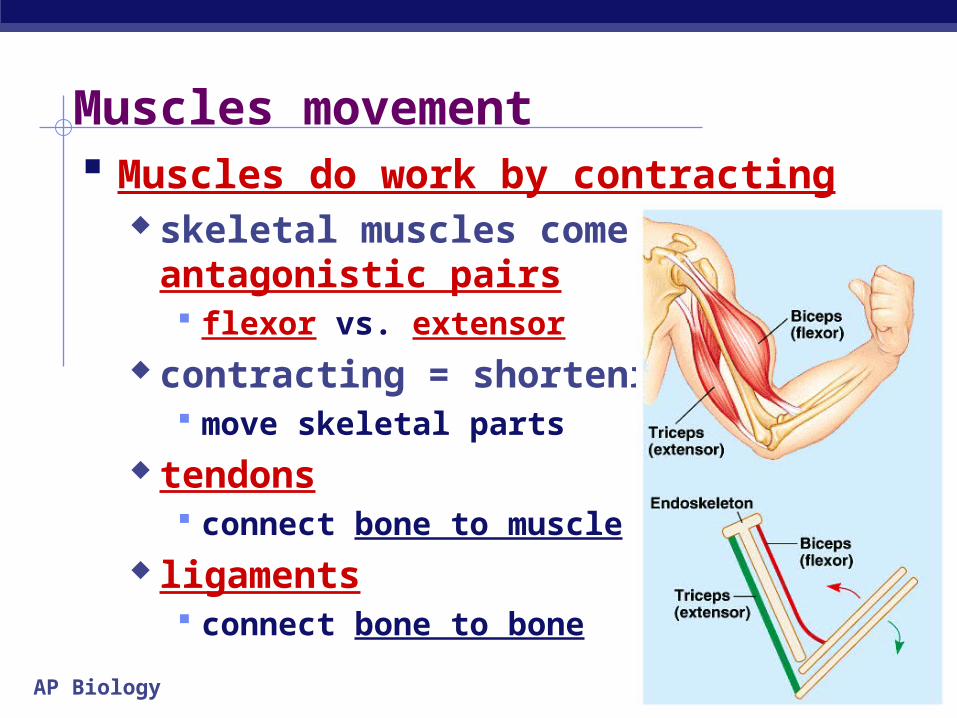
Muscles movement Muscles do work by contracting
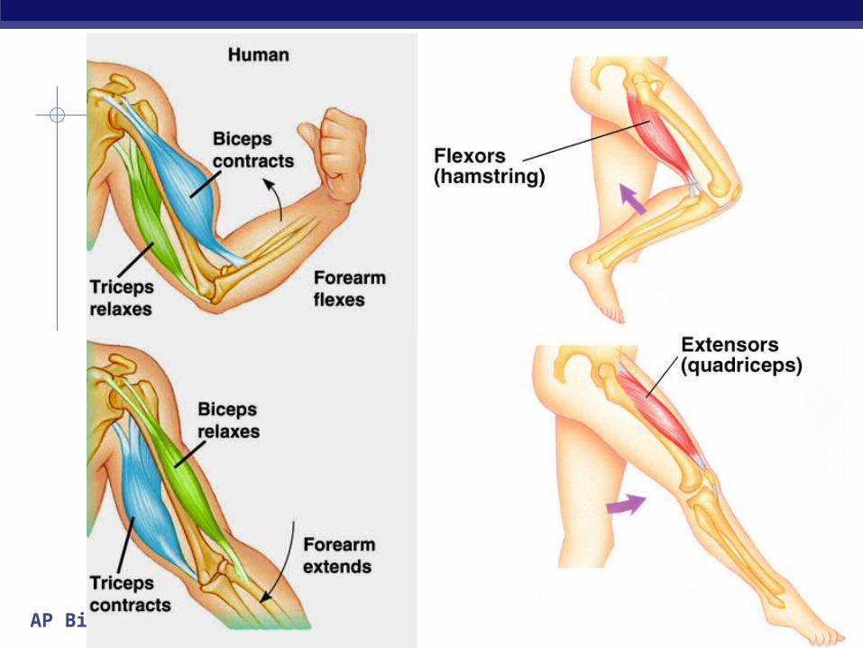
skeletal muscles come in antagonistic pairs flexor vs. extensor
contracting = shortening move skeletal parts
tendons connect bone to muscle
ligaments connect bone to bone
AP Biology

AP Biology
Structure of striated skeletal muscle Muscle Fiber
muscle cell divided into sections = sarcomeres
Sarcomere functional unit of muscle
contraction alternating bands of
thin (actin) & thick (myosin) protein filaments
AP Biology
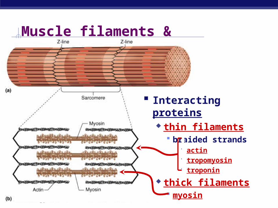
Muscle filaments & Sarcomere
Interacting proteins thin filaments
braided strands actin tropomyosin troponin
thick filaments myosin
AP Biology
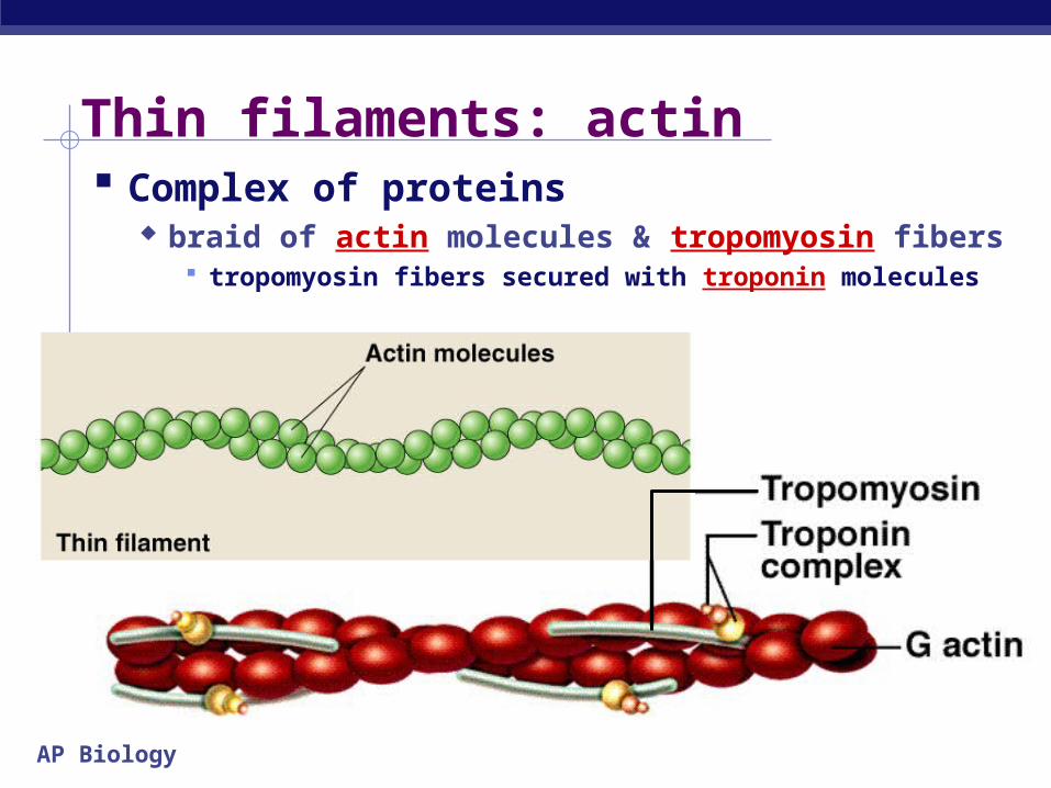
Thin filaments: actin Complex of proteins
braid of actin molecules & tropomyosin fibers tropomyosin fibers secured with troponin molecules
AP Biology
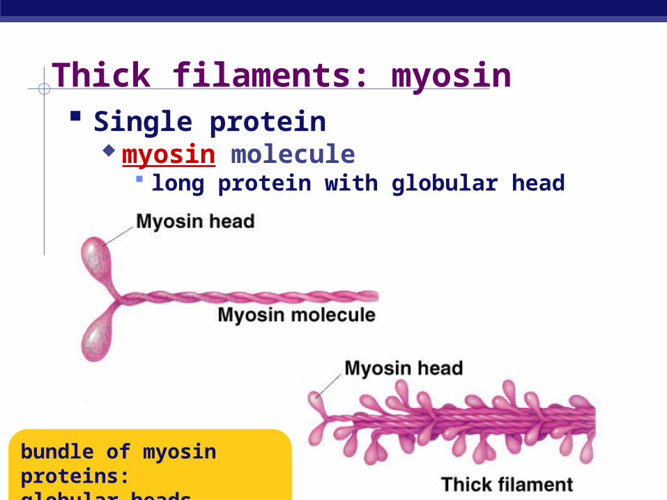
Thick filaments: myosin Single protein
myosin molecule long protein with globular head
bundle of myosin proteins:globular heads aligned
AP Biology
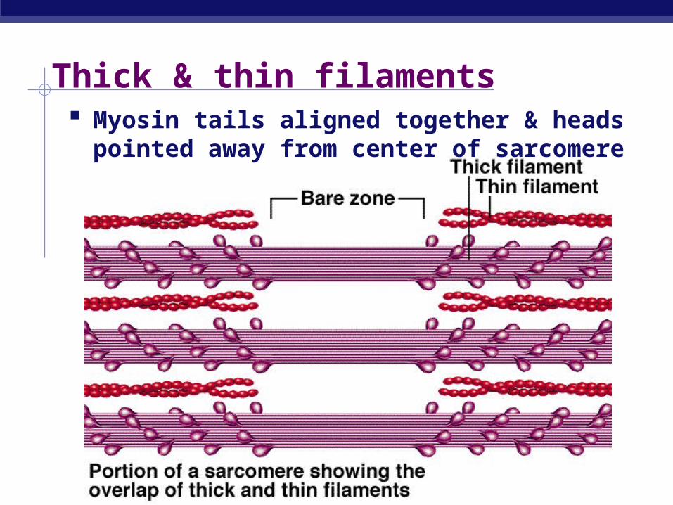
Thick & thin filaments Myosin tails aligned together & heads pointed
away from center of sarcomere
AP Biology
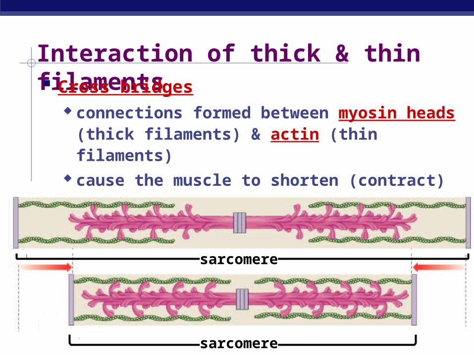
Interaction of thick & thin filaments Cross bridges
connections formed between myosin heads (thick filaments) & actin (thin filaments)
cause the muscle to shorten (contract)
sarcomere
sarcomere
AP Biology
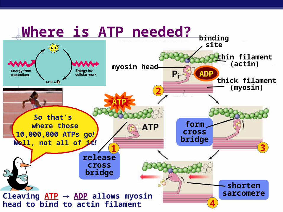
Where is ATP needed?
3
4
12
1
1
1
Cleaving ATP ADP allows myosin head to bind to actin filament
thin filament(actin)
thick filament(myosin)
ATP
myosin head
formcrossbridge
binding site
So that’s where those
10,000,000 ATPs go!Well, not all of it!
ADP
releasecrossbridge
shortensarcomere
1
AP Biology
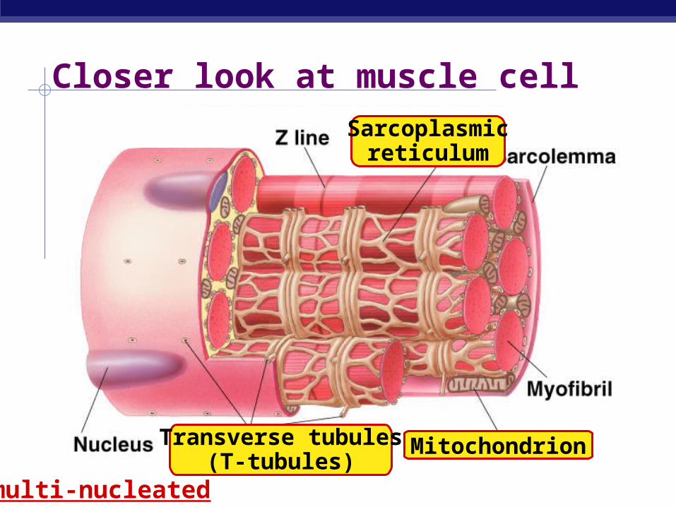
Closer look at muscle cell
multi-nucleated
Mitochondrion
Sarcoplasmicreticulum
Transverse tubules(T-tubules)
AP Biology
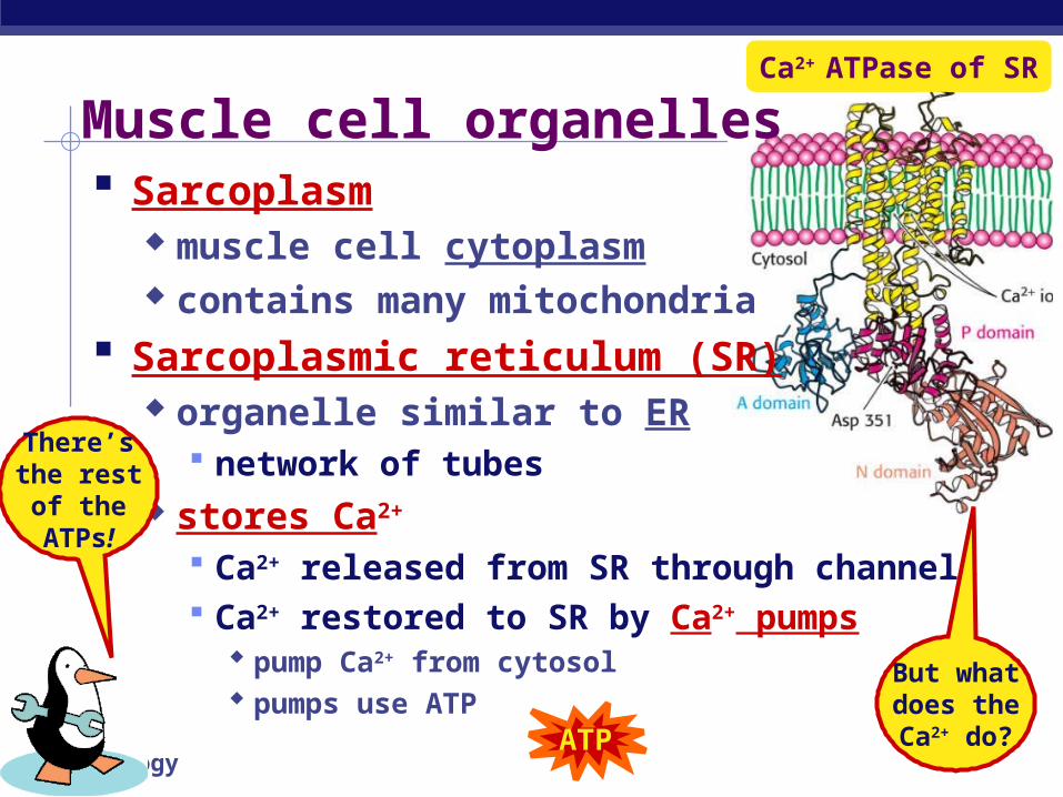
Muscle cell organelles Sarcoplasm
muscle cell cytoplasm contains many mitochondria
Sarcoplasmic reticulum (SR) organelle similar to ER
network of tubes stores Ca2+
Ca2+ released from SR through channels Ca2+ restored to SR by Ca2+ pumps
pump Ca2+ from cytosol pumps use ATP
Ca2+ ATPase of SR
ATP
There’sthe restof theATPs!
But whatdoes theCa2+ do?
AP Biology
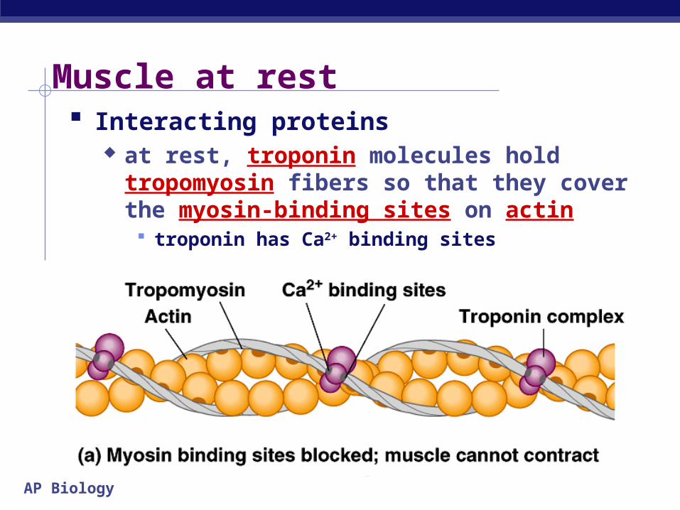
Muscle at rest Interacting proteins
at rest, troponin molecules hold tropomyosin fibers so that they cover the myosin-binding sites on actin troponin has Ca2+ binding sites
AP Biology
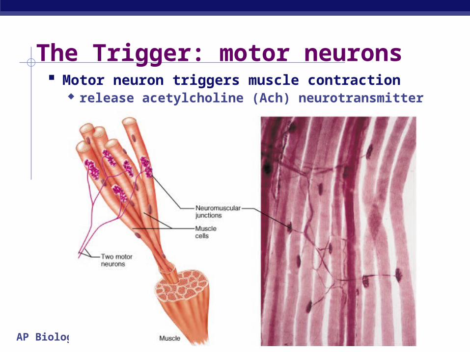
The Trigger: motor neurons Motor neuron triggers muscle contraction
release acetylcholine (Ach) neurotransmitter
AP Biology
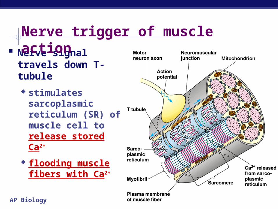
Nerve signal travels down T-tubule
stimulates sarcoplasmic reticulum (SR) of muscle cell to release stored Ca2+
flooding muscle fibers with Ca2+
Nerve trigger of muscle action

AP Biology
At rest, tropomyosin blocks myosin-binding sites on actin secured by troponin
Ca2+ binds to troponin shape change
causes movement of troponin
releasing tropomyosin exposes myosin-
binding sites on actin
Ca2+ triggers muscle action
AP Biology
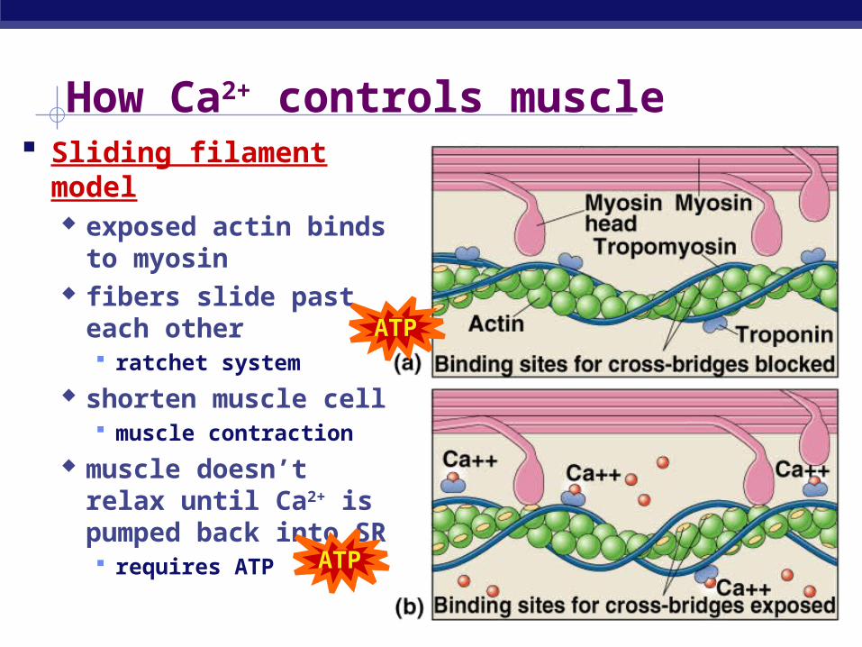
How Ca2+ controls muscle Sliding filament model
exposed actin binds to myosin
fibers slide past each other ratchet system
shorten muscle cell muscle contraction
muscle doesn’t relax until Ca2+ is pumped back into SR requires ATP
ATP
ATP
AP Biology
Put it all together…1
ATP
2
3
4
5
7
6
ATP
AP Biology
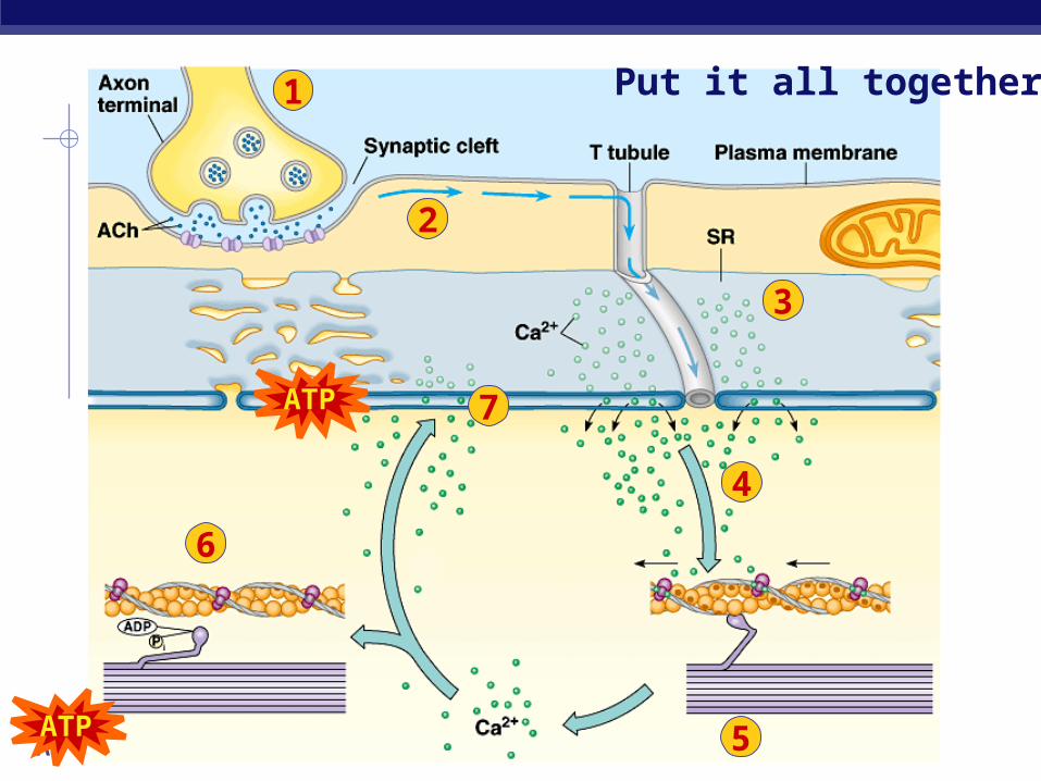
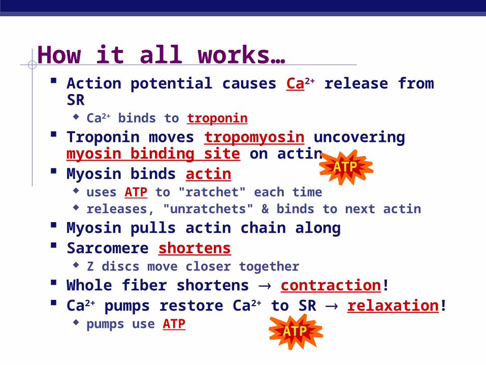
How it all works… Action potential causes Ca2+ release from SR
Ca2+ binds to troponin
Troponin moves tropomyosin uncovering myosin binding site on actin
Myosin binds actin uses ATP to "ratchet" each time releases, "unratchets" & binds to next actin
Myosin pulls actin chain along Sarcomere shortens
Z discs move closer together
Whole fiber shortens contraction! Ca2+ pumps restore Ca2+ to SR relaxation!
pumps use ATP
ATP
ATP
AP Biology
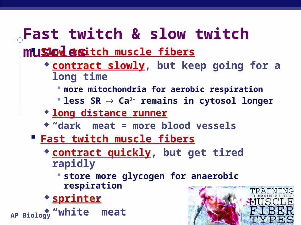
Fast twitch & slow twitch muscles Slow twitch muscle fibers
contract slowly, but keep going for a long time more mitochondria for aerobic respiration less SR Ca2+ remains in cytosol longer
long distance runner “dark” meat = more blood vessels
Fast twitch muscle fibers contract quickly, but get tired rapidly
store more glycogen for anaerobic respiration sprinter “white” meat
AP Biology
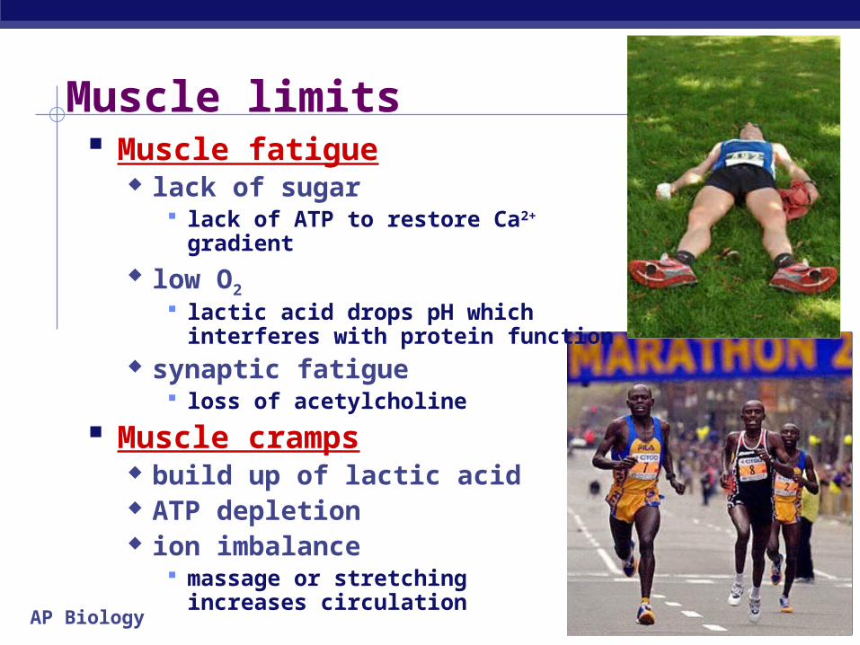
Muscle limits Muscle fatigue
lack of sugar lack of ATP to restore Ca2+ gradient
low O2 lactic acid drops pH which
interferes with protein function synaptic fatigue
loss of acetylcholine
Muscle cramps build up of lactic acid ATP depletion ion imbalance
massage or stretching increases circulation
AP Biology
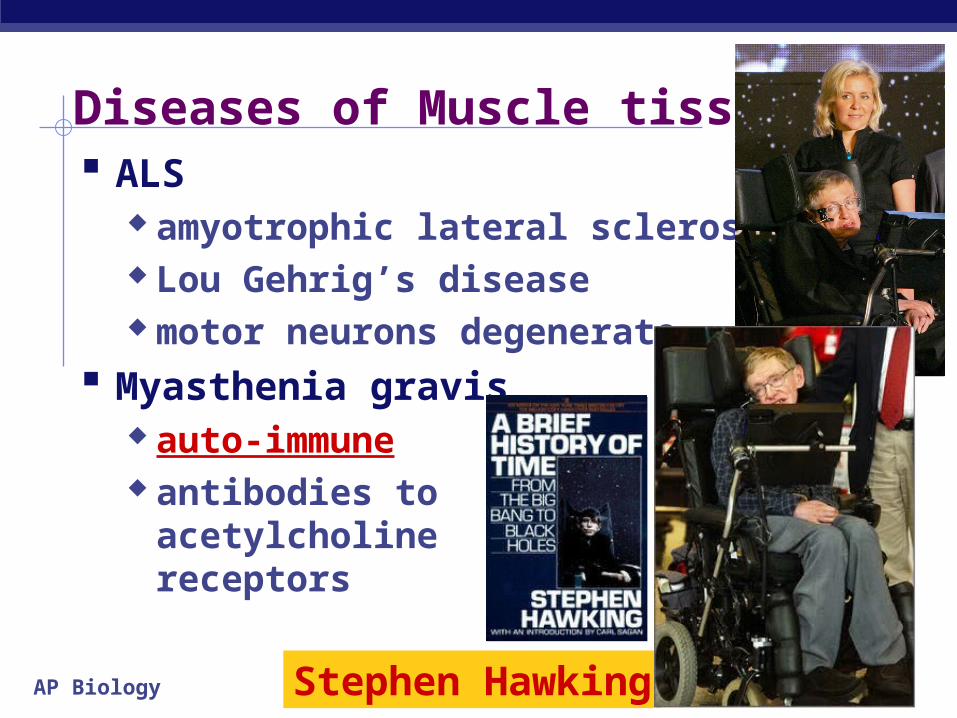
Diseases of Muscle tissue ALS
amyotrophic lateral sclerosis Lou Gehrig’s disease motor neurons degenerate
Myasthenia gravis auto-immune antibodies to
acetylcholine receptors
Stephen Hawking
AP Biology
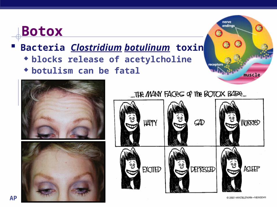
Botox Bacteria Clostridium botulinum toxin
blocks release of acetylcholine botulism can be fatal muscle
AP Biology
Rigor mortis So why are dead people “stiffs”?
no life, no breathing no breathing, no O2
no O2, no aerobic respiration no aerobic respiration, no ATP no ATP, no Ca2+ pumps Ca2+ stays in muscle cytoplasm muscle fibers continually
contract tetany or rigor mortis
eventually tissues breakdown& relax measure of time of death

2006-2007AP Biology
So don’t be a stiff!Ask Questions!!
AP Biology
The way it ISN’T: sensing brain analysis action.
The way it is: sensing, analysis, and action are ongoing and overlapping processes.
Sensations begin as different forms of energy that are detected by sensory receptors. This energy is converted to action potentials
that travel to appropriate regions of the brain. The limbic region plays a major role in determining
the importance of a particular sensory input.
Processing of input and output is cyclical
Copyright © 2002 Pearson Education, Inc., publishing as Benjamin Cummings
AP Biology
Sensations are action potentials that reach the brain via sensory neurons.
Perception is the awareness and interpretation of the sensation.
Copyright © 2002 Pearson Education, Inc., publishing as Benjamin Cummings
AP Biology
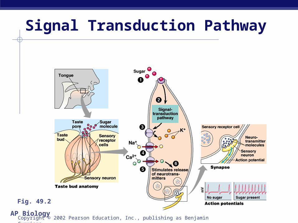
Signal Transduction Pathway
Copyright © 2002 Pearson Education, Inc., publishing as Benjamin Cummings
Fig. 49.2
AP Biology
Sensory reception begins with the detection of stimuli by sensory receptors. Exteroreceptors detect stimuli originating
outside the body. Interoreceptors detect stimuli originating
inside the body.
Copyright © 2002 Pearson Education, Inc., publishing as Benjamin Cummings
AP Biology
Transduction. The conversion of stimulus energy into a
change in membrane potential. Amplification.
The strengthening of stimulus energy that is can be detected by the nervous system.
Transmission. The conduction of sensory impulses to the
CNS. Integration.
The processing of sensory information.Copyright © 2002 Pearson Education, Inc., publishing as Benjamin Cummings
Sensory Processing
AP Biology
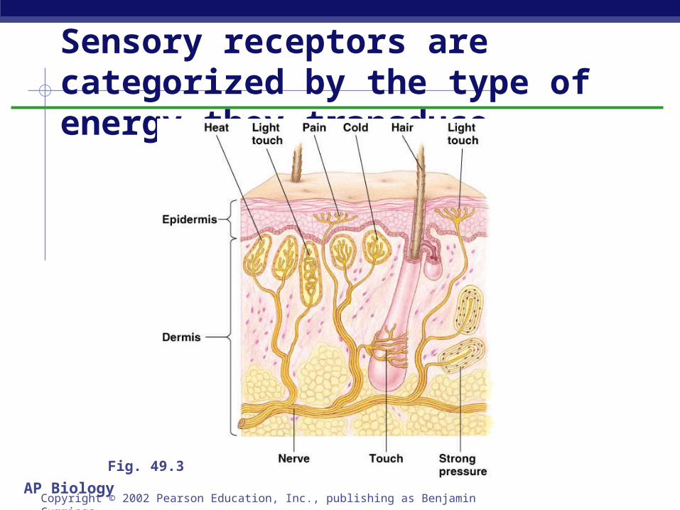
Sensory receptors are categorized by the type of energy they transduce
Copyright © 2002 Pearson Education, Inc., publishing as Benjamin Cummings
Fig. 49.3
AP Biology
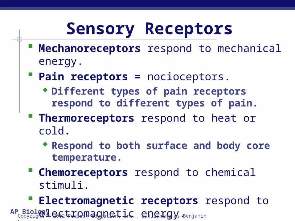
Mechanoreceptors respond to mechanical energy. Pain receptors = nocioceptors.
Different types of pain receptors respond to different types of pain.
Thermoreceptors respond to heat or cold. Respond to both surface and body core
temperature.
Chemoreceptors respond to chemical stimuli. Electromagnetic receptors respond to
electromagnetic energy. Photoreceptors respond to the radiation we know as
visible light.Copyright © 2002 Pearson Education, Inc., publishing as Benjamin Cummings
Sensory Receptors
AP Biology
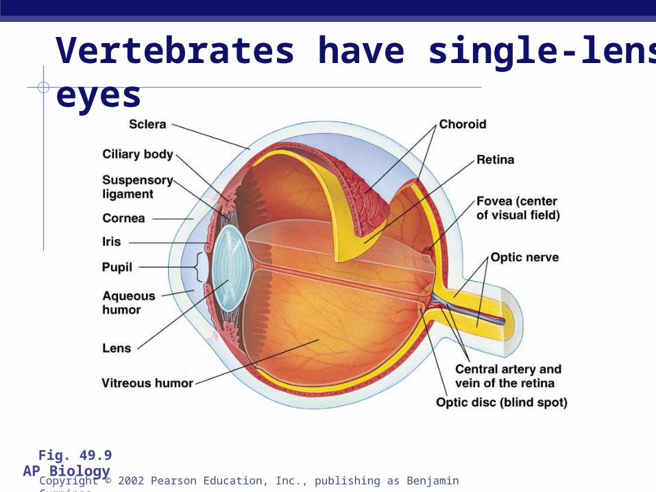
Vertebrates have single-lens eyes
Copyright © 2002 Pearson Education, Inc., publishing as Benjamin Cummings
Fig. 49.9
AP Biology
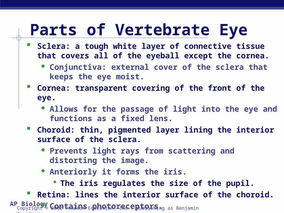
Sclera: a tough white layer of connective tissue that covers all of the eyeball except the cornea.
Conjunctiva: external cover of the sclera that keeps the eye moist.
Cornea: transparent covering of the front of the eye. Allows for the passage of light into the eye and functions as
a fixed lens. Choroid: thin, pigmented layer lining the interior surface of the
sclera. Prevents light rays from scattering and distorting the image. Anteriorly it forms the iris.
The iris regulates the size of the pupil. Retina: lines the interior surface of the choroid.
Contains photoreceptors.
Copyright © 2002 Pearson Education, Inc., publishing as Benjamin Cummings
Parts of Vertebrate Eye
AP Biology
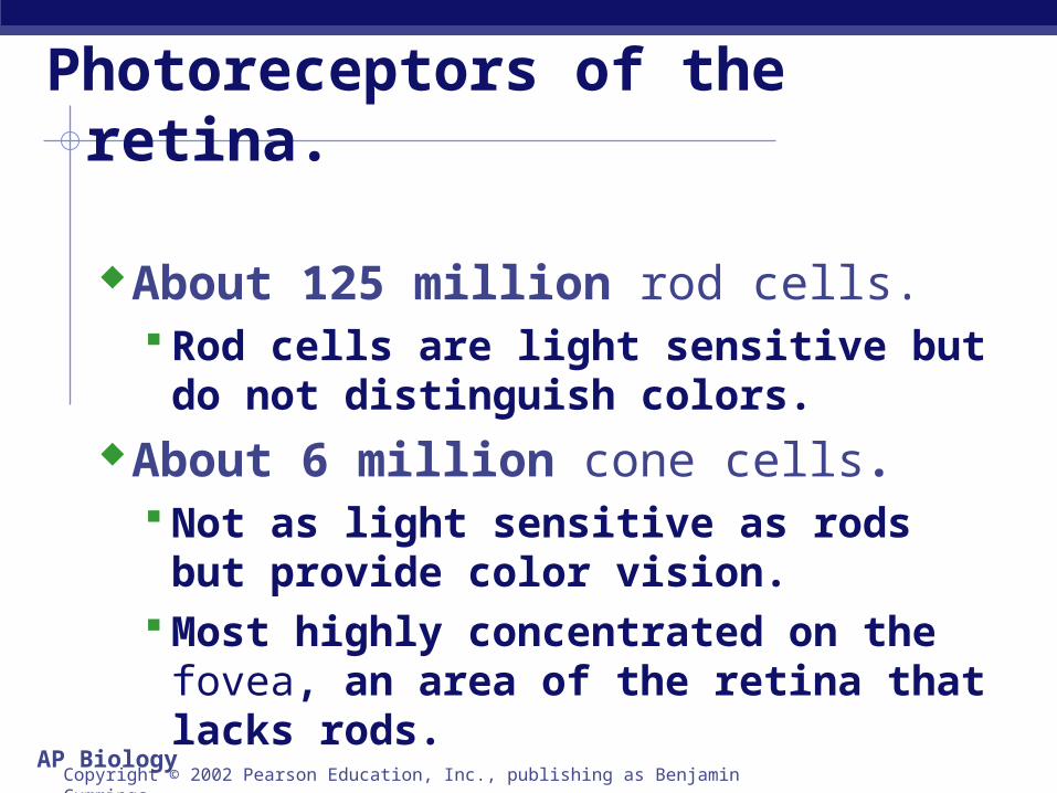
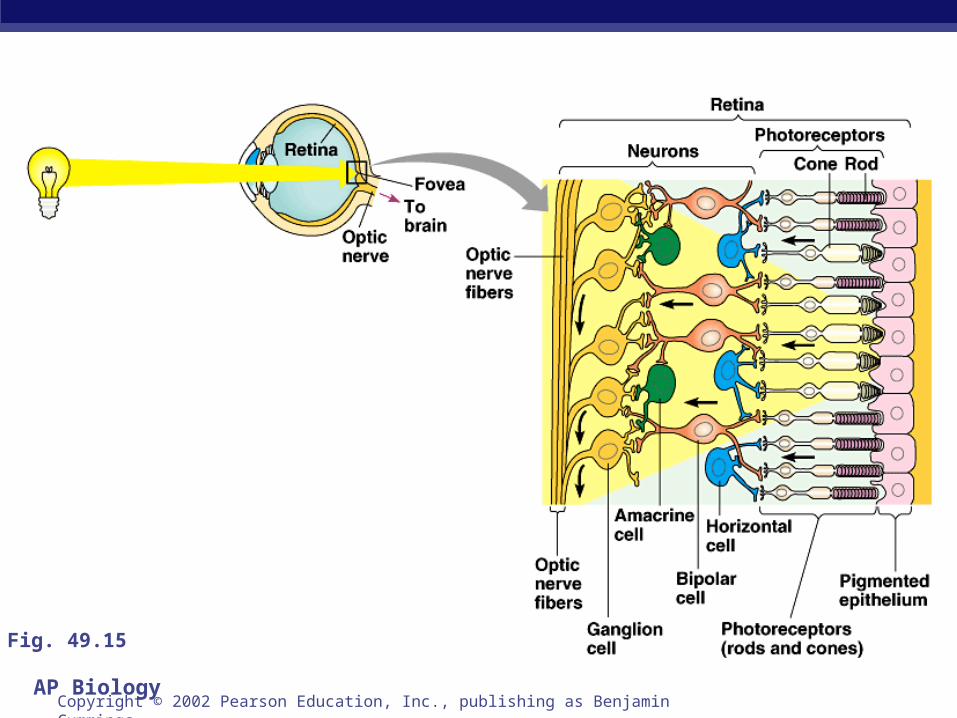
Photoreceptors of the retina.
About 125 million rod cells. Rod cells are light sensitive but do not
distinguish colors.About 6 million cone cells.
Not as light sensitive as rods but provide color vision.
Most highly concentrated on the fovea, an area of the retina that lacks rods.
Copyright © 2002 Pearson Education, Inc., publishing as Benjamin Cummings
AP BiologyCopyright © 2002 Pearson Education, Inc., publishing as Benjamin Cummings
Fig. 49.15
AP BiologyCopyright © 2002 Pearson Education, Inc., publishing as Benjamin Cummings
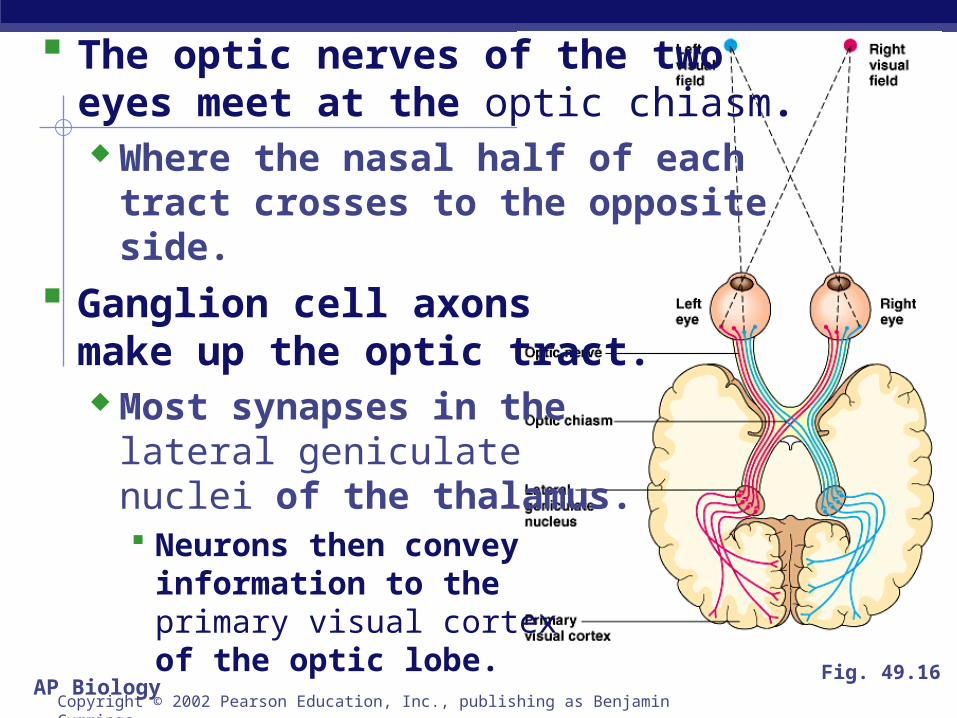
The optic nerves of the two eyes meet at the optic chiasm. Where the nasal half of each
tract crosses to the opposite side.
Ganglion cell axons make up the optic tract. Most synapses in the
lateral geniculate nuclei of the thalamus. Neurons then convey
information to the primary visual cortex of the optic lobe.
Fig. 49.16
AP Biology
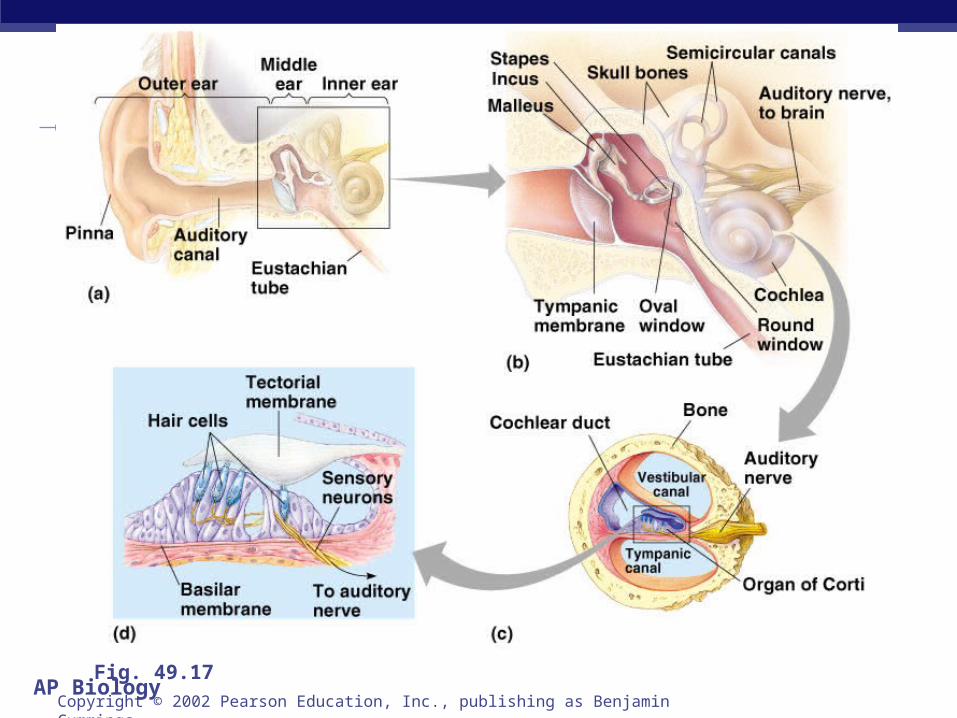
The outer ear includes the external pinna and the auditory canal.Collects sound waves and channels
them to the tympanic membrane (ear drum).
Hearing organ is within the ear
Copyright © 2002 Pearson Education, Inc., publishing as Benjamin Cummings
AP BiologyCopyright © 2002 Pearson Education, Inc., publishing as Benjamin Cummings
Fig. 49.17
AP Biology
From the tympanic membrane sound waves are transmitted through the middle ear.
Malleus incus stapes.From the stapes the sound wave is
transmitted to the oval window and on to the inner ear.
The eustachian tube connects the middle ear with the pharynx.
Copyright © 2002 Pearson Education, Inc., publishing as Benjamin Cummings
AP Biology
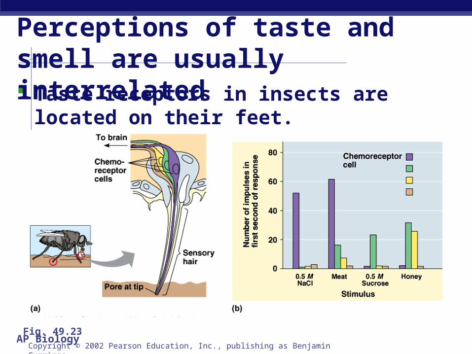
Taste receptors in insects are located on their feet.
Perceptions of taste and smell are usually interrelated
Copyright © 2002 Pearson Education, Inc., publishing as Benjamin Cummings
Fig. 49.23
AP Biology
In mammals, taste receptors are located in taste buds, most of which are on the surface of the tongue.
Each taste receptor responds to a wide array of chemicals. It is the pattern of taste receptor response
that determines perceived flavor.
Copyright © 2002 Pearson Education, Inc., publishing as Benjamin Cummings