Embed Size (px)
Citation preview
A Technique For Extracting All Six Otoliths From Fishes And A List Of Fishes From Lake Erie, Sandusky Bay, and Their Tributaries Located in North-Central Ohio,
U.S.A. From Which Otoliths Were Collected.
Michael James Norrocky 162 Duchess St., Vickery, Ohio
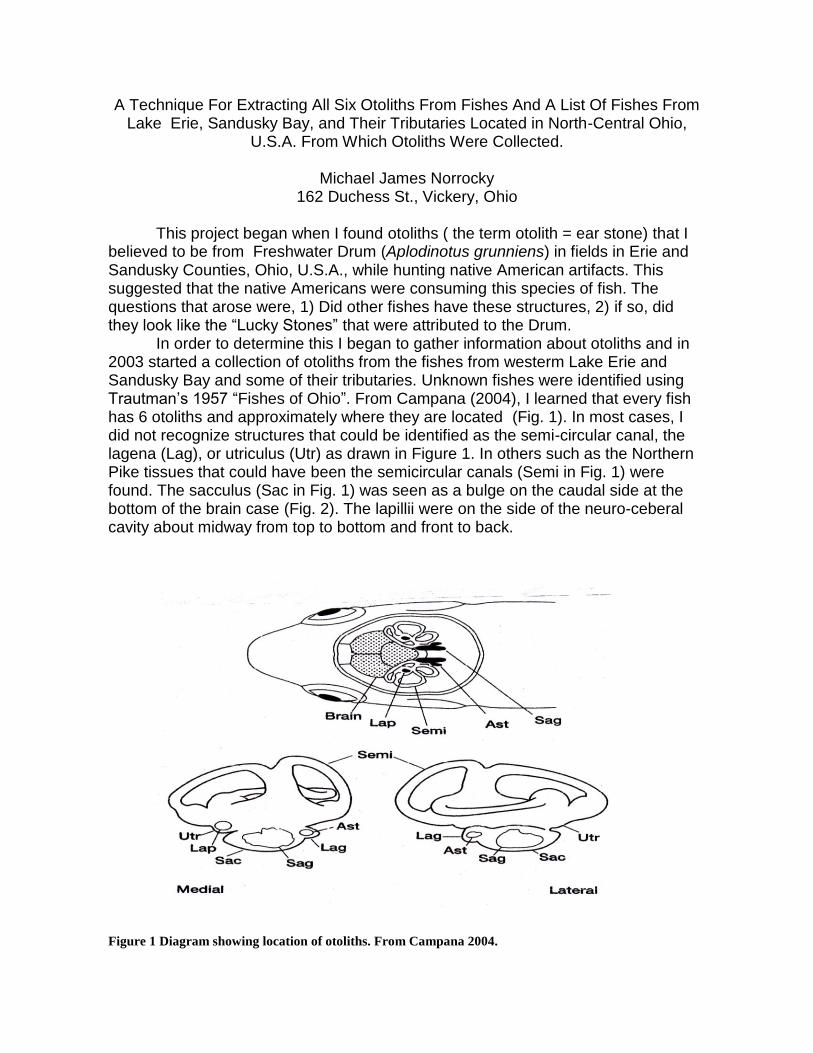
This project began when I found otoliths ( the term otolith = ear stone) that I believed to be from Freshwater Drum (Aplodinotus grunniens) in fields in Erie and Sandusky Counties, Ohio, U.S.A., while hunting native American artifacts. This suggested that the native Americans were consuming this species of fish. The questions that arose were, 1) Did other fishes have these structures, 2) if so, did they look like the “Lucky Stones” that were attributed to the Drum. In order to determine this I began to gather information about otoliths and in 2003 started a collection of otoliths from the fishes from westerm Lake Erie and Sandusky Bay and some of their tributaries. Unknown fishes were identified using Trautman’s 1957 “Fishes of Ohio”. From Campana (2004), I learned that every fish has 6 otoliths and approximately where they are located (Fig. 1). In most cases, I did not recognize structures that could be identified as the semi-circular canal, the lagena (Lag), or utriculus (Utr) as drawn in Figure 1. In others such as the Northern Pike tissues that could have been the semicircular canals (Semi in Fig. 1) were found. The sacculus (Sac in Fig. 1) was seen as a bulge on the caudal side at the bottom of the brain case (Fig. 2). The lapillii were on the side of the neuro-ceberal cavity about midway from top to bottom and front to back.
Figure 1 Diagram showing location of otoliths. From Campana 2004.
There are two basic groups of fishes, the ostariophysian (otophysan) which includes the catfishes, minnows and suckers. The non-ostariophysan non-otophysan) group includes the perch, pike, crappie and sunfishes. The otophysans have a structure called the Weberian ossicles that are located at the rear of the skull and are believed to make contact with the swim bladder and enhance hearing (Fig. 3). Notice the net- like structure. It is unique to the Suckers and the shape is species specific (Dr. Gerald Smith, University of Michigan, Personal Communication).
Figure 2. Ventral view of neuro-cranium of Northern Pike, Esox lucius. Slight bulge at point
of knife blade is sacculus which holds sagitta and astericus.
The otoliths function in a similar manner to our inner ears, aiding in hearing and balance. In addiltion, the lapilli are connected to nerves that are attached to the muscles that move the eyes and research has shown that as the fish swims, moving its head from side to side, the lalpilli are attached to nerve tissue that help the fish eye move so that it can constantly look at an object, such as a predator.

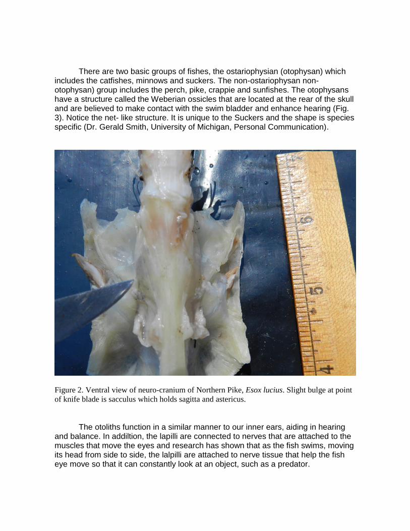
In non-otophysans, the largest otolith is the sagittae, the lapilli are usually next in size and mass, while the asteriscii are the smallest (Fig. 4). In the otophysan group the lapilli or asteriscii are more massive than the sagittae (Fig. 5). The otoliths are some form of calcium carbonate: calcite, aragonite, or vaterite and are composed of about 95% of this material by weight. The rest is 3-5% of an organic matrix and less than 1% non-organic trace impurities. Not only the mass is different in the two groups, the composition is different. In the non-ostariophysans the sagittae and lapilli are usually aragonite, which is milky white, the asteriscii are vaterite and have a glassy appearance (Figs. 4,5) (Campana, 2004) . In the otophysan group, the asteriscii and lapili are often both milky white (Figs. 5) and the sagittae glassy. Other combinations were found.
Figure 3. Bones known as Weberian processes at rear of skull of White Sucker (Catostomus
commersoni) and net-like structure species specific to the members of the Sucker family.
Note the lack of a bulge found in Fig. 2.
The otolith can be utilized in a variety of studies. Because the shape is species specific, it can be used to identify the fish species from which it came. The age and growth of a fish are recorded in the otolith as it is formed, layer by layer like rings of a tree. Daily growth increments formed in the first year of life record daily
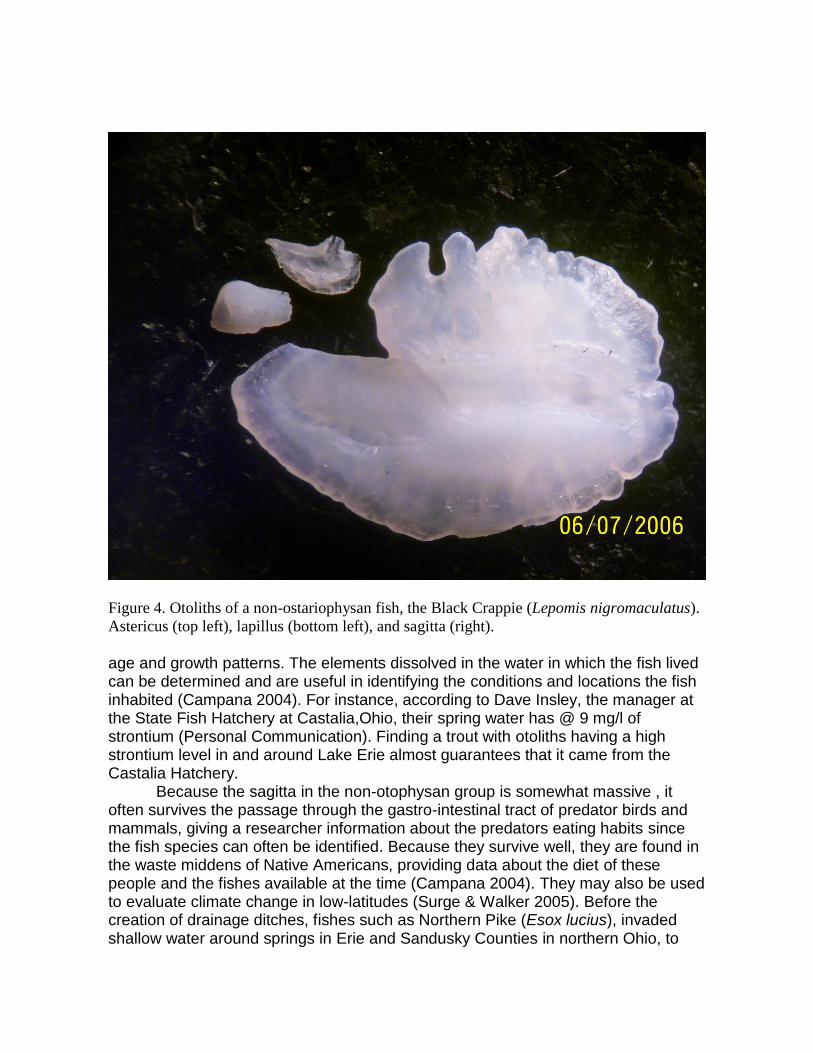
Figure 4. Otoliths of a non-ostariophysan fish, the Black Crappie (Lepomis nigromaculatus).
Astericus (top left), lapillus (bottom left), and sagitta (right). age and growth patterns. The elements dissolved in the water in which the fish lived can be determined and are useful in identifying the conditions and locations the fish inhabited (Campana 2004). For instance, according to Dave Insley, the manager at the State Fish Hatchery at Castalia,Ohio, their spring water has @ 9 mg/l of strontium (Personal Communication). Finding a trout with otoliths having a high strontium level in and around Lake Erie almost guarantees that it came from the Castalia Hatchery. Because the sagitta in the non-otophysan group is somewhat massive , it often survives the passage through the gastro-intestinal tract of predator birds and mammals, giving a researcher information about the predators eating habits since the fish species can often be identified. Because they survive well, they are found in the waste middens of Native Americans, providing data about the diet of these people and the fishes available at the time (Campana 2004). They may also be used to evaluate climate change in low-latitudes (Surge & Walker 2005). Before the creation of drainage ditches, fishes such as Northern Pike (Esox lucius), invaded shallow water around springs in Erie and Sandusky Counties in northern Ohio, to
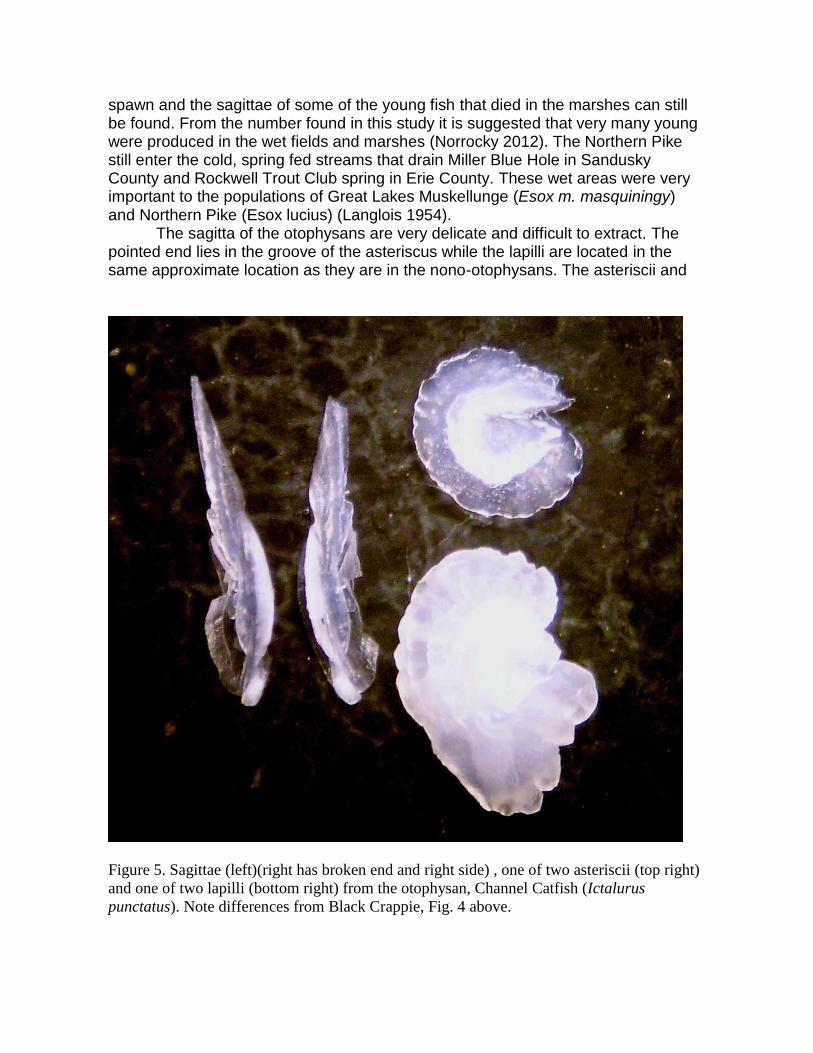
spawn and the sagittae of some of the young fish that died in the marshes can still be found. From the number found in this study it is suggested that very many young were produced in the wet fields and marshes (Norrocky 2012). The Northern Pike still enter the cold, spring fed streams that drain Miller Blue Hole in Sandusky County and Rockwell Trout Club spring in Erie County. These wet areas were very important to the populations of Great Lakes Muskellunge (Esox m. masquiningy) and Northern Pike (Esox lucius) (Langlois 1954). The sagitta of the otophysans are very delicate and difficult to extract. The pointed end lies in the groove of the asteriscus while the lapilli are located in the same approximate location as they are in the nono-otophysans. The asteriscii and
Figure 5. Sagittae (left)(right has broken end and right side) , one of two asteriscii (top right)
and one of two lapilli (bottom right) from the otophysan, Channel Catfish (Ictalurus
punctatus). Note differences from Black Crappie, Fig. 4 above.

lapilli in this group are different shapes than those of the non-otophysans and are not distinct enough that the species from which they came can be identified in my opinion. The sagittae in this group were seldom extracted without breaking or in the least a part being broken off. For this reason only a few photos are presented to give the reader some idea of what they look like. In the non-ostariophysans, the sacculus appeared as a bulge on the underside of the brain case. When the sagitta was extracted, it was the most massive and often the astericus was found embedded in tissue, on the caudo-lateral side of the sagitta. (Fig. 6). In the otophysans an obvious bulge was not found making it more difficult to locate the sagitta and astericus (Fig. 2). Retreiving the otoliths was a learning experience. I began by taking the head off the fish and trying to determine the best way to enter the skull where the otoliths were located. Removing the raw flesh proved difficult, so for fish larger than 3”, I put the head in a pan of water and heated it to near boiling. I cooked the head a couple of minutes ( time depending on size). This cooked the flesh just enough that it could be removed easily, leaving a bony skull. Cooking too long resulted in the skull coming apart, making it difficult to locate specific parts. The plates making up the cranial cavity (Fig. 7) could be removed rather easily, exposing the brain With a little teasing of the brain, the lapilli could be found along the side of the cranial cavity.
Figure 6. Distal surface of Bluegill (Lepomis macrochirus) sagitta with astericus, at top right
corner, embedded in tissue. Anterior end on left.

Figure 7. Dorsal view of Northern Pike (Esox lucius) skull. Note the knife blade under one of
the plates making up the top of the neuro-cranial cavity.
In some species, tube like strands that could be the lagena were seen. In some species these strands passed horizontally through a bony tube on the side of the skull, forming a loop that connected to other strands next to the brain that may have been vertical in the intact skull. Once the lapilli were retreived, the brain was taken out exposing the sacculi located on the bottom of the chamber (Fig. 1). In some species, tube like strands that could be the lagena were seen. In some species these strands passed horizontally through a bony tube on the side of the skull, forming a loop that connected to other strands next to the brain that may have been vertical in the intact skull. Once the lapilli were retreived, the brain was taken out exposing the sacculi located on the bottom of the chamber (Fig. 1). In a few species the bones of the skull are very flat (Channel Catfish {Ictalurus punctatus} or very complex such as the Bigmouth Buffalo (Ictiobus cyprenillus) (Fig. 8). Clearing away the flesh was very useful in retreiving the otoliths from these species.

Figure 8. Skull of the 17.75” Bigmouth Buffalo. The web like structure can be seen at the
right.
As the stones were removed they were put in a shallow, black pan that contained a 10% Chlorox® solution. This dissolved any tissue that might be attached. The stones were then put in a similar pan that had plain water which rinsed off the Chlorox® solution. After a few minutes in plain water, the otoliths were put on a plain paper sheet to dry. In some cases, a few scales from near the lateral line of the fish were removed and treated in the same manner as the otoliths. Dr. Gerald Smith of the University of Michigan, Museum of Zoology, commenting about the use of Chlorox® wrote, “The other thing is the use of chlorox. We have found that skulls that were prepared with bleach turn to powder after a few years because the chlorine stays in the bone and takes up moisture from the air, forming hydrochloric acid, which dissolves the bones. I have no idea whether that will happen to otoliths. Being single crystals, they might be resistant.” Small fishes, around 3” or less, were handled a little differently. Boiling did not prove to be efficient so a fish was put in a sandwich sized plastic bag and put in microwave oven for a few seconds. The head of the fish was then put in the pan with

the Clorox solution and teased apart carefully, while searching for the otoliths which would usually be seen as tiny white spots againt the black surface of the pan. In some cases the small otoliths of these fish floated on the surface in the pan, a condition which was not noticed at the beginning of the study. Handling of these stones was difficult, but an eyedropper was very helpful. In the event that the fish is to become a specimen in itself, the following method was presented in a note from Gerald Smith, University of Michigan, Museum of Zoology, “I have found that for museum specimens it is necessary to extract an otolith from one side of the fish without destroying or damaging the specimen. To do this, I lift the gill cover, bend the gills away from the lower side of the skull, and reach in with sharp forceps until I can feel and grasp an otolith, then pull it out. The gills and gill cover are then folded back into place and the specimen looks undamaged (except for the pro-otic and opisthotic or other nearby bones.” With practice this method could be used to extract sagittae from nonotophysans, however, I think it would be difficult to obtain the other stones. In the otophysan group, the lapillus is the most massive and might be obtained using this method. The other stones, as can be seen (Fig. 5), are small and in the case of the sagitta, very fragile. Once the otoliths were dry they were photographed. Initially a Digital Blue®, student, digital microscope was used. (Fig. 6). It had 10X, 60X, and 200X magnifaction. Problems with positioning the specimens and resolution were encountered. About midway through the project, a Bausch & Lomb® dissecting scope and Kodak Easyshare® camera was used. I constructed an extension tube from a plastic plumbing fitting that fit over one objective of the scope and the lens of the camera was inserted into the tube and manipulated to compose the picture (Fig. 7). Pictures were downloaded to a file on a computer. Getting clear photos was a challenge and usually multiple photos were taken.

Figure 9. Digital Blue® student digital microscope used initially to photograph otoliths.
The dry material was then placed in a folder made to hold coins. These were approximately 2.5” square with a cellophane window in the center. Once the otoliths and scales were in the folder, they were stapled on all four sides and labelled. The labels had the following information: fish common name, scientific name, length, date caught, location where caught, and collector’s name. The digital microscope was, however, the only way to take pictures of the very smallest otoliths. The fishes from which otoliths were collected are listed in Table 1. The otolliths were donated to the University of Michigan Museum of Zoology (UMMZ) and they have informed me that , the collection records will be added to the UMMZ Fish Division online catalogue (and to the FishNet2 database) during our next data update so they can be accessed online. I have sent them two CD’s with all the pictures I took and these may be available also.

Figure 10. Bausch & Lomb® dissecting scope with Kodak Easyshare® camera lens inserted
into plastic extension tube. Most of the photos are clear enough to determine species, so it is believed they could be useful. Some that are not clear have been omitted. As a fish ages, the shape and general morphology changes slightly (Campana 2004). Since these are the characters used to determine species, fish similar in size to those used in this study should be used for comparison when possible. During this study 30 species from 15 non-ostariophysan families and 23 species from 3 ostariophysan families were collected (Table 1).

Table 1. Fishes from Lake Erie, Sandusky Bay and nearby tributaries from which fish were taken for extraction of otoliths.
Family Genus Species Common Name
Non-ostariophysians Amiidae Amia calva Bowfin
Atherinidae Labidesther sicculus Brook Silverside
Centrachidae Amblooplites rupestris Rockbass
Lepomis gibbosus Pumpkinseed
Lepomis macrochirus Bluegill
Pomoxis annularis White Crappie
Micropterus salmoides Largemouth Bass
Micropterus dolomieu Smallmouth Bass
Lepomis gulosus Warmouth Sunfish
Clupeidae Dorosoma cepedianum Gizzard Shad
Coregonidae Coregonus clupeaformis Lake Whitefish
Esocidae Esox lucius Northern Pike
Fundilidae Fundulus notatus Black Striped Topminnow
Gasterosteidae Eucalia inconstans Brook Stickleback
Hiodontidae Hiodon tergisus Mooneye
Lepisosteidae Lepisosteus osseus Longnose Gar
Moronidae Morone americana White Perch
Morone chrysops White Bass
Osmeridae Osmerus mordax Rainbow Smelt
Percidae Etheostoma blennoides Rainbow Darter
Ethostoma nigrum Johnny Darter
Percina c. semifasciata Northern Logperch Darter
Percina c. caprodes Ohio Logperch Darter
Stizostedion vitreum Walleye
Perca flavescens Yellow Perch
Salmonidae Oncorhycus mykiss Rainbow Trout
Salvelinus fontanilis Brook Trout
Salmo trutta Brown Trout
Salvelinus namaycush Lake Trout
Sciaenidae Aplodinotus grunniens Freshwater Drum
Catostomidae Ictiobus cyprinellus Bigmouth Buffalo
Moxostoma m. macrolepidotum
Northern Shorthead Redhorse Sucker
Catostomus commersoni White Sucker
Hypentelium nigricans Hog Sucker
Cyprinidae Pimephales notatus Bluntnose Minnow
Semotilus a. atromaculatus
Northern Creek Chub
Cyprinidae
Cyprinus carpio Carp

Notropis atherinoides Emerald Shiner
Notemigonus crysoleucus Golden Shiner
Carrasius auratus Goldfish
Notropis v. volucellus Northern Mimic Shiner
Clinostumus elongatus Redside Dace
Macrhybobsis storeriana Silver Chub
Notropis buccatus Silverjaw Minnow
Cyprinella spiloptera Spotfin Shiner
Notropis hudsonius Spottail Shiner
Campostoma a. pullum Stoneroller Minnow
Luxilus chrysocephalus Striped Minnow
Ictaluridae Ameiurus natalis Yellow Bullhead
Ameiurus nebulosus Brown Bullhead
Ictalurus punctatus Channel Catfish
Pylodictis olivaris Flathead Catfish
Acknowledgements Ron Penrod for assistance in many aspects especially his work with the photos. Dean Koch of Dean Koch Fisheries, Crystal Rock, Ohio for providing some of the fish for this study. Dr. Gerald Smith of the University of Michigan, Museum of Zoology provided helpful comments concerning otolith extraction and the use of Chlorox® in cleaning otoliths. Some Alabama fishes were supplied by the Enterprise unit of the Alabama Department of Natural Resources.
Bibliography: Butler, J. L. 1992. Collection and Preservation of Material for Otolith Analysis, p. 13- 17. In D. K. Stevenson and S. E. Campana (ed.) Otolith Microst;ructure Examination and Analysis. Can. Spec. Publ. Fish. Aquat. Sci. 117. Campana, S. E. 1992. Measurement and Interpretation of the Microstructure of Fish Otoliths, p. 59-71. In D. K. Stevenson and S. E. Campana (ed.) Otolith Microst;ructure Examination and Analysis. Can. Spec. Publ. Fish. Aquat. Sci. 117.
Campana, S.E., G.A. Chouinard, J.M. Hanson, A. FreÂchet, J. Brattey. 2000 Otolith elemental fingerprints as biological tracers of fish stocks. Fisheries Research 46 (2000) 343-357.
Campana, S.E. 2001. REVIEW PAPER: Accuracy, precision and quality control in age determination, including a review of the use and abuse of age validation methods. Journal of Fish Biology (2001) 59, 197–242

Campana, Steven E., 2004. Photographic Atlas of Fish Otoliths of the Northwest Atlantic Ocean. NRC Research Press, Ottawa, Ontario. 284 pp. Grande, Terry, Mario de PINNA & Christopher BRAUN. 2003 The Development and Evolution of the Weberian Ossicles in Clupeocephalan Fishes. Abstract:
American Zoologist 41(6):1377-1655. 2001
Jones, C. M. 1992. Development and Application of the Otolith Increment Technique, p. 1-11. In D. K. Stevenson and S. E. Campana (ed.) Otolith Microst;ructure Examination and Analysis. Can. Spec. Publ. Fish. Aquat. Sci. 117.
Langlois, Thomas H., 1954 The Western End of Lake Erie and Its Ecology. J. W. Edwards, Publ. Inc. Ann Arbor, Michigan. Norrocky, M. J. Fossil Limpets and Fossil Otoliths as Indicators of the Extent of the Wet Prairies Around Castalia in Margaretta Township, Erie County and Townsend and Riley Townships in Sandusky County, Ohio, U.S.A. http://hoseco.spde.info/limpets/index.html Secor, D.H., J. M. Dean, and E.H. Laban. 1992. Otolith Removal and Preparation for Microstructural Examination, p. 19-57. In D. K. Stevenson and S. E. Campana (ed.) Otolith Microst;ructure Examination and Analysis. Can. Spec. Publ. Fish. Aquat. Sci. 117. Surge, Donna and Karen Jo Walker. 2005. Oxygen isotope composition of modern and archaeological otoliths from the estuarine hardhead catfish (Ariopsis felis) and their potential to record low-latitude climate change. Paleogeography, Paloeoclimatology, Paleoecology 228 (2005) pgs. 179-191 Thort-old, Simon R., Cynthia M. Jones, Steven E. Campana. 1997. Response of otolith microchemistry to environmental variations experienced by larval and juvenile Atlantic croaker (Micropogonias undulatus). Lirwu.d. Ocmrro~l:, 42( I ), 1997, 102-I I I Trautman, Milton B. 1957. The Fishes of Ohio. The Ohio State University Press. 683 pp.