Embed Size (px)
Citation preview

Photochemistry and Photobiology, 1996, 63(4): 547-552
Apoptosis is an Early Event During Phthalocyanine Photodynamic Therapy-Induced Ablation of Chemically Induced Squamous Papillomas in Mouse Skin
Rajesh Agarwal, Neil J. Korman, Rajiv R. Mohan, Denise K. Feyes, Seema Jawed, M. Tarif Zaim and Hasan Mukhtar* DeDarlment of Dermatoloav. Skin Diseases Research Center, University Hospitals of Cleveland, Cake Western Reserve Uzversity, Cleveland, OH, USA
Received 18 October 1995; accepted 2 January 1996
ABSTRACT
Photodynamic therapy (PDT) is a promising new modal- ity to treat malignant neoplasms including superficial skin cancers. In our search for an ideal photosensitizer for PDT, Pc 4, a silicon phthalocyanine, has shown prom- ising results both in in vitro assays and in implanted tu- mors. In this study we assessed the efficacy of Pc 4 PDT in the ablation of murine skin tumors; and the evidence for apoptosis during tumor ablation was also obtained. The Pc 4 was administered through tail vein injection to SENCAR mice bearing chemically induced squamous papillomas, and 24 h later the lesions were illuminated with an argon ion-pumped dye laser tuned at 675 nm for a total light dose of 135 J/cmZ. Within 72-96 h, almost complete tumor shrinkage occurred; no tumor regrowth was observed up to 90 days post-PDT. As evident by nu- cleosome-size DNA fragmentation, appearance of apop- totic bodies in hematoxylin and eosin staining and direct immunoperoxidase detection of digoxigenin-labeled ge- nomic DNA in sections, apoptosis was clearly evident 6 h post-PDT at which time tumor shrinkage was less than 30%. The apoptotic bodies, as evident by the condensa- tion of chromatin material around the periphery of the nucleus and increased vacuolization of the cytoplasm, were also observed in electron microscopic studies of the tumor tissues following Pc 4 PDT. The extent of apoptosis was greater at 15 h than at 6 and 10 h post-PDT. Taken together, our results clearly show that Pc 4 may be an effective photosensitizer for PDT of nonmelanoma skin cancer, and that apoptosis is an early event during this process.
INTRODUCTION
Photodynamic therapy (PDT)? is a promising new approach for selective eradication of tumor tissue (1-3). In our search
*To whom correspondence should be addressed at: Department of Dermatology, Skin Diseases Research Center, Case Western Re- serve University, 2074 Abington Road, Cleveland, OH 44106, USA. Fax: 216-368-0212; email: [email protected]
0 1996 American Society for Photobiology 0031-8655/96 $5.00+0.00
for an ideal photosensitizer for PDT, Pc 4 (a silicon phthal- ocyanine developed in our group) has shown encouraging results both in in vitro assays (43) and in implanted tumors (6). In addition, compared to porphyrin photosensitizers such as Photofrin-II@ (Pf-II), Pc 4 produces lower skin photosen- sitivity (7). Because of this advantage, further evaluation of PDT efficacy of Pc 4 is warranted.
Apoptosis, a normal programmed process of cell death, occurs during embryonic morphogenesis, metamorphosis and hormone-induced tissue remodeling, as well as after var- ious types of cellular damage (8,9). Some characteristics of apoptosis include cell shrinkage, chromatin condensation around the nuclear periphery, hyperconvolution of the nu- clear membrane and internucleosomal fragmentation (8). Various cells can be induced to undergo apoptosis in vitro by a wide variety of treatments including PDT (10-14). Our recent studies indicated that apoptosis occurs during PDT- induced shrinkage of RIF-I implanted tumors (15).
Photodynamic therapy is also showing promise in treat- ment of various dermatological disorders such as psoriasis, alopecia areata, nonmelanoma superficial skin cancer and patch and plaque stage cutaneous T-cell lymphoma (16-18). In earlier studies, we have shown that Pf-I1 and chloroalu- minum phthalocyanine tetrasulfonate PDT is effective in treating mouse skin tumors (19,20). Because PDT has sig- nificance in the treatment of cutaneous lesions (16-20), and because apoptosis also has relevance in dermatology (21), the purpose of this study was to assess whether Pc 4 PDT leads to ablation of murine skin tumors and that this phe- nomenon involves apoptosis.
MATERIALS AND METHODS Pc 4 and its formulation. Chemical synthesis of Pc 4 is recently described (4), and because it is insoluble in aqueous medium, it was formulated in 25% Cremophor EL ( 1 mg Pc 4, 0.7 mL normal saline, 0.25 mL Cremophor EL and 0.05 mL propylene glycol are mixed and vortexed thoroughly). All other chemicals were of the highest purity commercially available.
Induction of tumors in SENCAR mouse skin. Chemical carcino-
?Abbreviations: H&E, hematoxylin and eosin; Pc 4, SiPc- (OH)OSi(CH3),(CH,),N(CH,),I, silicon phthalocyanine; PDT, pho- todynamic therapy; Pf-11, Photofrin-IIB; TdT, terminal deoxynu- cleotidyl transferase.
547
548 Rajesh Agarwal et a/.
T
0 6 1 5 72 Time post-Pc 4-PDT (hours)
Figure 1. The Pc 4 PDT-mediated ablation of skin tumors in SEN- CAR mice. Tumor-bearing mice were injected through tail vein with 1 mgkg of Pc 4 formulated in 25% Cremophor EL and 24 h later tumors were irradiated with an argon-pumped dye laser for a total light dose of 135 J/cmZ. Following irradiation, at indicated time points, tumor volumes were measured using a caliper as described in the Materials and Methods. Each bar represents mean 2 SE of six skin tumors obtained from six mice.
gen-induced benign squamous papillomas were developed on SEN- CAR mouse skin employing the protocol described earlier ( I 9,20). In brief, 6 week old female SENCAR mice were obtained from Harlan-Sprague Dawley (Indianapolis, IN) and housed in CWRU’s Animal Resource Facility. The animals were acclimatized before use and subjected to a 12 h lighVl2 h dark cycle, housed at 24 2 2°C and 50 2 10% relative humidity in a room with 12-15 cycles of air exchangeslh and fed Purina chow diet and water ad libitum. One week after their arrival, animals were shaved with electric clippers and Nair depilatory was applied. Only those animals in resting phase of the hair cycle were used in the tumor protocol and treated topi- cally on the shaved area with a single topical application of 7,12- dimethylbenz(a)anthracene (DMBA) (20 pg in 0.2 mL acetone/ mouse). One week later, animals were treated twice a week with 12- 0-tetradecanoylphorbol-13-acetate (TPA) (2 pg in 0.2 mL acetone/ mouse). Using this protocol, at 12 weeks on test, about 30% mice had an average of two to three tumors per mouse; they were used in this study. Few tumors were randomly verified histopathologically as squamous papillomas.
Pc 4 PDT of murine skin tumors. Animals with closely matched tumor size were randomly divided into four groups that consisted of (1) an untreated group, (2) a laser irradiation alone group, (3) a Pc 4-treated alone group and (4) a Pc 4-treated-irradiated group. Formulated Pc 4 (1 mgkg body weight) was injected through tail vein, and 24 h later the tumors (one tumor/mouse) were irradiated with an argon-pumped dye laser for a total light dose of 135 J/cm2 as detailed earlier (15). The selection of 24 h time after Pc 4 injec- tion was based on our pharmacokinetics studies where this time period showed maximum tumor-skin ratio of Pc 4 (data not shown). Following irradiation, each tumor was measured with a caliper at several time periods up to complete tumor ablation. Tumor volume was calculated as described earlier (19,20). From the Pc PDT group, three mice were removed at 6 , 10 and 15 h postirradiation, animals were sacrificed and treated tumors removed. Tumors were also ex- cised from control groups identically. Each tumor was vertically divided into three sections, which were used for DNA fragmentation, histopathological and histochemical and electron microscopic stud- ies.
DNA isolation and agarose gel electrophoresis. Nucleosome-size DNA fragmentation was determined by the method described earlier (15). One part of excised tumor tissue was minced into small pieces, digested with proteinase K (0.5 mg/mL) in the presence of 1 % so- dium lauryl sarkosinate in Tris-HCI-EDTA buffer at 50°C overnight, and DNA was precipitated with two volumes of ice-cold ethanol and resuspended in 10 mM Tris-HC1, pH 8.0, containing 1 mM EDTA. The DNA solution was subjected to 1.5% agarose gel electrophoresis
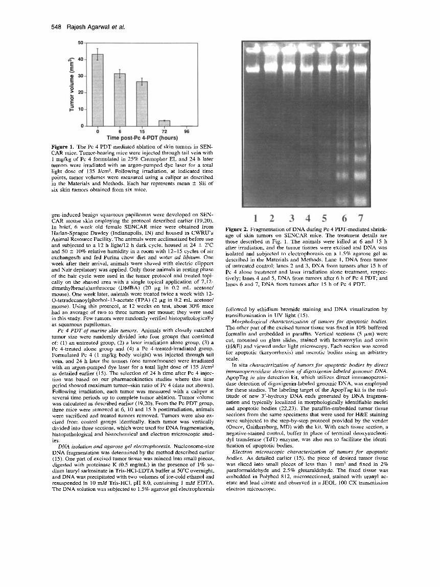
1 2 3 4 5 6 7 Figure 2. Fragmentation of DNA during Pc 4 PDT-mediated shrink- age of skin tumors on SENCAR mice. The treatment details are those described in Fig. 1. The animals were killed at 6 and 15 h after irradiation, and the tumor tissues were excised and DNA was isolated and subjected to electrophoresis on a 1.5% agarose gel as described in the Materials and Methods. Lane 1, DNA from tumor of untreated control; lanes 2 and 3, DNA from tumors after 15 h of Pc 4 alone treatment and laser irradiation alone treatment, respec- tively; lanes 4 and 5, DNA from tumors after 6 h of Pc 4 PDT; and lanes 6 and 7. DNA from tumors after 15 h of Pc 4 PDT.
followed by ethidium bromide staining and DNA visualization by transillumination in UV light (15).
Morphological characterization of tumors for apoptotic bodies. The other part of the excised tumor tissue was fixed in 10% buffered formalin and embedded in paraffin. Vertical sections (5 pm) were cut, mounted on glass slides, stained with hematoxylin and eosin (H&E) and viewed under light microscopy. Each section was scored for apoptotic (karyorrhexis) and necrotic bodies using an arbitrary scale.
In situ characterization of tumors for apoptotic bodies by direct immunoperoxidase detection of digoxigenin-labeled genomic DNA. ApopTag in situ detection kit, which utilizes direct immunoperoxi- dase detection of digoxigenin-labeled genomic DNA, was employed for these studies. The labeling target of the ApopTag kit is the mul- titude of new 3’-hydroxy DNA ends generated by DNA fragmen- tation and typically localized in morphologically identifiable nuclei and apoptotic bodies (22,23). The paraffin-embedded tumor tissue sections from the same specimens that were used for H&E staining were subjected to the step-by-step protocol provided by the vendor (Oncor, Gaithersburg, MD) with the kit. With each tissue section, a negative-stained control, buffer in place of terminal deoxynucleoti- dyl transferase (TdT) enzyme, was also run to facilitate the identi- fication of apoptotic bodies.
Electron microscopic characterization of tumors for apoptotic bodies. As detailed earlier (15), the piece of desired tumor tissue was sliced into small pieces of less than 1 mm3 and fixed in 2% paraformaldehyde and 2.5% glutaraldehyde. The fixed tissue was embedded in Polybed 812, microsectioned, stained with uranyl ac- etate and lead citrate and observed in a JEOL 100 CX transmission electron microscope.
Photochemistry and Photobiology, 1996, 63(4) 549
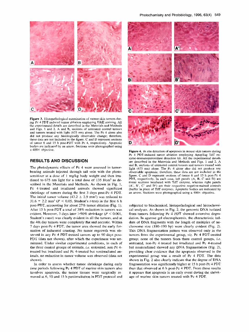
Figure 3. Histopathological examination of mouse skin tumors dur- ing Pc 4 PDT-induced tumor ablation employing H&E staining. All the experimental details are described in the Materials and Methods and Figs. 1 and 2. A and B, sections of untreated control tumors and tumors treated with light (675 nm) alone. The Pc 4 alone also did not produce any histologically observable change; therefore, these data are not included in the figure. C and D represent sections of tumor 6 and 1.5 h post-PDT with Pc 4, respectively. Apoptotic bodies are indicated by an arrow. Sections were photographed using a 400X objective.
RESULTS AND DISCUSSION The photodynamic effects of Pc 4 were assessed in tumor- bearing animals injected through tail vein with the photo- sensitizer at a dose of 1 mgkg body weight and then irra- diated to 675 nm light for a total dose of 135 J/cm2 as de- scribed in the Materials and Methods. As shown in Fig. 1, Pc 4-treated and irradiated animals showed significant shrinkage of tumors during the first 3 days post-Pc 4 PDT. The initial tumor volume (43.2 ? 2.9 mm3) was reduced to 31.6 ? 2.2 mm3 ( P < 0.05, Student’s t-test) in the first 6 h post-PDT, accounting for about 27% tumor ablation (Fig. 1). After 15 h post-PDT a total of 38% reduction in tumors was evident. However, 3 days later >go% shrinkage ( P < 0.001, Student’s t-test) was clearly evident in all the tumors, and at the 4th day tumors were completely flat and necrotic. Within 7 days post-Pc 4 PDT, the tumor area showed the early for- mation of indurated crusting. No tumor regrowth was ob- served in any Pc 4 PDT-treated tumors up to 90 days post- PDT (data not shown), after which the experiment was ter- minated. Under similar experimental conditions, in each of the three control groups of animals, i.e. untreated, non-Pc 4- treated but irradiated and Pc 4-treated but nonirradiated an- imals, no reduction in tumor volume was observed (data not shown).
In order to assess whether tumor shrinkage during early time periods following Pc 4 PDT of murine skin tumors also involves apoptosis, the tumor tissues were surgically re- moved at 6, 10 and 15 h postirradiation in PDT protocol and
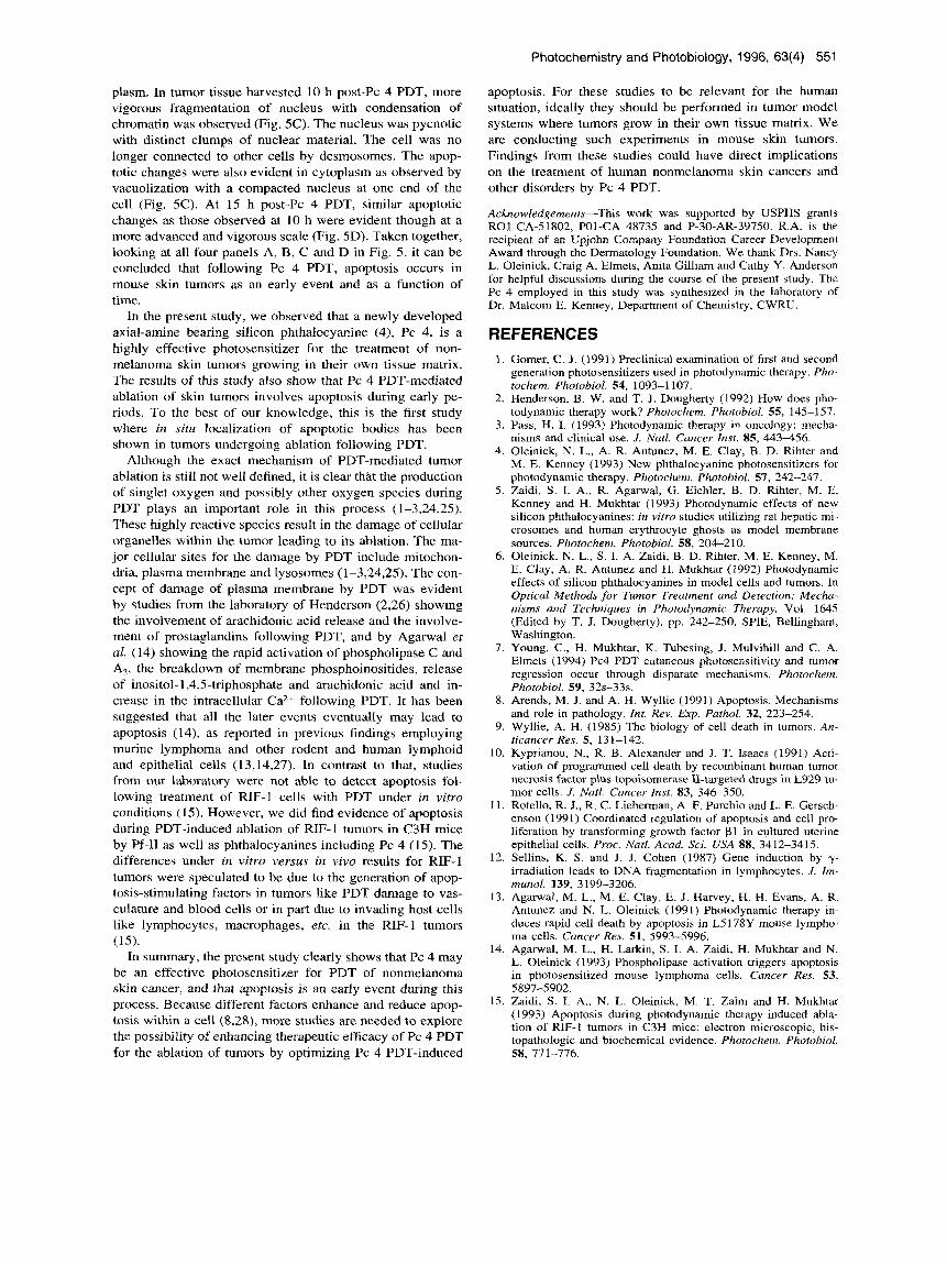
Figure 4. In situ detection of apoptosis in mouse skin tumors during Pc 4 PDT-induced tumor ablation employing ApopTag TdT en- zyme-immunoperoxidase detection kit. All the experimental details are described in the Materials and Methods and Figs. 1 and 2. A and B, sections of untreated control tumors and tumors treated with light (675 nm) alone. The Pc 4 alone also did not produce any observable apoptosis; therefore, these data are not included in the figure. C and D represent sections of tumor 6 and 15 h post-Pc 4 PDT, respectively. In each case, left panels (A, B, C and D) are tissue sections incubated with TdT enzyme, whereas right panels (A’, B’, C’ and D’) are their respective negative-stained controls (buffer in place of TdT enzyme). Apoptotic bodies are indicated by an arrow. Sections were photographed using a 400X objective.
subjected to biochemical, histopathological and histochemi- cal analyses. As shown in Fig. 2, the genomic DNA isolated from tumors following Pc 4 PDT showed extensive degra- dation. In agarose gel electrophoresis, the characteristic lad- ders of DNA fragments with the length of multiples of nu- cleosome size (180-190 bp) were clearly evident (Fig. 2). This DNA fragmentation pattern was observed only in the tumors from the experimental group, viz. Pc 4 PDT-treated group; none of the tumors from three control groups, i.e. untreated, non-Pc 4-treated but irradiated and Pc 4-treated but nonirradiated showed any DNA fragmentation (Fig. 2), providing clear evidence that the apoptosis observed in the experimental group was a result of Pc 4 PDT. The data shown in Fig. 2 also clearly indicate that the degree of DNA fragmentation was significantly higher at 15 h post-Pc 4 PDT than that observed at 6 h post-Pc 4 PDT. From these results it appears that apoptosis is an early event during the shnnk- age of murine skin tumors treated with Pc 4 PDT.

550 Rajesh Agarwal eta/.
Figure 5. Electron microscopic examination of mouse skin tumors during Pc 4 PDT-induced tumor ablation. All the experimental details are described in the Materials and Methods and Figs. 1 and 2. A, section of untreated control tumor. Light (675 nm) alone and Pc 4 alone treated tumors also did not produce any histologically observable change, and therefore, these data are not included in the figure. B, C and D represent sections of tumor 6 , 10 and 15 h post-Pc 4 PDT, respectively. Bar = 2 p,m.
When tumors were studied histologically after Pc 4 PDT, apoptotic bodies were observed in the sections (Fig. 3C,D). When stained with H&E, they had the classic light micro- scopic appearance of apoptotic cells: pycnotic nuclei, chro- matin condensation and intensely eosinophilic cytoplasm (Fig. 3C,D). The number of apoptotic bodies scored at 15 h post-PDT was greater than at 6 h post-PDT (data not shown). None of the tumors from three control groups showed apop- totic bodies in their tissue sections (Fig. 3A,B). When as- sessed employing the ApopTag in situ kit, which utilizes direct immunoperoxidase detection of digoxigenin-labeled genomic DNA, apoptotic bodies were observed in tumor tis- sue sections only after Pc 4 PDT (Fig. 4C,D). Evident by the specific staining, the light microscopy of apoptotic cells showed pycnotic nuclei and chromatin condensation as well as degradation (Fig. 4C,D). The number of apoptotic bodies scored at 15 h post-PDT was greater than that at 6 h post- PDT. No apoptotic bodies, however, were observed in either
untreated, or light- or Pc 4 alone-treated tumor sections (Fig. 4A,B).
In electron microscopic evaluation of mouse skin tumors collected from different control groups and Pc 4 PDT groups at different time points post-PDT, considerable apoptotic bodies were evident only in those samples that were exposed to Pc 4 PDT (Fig. 5). Figure 5A is from a control tumor. Three epithelial cells can be seen in the field shown. These cells have intact membranes with desmosomal attachments outlining their peripheral border. These cells also have nor- mal tonofilaments in the cytoplasm and an intact nucleus. When the electron micrograph of tumor tissue obtained 6 h post-Pc 4 PDT was examined (Fig. 5B), two early apoptotic epithelial cells shown in the field demonstrated condensation and margination of the chromatin material at the nuclear periphery. The other changes observed in cytoplasm include the disruption of tonofilaments compared to that of control tumor tissue samples suggesting a disorganization of cyto-
Photochemistry and Photobiology, 1996, 63(4) 551
plasm. In tumor tissue harvested 10 h post-Pc 4 PDT, more vigorous fragmentation of nucleus with condensation of chromatin was observed (Fig. 5C). The nucleus was pycnotic with distinct clumps of nuclear material. The cell was no longer connected to other cells by desmosomes. The apop- totic changes were also evident in cytoplasm as observed by vacuolization with a compacted nucleus at one end of the cell (Fig. 5C). At 15 h post-Pc 4 PDT, similar apoptotic changes as those observed at 10 h were evident though at a more advanced and vigorous scale (Fig. 5D). Taken together, looking at all four panels A, B, C and D in Fig. 5 , it can be concluded that following Pc 4 PDT, apoptosis occurs in mouse skin tumors as an early event and as a function of time.
In the present study, we observed that a newly developed axial-amine bearing silicon phthalocyanine (4), Pc 4, is a highly effective photosensitizer for the treatment of non- melanoma skin tumors growing in their own tissue matrix. The results of this study also show that Pc 4 PDT-mediated ablation of skin tumors involves apoptosis during early pe- riods. To the best of our knowledge, this is the first study where in situ localization of apoptotic bodies has been shown in tumors undergoing ablation following PDT.
Although the exact mechanism of PDT-mediated tumor ablation is still not well defined, it is clear that the production of singlet oxygen and possibly other oxygen species during PDT plays an important role in this process (1-3,24,25). These highly reactive species result in the damage of cellular organelles within the tumor leading to its ablation. The ma- jor cellular sites for the damage by PDT include mitochon- dria, plasma membrane and lysosomes (1-3,24,25). The con- cept of damage of plasma membrane by PDT was evident by studies from the laboratory of Henderson (2,26) showing the involvement of arachidonic acid release and the involve- ment of prostaglandins following PDT, and by Agarwal et al. (14) showing the rapid activation of phospholipase C and A2, the breakdown of membrane phosphoinositides, release of inositol- 1,4,5-triphosphate and arachidonic acid and in- crease in the intracellular Ca2+ following PDT. It has been suggested that all the later events eventually may lead to apoptosis (14), as reported in previous findings employing murine lymphoma and other rodent and human lymphoid and epithelial cells (13,14,27). In contrast to that, studies from our laboratory were not able to detect apoptosis fol- lowing treatment of RIF-1 cells with PDT under in vitro conditions (15). However, we did find evidence of apoptosis during PDT-induced ablation of RIF-1 tumors in C3H mice by Pf-I1 as well as phthalocyanines including Pc 4 (15). The differences under in vitro versus in vivo results for RIF-1 tumors were speculated to be due to the generation of apop- tosis-stimulating factors in tumors like PDT damage to vas- culature and blood cells or in part due to invading host cells like lymphocytes, macrophages, etc. in the RIF- 1 tumors
In summary, the present study clearly shows that Pc 4 may be an effective photosensitizer for PDT of nonmelanoma skin cancer, and that apoptosis is an early event during this process. Because different factors enhance and reduce apop- tosis within a cell (8,28), more studies are needed to explore the possibility of enhancing therapeutic efficacy of Pc 4 PDT for the ablation of tumors by optimizing Pc 4 PDT-induced
(15).
apoptosis. For these studies to be relevant for the human situation, ideally they should be performed in tumor model systems where tumors grow in their own tissue matrix. We are conducting such experiments in mouse skin tumors. Findings from these studies could have direct implications on the treatment of human nonmelanoma skin cancers and other disorders by Pc 4 PDT.
Acknowledgements-This work was supported by USPHS grants R01-CA-51802, POI-CA 48735 and P-30-AR-39750. R.A. is the recipient of an Upjohn Company Foundation Career Development Award through the Dermatology Foundation. We thank Drs. Nancy L. Oleinick, Craig A. Elmets, Anita Gilliam and Cathy Y. Anderson for helpful discussions during the course of the present study. The Pc 4 employed in this study was synthesized in the laboratory of Dr. Malcom E. Kenney, Department of Chemistry, CWRU.
REFERENCES 1.
2.
3.
4.
5.
6.
7.
8.
9.
10.
11.
12.
13.
14.
15.
Gomer, C . J. (1991) Preclinical examination of first and second generation photosensitizers used in photodynamic therapy. Pho- tochem. Photobiol. 54, 1093-1 107. Henderson, B. W. and T . J. Dougherty (1992) How does pho- todynamic therapy work? Photochem. Photobiol. 55, 145-1 57. Pass, H. I. (1993) Photodynamic therapy in oncology: rnecha- nisms and clinical use. J. Natl. Cancer Inst. 85, 443456. Oleinick, N. L., A. R. Antunez, M. E. Clay, B. D. Rihter and M. E. Kenney (1993) New phthalocyanine photosensitizers for photodynamic therapy. Photochem. Photobiol. 57, 242-247. Zaidi, S. I. A,, R. Agarwal, G . Eichler, B. D. Rihter, M. E. Kenney and H. Mukhtar (1993) Photodynamic effects of new silicon phthalocyanines: in vitro studies utilizing rat hepatic mi- crosomes and human erythrocyte ghosts as model membrane sources. Photochem. Photobiol. 58, 204-210. Oleinick, N. L., S. I. A. Zaidi, B. D. Rihter, M. E. Kenney, M. E. Clay, A. R. Antunez and H. Mukhtar (1992) Photodynamic effects of silicon phthalocyanines in model cells and tumors. In Optical Methods for Tumor Treatment and Detection: Mecha- nisms and Techniques in Photodynamic Therapy, Vol. 1645 (Edited by T. J. Dougherty), pp. 242-250. SPIE, Bellingham, Washington. Young, C., H. Mukhtar, K. Tubesing, J. Mulvihill and C. A. Elmets (1994) Pc4 PDT cutaneous photosensitivity and tumor regression occur through disparate mechanisms. Photochem. Photobiol. 59, 32s-33s. Arends, M. J. and A. H. Wyllie (1991) Apoptosis. Mechanisms and role in pathology. Int. Rev. Exp. Pathol. 32, 223-254. Wyllie, A. H. (1985) The biology of cell death in tumors. An- ticancer Res. 5, 131-142. Kyprianou, N., R. B. Alexander and J. T. Isaacs (1991) Acti- vation of programmed cell death by recombinant human tumor necrosis factor plus topoisomerase 11-targeted drugs in L929 tu- mor cells. J . Natl. Cancer Inst. 83, 346-350. Rotello, R. J., R. C . Lieberman, A. F. Purchio and L. E. Gersch- enson (1991) Coordinated regulation of apoptosis and cell pro- liferation by transforming growth factor p l in cultured uterine epithelial cells. Proc. Natl. Acad. Sci. USA 88, 3412-3415. Sellins, K. S. and J. J. Cohen (1987) Gene induction by y- irradiation leads to DNA fragmentation in lymphocytes. J. Im- munol. 139, 3199-3206. Agarwal, M. L., M. E. Clay, E. J. Harvey, H. H. Evans, A. R. Antunez and N. L. Oleinick (1991) Photodynamic therapy in- duces rapid cell death by apoptosis in L5 I78Y mouse lympho- ma cells. Cancer Res. 51, 5993-5996. Agarwal, M. L., H. Larkin, S. I. A. Zaidi, H. Mukhtar and N. L. Oleinick (1993) Phospholipase activation triggers apoptosis in photosensitized mouse lymphoma cells. Cancer Res. 53,
Zaidi, S. I. A,, N. L. Oleinick, M. T. Zaim and H. Mukhtar (1 993) Apoptosis during photodynamic therapy-induced abla- tion of RIF-1 tumors in C3H mice: electron microscopic, his- topathologic and biochemical evidence. Photochem. Photobiol.
5897-5902.
58, 771-776.

552 Rajesh Agarwal eta/.
16. Oseroff, A. R. (1993) Photodynamic therapy. In Clinical Pho- tomedicine (Edited by H. W. Lim and N. A. Soter) pp. 387- 402. Marcel Dekker, New York.
17. Wilson, B. D., T. S. Mang, H. Stoll et al. (1992) Photodynamic therapy for the treatment of basal cell carcinoma. Arch. Der-
18. Lui, H. and R. R. Anderson (1992) Photodynamic therapy in dermatology. Arch. Dermatol. 128, 1631-1636.
19. Mukhtar, H., R. Agarwal, M. Athar, R. L. Lewen, C . A. Elmets and D. R. Bickers (1991) Photodynamic therapy of murine skin tumors using PhotofrinB-11. Photodermatol. Photoimmunol. Photomed. 8, 169-175.
20. Agarwal, R., M. Athar, C. A. Elmets, D. R. Bickers and H. Mukhtar (1992) Photodynamic therapy of chemically- and ul- traviolet radiation-induced murine skin papillomas by chloroalu- minum phthalocyanine tetrasulfonate. Photochem. Photobiol.
21. Paus, R., T. Rosenbach, N. Haas and B. M. Czarnetski (1993) Patterns of cell death: the significance of apoptosis for derma- tology. Exp. Dermatol. 2, 3-1 l .
22. Gavrieli, Y., Y. Sherman and S. A. Ben-Sasson (1992) Identi- fication of programmed cell death in situ via specific labeling of nuclear DNA fragmentation. J. Cell Biol. 119, 493-501.
23. Thiry, M. (1992) Highly sensitive immunodetection of DNA on sections with exogenous terminal deoxynucleotidyl transferase
mtol . 128, 1597-1601.
56, 43-50.
and non-isotopic nucleotide analogs. J. Histochem. Cytochem. 40, 41 1 4 1 9 .
24. Girotti, A. W. (1990) Photodynamic lipid peroxidation in bio- logical systems. Photochem. Photobiol. 51, 497-509.
25. Aganval, R., S. I. A. Zaidi, M. Athar, D. R. Bickers and H. Mukhtar (1992) Photodynamic effects of chloroaluminum phthalocyanine tetrasulfonate are mediated by singlet oxygen: in vivo and in vitro studies utilizing hepatic microsomes as a model membrane source. Arch. Biochem. Biophys. 294, 30-37.
26. Henderson, B. W. and J. M. Donovan (1989) Release of pros- taglandin E, from cells by photodynamic treatment in vitro. Cancer Res. 49, 6896-6900.
27. Oleinick, N. L., M. L. Agarwal, N. A. Berger, S. J. Berger, M.- F. Cheng, S. Chatterjee, J. He, M. E. Kenney, H. E. Larkin, H. Mukhtar, B. D. Rihter and S. I. A. Zaidi (1993) Signal trans- duction and metabolic changes during tumor cell apoptosis fol- lowing phthalocyanine-sensitized photodynamic therapy. In Op- tical Methods for Tumor Treatment and Detection: Mechanisms and Techniques in Photodynamic Therapy, Vol. 188 (Edited by T. J. Dougherty), pp. 252-261. SPIE Proceedings, Bellingham, Washington.
28. Fuks, Z., R. S. Persaud, A. Alfieri, M. McLoughlin, D. Ehleiter, J. L. Schwartz, A. P. Seddon, C. Cordon-Cardo and A. Hai- movitz-Fnedman (1994) Basic fibroblast-growth factor protects endothelial cells against radiation-induced programmed cell death in vitro and in viva Cancer Res. 54, 2582-2590.