Embed Size (px)
Citation preview
Medical Hypotheses 18: 399-404, 1985
APOPTOSIS, LYMPHOCYTOTOXICITY AND THE CONTAINMENT OF VIRAL INFECTIONS
W.M. Clouston and J.F.R. Kerr, Departments of Medicine and Pathology, University of Queensland Medical School, Brisbane, Australia 4029
ABSTRACT
It is generally agreed that cellular immunity plays an important role in limiting certain primary viral infections. Morphological studies indicate that cell death induced by T cells, K cells and NK cells takes the form of apoptosis, not classical necrosis. Killing of a virus-infected cell by either of these means prior to the assembly of infectious virus would clearly contain the infection. Our hypothesis is that the exclusive involvement of apoptosis in lymphocytotoxicity may have additional advantages in preventing virus dissemination. Firstly, a very early event in apoptosis is activation of endogenous, non-lysosomal endonuclease, and this might destroy virus. Secondly, apoptosis results in the formation of membrane-bounded cell fragments, which are phagocytosed intact and digested within the lysosomes of adjacent cells. In contrast, necrosis is characteristically associated with rupture of the cell membrane and release of cellular contents; its induction by non-budding viruses aids in spread of the infection.
INTRODUCTION
Patients with impaired cellular immune competence show an increased susceptibility to many viruses, and atypical, disseminated patterns of infection are often seen (1). Conversely, agammaglobulinaemia does not appear to modify the course of primary infection with viruses such as varicella-zoster, even though lasting immunity does not develop, and recurrent infections are likely (2). In experimental animals, recovery from certain primary viral infections has been clearly demonstrated to depend on cell-mediated immunity (3, 4).
Cellular immune destruction of virus-infected cells soon after virus-coded antigens appear on their surface might prevent the assembly of infectious virus. However, we wish to propose other factors that may contribute to containment of infection, based on the features of the particular type of cell death that is induced by cell-mediated immune attack.
399
MODES OF CELL DEATH
Irreversible cellular damage produced by most varieties of injurious agents is characterised by progressive derangement of vital
cellular chemical activities and eventually by degeneration and disruption of the nucleus, organelles and plasma membrane, a process termed necrosis (5, 6). Cell death following some virus-cell interactions (7, 8), and death produced by the action of antibody and complement (9, 10) are both of this form. The plasma membrane rupture accompanying necrosis induced by viruses that do not bud from the cell surface membrane (adenovirus, poliovirus) (7, 11) is probably necessary for the dispersal of infectious particles.
Electron microscopic and microcinematographic studies have demonstrated that the cell death that is induced by attachment of T cells, K cells and NK cells is morphologically quite different from necrosis, and displays the characteristics of apoptosis (12-16). This second mode of cell death occurs under physiological as well as
pathological conditions, and the available evidence suggests an active process of self-destruction (17-20), not a process of degeneration as in necrosis. It is the sequence of structural (fig 1) and biochemical changes that occurs in apoptosis that is central to our hypothesis.
Cells affected by apoptosis undergo marked condensation, and this is associated with the development of numerous protuberances on the cell
surface (17, 18). The nucleus breaks up into discrete masses, and the surface protuberances separate with plasma membrane sealing so that the cell is converted into a number of fragments, or apoptotic bodies, which are surrounded by intact membranes. The closely packed organelles in the apoptotic bodies, including the lysosomes, appear well preserved.
This whole sequence of morphological events is completed within several minutes. One of the earliest biochemical events is activation of non-lysosomal endonuclease, with subsequent fragmentation of DNA into oligo-nucleosome chains (18-22).
In tissues, the apoptotic bodies are quickly phagocytosed by adjacent cells, and digested within phagolysosomes. There is no
exudative inflammation such as accompanies necrosis. In numerous
ultrastructural studies of apoptosis occurring in vivo, degenerate apoptotic bodies with ruptured membranes have not been observed in the intercellular spaces, indicating that they are phagocytosed while their membranes are still intact (18, 23). However, when apoptosis affects cells dispersed in culture medium, the apoptotic bodies may escape
phagocytosis, and then eventually undergo degeneration after a period of several hours (12, 18).
HYPOTHESIS
Fundamental to the process of apoptosis is the production of well preserved cell fragments. Our hypothesis is that an infected cell expressing virus-coded antigens on its surface, when killed by lymphocytes, would thus be converted into membrane-bounded packages of virus or provirus. These would then be rapidly degraded within phagolysosomes of adjacent tissue cells, without virus being released
400
APOPTOSIS
NECROSIS
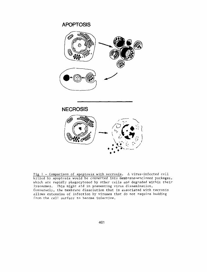
Fig 1 - Comparison of apoptosis with necrosis. A virus-infected cell killed by apoptosis would be converted into membrane-enclosed packages,
which are rapidly phagocytosed by other cells and degraded within their lysosomes. This might aid in preventing virus dissemination. Conversely, the membrane dissolution that is associated with necrosis
allows extension of infection by viruses that do not require budding from the cell surface to become infective.
401
into the intercellular space (fig 1). In this way, dissemination of the virus could be limited.
The recent biochemical studies of apoptosis demonstrating activation of non-ZysosomaZ endonuclease within affected cells coincident with development of the early structural changes suggest an additional possible mechanism for the destruction of viral nucleic acid. In this context, it would be of interest to study the fate of RNA as well as DNA at an early stage of apoptosis.
Our hypothesis is consistent with the time-course of the immune response to certain primary viral infections. For example, in primary ectromelia virus infection in mice, cytotoxic T lymphocytes are detectable 2 days after infection, peak on days 4-6, and decline to low levels by day 10 (24). The primary antibody response has a later onset - cytotoxic and virus-neutralising antibodies do not appear until day 4 and peak on days 8-10. Again, NK cell activity occurs early in the course of primary viral infections (25). Finally, it is known that antibody-dependent cell-mediated cytotoxicity (involving apoptosis) can occur at much lower antibody concentrations than those required for complement-mediated necrosis (26).
The fact that the occurrence of apoptosis does not evoke exudative inflammation might explain the absence of overt signs of tissue injury in mild primary viral infections.
Viruses that do not express cell surface antigens during replication, such as some picornaviruses (27), may not be susceptible to this mode of elimination, since they escape cellular inrmune surveillance. For such non-budding viruses, necrosis is probably required for dissemination, and humoral immunity is important for resolution of the infection.
TESTING THE HYPOTHESIS
There is clearly a need to look for the occurrence of apoptosis in mild, self-limiting viral infections. One situation in which this is well documented is mild human viral hepatitis - here "acidophilic" or "Councilman" bodies (classical apoptotic bodies (28)) occurring in association with lymphoid infiltration are a characteristic morphological feature of the disease. In experimental viral infections, the infectivity of virus contained within apoptotic bodies generated in vitro by lymphocyte activity might be investigated. Lastly, it would be relevant to study the susceptibility of selected viruses to the endogenous endonuclease(s) activated in apoptosis.
REFERENCES
1. Allison AC. Interactions of antibodies, complement components, and various cell types in immunity against viruses and pyogenic bacteria. Transplant Rev 19: 3, 1974.
402
2.
3.
4.
5.
6.
8.
9.
10.
11.
12.
13.
14.
Lawnton AR, Cooper MD. Immune deficiency diseases. p 326 in
Principles of Internal Medicine. 9th ed. (Isselbacher KJ,
Adams RD, Braunwald E, Petersdorf RG, Wilson JD, eds) McGraw Hill,
New York, 1980.
Blanden RV. T cell response to viral and bacterial infection.
Transplant Rev 19: 56, 1974.
Doherty PC, Solter D, Knowles BB. H-2 gene expression is required
for T cell-mediated lysis of virus-infected target cells. Nature
266: 361, 1977.
Trump BF, Ginn FL,. The pathogenesis of subcellular reaction to
lethal injury. p 1 in Methods and Achievements in Experimental
Pathology. Vol. 4. (Bajusz E, Jasmin G, eds) Karger, Base1 and New
York, 1969.
Croker BP, Saladino AJ, Trump BF. Ion movements in cell injury.
Relationship between energy metabolism and the pathogenesis ot lethal injury in the toad bladder. Am J Path01 59: 247, 1470.
Tamm I. Cell injury with viruses. Am J Path01 81: 163, 1975.
Amako K, Dales S. Cytopathology of mengovirus infection.
1. Relationship between cellular disintegration and virulence.
Virology 32: 184, 1967.
Goldberg B, Green H. The cytotoxic action of immune gamma glcbL.iin and complement on Krebs ascites tumor cells. 1. Ultrastructliral studies. J Exp Med 109: 505, 1959.
Hawkins HK, Ericsson JLE, Biberfeld P, Trump BF. Lysosome ax:d phagosome stability in lethal cell injury. Morphologic tracer studies in cell injury due to inhibition of energy metabolism,
immune cytolysis and photosensitization. Am 3 Path01 68: 255 1972. ,
Allison AC, Sandelin K. Activation of lysosomal enzymes .i~. virus-infected cells and its possible relation to cytopathic
effects. J Exp Med 117: 879, 1963.
Don MM, Ablett G, Bishop C-1, et al. Death of cells by spoptosis following attachment of specifically allergized lymphocytes in
vitro. Aust J Exp Biol Med Sci 55: 407, 1977.
Sanderson CJ, Glauert AM. The mec!lanism of 'I cell mediated cytotoxicity. V. Morphological studies by electron microscop\
Proc R Sot Lond (Biol) 198: 315, 1977.
Matter A. Microcinematographic and electron microsccpic analysis of target cell lysis induced by cytotoxic I' lymphocytes.
Immunology 36: 179, 1979.
403
15.
16.
17.
18.
19.
20.
21.
22.
23.
24.
25.
26.
27.
28.
Sanderson CJ, Thomas JA. The mechanism of K cell (antibody-dependent) cell mediated cytotoxicity. II. Characteristics of the effector cell and morphological changes in the target cell. Proc R Sot Lond (Biol) 197: 417, 1977.
Bishop CJ, Whiting VA. The role of natural killer cells in the intravascular death of intravenously injected murine tumour cells. Br J Cancer 48: 441, 1983.
Kerr JFR, Wyllie AH, Currie AR. Apoptosis: a basic biological phenomenon with wide-ranging implications in tissue kinetics. Br J Cancer 26: 239, 1972.
Wyllie AH, Kerr JFR, Currie AR. Cell death: the significance of apoptosis. Int Rev Cytol 68: 251, 1980.
Wyllie AH, Morris RG, Smith AL, Dunlop D. Chromatin cleavage in apoptosis: association with condensed chromatin morphology and dependence on macromolecular synthesis. J Path01 142: 67, 1984.
Cohen JJ, Duke RC. Glucocorticoid activation of a calcium-dependent endonuclease in thymocyte nuclei leads to cell death. J Immunol 132: 38, 1984.
Wyllie AH. Glucocorticoid-induced thymocyte apoptosis is associated with endogenous endonuclease activation. Nature 284: 555, 1980.
Duke RC, Chervenak R, Cohen JJ. Endogenous endonuclease-induced DNA fragmentation: an early event in cell-mediated cytolysis. Proc Nat1 Acad Sci USA 80: 6361, 1983.
Searle J, Kerr JFR, Bishop CJ. Necrosis and apoptosis: distinct modes of cell death with fundamentally different significance. Path01 Annu 17 Pt 2: 229, 1982.
Blanden RV, Gardner ID. The cell-mediated immune response to ectromelia virus infection. I. Kinetics and characteristics of the primary effector T cell response in vivo. Cell Immunol 22: 271, 1976.
Welsh RM. Natural cell-mediated immunity during viral infections. Curr Top Microbial Immunol 92: 83, 1981.
Sissons JGP, Oldstone MBA. Antibody-mediated destruction of virus-infected cells. Adv Immunol 29: 209, 1980.
Burns WH, Allison AC. Surface antigens on virus-infected cells. p 213 in Virus Infection and the Cell Surface. (Poste G, Nicolson GL, eds) North-Holland, Amsterdam, 1977.
Kerr JFR, Bishop CJ, Searle J. Apoptosis. p 1 in Recent Advances in Histopathology. Vol. 12. (Anthony PP, MacSween RNM, eds) Churchill Livingstone, Edinburgh, 1984.
404