Embed Size (px)
Citation preview

125
J. Phycol.
38,
125–134 (2002)
APPARENT LIGHT REQUIREMENT FOR ACTIVATION OF PHOTOSYNTHESIS UPON REHYDRATION OF DESICCATED BEACHROCK MICROBIAL MATS
1
Ulrich Schreiber, Rolf Gademann
Julius-von-Sachs-Institut für Biowissenschaften, Universität Würzburg, Julius-von-Sachs Platz 2, D-97082 Würzburg, Germany
Paul Bird
Centre of Marine Studies, University of Queensland, St. Lucia, 4072 Queensland, Australia
Peter J. Ralph
Multiscale Ecosystem Unit, University of Technology Sydney, Gore Hill, NSW, Australia
Anthony W. D. Larkum
School of Biological Sciences, University of Sydney, NSW 2006, Australia
and
Michael Kühl
2
Marine Biological Laboratory, University of Copenhagen, Strandpromenaden 5, DK-3000 Helsingør, Denmark
Photosynthetic electron transport of beachrockmicrobial mats growing in the intertidal zone ofHeron Island (Great Barrier Reef, Australia) was in-vestigated with a pulse amplitude modulation chl flu-orometer providing four different excitation wave-lengths for preferential excitation of the major algalgroups (cyanobacteria, green algae, diatoms/dinoflagel-lates). A new type of fiberoptic emitter-detector unit(PHYTO-EDF) was used to measure chl fluores-cence at the sample surface. Fluorescence signalsmainly originated from cyanobacteria, which couldbe almost selectively assessed by 640-nm excitation.Even after desiccation for long time periods underfull sunlight, beachrock showed rapid recovery of
photosynthesis after rehydration in the light (t
1/2
�
15 min). However, when rehydrated in the dark, thequantum yield of energy conversion of PSII re-mained zero over extended periods of time. Parallelmeasurements of O
2
concentration with an oxygenmicrooptode revealed zero oxygen concentration inthe surface layer of rehydrated beachrock in thedark. Upon illumination, O
2
concentration increasedin parallel with PSII quantum yield and decreasedagain to zero in the dark. It is proposed that oxygenis required for preventing complete dark reductionof the PSII acceptor pools via the NADPH-dehydro-genase/chlororespiration pathway. This hypothesisis supported by the observation that PSII quantumyield could be partially induced in the dark by flush-ing with molecular oxygen.
Key index words:
beachrock; cyanobacteria; photo-synthesis; oxygen; chl fluorescence; microsensor;state 1/state 2
Abbreviations:
EDF, emitter-detector unit; F
o
, fluor-escence yield of dark-adapted sample; F
m
, maximal
fluorescence yield measured during saturation pulse;F
v
, variable fluorescence yield; LED, light-emittingdiode; PAM, pulse amplitude modulation; PQ, plas-
toquinone
Beachrock consists of carbonate-cemented rock ofvarying composition occuring in the upper tidal zoneof many subtropical and tropical marine environ-ments. Beachrock is colonized by a variety of microor-ganisms with a predominance of epilithic, chasm-olithic, and endolithic cyanobacteria, which can forma dense microbial mat at the beachrock surface. Al-though geological and geochemical studies of beach-rock are relatively abundant in the literature, very lit-tle is known about the biology and biogeochemistry ofbeachrock. Biological studies have mostly focused onfaunistic and floristic accounts (e.g. Cribb 1966, Bratt-ström 1992) and of the possible role of microorgan-isms in the formation of beachrock (e.g. Neumeier1999, Webb et al. 1999). To our knowledge, the com-prehensive study of Krumbein (1979) represents themost detailed account of beachrock biogeochemistry,and very little is known about the ecophysiology of mi-crobial communities in beachrock.
The beachrock on Heron Island, Great BarrierReef consists of microbialites and micritic aragonitecement, the formation of which is induced by biologi-cal activity (Davies and Kinsey 1973, Webb et al.1999). Three characteristic types of beachrock havebeen identified (Stephenson and Searles 1960, Cribb1966): a dark brownish-black colored zone in the up-per littoral, a pale whitish-pink zone in the intermedi-ate tidal zone, and a pale greenish-white zone extend-ing across the low tide mark. The different zones are
1
Received 8 June 2001. Accepted 2 November 2001.
2
Author for correspondence: e-mail [email protected].

126
ULRICH SCHREIBER ET AL.
characterized by a different composition of cyanobac-teria and microalgae (see Cribb 1966).
A prominent feature of beachrock is periodicaldessication and exposure to extreme irradiance. Inthis study we investigated the photosynthesis of micro-bial mats covering the uppermost brownish-blackzone, which is most prone to dessication and irradi-ance stress. The goal of the present study was to inves-tigate physiological mechanisms involved in the recov-ery of photosynthesis of beachrock cyanobacteria whenrehydrated after a period of desiccation. The photo-synthesis of intact beachrock microbial mats was anal-ysed by noninvasive pulse amplitude modulated (PAM)chl fluorescence measurements (Schreiber et al. 1986)in combination with fiberoptic oxygen microsensors(Klimant et al. 1995). Both techniques are used in-creasingly in aquatic photosynthesis studies (e.g. Kühlet al. 2001) and are applied here to epilithic cyano-bacterial communities of beachrock for the first time.
Chl fluorescence provides detailed information onphotosynthetic reactions (Krause and Weis 1991,Schreiber et al. 1994). In particular, PAM fluorescencemeasurements allow the assessment of the effectivequantum yield of energy conversion at PSII reactioncenters with the help of short pulses of saturating light(so-called saturation pulses) (Schreiber et al. 1986,1994, Genty et al. 1989). Fluorescence yield is con-trolled by two major types of quenching mechanisms.One mechanism is photochemical quenching bycharge separation at PSII, which is maximal when allreaction centers are open (i.e. all primary acceptorsare oxidized) and minimal when all centers are closed(i.e. all primary acceptors are reduced). Thus, at max-imal photochemical quenching the minimal fluores-cence yield (F
o
) is observed, whereas at minimal pho-tochemical quenching the maximal fluorescenceyield (F
m
) is observed. Another mechanism is non-photochemical quenching, which controls fluores-cence yield at the pigment level independently of theopenness of the reaction centers. Hence, nonphoto-chemical quenching also affects the F
m
level, whichcan be selectively assessed by a saturation pulse, whichtransiently closes all reaction centers.
Nonphotochemical quenching can reflect variousprocesses that withdraw excitation energy from PSII,including the controlled dissipation of excess energyinto heat (so-called energy-dependent quenching)(Krause et al. 1982, Demmig-Adams and Adams 1992)and the regulated transfer of energy from PSII to PSI,associated with the so-called pigment state 2 (Bona-ventura and Myers 1969, Williams and Allen 1987,Quick and Stitt 1989, Allen 1992). The latter aspect isparticularly important for the interpretation of chlfluorescence changes in cyanobacteria (Allen 1992,Schreiber et al. 1995), which constitute the dominantcomponent of the beachrock microbial mats investi-gated in the present study. In cyanobacteria the majorantenna pigments of PSI (chl
a
) and PSII (phycobi-lins) display markedly different excitation spectra, andthe regulatory mechanism of pigment state shifts is
particularly important to ensure a balanced excitationof the two photosystems. State 2 is induced when ahigh reduction level of the plastoquinone (PQ) poolis reached for a significant period of time (severalminutes). As a consequence of the shift to state 2,photon capture by PSII is down-regulated with respectto PSI and the PQ pool is oxidized. Conversely, a shiftto state 1 is induced when the PQ pool is oxidized inthe light due to preferential PSI excitation (Allen1992). In cyanobacteria, the light-induced state 2–state1 shift is relatively fast (t
1/2
�
1 min) (Schreiber et al.1995).
In the present investigation of beachrock, a newtype of fiberoptic chl fluorometer PHYTO-PAM EDF(Heinz Walz GmbH, Effeltrich, Germany) was applied,which was specifically developed (by the first authorof the present report) for the study of microphytob-enthos, periphyton, and microbial mats with hetero-geneous populations of photosynthetically activeorganisms. With this new device, chl fluorescence is ex-cited simultaneously at four different wavelengths, thusproviding coarse excitation spectra that contain infor-mation on the contribution of various types of pig-mented organisms to the overall fluorescence signal(Kolbowski and Schreiber 1995, Schreiber 1998).
materials and methods
Beachrock samples.
Beachrock was collected from the upperbrownish-black zone at the southern shore of Heron Island (152
�
6
�
E, 20
�
29
�
S). (See Webb et al. [1999] for a detailed descrip-tion and pictures of the sampling site.) The beachrock was cov-ered by an
�
1- to 1.5-mm-thick microbial mat with a dense crum-bly structure, which was predominantly composed of cyanobacteria(mainly
Entophysalis
sp.,
Calothrix
sp., and
Lyngbya
sp.). An
�
30-mm-thick slab of the surface was separated from a larger rock with thehelp of a seawater-cooled diamond-tipped circular saw, from whichreplicate samples of approximately 25
�
25
�
30 mm were cut.The samples were kept outdoors in a bath of continuously renewedseawater in natural daylight (up to
�
2500
�
mol quanta
�
m
�
2
�
s
�
1
)for several hours every day and were exposed to dry air for the restof the time. Experiments were carried out with samples desiccatedfor at least 8 h. For measurements, the samples were transferred toa darkened dish that could be filled with filtered aerated seawa-ter covering the sample. In some experiments the seawater wasflushed for 15 min with pure oxygen or nitrogen before additionto the samples.
Chl fluorescence measurements.
Chl fluorescence was measuredwith a PHYTO-PAM chl fluorometer (commercially availablefrom Heinz Walz GmbH, Effeltrich, Germany) equipped with aspecial emitter-detector unit (PHYTO-EDF) designed for the in-vestigation of microphytobenthos, periphyton, and microbialmats. The PHYTO-EDF features polyfurcated fiberoptics con-nected to various measuring and actinic light-emitting diode(LED) light sources via miniature fiber couplers and to a pho-tomultiplier detector. Single 1-mm plastic fibers were coupledto 470, 525, 640, and 665 nm measuring light LEDs, whereasfour 1-mm fiber branches were coupled to actinic LEDs (peak660 nm). A single 1.5-mm plastic fiber carried the fluorescencesignal to the detector, which was protected by a long-pass filter(
�
700 nm). The combined fiberoptics had a 4-mm active di-ameter at their joint end. A perspex cylinder (50 mm long and4 mm in diameter) served for randomization of the variouslight beams. Measurements were carried out with a distance of
�
2 mm between the tip of the perspex cylinder and the surfaceof the beachrock sample.
Ambient light was prevented from reaching the sample withthe help of a darkening hood. The minimal measuring light

127
ACTIVATION OF PHOTOSYNTHESIS IN BEACHROCK
frequency was applied, corresponding to an integrated inten-sity of 0.15
�
mol quanta
�
m
�
2
�
s
�
1
PAR, the actinic effect ofwhich could be neglected. The measuring light intensity of thedifferent wavelengths was similar. When actinic illuminationwas applied, this amounted to 120
�
mol quanta
�
m
�
2
�
s
�
1
of 660nm light. Full
in situ
irradiance of the beachrock amounted to
�
2500
�
mol photons
�
m
�
2
�
s
�
1
, and the actinic light did notlead to any photoinhibition. During actinic illumination andapplication of saturation pulses, the measuring frequency wasautomatically increased by a factor of 128 to improve the signal-to-noise ratio and time resolution. Saturation pulses were ap-plied with the actinic light LEDs (660 nm). Saturation pulseshad an intensity of
�
1800
�
mol quanta
�
m
�
2
�
s
�
1
, which provedenough for full PSII closure.
The PHYTO-PAM was operated in conjunction with a note-book PC and the PhytoWin data acquisition software (HeinzWalz GmbH) provided with the instrument. In applicationswith phytoplankton samples, a major purpose of this system isthe deconvolution of fluorescence responses of the major algalgroups. In the case of beachrock, this aspect was of minor im-portance, because the signal was dominated by cyanobacterialfluorescence. Hence, the presented data were mostly derivedfrom signals obtained with the 640-nm measuring light, wheremaximal responses were obtained. In the few experiments wherefluorescence signals were deconvoluted, “reference spectra” ofthe following organisms were used:
Synechococcus
sp. (blue),
Anki-strodesmus braunii
(green), and
Phaeodactylum tricornutum
(brown).The reference spectra were recorded in suspensions with a highchl contents (
�
10
�
g chl
�
mL
�
1
) to approach the state of thehighly pigmented organisms in microbial mats.
The PHYTO-PAM monitors continuously the chl fluores-cence yield (F) using four different excitation wavelengths si-multaneously. Hence, there are four independent primary sig-nals, F
470
, F
525
, F
640
, and F
665
. In the absence of actinic light,these signals correspond to F
o
, the fluorescence yield of a dark-adapted sample, because the integrated intensity of the measur-ing light does not induce the accumulation of reduced PSIIelectron acceptors. Each actual “measurement” by the PHYTO-PAM (the data being saved in a “report file”) involves the appli-cation of a saturation pulse to determine the maximum fluores-cence yield (F
m
) and thus the increase of fluorescence yield in-duced by the saturation pulse (
F, denoted as dF by thePhytoWin software). Briefly, before the saturation pulse, themomentary fluorescence yield (F
�
F
t
) is measured, which maycorrespond to F
o
if the sample is dark adapted or any otherfluorescence yield between F
o
and F
m
if the sample is illumi-nated. In any case, a
F is measured that when related to themaximal fluorescence yield in the momentary state of the sam-ple gives a reliable measure of the quantum yield of energy con-version of PSII: Y
�
F/F
m
(Genty et al. 1989). Formally, with adark-adapted sample,
F/F
m
corresponds to F
v
/F
m
, a fre-quently used parameter for the assessment of the maximalquantum conversion efficiency of PSII. However, although thisparameter is well defined and readily accessible in the study ofgreen plants, this is not the case with cyanobacteria, which aremainly dealt with in the present study.
In contrast to green plants, the dark-adapted state in cyano-bacteria is not characterized by maximal photochemical andminimal nonphotochemical quenching (see e.g. Schreiber etal. 1995). Therefore, when dealing with cyanobacteria, the useof conventional F
v
and F
v
/F
m
parameters (defined in vanKooten and Snel 1990) are problematic. It does not make senseto distinguish between F
m
(maximal fluorescence yield in thedark-adapted state) and F
m
�
(maximal fluorescence yield in theilluminated state) because the “true F
m
” is not well defined.Consequently, and for the sake of simplicity, in the present re-port all maximal fluorescence yields determined with the helpof a saturation pulse are denoted as F
m
. Likewise, all fluores-cence yields measured shortly before a saturation pulse are de-noted as F. If not stated otherwise, the data presented in thiscommunication correspond to F, F
m
,
F, and Y as measuredwith 640-nm excitation, which is absorbed preferentially by phy-cobilins in cyanobacteria.
Measurements of oxygen concentration.
Oxygen concentration wasmeasured with an oxygen microoptode (Klimant et al. 1995; Sen-sor type B, Presense GmbH, Germany) coupled to a portablefluorescence lifetime measuring system (Microx TX, PresenseGmbH, Germany). The system was connected to a PC via a se-rial interface. Calibration of the microoptode signal and dataacquisition was controlled by the supplied software for the in-strument. The oxygen microoptode was connected to the instru-ment via a standard ST-fiber connector, and a two-point calibra-tion was performed at experimental temperature and salinityby recording the sensor signal in fully aerated seawater and inseawater made anoxic by addition of sodium dithionite. Theresponse time of the microoptode was
�
5 s. For measure-ments on the beachrock, the microoptode was mounted in amanually operated micromanipulator (MM33, MärtzhäuserGmbH, Germany) and the measuring tip was carefully posi-tioned onto the surface of the beachrock at an insertion angle,which still allowed the perspex rod of the PHYTO-PAM EDF fi-ber to keep a distance of
�
2 mm relative to the surface (seealso above). The sample surface was covered with seawater, sothat the tip of the microoptode and the end of the perspex rodwere immersed.
results
Basic information provided by the PHYTO-PAM.
Figure1 shows the fluorescence responses of a beachrocksample after rehydration in natural sunlight, as dis-played on the PC monitor screen by the PhytoWinsoftware. The fluorescence yields, measured simulta-neously with 470-, 525-, 640-, and 665-nm excitation,are displayed numerically and graphically (bars). Al-though the signal excited at 640 nm was distinctlyhigher than that at 665 nm, relatively low signals wereobserved with 470- and 525-nm excitation. This excita-tion pattern is typical for cyanobacteria, where phyco-cyanin and allophycocyanin (strong absorption at620–640 nm) constitute the major accessory antennapigments transferring energy to chl
a
in PSII. The val-ues of the parameter F, as well as the correspondingbars, represent the momentary fluorescence yields.Upon application of a saturation pulse, first the mo-mentary fluorescence yield (F) and briefly afterwardthe maximal fluorescence yield (F
m
) are sampled.The PhytoWin software saves the momentary fluores-cence yield under F
t
and the saturation pulse inducedincrease of fluorescence yield (
F
�
F
m
�
F
t
) underdF. Due to the high signal-to-noise ratio in the givenapplication, the values of F
t
and F are identical; there-fore, for the sake of simplicity the momentary fluores-cence yield will be generally denoted by F in thepresent study (see also Materials and Methods). Theyield parameter is derived from the expression dF/(F
�
dF), which corresponds to the commonly knownexpressions F
v
/F
m
or
F/F
m
for the effective quantumyield of energy conversion in PSII (Genty et al. 1989,Schreiber et al. 1994) (for nomenclature see Materi-als and Methods). The relatively low apparent quan-tum yield of 0.37 at 640-nm excitation is typical for cy-anobacteria (Schreiber et al. 1995). The somewhathigher value at 665-nm excitation indicates an addi-tional, albeit low, fluorescence contribution of greenalgae, which absorb strongly at that wavelength anddisplays higher values of variable fluorescence.

128 ULRICH SCHREIBER ET AL.
Figure 2 shows the result of the deconvolution rou-tine. The data indicate that about 9/10 of the fluores-cence measured from the top layer of the investigatedbeachrock was originating from cyanobacteria, whereasthe remaining 1/10 was due to green algae. Micro-scopic observations revealed the presence of smallquantities of Enteromorpha intestinalis; no diatoms ordinoflagellates could be detected either in the decon-voluted fluorescence data or by microscopic observa-tions. Although the apparent quantum yield of thegreen algae was almost twice that of the cyanobacte-ria, their contribution to the 640-nm signal was negli-gible, as suggested by the almost identical yield valuesfor cyanobacteria (yield � 0.35 for “blue”) and 640-
nm excitation (yield � 0.37). The overall signal wasthus dominated by cyanobacteria, particularly whenmeasured with 640-nm excitation. Therefore, in thefollowing experiments on rehydration of beachrockand on the apparent light activation of photosyntheticelectron transport, we focused on the original fluores-cence parameters measured with 640-nm excitation.
Fluorescence changes upon rehydration. In Fig. 3 the fluo-rescence changes upon rehydration of beachrock arepresented, as measured with 640-nm excitation and re-petitive application of saturation pulses at 60-s intervalsfor assessment of variable fluorescence yield. Desiccatedbeachrock exhibited a rather low fluorescence yield,which upon addition of seawater rapidly increased by afactor of �6 (t1/2 � 1 min) before it declined moreslowly to an intermediate level (t1/2 � 2 min). No vari-able fluorescence could be induced by saturation pulsesneither in the desiccated state nor after rehydration inthe dark (Fig. 3A). However, when rehydration tookplace in the presence of continuous actinic light (Fig.3B), variable fluorescence (i.e. a difference between Fand Fm) was slowly induced, thus reflecting the recov-ery of PSII activity.
Light-induced changes of fluorescence and effective quan-tum yield. When rehydrated beachrock samples werekept in the dark, no variable fluorescence could be in-duced by saturation pulses over extended periods oftime (from minutes to hours), thus indicating a per-
Fig. 1. Fluorescence responses of a beachrock sample af-ter rehydration in natural sunlight and 5 min dark adaptation.The Channel screen of the PhytoWin user interface is depicted.A saturation pulse was applied to assess the effective quantumyield of PSII (yield parameter) via the induced fluorescence in-crease (dFt). Ft is the fluorescence yield briefly before the satu-ration pulse. Ft � dFt corresponds to the maximal fluorescenceyield reached during the saturation pulse, Fm. The bar diagramshows the momentary fluorescence yields, F, with 470-, 525-,640-, and 665-nm excitation (from left to right), respectively.
Fig. 2. Deconvoluted fluorescence data of a beachrocksample rehydrated in natural sunlight, as displayed on the Al-gae screen of the PhytoWin user interface. The bar diagramshows the deconvoluted momentary fluorescence yields, F, ofcyanobacteria (blue), green algae (green), and diatoms and di-noflagellates (brown) (from left to right), respectively. The cor-responding original data are presented in Fig. 1. For furtherexplanations, see legend to Fig. 1 and text.
Fig. 3. Time-dependent changes of fluorescence yieldupon rehydration of desiccated beachrock samples. (A) Rehy-dration of sample kept in the dark, except for the weak measur-ing light. (B) Rehydration of sample illuminated with 120 �molquanta�m�2�s�1 of 660-nm actinic light. Aerated filtered seawa-ter was added where indicated and covered the complete sam-ple. Saturation pulses where applied repetitively at 1-min inter-vals to assess variable fluorescence.

129ACTIVATION OF PHOTOSYNTHESIS IN BEACHROCK
sistent blockage of PSII activity (Fig. 3A). When rehy-dration occurred in the light, variable fluorescencedeveloped after about 5 min and reached a maximumafter about 25 min (Fig. 3B). To elucidate the under-lying mechanisms of inhibition and recovery, thedark–light–dark induction kinetics of fluorescencewere investigated. Figure 4A shows the fluorescenceresponses of a beachrock sample, which had been re-hydrated in the dark for 20 min. After this time,changes in fluorescence caused by the hydration pro-cess were largely terminated (see Fig. 3A), but no vari-able fluorescence was observable (i.e. the values of Fand Fm were identical). Immediately after onset of ac-tinic illumination, variable fluorescence developed,thus indicating the recovery of photosynthetic activity.The light-on response of the fluorescence yield (F)consisted of a small rise followed by a large decline. Inthe light-on response of Fm, the initial rise was morepronounced, whereas the subsequent decline did notfall much below the initial dark level. At light-off, bothsignals displayed a rapid drop. In the following darkperiod, the F value showed a large amplitude slowrise, whereas Fm remained almost unchanged.
In Fig. 4B the corresponding changes of the effec-tive PSII quantum yield are shown. In the given exam-ple, the deconvoluted quantum yields for the cyano-bacteria (Yblue) and green algae (Ygreen) are displayed
as well as those determined with 640-nm excitation(Y640), which were derived from the original fluores-cence data in Fig. 4A. Comparison of the light-inducedY640 and Yblue responses confirmed that the cyanobac-terial signal was well described by the 640-nm signal.However, it was also apparent that the Ygreen responsediffered from the Yblue response, as it increased distinctlyfaster (t1/2 � 1 min as compared with t1/2 � 15 min)and reached a substantially higher level (0.55 as com-pared with 0.27). After light-off, both quantum yieldsshowed a transitory increase, as may be expectedupon dark reoxidation of the intersystem electrontransport chain. Thereafter, both quantum yields de-clined with similar kinetics (t1/2 � 20 min). It can beseen that the slow decline matched the slow rise of thefluorescence yield (F) (Fig. 4A).
Green algae are generally known to display highervalues of Y � F/Fm than cyanobacteria, which is inline with the results presented in Fig. 4B. Hence, it ap-pears likely that the Ygreen response indeed reflects apopulation of green algae in the microbial mat. How-ever, because this population was very small as com-pared with the dominating cyanobacteria, we concen-trated our efforts on the cyanobacteria, for assessmentof which deconvolution was not required. Therefore,in the following experiments only the responses with640-nm excitation are presented, which closely followthe responses of the dominating cyanobacteria (com-pare Y640 and Yblue in Fig. 4B).
The dark–light–dark induction kinetics of rehy-drated beachrock displayed in Fig. 4 differs consider-ably from corresponding kinetics known for higherplants or for cyanobacteria and green algae in suspen-sion. Normally, the fluorescence yield of a dark-adaptedsample is minimal and dark–light induction involves arapid rise of fluorescence yield to a high level, fol-lowed by a slow decline to a lower stationary yield.This transition is also known as the Kautsky effect(Kautsky and Hirsch 1931). The observed inductionkinetics of beachrock are reminiscent of that de-scribed for Scenedesmus obliquus under conditions ofextreme anaerobiosis after prolonged dark adapta-tion (Schreiber and Vidaver 1974, 1975). This earlierwork suggested that a hydrogenase is activated underanaerobic conditions, which causes slow reduction ofthe intersystem electron transport chain in the dark,accompanied by a rise in fluorescence yield from aminimal level, Fo, to a maximal level, Fm. We hypothe-sized that after rehydration in the dark, the beachrockmicrobial mat became anaerobic due to respiratoryactivity in combination with a slow rate of O2 diffusionacross the diffusive boundary layer overlaying the wet-ted beachrock. The role of oxygen was now investi-gated using an O2 microelectrode in combinationwith the PAM fiberoptics sensor.
Parallel measurements of O2 concentration and chlfluorescence. To test the working hypothesis that pho-tosynthesis of dark-rehydrated beachrock is blockeddue to anaerobiosis, parallel measurements of O2 con-centration and chl fluorescence were carried out. O2
Fig. 4. Dark–light–dark induction transients of beach rocksample previously rehydrated in the dark. (A) Induction tran-sients of fluorescence yield. (B) Induction transients of the ef-fective PSII quantum yield measured with 640-nm excitation(Y640) in comparison with the deconvoluted effective PSII quan-tum yields in cyanobacteria (Yblue) and green algae (Ygreen). Forother conditions, see legend to Fig. 1. AL, actinic light (120�mol photons�m�2�s�1).
130 ULRICH SCHREIBER ET AL.
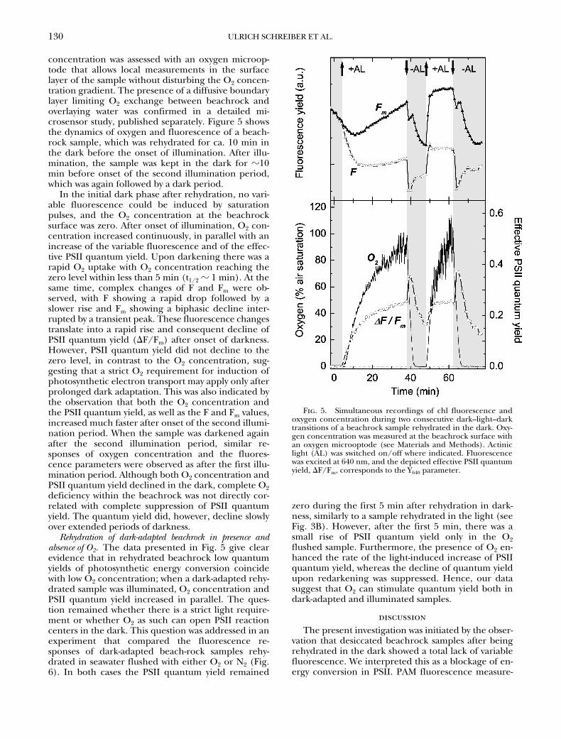
concentration was assessed with an oxygen microop-tode that allows local measurements in the surfacelayer of the sample without disturbing the O2 concen-tration gradient. The presence of a diffusive boundarylayer limiting O2 exchange between beachrock andoverlaying water was confirmed in a detailed mi-crosensor study, published separately. Figure 5 showsthe dynamics of oxygen and fluorescence of a beach-rock sample, which was rehydrated for ca. 10 min inthe dark before the onset of illumination. After illu-mination, the sample was kept in the dark for �10min before onset of the second illumination period,which was again followed by a dark period.
In the initial dark phase after rehydration, no vari-able fluorescence could be induced by saturationpulses, and the O2 concentration at the beachrocksurface was zero. After onset of illumination, O2 con-centration increased continuously, in parallel with anincrease of the variable fluorescence and of the effec-tive PSII quantum yield. Upon darkening there was arapid O2 uptake with O2 concentration reaching thezero level within less than 5 min (t1/2 � 1 min). At thesame time, complex changes of F and Fm were ob-served, with F showing a rapid drop followed by aslower rise and Fm showing a biphasic decline inter-rupted by a transient peak. These fluorescence changestranslate into a rapid rise and consequent decline ofPSII quantum yield (F/Fm) after onset of darkness.However, PSII quantum yield did not decline to thezero level, in contrast to the O2 concentration, sug-gesting that a strict O2 requirement for induction ofphotosynthetic electron transport may apply only afterprolonged dark adaptation. This was also indicated bythe observation that both the O2 concentration andthe PSII quantum yield, as well as the F and Fm values,increased much faster after onset of the second illumi-nation period. When the sample was darkened againafter the second illumination period, similar re-sponses of oxygen concentration and the fluores-cence parameters were observed as after the first illu-mination period. Although both O2 concentration andPSII quantum yield declined in the dark, complete O2deficiency within the beachrock was not directly cor-related with complete suppression of PSII quantumyield. The quantum yield did, however, decline slowlyover extended periods of darkness.
Rehydration of dark-adapted beachrock in presence andabsence of O2. The data presented in Fig. 5 give clearevidence that in rehydrated beachrock low quantumyields of photosynthetic energy conversion coincidewith low O2 concentration; when a dark-adapted rehy-drated sample was illuminated, O2 concentration andPSII quantum yield increased in parallel. The ques-tion remained whether there is a strict light require-ment or whether O2 as such can open PSII reactioncenters in the dark. This question was addressed in anexperiment that compared the fluorescence re-sponses of dark-adapted beach-rock samples rehy-drated in seawater flushed with either O2 or N2 (Fig.6). In both cases the PSII quantum yield remained
zero during the first 5 min after rehydration in dark-ness, similarly to a sample rehydrated in the light (seeFig. 3B). However, after the first 5 min, there was asmall rise of PSII quantum yield only in the O2flushed sample. Furthermore, the presence of O2 en-hanced the rate of the light-induced increase of PSIIquantum yield, whereas the decline of quantum yieldupon redarkening was suppressed. Hence, our datasuggest that O2 can stimulate quantum yield both indark-adapted and illuminated samples.
discussionThe present investigation was initiated by the obser-
vation that desiccated beachrock samples after beingrehydrated in the dark showed a total lack of variablefluorescence. We interpreted this as a blockage of en-ergy conversion in PSII. PAM fluorescence measure-
Fig. 5. Simultaneous recordings of chl fluorescence andoxygen concentration during two consecutive dark–light–darktransitions of a beachrock sample rehydrated in the dark. Oxy-gen concentration was measured at the beachrock surface withan oxygen microoptode (see Materials and Methods). Actiniclight (AL) was switched on/off where indicated. Fluorescencewas excited at 640 nm, and the depicted effective PSII quantumyield, F/Fm, corresponds to the Y640 parameter.

131ACTIVATION OF PHOTOSYNTHESIS IN BEACHROCK
ments use very low intensity pulse-modulated measuringlight for assessment of the momentary fluorescenceyield, F, and the application of brief pulses of saturat-ing light (saturation pulses) for determination of themaximal fluorescence yield, Fm. On the basis of thisinformation, the effective quantum yield of energyconversion in PSII can be calculated (Genty et al.1989, Schreiber et al. 1994). Because of the low mea-suring light intensity and the shortness of the satura-tion pulses, such measurements may be considerednonintrusive (i.e. they do not affect the state of thephotosynthetic apparatus). On the other hand, ac-tinic light can cause substantial changes in the state ofthe photosynthetic apparatus, particularly the redoxstate of primary acceptors, the first stable acceptor ofPSII, which in turn controls the rate of electron trans-port through PSII. When desiccated beachrock wasrehydrated in the presence of actinic illumination, incontrast to dark rehydration the PSII activity (as mea-sured via Fv) increased to high values within 15 min.Hence, there appears to be a light requirement forthe recovery of activity. It was the aim of the presentstudy to obtain information on the causes of the ob-served dark inhibition and light recovery.
By separating the rehydration step from the light-recovery step, it was possible to analyze the kinetics oflight recovery. When a rehydrated sample was kept inthe dark, the inhibited state could be maintained overextended periods of time and was not affected by theapplied weak measuring light and brief pulses of satu-rating light. Upon onset of actinic light, complexchanges of the momentary fluorescence yield (F) andthe maximal fluorescence yield (Fm) were induced. Inthe course of actinic illumination, variable fluores-
cence recovered, thus reflecting an opening of PSIIreaction centers. Notably, this recovery primarily in-volved a decrease of F, whereas Fm remained at a highlevel. This may be considered unusual, as with mostother types of samples, like algal suspensions orleaves, variable fluorescence, Fv � F � Fm – F, is max-imal after dark adaptation (where F � Fo) and de-creases with increasing actinic intensity. This situationresults from two major types of deviation from normalbehavior, both of which have been reported previ-ously. First, in certain unicellular microalgae, like S.obliquus, electron carriers at the acceptor side of PSIIcan become reduced in the dark, in particular whendark reoxidation by O2 is prevented under anaerobicconditions (Schreiber and Vidaver 1974, 1975). Sec-ond, in contrast to higher plant leaves, cyanobacteriaand certain unicellular algae tend to attain the so-called state 2 after dark adaptation (for definition ofstate 1–state 2, see above), which is characterized bylow values of Fm and Fv (Schreiber et al. 1995, Schreiber1998). Both aspects may be linked, because state tran-sitions are controlled by the redox state of the PQpool and/or of components of the cyt b/f complex(Allen 1992, Allen et al. 1995). Transition to state 2may be induced in the dark, when the PQ pool is re-duced via a thylakoid membrane-bound NADPH-dehydrogenase (Mi et al. 1992, Schreiber et al. 1995).In view of these previous investigations, it appears rea-sonable to assume that the unusual dark–light–darkfluorescence transients observed with rehydratedbeachrock are related to reduction of the PQ pool un-der anaerobiosis in the dark and subsequent attain-ment of state 2.
In principle, the inactive state observed after rehy-dration could also be due to inactivation of the water-splitting complex upon desiccation, the reactivationof which requires light. However, a block at the PSIIdonor side generally is correlated with quenching ofFm (donor-side dependent quenching), and when thisis relieved, the increase of variable fluorescence (andquantum yield) should be paralleled by a rise in Fmand not by a decrease of F, as observed in our experi-ments. Therefore, although a possible role of watersplitting inactivation/reactivation cannot be ruledout, it is not considered further here.
Simultaneous measurements of O2 concentrationand fluorescence gave direct evidence for a decisiverole of anaerobiosis. Application of an O2 microop-tode allowed minimally invasive measurements of theO2 concentration in the surface layer of the beach-rock submersed in seawater. Although the water col-umn above the beachrock surface was in equilibriumwith air, the surface O2 concentration dropped tozero in darkness due to the resistance to oxygen masstransfer imposed by the presence of a diffusive bound-ary layer, in combination with intense oxygen con-sumption of the beachrock microbial community.(For detailed accounts of diffusive boundary layers,see e.g. Jørgensen and Revsbech [1985] or Boudreauand Jørgensen [2001].) Obviously, after rehydration
Fig. 6. Effect of molecular oxygen on the time-dependentchanges of PSII quantum yield following rehydration of desic-cated beachrock samples. Time zero corresponds to the mo-ment at which the desiccated sample was covered with seawater,which previously had been flushed for 5 min either with O2(�O2) or with N2 (�O2). Where indicated, actinic light (AL)was switched on or off. The Y640-parameter is depicted.

132 ULRICH SCHREIBER ET AL.
the respiratory activity was rapidly restored and therate of O2 uptake exceeded the rate of diffusive oxy-gen supply from the bulk water phase. The high rateof net O2 uptake was evident from the rapid decline ofO2 concentration after switching the illumination off.
From our combined oxygen and PAM fluorescencemeasurements, we put forward a model for the actionof O2 on the activity of PSII in the beachrock samples,which is described below. The measured fluorescencesignals were dominated by cyanobacteria, particularlywhen 640-nm excitation was applied. Therefore, theobservations must be explained in terms of O2 effectson cyanobacteria, which have previously only beenstudied in laboratory strains of Synechocystis PCC 6803(Mi et al. 1992, 2000, Schreiber et al. 1995). In cyano-bacteria, the photosynthetic membranes, which mayor may not be discrete thylakoid membranes, arecharacterized by a unique assembly of electron trans-port components, which on one hand are involved inoxygenic photosynthesis and on the other hand carryout oxidative phosphorylation, with some compo-nents being shared by the two processes ( Jones andMyers 1963, Peschek and Schmetterer 1982). NADPH-dehydrogenase in cyanobacteria has been suggestedto participate in the donation of electrons from respi-ratory substrates to the photosynthetic electron trans-port chain (Sandmann and Malkin 1983, Mi et al.1992). In cyanobacteria, the oxidative pentose phos-phate cycle is quantitatively more important than glyc-olysis in the breakdown of sugars and hence is mainlyresponsible for the reduction of NADP in the dark(Peschek 1999). Under aerobic conditions in thedark, electrons fed via the NADPH-dehydrogenaseinto the PQ pool normally end up in the reduction ofO2, catalyzed by a still unknown oxidase in the so-called process of chlororespiration (Bennoun 1982,Scherer 1990). In the absence of O2, the dark reduc-tion of the PQ pool cannot be balanced by the oxi-dase reaction anymore, and the acceptor side of PSIIbecomes reduced, with a concomitant increase offluorescence yield, F, and decrease of the energy con-version efficiency of PSII.
As apparent in Fig. 5, the dark rise of F after therapid drop after light-off can be accompanied by apronounced decline of Fm, which is most readily ex-plained by a state 1–state 2 transition. Both the in-crease in F and the decrease in Fm contribute to theapparent decrease in effective PSII quantum yield.Upon a dark–light transition, PSI oxidizes the PQpool, but at the same time PQ is also reduced by PSIIactivity. The steady-state equilibrium redox level is de-termined mainly by i) the activity of processes on thePSI acceptor side, ii) water-splitting activity, and iii)the distribution of quanta between the two photosys-tems. Oxygen has an important influence on the PQredox state upon a dark–light transition, because itserves as the major electron acceptor of PSI beforethe Calvin cycle is activated and CO2 becomes the ma-jor acceptor (Schreiber and Vidaver 1974, Radmerand Kok 1976). This explains the different responses
of oxygen and fluorescence parameters between thefirst and second illumination period of Fig. 5. In bothcases, the O2 concentration before illumination waszero, but in the case of the second illumination pe-riod the Calvin cycle enzymes were not yet fully darkinactivated, and consequently photosynthetic elec-tron flow could be initiated immediately even in theabsence of O2 upon illumination.
The observed fluorescence changes upon onset ofthe second illumination point to a pronounced state2–state 1 transition, a prerequisite for which is the ox-idation of the PQ pool by PSI activity (Allen 1992).On the other hand, upon onset of the first illumina-tion, first the system was in state 2 (after being in thedark under anaerobic conditions) and second neithersubstantial amounts of O2 nor CO2 were available asacceptors of PSI. Under these circumstances, it is notsurprising that oxygen evolution and photochemicalquenching in PSII developed slowly. The limitation atthe PSI acceptor side is reflected by the slow rise of Fm(t1/2 � 20 min as compared with t1/2 � 1 min afterpreillumination), which indicates a slow transitionfrom state 2 to state 1, as the oxidation level of the PQpool is only slowly increasing.
The pivotal role of O2 was confirmed by the obser-vation that flushing with O2 caused an increase of thePSII quantum yield in the dark, suggesting a reoxida-tion of the PSII acceptor side (i.e. mainly the PQpool) by O2. However, the relatively small increase ofthe observed quantum yield indicates that the dark-reduction rate was high and could hardly be matchedby the diffusive O2 supply and/or the rate of the oxy-genase reaction. Although illumination plays the ma-jor role in the restoration of PSII activity, the illumina-tion effect is enhanced by O2. As outlined above, itmay therefore be assumed that before light activationof CO2 reduction in the Calvin cycle, O2 serves as anelectron acceptor at the PSI acceptor side. Presum-ably, it is this O2-dependent electron flow, togetherwith cyclic electron flow around PSI, that gives rise toa proton gradient sufficiently high for ATP formation,which is essential for CO2 reduction. Because O2-dependent electron flow as such is associated withzero net O2 exchange (see e.g. Schreiber et al. 1994),the role of O2 may be visualized as equivalent to a cat-alyst in an overall autocatalytic activation process. Be-fore activation of the Calvin cycle, net O2 evolutioncan be explained only by electron flow to the rela-tively small pool of ferredoxin (Fd) and NADP, whichare the only electron carriers with sufficiently low re-dox potentials to stay oxidized under dark anaerobicconditions. Obviously, the involved physiological reac-tions are rather complex, and it is out of the scope ofthe present communication to go into more details.
In Figure 7 the three major states of the cyanobac-teria photosynthetic apparatus in beachrock are illus-trated by highly simplified schemes, which may serveto explain the observed photosynthesis responses to-ward illumination and O2 in beachrock. In Fig. 7A,the dark rehydrated state is characterized by anoxic

133ACTIVATION OF PHOTOSYNTHESIS IN BEACHROCK
conditions associated with full reduction of the PQpool and of all other electron carriers, except for Fdand presumably also some NADP. NADPH-dehydro-genase is responsible for keeping the PQ pool fully re-duced, with the electrons originally derived from thebreakdown of sugars. The system is in pigment state 2,which is characterized by weak excitation of PSII. Im-mediately after onset of illumination (Fig. 7B), thesystem is still in state 2, as the PQ pool initially re-mains highly reduced. Due to PSI driven reduction ofthe available Fd and NADP, a small amount of PQH2becomes oxidized, which immediately is re-reducedby PSII with concomitant evolution of a small amount
of O2. By diffusion, this O2 becomes available as elec-tron acceptor at the PSI acceptor side (Mehler reac-tion) and also at the level of the PQ pool (chlororespi-ration). These two types of O2-dependent electronflow may provide ATP required for CO2 fixation,which will lead to net O2 evolution, once the Calvin cy-cle has become light activated, which may be expectedwithin a time range of several minutes. In Fig. 7C a sus-tained light state is reached after longer illumination.This state is characterized by continuous electron flowto CO2, the fixation of which is supported by ATP for-mation associated with O2-dependent electron flow,which again is enhanced by the increased level of O2concentration created by CO2-dependent electronflow. As the rate of PQH2 oxidation by PSI and chloro-respiration increases, the PQ pool reaches a suffi-ciently oxidized state for a transition from state 2 tostate 1. Hence, eventually a sustained light state isreached that optimizes the photosynthesis rate undergiven environmental conditions.
This study was carried out as part of the advanced researchworkshop Bioreactive surfaces in tropical marine environments,Heron Island Research Station, February 1–13, 2001. We thankOve Hoegh-Guldberg and the staff of HIRS for organizing anexcellent workshop and for tireless logistic and technical sup-port. Financial support of the Danish Natural Science ResearchFoundation (to M. K., contract no. 9700549) and of the Austra-lian Research Council (to A. W. D. L. and P. R.) is gratefully ac-knowledged. Thanks are also due to the Heinz Walz GmbH andPresense GmbH for providing the instrumentation and sensorsused in this study.
Allen, J. F. 1992. Protein phosphorylation in regulation of photo-synthesis. Biochim. Biophys. Acta 1098:275–335.
Allen, J. F., Alexciev, K. & Hakansson, G. 1995. Regulation by redoxsignalling. Curr. Biol. 5:869–72.
Bennoun, P. 1982. Evidence for a respiratory chain in the chloro-plast. Proc. Natl. Acad. Sci. USA 79:4352–6.
Bonaventura, C. & Myers, J. 1969. Fluorescence and oxygen evolu-tion from Chlorella pyrenoidosa. Biochim. Biophys. Acta 189:366–89.
Boudreau, B. P. & Jørgensen, B. B. [Eds.] 2001. The Benthic Bound-ary Layer, Transport Processes and Biogeochemistry. Oxford Univer-sity Press, New York, 404 pp.
Brattström, H. 1992. Marine biological investigations in the Baha-mas. Littoral zonation at three Bahamian beachrock localities.Sarsia 77:81–109.
Cribb, A. B. 1966. The algae of Heron Island, Great Barrier Reef,Australia, part 1. A general account. Univ. Queensland Papers,Gt. Barrier Reef Committee., Heron Island Res. Station 1:3–23.
Davies, P. J. & Kinsey, D.-W. 1973. Organic and inorganic factors inrecent beachrock formation, Heron Island, Great Barrier Reef.J. Sed. Petrol. 43:59–81.
Demmig-Adams, B. & Adams, W. W. III. 1992. Photoprotection andother responses of plants to high light stress. Annu. Rev. PlantPhysiol. Plant Mol. Biol. 43:599–626.
Genty, B., Briantais, J. M. & Baker, N. 1989. The relationship betweenthe quantum yield of photosynthetic electron transport andquenching of chl fluorescence. Biochim. Biophys. Acta 990:87–92.
Jones, L. W. & Myers, J. 1963. A common link between photosyn-thesis and respiration in a blue-green alga. Nature 199:670–2.
Jørgensen, B. B. & Revsbech, N. P. 1985. Diffusive boundary layersand the oxygen uptake of sediment and detritus. Limnol.Oceanogr. 30:111–22.
Kautsky, H. & Hirsch, A. 1931. Neue Versuche zur Kohlensäureas-similation. Naturwissenschaften 19:964.
Klimant, I., Meyer, V. & Kühl, M. 1995. Fiber-optic oxygen mi-
Fig. 7. Schemes characterizing three major states of the cy-anobacterial photosynthetic apparatus in beachrock : A. DARK,after rehydration in the dark B. INITIAL LIGHT, briefly (up toa few minutes) after onset of illumination C. SUSTAINEDLIGHT, after ca. 10–20 min of illumination. Electron flow is in-dicated by solid arrows, while oxygen production and diffusionis indicated by dotted arrows. See text for further explanations.
134 ULRICH SCHREIBER ET AL.
crosensors, a new tool in aquatic biology. Limnol. Oceanogr.40:1159–65.
Kolbowski, J. & Schreiber, U. 1995. Computer-controlled phy-toplankton analyzer based on 4-wavelengths PAM chlorophyllfluorometer. In Mathis P. [Ed.] Photosynthesis: from Light to Bio-sphere. Vol. V., Kluwer Academic Publishers, Dordrecht, TheNetherlands, pp. 825–8.
Krause, G. H., Briantais, J. M. & Vernotte, C. 1982. Photoinducedquenching of chlorophyll fluorescence in intact chloroplastsand algae. Resolution into two components. Biochim. Biophys.Acta 679:116–24.
Krause, G. & Weis, E. 1991. Chlorophyll fluorescence and photo-synthesis: the basics. Annu. Rev. Plant Physiol. Plant Mol. Biol.42:313–49.
Krumbein, W. E. 1979. Photolithotrophic and chemoorgan-otrophic activity of bacteria and algae as related to beachrockformation and degradation (Gulf of Aqaba, Sinai). Geomicro-biol. J. 1:139–203.
Kühl, M., Glud, R. N., Borum, J., Roberts, R. & Rysgaard, S. 2001.Photosynthetic performance of surface associated algae belowsea ice as measured with a pulse amplitude modulated (PAM)fluorometer and O2 microsensors. Mar. Ecol. Progr. Ser. 223:1–14.
Mi, H., Endo T., Schreiber, U., Ogawa, T. & Asada, K. 1992. Elec-tron donation from cyclic and respiratory flows to photosyn-thetic intersystem chain is mediated by pyridine nucleotide de-hydrogenase in the cyanobacterium Synechocystic PCC 6803.Plant Cell Physiol. 33:1233–7.
Mi, H., Klughammer, C. & Schreiber, U. 2000. Light-induced dy-namic changes of NADPH-fluorescence in Synechocystis PCC6803 and its ndhB defective mutant M55. Plant Cell Physiol.41:1129–35.
Neumeier, U. 1999. Experimental modelling of beachrock cemen-tation under microbial influence. Sed. Geol. 126:35–46.
Peschek, G. A. 1999. Photosynthesis and respiration of cyanobacte-ria. In Peschek, G. A., Löffelhardt, W. & Schmetterer, G. [Eds.]The Phototrophic Prokaryotes. Kluwer Academic/Plenum Publish-ers, Dordrecht, The Netherlands, pp. 201–9.
Peschek, G. A. & Schmetterer, G. 1982. Evidence for plasto-quinonol-cytochrome f/b-563 reductase as a common electrondonor to P700 and cytochrome oxidase in cyanobacteria. Bio-chem. Biophys. Res. Commun. 108:1188–95.
Quick, W. P. & Stitt, M. 1989. An examination of factors contribut-
ing to non-photochemical quenching of chlorophyll fluores-cence in barley leaves. Biochim. Biophys. Acta 977:287–96.
Radmer, R. J. & Kok, B. 1976. Photoreduction of O2 primes and re-places CO2 assimilation. Plant Physiol. 58:336–40.
Sandmann, G. & Malkin, R. 1983. NADH and NADPH as electron do-nors to respiratory and photosynthetic electron transport in theblue-green alga Aphanocapsa. Biochim. Biophys. Acta 234: 105–11.
Scherer, S. 1990. Do photosynthetic and respiratory electron trans-port chains share redox proteins? Trends Biochem. Sci. 15:458–62.
Schreiber, U. & Vidaver, W. 1974. Chlorophyll fluorescence induc-tion in anaerobic Scenedesmus obliquus. Biochim. Biophys. Acta386:97–112.
Schreiber, U. & Vidaver, W. 1975. Analysis of anaerobic fluores-cence decay in Scenedesmus obliquus. Biochim. Biophys. Acta386:37–51.
Schreiber, U., Bilger, W. & Schliwa, U. 1986. Continuous recordingof photochemical and non-photochemical chlorophyll fluores-cence quenching with a new type of modulation fluorometer.Photosynth. Res. 10:51–62.
Schreiber, U., Bilger, W. & Neubauer, C. 1994. Chlorophyll fluores-cence as a non-intrusive indicator for rapid assessment of invivo photosynthesis. Ecol. Stud. 100:49–70.
Schreiber, U., Endo, T., Mi, H. & Asada, K. 1995. Quenching analy-sis of chlorophyll fluorescence by the saturation pulse method:particular aspects relating to the study of eucaryotic algae andcyanobacteria. Plant Cell Physiol. 36:873–82.
Schreiber, U. 1998. Chlorophyll fluorescence: new instruments forspecial applications. In Garab, G. [Ed.] Photosynthesis: Mecha-nisms and Effects. Vol. V. Kluwer Academic Publishers, Dor-drecht, pp. 4253–8.
Stephenson, W. & Searles, R. B. 1960. Experimental studies on theecology of intertidal environments at Heron Island. I. Exclu-sion of fish from beachrock. Aust. J. Mar. Freshw. Res. 11:241–67.
van Kooten, O. & Snel, J. F. H. 1990. The use of chlorophyll fluores-cence nomenclature in plant stress physiology. Photosynth. Res.25:147–50.
Webb, G. E., Jell, J. S. & Baker, J. C. 1999. Cryptic intertidal micro-bialites in beachrock, Heron Island, Great Barrier Reef: impli-cations for the origin of microcrystalline beachrock cement.Sed. Geol. 126:317–34.
Williams, W. P. & Allen, J. F. 1987. State 1/state 2 changes in higherplants and algae. Photosynth. Res. 13:19–45.