Embed Size (px)
Citation preview

Appendix 1 to Sehmbi M, Rowley CD, Minuzzi L, et al. Age-related deficits in intracortical
myelination in young adults with bipolar disorder type I. J Psychiatry Neurosci 2019.
DOI: 10.1503/jpn.170220 © Joule Inc. Online appendices are unedited and posted as supplied by the authors.
1
SUPPLEMENTARY MATERIAL
Supplementary Methods
Intracortical Myelin (ICM) Image Processing Methods
Image processing was performed predominantly in MIPAV v7.0.1
software (mipav.cit.nih.gov) using the JIST v3.0
(www.nitrc.org/projects/jist, TOADS-CRUISE vR3c
(www.nitrc.org/projects/toads-cruise), and CBS High-Res Brain
Processing Tools Version v3.0 (www.nitrc.org/projects/cbs-tools)
plug-ins, and Amira v5.2 software (Visage Imaging). All
processing was performed on the ratio image.
First, a mask of the cerebrum created from the SPECTRE 2010
algorithm 1 in MIPAV, was used to skull strip the ratio image.
The skull-stripped ratio image was used as input to the Multiple
Object Geometric Deformable Model (MGDM) Multi-contrast Brain
Segmentation algorithm 2, 3 in MIPAV to generate initial
probabilistic labels for tissue classes for each hemisphere of
the brain. The probability labels generated for cerebral gray
matter (GM) and white matter (WM) were used as the input to the
CRUISE algorithm 4 to generate smoothed, topologically correct

Appendix 1 to Sehmbi M, Rowley CD, Minuzzi L, et al. Age-related deficits in intracortical
myelination in young adults with bipolar disorder type I. J Psychiatry Neurosci 2019.
DOI: 10.1503/jpn.170220 © Joule Inc. Online appendices are unedited and posted as supplied by the authors.
2
labels for the cerebrum. The CRUISE algorithm was performed in
each hemisphere separately.
Subcortical structures and ventricles (as identified by the
MGDM algorithm) were then removed from the labels for the left
and right cerebrums and the labels remaining were combined. At
this point, all GM segmentations were inspected and manual edits
were made to replace missing cortex and to remove remaining dura
mater arising from potential errors in the MGDM and CRUISE
algorithms to ensure an accurate pial surface. This label for
the entire cerebrum without the subcortical structures was then
used to mask the Ratio image such that it only contained the
cerebral cortex and underlying major white matter tracts.
The manually corrected, cerebrum-masked Ratio image was next
segmented into two main tissue classes: WM and GM, taking into
account lightly and heavily myelinated components of GM 5 using
the FANTASM algorithm 6 in MIPAV, as we found that this yields a
more accurate WM label for our ICM analysis than the output from
CRUISE. The hemispheres were segmented together to avoid
potential hemispheric bias, such that the algorithm used the
entire cerebrum for its classification. Following segmentations,
Appendix 1 to Sehmbi M, Rowley CD, Minuzzi L, et al. Age-related deficits in intracortical
myelination in young adults with bipolar disorder type I. J Psychiatry Neurosci 2019.
DOI: 10.1503/jpn.170220 © Joule Inc. Online appendices are unedited and posted as supplied by the authors.
3
labels for each tissue class were split back into left and right
hemispheres for subsequent processing. The labels near the
GM/WM-boundary were morphologically processed to remove all WM
tissue not connected to the largest WM mass.
The corrected GM labels from CRUISE representing the pial
surface, and WM labels from FANTASM representing the GM/WM
boundary surface were used as inputs to a volume-preserving
cortical depth model to generate an intracortical surface at the
½ cortical depth to sample the T1-weighted image signal 7, 8
(Figure S1). Each subject’s ½ depth surface was registered to
the ½ depth surface generated from the MNI-152 atlas using a
multi-contrast multi-scale surface registration approach 9. In
the analysis, the signal was first sampled onto the individual
subject’s surface, which was then deformed to be in register
with the MNI-152 equivalent surface.

Appendix 1 to Sehmbi M, Rowley CD, Minuzzi L, et al. Age-related deficits in intracortical
myelination in young adults with bipolar disorder type I. J Psychiatry Neurosci 2019.
DOI: 10.1503/jpn.170220 © Joule Inc. Online appendices are unedited and posted as supplied by the authors.
4
References
1. Carass A, Cuzzocreo J, Wheeler MB, Bazin P-L, Resnick SM,
Prince JL. Simple paradigm for extra-cerebral tissue removal:
algorithm and analysis. NeuroImage. 2011;56(4):1982-92.
2. Bazin P-L, Weiss M, Dinse J, Schäfer A, Trampel R, Turner
R. A computational framework for ultra-high resolution cortical
segmentation at 7Tesla. Neuroimage. 2014;93:201-9.

Appendix 1 to Sehmbi M, Rowley CD, Minuzzi L, et al. Age-related deficits in intracortical
myelination in young adults with bipolar disorder type I. J Psychiatry Neurosci 2019.
DOI: 10.1503/jpn.170220 © Joule Inc. Online appendices are unedited and posted as supplied by the authors.
5
3. Bogovic JA, Prince JL, Bazin P-L. A multiple object
geometric deformable model for image segmentation. Computer
Vision and Image Understanding. 2013;117(2):145-57.
4. Han X, Pham DL, Tosun D, Rettmann ME, Xu C, Prince JL.
CRUISE: cortical reconstruction using implicit surface
evolution. NeuroImage. 2004;23(3):997-1012.
5. Rowley CD, Bazin P-L, Tardif CL, Sehmbi M, Hashim E,
Zaharieva N, et al. Assessing intracortical myelin in the living
human brain using myelinated cortical thickness. Frontiers in
neuroscience. 2015;9.
6. Pham DL, editor Robust fuzzy segmentation of magnetic
resonance images. Computer-Based Medical Systems, 2001 CBMS 2001
Proceedings 14th IEEE Symposium on; 2001: IEEE.
7. Waehnert M, Dinse J, Weiss M, Streicher MN, Waehnert P,
Geyer S, et al. Anatomically motivated modeling of cortical
laminae. Neuroimage. 2014;93:210-20.
8. Rowley CD, Sehmbi M, Bazin PL, Tardif CL, Minuzzi L, Frey
BN, et al. Age‐ related mapping of intracortical myelin from late adolescence to middle adulthood using T1‐ weighted MRI. Human Brain Mapping. 2017.
9. Tardif CL, Schäfer A, Waehnert M, Dinse J, Turner R, Bazin
P-L. Multi-contrast multi-scale surface registration for
improved alignment of cortical areas. Neuroimage. 2015;111:107-
22.
Figure S1. Half depth metric visualization.

Appendix 1 to Sehmbi M, Rowley CD, Minuzzi L, et al. Age-related deficits in intracortical
myelination in young adults with bipolar disorder type I. J Psychiatry Neurosci 2019.
DOI: 10.1503/jpn.170220 © Joule Inc. Online appendices are unedited and posted as supplied by the authors.
6
Left: T1-weighted image with high intracortical contrast.
Right: Illustration of the half depth signal surface, which is
calculated from the pial and white matter surfaces.

Appendix 1 to Sehmbi M, Rowley CD, Minuzzi L, et al. Age-related deficits in intracortical myelination in young adults with bipolar
disorder type I. J Psychiatry Neurosci 2019.
DOI: 10.1503/jpn.170220 © Joule Inc. Online appendices are unedited and posted as supplied by the authors.
7
Table S1. Significance of the Age*DX2 term with and without bootstrapping (10 000
iterations, with replacement).
Cortical Region Left Hemisphere Right Hemisphere
2.5% CI 97.5% CI Significance
of Age*DX2
with
bootstrapping
Significance
of Age*DX2
without
bootstrapping
2.5% CI 97.5% CI Significance
of Age*DX2
with
bootstrapping
Significance
of Age*DX2
without
bootstrapping
Caudal Medial
Visual Cortex
-0.000078 0.000001 NS NS -0.000064 0.000012 NS NS
Lateral Visual
Cortex
-0.000065 0.000007 NS NS -0.000078 -0.000007 * *
Superior Visual
Cortex
-0.000071 -0.000005 * NS -0.000071 -0.000001 * NS
Cuneus -0.000071 0.000006 NS NS -0.000071 0.000000 NS NS
Caudal Middle
Temporal Cortex
-0.000072 -0.000002 * NS -0.000083 -0.000011 * *
Caudal Superior
Temporal Cortex
-0.000087 -0.000005 * * -0.000085 -0.000004 * *
Rostral Middle -0.000076 0.000007 NS NS -0.000082 0.000002 NS *

Appendix 1 to Sehmbi M, Rowley CD, Minuzzi L, et al. Age-related deficits in intracortical myelination in young adults with bipolar
disorder type I. J Psychiatry Neurosci 2019.
DOI: 10.1503/jpn.170220 © Joule Inc. Online appendices are unedited and posted as supplied by the authors.
8
Temporal Cortex
Ventral
Inferior
Parietal Cortex
-0.000079 -0.000005 * * -0.000082 -0.000003 * *
Dorsal Inferior
Parietal Cortex
-0.000071 -0.000002 * NS -0.000080 -0.000010 * *
Superior
Parietal Cortex
-0.000073 0.000000 NS NS -0.000070 0.000001 NS NS
Medial Superior
Parietal Cortex
-0.000074 -0.000005 * * -0.000079 -0.000009 * *
Medial Parietal
Cortex
-0.000082 0.000006 NS NS -0.000081 0.000003 NS NS
Posterior
Cingulate
Cortex
-0.000092 0.000005 NS NS -0.000083 0.000017 NS NS
Ventral
Somatosensory
Cortex
-0.000077 0.000003 NS NS -0.000089 -0.000007 * *
Dorsolateral
Somatosensory
Cortex
-0.000069 0.000003 NS NS -0.000083 -0.000008 * *

Appendix 1 to Sehmbi M, Rowley CD, Minuzzi L, et al. Age-related deficits in intracortical myelination in young adults with bipolar
disorder type I. J Psychiatry Neurosci 2019.
DOI: 10.1503/jpn.170220 © Joule Inc. Online appendices are unedited and posted as supplied by the authors.
9
Dorsomedial
Somatosensory
Cortex
-0.000079 -0.000006 * * -0.000062 0.000011 NS NS
Ventral Motor
Cortex
-0.000080 0.000003 NS NS -0.000089 -0.000006 * *
Dorsolateral
Motor Cortex
-0.000099 -0.000019 * * -0.000108 -0.000023 * *
Dorsomedial
Motor Cortex
-0.000102 -0.000018 * * -0.000099 -0.000010 * *
Rostral Ventral
Premotor Cortex
-0.000091 -0.000006 * * -0.000088 0.000001 NS NS
Dorsolateral
Premotor Cortex
-0.000104 -0.000024 * * -0.000096 -0.000012 * *
Dorsomedial
Premotor Cortex
-0.000099 -0.000011 * * -0.000106 -0.000017 * *
Caudal
Dorsolateral
Prefrontal
Cortex
-0.000087 -0.000007 * * -0.000096 -0.000008 * *
Caudal
Dorsomedial
-0.000103 -0.000025 * * -0.000089 -0.000005 * *

Appendix 1 to Sehmbi M, Rowley CD, Minuzzi L, et al. Age-related deficits in intracortical myelination in young adults with bipolar
disorder type I. J Psychiatry Neurosci 2019.
DOI: 10.1503/jpn.170220 © Joule Inc. Online appendices are unedited and posted as supplied by the authors.
10
Prefrontal
Cortex
Mid Cingulate
Cortex
-0.000087 0.000009 NS NS -0.000094 0.000012 NS NS
Rostral
Ventrolateral
Prefrontal
Cortex
-0.000086 0.000000 NS NS -0.000084 -0.000005 * *
Rostral
Dorsolateral
Inferior
Prefrontal
Cortex
-0.000071 0.000008 NS NS -0.000098 -0.000020 * *
Rostral
Dorsolateral
Superior
Prefrontal
Cortex
-0.000073 0.000000 NS NS -0.000086 -0.000011 * *
Rostral Dorsal
Prefrontal
Cortex
-0.000085 -0.000009 * * -0.000071 -0.000001 * NS
Rostral Medial
Prefrontal
-0.000089 -0.000011 * * -0.000072 0.000007 NS NS

Appendix 1 to Sehmbi M, Rowley CD, Minuzzi L, et al. Age-related deficits in intracortical myelination in young adults with bipolar
disorder type I. J Psychiatry Neurosci 2019.
DOI: 10.1503/jpn.170220 © Joule Inc. Online appendices are unedited and posted as supplied by the authors.
11
Cortex
Ventrolateral
Orbito Frontal
Cortex
-0.000095 -0.000005 * * -0.000091 0.000002 NS NS
Ventral Orbito
Frontal Cortex
-0.000076 0.000003 NS NS -0.000081 -0.000005 * *
Ventromedial
Orbito Frontal
Cortex
-0.000071 0.000012 NS NS -0.000074 -0.000001 * NS
Ventromedial
Prefrontal
Cortex
-0.000068 0.000007 NS NS -0.000062 0.000010 NS NS
Anterior
Cingulate
Cortex
-0.000071 0.000013 NS NS -0.000086 0.000025 NS NS
CI: Confidence Interval

Appendix 1 to Sehmbi M, Rowley CD, Minuzzi L, et al. Age-related deficits in intracortical myelination in young adults with bipolar
disorder type I. J Psychiatry Neurosci 2019.
DOI: 10.1503/jpn.170220 © Joule Inc. Online appendices are unedited and posted as supplied by the authors.
12
Table S2. Partial correlations with age of onset (years) and duration of illness (years)
in BD subjects (N=45).
Cortical
region
Left hemisphere Right hemisphere
Partial
R
P,
uncorrected
P,
corrected
Partial
R
P,
uncorrected
P,
corrected
Age of onset
Superior
parietal
cortex
0.17 0.16 1 0.25 0.04* 1
Medial
parietal
cortex
0.27 0.02* 1 0.34 0.006** 0.38
Dorsomedial
somatosensory
cortex
0.26 0.03* 1 0.20 0.10 1
Dorsolateral
motor cortex
0.23 0.06 1 0.25 0.04* 1
Dorsomedial
motor cortex
0.22 0.07 1 0.26 0.03* 1
Rostral
ventral
premotor
cortex
0.15 0.21 1 0.26 0.03* 1
Dorsolateral
premotor
0.26 0.03* 1 0.22 0.07 1
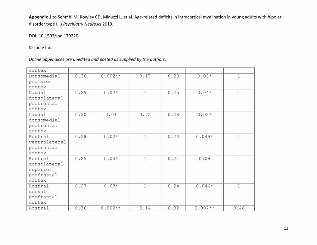
Appendix 1 to Sehmbi M, Rowley CD, Minuzzi L, et al. Age-related deficits in intracortical myelination in young adults with bipolar
disorder type I. J Psychiatry Neurosci 2019.
DOI: 10.1503/jpn.170220 © Joule Inc. Online appendices are unedited and posted as supplied by the authors.
13
cortex
Dorsomedial
premotor
cortex
0.36 0.002** 0.17 0.28 0.02* 1
Caudal
dorsolateral
prefrontal
cortex
0.29 0.02* 1 0.25 0.04* 1
Caudal
dorsomedial
prefrontal
cortex
0.30 0.01 0.72 0.28 0.02* 1
Rostral
ventrolateral
prefrontal
cortex
0.28 0.02* 1 0.24 0.049* 1
Rostral
dorsolateral
superior
prefrontal
cortex
0.25 0.04* 1 0.21 0.08 1
Rostral
dorsal
prefrontal
cortex
0.27 0.03* 1 0.24 0.046* 1
Rostral 0.36 0.002** 0.14 0.32 0.007** 0.48

Appendix 1 to Sehmbi M, Rowley CD, Minuzzi L, et al. Age-related deficits in intracortical myelination in young adults with bipolar
disorder type I. J Psychiatry Neurosci 2019.
DOI: 10.1503/jpn.170220 © Joule Inc. Online appendices are unedited and posted as supplied by the authors.
14
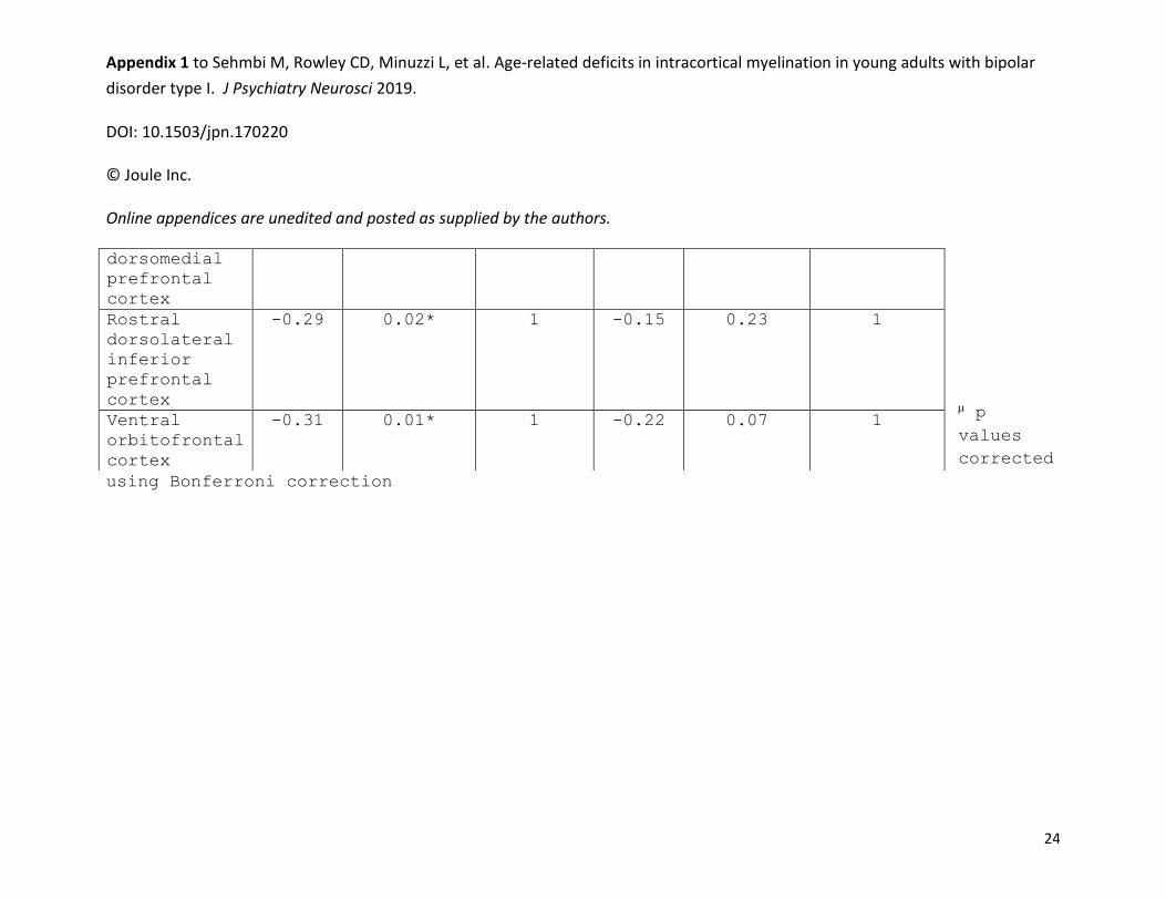
medial
prefrontal
cortex
Duration of illness (years)
Caudal medial
visual cortex
-0.33 0.005** 0.33 -0.31 0.009** 0.56
Lateral
visual cortex
-0.26 0.03 1 -0.32 0.007** 0.43
Superior
visual cortex
-0.26 0.03 1 -0.19 0.11 1
Cuneus -0.33 0.006** 0.38 -0.26 0.03* 1
Caudal
superior
temporal
cortex
-0.31 0.01* 0.60 -0.30 0.01 0.63
Dorsal
inferior
parietal
cortex
-0.16 0.19 1 -0.25 0.03* 1
Superior
parietal
cortex
-0.21 0..09 1 -0.29 0.02* 0.84
Medial
superior
parietal
cortex
-0.27 0.02* 1 -0.28 002* 0.85

Appendix 1 to Sehmbi M, Rowley CD, Minuzzi L, et al. Age-related deficits in intracortical myelination in young adults with bipolar
disorder type I. J Psychiatry Neurosci 2019.
DOI: 10.1503/jpn.170220 © Joule Inc. Online appendices are unedited and posted as supplied by the authors.
15
Medial
parietal
cortex
-0.33 0.005** 0.36 -0.37 0.002** 0.12
Posterior
cingulate
cortex
-0.33 0.005* 0.37 -0.30 0.01* 0.71
Dorsomedial
somatosensory
cortex
-0.30 0.01* 0.70 -0.18 0.13 1
Ventral motor
cortex
-0.23 0.05 1 -0.25 0.04* 1
Dorsolateral
motor cortex
-0.23 0.06 1 -030 0.01* 0.73
Dorsomedial
motor cortex
-0.28 0.02* 0.87 -0.23 0.06 1
Rostral
ventral
premotor
cortex
-0.24 0.049* 1 -0.23 0.06 1
Dorsolateral
premotor
cortex
-0.27 0.02* 1 -0.22 0.07 1
Dorsomedial
premotor
cortex
-0.29 0.01* 0.75 -0.27 0.03* 1
Caudal -0.29 0.01* 0.75 -0.20 0.09 1

Appendix 1 to Sehmbi M, Rowley CD, Minuzzi L, et al. Age-related deficits in intracortical myelination in young adults with bipolar
disorder type I. J Psychiatry Neurosci 2019.
DOI: 10.1503/jpn.170220 © Joule Inc. Online appendices are unedited and posted as supplied by the authors.
16
dorsomedial
prefrontal
cortex
Midcingulate
cortex
-0.33 0.005** 0.37 -0.27 0.02* 1
Rostral
ventrolateral
prefrontal
cortex
-0.24 0.04 1 -0.18 0.13 1
Rostral
dorsolateral
inferior
prefrontal
cortex
-0.25 0.04* 1 -0.29 0.02* 0.84
Rostral
medial
prefrontal
cortex
-0.29 0.02* 0.85 -0.22 0.06 1
Ventrolateral
orbitofrontal
cortex
-0.24 0.048* 1 -0.20 0.09 1
Anterior
cingulate
cortex
-0.33 0.005** 0.33 -0.31 0.008** 0.53
p values corrected using Bonferroni correction.

Appendix 1 to Sehmbi M, Rowley CD, Minuzzi L, et al. Age-related deficits in intracortical myelination in young adults with bipolar
disorder type I. J Psychiatry Neurosci 2019.
DOI: 10.1503/jpn.170220 © Joule Inc. Online appendices are unedited and posted as supplied by the authors.
17
Table S3. Partial correlations with number of mood episodes in BD subjects (N=45).
Cortical
Region
Left hemisphere Right hemisphere
Partial
R
P,
uncorrected
P,
corrected
Partial
R
P,
uncorrected
P,
corrected
Number of manic episodes
Rostral
middle
temporal
cortex
-0.17 0.15 1 -0.24 0.047* 1
Number of hypomanic episodes
Posterior
cingulate
cortex
-0.27 0.03* 1 -0.31 0.009** 0.67
Anterior
cingulate
cortex
-0.16 0.17 1 -0.27 0.02* 1

Appendix 1 to Sehmbi M, Rowley CD, Minuzzi L, et al. Age-related deficits in intracortical myelination in young adults with bipolar
disorder type I. J Psychiatry Neurosci 2019.
DOI: 10.1503/jpn.170220 © Joule Inc. Online appendices are unedited and posted as supplied by the authors.
18
Number of mixed episodes
Lateral
visual cortex
0.08 0.51 1 0.26 0.03* 1
Superior
visual cortex
0.09 0.46 1 0.27 0.03* 1
Caudal middle
temporal
cortex
0.20 0.09 1 0.31 0.009** 1
Rostral
middle
temporal
cortex
0.29 0.01* 1 0.19 0.10 1
Ventral
inferior
parietal
cortex
0.12 0.31 1 0.27 0.02* 1
Dorsal
inferior
parietal
cortex
0.10 0.40 1 0.26 0.03* 1
Superior
parietal
cortex
0.04 0.77 1 0.24 0.045* 1
Ventral
somatosensory
cortex
0.0009 0.99 1 0.29 0.02* 1

Appendix 1 to Sehmbi M, Rowley CD, Minuzzi L, et al. Age-related deficits in intracortical myelination in young adults with bipolar
disorder type I. J Psychiatry Neurosci 2019.
DOI: 10.1503/jpn.170220 © Joule Inc. Online appendices are unedited and posted as supplied by the authors.
19
Dorsolateral
somatosensory
cortex
0.18 0.14 1 0.26 0.03* 1
Rostral
dorsolateral
inferior
parietal
cortex
0.22 0.06 1 0.26 0.03* 1
Ventral
orbitofrontal
cortex
0.33 0.005** 1 0.29 0.01* 1
Ventromedial
orbitofrontal
cortex
0.37 0.001** 1 0.15 0.21 1
Ventromedial
prefrontal
cortex
0.42 0.0003*** 0.04* 0.18 0.13 1

Appendix 1 to Sehmbi M, Rowley CD, Minuzzi L, et al. Age-related deficits in intracortical myelination in young adults with bipolar
disorder type I. J Psychiatry Neurosci 2019.
DOI: 10.1503/jpn.170220 © Joule Inc. Online appendices are unedited and posted as supplied by the authors.
20
p values corrected using Bonferroni correction.
Table S4. Partial correlations with medication use in BD subjects (N=45).
Cortical
Region
Left hemisphere Right hemisphere
Partial
R
P,
uncorrected
P,
corrected
Partial
R
P,
uncorrected
P,
corrected
Anticonvulsant use
Rostral
dorsolateral
inferior
prefrontal
cortex
-0.24 0.04* 1 -0.09 0.46 1
Rostral
dorsal
prefrontal
cortex
-0.25 0.04* 1 -0.25 0.03* 1
Ventromedial -0.12 0.34 1 -0.25 0.04* 1

Appendix 1 to Sehmbi M, Rowley CD, Minuzzi L, et al. Age-related deficits in intracortical myelination in young adults with bipolar
disorder type I. J Psychiatry Neurosci 2019.
DOI: 10.1503/jpn.170220 © Joule Inc. Online appendices are unedited and posted as supplied by the authors.
21
orbitofrontal
cortex
Antipsychotic use
Caudal medial
visual cortex
-0.33 0.005** 1 -0.16 0.18 1
Cuneus -0.23 0.06 1 -0.30 0.01* 1
Medial
superior
parietal
cortex
-0.25 0.04* 1 -0.27 0.02* 1
Medial
parietal
cortex
-0.14 0.24 1 -0.25 0.04* 1
Posterior
cingulate
cortex
-0.29 0.02* 1 -0.26 0.03* 1
Dorsolateral
motor cortex
-0.27 0.02* 1 -0.25 0.04* 1
Midcingulate
cortex
-0.26 0.03* 1 -0.32 0.008** 1
Anterior
cingulate
cortex
-0.10 0.39 1 -0.35 0.003** 1
Total medication load
Caudal medial -0.38 0.001** 0.70 -0.27 0.02* 1

Appendix 1 to Sehmbi M, Rowley CD, Minuzzi L, et al. Age-related deficits in intracortical myelination in young adults with bipolar
disorder type I. J Psychiatry Neurosci 2019.
DOI: 10.1503/jpn.170220 © Joule Inc. Online appendices are unedited and posted as supplied by the authors.
22
visual cortex
Cuneus -0.17 0.16 1 -0.27 0.03* 1
Medial
parietal
cortex
-0.13 0.25 1 -0.28 0.02* 1
Posterior
cingulate
cortex
-0.25 0.04* 1 -0.22 0.07 1
Dorsolateral
somatosensory
cortex
-0.13 0.28 1 -0.24 0.04* 1
Dorsolateral
motor cortex
-0.30 0.01* 1 -0.30 0.01* 1
Dorsomedial
motor cortex
-0.28 0.02* 1 -0.28 0.02* 1
Total medication count
Caudal medial
visual cortex
-0.33 0.006** 1 -0.22 0.06 1
Caudal middle
temporal
cortex
-0.06 0.62 1 -0.26 0.03* 1
Ventral
inferior
parietal
cortex
-0.14 0.26 1 -0.27 0.03* 1
Dorsal -0.16 0.19 1 -0.26 0.03* 1

Appendix 1 to Sehmbi M, Rowley CD, Minuzzi L, et al. Age-related deficits in intracortical myelination in young adults with bipolar
disorder type I. J Psychiatry Neurosci 2019.
DOI: 10.1503/jpn.170220 © Joule Inc. Online appendices are unedited and posted as supplied by the authors.
23
inferior
parietal
cortex
Superior
parietal
cortex
-0.13 0.28 1 -0.24 0.049* 1
Medial
parietal
cortex
-0.08 0.50 1 -0.25 0.03* 1
Dorsolateral
somatosensory
cortex
-0.18 0.12 1 -0.33 0.005** 1
Dorsomedial
somatosensory
cortex
-0.17 0.17 1 -0.25 0.03* 1
Dorsolateral
motor cortex
-0.23 0.05 1 -0.29 0.02* 1
Dorsomedial
motor cortex
-0.26 0.03* 1 -0.32 0.008** 1
Dorsolateral
premotor
cortex
-0.20 0.10 1 -0.25 0.04* 1
Dorsomedial
premotor
cortex
-0.25 0.03* 1 -0.31 0.009** 1
Caudal -0.22 0.06 1 -0.25 0.04* 1
Appendix 1 to Sehmbi M, Rowley CD, Minuzzi L, et al. Age-related deficits in intracortical myelination in young adults with bipolar
disorder type I. J Psychiatry Neurosci 2019.
DOI: 10.1503/jpn.170220 © Joule Inc. Online appendices are unedited and posted as supplied by the authors.
24
p
values
corrected
using Bonferroni correction
dorsomedial
prefrontal
cortex
Rostral
dorsolateral
inferior
prefrontal
cortex
-0.29 0.02* 1 -0.15 0.23 1
Ventral
orbitofrontal
cortex
-0.31 0.01* 1 -0.22 0.07 1