Embed Size (px)
Citation preview
461
Apple Snail Perivitellin Precursor Properties Help Explain
Predators’ Feeding Behavior
María Pilar Cadierno1
Marcos Sebastián Dreon1,2
Horacio Heras1,3,*1Instituto de Investigaciones Bioquímicas de La Plata(INIBIOLP), Consejo Nacional de Investigaciones Científicasy Técnicas–Universidad Nacional de La Plata (CONICET-UNLP), La Plata, Argentina; 2Cátedra de Bioquímica yBiología Molecular, Facultad de Ciencias Médicas, UNLP, LaPlata, Argentina; 3Cátedra de Química Biológica, Facultad deCiencias Naturales y Museo, UNLP, La Plata, Argentina
Accepted 12/28/2016; Electronically Published 4/12/2017
Online enhancements: appendix figures.
ABSTRACT
In contrast with vitellogenin maturation, it is unknown whethergastropod perivitellin precursors are subject to large structuralchanges. The gastropod reproductive tract includes an accessoryorgan, the albumen gland (AG), that produces and secretes peri-vitelline fluid. In the apple snail Pomacea canaliculata, the large,reddish-pink AG provides eggs with perivitellins that are defensiveagainst predators. Although the AG makes a considerable con-tribution to apple snail biomass, field observations indicate thatit is rejected by avian and mammalian predators, although theunderlying reason remains unknown. By analyzing the structure-function properties of P. canaliculata perivitellin precursors, weprovide insight into perivitellin maturation and its relationshipwith apple snail predator feeding behavior. Structural analysisusing small-angle X-ray scattering, absorption and fluorescencespectroscopy, circular dichroism, electrophoresis, chromatog-raphy, and partial proteolysis showed that the size, shape, andstructure of perivitellin precursors resemble those of egg matureforms. Functional analysis indicates that the precursors of thedefensive perivitellins ovorubin (PcOvo) and perivitellin-2 (PcPV2)are highly stable and antinutritive, withstanding proteinase di-gestion and displaying structural stability of their quaternary struc-ture under a wide pH range (4.0–10.0). Furthermore, AG ex-tracts limit a predator’s ability to digest nutrients and are toxicto mice (median lethal concentration 96 h after administration:5.9 mg/kg). Treated mice displayed neurologic signs similar tothose produced by egg PcPV2. Results indicate that apple snails
*Corresponding author; e-mail: [email protected].
Physiological and Biochemical Zoology 90(4):461–470. 2017. q 2017 by TheUniversity of Chicago. All rights reserved. 1522-2152/2017/9004-6084$15.00.DOI: 10.1086/691526
This content downloaded from 132.All use subject to University of Chicago Press Term
store active precursors of egg proteins inside the AG, providingevidence that gastropod perivitellin precursors do not experiencethe large structural processing of invertebrate vitellogenin mat-uration. These defensive proteins provide the apple snail AG withneurotoxic, antinutritive, and antidigestive activity, a likely ex-planation for the predators’ feeding behavior.
Keywords: neurotoxin, antinutritive protein, snail kite, animaldefense, structure-function, defensive protein, toxicity, peri-vitellin precursor.
Introduction
A molecular approach was implemented to gain insight intogastropod perivitellin precursor characteristics and to inves-tigate for possible causes of Pomacea canaliculata predatorbehavior of discarding the albumen gland (AG). We analyzedthe structural and functional properties of the precursors ofthe major egg perivitellins. We hypothesize (1) that unlikevitellogenins, they do not undergo any large posttranslationalprocessing before being incorporated into eggs and (2) thatthese particles are stored already functional and noxious in theAG, which could explain the predators’ discarding behavior.
Vitellogenic Mechanisms in Oviparous Animalsand Gastropod Singularities
Many animal life-history traits are constrained by reproductive-tract structure and taxon-specific vitellogenic mechanisms thatdetermine the quantity, quality, and rate of energy (yolk) in-corporation into egg (Eckelbarger 1994). During vitellogenesis,oviparous species synthesize lipids, proteins, and carbohydrateseither outside or inside the ovary and incorporate them into pri-mary oocytes to serve mainly as energetic and structural sourcesfor embryo development (de Jong-Brink et al. 1983). The majoregg yolk proteins usually form complex water-soluble particlescalled vitellins (Wallace et al. 1967). Vitellin precursors, collec-tively referred to as vitellogenins, undergo substantial structuralchanges involving proteolysis before acquiring their mature vi-tellin conformation (Raikhel and Sappington 2002). However, aphysiologically distinct vitellogenic pathway is present in manygastropods. They have evolved an alternative mechanism inwhich a limited amount of vitellus is included in the eggs, as theycoat the fertilized oocyte with a nutritive and protective layer orcapsule (termed perivitelline fluid) produced by the AG, a femaleaccessory reproductive organ that is exclusive to gastropods (Run-ham 1988). Proteins present in the perivitelline fluid have been
239.001.231 on April 18, 2017 06:21:11 AMs and Conditions (http://www.journals.uchicago.edu/t-and-c).

462 M. P. Cadierno, M. S. Dreon, and H. Heras
termed perivitellins and would be equivalent to vitellins. How-ever, in contrast with vitellogenins, it is unknown whether peri-vitellin precursors are active or subject to large structural changesbefore they reach their final destination. This knowledge gapin gastropods is remarkable, considering the central role ofperivitellins in reproduction.
Apple Snails as a Model for the Study of Gastropod Perivitellins
Apple snails (Ampullariidae) are among the largest and mostecologically important freshwater snails, with a large and di-verse body of research across several fields, including mostmetabolic, structural, and functional studies on gastropod peri-vitellins (Hayes et al. 2015). Most research concerns a singlespecies, P. canaliculata (Lamarck, 1822), that possesses the po-tential to become a model organism (Hayes et al. 2015). Pomaceacanaliculata is a South American snail introduced to Asianwetlands and the Pacific islands, where they became an invasivespecies, a serious pest of aquatic crops, and a vector for humaneosinophilic meningoencephalitis, a parasitic disease (Cowie 2002;Lv et al. 2009).Pomacea canaliculata lay their eggs above the water level
and have virtually no reported egg predators worldwide, withthe exception of the fire ant Solenopsis geminata (Yusa 2001).This is probably related to the presence of an unusual set of de-fensive perivitellins, including an aposematic signal as a predatorwarning (Dreon et al. 2002, 2010, 2013, 2014; Heras et al. 2007).Recently, transcriptomic and proteomic analysis revealed a largematernal investment on perivitelline-fluid proteins (Sun et al.2012a). The precursors of the most abundant perivitellins, ovo-rubin (PcOvo) and perivitellin-2 (PcPV2), are studied in Heraset al. (1998). These are exclusively synthesized and stored in theAG (Dreon et al. 2002, 2003; Catalán et al. 2006; Sun et al. 2012b),and they account for 65% of egg proteins, playing multipleroles, such as nourishing and protecting embryos. PcOvo is anoligomeric protein, composed of subunits ranging from 28 to35 kDa (Garín et al. 1996), that carries and stabilizes carot-enoids, supplying the embryos with a potent membrane anti-oxidant molecule and giving the reddish-pink pigmentation toeggs (Dreon et al. 2004; Heras et al. 2007). This perivitellin re-sists digestion and reaches the predator small intestine in a fullyactive form, as an antinutritive that diminishes rat growth rate(Dreon et al. 2010). The second-most-abundant protein, PcPV2,is also an antinutritive factor. In addition, it displays a potentneurotoxin activity, with a lethal effect on mice. Its structure isunique in nature, combining a lectin (31 kDa) and a pore-forming subunit (67 kDa) linked by disulfide bridges (Heraset al. 2008; Dreon et al. 2013). PcOvo and PcPV2 are good can-didates for the study of perivitellin precursors, because their func-tionality is relatively easy to determine.
Apple Snail AG, Perivitellin Precursors, and Adult-SnailPredator Behavior
In contrast with the scarce egg predators, adult apple snailsare preyed on by several species. Among them, the snail kite
This content downloaded from 132.All use subject to University of Chicago Press Term
Rostrhamus sociabilis, the limpkin Aramus guarauna, and theNorway rat Rattus norvegicus have been the most studied ones(Snyder and Kale 1983; Sykes 1987; Yusa et al. 2000). Kites andlimpkins are diurnal hunters that concentrate their foragingactivity on lentic water bodies. After finding and capturing asnail, they carry them to feeding perches (kites) or to the shore(limpkins) to consume the soft parts (Tanaka et al. 2006). Ratsprey on snails by peeling off the shells with their teeth untilthey reach the attachment site of the columellar muscle (Yusaet al. 2000). Field observations indicate that these avian andmammalian predators display an intriguing behavior, discard-ing the AG when feeding on female snails (Snyder and Kale1983; Sykes 1987; Yusa et al. 2000). This is remarkable, con-sidering that the AG represents a great contribution to femalesnail body biomass (Estoy et al. 2002), which implies a signif-icant loss of energy and nutrients (M. P. Cadierno, S. Burela,M. S. Dreon, P. R. Martín, and H. Hera, unpublished manuscript).The cause of this behavior is, however, unknown.
Methods
Sample Preparation
Females and egg masses with embryos up to morula stage ofPomacea canaliculata were collected in the wild from streamsand ponds near the city of La Plata, Buenos Aires Province, Ar-gentina, during its reproductive period (November–March). Toget the soluble fraction of these samples, a number of egg massesand dissected AGs were pooled and homogenized in ice-cold20 mM Tris/HCl buffer, pH 7.5, on a Potter-type homogenizer(Thomas Scientific, Swedesboro, NJ) with a buffer∶sample ratioof 3∶1 v/w. The crude homogenate was sequentially centrifugedat 10,000 g for 20 min and at 100,000 g for 50 min; the pelletswere discarded. Total soluble protein concentration was deter-mined colorimetrically (Lowry et al. 1951), with bovine serumalbumin (BSA) as standard.For perivitellin purification, the AG and egg soluble frac-
tions were subjected to density gradient ultracentrifugation, asdescribed by Garín et al. (1996). Aliquots containing either theprecursors or the mature forms of PcPV2 and PcOvo werepooled, desalted, and concentrated by ultrafiltration in an Ami-con Ultra-4 Centrifugal Filter Unit (Millipore, Billerica, MA).Both perivitellin fractions were purified by anion exchange liquidchromatography in a Mono Q HR 10/100 column (Amersham-Pharmacia, Uppsala, Sweden) coupled to an Agilent 1260 high-performance liquid chromatography (HPLC) system (AgilentTechnologies), with a gradient of 0–1 M NaCl in a 20 mM Tris/HCl buffer, pH 8.5. The PcOvo peak was further purified by sizeexclusion chromatography in a Superdex 200 10/300 GL column(Amersham-Pharmacia) with a 20 mM Tris/HCl buffer, pH 8.5.Samples were desalted and concentrated after each chromatog-raphy step, as described above. The purity of the single peakswas checked by native electrophoresis, and the total protein con-tent was quantified (Lowry et al. 1951). To identify perivitellinorigin, we refer to AG perivitellin precursors as agPcOvo andagPcPV2 and to mature egg forms of perivitellins as ePcOvo andePcPV2.
239.001.231 on April 18, 2017 06:21:11 AMs and Conditions (http://www.journals.uchicago.edu/t-and-c).
Apple Snail Albumen Gland Has Harmful Proteins 463
Structural Studies of Egg and AG Perivitellins
Polyacrylamide Gel Electrophoresis (PAGE). Native proteins wereanalyzed by nondissociating PAGE using a gradient of 4%–20%acrylamide (Davis 1964; Margolis and Wrigley 1975) and proteinsubunits by sodium dodecyl sulfate (SDS)-PAGE using a 4%–20% polyacrylamide gradient (Laemmli 1970). Gels were stainedwith Coomassie Brilliant Blue R-250 (Sigma Chemical).
Anion Exchange and Size Exclusion Liquid Chromatography.The hydrodynamic volume and the content of superficial chargesof the perivitellins were evaluated by comparing their reten-tion time through a Superdex 200 10/300 GL column (Amersham-Pharmacia) and a Mono Q HR 10/100 column (Amersham-Pharmacia), respectively. Both columns were coupled to an Agilent1260 HPLC system (Agilent Technologies). Running conditionswere those mentioned for perivitellin purification.
Absorption Spectroscopy. Absorption ultraviolet (UV)-visiblespectra were recorded in 20 mMTris/HCl buffer, pH 8.5, at 257Cwith an Agilent 8453 UV-visible spectrophotometer (AgilentTechnologies). Spectra were also recorded for agPcOvo prein-cubated at different pH values, in order to evaluate the effect ofpH over the carotenoid moiety. Spectral scanning was performedat 1-nm bandwidth, and each spectrum was the average of 10 in-dividual runs. Buffer and light-scattering contributions to rawdata values were subtracted.
Circular Dichroism (CD). Far-UV CD spectra (195–250 nm)were performed in a J-810 spectropolarimeter (Jasco) using0.1-cm cells at 257C. Samples were measured at a concentra-tion of 0.5 mg/mL in 20 mM Tris/HCl buffer, pH 8.5. Scanningwas performed with 0.1-nm bandwidth, 20‐nm-min21 scanspeed, and 1-s average time. Each spectrum was obtained byaveraging at least five individual runs and corrected for bufferoptical activity.
Fluorescence Spectroscopy. Fluorescence spectra were collectedon a Perkin-Elmer LS55 Luminescence Spectrometer (Perkin-Elmer, Norwalk, CT). Tryptophan (Trp) residues were excitedat 285 nm (8-nm slit), and their emissions were recorded be-tween 295 and 440 nm (8-nm slit). All samples were measuredin 20 mMTris/HCl buffer, pH 8.5, at 257Cwith a 10-mm opticalpath length quartz cell. Each spectrum was obtained by aver-aging 10 individual runs. To evaluate perivitellin structural sta-bility at different pH conditions, solutions of purified agPcOvoand agPCPV2 in ultrapure water were prepared. They were in-cubated overnight in a series of buffers with a pH range between2 and 12. Emission fluorescence for these samples was also re-corded as described above.
Small-Angle X-ray Scattering (SAXS). SAXS experiments wereperformed at the D02A-SAXS2 line in the Brazilian Synchro-tron Light Laboratory, Campinas (Sao Paulo). The scatteringpattern was detected with a MarCCD bidimensional charge-coupled device assisted by FIT2D software (http://www.esrf.fr
This content downloaded from 132.All use subject to University of Chicago Press Term
/computing/scientific/FIT2D). The sample cell was built withtwo parallel mica windows 1 mm apart, leaving 300 mL of freevolume for the sample. The wavelength used was 1.55 Å, andthe nominal q-range was between 0.09 and 2.2 nm21 (nominalDmax ≤ 26 nm). All experiments were performed at 257C.Corrections of beam intensity, detector homogeneity, and
sample absorption were performed, following standard pro-cedures. Experiments were performed in a protein concentrationrange of 2.40–0.50 mg/mL to rule out a concentration effect. Atleast five independent curves were averaged for each single ex-periment. The size of proteins was determined from the gyrationradii (RG) obtained by analysis of SAXS patterns as Guinierplots (ln(I)p ln(I0)2 (1=3) � RG2 � q2, qp 4p sin(V)=l, RG �q ≤ 1), and the globularity was evaluated by inspecting theKratky plots (I(q) � q2 vs. q). A pair-distance distribution func-tion (PDDF), obtained from scattering data, was calculated viaregularized Fourier transform in the program GNOM 4.6 (Sver-gun 1992). A low-resolution three-dimensional model of the peri-vitellins was obtained with simulated annealing algorithms im-plemented in the program DAMMIF (Svergun 1999). SAXS dataanalysis was performed with ATSAS software, version 2.7.1(Petoukhov et al. 2012). Data were deposited in the Small AngleScattering Biological Data Bank (SASBDB).
Limited Proteolysis Using Proteinase K. To evaluate the sus-ceptibility of native AG and egg perivitellins to aggressive pro-tease digestion, a limited proteolysis with proteinase K was per-formed. Solutions containing 1 mg/mL of each perivitellin in20 mM Tris/HCl buffer (pH 8.5), 25 mM CaCl2, and 10 mg/mLproteinase K (Promega) were incubated at 377C. Aliquots weretaken at 10, 20, and 40 min, and enzyme activity was stopped byadding SDS sample buffer and boiling the mixture for 5 min at1007C. Samples were analyzed by SDS-PAGE with a 4%–20%polyacrylamide gradient.
Functional Studies
Either the AG soluble fraction or purified agPcOvo and agPcPV2were subjected to the following functional experiments and com-pared with egg perivitellins.
Trypsin Inhibition Assay. Aliquots of the AG soluble fractioncontaining 79 mg of protein were incubated with 4 mg of trypsinfor 5 min, and trypsin inhibition was determined as previouslydescribed (Dreon et al. 2010), with slight modifications. In short,N-benzoil-L-arginine ethyl ester (BAEE) was hydrolyzed by tryp-sin at the ester linkage, causing an absorbance increase at 253 nmat 257C. Results are expressed as units of activity (the amount ofenzyme that causes an absorbance increase of 0.003/min).
Simulated Gastrointestinal Digestion. In vitro simulated gas-trointestinal digestion of the purified agPcOvo and agPcPV2,followed the method described by Moreno et al. (2005), withslight modifications. Briefly, gastric digestion was performed at377C for 120 min at pH 2.5 in the presence of porcine pepsin(Sigma, catalog no. P 6887) at an enzyme∶substrate ratio of
239.001.231 on April 18, 2017 06:21:11 AMs and Conditions (http://www.journals.uchicago.edu/t-and-c).

464 M. P. Cadierno, M. S. Dreon, and H. Heras
1∶20 (w/w). Aliquots were taken at 0, 60, and 120 min andanalyzed by SDS-PAGE. For in vitro duodenal digestion, the120-min gastric digest was used as starting material. Pepsin ac-tivity was stopped by raising the pH to 8.5 with 0.1 M NaOH and150 mM Tris/HCl buffer. The duodenal digestion was simulatedby adding 0.25 M taurocholic acid and trypsin from bovinez pan-creas (Sigma, catalog no. T 9935) at an enzyme∶substrate ratioof 1∶2.7 (w/w), at 377C. Aliquots were taken at 0, 60, and 120 minfor SDS-PAGE analysis. Albumin was used as a positive (withenzyme) or negative (without enzyme) control in both gastric andduodenal digestion.
Toxicity Assay. All studies performed with murine animals wereapproved by the Comité Institucional para el Cuidado y Usode Animales de Laboratorio (CICUAL; i.e., Institutional AnimalCare and Use Committee) of the School of Medicine, Univer-sidad Nacional de La Plata (UNLP; protocol P 01012016) andwere carried out in accordance with the Guide for the Care andUse of Laboratory Animals (NIH 2011).Female BALBc mice, 4 wk old and weighing 19–21 g, from
a colony started with a stock provided by the US NationalInstitutes of Health and bred in a specific pathogen-free en-vironment were used. Groups of 10 animals were subjected tointraperitoneal injection with a single dose of 300 mL phosphate-buffered saline or the same volume of five serial dilutions of theAG soluble fraction (20, 40, 80, 160, and 320 mg of total pro-tein). The median lethal concentration (LD50) was determinedby a lethality test 96 h after administration, and the symptom-monitoring behavior was chosen according to experimental datapreviously published (Heras 2008).Data were analyzed by t-test with the software Instat 2.0,
with a 95% confidence interval. The bioassay statistics was per-formedwithprobit analysis implemented in the programTOXDAT(US Environmental Protection Agency), with 95% of confidencelimits.
Results
Structural Characterization of AG Perivitellin Precursors
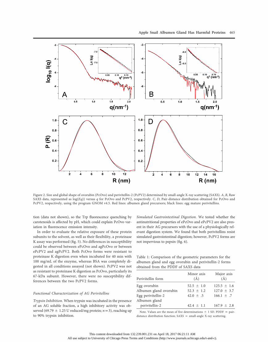
Native Conformation, Size, and Global Shape. The electro-phoretical mobility of ePcOvo and agPcOvo, as well as that ofePcPV2 and agPcPV2, showed no differences in native con-ditions. Furthermore, SDS-PAGE shows the same subunit com-position for these perivitellins regardless of their origin (fig. 1).In addition, no differences could be detected between ePcOvoand agPcOvo or between ePcPV2 and agPcPV2 either in theircharge surface or in their hydrodynamic volume, in anion ex-change and size exclusion liquid chromatography, respectively(not shown).The size and global shape of PcOvo and PcPV2 from eggs
and AGwere determined by SAXS. Experimental data normalizedfor protein concentration are presented as log10(I(q))-versus-qplots for PcOvo (fig. 2A) and PcPV2 (fig. 2B) forms. Clearly, thecurves for both perivitellins virtually overlap, indicating a com-parable shape and size regardless of the origin. Moreover, fromthe Guinier-linearized region it was possible to fit the RG for the
This content downloaded from 132.All use subject to University of Chicago Press Term
FeAaBmsula
proteins in a q range between 0.1 and 0.38 nm21 (insets in fig. 2A,2B). Slight significant differences were detected between ePcOvo(42.30 5 0.07 Å) and agPcOvo (43.03 5 0.25 Å), but not be-tween ePcPV2 (44.435 0.36 Å) and agPcPv2 (44.545 0.28 Å).The Kratky plots for the proteins (fig. A1; figs. A1, A2 availableonline) are bell shaped, as expected for globular proteins. Bothperivitellins appear as anisometric particles with well-definedminor and major axes, as shown by their PDDFs (fig. 2C, 2D),where no significant differences were observed regarding theirdifferent origins (table 1). Both AG perivitellin models werequite similar to those of the egg forms (Dreon et al. 2008). SAXSdata and models were deposited in the SASBDB, with accessioncodes SASDBW4 for agPcOvo and SASDBX4 for agPcPV2.
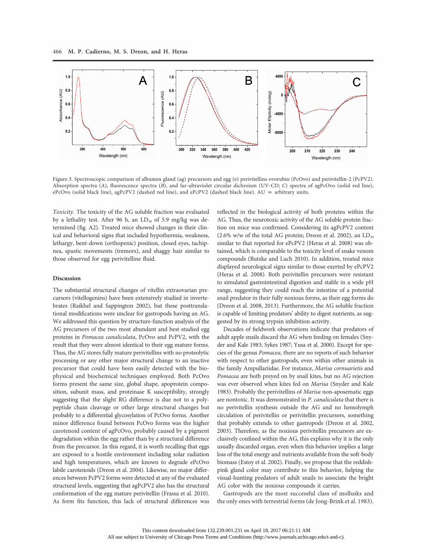
Secondary and Tertiary Structure. Absorption spectra of ePcOvoand agPcOvo in the UV-visible region were almost coincident,presenting four absorption maxima at 433, 477, 508, and 541 nm,indicating no differences in their carotenoid-binding site. Nev-ertheless, a higher intensity in agPcOvo spectra may be indic-ative of a higher carotenoid content (fig. 3A). Figure 3B showsthe fluorescence emission spectra of precursor and mature formsof PcOvo and PcPV2. Each perivitellin has a distinctive emissionmaximum, 325 and 332 nm for PcOvo and PcPV2, respectively,and presents nearly the same spectrum regardless of its origin,indicating no major differences in tertiary structure. Finally, far-UV CD spectra showed no significant differences in the second-ary structure between agPcOvo and ePcOvo or between agPcPV2and ePcPV2 (fig. 3C).
Stability against pH and Aggressive Protease Treatment. TheTrp fluorescence emission maxima observed for agPcOvo andagPcPV2 did not vary along the studied pH range (2.0–12.0),indicating that the Trp electronic environment is not affected(fig. 4). Interestingly, there was a variation in the fluorescenceemission intensity between the recorded spectra for the twoperivitellins. In a similar fashion, the absorbance intensity ofcarotenoid spectra decreased according to the pH value reduc-
igure 1. Electrophoretical comparison of albumen gland (ag) andgg (e) perivitellins ovorubin (PcOvo) and perivitellin-2 (PcPV2)., Native 4%–20%polyacrylamide gel electrophoresis (PAGE). Lane 1:gPcPV2; lane 2: ePcPV2; and lane 3: molecular weight markers., Native 4%–20% PAGE. Lane 1: agPcOvo; lane 2: ePcOvo; and lane 3:olecular weight markers. C, Dissociating 4%–20% sodium dodecyllfate (SDS)-PAGE. Lane 1: agPcOvo; lane 2: ePcOvo; lane 3: agPcPV2;ne 4: ePcPV2; and lane 5: molecular weight markers.
239.001.231 on April 18, 2017 06:21:11 AMs and Conditions (http://www.journals.uchicago.edu/t-and-c).
Apple Snail Albumen Gland Has Harmful Proteins 465
tion (data not shown), so the Trp fluorescence quenching bycarotenoids is affected by pH, which could explain PcOvo var-iation in fluorescence emission intensity.In order to evaluate the relative exposure of these protein
subunits to the solvent, as well as their flexibility, a proteinaseK assay was performed (fig. 5). No differences in susceptibilitycould be observed between ePcOvo and agPcOvo or betweenePcPV2 and agPcPV2. Both PcOvo forms were resistant toproteinase K digestion even when incubated for 40 min with100 mg/mL of the enzyme, whereas BSA was completely di-gested in all conditions assayed (not shown). PcPV2 was notas resistant to proteinase K digestion as PcOvo, particularly its67-kDa subunit. However, there were no susceptibility dif-ferences between the two PcPV2 forms.
Functional Characterization of AG Perivitellins
Trypsin Inhibition.When trypsin was incubated in the presenceof an AG soluble fraction, a high inhibitory activity was ob-served (69.795 1.25 U reduced/mg protein; np3), reaching upto 90% trypsin inhibition.
This content downloaded from 132.All use subject to University of Chicago Press Term
Simulated Gastrointestinal Digestion. We tested whether theantinutritional properties of ePcOvo and ePcPV2 are also pres-ent in their AG precursors with the use of a physiologically rel-evant digestion system. We found that both perivitellins resistsimulated gastrointestinal digestion; however, PcPV2 forms arenot impervious to pepsin (fig. 6).
Figure 2. Size and global shape of ovorubin (PcOvo) and perivitellin-2 (PcPV2) determined by small-angle X-ray scattering (SAXS). A, B, RawSAXS data, represented as log(I(q)) versus q for PcOvo and PcPV2, respectively. C, D, Pair-distance distribution obtained for PcOvo andPcPV2, respectively, using the program GNOM v4.5. Red lines: albumen gland precursors; black lines: egg mature perivitellins.
Table 1: Comparison of the geometric parameters for thealbumen gland and egg ovorubin and perivitellin-2 formsobtained from the PDDF of SAXS data
Perivitellin form
239.001.231 on April 18, 2017 06:21s and Conditions (http://www.journa
Minor axis(Å)
:11 AMls.uchicago.edu/t-and
Major axis(Å)
Egg ovorubin
52.5 5 1.0 125.5 5 1.6 Albumen gland ovorubin 52.3 5 1.2 127.0 5 3.7 Egg perivitellin-2 42.0 5 .5 166.1 5 .7 Albumen glandperivitellin-2 42.4 5 1.1 167.9 5 2.8Note. Values are the mean of five determinations 5 1 SD. PDDF p pair-distance distribution function; SAXS p small-angle X-ray scattering.
-c).
466 M. P. Cadierno, M. S. Dreon, and H. Heras
Toxicity. The toxicity of the AG soluble fraction was evaluatedby a lethality test. After 96 h, an LD50 of 5.9 mg/kg was de-termined (fig. A2). Treated mice showed changes in their clin-ical and behavioral signs that included hypothermia, weakness,lethargy, bent-down (orthopenic) position, closed eyes, tachip-nea, spastic movements (tremors), and shaggy hair similar tothose observed for egg perivitelline fluid.
Discussion
The substantial structural changes of vitellin extraovarian pre-cursors (vitellogenins) have been extensively studied in inverte-brates (Raikhel and Sappington 2002), but these posttransla-tional modifications were unclear for gastropods having an AG.We addressed this question by structure-function analysis of theAG precursors of the two most abundant and best-studied eggproteins in Pomacea canaliculata, PcOvo and PcPV2, with theresult that they were almost identical to their egg mature forms.Thus, the AG stores fully mature perivitellins with no proteolyticprocessing or any other major structural change to an inactiveprecursor that could have been easily detected with the bio-physical and biochemical techniques employed. Both PcOvoforms present the same size, global shape, apoprotein compo-sition, subunit mass, and proteinase K susceptibility, stronglysuggesting that the slight RG difference is due not to a poly-peptide chain cleavage or other large structural changes butprobably to a differential glycosylation of PcOvo forms. Anotherminor difference found between PcOvo forms was the highercarotenoid content of agPcOvo, probably caused by a pigmentdegradation within the egg rather than by a structural differencefrom the precursor. In this regard, it is worth recalling that eggsare exposed to a hostile environment including solar radiationand high temperatures, which are known to degrade ePcOvolabile carotenoids (Dreon et al. 2004). Likewise, no major differ-ences between PcPV2 forms were detected at any of the evaluatedstructural levels, suggesting that agPcPV2 also has the structuralconformation of the egg mature perivitellin (Frassa et al. 2010).As form fits function, this lack of structural differences was
This content downloaded from 132.All use subject to University of Chicago Press Term
reflected in the biological activity of both proteins within theAG. Thus, the neurotoxic activity of the AG soluble protein frac-tion on mice was confirmed. Considering its agPcPV2 content(2.6% w/w of the total AG protein; Dreon et al. 2002), an LD50
similar to that reported for ePcPV2 (Heras et al. 2008) was ob-tained, which is comparable to the toxicity level of snake venomcompounds (Butzke and Luch 2010). In addition, treated micedisplayed neurological signs similar to those exerted by ePcPV2(Heras et al. 2008). Both perivitellin precursors were resistantto simulated gastrointestinal digestion and stable in a wide pHrange, suggesting they could reach the intestine of a potentialsnail predator in their fully noxious forms, as their egg forms do(Dreon et al. 2008, 2013). Furthermore, the AG soluble fractionis capable of limiting predators’ ability to digest nutrients, as sug-gested by its strong trypsin inhibition activity.Decades of fieldwork observations indicate that predators of
adult apple snails discard the AG when feeding on females (Sny-der and Kale 1983; Sykes 1987; Yusa et al. 2000). Except for spe-cies of the genus Pomacea, there are no reports of such behaviorwith respect to other gastropods, even within other animals inthe family Ampullariidae. For instance, Marisa cornuarietis andPomacea are both preyed on by snail kites, but no AG rejectionwas ever observed when kites fed on Marisa (Snyder and Kale1983). Probably the perivitellins of Marisa non-aposematic eggsare nontoxic. It was demonstrated in P. canaliculata that there isno perivitellin synthesis outside the AG and no hemolymphcirculation of perivitellin or perivitellin precursors, somethingthat probably extends to other gastropods (Dreon et al. 2002,2003). Therefore, as the noxious perivitellin precursors are ex-clusively confined within the AG, this explains why it is the onlyusually discarded organ, even when this behavior implies a largeloss of the total energy and nutrients available from the soft-bodybiomass (Estoy et al. 2002). Finally, we propose that the reddish-pink gland color may contribute to this behavior, helping thevisual-hunting predators of adult snails to associate the brightAG color with the noxious compounds it carries.Gastropods are the most successful class of mollusks and
the only ones with terrestrial forms (de Jong-Brink et al. 1983).
Figure 3. Spectroscopic comparison of albumen gland (ag) precursors and egg (e) perivitellins ovorubin (PcOvo) and perivitellin-2 (PcPV2).Absorption spectra (A), fluorescence spectra (B), and far-ultraviolet circular dichroism (UV-CD; C) spectra of agPcOvo (solid red line),ePcOvo (solid black line), agPcPV2 (dashed red line), and ePcPV2 (dashed black line). AU p arbitrary units.
239.001.231 on April 18, 2017 06:21:11 AMs and Conditions (http://www.journals.uchicago.edu/t-and-c).
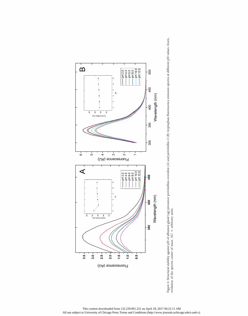
Figu
re4.Structuralstability
againstpH
ofalbu
men
glan
d(ag)
precursorperivitellinsovorub
in(A
)andperivitellin-2
(B):tryptoph
anfluo
rescence
emission
spectraat
differentpH
values.Insets,
evolutionof
thespectracenterof
mass.AU
parbitraryun
its.
This content downloaded from 132.239.001.231 on April 18, 2017 06:21:11 AMAll use subject to University of Chicago Press Terms and Conditions (http://www.journals.uchicago.edu/t-and-c).

468 M. P. Cadierno, M. S. Dreon, and H. Heras
It has been well established that internal fertilization, directdevelopment, and a female accessory AG that secretes an eggcoat of perivitelline fluid allowed gastropod success in a widerange of environments (Runham 1988). To the best of ourknowledge there is, however, no other report demonstratingby comparative structure-function analysis that perivitellin pre-cursors in AG do not experience large structural processing. Aliterature survey showed that functional perivitellin precursorswere reported in the AGs of a few other snails from marine,freshwater, and terrestrial environments that belong to the Pul-monata and Opisthobranchia clades (Takamatsu et al. 1995; Nagleet al. 2001; Mukai et al. 2004; Sanchez et al. 2006; Hathaway et al.2010). Though from a different context, these findings suggestthat the vitellogenic mechanism found in apple snails, whichbelong to the clade Caenogastropoda, may be more widespreadamong gastropods, contributing to the reproductive success ofthe group, currently the second most species-rich group of ani-mals after insects.
Conclusion
To comprehend the evolutionary forces that shaped metazoanlife-history patterns, one must consider the role played by re-productive strategies, especially the diverse mechanisms of eggyolk synthesis that have arisen through selection. This studyprovides the first piece of evidence that the AG produces peri-vitellin precursors functionally and structurally similar to ma-ture perivitellins. This suggests that those gastropods with an AGhave developed an alternative vitellogenic mechanism withoutthe large perivitellin precursor processing typical of invertebrateegg yolk vitellin maturation. This is probably a more efficientand faster mechanism for food conversion into egg production.By this mechanism Pomacea canaliculata stores the noxious pro-teins of the eggs inside the AG, a likely explanation for the feeding
Fpfepli
This content downloaded from 132.All use subject to University of Chicago Press Term
behavior of predators that avoid eating the gland while consumingthe rest of the snail female body.Further perivitellin structure-function studies in other gas-
tropods with the same and different oviposition strategies arenecessary to confirm this finding. Among apple snails, morework regarding comparative reproductive biology at the mo-lecular level would be valuable, considering that reproductionhas a high significance for understanding distribution pat-terns, invasion biology, and possibly the evolution of the am-phibious trait seen in most members of Ampullariidae.
Acknowledgments
H.H. and M.S.D. are members of the Carrera del InvestigadorCONICET, Argentina, and the Centro de Investigaciones, Ins-tituto Clínico de Buenos Aires (CICBA), Argentina, respectively.M.P.C. is a doctoral fellow at CONICET, Argentina. We thankthe Laboratório Nacional de Luz Síncrotron, the Brazilian Syn-chrotron Light Laboratory (project SAXS1-15958). We thankDr. J. M. Lofeudo for his help in the toxicity bioassay, Dr. G.Franchini for her suggestions, and Dr. M. Ermacora for hisfruitful comments. This work was supported by a grant from theAgencia Nacional de Promoción Científica y Técnica, Argentina(project PICT 1428). The funders had no role in study design,
Figure 6. Simulated gastrointestinal digestion of albumen gland (ag)perivitellins ovorubin (PcOvo) and perivitellin-2 (PcPV2). Lanes 1, 2:Bovine serum albumin (BSA)-negative and BSA-positive controls ofgastric digestion; lanes 3–5: agPcPV2 subunits (31 and 67 KDa) andagPcOvo subunits (28–35-KDa range) after incubation with pepsinfor 0, 60, and 120 min. Lanes 6, 7: BSA-negative and BSA-positivecontrols of duodenal digestion; lanes 8–10: agPcPV2 and agPcOvodigested with trypsin for 0, 60, and 120 min. Note the faint trypsinband under the 31-KDa agPcPV2 in lanes 8–10.
igure 5. Limited proteolysis analysis by proteinase K digestion. Therotein solution (1 mg/mL) was incubated with proteinase K at dif-rent times. Products were analyzed on an sodium dodecyl sulfateolyacrylamide gel (4%–20%) and visualized with Coomassie Bril-ant Blue R-250. A, ovorubin forms (ag p albumen gland precursor;
e p egg mature); B, perivitellin-2 forms.
239.001.231 on April 18, 2017 06:21:11 AMs and Conditions (http://www.journals.uchicago.edu/t-and-c).
Apple Snail Albumen Gland Has Harmful Proteins 469
data collection and analysis, decision to publish, or preparationof the manuscript.
Literature Cited
Butzke D. and A. Luch. 2010. High-molecular weight proteintoxins of marine invertebrates and their elaborate modes ofaction. Pp. 213–232 in A. Luch, ed. Clinical toxicology. Vol. 2of Molecular, clinical and environmental toxicology. Birk-häuser, Basel, Switzerland.
Catalán M., M.S. Dreon, H. Heras, R.J. Pollero, S.N. Fernández,and B. Winik. 2006. Pallial oviduct of Pomacea canaliculata(Gastropoda): ultrastructural studies of the parenchymal cel-lular types involved in the metabolism of perivitellins. CellTissue Res 324:523–533.
Cowie R.H. 2002. Apple snails (Ampullariidae) as agriculturalpests: their biology, impacts, and management. Pp. 145–192in G.M. Baker, ed. Molluscs as crop pests. CABI, Walling-ford, UK.
Davis B. 1964. Disc electrophoresis. II. Method and applicationto human serum proteins. Ann NY Acad Sci 121:404–428.
de Jong-Brink M., H.H. Boer, and J. Joosse. 1983. Mollusca.Pp. 297–355 in K.G. Adiyodi and R.G. Adiyodi, eds. Oo-genesis, oviposition and oosorption. Vol. I of Reproductivebiology of invertebrates. Wiley, New York.
Dreon M., S. Lavarias, C.F. Garin, H. Heras, and R.J. Pollero.2002. Synthesis, distribution, and levels of an egg lipo-protein from the apple snail Pomacea canaliculata (Mol-lusca: Gastropoda). J Exp Zool 292:323–330.
Dreon M.S., P.E. Fernández, E.J. Gimeno, and H. Heras. 2014.Insights into embryo defenses of the invasive apple snailPomacea canaliculata: egg mass ingestion affects rat in-testine morphology and growth. PLoS Negl Trop Dis 8(6):e2961. doi:10.1371/journal.pntd.0002961.
Dreon M.S., M.V. Frassa, M. Ceolin, S. Ituarte, J.W. Qiu, J.Sun, P.E. Fernández, and H. Heras. 2013. Novel animaldefenses against predation: a snail egg neurotoxin combin-ing lectin and pore-forming chains that resembles plant de-fense and bacteria attack toxins. PLoS ONE 8(5):e63782. doi:10.1371/journal.pone.0063782.
Dreon M.S., H. Heras, and R.J. Pollero. 2003. Metabolism ofovorubin, the major egg lipoprotein from the apple snail.Mol Cell Biochem 243:9–14.
Dreon M.S., S. Ituarte, M. Ceolín, and H. Heras. 2008. Globalshape and pH stability of ovorubin, an oligomeric protein fromthe eggs of Pomacea canaliculata. FEBS J 275:4522–4530.
Dreon M.S., S. Ituarte, and H. Heras. 2010. The role of theproteinase inhibitor ovorubin in apple snail eggs resemblesplant embryo defense against predation. PLoS ONE 5(12):e15059. doi:10.1371/journal.pone.0015059.
Dreon M.S., G. Schinella, H. Heras, and R.J. Pollero. 2004.Antioxidant defense system in the apple snail eggs, the roleof ovorubin. Arch Biochem Biophys 422:1–8.
Eckelbarger K.J. 1994. Diversity of metazoan ovaries and vi-tellogenic mechanisms: implications for life history theory.Proc Biol Soc Wash 107:193–218.
This content downloaded from 132.All use subject to University of Chicago Press Term
Estoy J., Y. Yusa, T. Wada, H. Sakurai, and K. Tsuchida. 2002.Effects of food availability and age on the reproductive ef-fort of the apple snail, Pomacea canaliculata (Lamarck) (Gas-tropoda: Ampullariidae). Appl Entomol Zool 37:543–550.
Frassa M.V., M. Ceolín, M.S. Dreon, and H. Heras. 2010.Structure and stability of the neurotoxin PV2 from the eggsof the apple snail Pomacea canaliculata. Biochim BiophysActa 1804:1492–1499.
Garín C.F., H. Heras, and R.J. Pollero. 1996. Lipoproteins ofthe egg perivitellin fluid of Pomacea canaliculata snails (Mol-lusca: Gastropoda). J Exp Zool 276:307–314.
Hathaway J.J., C.M. Adema, B.A. Stout, C.D. Mobarak, andE.S. Loker. 2010. Identification of protein components ofegg masses indicates parental investment in immunopro-tection of offspring by Biomphalaria glabrata (Gastropoda,Mollusca). Dev Comp Immunol 34:425–435.
Hayes K.A., R.L. Burks, A. Castro-Vazquez, P.C. Darby, H.Heras, P.R. Martín, J.W. Qiu, et al. 2015. Insights from anintegrated view of the biology of apple snails (Caenogas-tropoda: Ampullariidae). Malacologia 58:245–302.
Heras H., M.S. Dreon, S. Ituarte, and R.J. Pollero. 2007. Eggcarotenoproteins in Neotropical Ampullariidae (Gastropoda:Architaenioglossa). Comp Biochem Physiol C 146:158–167.
Heras H., M.V. Frassa, P.E. Fernández, C.M. Galosi, E.J.Gimeno, and M.S. Dreon. 2008. First egg protein with a neu-rotoxic effect on mice. Toxicon 52:481–488.
Heras H., C.F. Garín, and R.J. Pollero. 1998. Biochemicalcomposition and energy sources during embryo developmentand in early juveniles of the snail Pomacea canaliculata (Mol-lusca: Gastropoda). J Exp Zool 280:375–383.
Laemmli U.K. 1970. Cleavage of structural proteins during theassembly of the head of bacteriophage T4. Nature 227:680–685.
Lowry O.H., N.J. Rosenbrough, A.L. Farr, and R. Randall.1951. Protein measurement with the Folin phenol reagent.J Biol Chem 193:265–275.
Lv S., Y. Zhang, H.X. Liu, C.W. Zhang, P. Steinmann, X.N.Zhou, and J. Utzinger. 2009. Angiostrongylus cantonensis:morphological and behavioral investigation within the fresh-water snail Pomacea canaliculata. Parasitol Res 104:1351–1359.
Margolis J. and C.W. Wrigley. 1975. Improvement of poregradient electrophoresis by increasing the degree of cross-linking at high acrylamide concentration. J Chromatogr 105:204–209.
Moreno F.J., F.A. Mellon, M.S. Wickham, A.R. Bottrill, andE.N. Mills. 2005. Stability of the major allergen Brazil nut 2Salbumin (Ber e 1) to physiologically relevant in vitro gas-trointestinal digestion. FEBS J 272:341–352.
Mukai S.T., T. Hoque, F. Morishita, and A.S. Saleuddin. 2004.Cloning and characterization of a candidate nutritive gly-coprotein from the albumen gland of the freshwater snail,Helisoma duryi (Mollusca: Pulmonata). Invertebr Biol 123:83–92.
Nagle G.T., M. de Jong-Brink, S.D. Painter, and K.W. Li. 2001.Structure, localization and potential role of a novel mol-
239.001.231 on April 18, 2017 06:21:11 AMs and Conditions (http://www.journals.uchicago.edu/t-and-c).

470 M. P. Cadierno, M. S. Dreon, and H. Heras
luscan trypsin inhibitor in Lymnaea. Eur J Biochem 268:1213–1221.
NIH (National Institutes of Health). 2011. Guide for care anduse of laboratory animals. Academic Press, Washington.
Petoukhov M.V., D. Franke, A.V. Shkumatov, G. Tria, A.G.Kikhney, M. Gajda, C. Gorba, et al. 2012. New develop-ments in the ATSAS program package for small-angle scat-tering data analysis. J Appl Crystallogr 45:342–350.
Raikhel A.S. and T.W. Sappington, eds. 2002. Progress invitellogenesis. Vol. XII, Pt. A of Reproductive biology of in-vertebrates. Science, Enfield, NH.
Runham N. 1988. Mollusca. Pp. 1–28 in K.G. Adiyodi andR.G. Adiyodi, eds. Accessory sex glands. Vol. III of Re-productive biology of invertebrates. Wiley, New York.
Sanchez J.F., J. Lescar, V. Chazalet, A. Audfray, J. Gagnon, R.Alvarez, C. Breton, A. Imberty, and E.P. Mitchell. 2006.Biochemical and structural analysis of Helix pomatia ag-glutinin: a hexameric lectin with a novel fold. J Biol Chem281:20171–20180.
Snyder N.F.R. and H.W. Kale. 1983. Mollusk predation bysnail kites in Colombia. Auk 100:93–97.
Sun J., M. Wang, H. Wang, H. Zhang, X. Zhang, V. Thiyaga-rajan, P.Y. Qian, and J.W. Qiu. 2012a. De novo assembly ofthe transcriptome of an invasive snail and its multiple eco-logical applications. Mol Ecol Resour 12:1133–1144.
Sun J., H. Zhang, H. Wang, H. Heras, M.S. Dreon, S. Ituarte, T.Ravasi, P.Y. Qian, and J.W. Qiu. 2012b. First proteome of
This content downloaded from 132.All use subject to University of Chicago Press Term
the egg perivitelline fluid of a freshwater gastropod withaerial oviposition. J Proteome Res 11:4240–4248.
Svergun D.I. 1992. Determination of the regularization pa-rameter in indirect-transform methods using perceptualcriteria. J Appl Crystallogr 25:495–503.
———. 1999. Restoring low resolution structure of biologicalmacromolecules from solution scattering using simulatedannealing. Biophys J 76:2879–2886.
Sykes P.W., Jr. 1987. The feeding habits of the snail kite inFlorida, USA. Colon Waterbirds 10:84–92.
Takamatsu N., T. Shiba, K. Muramoto, and H. Kamiya. 1995.Molecular cloning of the defense factor in the albumen glandof the sea hare Aplysia kurodai. FEBS Lett 377:373–376.
Tanaka M.O., A.L.T. Souza, and E.S. Modena. 2006. Habitatstructure effects on size selection of snail kites (Rostrhamussociabilis) and limpkins (Aramus guarauna) when feedingon apple snails (Pomacea spp.). Acta Oecol 30:88–96.
Wallace R.A., S.L. Walker, and P.V. Hauschka. 1967. Crus-tacean lipovitellin: isolation and characterization of themajor high-density lipoprotein from eggs of decapods. Bio-chemistry 6:1582–1590.
Yusa Y. 2001. Predation on eggs of the apple snail Pomaceacanaliculata (Gastropoda: Ampullariidae) by the fire antSolenopsis geminata. J Molluscan Stud 67:275–279.
Yusa Y., N. Sugiura, and K. Ichinose. 2000. Predation on the ap-ple snail, Pomacea canaliculata (Ampullariidae), by the Nor-way rat, Rattus norvegicus, in the field. Veliger 43:349–353.
239.001.231 on April 18, 2017 06:21:11 AMs and Conditions (http://www.journals.uchicago.edu/t-and-c).