Embed Size (px)
Citation preview

Application and Development of Aptamer in Cancer: From Clinical Diagnosis to 1
Cancer Therapy 2
Jing Han2#, Liang Gao3#, Jinsheng Wang4*, Jia Wang1* 3
1. Department of Immunology, Changzhi Medical College, Changzhi, Shanxi, 046000 4
China. 5
2. Department of Reproductive Medicine, Heping Hospital Affiliated to Changzhi 6
Medical College, Changzhi, Shanxi, 046000 China. 7
3. Department of Dermatology, Heji Hospital Affiliated to Changzhi Medical College, 8
Changzhi, Shanxi, 046000 China. 9
4. Department of Pathology, Changzhi Medical College, Changzhi, Shanxi, 046000 10
China. 11
# These authors contributed equally to this study. 12
*Correspondence to: 13
Jia Wang, E-mail: [email protected], from Department of Immunology, Changzhi 14
Medical College, Changzhi, Shanxi, 046000 China. 15
Jinsheng Wang, Email: [email protected], Changzhi Medical College, Changzhi, 16
Shanxi, 046000 China. 17
18
19
20
21
22
23
24
25
26
27
28
29
30

Abstract 31
Traditional anticancer therapies can cause serious side effects in clinical treatment due 32
to their nonspecific of tumor cells.Aptamers, also termed as ‘‘chemical antibodies, 33
are short DNA or RNA oligonucleotides selected from the synthetic large random 34
single-strand oligonucleotide library by systematic evolution of ligands by 35
exponential enrichment (SELEX) to bind to lots of different targets, such as proteins 36
or nucleic acid structures. Aptamers have good affinities and high specificity with 37
target molecules, thus may be able to act as drugs themselves to directly inhibit the 38
proliferation of tumor cells, or own great potentialities in the targeted drug delivery 39
systems which can be used in tumor diagnosis and target specific tumor cells, thereby 40
minimizing the toxicity to normal cells.Here we review the unique properties of 41
aptamer represents a great opportunity when applied to the rapidly developing fields 42
of biotechnology and discuss the recent developments in the use of aptamers as 43
powerful tools for analytic, diagnostic and therapeutic applications for cancer. 44
45
Key Words: Aptamers; Cancer; Cancer diagnosis; Targeted therapy; Drug delivery; 46
47
Key Points 48
49
50
51
52
53
54
55
56
57
58
59
60

Introduction 61
In recent years, many progresses have been made in treating diseases including cancer 62
due to rapid development in many aspects. Globally, The high mortality rate of 63
malignant tumors still seriously endangers people's lives and health because of the 64
challenges arising in the clinical management and diagnosis of cancer[1, 2]. 65
Traditional tumor treatment methods include chemotherapy, radiotherapy and surgery, 66
but these therapies have their own limitations in practical clinical applications. 67
Surgical treatment is very effective for early solid tumors, but it is less effective for 68
advanced tumors and unfortunately is not as good as non-solid tumors such as 69
leukemia. Chemotherapy is currently an important method for treating tumors. 70
Generally, it is distributed throughout the body after intravenous injection to achieve 71
therapeutic effects. However, this treatment method lacks selectivity. It will kill 72
normal tissues while killing tumor tissues, causing non-specific damage. In addition, 73
long-term use of chemotherapeutic drugs can cause patients to develop drug 74
resistance and reduce the effectiveness of treatment. In radiotherapy, ionizing 75
radiation is used to treat malignant tumors. Systemic and local radiotherapy often 76
produces serious adverse reactions, such as radiation osteonecrosis, radiation 77
pneumonia and systemic reactions [3, 4]. Therefore, it is urgent to significantly reduce 78
cancer mortality by exploring effective methods for cancer diagnosis, early detection 79
and treatment strategies [5]. the unique properties of aptamers that are compatible 80
with multiple platform designs in the growing field of biotechnology contributing to 81
the rapid cancer analysis, diagnosis, and treatment[6]. 82
Aptamers are single stranded DNA, RNA, or altered nucleic acids sequences with 83
strong affinity for specific target binding generated by the systematic evolution of 84
ligands by exponential enrichment(SELEX) technology[7, 8]. Aptamers have a unique 85
three-dimensional spatial structure, which can bind to the target with high specific and 86
affinity. Some aptamers have the function of regulatory proteins and can regulate the 87
function of target proteins by binding to target proteins. Targets of aptamers can be 88
various, such as proteins, ions, cells, etc [9, 10]. Compared with antibodies, aptamers 89
have the following advantages: stronger affinity with the target, higher specificity, 90

easy preparation and modification, and good stability and easy to store[11]. So far, a 91
variety of aptamers targeting different targets have been screened. Due to the unique 92
characteristics of aptamers, it has been widely used in detection, diagnosis imaging 93
analysis and drug targeted therapy for tumor[12]. 94
As synthetic molecules, aptamers readily modifications toward a specific purpose. Up 95
to now, lots of modifications to aptamers have been applied to make them more 96
versatile, such as modified with biotin, florescent dyes or radionuclides. In generally, 97
the physical and biochemical properties of aptamer influence its biodistribution or 98
pharmacokinetics [13]. Therefore, to meet clinical purposes and improve the 99
characteristics of aptamer, it is prerequisite to enhance aptamer’s biocompatibility, 100
stability and biovailability by grafting modification to improve its biochemical 101
properties [14, 15]. In addition, aptamers can efficiently deliver proteins, drugs or 102
nucleic acids into specific structures in cells by conjugating to small interfering RNAs 103
(siRNAs), drug molecules or nanoparticles, thereby reducing toxic and side 104
effects[16-18]. 105
Nano-sized particles or materials are made of biocompatible and biodegradable 106
materials. It can efficiently deliver proteins, peptides or nucleic acids into specific 107
cells[19]. As a drug carrier, through the enhanced permeability and retention effect 108
(EPR, that is, compared with normal tissues, molecules or particles of a specific size 109
are more likely to accumulate in cancer tissues) of solid tumors, the nanomaterial can 110
be non-specifically accumulated in the cancer tissue site [20-22]. However, the 111
passive targeting efficiency of the EPR effect is low. By combining aptamers that can 112
specifically recognize antigens or biomarkers of tumor cell with nanomaterials, active 113
targeted aggregation of nanomaterials in tumor tissue can be achieved, and toxicity to 114
normal tissues can be reduced[23]. Recently, applications of aptamer-nanomaterial 115
conjugates for cancer diagnosis and therapy have attracted increasing attention. 116
Aptamers-nanomaterial complex with high affinity to tumor biomarkers are widely 117
applied for the detection of circulating tumor cells (CTCs), providing early diagnosis 118
and targeted therapy, opening a new choice for personalized medicine of 119
cancer[24-26]. Here, we summarize the development and applications of aptamers in 120

cancer diagnostics and therapeutics and discuss an outlook of current problems and 121
future perspectives in this field. 122
123
124
125
126
127
128
129
130
131
132
133
134
135
136
137
138
139
140
141
142
143
144
145
146
147
148
149
150

Structure and properties of aptamers 151
The term “aptamer”, derived from the Latin "aptus" meaning "fit", was first reported 152
in 1990 by Ellington et al[27]. Aptamers are short-stranded oligonucleotides, that is, 153
single-stranded DNA or RNA sequences, which can bind specific targets and have 154
great potential for targeted diagnosis and treatment of tumors[28]. Aptamers usually 155
fold into a unique three-dimensional structure and can specifically recognize targets 156
such as small organic molecules, proteins, and cells [29-31]. The three-dimensional 157
structure of an aptamer is characterized by stem, inner ring, bulge, hairpin, 158
pseudoknot, triplicate or G-quadruplex structure. The combination of aptamer and its 159
target is due to the complementarity of the combined geometry, the stacking 160
interaction of the aromatic ring with the aptamer nucleobase, the electrostatic 161
interaction between the charged groups, the van der waals interaction, and the 162
hydrogen bond[32]. Aptamers have received increasing attention since their discovery 163
and are used as diagnostic and therapeutic targeting ligands [33-35]. Compared with 164
antibodies, it has a small molecular weight, stable structure, plasticity of chemical 165
groups, fast blood clearance, and non-immunogenicity[36](Table1). Studies have 166
shown that the dissociation constant of some RNA aptamer-target complexes can 167
reach picomolar levels, showing that the aptamers have stronger affinity for binding 168
targets than antibodies. Aptamers have high specificity, and can strictly recognize the 169
difference between a target molecule and non-target molecules or even a group and 170
different amino acids[37]. Aptamers as therapeutic drugs and targeted ligands for drug 171
delivery, binding with cancer cells and related protein targets can improve the efficacy 172
of tumor treatment and reduce toxic and side effects. 173
174
Screening of aptamer 175
Aptamers are selected by SELEX technology, which begins with a 176
chemically-synthesized oligonucleotide library that contains 1013-1016 different 177
sequences[38]. The SELEX technology mainly consists of three steps: First, construct 178
a random screening library of DNA or RNA in vitro by molecular biology technology, 179
and then incubate the random library with the target to iso late the binding sequence. 180

After that, the PCR amplification product can be used for the next round of screening, 181
which can be enriched after several rounds of screening. A nucleic acid library with 182
high affinity to the target is collected, and a specific aptamer sequence can be 183
obtained after cloning and sequencing[39, 40](Figure 1). Although the conventional 184
SELEX can effectively screen aptamers in vitro, a typical SELEX process can take 185
weeks or even months. In recent years, with the emergence of various new, efficient 186
and high- throughput screening methods, including automation technology, capillary 187
electrophoresis technology, microfluidic chip technology, nanotechnology, 188
high-throughput sequencing technology, etc. there has been significant development 189
both in in screening objects and screening efficiency[41-44]. For example, capillary 190
electrophoresis SELEX technology. Capillary electrophoresis has the characteristics 191
of less sample injection, high separation efficiency, economy, and high degree of 192
automation. It is widely used for material separation and analysis. In 2004, Bowser's 193
research group firstly introduced capillary electrophoresis technology to the screening 194
of aptamers. Capillary electrophoresis technology can efficiently separate the bound 195
and unbound aptamer molecules with only a few round screening, which has greatly 196
improved the screening efficiency and greatly shortened the screening cycle [45]. 197
Berezovski et al. further improved capillary electrophoresis SELEX by constructing a 198
“non-equilibrium capillary electrophoresis of equilibrium mixtures (NECEEM)” for 199
screening aptamers. The biggest advantage of this method is the low background, 200
which is 100 ~ 1000 times lower than that of the traditional method[46, 47]. Besides 201
the capillary electrophoresis SELEX that tailored or primer free SELEX[43, 48], 202
toggle SELEX[49], expression cassette SELEX[42], photo SELEX[50] and automated 203
SELEX[51] have also greatly improved the selection and screening of aptamers. 204
In addition, cancer cells are characterized by cell surface molecules that are 205
over-expressed or altered due to multiple oncogenic mutations, so scientists have 206
further developed cell-SELEX aptamer screening technology[52, 53] (Table2). The 207
cell-SELEX screening process is similar to traditional SELEX, including incubation, 208
isolation, and amplification. Unlike traditional SELEX targeting individual target 209
molecules, the target of cell-SELEX is intact living cells, retaining the natural 210

conformation of cell surface proteins, and the screened aptamers are more suitable for 211
biological applications. Since there is no need to first detect the molecular markers on 212
the cell surface, it brings great convenience to the aptamer selection process[54, 55]. 213
214
Modification of aptamer 215
Considering the wild-type aptamer molecules have very short half- life due to the 216
clearance from the body by kidney and nuclease-mediated degradation, which limited 217
their application under physiological conditions. Therefore, a number of modified 218
aptamers through biochemistry approaches have been used to overcome aptamer’s 219
instability and optimize the pharmacokinetic and pharmacodynamic properties of 220
aptamer[56](Figure 2). Specific modifications facilitate the delivery aptamers into 221
target cells with precised specificity. Some modifications of aptamers by functional 222
optimization, multimerization or truncation have been shown to enhance the stability 223
and binding efficacy. Various modification designs, conjugation strategies and linkage 224
approaches are prevalent in aptamer technology[56]. Chemical modification of 225
aptamers is common to enhance its stability and functionality. A strategy is to use a 226
pool of oligonucleotides with the chemical substitutions already exist in SELEX 227
process. When amplified, aptamers can be generated which are partially or completely 228
substituted with one or more modifications, including 5 -́bromide[57], 2’-amino[58, 229
59], 2’- fluoro[18, 60, 61], 2’-O-methyl nucleotides[62, 63], cholesterol[64, 65] and 230
polyethylene glycol (PEG)[66] etc. 231
In recent years, chimeric aptamers have become an insight because of their greater 232
stability and nuclease resistance. Chimerization of aptamers refers to its combination 233
with another aptamer, siRNA, protein, enzyme, biomacromolecules, drug, imaging 234
agents or dyes[67]. the aptamers chimerization with two or more different chemical or 235
biological components provides a great plausibility for engineering new 236
multifunctional aptamers-based structures[68]. 237
With the progress of research in this field, it is very clear that chimeric aptamers are 238
not only highly stable and efficient but are also able to deliver drugs. For instance, 239

The anticancer aptamers chimera systems are able to specifically bind to tumor cells 240
and deliver their drugs to the target cells[69]. Aptamers chimera systems also can be 241
designed with the capability of concurrent binding to cancer cells and lymphocytes, 242
which can induce an enhanced antitumor and cellular immunity by simultaneous 243
targeting of cancer cells and the immune cells[70]. 244
245
Applications of aptamers in cancer 246
Most of the traditional anti-tumor drugs are not selective and have caused serious 247
toxic and side effects in clinical treatment[71]. Aptamers are a class of small nucleic 248
acid ligands that have high affinity and specificity for their targets[7]. Aptamers 249
screened by using biomarkers closely related to the development of cancer as targets 250
can be used as drugs themselves, and can also act as a targeted drug delivery system 251
by conjugating with drugs, siRNA, nanoparticles, etc. to form a targeted drug delivery 252
system which can target specific tumor cells,thus minimizing the toxicity to normal 253
cells,reducing the dose needed for treatment and enhancing therapeutic efficacy. 254
Currently, a variety of aptamers targeting tumor cells have been screened by SELEX. 255
For example, A10, anti-prostate-specific membrane antigen(PSMA) aptamer[72], 256
AS1411, anti-nucleolin aptamer[73, 74], EpCAM, anti-epithelial cell adhesion 257
molecule aptamer[75, 76], Sgc8, anti-protein tyrosine kinase 7(PTK7) aptamer[77, 258
78], MUC1, anti-mucin1 aptamer[79], etc. Moreover, for these aptamers, a variety of 259
drug delivery systems have been developed for targeted treatment of specific tumor 260
cells. 261
262
Roles of aptamers for biosensors 263
In recent years, aptasensors, a biosensors that use aptamers as bio-receptors, have 264
been attracted noticeable attention for cancer biomarker detection[80]. Most 265
biosensors are designed based on the principle of antibody-antigen binding[81]. Two 266
different types of antibodies are used to detect target molecules. However, due to the 267
different affinity of these antibodies, the use of biosensors for disease diagnosis has 268
caused problems. Compared with antibodies, aptamers have many excellent properties, 269

the conformation of the aptamer changes after binding to the target molecule, which 270
provides the possibility to design unique and variable sensors. Therefore, aptamers, as 271
a promising biosensor, have been used in the design of tumor-related biomarker 272
sensors, including aptamer-based optical sensors, electrochemical sensors, etc[82-85]. 273
Aptamer-based optical sensors had made great improvements in a variety of fields 274
such as life science, medicine and environmental monitoring[86]. Among 275
aptamer-based optical techniques, mainly including colorimetric based biosensors[87], 276
surface enhanced raman scattering (SERS) based optical biosensors[88], fluorescent 277
based optical biosensors[89], luminescent based optical biosensors[90], 278
chemiluminescence based optical biosensors[91] and electrochemiluminescence based 279
optical biosensors[92]. For example, Hu et al. used fluorescein isothiocyanate(FITC) 280
labeled aptamer as energy donor to construct a highly sensitive fluorescence 281
resonance energy transfer (FRET) aptasensor for sensing immunoglobulin E(IgE)[84]. 282
Aptamer-based electrochemical sensors, as an important sub-group of sensors, are 283
highly attractive and applicable for diagnosis of cancer and use in point-of-care tools, 284
in addition to multi-analyte detection[93]. Among aptamer-based electrochemical 285
techniques, mainly including amperometric, impedance, surface charge using 286
field-effect transistors (FETs) and voltammetric sensors based on electrochemical 287
transduction[94]. For example, Liao et al. used nanomaterials as a sensing probe for 288
construction of an electrical impedance spectroscopy (EIS) electrochemical 289
aptasensor for the detection of platelet-derived growth factor (PGDF), which is an 290
important protein biomarker of cancer. They synthesized Co3(PO4)2-based 291
nano-complex through a simple self-assembly method aided by aptamer and BSA. 292
These composite could detect efficiently PDGF concentrations as low as 3.7 293
pg/mL[95]. 294
295
Roles of aptamers in cancer diagnostics 296
Cancer is the second leading cause of death worldwide[96, 97]. Accurate tumor 297
diagnosis technology has positive clinical significance, which can help doctors to 298
propose treatment strategies early, evaluate treatment effects, monitor tumor 299

recurrence and metastasis, and assess prognosis. At present, antibodies are widely 300
used in clinical diagnosis of tumors, such as flow cytometry, tumor marker detection, 301
immunohistochemistry, in vivo imaging, etc.[98]. However, due to the shortcomings 302
of antibodies such as high immunogenicity, poor stability, difficult chemical 303
modification, limited production methods and expensive production costs, their 304
clinical applications have been limited to a certain extent. Compared with antibodies, 305
aptamers also have the ability of high affinity, high specificity to bind to targets, and 306
have obvious advantages in chemical modification, stability, and production cost. It 307
has been widely used in various fields of tumor diagnosis, such as circulating tumor 308
cells (CTCs) detection, immunohistochemical analysis, and in vivo imaging. 309
310
Aptamers for cancer detection 311
Sensitive detection of cancer cells plays an important role in in cancer diagnosis and 312
prognosis[99]. At various stages of tumorigenesis and development, some specific 313
tumor markers, such as CTCs, produced or secreted by tumor cells into the blood 314
system are good targets for liquid biopsy[100]. Accurate and efficient detection of 315
CTCs in the early stages of cancer when the concentration of CTCs is low, will 316
significantly help monitor the patient’s condition and progression of cancer[101]. 317
However, CTCs detection is highly challenging field due to very few cancer cells in 318
blood compared to very majority blood cells[102, 103]. In recent years, a series of 319
analysis methods based on aptamers have been applied to CTCs detection by 320
combining or conjugating different signal reporting technologies such as fluorescence, 321
colorimetric, magnetic, and electrochemical technologies[104-107]. For example, 322
Karnik et al. developed a platform to isolate and capture CTCs using a DNA network 323
comprising repeating adhesive aptamer domains[108]. Kashefi-Kheyrabadi et al. 324
established an electrochemical technique for liver cancer HepG2 cell-specific aptamer 325
conjugated to a gold plane electrode for CTCs detection of liver cancer[105]. Fan et al. 326
developed a platform linking multivalent DNA aptamer nanomaterials with 327
microfluidic devices for isolating of cancer cells from blood[109]. 328
Aptamer-functionalized nanostructures have also been developed for cell catch and 329

isolate. Wang et al. established a dual-aptamer-targeted high-sensitivity CTCs 330
detection platform, that is, conjugating VEGF aptamers with magnetic beads for 331
capturing and concentrating CTCs, and then using MUC1 aptamer-conjugated Pt-Au 332
nanoparticles. The nanoparticles were incubated with concentrated CTCs. In the 333
presence of TMB(3’, 3’, 5’, 5’-tetramethylbenzidine) and hydrogen peroxide(H2O2), 334
Pt-Au nanoparticles quickly catalyzed the colorimetric reaction and transmit a 335
sensitive signal. The system has a detection limit of 10 cells/mL and a linear range of 336
10 to 105 cells/mL[107]. 337
338
Aptamers for cancer imaging 339
Molecular imaging is of great significance in disease detection, surveillance and 340
prognosis[110]. By combining fluorescent molecules[111, 112], radionuclides[113] or 341
other imaging molecules[114], aptamers are becoming important tool for cancer 342
diagnosis. In vivo imaging technologies for tumor diagnosis include luminescence 343
imaging, computed tomography (CT), magnetic resonance imaging (MRI), 344
radionuclide-based positron emission tomography (PET), and single photon emission 345
computed tomography (SPECT). For example, IRD800CW labeled CD30 aptamer is 346
used for in vivo imaging of lymphoma[115], Cy5 labeled pancreatic cancer-specific 347
aptamer is used for in vivo imaging of pancreatic cancer[116], AS1411 aptamer 348
coupled with blood-brain barrier targeting peptide for in vivo imaging of 349
gliomas[117]),etc. All of them show fast and specific tumor targeting ability, high 350
signal-to-noise ratio and good pharmacokinetic properties. In addition to imaging with 351
fluorescent molecules, aptamers can be conjugated to magnetic beads to enhance 352
MRI[114]. Such as vascular endothelial growth factor receptor 2(VEGFR2) aptamer 353
conjugated with magnetic nanomaterials for glioma MRI imaging[118]. 354
Epidermal adhesion molecule (EpCAM) aptamer conjugated with magnetic 355
nanomaterials for MRI imaging of gastric cancer[119], etc. All significantly improve 356
the tumor-targeted imaging capability, sensitivity and biocompatibility, and reduce 357
cytotoxicity, further improving the clinical application potential of MRI. 358
359

Aptamers for immunohistochemical (IHC) 360
In recent years, aptamers have been widely used in IHC analysis of tumor tissues, and 361
have shown some application characteristics superior to antibodies. Zu’s group 362
performed IHC analysis of formalin-fixed-paraffin-embedded lymphoma samples and 363
found that the CD30 aptamer showed almost the same staining pattern as the CD30 364
antibody, but its reaction conditions were simpler than antibody. Such as antigen 365
retrieval temperature is 37℃ (CD30 antibody is 95℃), incubation time is 20 minutes 366
(CD30 antibody is 90 minutes). In addition, unlike antibodies, aptamers do not cause 367
non-specific staining of necrotic areas in tissue samples when applied to IHC[120]. In 368
addition, Li X et al. screened a group of aptamers that can specifically identify 369
metastatic lymph node tissues of colon cancer, that is, using aptamers as probes, 370
immunohistochemical analysis of different type of tumor tissue samples and found 371
that aptamers can specifically identify colon cancer tissues with lymph node 372
metastasis and lymph node tissues with colon cancer metastasis, but no signal was 373
reported in non-metastatic colon cancer tissue samples or other control tissues, 374
suggesting that the that the target recognized by the aptamer is related to the process 375
of colon cancer metastasis and can be used as an early diagnostic tool for colon 376
cancer[121]. 377
378
Roles of aptamers for cancer therapy 379
At present, many chemical drugs can effectively kill cancer cells, but also destroy 380
normal tissue cells and cause serious adverse reactions. Therefore, targeted drug 381
delivery is the key to current tumor treatment. Aptamers have become a new direction 382
for tumor-targeted drug therapy and an ideal tool for therapeutic applications due to 383
their unique physical and chemical properties[35, 122]. Such as therapeutic aptamers, 384
aptamer-drug conjugates (AptDC), aptamer- functionalized nanoparticles, and 385
aptamer-mediated immunotherapy (Figure 3). 386
387
Therapeutic aptamers for cancer therapy 388
With development of aptamers selected from Cell-SELEX, increasing number of 389

aptamers are capable to be as therapeutic drugs for disease(Table3). For example, the 390
first aptamer targeted to human VEGF for the treatment of age-related macular 391
degeneration was approved by food and drug administration(FDA) in 2004[123]. 392
Recently, aptamers were also developed as therapeutic agents for cancer treatment. 393
The aptamer AS1411 (developed by Antisoma)[124, 125] and olaptesed pegol (also 394
known as NOX-A12, developed by NOXXON Pharm)[126, 127] are currently in the 395
phase II clinical research stage. AS1411, a unmodified DNA aptamer with 396
G-quadruplex structure known as a non-SELEX aptamer that binds to nucleolin, was 397
discovered serendipitously by Bates et al[124]. AS1411 has shown growth-inhibitory 398
functions against a broad range of cancer cells in vitro[128]. AS1411 could also link 399
with nuclear factor-κB(NF-κB) to inhibit its activity and destabilize BCL-2 mRNA 400
that all can suppress cell proliferation[129]. Compared with monovalent aptamers, 401
multivalent aptamers have stronger antitumor activity because of they can further 402
increase the affinity with the target, multimerize the receptor, and activate 403
downstream signals. Recently, Mahlknecht et al. screened a HER2 aptamer and 404
constructed its trivalent structure. The results showed that the trivalent HER2 aptamer 405
had 2 times higher antitumor activity than the HER2 antibody[130]. The results of 406
Parekh et al. also showed that the trivalent CD30 aptamer has stronger anti-tumor 407
proliferation ability[131]. 408
409
Aptamer–based conjugates for cancer therapy 410
Besides the direct therapeutic effect of aptamers for cancer, most of the aptamer-based 411
investigations for cancer treatment focus on the specific targeting ability to different 412
cancer cells. As a result, A series of aptamer-drug conjugates (AptDC) models have 413
also been successfully established in recent years and are undergoing extensive 414
preclinical evaluation[132]. 415
416
Aptamer–siRNA conjugates 417
Gene drugs such as siRNA and microRNA also have important clinical application 418
value. However, the lack of targeting and inability to effectively enter tumor cells has 419

greatly reduced the feasibility of its clinical application. Conjugating with aptamers 420
can increase many application advantages of gene drugs, such as making it obtain 421
tumor targeting ability, and improving the efficiency of entering cells by 422
receptor-mediated internalization pathways. In recent years, many aptamer-conjugated 423
gene drug models have been established, and extensive preclinical evaluations have 424
confirmed their feasibility for tumor treatment, such as PSMA aptamer conjugated 425
with Plk1 or Bcl2 siRNA for prostate cancer treatment[18], HER2 aptamer conjugated 426
with Bcl2 siRNA and cisplatin for breast cancer treatment[133], MUC1 aptamer 427
conjugated microRNA-29b for ovarian cancer treatment [134] et al. 428
429
Aptamer–chemotherapy drugs conjugates 430
Chemotherapy is primary approach for cancer treatment. However, the shortcomings 431
of chemotherapeutic drugs are inevitable owing to serious toxic side effects, such as 432
their poor water solubility, nonspecific distribution and systemic toxicity greatly 433
reduce delivery efficiency, which limits their use in clinic. Up to now, targeted 434
chemotherapy is vital to avoid its side effects and enhance its therapeutic 435
efficiency[135]. Aptamers conjugated with chemotherapeutic drugs as a kind of 436
targeted therapeutic mean would increase drugs delivery effect and reduce 437
cytotoxicity of drugs in normal tissue. For instance, Bagalkot et al. specifically 438
delivered doxorubicine(Dox) into LNCaP cells through aptamers’ binding capability 439
to the prostate-specific membrane antigen (PSMA) on LNCaP cells[16]. CD117 440
aptamer covalently coupled with methotrexate for the treatment of acute myeloid 441
lymphoma[136]. Protein tyrosine kinase7(PTK7) aptamer covalently coupled with 442
Dox for treatment of lymphocytic leukemia via an acid-sensitive linker[137], etc. 443
These conjugates have shown good specificity of tumor targeting ability, fast drug 444
release efficiency, significantly reduced off-target effects, and significantly improved 445
therapeutic effects in both in vivo and in vitro studies. In addition to covalent 446
coupling, for some aptamers containing consecutive paired GC/CG sequences in a 447
three-dimensional structure, anthracycline antitumor drugs (such as Dox) can be 448
incorporated by non-covalent binding in the GC/CG structure of aptamers, forming a 449

simple and effective AptDC model, such as epidermal adhesion molecule (EpCAM) 450
aptamer coupled with doxorubicin for the treatment of retinoblastoma[138]. 451
Ligand-coupled doxorubicin for the treatment of breast cancer[139]. 452
453
Aptamer-functionalized nanoparticles conjugates 454
Due to the small molecular weight of the aptamers, the half- life in the blood 455
circulation is short, which seriously affects the efficacy. nanomaterials with a certain 456
range of diameters can target to tumor cells via the enhanced permeability and 457
retention (EPR) effect. However, this way of increasing drug enrichment through the 458
EPR effect is still a “passive” targeting mode, which is susceptible to some factors 459
such as structural differences in new blood vessels, blood pressure, and the 460
pathological type and location of tumors, resulting in significant individual effects 461
difference[140]. Combining aptamers with nanomaterials can not only effectively 462
extend the half- life in blood circulation, but also the large specific surface area of 463
nanomaterials can also increase the load of drugs and aptamers, and the uniform 464
morphology also makes them exhibit good biological distribution. Therefore, 465
aptamer-nanoparticles are a targeted drug delivery system with promising 466
applications[141-146]. In recent years, based on different inorganic or organic 467
nanomaterials, such as gold nanomaterials, magnetic nanomaterials, singe-walled 468
carbon nanotubes, silica nanoparticles (MSNs), quantum dots, liposomes, copolymers, 469
and nucleic 470
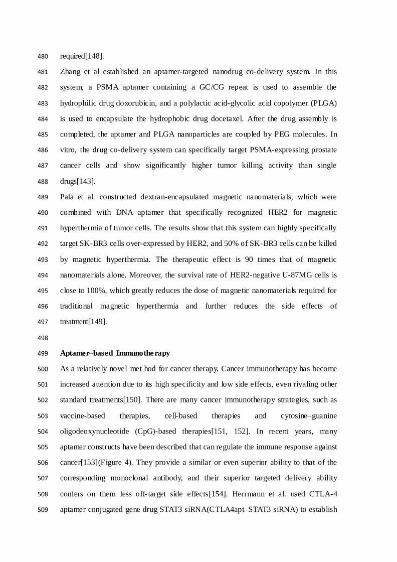
acid-based and protein-based nanomaterials. A series of aptamer-targeted nanodrug 471
models have been successfully constructed[147] (Table4). 472
Due to the special photothermal effect of gold-silver nanoparticles, Huang et al. 473
conjugated it with the aptamer sgc8c that specifically recognizes CCCRF-CEM cells 474
to form an Apt-NPs complex, which can be used for photothermal treatment. The 475
results show that the Apt-NPs complex can not only target tumor cells, but the 476
temperature of AuNPs will continue to increase under near- infrared light to kill tumor 477
cells, but it will not cause damage to normal cells (survival rate is 87%), reduced 478
required laser exposure, and greatly reduce the amount of laser irradiation 479
required[148]. 480
Zhang et al established an aptamer-targeted nanodrug co-delivery system. In this 481
system, a PSMA aptamer containing a GC/CG repeat is used to assemble the 482
hydrophilic drug doxorubicin, and a polylactic acid-glycolic acid copolymer (PLGA) 483
is used to encapsulate the hydrophobic drug docetaxel. After the drug assembly is 484
completed, the aptamer and PLGA nanoparticles are coupled by PEG molecules. In 485
vitro, the drug co-delivery system can specifically target PSMA-expressing prostate 486
cancer cells and show significantly higher tumor killing activity than single 487
drugs[143]. 488
Pala et al. constructed dextran-encapsulated magnetic nanomaterials, which were 489
combined with DNA aptamer that specifically recognized HER2 for magnetic 490
hyperthermia of tumor cells. The results show that this system can highly specifically 491
target SK-BR3 cells over-expressed by HER2, and 50% of SK-BR3 cells can be killed 492
by magnetic hyperthermia. The therapeutic effect is 90 times that of magnetic 493
nanomaterials alone. Moreover, the survival rate of HER2-negative U-87MG cells is 494
close to 100%, which greatly reduces the dose of magnetic nanomaterials required for 495
traditional magnetic hyperthermia and further reduces the side effects of 496
treatment[149]. 497
498
Aptamer–based Immunotherapy 499
As a relatively novel met hod for cancer therapy, Cancer immunotherapy has become 500
increased attention due to its high specificity and low side effects, even rivaling other 501
standard treatments[150]. There are many cancer immunotherapy strategies, such as 502
vaccine-based therapies, cell-based therapies and cytosine–guanine 503
oligodeoxynucleotide (CpG)-based therapies[151, 152]. In recent years, many 504
aptamer constructs have been described that can regulate the immune response against 505
cancer[153](Figure 4). They provide a similar or even superior ability to that of the 506
corresponding monoclonal antibody, and their superior targeted delivery ability 507
confers on them less off- target side effects[154]. Herrmann et al. used CTLA-4 508
aptamer conjugated gene drug STAT3 siRNA(CTLA4apt–STAT3 siRNA) to establish 509

an innovative immune checkpoint gene therapy method. The results show that this 510
method can significantly activate anti-tumor immunity and suppress tumors growth 511
and metastasis[155]. CTLA4apt–STAT3 siRNA can lead to internalization into 512
tumor-associated CD8+ T cells and inhibit the expression of STAT3, which can 513
activate the tumor antigen–specific T cells. Furthermore, CTLA4apt–STAT3 siRNA 514
can dramatically reduce tumor-associated Tregs. In addition, Zhang et al. used nature 515
killer (NK) cells as anti-tumor immune cells to eliminate residual tumor cells after 516
photothermal therapy(PTT), and the NK cells were modified by TLS11a aptamer 517
against hepatocellular carcinoma(HCC) cells on the cell surface to enhance 518
immunotherapy efficiency[156]. PD-1 is expressed in some cell types including T 519
cells, specifically in CD8 tumor- infiltrating lymphocytes (TILs) which are responsible 520
for killing tumor cells[157]. Prodeus et al. screened a mouse-derived PD-1 aptamer 521
that can specifically block the binding of PD-1 and PD-L1, thereby reversing the 522
immunosuppressive state of the tumor and activating anti-tumor immunity. In the 523
tumor-bearing models of PD-1 positive colon cancer, PD-1 aptamers can significantly 524
inhibit tumor growth, and the treatment effect is similar to that of PD-1 525
antibodies[158]. 526
527
528
529
530
531
532
533
534
535
536
537
538
539

Conclusion and perspectives 540
Aptamer, a novel specific combining tool to various types of target, has attracted a n 541
increasing attention for cancer diagnosis and therapy. In this review, we review recent 542
advances in this promising field of aptamer, including the screen of aptamer by 543
SELEX process, modification of aptamers and applications of aptamenr for 544
biosensing, bioimaging and therapy in cancer. Although aptamers have made great 545
progress in tumor application research, they still need to continue to improve of drug 546
loading rate, targeting efficiency, circulation time, and affinity, etc. The combination 547
of aptamers and drugs and the modification of nanocarriers by aptamers still need to 548
be improved. With the continuous advancement of SELEX technology and chemical 549
modification methods, we believe that aptamers will definitely play a more and more 550
important role in future oncology applications. 551
552
List of abbreviations 553
SELEX, Systematic Evolution of Ligands by Exponential Enrichment 554
CTCs, Circulating tumor cells 555
PCR, Polymerase Chain Reaction 556
ChIP, Chromatin immunoprecipitation 557
NECEEM, Non-equilibrium capillary electrophoresis of equilibrium mixtures 558
PEG, polyethylene glycol 559
PSMA, Prostate-specific membrane antigen 560
EpCAM, Epithelial cell adhesion molecule 561
PTK7, Protein tyrosine kinase 7 562
MUC1, Mucin1 563
SERS, Surface enhanced raman scattering 564
IgE, Immunoglobulin E 565
FITC, Fluorescein isothiocyanate 566
FRET, Fluorescence resonance energy transfer 567
FETs, Field-effect transistors 568
EIS, Electrical impedance spectroscopy 569

PGDF, Platelet-derived growth factor 570
BSA, Albumin from bovine serum 571
CT, Computed tomography 572
MRI, Magnetic resonance imaging 573
PET, Positron emission tomography 574
SPECT, Single photon emission computed tomography 575
VEGFR2, Vascular endothelial growth factor receptor 2 576
IHC, Immunohistochemical 577
FDA, Food and drug administration 578
NF-κB, Nuclear factor-κB 579
AptDC, Aptamer-drug conjugates 580
EPR, Enhanced permeability and retention 581
PLGA, Polylactic acid-glycolic acid copolymer 582
PTT, Photothermal therapy 583
HCC, Hepatocellular carcinoma 584
TILs, Tumor-infiltrating lymphocytes 585
NK, Nature killer 586
PD-1, Programmed cell death 1 587
CTLA-4, Cytotoxic T-Lymphocyte Associated Protein 4 588
HER2, Erb-B2 Receptor Tyrosine Kinase 2 589
Dox, Doxorubicine 590
Bcl2, BCL2 Apoptosis Regulator 591
Plk1, Polo Like Kinase 1 592
593
594
595
596
597
598
599

Ethics approval and consent to participate 600
Not applicable. 601
Consent for publication 602
Not applicable. 603
Availability of data and materials 604
Not applicable. 605
Competing interests 606
The authors declare that they have no competing interests. 607
Funding 608
This work was supported in part by grants from the Applied Basic Research Programs 609
of Science and Technology Commission Foundation of Shanxi Province (Grant No. 610
201901D211472), Shanxi Province Service Industry Innovation Discipline Group 611
Construction Plan (Grant No. 201809).Science and technology innovation plan of 612
Shanxi Higher Education Institutions (Grant No.2020L0390, 2020L0398) 613
Authors' contributions 614
Jia Wang and Jinsheng Wang designed this study. Jing Han, Liang Gao, Jia Wang and 615
Jinsheng Wang drafted the manuscript. All authors read and approved the final 616
manuscript. 617
Acknowledgements 618
Not applicable. 619
620
621
622
623
624

Referances 625
626
1. Bray F, Ferlay J, Soerjomataram I, Siegel RL, Torre LA, Jemal A. Global cancer statistics 2018: 627
GLOBOCAN estimates of incidence and mortality worldwide for 36 cancers in 185 countries . CA: 628
a cancer journal for clinicians. 2018; 68: 394-424. 629
2. Siegel RL, Miller KD, Jemal A. Cancer statistics, 2018. CA: a cancer journal for clinicians. 630
2018; 68: 7-30. 631
3. Vermes A, Guchelaar HJ, Dankert J. Flucytosine: a review of its pharmacology, clinical 632
indications, pharmacokinetics, toxicity and drug interactions . The Journal of antimicrobial 633
chemotherapy. 2000; 46: 171-9. 634
4. Reuther T, Schuster T, Mende U, Kubler A. Osteoradionecrosis of the jaws as a side effect of 635
radiotherapy of head and neck tumour patients --a report of a thirty year retros pective review. 636
International journal of oral and maxillofacial surgery . 2003; 32: 289-95. 637
5. Smith RA, Andrews KS, Brooks D, Fedewa SA, Manas saram-Baptiste D, Saslow D, et al. Cancer 638
screening in the United States, 2018: A review of current American Cancer Society guidelines and 639
current issues in cancer screening. CA: a cancer journal for clinicians. 2018; 68: 297-316. 640
6. Bruno JG. Predicting the Uncertain Future of Aptamer-Based Diagnostics and Therapeutics . 641
Molecules. 2015; 20: 6866-87. 642
7. Tuerk C, Gold L. Systematic evolution of ligands by exponential enrichment: RNA ligands to 643
bacteriophage T4 DNA polymerase. Science. 1990; 249: 505-10. 644
8. Osborne SE, Matsumura I, Ellington AD. Aptamers as therapeutic and diagnostic reagents: 645
problems and prospects . Current opinion in chemical biology. 1997; 1: 5-9. 646
9. Mayer G. The chemical biology of aptamers . Angewandte Chemie. 2009; 48: 2672-89. 647
10. Gelinas AD, Davies DR, Jan jic N. Embracing proteins: structural themes in aptamer-protein 648
complexes . Current opinion in structural biology. 2016; 36: 122-32. 649
11. Xi Z, Huang R, Deng Y, He N. Progress in selection and biomedical applications of aptamers . 650
Journal of biomedical nanotechnology. 2014; 10: 3043-62. 651
12. Ma H, Liu J, Ali MM, Mahmood MA, Labanieh L, Lu M, et al. Nucleic acid aptamers in cancer 652
research, diagnosis and therapy. Chemical Society reviews. 2015; 44: 1240-56. 653
13. Xuan W, Peng Y, Deng Z, Peng T, Kuai H, Li Y, et al. A basic insight into aptamer-drug 654
conjugates (ApDCs). Biomaterials. 2018; 182: 216-26. 655
14. Hirao I, Kimoto M, Lee KH. DNA aptamer generation by ExS ELEX using genetic alphabet 656
expansion with a mini-hairpin DNA stabilization method. Biochimie. 2018; 145: 15-21. 657
15. Rothlisberger P, Hollenstein M. Aptamer chemistry. Advanced drug delivery reviews. 2018; 134: 658
3-21. 659
16. Bagalkot V, Farokhzad OC, Langer R, Jon S. An aptamer-doxorubicin physical conjugate as a 660
novel targeted drug-delivery platform. Angewandte Chemie. 2006; 45: 8149-52. 661
17. Farokhzad OC, Cheng J, Tep ly BA, Sherifi I, Jon S, Kantoff PW, et al. Targeted 662
nanoparticle-aptamer bioconjugates for cancer chemotherapy in vivo. Proceedings of the National 663
Academy of Sciences of the United States of America. 2006; 103: 6315-20. 664
18. McNamara JO, 2nd, Andrechek ER, Wang Y, Viles KD, Rempel RE, Gilboa E, et al. Cell 665
type-s pecific delivery of siRNAs with aptamer-siRNA chimeras . Nature biotechnology. 2006; 24: 666
1005-15. 667
19. Borzabadi-Farahani A, Borzabadi E, Lynch E. Nanoparticles in orthodontics, a review of 668

antimicrobial and anti-caries applications . Acta odontologica Scandinavica. 2014; 72: 413-7. 669
20. Maeda H, Wu J, Sawa T, Matsumura Y, Hori K. Tumor vascular permeability and the EPR 670
effect in macromolecular therapeutics: a review. Journal of controlled release : o fficial journal of 671
the Controlled Release Society. 2000; 65: 271-84. 672
21. Fang J, Nakamura H, Maeda H. The EPR effect: Unique features of tumor blood vessels for 673
drug delivery, factors involved, and limitations and augmentation of the effect . Advanced drug 674
delivery reviews. 2011; 63: 136-51. 675
22. Wieser A. Review of reconstruction of radiation incident air kerma by measurement of 676
absorbed dose in tooth enamel with EPR. Radiation protection dosimetry. 2012; 149: 71-8. 677
23. Tian J, Ding L, Ju H, Yang Y, Li X, Shen Z, et al. A multifunctional nanomicelle for real-time 678
targeted imaging and precise near-infrared cancer therapy. Angewandte Chemie. 2014; 53: 9544-9. 679
24. Hua X, Zhou Z, Yuan L, Liu S. Selective collection and detection of MCF-7 breast cancer cells 680
using aptamer-functionalized magnetic beads and quantum dots based nano-bio-probes . 681
Analytica chimica acta. 2013; 788: 135-40. 682
25. Niazi JH, Verma SK, Niazi S, Qureshi A. In vitro HER2 protein-induced affinity dissociation 683
of carbon nanotube-wrapped anti-HER2 aptamers for HER2 protein detection. The Analyst. 2015; 684
140: 243-9. 685
26. Eikrem OS, Strauss P, Beisland C, Scherer A, Landolt L, Flatberg A, et al. Development and 686
confirmation of potential gene classifiers of human clear cell renal cell carcinoma using 687
next-generation RNA sequencing. Scandinavian journal of urology. 2016; 50: 452-62. 688
27. Ellington AD, Szostak JW. In vitro selection of RNA molecules that bind s pecific ligands . 689
Nature. 1990; 346: 818-22. 690
28. Mascini M, Palchetti I, Tombelli S. Nucleic acid and peptide aptamers: fundamentals and 691
bioanalytical aspects . Angewandte Chemie. 2012; 51: 1316-32. 692
29. Stoltenburg R, Reinemann C, Strehlitz B. S ELEX--a (r)evolutionary method to generate 693
high-affinity nucleic acid ligands . Biomolecular engineering. 2007; 24: 381-403. 694
30. Radom F, Jurek PM, Mazurek MP, Otlewski J, Jelen F. Aptamers: molecules of great potential. 695
Biotechnology advances. 2013; 31: 1260-74. 696
31. Caro li J, Taccioli C, De La Fuente A, Serafin i P, Bicciato S. APTANI: a computational tool to 697
select aptamers through sequence-structure moti f analysis of HT-S ELEX data. Bioinformatics. 698
2016; 32: 161-4. 699
32. Hermann T, Patel DJ. Adaptive recognition by nucleic acid aptamers . Science. 2000; 287: 700
820-5. 701
33. Shum KT, Zhou J, Rossi JJ. Aptamer-based therapeutics: new approaches to combat human 702
viral diseases . Pharmaceuticals. 2013; 6: 1507-42. 703
34. Martinez O, Bellard E, Golzio M, Mechiche-Alami S, Rols MP, Teissie J, et al. Direct validation 704
of aptamers as powerful tools to image solid tumor. Nucleic acid therapeutics. 2014; 24: 217-25. 705
35. Reinemann C, Strehlitz B. Aptamer-modified nanoparticles and their use in cancer 706
diagnostics and treatment. Swiss medical weekly. 2014; 144: w13908. 707
36. Keefe AD, Pai S, Ellington A. Aptamers as therapeutics . Nature reviews Drug discovery. 2010; 708
9: 537-50. 709
37. Wilson DS, Szostak JW. In vi tro selection of functional nucleic acids . Annual review of 710
biochemistry. 1999; 68: 611-47. 711
38. Duan N, Wu S, Chen X, Huang Y, Xia Y, Ma X, et al. Selection and characterization of 712

aptamers against Salmonella typhimurium using whole-bacterium Systemic Evolution of Ligands 713
by Exponential Enrichment (S ELEX). Journal of agricultural and food chemistry . 2013; 61: 714
3229-34. 715
39. Dhar S, Gu FX, Langer R, Farokhzad OC, Lippard SJ. Targeted delivery of cis platin to prostate 716
cancer cells by aptamer functionalized Pt(IV) prodrug-PLGA-PEG nanoparticles . Proceedings of 717
the National Academy of Sciences of the United States of America . 2008; 105: 17356-61. 718
40. Bayat P, Nosrati R, A libolandi M, Rafatpanah H, Abnous K, Khedri M, et al. S ELEX methods on 719
the road to protein targeting with nucleic acid aptamers . Biochimie. 2018; 154: 132-55. 720
41. Klussmann S, Nolte A, Bald R, Erdmann VA, Furste JP. Mirror-image RNA that binds 721
D-adenosine. Nature biotechnology. 1996; 14: 1112-5. 722
42. Martell RE, Nevins JR, Su llenger BA. Optimizing aptamer activi ty for gene therapy 723
applications using expression cassette S ELEX . Molecular therapy : the journal of the American 724
Society of Gene Therapy. 2002; 6: 30-4. 725
43. Vater A, Jarosch F, Buchner K, Klussmann S. Short bioactive S piegelmers to 726
migraine-associated calcitonin gene-related peptide rapidly identified by a novel approach: 727
tailored-SELEX. Nucleic acids research. 2003; 31: e130. 728
44. Huang CJ, Lin HI, Sh iesh SC, Lee GB. Integrated microfluidic system for rapid screening of 729
CRP aptamers utilizing systematic evolution of ligands by exponential enrichment (S ELEX) . 730
Biosensors & bioelectronics. 2010; 25: 1761-6. 731
45. Lu J, Ho DM, Vogelaar NJ, Kraml CM, Pascal RA, Jr. A pentacene with a 144 degrees twist. 732
Journal of the American Chemical Society. 2004; 126: 11168-9. 733
46. Drabovich A, Berezovski M, Krylov SN. Selection of smart aptamers by equilibrium capillary 734
electrophoresis of equilibrium mixtures (ECEEM) . Journal of the American Chemical Society. 2005; 735
127: 11224-5. 736
47. Berezovski M, Musheev M, Drabovich A, Kry lov SN. Non-S ELEX selection of aptamers . 737
Journal of the American Chemical Society. 2006; 128: 1410-1. 738
48. Wen JD, Gray DM. Selection of genomic sequences that bind tightly to Ff gene 5 protein: 739
primer-free genomic SELEX. Nucleic acids research. 2004; 32: e182. 740
49. White R, Rusconi C, Scardino E, Wolberg A, Lawson J, Hoffman M, et al. Generation of species 741
cross-reactive aptamers using "toggle" S ELEX . Molecular therapy : the journal of the American 742
Society of Gene Therapy. 2001; 4: 567-73. 743
50. Golden MC, Collins BD, Willis MC, Koch TH. Diagnostic potential of PhotoS ELEX-evolved 744
ssDNA aptamers . Journal of biotechnology. 2000; 81: 167-78. 745
51. Cox JC, Hayhurst A, Hesselberth J, Bayer TS, Georgiou G, Ellington AD. Automated selection 746
of aptamers against protein targets translated in vi tro: from gene to aptamer . Nucleic acids 747
research. 2002; 30: e108. 748
52. Cerch ia L, de Franciscis V. Targeting cancer cells with nucleic acid aptamers . Trends in 749
biotechnology. 2010; 28: 517-25. 750
53. Camoran i S, Cerchia L. Oligonucleotide aptamers for glioma targeting: an update . Central 751
nervous system agents in medicinal chemistry. 2015; 15: 126-37. 752
54. Sefah K, Shangguan D, Xiong X, O'Donoghue MB, Tan W. Development of DNA aptamers 753
using Cell-SELEX. Nature protocols. 2010; 5: 1169-85. 754
55. Tan W, Donovan MJ, Jiang J. Aptamers from cell-based selection for bioanalytical 755
applications . Chemical reviews. 2013; 113: 2842-62. 756

56. Keefe AD, Cload ST. S ELEX with modified nucleotides . Current opinion in chemical biology. 757
2008; 12: 448-56. 758
57. Cheung CH, Sun X, Kanwar JR, Bai JZ, Cheng L, Krissansen GW. A cell-permeable 759
dominant-negative survivin protein induces apoptosis and sensitizes prostate cancer cells to 760
TNF-alpha therapy. Cancer cell international. 2010; 10: 36. 761
58. Lin Y, Nieuwlandt D, Magallanez A, Feistner B, Jayasena SD. High-affinity and specific 762
recognition of human thyroid stimulating hormone (hTS H) by in vi tro-selected 763
2'-amino-modified RNA. Nucleic acids research. 1996; 24: 3407-14. 764
59. Kujau MJ, Wolfl S. Intramolecular derivatization of 2'-amino-pyrimidine modified RNA with 765
functional groups that is compatible with re-amplification. Nucleic acids research. 1998; 26: 766
1851-3. 767
60. Ruckman J, Green LS, Beeson J, Waugh S, Gillette W L, Henninger DD, et al. 768
2'-Fluoropyrimidine RNA-based aptamers to the 165-amino acid form of vascular endothelial 769
growth factor (VEGF165). Inhibition of receptor binding and VEGF-induced vascular 770
permeability through interactions requiring the exon 7-encoded domain . The Journal of biological 771
chemistry. 1998; 273: 20556-67. 772
61. Rusconi CP, Scardino E, Layzer J, Pitoc GA, Ortel TL, Monroe D, et al. RNA aptamers as 773
reversible antagonists of coagulation factor IXa. Nature. 2002; 419: 90-4. 774
62. Burmeister PE, Lewis SD, Silva RF, Preiss JR, Horwitz LR, Pendergrast PS, et al. Direct in vi tro 775
selection of a 2'-O-methyl aptamer to VEGF. Chemistry & biology. 2005; 12: 25-33. 776
63. Burmeister PE, Wang C, Killough JR, Lewis SD, Horwitz LR, Ferguson A, et al. 2'-Deoxy purine, 777
2'-O-methyl pyrimidine (dRmY) aptamers as candidate therapeutics . Oligonucleotides. 2006; 16: 778
337-51. 779
64. Healy JM, Lewis SD, Kurz M, Boomer RM, Thomps on KM, Wilson C, et al. Pharmacokinetics 780
and biodistribution of novel aptamer compositions . Pharmaceutical research. 2004; 21: 2234-46. 781
65. Rusconi CP, Roberts JD, Pitoc GA, Nimjee SM, White RR, Quick G, Jr., et al. 782
Antidote-mediated control of an anticoagulant aptamer in vivo. Nature biotechnology. 2004; 22: 783
1423-8. 784
66. Boomer RM, Lewis SD, Healy JM, Kurz M, Wilson C, McCauley TG. Conjugation to 785
polyethylene glycol polymer promotes aptamer biodistribution to healthy and inflamed tissues . 786
Oligonucleotides. 2005; 15: 183-95. 787
67. Kanwar JR, Roy K, Kanwar RK. Chimeric aptamers in cancer cell-targeted drug delivery. 788
Critical reviews in biochemistry and molecular biology . 2011; 46: 459-77. 789
68. Vandghanooni S, Eskandani M, Barar J, Omid i Y. Bispecific therapeutic aptamers for targeted 790
therapy of cancer: a review on cellular perspective . Journal of molecular medicine. 2018; 96: 791
885-902. 792
69. Cho Y, Lee YB, Lee JH, Lee DH, Cho EJ, Yu SJ, et al. Modified AS1411 Aptamer Suppresses 793
Hepatocellular Carcinoma by Up-Regulating Galectin-14. PloS one. 2016; 11: e0160822. 794
70. Pastor F. Aptamers: A New Technological Platform in Cancer Immunotherapy. 795
Pharmaceuticals. 2016; 9. 796
71. Soldevilla MM, Villanueva H, Pastor F. Aptamers: A Feasible Technology in Cancer 797
Immunotherapy. Journal of immunology research . 2016; 2016: 1083738. 798
72. Lupold SE, Hicke BJ, Lin Y, Coffey DS. Identification and characterization of 799
nuclease-stabilized RNA molecules that bind human prostate cancer cells via the prostate -specific 800

membrane antigen. Cancer research. 2002; 62: 4029-33. 801
73. Shieh YA, Yang SJ, Wei MF, Sh ieh MJ. Aptamer-based tumor-targeted drug delivery for 802
photodynamic therapy. ACS nano. 2010; 4: 1433-42. 803
74. Cao Z, Tong R, Mishra A, Xu W, Wong GC, Cheng J, et al. Reversible cell-specific drug 804
delivery with aptamer-functionalized liposomes . Angewandte Chemie. 2009; 48: 6494-8. 805
75. Song Y, Zhu Z, An Y, Zhang W, Zhang H, Liu D, et al. Selection of DNA aptamers against 806
epithelial cell adhesion molecule for cancer cell imaging and circulating tumor cell capture. 807
Analytical chemistry. 2013; 85: 4141-9. 808
76. Xiang D, Sh igdar S, Qiao G, Wang T, Kouzan i AZ, Zhou SF, et al. Nucleic acid aptamer-guided 809
cancer therapeutics and diagnostics: the next generation of cancer medicine . Theranostics. 2015; 5: 810
23-42. 811
77. Zhu G, Hu R, Zhao Z, Chen Z, Zhang X, Tan W. Noncanonical self-assembly of multi functional 812
DNA nanoflowers for biomedical applications . Journal of the American Chemical Society. 2013; 135: 813
16438-45. 814
78. Zhu G, Zheng J, Song E, Donovan M, Zhang K, Liu C, et al. Self-assembled, aptamer-tethered 815
DNA nanotrains for targeted trans port of molecular drugs in cancer theranostics . Proceedings of 816
the National Academy of Sciences of the United States of America . 2013; 110: 7998-8003. 817
79. Ferreira CS, Matthews CS, Missailid is S. DNA aptamers that bind to MUC1 tumour marker: 818
design and characterization of MUC1-binding single-stranded DNA aptamers . Tumour biology : 819
the journal of the International Society for Oncodevelopmental Biology and Medicine . 2006; 27: 820
289-301. 821
80. Chang CC, Wei SC, Wu TH, Lee CH, Lin CW. Aptamer-based colorimetric detection of 822
platelet-derived growth factor using unmodified gold nanoparticles . Biosensors & bioelectronics. 823
2013; 42: 119-23. 824
81. Mackey D, Kelly E, Nooney R. Modelling random antibody adsorption and immunoassay 825
activity. Mathematical biosciences and engineering : MBE. 2016; 13: 1159-68. 826
82. Qu F, Lu H, Yang M, Deng C. Electrochemical immunosensor based on electron transfer 827
mediated by graphene oxide initiated silver enhancement. Biosensors & bioelectronics. 2011; 26: 828
4810-4. 829
83. Wei H, Wang E. Electrochemiluminescence of tris(2,2'-bipyridyl)ruthenium and i ts 830
applications in bioanalysis: a review. Luminescence : the journal of biological and chemical 831
luminescence. 2011; 26: 77-85. 832
84. Hu K, Yang H, Zhou J, Zhao S, Tian J. Aptasensor for amplified IgE sensing based on 833
fluorescence quenching by graphene oxide. Luminescence : the journal of biological and chemical 834
luminescence. 2013; 28: 662-6. 835
85. Hosseini M, Mehrabi F, Ganjali M R, Norouzi P. A fluorescent aptasensor for sensitive analysis 836
oxytetracycline based on silver nanoclusters . Luminescence : the journal of biological and chemical 837
luminescence. 2016; 31: 1339-43. 838
86. Razmi N, Baradaran B, Hejazi M, Hasanzadeh M, Mosafer J, Mokhtarzadeh A, et al. Recent 839
advances on aptamer-based biosensors to detection of platelet-derived growth factor. Biosensors 840
& bioelectronics. 2018; 113: 58-71. 841
87. Huang CC, Huang YF, Cao Z, Tan W, Chang HT. Aptamer-modified gold nanoparticles for 842
colorimetric determination of platelet-derived growth factors and their receptors . Analytical 843
chemistry. 2005; 77: 5735-41. 844

88. Ye S, Zhai X, Wu Y, Kuang S. Dual-primer self-generation S ERS signal ampli fication assay 845
for PDGF-BB using label-free aptamer. Biosensors & bioelectronics. 2016; 79: 130-5. 846
89. Li H, Wang M, Wang C, Li W, Qiang W, Xu D. Silver nanoparticle-enhanced fluorescence 847
resonance energy transfer sensor for human platelet-derived growth factor-BB detection. 848
Analytical chemistry. 2013; 85: 4492-9. 849
90. Jiang Y, Fang X, Bai C. Signaling aptamer/protein binding by a molecular light s witch 850
complex. Analytical chemistry. 2004; 76: 5230-5. 851
91. Cao ZJ, Peng QW, Qiu X, Liu CY, Lu JZ. Highly sensitive chemiluminescence technology for 852
protein detection using aptamer-based rolling circle amplification platform. Journal of 853
pharmaceutical analysis. 2011; 1: 159-65. 854
92. Chai Y, Tian D, Gu J, Cui H. A novel electrochemiluminescence aptasensor for protein based 855
on a sensitive N-(aminobutyl)-N-ethylisoluminol-functionalized gold nanoprobe. The Analyst. 2011; 856
136: 3244-51. 857
93. Meirinho SG, Dias LG, Peres AM, Rodrigues LR. Voltammetric aptasensors for protein 858
disease biomarkers detection: A review. Biotechnology advances. 2016; 34: 941-53. 859
94. Thevenot DR, Toth K, Durst RA, Wilson GS. Electrochemical biosensors: recommended 860
definitions and classification. Biosensors & bioelectronics. 2001; 16: 121-31. 861
95. He L, Zhang S, Ji H, Wang M, Peng D, Yan F, et al. Protein-templated cobaltous phos phate 862
nanocomposites for the highly sensitive and selective detection of platelet-derived growth 863
factor-BB. Biosensors & bioelectronics. 2016; 79: 553-60. 864
96. Chen W, Zheng R, Baade PD, Zhang S, Zeng H, Bray F, et al. Cancer statistics in China, 2015. 865
CA: a cancer journal for clinicians. 2016; 66: 115-32. 866
97. Siegel RL, Miller KD, Jemal A. Cancer statistics, 2016. CA: a cancer journal for clinicians. 867
2016; 66: 7-30. 868
98. Zhang X, Soori G, Dobleman TJ, Xiao GG. The application of monoclonal antibodies in 869
cancer diagnosis . Expert review of molecular diagnostics. 2014; 14: 97-106. 870
99. Nagrath S, Sequist LV, Maheswaran S, Bell DW, Irimia D, Ulkus L, et al. Isolation of rare 871
circulating tumour cells in cancer patients by microchip technology. Nature. 2007; 450: 1235-9. 872
100. Alix-Panabieres C, Pantel K. Clinical Applications of Circulating Tumor Cells and Circulating 873
Tumor DNA as Liquid Biopsy. Cancer discovery. 2016; 6: 479-91. 874
101. Jacob K, Sollier C, Jabado N. Circulating tumor cells: detection, molecular profiling and 875
future prospects . Expert review of proteomics. 2007; 4: 741-56. 876
102. Yoon HJ, Kim TH, Zhang Z, Azizi E, Pham TM, Paoletti C, et al. Sensitive capture of 877
circulating tumour cells by functionalized graphene oxide nanosheets . Nature nanotechnology. 878
2013; 8: 735-41. 879
103. Yu M, Bardia A, Wittner BS, Stott SL, Smas ME, Ting DT, et al. Circulating breast tumor cells 880
exhibit dynamic changes in epithelial and mesenchymal composition. Science. 2013; 339: 580-4. 881
104. Bi S, Ji B, Zhang Z, Zhang S. A chemiluminescence imaging array for the detection of cancer 882
cells by dual-aptamer recognition and bio-bar-code nanoprobe-based rolling circle amplification. 883
Chemical communications. 2013; 49: 3452-4. 884
105. Kashefi-Kheyrabadi L, Mehrgardi MA, Wiechec E, Turner AP, Tiwari A. Ultrasensitive detection 885
of human liver hepatocellular carcinoma cells using a label-free aptasensor. Analytical chemistry. 886
2014; 86: 4956-60. 887
106. Zeng Z, Tung CH, Zu Y. A cancer cell-activatable aptamer-reporter system for one-step assay 888

of circulating tumor cells . Molecular therapy Nucleic acids. 2014; 3: e184. 889
107. Wang K, Fan D, Liu Y, Wang E. Highly sensitive and s pecific colorimetric detection of cancer 890
cells via dual-aptamer target binding strategy. Biosensors & bioelectronics. 2015; 73: 1-6. 891
108. Zhao W, Cui CH, Bose S, Guo D, Shen C, Wong WP, et al. Bioinspired multivalent DNA 892
network for capture and release of cells . Proceedings of the National Academy of Sciences of the 893
United States of America. 2012; 109: 19626-31. 894
109. Sheng W, Chen T, Tan W, Fan ZH. Multivalent DNA nanos pheres for enhanced capture of 895
cancer cells in microfluidic devices . ACS nano. 2013; 7: 7067-76. 896
110. Massoud TF, Paulmurugan R, Gambhir SS. Molecular imaging of homodimeric protein-protein 897
interactions in living subjects . FASEB journal : official publication of the Federation of American 898
Societies for Experimental Biology. 2004; 18: 1105-7. 899
111. Shi H, Tang Z, Kim Y, Nie H, Huang YF, He X, et al. In vivo fluorescence imaging of tumors 900
using molecular aptamers generated by cell-SELEX. Chemistry, an Asian journal. 2010; 5: 2209-13. 901
112. Zhong H, Zhang Q, Zhang S. High-intensity fluorescence imaging and sensitive 902
electrochemical detection of cancer cells by using an extracellular supramolecular reticular 903
DNA-quantum dot sheath. Chemistry. 2011; 17: 8388-94. 904
113. Rockey WM, Huang L, Kloepping KC, Baumhover NJ, Giangrande PH, Schultz MK. Synthesis 905
and radiolabeling of chelator-RNA aptamer bioconjugates with copper-64 for targeted molecular 906
imaging. Bioorganic & medicinal chemistry. 2011; 19: 4080-90. 907
114. Yu MK, Kim D, Lee IH, So JS, Jeong YY, Jon S. Image-guided prostate cancer therapy using 908
aptamer-functionalized thermally cross-linked superparamagnetic iron oxide nanoparticles . 909
Small. 2011; 7: 2241-9. 910
115. Zeng Z, Parekh P, Li Z, Shi ZZ, Tung CH, Zu Y. S pecific and sensitive tumor imaging using 911
biostable oligonucleotide aptamer probes . Theranostics. 2014; 4: 945-52. 912
116. Wu X, Zhao Z, Bai H, Fu T, Yang C, Hu X, et al. DNA Aptamer Selected against Pancreatic 913
Ductal Adenocarcinoma for in vivo Imaging and Clinical Tissue Recognition. Theranostics. 2015; 914
5: 985-94. 915
117. Ma H, Gao Z, Yu P, Shen S, Liu Y, Xu B. A dual functional fluorescent probe for glioma 916
imaging mediated by blood-brain barrier penetration and glioma cell targeting. Biochemical and 917
biophysical research communications. 2014; 449: 44-8. 918
118. Kim B, Yang J, Hwang M, Choi J, Kim HO, Jang E, et al. Aptamer-modified magnetic 919
nanoprobe for molecular MR imaging of VEGFR2 on angiogenic vasculature . Nanoscale research 920
letters. 2013; 8: 399. 921
119. Heo D, Lee E, Ku M, Hwang S, Kim B, Park Y, et al. Maleimidyl magnetic nanoplatform for 922
facile molecular MRI. Nanotechnology. 2014; 25: 275102. 923
120. Zeng Z, Zhang P, Zhao N, Sheehan AM, Tung CH, Chang CC, et al. Using oligonucleotide 924
aptamer probes for immunostaining of formalin-fixed and paraffin-embedded tissues . Modern 925
pathology : an official journal of the United States and Canadian Academy of Pathology, Inc . 2010; 23: 926
1553-8. 927
121. Li X, An Y, Jin J, Zhu Z, Hao L, Liu L, et al. Evolution of DNA aptamers through in vi tro 928
metastatic-cell-based systematic evolution of ligands by exponential enrichment for metastatic 929
cancer recognition and imaging. Analytical chemistry. 2015; 87: 4941-8. 930
122. Zhang Z, Ali MM, Eckert MA, Kang DK, Chen YY, Sender LS, et al. A polyvalent aptamer 931
system for targeted drug delivery. Biomaterials. 2013; 34: 9728-35. 932

123. Ng EW, Sh ima DT, Calias P, Cunningham ET, Jr., Guyer DR, Adamis AP. Pegaptanib, a targeted 933
anti-VEGF aptamer for ocular vascular disease. Nature reviews Drug discovery. 2006; 5: 123-32. 934
124. Bates PJ, Laber DA, Miller DM, Thomas SD, Trent JO. Discovery and development of the 935
G-rich oligonucleotide AS1411 as a novel treatment for cancer . Experimental and molecular 936
pathology. 2009; 86: 151-64. 937
125. Mongelard F, Bouvet P. AS-1411, a guanosine-rich oligonucleotide aptamer targeting 938
nucleolin for the potential treatment of cancer, including acute myeloid leukemia. Current opinion 939
in molecular therapeutics. 2010; 12: 107-14. 940
126. Darisipudi MN, Kulkarn i OP, Sayyed SG, Ryu M, Migliorini A, Sagrinati C, et al. Dual blockade 941
of the homeostatic chemokine CXCL12 and the proinflammatory chemokine CCL2 has additive 942
protective effects on diabetic kidney disease. The American journal of pathology. 2011; 179: 116-24. 943
127. Duda DG, Kozin SV, Kirkpatrick ND, Xu L, Fukumura D, Jain RK. CXCL12 944
(SDF1alpha)-CXCR4/CXCR7 pathway inhibition: an emerging sensitizer for anticancer 945
therapies? Clinical cancer research : an official journal of the American Association for Cancer 946
Research. 2011; 17: 2074-80. 947
128. Bates PJ, Kahlon JB, Thomas SD, Trent JO, Miller DM. Antiproliferative activi ty of G-rich 948
oligonucleotides correlates with protein binding . The Journal of biological chemistry. 1999; 274: 949
26369-77. 950
129. Soundararajan S, Chen W, Spicer EK, Courtenay-Luck N, Fernandes DJ. The nucleolin targeting 951
aptamer AS1411 destabilizes Bcl-2 messenger RNA in human breast cancer cells . Cancer research. 952
2008; 68: 2358-65. 953
130. Mahlknecht G, Maron R, Mancini M, Schechter B, Sela M, Yarden Y. Aptamer to ErbB-2/HER2 954
enhances degradation of the target and inhibits tumorigenic growth . Proceedings of the National 955
Academy of Sciences of the United States of America . 2013; 110: 8170-5. 956
131. Parekh P, Kamble S, Zhao N, Zeng Z, Portier BP, Zu Y. Immunotherapy of CD30-expressing 957
lymphoma using a highly stable ssDNA aptamer. Biomaterials. 2013; 34: 8909-17. 958
132. Wu X, Chen J, Wu M, Zhao JX. Aptamers: active targeting ligands for cancer diagnosis and 959
therapy. Theranostics. 2015; 5: 322-44. 960
133. Thiel KW, Hernandez LI, Dassie JP, Thiel WH, Liu X, Stockdale KR, et al. Delivery of 961
chemo-sensitizing siRNAs to HER2+-breast cancer cells using RNA aptamers . Nucleic acids 962
research. 2012; 40: 6319-37. 963
134. Dai F, Zhang Y, Zhu X, Shan N, Chen Y. Anticancer role of MUC1 aptamer-miR-29b chimera 964
in epithelial ovarian carcinoma cells through regulation of PTEN methylation . Targeted oncology. 965
2012; 7: 217-25. 966
135. Liu M, Yu X, Chen Z, Yang T, Yang D, Liu Q, et al. Aptamer selection and applications for 967
breast cancer diagnostics and therapy. Journal of nanobiotechnology. 2017; 15: 81. 968
136. Zhao N, Pei SN, Qi J, Zeng Z, Iyer SP, Lin P, et al. Oligonucleotide aptamer-drug conjugates 969
for targeted therapy of acute myeloid leukemia. Biomaterials. 2015; 67: 42-51. 970
137. Huang YF, Shangguan D, Liu H, Phillips JA, Zhang X, Chen Y, et al. Molecular assembly of an 971
aptamer-drug conjugate for targeted drug delivery to tumor cells . Chembiochem : a European 972
journal of chemical biology. 2009; 10: 862-8. 973
138. Subramanian N, Raghunathan V, Kanwar JR, Kanwar RK, Elchuri SV, Khetan V, et al. 974
Target-specific delivery of doxorubicin to retinoblastoma using epithelial cell adhesion molecule 975
aptamer. Molecular vision. 2012; 18: 2783-95. 976

139. Liu Z, Duan JH, Song YM, Ma J, Wang FD, Lu X, et al. Novel HER2 aptamer selectively 977
delivers cytotoxic drug to HER2 -positive breast cancer cells in vitro. Journal of translational 978
medicine. 2012; 10: 148. 979
140. Rink JS, Plebanek MP, Tripathy S, Thaxton CS. Update on current and potential nanoparticle 980
cancer therapies . Current opinion in oncology. 2013; 25: 646-51. 981
141. Xiao Z, Farokhzad OC. Aptamer-functionalized nanoparticles for medical applications: 982
challenges and opportunities . ACS nano. 2012; 6: 3670-6. 983
142. Yang L, Zhang X, Ye M, Jiang J, Yang R, Fu T, et al. Aptamer-conjugated nanomaterials and 984
their applications . Advanced drug delivery reviews. 2011; 63: 1361-70. 985
143. Zhang L, Radovic-Moreno AF, A lexis F, Gu FX, Basto PA, Bagalkot V, et al. Co-delivery of 986
hydrophobic and hydrophilic drugs from nanoparticle-aptamer bioconjugates . ChemMedChem. 987
2007; 2: 1268-71. 988
144. Xing H, Tang L, Yang X, Hwang K, Wang W, Yin Q, et al. Selective Delivery of an Anticancer 989
Drug with Aptamer-Functionalized Liposomes to Breast Cancer Cells in Vitro and in Vivo. 990
Journal of materials chemistry B. 2013; 1: 5288-97. 991
145. Xiao Z, Levy-Nissenbaum E, Alexis F, Luptak A, Teply BA, Chan JM, et al. Engineering of 992
targeted nanoparticles for cancer therapy using internalizing aptamers isolated by cell-uptake 993
selection. ACS nano. 2012; 6: 696-704. 994
146. Tong GJ, Hsiao SC, Carrico ZM, Francis MB. Viral capsid DNA aptamer conjugates as 995
multivalent cell-targeting vehicles . Journal of the American Chemical Society. 2009; 131: 11174-8. 996
147. Sun H, Zu Y. Aptamers and their applications in nanomedicine. Small. 2015; 11: 2352-64. 997
148. Huang YF, Sefah K, Bamrungsap S, Chang HT, Tan W. Selective photothermal therapy for 998
mixed cancer cells using aptamer-conjugated nanorods . Langmuir : the ACS journal of surfaces and 999
colloids. 2008; 24: 11860-5. 1000
149. Pala K, Serwotka A, Jelen F, Jakimowicz P, Ot lewski J. Tumor-s pecific hyperthermia with 1001
aptamer-tagged superparamagnetic nanoparticles . International journal of nanomedicine. 2014; 9: 1002
67-76. 1003
150. Borghaei H, Paz-Ares L, Horn L, Spigel DR, Steins M, Ready NE, et al. Nivolumab versus 1004
Docetaxel in Advanced Nons quamous Non-S mall-Cell Lung Cancer. The New England journal of 1005
medicine. 2015; 373: 1627-39. 1006
151. Mohri K, Nishikawa M, Takahashi N, Shiomi T, Mats uoka N, Ogawa K, et al. Design and 1007
development of nanosized DNA assemblies in polypod-like structures as efficient vehicles for 1008
immunostimulatory CpG motifs to immune cells . ACS nano. 2012; 6: 5931-40. 1009
152. Liu H, Kwong B, Irvine DJ. Membrane anchored immunostimulatory oligonucleotides for in 1010
vivo cell modification and localized immunotherapy. Angewandte Chemie. 2011; 50: 7052-5. 1011
153. Gilboa E, McNamara J, 2nd, Pastor F. Use of oligonucleotide aptamer ligands to modulate the 1012
function of immune receptors . Clinical cancer research : an official journal of the American 1013
Association for Cancer Research. 2013; 19: 1054-62. 1014
154. Gilboa E, Berezhnoy A, Schrand B. Reducing Toxicity of Immune Therapy Using 1015
Aptamer-Targeted Drug Delivery. Cancer immunology research. 2015; 3: 1195-200. 1016
155. Herrmann A, Priceman SJ, Swiderski P, Kujawski M, Xin H, Cherryholmes GA, et al. CTLA4 1017
aptamer delivers STAT3 siRNA to tumor-associated and malignant T cells . The Journal of clinical 1018
investigation. 2014; 124: 2977-87. 1019
156. Zhang D, Zheng Y, Lin Z, Lan S, Zhang X, Zheng A, et al. Artificial Engineered Natural Killer 1020

Cells Combined with Antiheat Endurance as a Powerful Strategy for Enhancing 1021
Photothermal-Immunotherapy Efficiency of Solid Tumors . Small. 2019; 15: e1902636. 1022
157. Gros A, Robbins PF, Yao X, Li YF, Turcotte S, Tran E, et al. PD-1 identifies the patient-specific 1023
CD8(+) tumor-reactive repertoire infiltrating human tumors . The Journal of clinical investigation . 1024
2014; 124: 2246-59. 1025
158. Prodeus A, Abdul-Wahid A, Fischer NW, Huang EH, Cydzik M, Gariepy J. Targeting the 1026
PD-1/PD-L1 Immune Evasion Axis With DNA Aptamers as a Novel Therapeutic Strategy for the 1027
Treatment of Disseminated Cancers . Molecular therapy Nucleic acids. 2015; 4: e237. 1028
159. Shigdar S, Lin J, Yu Y, Pastuovic M, Wei M, Duan W. RNA aptamer against a cancer stem cell 1029
marker epithelial cell adhesion molecule. Cancer science. 2011; 102: 991-8. 1030
160. Shigdar S, Qiao L, Zhou SF, Xiang D, Wang T, Li Y, et al. RNA aptamers targeting cancer stem 1031
cell marker CD133. Cancer letters. 2013; 330: 84-95. 1032
161. Bayat P, Taghdisi SM, Rafatpanah H, Abnous K, Ramezani M. In vitro selection of CD70 1033
binding aptamer and its application in a biosensor design for sensitive detection of S KOV-3 1034
ovarian cells. Talanta. 2019; 194: 399-405. 1035
162. Almasi F, Mousavi Gargari SL, Bitaraf F, Rasoulinejad S. Development of a Single Stranded 1036
DNA Aptamer as a Molecular Probe for LNCap Cells Using Cell -S ELEX. Avicenna journal of 1037
medical biotechnology. 2016; 8: 104-11. 1038
163. Daniels DA, Chen H, Hicke BJ, Swiderek KM, Gold L. A tenascin-C aptamer identi fied by 1039
tumor cell SELEX: systematic evolution of ligands by exponential enrichment . Proceedings of the 1040
National Academy of Sciences of the United States of America . 2003; 100: 15416-21. 1041
164. Alshaer W, Hillaireau H, Vergnaud J, Ismail S, Fattal E. Functionalizing Liposomes with 1042
anti-CD44 Aptamer for Selective Targeting of Cancer Cells . Bioconjugate chemistry. 2015; 26: 1043
1307-13. 1044
165. Haghighi FH, Binaymotlagh R, Mirahmadi-Zare SZ, Hadadzadeh H. Aptamer/magnetic 1045
nanoparticles decorated with fluorescent gold nanoclusters for selective detection and collection 1046
of human promyelocytic leukemia (HL-60) cells from a mixture. Nanotechnology. 2020; 31: 1047
025605. 1048
166. Fafinska J, Czech A, Sitz T, Ignatova Z, Hahn U. DNA Aptamers for the Malignant 1049
Transformation Marker CD24. Nucleic acid therapeutics. 2018; 28: 326-34. 1050
167. Mallikaratchy P, Tang Z, Kwame S, Meng L, Shangguan D, Tan W. Aptamer directly evolved 1051
from live cells recognizes membrane bound immunoglobin heavy mu chain in Burkitt's 1052
lymphoma cells . Molecular & cellular proteomics : MCP. 2007; 6: 2230-8. 1053
168. Vinores SA. Pegaptanib in the treatment of wet, age-related macular degeneration. 1054
International journal of nanomedicine. 2006; 1: 263-8. 1055
169. Jaffe GJ, Ciulla TA, Ciardella AP, Devin F, Dugel PU, Eandi CM, et al. Dual Antagonism of 1056
PDGF and VEGF in Neovascular Age-Related Macular Degeneration: A Phase IIb, Multicenter, 1057
Randomized Controlled Trial. Ophthalmology. 2017; 124: 224-34. 1058
170. Spiel AO, Mayr FB, Ladani N, Wagner PG, Schaub RG, Gilbert JC, et al. The aptamer 1059
ARC1779 is a potent and s pecific inhibitor of von Willebrand Factor mediated ex vivo platelet 1060
function in acute myocardial infarction. Platelets. 2009; 20: 334-40. 1061
171. Buff MC, Schafer F, Wulffen B, Muller J, Potzsch B, Heckel A, et al. Dependence of aptamer 1062
activi ty on opposed terminal extensions: improvement of light-regulation efficiency. Nucleic acids 1063
research. 2010; 38: 2111-8. 1064

172. Vater A, Klussmann S. Turning mirror-image oligonucleotides into drugs: the evolution of 1065
Spiegelmer((R)) therapeutics . Drug discovery today. 2015; 20: 147-55. 1066
173. van Eijk LT, John AS, Schwoebel F, Summo L, Vauleon S, Zollner S, et al. Effect of the 1067
antihepcidin S piegelmer lexaptepid on inflammation-induced decrease in serum iron in humans . 1068
Blood. 2014; 124: 2643-6. 1069
174. Leung E, Landa G. Update on current and future novel therapies for dry age-related macular 1070
degeneration. Expert review of clinical pharmacology. 2013; 6: 565-79. 1071
175. Povsic TJ, Vavalle JP, Alexander JH, Aberle LH, Zelenkofske SL, Becker RC, et al. Use of the 1072
REG1 anticoagulation system in patients with acute coronary syndromes undergoing 1073
percutaneous coronary intervention: results from the phase II RADAR-PCI study. 1074
EuroIntervention : journal of EuroPCR in collaboration with the Working Group on Interventional 1075
Cardiology of the European Society of Cardiology . 2014; 10: 431-8. 1076
176. Waters EK, Genga RM, Schwartz MC, Nelson JA, Schaub RG, Olson KA, et al. Aptamer 1077
ARC19499 mediates a procoagulant hemostatic effect by inhibiting tissue factor pathway 1078
inhibitor. Blood. 2011; 117: 5514-22. 1079
177. Taghavi S, Nia AH, Abnous K, Ramezani M. Polyethylenimine-functionalized carbon 1080
nanotubes tagged with AS1411 aptamer for combination gene and drug delivery into human 1081
gastric cancer cells . International journal of pharmaceutics. 2017; 516: 301-12. 1082
178. Alibolandi M, Ramezani M, Abnous K, Hadizadeh F. AS1411 Aptamer-Decorated 1083
Biodegradable Polyethylene Glycol-Poly(lactic-co-glycolic acid) Nanopolymersomes for the 1084
Targeted Delivery of Gemcitabine to Non-Small Cell Lung Cancer In Vitro. Journal of 1085
pharmaceutical sciences. 2016; 105: 1741-50. 1086
179. Xie X, Li F, Zhang H, Lu Y, Lian S, Lin H, et al. EpCAM aptamer-functionalized mesoporous 1087
silica nanoparticles for efficient colon cancer cell-targeted drug delivery. European journal of 1088
pharmaceutical sciences : official journal of the European Federation for Pharmaceutical Sciences . 1089
2016; 83: 28-35. 1090
180. Leach JC, Wang A, Ye K, Jin S. A RNA-DNA Hybrid Aptamer for Nanoparticle-Based 1091
Prostate Tumor Targeted Drug Delivery. International journal of molecular sciences. 2016; 17: 380. 1092
181. Taghdisi SM, Danesh NM, Lavaee P, Emran i AS, Hassanabad KY, Ramezani M, et al. Double 1093
targeting, controlled release and reversible delivery of daunorubicin to cancer cells by polyvalent 1094
aptamers-modified gold nanoparticles . Materials science & engineering C, Materials for biological 1095
applications. 2016; 61: 753-61. 1096
182. Moosavian SA, Abnous K, Badiee A, Jaafari MR. Improvement in the drug delivery and 1097
anti-tumor efficacy of PEGylated liposomal doxorubicin by targeting RNA aptamers in mice 1098
bearing breast tumor model. Colloids and surfaces B, Biointerfaces. 2016; 139: 228-36. 1099
183. Taghavi S, Ramezani M, A libolandi M, Abnous K, Taghdisi SM. Chitosan-modified PLGA 1100
nanoparticles tagged with 5TR1 aptamer for in vivo tumor-targeted drug delivery. Cancer letters. 1101
2017; 400: 1-8. 1102
184. Pi F, Zhang H, Li H, Th iviyanathan V, Gorenstein DG, Sood AK, et al. RNA nanoparticles 1103
harboring annexin A2 aptamer can target ovarian cancer for tumor-s pecific doxorubicin delivery. 1104
Nanomedicine : nanotechnology, biology, and medicine . 2017; 13: 1183-93. 1105
185. Chen Y, Wang J, Wang J, Wang L, Tan X, Tu K, et al. Aptamer Functionalized 1106
Cisplatin-Albumin Nanoparticles for Targeted Delivery to Epidermal Growth Factor Receptor 1107
Positive Cervical Cancer. Journal of biomedical nanotechnology. 2016; 12: 656-66. 1108

186. Jiao J, Zou Q, Zou MH, Guo RM, Zhu S, Zhang Y. Aptamer-modified PLGA nanoparticle 1109
delivery of triplex forming oligonucleotide for targeted prostate cancer therapy. Neoplasma. 2016; 1110
63: 569-75. 1111
1112
1113
1114
1115
1116
1117
1118
1119
1120
1121
1122
1123
1124
1125
1126
1127
1128
1129
1130
1131
1132
1133
1134
1135
1136
1137
1138
1139
1140
1141
1142
1143
1144
1145
1146
1147
1148
1149
1150
1151
1152

Tables 1153
Table 1 Comparison the characteristics of aptamers and antibodies 1154
Characteristics Aptamers Antibodies
Targets
Size
Affinity
Immunogenicity
Tissue
penetration
Production
process
Stability
Toxicity
Cost
Widely
15~30 kD
High
Low
Fast
Chemical
High
Low
Low
Mostly immunogenic macromolecular
targets
50~100 kD
High
High
Slow
In vivo production
Low
High
Expensive
1155
1156
1157
1158
1159
1160
1161
1162
1163
1164
1165
1166
1167
1168
1169

Table 2. Aptamers targeting markers of cancer cells 1170
Aptamers Targets Target cells References
SYL3C
A15
Apt928
A10
GBI-10
Apt1
KH1C12 aptamer
CD24 aptamer
TD05
EpCAM
CD133
CD70
PSMA
Tenascin-C
CD44
KH1C12
CD24
IGHM
MCF7(breast cancer)
Hep3B(liver cancer)
SKOV-3(ovarian cancer)
LNCaP(prostate cancer)
U251(glioblastoma)
A549(lung cancer)
HL-60(leukemia)
HT-29(colon cancer)
Ramos(Burkitt’s lymphoma)
[159]
[160]
[161]
[162]
[163]
[164]
[165]
[166]
[167]
1171
1172
1173
1174
1175
1176
1177
1178
1179
1180
1181
1182
1183
1184
1185
1186
1187
1188

Table 3 Application of aptamers in clinical 1189
Aptamers Targets Phase References
Macugen
E10030
AS1411
ARC1779
NU172
NOX-A12
NOX-E36
NOX-H94
ARC1905
REG1
ARC19499
VEGF
PDGF
Nucleolin
vWF
Thrombin
SDF-1
MCP-1
Hepcidin
Complement component
5(C5)
Coagulation factor IXa
Hemophilia
Approved
Phase III
Phase II
Phase II
Phase II
Phase II
Phase II
Phase I
Phase II
Phase III
Phase I
[168]
[169]
[124]
[170]
[171]
[172]
[172]
[173]
[174]
[175]
[176]
1190
1191
1192
1193
1194
1195
1196
1197
1198
1199
1200
1201
1202
1203
1204
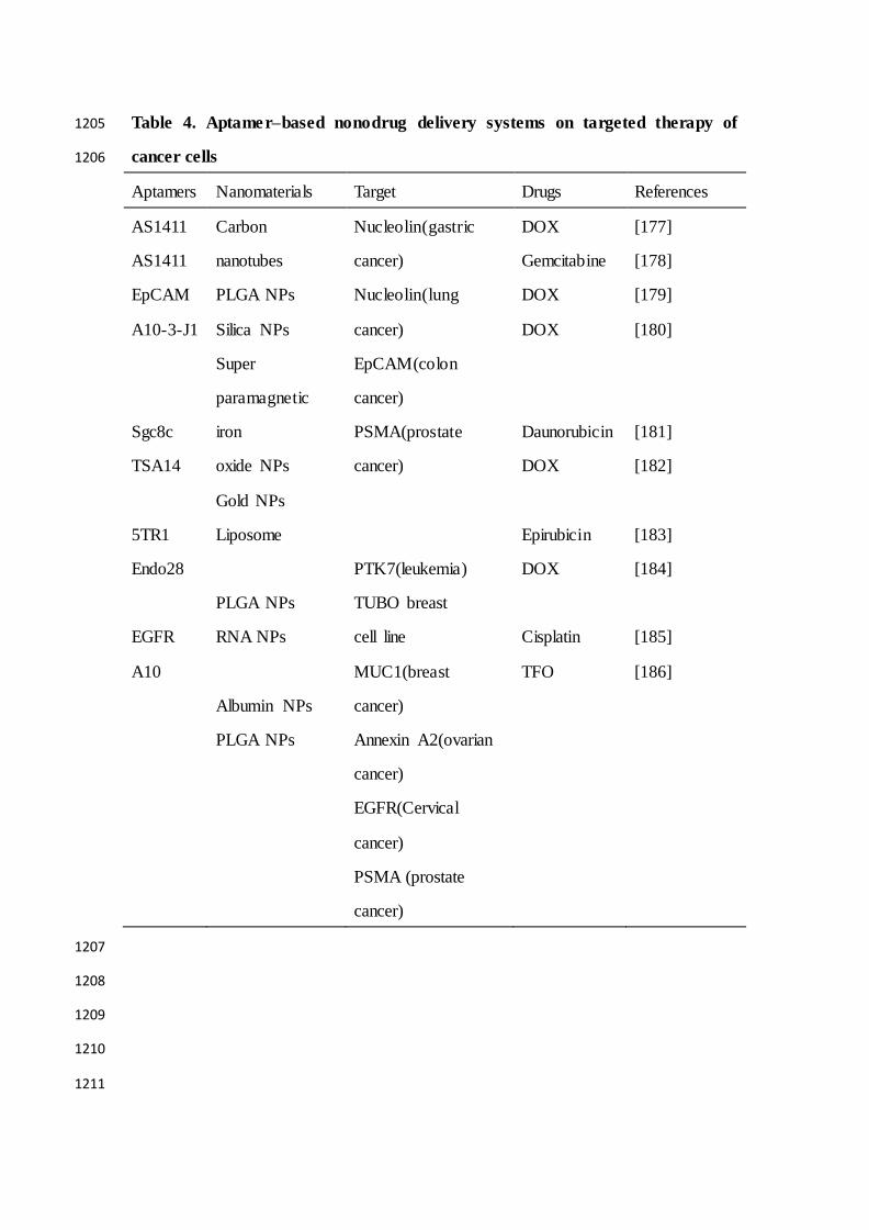
Table 4. Aptamer–based nonodrug delivery systems on targeted therapy of 1205
cancer cells 1206
Aptamers Nanomaterials Target Drugs References
AS1411
AS1411
EpCAM
A10-3-J1
Sgc8c
TSA14
5TR1
Endo28
EGFR
A10
Carbon
nanotubes
PLGA NPs
Silica NPs
Super
paramagnetic
iron
oxide NPs
Gold NPs
Liposome
PLGA NPs
RNA NPs
Albumin NPs
PLGA NPs
Nucleolin(gastric
cancer)
Nucleolin(lung
cancer)
EpCAM(colon
cancer)
PSMA(prostate
cancer)
PTK7(leukemia)
TUBO breast
cell line
MUC1(breast
cancer)
Annexin A2(ovarian
cancer)
EGFR(Cervical
cancer)
PSMA (prostate
cancer)
DOX
Gemcitabine
DOX
DOX
Daunorubicin
DOX
Epirubicin
DOX
Cisplatin
TFO
[177]
[178]
[179]
[180]
[181]
[182]
[183]
[184]
[185]
[186]
1207
1208
1209
1210
1211

Figures and figure legends 1212
1213
Figure 1. A schematic representation of the SELEX strategy of aptamer selection. 1214
The targets are used to screen and enrich the random DNA oligonucleotide library 1215
pools, and cloning and sequencing are performed to obtain the fittest aptamers. 1216
1217
1218
1219
1220
1221
1222

1223
Figure 2. Chemical modifications of aptamers. Figure represents the various 1224
modifications of aptamer to increase its stability and potential applications. 1225
1226
1227
1228
1229
1230

1231
Figure 3. Applications of aptamers-based for cancer therapy. The figure shows 1232
that in addition to being used as a therapeutic drug, aptamers can also be co ngujated 1233
with siRNA, chemotherapy agents and nanopaticles to kill cancer cells. 1234
1235
1236
1237
1238
Figure 4: Applications of aptamers in cancer immunotherapy. The figure shows 1239
that specific aptamers that target molecular markers on the surface of tumor cells, 1240
dendritic cells, T cells, natural killer cells and macrophage cells can be used for tumor 1241
immunotherapy. 1242
1243
1244
1245