Embed Size (px)
Citation preview

Application of Raman Spectroscopy for the Differentiation of Lipstick Traces
Fatma Salahioglu, Michael J. Went, Stuart J. Gibson
School of Physical Sciences, University of Kent, Canterbury, Kent, CT2 7NH, UK
Corresponding author. Tel +44 1227 823540; fax +44 1227 827558
E-mail address: [email protected]
Abstract
This study demonstrates that Raman spectroscopy is a valuable tool for discriminating
between lipstick samples under a range of forensically relevant situations. Trace amounts of
lipstick smears deposited on textile fibres, cigarette butts and paper tissues were analysed.
Differentiation of lipstick smears could be achieved with little or no interference from the
underlying medium. Lipstick smears on glass slides, cigarette butts and tissues could also be
analysed and identified in situ through evidence bags. Using a range of excitation frequencies
(473, 633 and 784 nm) was effective in overcoming problems with fluorescent lipstick
samples. The majority of the spectra of deposited lipstick samples remained unchanged over
a period of up to two years. In some of the aged lipstick spectra, the (C=C) band at 1655 cm-1
and the (=CH) band at 3011 cm-1
were found to decrease in intensity and disappear over time.
The use of chemometrics for the characterisation of large numbers of lipstick spectra was
explored. Thirty spectra each from ten different lipsticks were analysed by Principal
Components Analysis (PCA) and classified using the K-Nearest Neighbours (KNN)
classifier. Up to 98.7% correct classification was achieved. Spectra from trace amounts of
lipstick smears deposited on fibres were also analysed and classified using the same
technique. 100% correct classification of these samples was achieved.
Keywords: Raman spectroscopy, lipsticks, trace evidence, chemometrics, forensic
Introduction
Identifying and establishing of the source of trace evidence in forensic science is an important
task. There are many types of trace evidence that can be encountered by a forensic scientist at
a crime scene, and one of these is cosmetic evidence such as lipstick smears. Lipsticks can

easily be transferred and, like other forms of evidence, they can provide a link between a
suspect and a victim, as well as between individuals and a crime scene.
Several methods for the forensic examination of lipsticks have been reported in which the
sample masses are typically less than 1 mg. For example, one study considered 117 lipsticks
and found good discrimination could be obtained by first visually comparing their colours.1 If
the samples were found to be indistinguishable, energy dispersive X-ray analysis was carried
out. If no significant difference in elemental composition was observed, then the colour
additives were examined by thin-layer chromatography (TLC). Finally if discrimination was
still not observed the samples were analysed by high performance liquid chromatography
(HPLC). A further study involving over 300 lipstick samples found that TLC and gas
chromatography were suitable for the analysis, characterisation and discrimination of small
quantities of lipstick found in casework.2 The combination of microspectrophotometry and
scanning electron microscopy/energy dispersive spectroscopy has also been found to be an
effective combination for characterising colour and elemental composition.3 Elemental
analysis data can also be obtained from neutron activation analysis using γ-ray spectrometry.4
Another successful combination of techniques is fluorescence observation and purge-and-trap
gas chromatography.5
These techniques, however, have some disadvantages. Some involve human opinion (e.g.
microscopy) and some are destructive involving extraction processes. Ideally the analysis
should be performed non-destructively on trace amounts of samples with the minimum of
sample preparation and the avoidance of contamination. Raman spectroscopy is an easy,
rapid and non-destructive method which requires no sample preparation and can analyse
samples contained in evidence bags.6 The unique vibrational spectra of molecules result in a
high degree of confidence in identification. Surface enhanced resonance Raman spectroscopy
has been used for the in-situ characterisation of chromophores in six red lipstick smears on
glass and cotton surfaces7,8
and dispersive Raman spectroscopy gave chemical fingerprints
from four (damson, champagne, pink frost and mango) lipsticks.9
We have previously demonstrated that Raman spectroscopy is a valuable technique for the
identification and differentiation of a large range of lipstick samples under controlled
laboratory conditions.10
However forensic evidence is rarely found in ideal conditions. It is
often found in trace amounts on different substrates which have been exposed to variable
environmental conditions. In this paper we report the acquisition of Raman spectra from
lipstick smears deposited on a variety of surfaces such as cigarette butts, garments and tissue
paper. The effect of ageing on lipstick evidence and the use of chemometrics in the
classification of lipstick spectra are also reported.
Experimental
The experiments were carried out using a Horiba LabRAM-HR Raman spectrometer utilising
three different lasers operating at wavelengths of 473, 633 and 784 nm. The spectrometer
incorporated a Peltier cooled charge coupled device (CCD) detector which operated at -70
C. A ×50 objective lens was used giving a beam diameter of approximately 2 µm on the
sample. The spectrometer was calibrated at the start of each session against the silicon line at
520.6 cm-1
.
The laser power at the sample was 4.9 mW with the 473 nm laser, 1.7 mW with the 633 nm
laser and 20.1 mW with the 784 nm laser at full power (measured using an ASSY
LaserCheck handheld power meter, accurate to ±5%). Even though Raman spectroscopy is a
non-destructive technique, such powerful lasers focused onto the sample can increase the
chance of causing decomposition during the analysis. Decomposition was observed with
some of the lipstick smears; therefore, neutral density filters were used to decrease the laser
intensity at the sample. The diameter of the confocal aperture ("hole size") was adjusted
depending on the experiment. A smaller hole size meant sampling of a thinner layer whereas
a larger hole sampled a thicker area of the sample. Spectra were exported to Labspec version
5 for processing, analysis and presentation.
The variations in other parameters (such as accumulation times) used for each experiment are
given in the respective sections.
The lipsticks used were:

Bourjois: Docteur Glamour 15 'Fuchsia 0 bobo', Lovely Rouge 16 'Brique Exclusif', So
Rouge 32 'Fashion Rouge', Sweet Kiss 54 'Rouge Glamour' (purchased online at
www.cosmetics4less.net); Sweet Kiss 47 'Rose paré' (obtained from the local Boots store at
Canterbury, Kent, UK)
Barry M: Lip Paint No. 52, 53, 101, 121, 136, 140 and 144 (obtained from Boots)
Elizabeth Arden: Exceptional Lipstick 17 'Breathless' (purchased at
www.cosmetics4less.net)
Rimmel: Volume 080 'Screamer' (purchased at www.cosmetics4less.net); Colour Show Off
130 'Love Me', Lasting Finish 170 'Alarm', Lasting Finish 210 'Coral in Gold', Colour Show
Off 230 'Red Fever', 320 'Funtime Fuchsia', Moisture Renew 210 'Fancy', Lasting Finish 272
'Frosted' (obtained from Boots)
Revlon: Matte 004 'Pink About It', Matte 006 'Really Red', Matte 009 'Fabulous Fig',
Colorburst 025 'Carnation', Revlon Colorburst 075 'Peach', Super Lustrous Pearl 430
'Softsilver Rose', Super Lustrous Pearl 450 'Gentlemen Prefer Pink' (obtained at Boots) and
Absolutely Fabulous Lipcream 07 'Cherish' (purchased at www.cosmetics4less.net)
Max Factor: Hyperfull 210 'Vigorous' (purchased at www.cosmetics4less.net)
La Femme: 29 'Dream Rose' (purchased at www.cosmetics4less.net)
Results and Discussion
Effects of ageing on the Raman spectra of lipsticks
A systematic investigation of the effects of ageing on evidential material can shed light onto
how materials change over time, and allow any such changes to be quantified as a function of
time. In forensic science it is especially important to know whether a piece of evidence
undergoes changes over time, in order to be able to accurately analyse it and determine its
history. In the context of Raman spectroscopic analyses, some materials can degrade which
can cause some peaks to disappear from their Raman spectra. In the same way, the products
of degradation can cause new peaks to appear in the spectra.
The effects of ageing on 20 different lipsticks were investigated. Each lipstick was smeared
on a glass slide and left on a laboratory bench at room temperature, its spectrum being
recorded periodically. The samples were analysed using a ×50 objective lens and the 633 nm

laser with the confocal hole diameter being kept constant at 200 µm. Each sample was
analysed using the same parameters of fifteen accumulations and two seconds per
accumulation. The samples were not subjected to any special storage conditions and only the
effect of time on the samples was investigated.
The lipsticks used were: Bourjois (No.s 15, 16, 32, 47 and 54), Barry M (No.s 53, 101, 121,
136 and 140), Rimmel (No.s 080, 130, 170, 210 and 320) and Revlon (No.s 004, 07, 009, 430
and 450).
Fifteen out of the twenty analysed samples were found to give the same spectra when
analysed up to two years after the deposition of the sample. These were: Barry M No. 121,
Bourjois (No.s 15, 16, 32, 47, 54), Revlon (No.s 07, 009, 430, 450), and Rimmel (No.s 080,
130, 170, 320). However, for the lipsticks Barry M 53, Barry M 101, Barry M 136, Barry M
140 and Rimmel 210 some changes in the spectra were observed. In the spectra of these
lipsticks the peak observed at 1655 cm-1
decreased in intensity over time. The peak at 3011
cm-1
found in the spectra of Barry M 53, Barry M 101, Barry M 136 and Barry M 140 also
decreased in intensity and eventually disappeared within a few months (Fig. 1).
The weak peak at around 1655 cm-1
usually arises due to the C=C stretching in the fatty acids
that are found in the waxy composition of lipsticks. It has been shown that the C=C bond
undergoes oxidation over time which results in the decreased intensity of its Raman band.12
The weak band at around 3011 cm-1
arises from (=CH) vibrations which has also been shown
to disappear in the Raman spectra of aged lipids.12
It was found that it was only these two peaks that changed over time within the analysed set.
Therefore it is possible to identify and differentiate between lipstick smears even a year or
two years after the deposition of the smear since a great majority of the peaks remain
unchanged. Care must be taken when making comparisons between lipsticks that have peaks
at around 1655 and 3011 cm-1
, since these peaks were shown to change and eventually
disappear.
Effects of changing the excitation wavelength on fluorescent spectra
Lasers of different wavelengths can be used for the analysis of samples that are too
fluorescent when analysed with a certain excitation wavelength. Fluorescence occurs when
the excitation source has enough energy to cause electronic transition within the sample. The
problem can be avoided by selecting an excitation wavelength that has a lower energy than
any electronic transitions in the sample. For this purpose, lasers with longer wavelengths such
as those operating in the near-infrared region are generally preferred.13,14
In order to determine whether the use of different lasers could help analyse and differentiate
between fluorescent lipstick samples, three lipsticks with very fluorescent spectra and no
discernible peaks (when illuminated with a 633 nm laser) were chosen. These were Barry M
144, Elizabeth Arden 17 and Max Factor 210. Each sample was smeared on a glass slide and
analysed using the 473 nm (blue), 633 nm (red) and 784 nm (near-infrared) lasers with fifteen
accumulations for two seconds for each laser.
It was found that the lipsticks gave less fluorescent spectra when analysed using the 473 nm
laser. Each spectrum showed characteristic peaks between 900 and 1800 cm-1
, peaks due to
C-H vibrations between 2800 and 3000 cm-1
, and peaks due to the anatase form of titanium
dioxide (in most cases only the most intense anatase peak at 143 cm-1
was observable). In the
case of Elizabeth Arden 17 lipstick, the characteristic peaks between 1100 and 1700 cm-1
were identified as belonging to D&C Red No. 7 Calcium lake dye, which is commonly used
in the manufacture of lipsticks. This lipstick displayed strong fluorescence beyond 2000 cm-1
when analysed with the 473 nm laser, obscuring the C-H peaks (Fig. 2).
Lipsticks are complicated mixtures of components and it is often difficult to make peak
assignments. Components of such mixtures can be identified by comparing their spectra to
that of pure constituent compounds such as beeswax and a variety of dyes and pigments.
Previous work has reported assignments of peaks to some of the compounds commonly
found in lipsticks.10
Further identification of components and assignment of peaks have also
been published.11
Fluorescence due to the underlying glass was observed between 1200 and 2000 cm-1
when
the smears were analysed using the 784 nm laser.15
The C-H peaks between 2800 and 3000
cm-1
appeared very weak due to the insensitivity of the CCD detector at large shifts away
from the near-infrared excitation line.

Overall, it was possible to obtain spectra from the samples that were too fluorescent when
analysed with the red laser by using different wavelength lasers. The lipsticks analysed
displayed characteristic peaks that could be used to differentiate them from each other.
Detection of lipstick smears on textile fibres
Lipstick smears can easily be transferred by contact and one of the surfaces they can be
encountered on is textile materials, such as clothing and bedding. Raman spectroscopy is a
powerful technique for the detection and analysis of trace evidence materials on textile fibres,
such as drugs-of-abuse,16-18
explosives particle,19-20
and lipsticks.7 These types of trace
evidence materials can be analysed in situ without requiring any sample extraction or
preparation. Furthermore, confocal Raman microscopy can be used to avoid interference
from the fibre by selectively focusing on the sample surface and rejecting the signals that
arise from the material around and underneath.
For the purposes of this study a variety of different types and colours of textile fibres (in the
form of threads) were used. These were: DMC Laine Colbert Tapestry Wool (7785, yellow);
DMC Laine Colbert Tapestry Wool (7798, light blue); DMC Laine Colbert Tapestry Wool
(7309, very dark navy blue); DMC 25 Mouliné Spécial Cotton (307, yellow); DMC 25
Mouliné Spécial Cotton (912, green); DMC Mouliné Lin Linen Embroidery Floss (L778,
very light pink); and DMC Mouliné Lin Linen Embroidery Floss (L435, burnt orange).
The lipsticks used were La Femme 29, Revlon 07, Barry M 101 and Rimmel 230. These
lipsticks were chosen because of the similarities in their spectra: the majority of the peaks of
La Femme 29 and Rimmel 230 were common to both lipsticks, with a few clear differences
in terms of the presence, or absence, of peaks. The spectra for Barry M 101 and Revlon 07
also displayed peaks that were common to both lipsticks, but different from the other two
lipsticks. This helps determine whether lipsticks with similar spectra can still be
differentiated from each other when analysed on media such as textile fibres, where
interference from the medium (in the form of fluorescence, or peaks from the media's own
Raman spectrum) is highly probable.

The samples were prepared by very lightly dabbing the lipsticks onto the threads so that only
a trace amount was transferred. Each sample was analysed using the 473 and 633 nm lasers.
The confocal hole size was adjusted where necessary: if interference from the fibre was
encountered, the size of the hole was decreased. If there was little or no interference from the
fibre, larger hole sizes were used. The parameters used for each sample typically varied from
10 to 15 accumulations for 2 to 5 seconds with the 473 nm laser; and 15 to 25 accumulations
for 2 to 5 seconds with the 633 nm laser. The confocal hole sizes used ranged from 50 µm for
more fluorescent or damage-prone samples, to 300 µm for less fluorescent samples. Different
neutral density filters were used depending on sample fluorescence or degradation.
Each sample was analysed at five different points. Difficulties arising from the detection of
trace amounts of lipstick smear on fibres extended the overall time for the analyses.
Fluorescent interference from the fibres was observed in many cases which made it difficult
to obtain a spectrum of the lipstick, especially when the 633 nm laser was used. In many
cases more than five spectra per sample had to be recorded in order to obtain a good spectrum
of the lipstick with minimum interference from the fibre.
The lipsticks La Femme 29 and Rimmel 230 gave good signal-to-noise ratios when analysed
with the 633 nm laser. However, with the 473 nm laser the spectra started to fluoresce around
2000 cm-1
and the baseline intensity rose very quickly at higher Raman shifts, reaching very
high intensities at approximately 3100 cm-1
. This made the spectra effectively unusable
beyond 2000 cm-1
. As a result, the range to be analysed when the 473 nm laser was used
(with these two lipsticks) was decided to be 90 to 2000 cm-1
. The lipsticks Barry M 101 and
Revlon 07 gave very good signal-to-noise ratios when analysed using both lasers, so the
wavenumber range of 90 to 3100 cm-1
was used in both cases.
In general, all samples could readily be analysed and identified using the 473 nm laser (Fig.
3). The fibres gave characteristic peaks when analysed with this laser. However, these peaks
did not interfere with the lipstick spectra (except in the case of Revlon 07 lipstick deposited
on orange linen where some of the fibre peaks were observed in the lipstick spectrum).
The analysis was more difficult when the 633 nm laser was used. Smears deposited on yellow
wool and yellow cotton were found to be easiest to analyse using this excitation frequency.
Despite fluorescence observed with the orange linen, the lipstick smears deposited on this

fibre could still be analysed and identified (Fig. 4). It was more difficult, however, to obtain
spectra from lipsticks deposited on light pink linen and useful spectra could only be obtained
for the La Femme 29 and Rimmel 230 lipsticks. Due to intense fluorescence arising from
light blue wool, dark navy wool and green cotton, it was not possible to obtain spectra from
deposited lipstick smears using this excitation frequency.
In some cases, a decrease in the intensity of peaks between 1100 and 1800 cm-1
, as well as
the C-H peaks between 2800 and 3000 cm-1
was observed with the Revlon 07 lipstick. This
selective loss of peaks could have been due to the compounds giving rise to these peaks
getting soaked into the fibre. It could also be a result of some of the components of lipsticks
decomposing due to the heat energy from the laser. The reason this effect was not observed
when glass slides were used could be attributed to the higher thermal conductivity of glass
compared to that of textile fibres. Whereas the heat from the laser was dissipated more
efficiently by the underlying glass, this may not have been the case for the fibres, which
resulted in localised heating and a change in the form/composition of the smear.
Overall, it was found that the blue laser was more suitable for the analysis of trace amounts of
lipstick smears on fibres. The only disadvantage of the blue laser was found to be the very
intense fluorescence it caused between 2000 and 3100 cm-1
with two of the lipsticks, which
made it impossible to determine any spectral peaks within this region. However, the peaks of
interest were found between 90 and 1800 cm-1
, therefore this was not a major impediment.
Analyses with the red laser on the other hand were more difficult to perform due to intense
fluorescent interference from the fibres. The smears were analysed the best with this laser
when they were found on yellow fibres. The results are summarised in Table 1.
It was possible to differentiate between La Femme 29 and Rimmel 230 deposited on fibres.
When good spectra could be obtained using the red laser, differentiation between the two
lipsticks could be achieved (Fig. 5). In cases where the red laser could not be used to obtain
spectra from the smears, differentiation of the lipsticks using the blue laser could be achieved
(Fig. 6). It was also possible to differentiate between Barry M 101 and Revlon 07 using the
red laser when good lipstick spectra could be obtained with this laser (Fig. 7). In cases where
lipstick spectra with discernible peaks could not be obtained with the red laser, spectra
obtained using the blue laser were compared and differentiation between the two lipsticks
was achieved (Fig. 8).

Detection of lipstick smears on cigarette butts
One of the surfaces lipstick smears are commonly encountered on is cigarette butts, which
have been recognised as forensic evidence for a long time. A variety of evidential materials
can be found on a cigarette butt, such as saliva, which is used for DNA analysis,21
and
fingerprints.22
Lipstick is easily transferred onto the cigarette butt during smoking, leaving
another source of evidence on the butt. Analysis and identification of these smears can
therefore provide valuable information for use in a forensic investigation.
The lipsticks used were La Femme 29, Revlon 07, Barry M 101 and Rimmel 230. Volunteers
were asked to smoke cigarettes after putting on a given lipstick. The smoked cigarette butts
were then collected and analysed. Spectra of the lipstick smears on cigarette butts, as well as
that of 'blank' cigarette butts, were obtained and the results were compared. Each sample was
analysed at five different points using the 633 nm laser. The parameters used for each sample
ranged from 15 to 20 accumulations for 2 to 4 seconds and the confocal hole sizes ranged
from 100 to 200 µm
The filter of a cigarette consists of cellulose acetate fibres. Each fibre is treated with titanium
dioxide (whitening agent) and over 15000 fibres are packed tightly together to create a single
filter. This is then wrapped with paper and/or rayon wrapping, which is also treated with
chemicals such as glues and alkali metal salts.23-25
The analysis of cigarette butts using
Raman spectroscopy revealed that the most intense peaks found in their spectra were those of
titanium dioxide in anatase form. The strong presence of titanium dioxide in the spectra of
cigarette butts presented a challenge in terms of the analysis of lipsticks that had titanium
dioxide as the main peaks in their spectra, such as the Barry M 101 lipstick on a B&H Silver
cigarette butt (Fig. 9). In this case it was not possible to reach a valid conclusion in terms of
detection of this lipstick smear on the cigarette butt using Raman spectroscopy.
Revlon 07 also contained strong titanium dioxide peaks. However, it was still possible to
analyse and identify the lipstick smear because the spectrum of the cigarette butt used did not
display strong peaks. Even though it was possible to detect the lipstick peaks between 90 and
800 cm-1
, some of the characteristic peaks between 1000 and 1800 cm-1
, as well as the C-H
peaks between 2800 and 3000 cm-1
, could not be detected (Fig. 10). This selective loss of

peaks could be arising from some components decomposing, reacting with, or soaking into
the cigarette butt.
With the lipsticks La Femme 29 and Rimmel 230, the spectra of the cigarette butts did not
cause as much interference except for the strong anatase peak at 143 cm-1
that appeared in the
spectra of the smears. A comparison of the spectra of these two lipsticks on cigarette butts
showed that it was possible to differentiate between lipsticks with similar spectra even when
they are found on cigarette butts (Fig. 11).
Detection of lipstick smears on tissues
Tissues and handkerchiefs are frequently used in daily life. Lipstick smears can be transferred
onto tissues when the mouth is wiped onto the tissue after a meal or when tissues are used to
remove makeup. Therefore the identification and characterisation of lipstick smears on
tissues could potentially aid in a forensic investigation by linking the evidence to a suspect or
a victim.
The lipsticks used for this study were La Femme 29, Rimmel 230, Barry M 101 and Revlon
07. Each sample was prepared by lightly dabbing a piece of tissue (Tempo Petit Jasmine
Handkerchiefs) onto the lips with lipstick on. Spectra were taken from five different positions
on each sample using the 633 nm laser. Both the 'blank' tissue and the lipstick smear on the
tissue were analysed and compared. Each sample was analysed using 20 accumulations for 3
to 4 seconds, with the confocal hole sizes of either 100 or 200 µm being employed when
necessary.
The Raman spectrum of the blank tissue displayed peaks that arose from cellulose found in
the makeup of the tissue. The characteristic peaks of cellulose are found at 380, 1098 and
2900 cm-1
.26
Despite some interference from the tissue, Raman spectra of the lipstick smears
could still be obtained. La Femme 29 and Rimmel 230 lipsticks were easier to analyse on the
tissues and they produced spectra without interference from the underlying tissue. It was
possible to differentiate between the two lipsticks deposited on tissues using Raman
spectroscopy (Fig. 12).

The Barry M 101 lipstick could also be analysed without much interference from the tissue.
However, it was more difficult to obtain a good spectrum of Revlon 07 on the tissue. Some of
the characteristic peaks of this lipstick between 1100 and 1700 cm-1
, and the C-H bond
vibrations between 2800 and 3000 cm-1
could not be detected when the smear deposited on
the tissue was analysed. Despite these difficulties, it was possible to differentiate between the
two lipsticks deposited on tissues using Raman spectroscopy (Fig. 13).
Analysis of lipstick smears through evidence bags
In forensic science, it is very important to preserve the integrity of evidential material.
Recovered evidence is stored in evidence bags to allow secure transit and to preserve the
chain of custody. It is therefore extremely valuable to be able to analyse the evidence without
removing it from the evidence bag in order to minimise the risk of contamination. Raman
spectroscopy has been successfully used for the analysis of samples contained within
packaging materials and evidence bags that are transparent to the laser.6,27-29
The evidence bag used in this study was a polyethylene Tek-Niche Police Evidence Bag and
the lipstick used was La Femme 29. The samples prepared and analysed were: La Femme 29
lipstick smear on a glass slide, La Femme 29 lipstick smear on a Pall Mall cigarette butt, and
La Femme 29 lipstick smear on a piece of Tempo Petit Jasmine Handkerchiefs tissue. Each
sample was placed in the evidence bag and analysed under the Raman microscope without
any sample preparation or treatment. Each sample was analysed using the 633 nm laser and a
×50 objective lens. Five spectra were obtained from different parts of each sample. The
parameters used for each sample ranged from 20 to 25 accumulations for 4 to 5 seconds, with
a confocal hole size of either 100 or 200 µm depending on the sample.
The polyethylene evidence bag displayed strong Raman peaks. However, this could mostly
be avoided by ensuring that the laser was properly focused on the sample rather than the
evidence bag. The laser was focused through the clear part of the evidence bag.
Figure 14 compares the spectrum of La Femme 29 on glass slide with and without the
evidence bag. The lipstick smear could easily be detected and identified through the evidence
bag without interference from the polyethylene peaks. However, during the analysis the
evidence bag frequently came in contact with the cigarette butt. Therefore, the spectra

obtained of the lipstick smear had some interference from the evidence bag (as indicated in
Fig. 15). Despite this, it was still possible to analyse and identify the lipstick smear. When the
smear on a tissue was analysed through the evidence bag, the Raman signals from the lipstick
were found to be less intense. However, a comparison with the spectrum of lipstick on the
glass slide showed that the smear on the tissue could still be identified as La Femme 29 (Fig.
16).
Use of chemometrics for the characterisation of lipstick spectra
Modern Raman spectrometers can acquire a large number of high quality spectra very
quickly, decreasing the analysis time and leading to fast identification of samples.
Chemometric methods offer an efficient approach to the analysis of large datasets. Studies on
the application of chemometric methods such as PCA to the analysis of spectra include
discrimination of carbon nanotubes,30
detection of counterfeit medicines,31
discrimination
between authentic and counterfeit banknotes,32
quantitative analysis of narcotics in
mixtures,33-34
discrimination between synthetic and natural artist's pigments35
as well as
historic pigments used in ancient paintings,36
and forensic analysis of paint samples37
and
lipsticks.38
The lipstick data was pretreated to reduce noise and remove the baseline, and then PCA was
applied to reduce the dimensionality. The data were subsequently used to construct a simple
k-nearest neighbours (kNN) classifier. Our analysis was implemented in MATLAB R2012a
using the Statistics Toolbox.
The classifier was trained on ten different lipsticks of various colours and brands: Barry M
No.s 52, 53, 121; La Femme 29; Rimmel No.s 210, 272; Revlon No.s 006, 025, 075 and 07.
Each lipstick was smeared onto a separate glass slide and analysed using a Horiba LabRAM-
HR Raman spectrometer using fifteen accumulations over a period of two seconds per
spectrum. A ×50 objective lens and an excitation wavelength of 633 nm were employed. For
each lipstick, spectra were obtained over the full range (90 – 3100 cm-1
) from thirty different
positions on the smear, giving a total of 300 spectra. In some cases a very intense peak was
observed at 143 cm-1
due to the presence of titanium dioxide (in anatase form) which
dominated the spectrum and made characterisation difficult. Therefore, the anatase peak was
disregarded and spectra were analysed over the range 180 – 3100 cm-1
instead. High

frequency noise was removed using a moving average filter of width 20 cm-1
. Each spectrum
was then down-sampled to include 5280 equally spaced data points. In most spectra the data
exhibited curved fluorescence baselines that could not be removed effectively by calculating
a spectrum’s derivative. Instead, we chose to remove the baseline by subtracting a 4th
order
polynomial fit from each spectrum. This was achieved in computer software without human
intervention. Each spectrum was then normalised using the standard normal variate method
and then inserted as a column in a data matrix. To reduce the dimensionality of the data and
consequently aid interpretation, a principal components analysis was performed by
determining the singular value decomposition of the data matrix. A scree plot was used to
indicate the most descriptive principal components. Ten principal components were retained
after following the standard practice of truncating at the elbow of the scree plot. Hence the
dimensionality of the data was reduced from 5280 to 10 dimensions.
A leave one out cross validation (LOO CV) was performed on the training data using kNN
with k = 5, k = 7, k = 9, k = 11, k = 15 resulting in correct classification rates of 95.3%,
97.6%, 98.0%, 98.3% and 98.7% respectively.
The non-destructive nature of Raman spectroscopy is ideally suited to analysing substances
on evidential materials such as clothing fibres. Accordingly, two lipsticks, chosen at random,
were smeared on a variety of different fibres and 13 spectra were obtained using the same
method as before. The following fibre/lipstick combinations were considered:
La Femme 29 smear on orange linen (×2 spectra)
La Femme 29 smear on pink linen (×3 spectra)
La Femme 29 smear on yellow cotton (×3 spectra)
La Femme 29 smear on yellow wool (×3 spectra)
Revlon 07 on orange linen (1 spectrum)
Revlon 07 on yellow cotton (1 spectrum)
These ‘evidential’ test samples were pretreated and included in the PCA model. Application
of the kNN classifier (k = 15), based on the 300 training samples (anatase peak removed),
resulted in 100% correct classification of the test spectra.
Conclusions

Overall it can be stated that lipstick traces on various substrates can be differentiated non-
destructively by Raman spectroscopy using excitation wavelengths at 784, 473 and 633 nm.
Fluorescence problems were mainly encountered with the 633 nm laser, thus the
recommended approach is using 784 and/or 473nm lasers.
Effects of ageing on the spectra of lipsticks were investigated. It was found that the spectra of
15 out of the 20 samples analysed did not change over the course of up to two years. In the
spectra of five of the lipsticks analysed, changes were observed in terms of the intensity of
peaks found at 1655 and 3011 cm-1
, which were attributed to C=C and (=CH) vibrations of
fatty acids (found in the waxy composition of lipsticks) respectively. It was determined that
these peaks decreased in intensity and disappeared over the course of a few months.
Raman spectroscopy was applied to the detection and analysis of trace amounts of lipstick
smears on textile fibres. It was found that the analyses were more difficult with the 633 nm
laser due to intense fluorescent interference from the fibres. However, almost all of the
samples could be analysed and differentiated from each other using the 473 nm laser. It was
determined that each sample could easily be analysed and differentiated from each other by
using both lasers together. Therefore, Raman spectroscopy was found to be an effective tool
for the analysis of trace amounts of lipsticks on fibres.
Lipstick smears on smoked cigarette butts were also analysed using Raman spectroscopy.
The presence of titanium dioxide peaks in the spectra of cigarette butts presented a challenge
during the analysis of lipsticks with strong titanium dioxide peaks. However, it was possible
to identify and differentiate between lipstick smears that did not have titanium dioxide peaks
as the main peaks in their spectra. Therefore Raman spectroscopy was found to be effective
for the analysis of lipstick smears on cigarette butts, where either (a) the lipstick spectrum
does not contain strong titanium dioxide peaks, or (b) the cigarette butt spectrum does not
have strong titanium dioxide peaks.
Lipstick smears deposited on tissues were analysed using Raman spectroscopy. The smears
could be identified and differentiated from each other with minimum interference from the
tissue. Therefore Raman spectroscopy was found to be effective for the analysis and
discrimination of trace amounts of lipsticks deposited on tissues.

Applicability of Raman spectroscopy to the analysis of lipstick smears deposited on glass
slides, cigarette butts and tissues through evidence bags was investigated. It was found that
Raman spectroscopy could be used to obtain spectra of the lipstick smears deposited on a
variety of surfaces even through evidence bags, with little or no interference from the bag.
Use of chemometrics for the characterisation of large numbers of lipstick spectra was
explored. It was found that Principal Components Analysis (PCA) was an effective technique
for reducing the dimensionality of data while preserving the variance within the data. The
spectra were classified using the K-Nearest Neighbours (KNN) method. Up to 98.7% correct
classification was achieved. Spectra from trace amounts of lipstick smears deposited on fibres
were also analysed and classified using the same technique. 100% correct classification of
these samples was achieved. This demonstrated that Raman spectroscopy combined with
PCA could be a potentially powerful forensic technique for the characterisation and
identification of lipstick smears recovered from crime scenes.
References
(1) J. Andrasko, Forensic Sci. Int., 1981, 17, 235-251.
(2) L.W. Russell and A.E. Welch, Forensic Sci. Int., 1984, 25, 105-116.
(3) M.Y. Choudhry, J. Forensic Sci., 1991, 36, 366-375.
(4) G. Misra and V. K. Mittal, J. Appl. Spectrosc., 2004, 71, 270-274
(5) Y. Ehara and Y. Marumo, Forensic Sci. Int., 1998, 96, 1-10.
(6) M. J. West and M. J. Went, Forensic Sci. Int., 2008, 174, 1-5.
(7) C. Rodger, V. Rutherford, D. Broughton, P. C. White and W. E. Smith, Analyst, 1998,
123, 1823-1826.
(8) P.C. White, Science & Justice, 2000, 40, 113-119.
(9) R. Goodacre, N. Kaderbhai, A.C. McGovern and E.A. Goodacre, Kohonen Maps,
1999, 335-347.
(10) F. Salahioglu and M. J. Went, Forensic Sci. Int., 2012, 223, 148-152.
(11) F. Salahioglu, PhD Thesis, University of Kent, 2013(12) M. S. Maier, D. L. A. de
Faria, M. T. Boschín and S. D. Parera, ARKIVOC, 2005, 12, 311-318.
(13) B. Schrader, A. Hoffmann and S. Keller, Spectrochim. Acta, Part A, 1991, 47, 1135-
1148.
(14) F. C. Thorley, K. J. Baldwin, D. C. Lee and D. N. Batchelder, J. Raman Spectrosc.,
2006, 37, 335-341.
(15) HORIBA Scientific Application Note, RA19, Raman.
(16) E. M. A. Ali, H. G. M. Edwards, M. D. Hargreaves and I. J. Scowen, J. Raman
Spectrosc., 2010, 41, 938-943.
(17) E. M. A. Ali, H. G. M. Edwards, M. D. Hargreaves and I. J. Scowen, Anal. Chim.
Acta, 2008, 615, 63-72.
(18) M. J. West and M. J. Went, Forensic Sci. Int., 2009, 189, 100-103.
(19) E. M. A. Ali, H. G. M. Edwards and I. J. Scowen, Talanta, 2009, 78, 1201-1203.
(20) E. M. A. Ali, H. G. M. Edwards and I. J. Scowen, J. Raman Spectrosc., 2009, 40,
2009-2014 (DOI:10.1002/jrs.2360).
(21) S. A. Montpetit, I. T. Fitch and P. T. O’Donnell, J. Forensic Sci., 2005, 50, 1-9.
(22) N. J. Crane, E. G. Bartick, R. S. Perlman and S. Huffman, J. Forensic Sci., 2007, 52,
48-53.
(23) E. Slaughter, R. M. Gersberg, K. Watanabe, J. Rudolph, C. Stransky and T. E.
Novotny, Tobacco Control, 2011, 20, i25-i29.

(24) E. Slaughter, Toxicity of cigarette butts and their chemical components to the marine
and freshwater fishes, atherinops affinis and pimephales promelas, 2010.
(25) O. Geiss and D. Kotzias, Tobacco, Cigarettes and Cigarette Smoke: An Overview,
European Communities, Italy, 2007.
(26) U. P. Agarwal, Planta, 2006, 224, 1141-1153.
(27) A. K. Deisingh, Analyst, 2005, 130, 271-279.
(28) C. Eliasson and P. Matousek, Anal. Chem., 2007, 79, 1696-1701.
(29) M. J. West and M. J. Went, Spectrochim. Acta, Part A, 2009, 71, 1984-1988.
(30) R. Y. Sato-Berru, E. V. Basiuk and J. M. Saniger, J. Raman Spectrosc., 2006, 37,
1302-1306.
(31) K. Dégardin, Y. Roggo, F. Been and P. Margot, Anal. Chim. Acta, 2011, 705, 334-
341.
(32) M. R. de Almeida, D. N. Correa, W. F. C. Rocha, F. J. O. Scafi and R. J. Poppi,
Microchem. J., 2012, (in press).
(33) A. G. Ryder, G. M. O'Connor and T. J. Glynn, J. Forensic Sci., 1999, 44, 1013-1019.
(34) A. G. Ryder, G. M. O'Connor and T. J. Glynn, J. Raman Spectrosc., 2000, 31, 221-
227.
(35) P. Vandenabeele and L. Moens, Analyst, 2003, 128, 187-193.
(36) N. Navas, J. Romero-Pastor, E. Manzano and C. Cardell, J. Raman Spectrosc., 2010,
41, 1486-1493.
(37) C. Muehlethaler, G. Massonnet and P. Esseiva, Forensic Sci. Int., 2011, 209, 173-182.
(38) R. Goodacre, N. Kaderbhai, A. C. McGovern and E. A. Goodacre, in Kohonen Maps,
ed. E. Oja and S. Kaski, Elsevier Science B.V., Netherlands, 1999, p. 335-347.
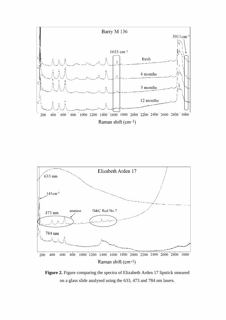
Figure 2. Figure comparing the spectra of Elizabeth Arden 17 lipstick smeared
on a glass slide analysed using the 633, 473 and 784 nm lasers.
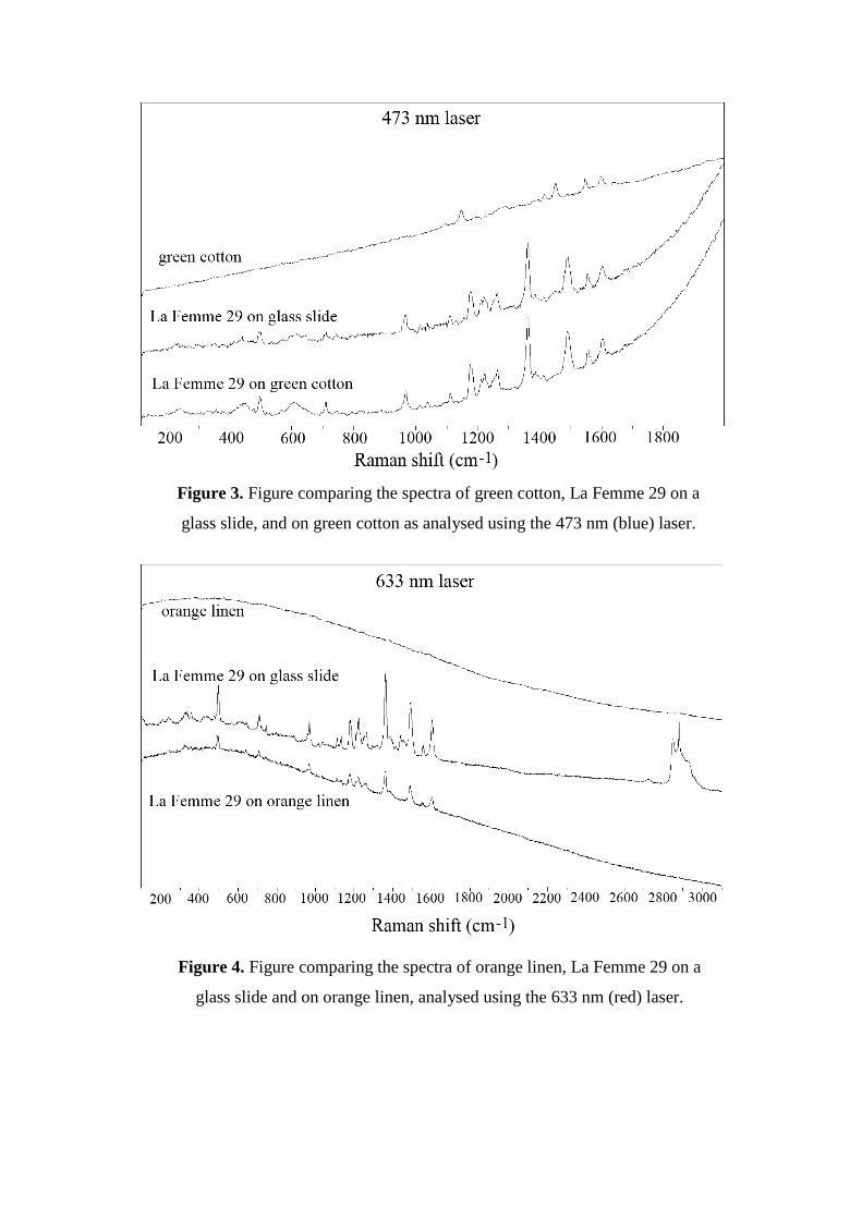
Figure 3. Figure comparing the spectra of green cotton, La Femme 29 on a
glass slide, and on green cotton as analysed using the 473 nm (blue) laser.
Figure 4. Figure comparing the spectra of orange linen, La Femme 29 on a
glass slide and on orange linen, analysed using the 633 nm (red) laser.

Figure 5. Figure comparing the spectra of Rimmel 230 and La Femme 29 on
yellow cotton, analysed using the 633 nm (red) laser. The main points of
difference between the two spectra are marked. It was possible to differentiate
between the two lipstick smears.
Figure 6. Figure comparing the spectra of La Femme 29 and Rimmel 230 on
dark navy wool, analysed using the 473 nm (blue) laser. The main points of
difference between the two spectra are marked.

Figure 7. Figure comparing the spectra of Barry M 101 and Revlon 07 on
yellow cotton, analysed using the 633 nm (red) laser. The main points of
difference between the two spectra are marked.
Figure 8. Figure comparing the spectra of Revlon 07 and Barry M 101 on dark
navy wool, analysed using the 473 nm (blue) laser. The main points of
difference between the two spectra are marked.

Figure 9. Figure comparing the spectra of B&H Silver cigarette butt, Barry M
101 on the cigarette butt and on glass slide analysed using the 633 nm (red)
laser.
Figure 10. Figure comparing the spectra of Pall Mall cigarette butt, Revlon 07
on the cigarette butt and on glass slide analysed using the 633 nm (blue) laser.

Figure 11. Figure comparing the spectra of Rimmel 230 and La Femme 29 on
cigarette butts analysed using the 633 nm (red) laser. The main points of
difference between the two lipsticks are highlighted in the boxes.
Figure 12. Figure comparing the spectra of La Femme 29 and Rimmel 230 on
tissues analysed using the 633 nm (red) laser. The main points of difference
between the two lipstick spectra are indicated.

Figure 13. Figure comparing the spectra for Revlon 07 and Barry M 101 on the
tissue analysed using the 633 nm (red) laser. One of the main points of
difference between the two spectra is indicated.
Figure 14. (a) Spectrum of the evidence bag (polyethylene) (b) Spectrum of La
Femme 29 on glass slide through the evidence bag (c) Spectrum of La Femme
29 on glass slide without the evidence bag (633 nm laser). Guide lines indicate
the positions of major evidence bag peaks.
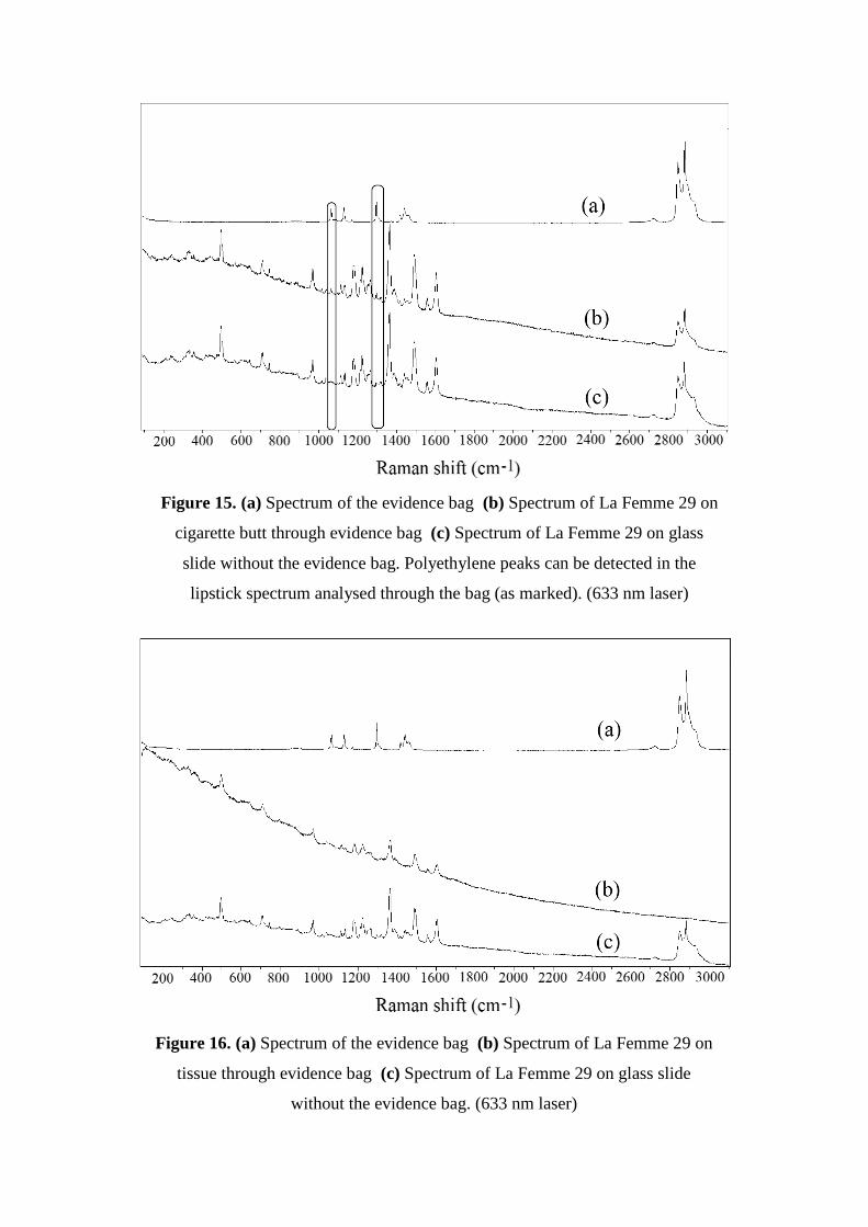
Figure 15. (a) Spectrum of the evidence bag (b) Spectrum of La Femme 29 on
cigarette butt through evidence bag (c) Spectrum of La Femme 29 on glass
slide without the evidence bag. Polyethylene peaks can be detected in the
lipstick spectrum analysed through the bag (as marked). (633 nm laser)
Figure 16. (a) Spectrum of the evidence bag (b) Spectrum of La Femme 29 on
tissue through evidence bag (c) Spectrum of La Femme 29 on glass slide
without the evidence bag. (633 nm laser)

473 nm Laser
yellow wool
light blue wool
dark navy wool
yellow cotton
green cotton
light pink linen
orange linen
La Femme 29
Rimmel 230
Barry M 101
Revlon 07 *
633 nm Laser
yellow wool
light blue wool
dark navy wool
yellow cotton
green cotton
light pink linen
orange linen
La Femme 29
Rimmel 230 *
Barry M 101
Revlon 07 *
Table 1. A '' indicates that the smear could be analysed and identified on the
corresponding fibre with the laser used. A '' indicates that the smear could not
be analysed or identified. A '*' indicates that the smear could sometimes be
identified, or identified to a certain degree (i.e. could not detect all of the peaks
in the spectrum).