Embed Size (px)
Citation preview
Interview
Applying the Density Gradient Ultracentrifugal Isolation Method in
Exosome Subclass Analysis
Dr. Kiyotaka ShibaDivision Head, Division of Protein
Engineering, Cancer Institute, Japanese
Foundation for Cancer Research
Dr. Satoshi Yamamoto
Research Scholar, Division of Protein
Engineering, Cancer Institute, Japanese
Foundation for Cancer Research
Tokyo Dental College
Department of Oral Implantology
Dr. Kazuya Iwai Research Scholar, Division of Protein
Engineering, Cancer Institute, Japanese
Foundation for Cancer Research
Tokyo Dental College
Department of Oral Implantology
11 Introduction
Interview
Applying the Density Gradient Ultracentrifugal Isolation Method in
Exosome Subclass Analysis
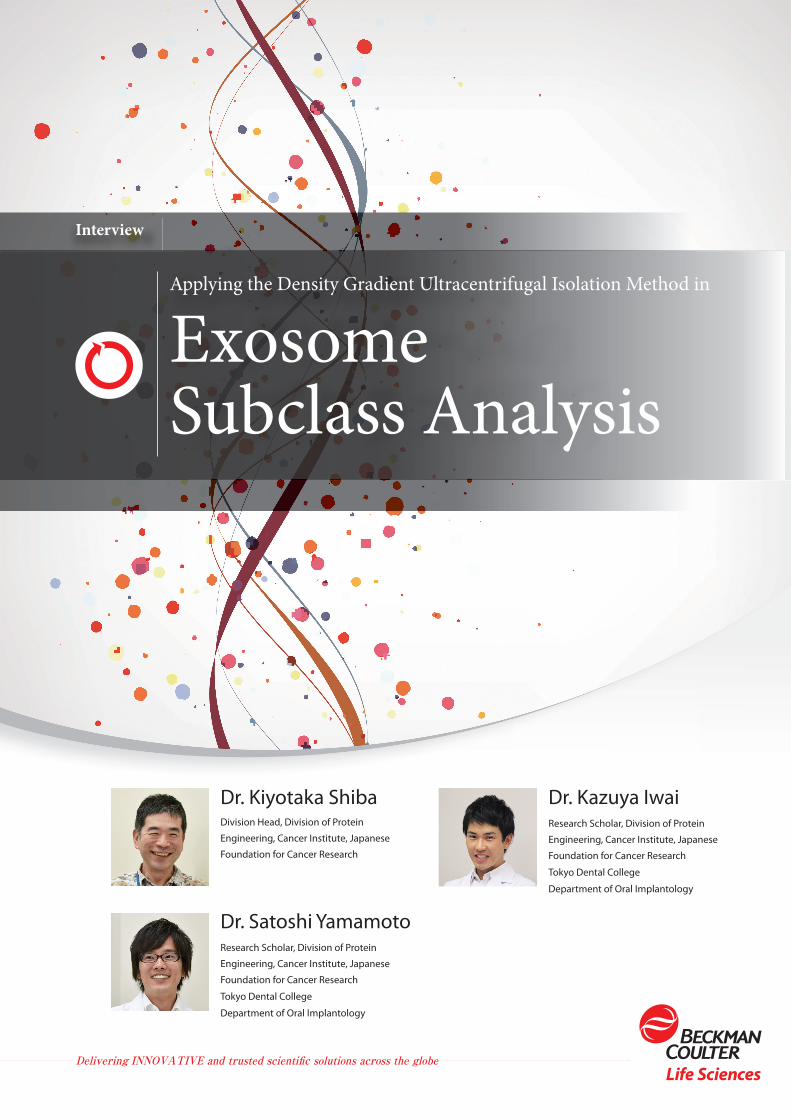
Exosomes are extracellular vesicles that have attracted attention since the late 2000s as communication agents between cells,
as well as potential diagnostic/therapeutic tools for various disorders. Much effort has been put into research on their secretion/
transport modality, the substance contained within and other characteristics. The reality, however, is that there is no consensus on
the fundamental question, "What are exosomes?" In order to define exosomes, research on their isolation method is essential.
Dr. Kiyotaka Shiba conducts research focusing
on exosome and leads the Protein Engineering
Division at the Cancer Institute of the Japanese
Foundation for Cancer Research. Along with
research scholars at the institute, Dr. Kazuya Iwai
and Dr. Satoshi Yamamoto, from the Department
of Oral Implantology at the Tokyo Dental College,
Dr. Shiba discusses subclass analysis based on
exosome density, as well as purification methods
and cautions.
2
CONTENTS
P.3 On exosome research
P.3 Density-based exosome subclass analysis
P.4 Exosome purification method and cautions in using the density gradient ultracentrifugal isolation approach
P.6 Handling purified exosomes
P.6 Final remarks
Dr. Kiyotaka ShibaDivision Head, Division of Protein Engineering, Cancer Institute, Japanese Foundation for Cancer Research
Profile
1981 • Graduated from the Faculty of Science, Kyoto University
1986 • Completed the second half of the doctoral program at the Graduate School of Science, Kyoto University; obtained a Ph.D. in Science
1986 • Instructor, National Institute of Multimedia Education Development Center
1987 • Assistant (RNA molecular biology), Department of Biophysics and Biochemistry, Faculty of Science, University of Tokyo
1989 • Doctoral fellow (Protein engineering), Department of Biology, Massachusetts Institute of Technology
1991 • Researcher, Division of Cell Biology, Cancer Institute, Japanese Foundation for Cancer Research
2001 • Division Head, Division of Protein Engineering, Cancer Institute, Japanese Foundation for Cancer Research (to the present)
Dr. Kazuya Iwai Research scholar, Division of Protein Engineering, Cancer Institute, Japanese
Foundation for Cancer Research Department of Oral Implantology, Tokyo Dental
College
Profile
2011 • Graduated from the Tokyo Dental College
2012 • Completed the clinical training in dentistry at the Tokyo Dental College
2012 • Graduate School of Dental Research (Oral Implantation), Tokyo Dental College
2012 • (Research scholar, Division of Protein Engineering) Cancer Institute, Japanese Foundation for Cancer Research
2016 • Completed the doctoral program at the Graduate School of Dental Research, Tokyo Dental College; Ph.D. (Dentistry)
2016 • (Resident, Division of Oral Implantation) Tokyo Dental College Suidobashi Hospital (to the present)
Dr. Satoshi YamamotoResearch scholar, Division of Protein Engineering, Cancer Institute, Japanese
Foundation for Cancer Research Department of Oral Implantology, Tokyo Dental
College
Profile
2013 • Graduated from the Tokyo Dental College
2014 • Completed the clinical training in dentistry at the Tokyo Dental College
2014 • Graduate School of Dental Research (Oral Implantation), Tokyo Dental College
2014 • (Research scholar, Division of Protein Engineering) Cancer Institute, Japanese Foundation for Cancer Research (to the present)
3 Applying the Density Gradient Ultracentrifugal Isolation Method in Exosome Subclass Analysis
Interview
Applying the Density Gradient Ultracentrifugal Isolation Method for Exosome Subclass Analysis
On Exosome Research
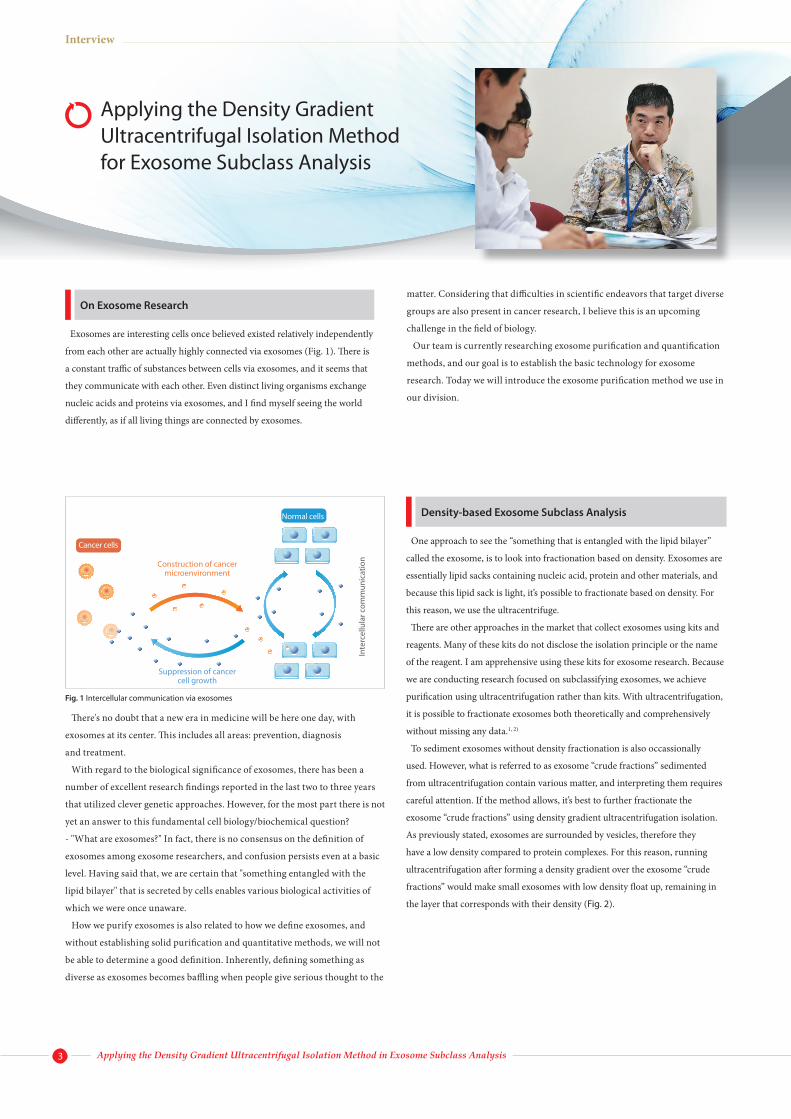
Exosomes are interesting cells once believed existed relatively independently
from each other are actually highly connected via exosomes (Fig. 1). There is
a constant traffic of substances between cells via exosomes, and it seems that
they communicate with each other. Even distinct living organisms exchange
nucleic acids and proteins via exosomes, and I find myself seeing the world
differently, as if all living things are connected by exosomes.
There's no doubt that a new era in medicine will be here one day, with
exosomes at its center. This includes all areas: prevention, diagnosis
and treatment.
With regard to the biological significance of exosomes, there has been a
number of excellent research findings reported in the last two to three years
that utilized clever genetic approaches. However, for the most part there is not
yet an answer to this fundamental cell biology/biochemical question?
- "What are exosomes?" In fact, there is no consensus on the definition of
exosomes among exosome researchers, and confusion persists even at a basic
level. Having said that, we are certain that "something entangled with the
lipid bilayer" that is secreted by cells enables various biological activities of
which we were once unaware.
How we purify exosomes is also related to how we define exosomes, and
without establishing solid purification and quantitative methods, we will not
be able to determine a good definition. Inherently, defining something as
diverse as exosomes becomes baffling when people give serious thought to the
matter. Considering that difficulties in scientific endeavors that target diverse
groups are also present in cancer research, I believe this is an upcoming
challenge in the field of biology.
Our team is currently researching exosome purification and quantification
methods, and our goal is to establish the basic technology for exosome
research. Today we will introduce the exosome purification method we use in
our division.
Density-based Exosome Subclass Analysis
One approach to see the “something that is entangled with the lipid bilayer”
called the exosome, is to look into fractionation based on density. Exosomes are
essentially lipid sacks containing nucleic acid, protein and other materials, and
because this lipid sack is light, it’s possible to fractionate based on density. For
this reason, we use the ultracentrifuge.
There are other approaches in the market that collect exosomes using kits and
reagents. Many of these kits do not disclose the isolation principle or the name
of the reagent. I am apprehensive using these kits for exosome research. Because
we are conducting research focused on subclassifying exosomes, we achieve
purification using ultracentrifugation rather than kits. With ultracentrifugation,
it is possible to fractionate exosomes both theoretically and comprehensively
without missing any data.1, 2)
To sediment exosomes without density fractionation is also occassionally
used. However, what is referred to as exosome “crude fractions” sedimented
from ultracentrifugation contain various matter, and interpreting them requires
careful attention. If the method allows, it’s best to further fractionate the
exosome “crude fractions” using density gradient ultracentrifugation isolation.
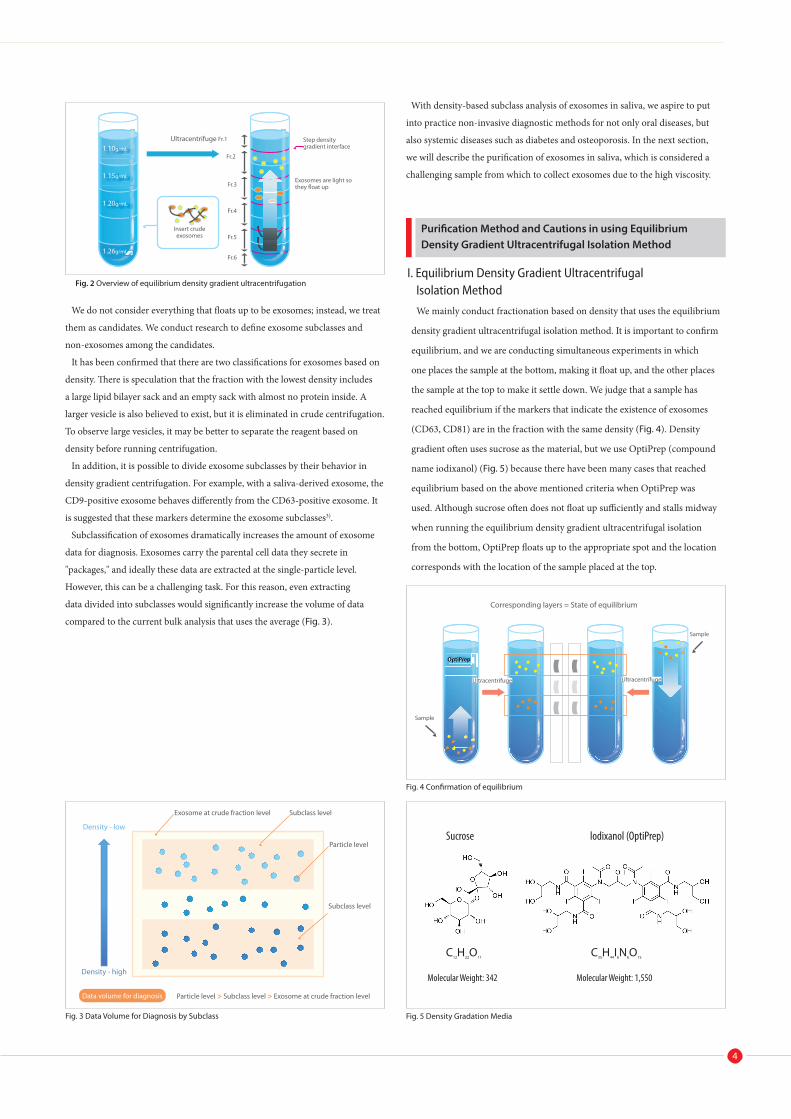
As previously stated, exosomes are surrounded by vesicles, therefore they
have a low density compared to protein complexes. For this reason, running
ultracentrifugation after forming a density gradient over the exosome “crude
fractions” would make small exosomes with low density float up, remaining in
the layer that corresponds with their density (Fig. 2).
Cancer cells
Construction of cancer microenvironment
Suppression of cancer cell growth
Normal cells
Inte
rcel
lula
r com
mun
icat
ion
Fig. 1 Intercellular communication via exosomes
Data volume for diagnosis
Purification Method and Cautions in using Equilibrium Density Gradient Ultracentrifugal Isolation Method
1.10g/mL
1.15g/mL
1.20g/mL
1.26g/mL
Ultracentrifuge Fr.1
Fr.2
Insert crude exosomes
Fr.3
Fr.4
Fr.5
Fr.6
Step density gradient interface
Exosomes are light so they float up
Sample
OptiPrep
Ultracentrifuge
Corresponding layers = State of equilibrium
Sample
We do not consider everything that floats up to be exosomes; instead, we treat
them as candidates. We conduct research to define exosome subclasses and
non-exosomes among the candidates.
It has been confirmed that there are two classifications for exosomes based on
density. There is speculation that the fraction with the lowest density includes
a large lipid bilayer sack and an empty sack with almost no protein inside. A
larger vesicle is also believed to exist, but it is eliminated in crude centrifugation.
To observe large vesicles, it may be better to separate the reagent based on
density before running centrifugation.
In addition, it is possible to divide exosome subclasses by their behavior in
density gradient centrifugation. For example, with a saliva-derived exosome, the
CD9-positive exosome behaves differently from the CD63-positive exosome. It
is suggested that these markers determine the exosome subclasses3).
Subclassification of exosomes dramatically increases the amount of exosome
data for diagnosis. Exosomes carry the parental cell data they secrete in
"packages," and ideally these data are extracted at the single-particle level.
However, this can be a challenging task. For this reason, even extracting
data divided into subclasses would significantly increase the volume of data
compared to the current bulk analysis that uses the average (Fig. 3).
Density - low
Density - high
Exosome at crude fraction level Subclass level
Particle level
Subclass level
Particle level > Subclass level > Exosome at crude fraction level
Fig. 3 Data Volume for Diagnosis by Subclass
With density-based subclass analysis of exosomes in saliva, we aspire to put
into practice non-invasive diagnostic methods for not only oral diseases, but
also systemic diseases such as diabetes and osteoporosis. In the next section,
we will describe the purification of exosomes in saliva, which is considered a
challenging sample from which to collect exosomes due to the high viscosity.
I. Equilibrium Density Gradient Ultracentrifugal Isolation Method
We mainly conduct fractionation based on density that uses the equilibrium
density gradient ultracentrifugal isolation method. It is important to confirm
equilibrium, and we are conducting simultaneous experiments in which
one places the sample at the bottom, making it float up, and the other places
the sample at the top to make it settle down. We judge that a sample has
reached equilibrium if the markers that indicate the existence of exosomes
(CD63, CD81) are in the fraction with the same density (Fig. 4). Density
gradient often uses sucrose as the material, but we use OptiPrep (compound
name iodixanol) (Fig. 5) because there have been many cases that reached
equilibrium based on the above mentioned criteria when OptiPrep was
used. Although sucrose often does not float up sufficiently and stalls midway
when running the equilibrium density gradient ultracentrifugal isolation
from the bottom, OptiPrep floats up to the appropriate spot and the location
corresponds with the location of the sample placed at the top.
Fig. 4 Confirmation of equilibrium
Sucrose lodixanol (OptiPrep)
C12H22O11 C35H44I6N6O15
Molecular Weight: 342 Molecular Weight: 1,550
Fig. 5 Density Gradation Media
4
Fig. 2 Overview of equilibrium density gradient ultracentrifugation
Ultracentrifuge
5
5 mL saliva
Preparation Enclosed ultrasonication
2,600 x g, 30 min
Pellet Supernatant
160,000 x g, 20 min (TLA-110)
Pellet (exosome crude fraction) Supernatant
Wash using PBS 160,000 x g, 20 min (TLA-110)
Pellet (exosome crude fraction) Supernatant
Add 640 μL of 47% OptiPrep and suspend by pippetting
Prepared density gradient show multiple layers in corresponding locations (Fig. 2)
160,000 x g, 4 hr (TLA-110)
Aliquot into 6 fractions
Various analysis
Fig. 7 Equilibrium Density Gradient Centrifugation Protocol using Angle Rotor
15 mL saliva
Add 15 mL PBS and mix
Preparation Enclosed ultrasonication
2,600 x g, 30 min
Pellet Supernatant
160,000 x g, 70 min (SW 32 Ti)
Pellet (exosome crude fraction)
Add 2.5 mL of 47% OptiPrep and suspend by pipetting
Supernatant
Prepared density gradient shows multiple layers in corresponding locations (Fig. 2)
160,000 x g, 17 hr (SW 40 Ti)
Aliquot into 6 fractions
Various analysis
Fig. 6 Equilibrium Density Gradient Centrifugation Protocol using Swing Rotor
Interview
II. Purification using Swing Rotor
Fig. 6 indicates the protocol for the equilibrium density gradient
ultracentrifugal isolation from saliva that we performed.3) In obtaining
exosomes from saliva, it was not possible to directly adopt the protocol for
cultured cells. If used without adjustments, the samples would not separate
successfully in the density gradient stage. They would also stall midway even
if we try to make them float up. We have also confirmed that the fraction that
gets stuck shows a high degree of viscosity. For this reason, we treat saliva with
ultrasound. Filtration also works, but considering the loss of samples, we have
decided to use ultrasound.
After a crude centrifuge of saliva that was treated with ultrasound at 2,600 x
g, the supernatant is ultracentrifuged under 160,000 x g for 70 minutes at 4°C
on an Optima L-90K ultracentrifuge (Beckman Coulter) with swing rotor SW
32 Ti (38.5 mL Ultra-Clear tubes, #344058). The obtained pellet presents the
exosome crude fraction (crude purified fraction, exosome + contaminants). We
then suspend this pellet in buffer (47% iodixanol, 0.02 M HEPES/NaOH, pH
7.2) and placed it in the step density gradient buffer (47, 37, 28, 18% iodixanol,
0.02 M HEPES/ NaOH, pH 7.2). In this process we use 14 mL ultra-clear tubes
(#344060, Beckman Coulter) and ultracentrifuge at 160,000 x g for 17 hours at
4°C using swing rotor SW 40 Ti. Density gradient may also be prepared using a
continuous density gradient preparation device. When there are many samples,
the experiment becomes easier when they are prepared in steps. Following
centrifugation aliquot fractions from the top every 2.5 mL and confirm using
nanoparticle tracking analysis (NTA), western blot and atomic force
microscope (AFM).
The fractions obtained may be used in the next experiment as they are, or after
running them on an ultracentrifuge, they may be re-suspended in PBS Details
are presented later, but because most exosomes are lost when they go through
washing, the sample moves straight to the proceeding analysis when miRNA
etc. are analyzed. In analyzing NTA etc. as is, it is necessary to correct the
viscosity, and because the process requires care, the samples are replaced with
PBS before the analysis.
III. Purification using Angle Rotor The biggest weakness of ultracentrifugation is the throughput. In order to
improve the outcome, we have tried to run a density gradient centrifuge with an
angle rotor (fixed-angle rotor). Regular swing rotors are able to centrifuge 4 or
6 tubes, and angle rotors are capable of centrifuging more. The tabletop Optima
MAX-XP ultracentrifuge and TLA-110 angle rotor enable reductions in sample
volume and time. Its protocol is represented in Fig. 7. The procedure is as
previously outlined, but we were able to considerably reduce the centrifugation
time, and the capacity increased from 6 to 8. We had been limited to one
purification a day with the swing rotor but we could run two purifications
a day with this. The level of purification is similar to that achieved with the
swing rotor, and purification can be performed with no issues.
IV. Cautions As a note of caution, there have been some reports that the results differ
depending on slight differences in ultracentrifuge conditions. Accordingly, ISEV
(International Society for Extracellular Vesicles) and others recommend stating
not only the x g and the centrifugation time but also the type of rotor and the
tubes used.
One group attempting to precipitate exosomes from breast milk is also creating
its own protocol4), indicating a possible need to develop protocols tailored to
each body fluid.
Finally, in conducting exosome research, it is essential to be cognizant of the
fact that we are handling highly heterogeneous matter. In conducting basic
research, it is necessary to use a purification method that does not miss any-
thing. And this makes an ultracentrifuge the prime choice at this point in time.
Applying the Density Gradient Ultracentrifugal Isolation Method in Exosome Subclass Analysis
6
Handling Purified Exosomes
Final Remarks
About the institution
Japanese Foundation for Cancer Research
Cancer Institute Location: 3-8-31 Ariake, Koto-ku, Tokyo
The Japan Foundation for Cancer Research was established in 1908 with the mission to "contribute to the welfare of the humanity through eradication of cancer." It is a private non-profit organization. After extensive fundraising efforts, the first cancer institute and hospital in Japan were founded in 1934. Ever since, the Japan Foundation for Cancer Research has been active at the forefront of cancer research and promotion of cancer treatments in Japan.
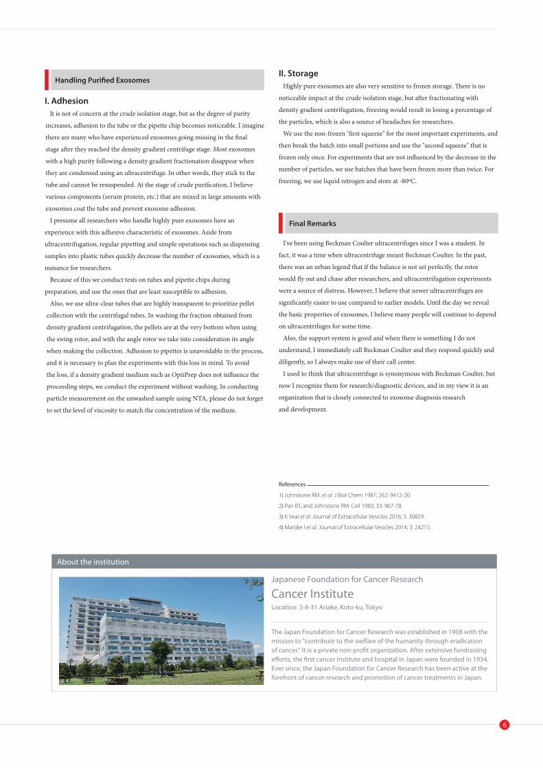
I. Adhesion It is not of concern at the crude isolation stage, but as the degree of purity
increases, adhesion to the tube or the pipette chip becomes noticeable. I imagine
there are many who have experienced exosomes going missing in the final
stage after they reached the density gradient centrifuge stage. Most exosomes
with a high purity following a density gradient fractionation disappear when
they are condensed using an ultracentrifuge. In other words, they stick to the
tube and cannot be resuspended. At the stage of crude purification, I believe
various components (serum protein, etc.) that are mixed in large amounts with
exosomes coat the tube and prevent exosome adhesion.
I presume all researchers who handle highly pure exosomes have an
experience with this adhesive characteristic of exosomes. Aside from
ultracentrifugation, regular pipetting and simple operations such as dispensing
samples into plastic tubes quickly decrease the number of exosomes, which is a
nuisance for researchers.
Because of this we conduct tests on tubes and pipette chips during
preparation, and use the ones that are least susceptible to adhesion.
Also, we use ultra-clear tubes that are highly transparent to prioritize pellet
collection with the centrifugal tubes. In washing the fraction obtained from
density gradient centrifugation, the pellets are at the very bottom when using
the swing rotor, and with the angle rotor we take into consideration its angle
when making the collection. Adhesion to pipettes is unavoidable in the process,
and it is necessary to plan the experiments with this loss in mind. To avoid
the loss, if a density gradient medium such as OptiPrep does not influence the
proceeding steps, we conduct the experiment without washing. In conducting
particle measurement on the unwashed sample using NTA, please do not forget
to set the level of viscosity to match the concentration of the medium.
II. Storage Highly pure exosomes are also very sensitive to frozen storage. There is no
noticeable impact at the crude isolation stage, but after fractionating with
density gradient centrifugation, freezing would result in losing a percentage of
the particles, which is also a source of headaches for researchers.
We use the non-frozen "first squeeze" for the most important experiments, and
then break the batch into small portions and use the "second squeeze" that is
frozen only once. For experiments that are not influenced by the decrease in the
number of particles, we use batches that have been frozen more than twice. For
freezing, we use liquid nitrogen and store at -80oC.
I've been using Beckman Coulter ultracentrifuges since I was a student. In
fact, it was a time when ultracentrifuge meant Beckman Coulter. In the past,
there was an urban legend that if the balance is not set perfectly, the rotor
would fly out and chase after researchers, and ultracentrifugation experiments
were a source of distress. However, I believe that newer ultracentrifuges are
significantly easier to use compared to earlier models. Until the day we reveal
the basic properties of exosomes, I believe many people will continue to depend
on ultracentrifuges for some time.
Also, the support system is good and when there is something I do not
understand, I immediately call Beckman Coulter and they respond quickly and
diligently, so I always make use of their call center.
I used to think that ultracentrifuge is synonymous with Beckman Coulter, but
now I recognize them for research/diagnostic devices, and in my view it is an
organization that is closely connected to exosome diagnosis research
and development.
References
1) Johnstone RM, et al. J Biol Chem 1987; 262: 9412-20.
2) Pan BT, and Johnstone RM. Cell 1983; 33: 967-78.
3) K Iwai et al. Journal of Extracellular Vesicles 2016; 5: 30829.
4) Marijke I et al. Journal of Extracellular Vesicles 2014; 3: 24215.
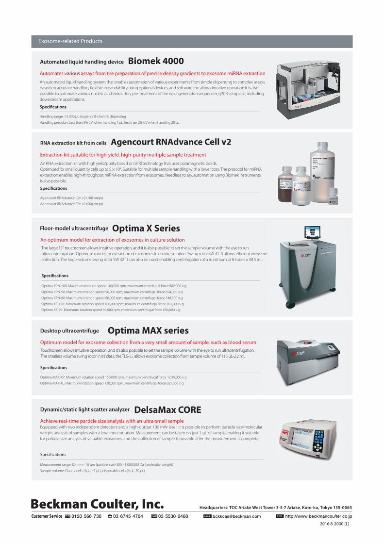
The large 15" touchscreen allows intuitive operation, and it is also possible to set the sample volume with the eye to run ultracentrifugation. Optimum model for extraction of exosomes in culture solution. Swing rotor SW 41 Ti allows efficient exosome collection. The large-volume swing rotor SW 32 Ti can also be used, enabling centrifugation of a maximum of 6 tubes x 38.5 mL.
Specifications
Optima XPN-100: Maximum rotation speed 100,000 rpm, maximum centrifugal force 802,000 x g
Optima XPN-90: Maximum rotation speed 90,000 rpm, maximum centrifugal force 694,000 x g
Optima XPN-80: Maximum rotation speed 80,000 rpm, maximum centrifugal force 548,300 x g
Optima XE-100: Maximum rotation speed 100,000 rpm, maximum centrifugal force 802,000 x g
Optima XE-90: Maximum rotation speed 90,000 rpm, maximum centrifugal force 694,000 x g
Headquarters: TOC Ariake West Tower 3-5-7 Ariake, Koto-ku, Tokyo 135-0063
Customer Service
Dynamic/static light scatter analyzer DelsaMax CORE Achieve real-time particle size analysis with an ultra-small sample Equipped with two independent detectors and a high-output 100 mW laser, it is possible to perform particle size/molecular weight analysis of samples with a low concentration. Measurement can be taken on just 1 μL of sample, making it suitable for particle size analysis of valuable exosomes, and the collection of sample is possible after the measurement is complete.
Specifications
Measurement range: 0.4 nm - 10 μm (particle size) 300 - 1,000,000 Da (molecular weight)
Sample volume: Quartz cells (1μL, 45 μL), disposable cells (4 μL, 50 μL)
Exosome-related Products
Automated liquid handling device Biomek 4000Automates various assays from the preparation of precise density gradients to exosome miRNA extraction
An automated liquid handling system that enables automation of various experiments from simple dispensing to complex assays based on accurate handling, flexible expandability using optional devices, and software the allows intuitive operation.It is also possible to automate various nucleic acid extraction, pre-treatment of the next-generation sequencer, qPCR setup etc., including downstream applications.
Specifications
Handling range: 1-1,000 μL single- or 8-channel dispensing
Handling precision: Less than 5% CV when handling 1 μL, less than 2% CV when handling 20 μL
RNA extraction kit from cells Agencourt RNAdvance Cell v2Extraction kit suitable for high-yield, high-purity multiple sample treatment
An RNA extraction kit with high yield/purity based on SPRI technology that uses paramagnetic beads.Optimized for small quantity cells up to 5 × 10⁴. Suitable for multiple sample handling with a lower cost. The protocol for miRNA extraction enables high-throughput miRNA extraction from exosomes. Needless to say, automation using Biomek instruments is also possible.
Specifications
Agencourt RNAdvance Cell v2 (100 preps)
Agencourt RNAdvance Cell v2 (960 preps)
Floor-model ultracentrifuge Optima X Series An optimum model for extraction of exosomes in culture solution
Desktop ultracentrifuge Optima MAX seriesOptimum model for exosome collection from a very small amount of sample, such as blood serum
Specifications
Optima MAX-XP: Maximum rotation speed 150,000 rpm, maximum centrifugal force 1,019,000 x g
Optima MAX-TL: Maximum rotation speed 120,000 rpm, maximum centrifugal force 657,000 x g
2016.8-2000 (L)
Touchscreen allows intuitive operation, and it's also possible to set the sample volume with the eye to run ultracentrifugation. The smallest volume swing rotor in its class, the TLS-55 allows exosome collection from sample volume of 115 μL-2.2 mL
Beckman Coulter, Inc.

![The Conjugate Gradient Method...Conjugate Gradient Algorithm [Conjugate Gradient Iteration] The positive definite linear system Ax = b is solved by the conjugate gradient method](https://img.pdfslide.net/doc/110x75/5e95c1e7f0d0d02fb330942a/the-conjugate-gradient-method-conjugate-gradient-algorithm-conjugate-gradient.jpg)