Embed Size (px)
Citation preview
1
APPROACH TO THE APPROACH TO THE DIAGNOSIS OF THE DIAGNOSIS OF THE DIFFICULT LIVER DIFFICULT LIVER LESION WITHLESION WITH MRIMRI
Richard C. SemelkaRichard C. Semelka
Clinical historyClinical history
HistoryHistory
Chronic Primary No KnownChronic Primary No KnownLiver Malignancy DiseaseLiver Malignancy DiseaseDiseaseDisease
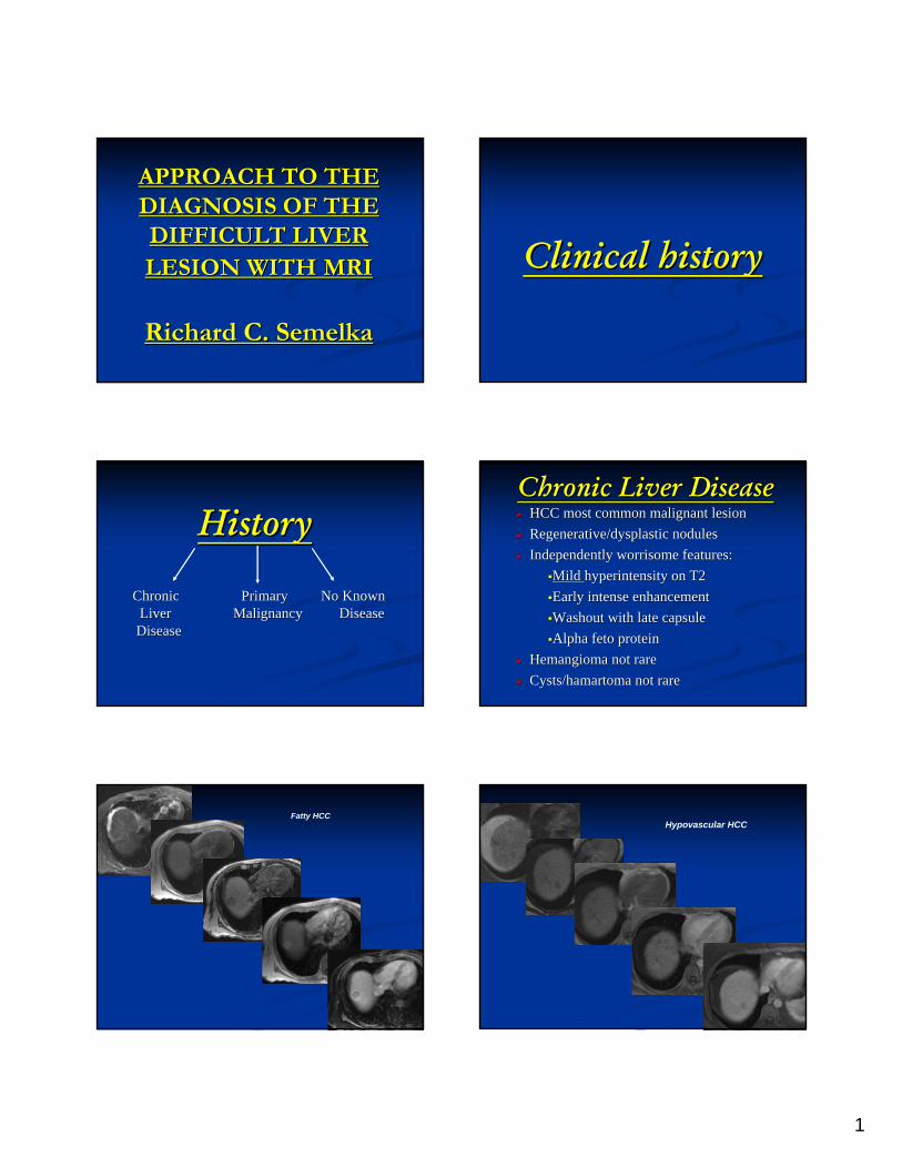
Chronic Liver DiseaseChronic Liver DiseaseHCC most common malignant lesion HCC most common malignant lesion Regenerative/Regenerative/dysplasticdysplastic nodulesnodulesIndependently worrisome features:Independently worrisome features:
Mild Mild hyperintensityhyperintensity on T2on T2Early intense enhancementEarly intense enhancementWashout with late capsuleWashout with late capsuleAlpha Alpha fetofeto proteinprotein
HemangiomaHemangioma not rarenot rareCysts/Cysts/hamartomahamartoma not rarenot rare
Fatty HCCHypovascular HCC
2
Hypervascular HCC
Multiple HCC
Diffuse HCC
Regenerative nodules
Low grade dysplastic nodule High grade dysplastic nodule
3
Acute on Chronic Hepatitis Portal vein thrombosis
Acute Budd-Chiari
Primary MalignancyPrimary Malignancy
HistologicHistologic type and locationtype and locationClues to Clues to vascularityvascularity of liver metastasesof liver metastases
Chemotherapy administration and whenChemotherapy administration and whenLiver metastases can adopt a variety of Liver metastases can adopt a variety of appearances in acute, appearances in acute, subacutesubacute and and chronic response to chemotherapychronic response to chemotherapy
Hypervascular carcinoid metastases
Hypervascular liver metastases
4
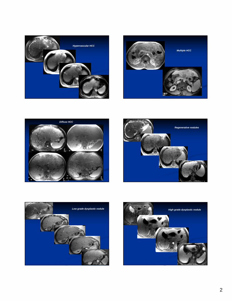
Colon Cancer Metastases Colon Cancer Metastases
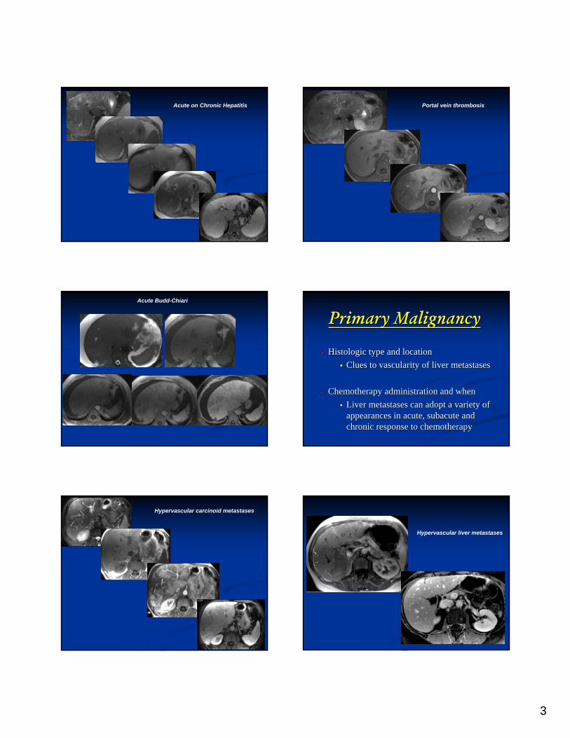
No Known DiseaseNo Known DiseaseBBenign lesions 10x to 100x more common enign lesions 10x to 100x more common than malignant, based on actuarial data than malignant, based on actuarial data BBenign lesions are common (20% population)enign lesions are common (20% population)Normal background liver?Normal background liver?Could the patient have cirrhosis?Could the patient have cirrhosis?
MRI appearance of background liverMRI appearance of background liverHCC uncommon in the absence of HCC uncommon in the absence of underlying chronic disease(< 1 in 100)underlying chronic disease(< 1 in 100)
Could the patient have an unsuspected Could the patient have an unsuspected primary malignancy?primary malignancy?
Uncommon (< 1 in 200Uncommon (< 1 in 200))
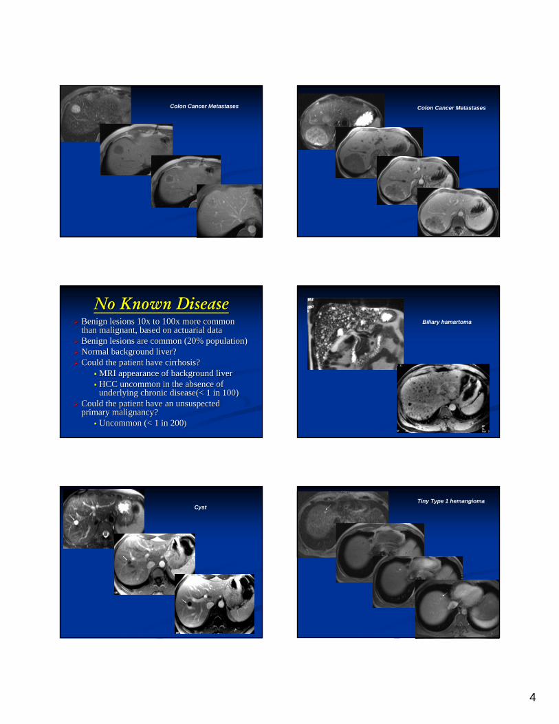
Biliary hamartoma
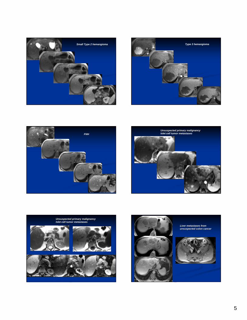
CystTiny Type 1 hemangioma
5
Small Type 2 hemangioma Type 3 hemangioma
FNHUnsuspected primary malignancyUnsuspected primary malignancyIslet cell tumor metastases
Unsuspected primary malignancyUnsuspected primary malignancyIslet cell tumor metastases
Liver metastases from Liver metastases from unsuspected colon cancerunsuspected colon cancer
6
MRI findingsMRI findingsLesion appearance onLesion appearance on::
T1, T2, T1, T2,
early and late postearly and late post--GdGd images images
±± hepatocytehepatocyte uptakeuptake
MRI appearanceMRI appearanceMarginsMarginsSI on SI on noncontrastnoncontrast imagesimages
Qualify Qualify SISI e.g. mild, mod, marked S1e.g. mild, mod, marked S1e.g. HCC mild T2, never mod or markede.g. HCC mild T2, never mod or markede.g. e.g. hemangiomahemangioma mod or marked T2mod or marked T2
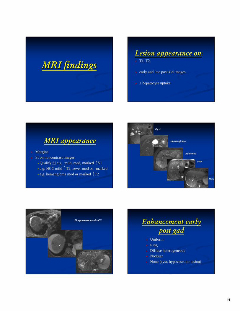
Cyst
Hemangioma
Adenoma
FNH
HCC
T2 appearances of HCC
UniformUniformRingRingDiffuse heterogeneousDiffuse heterogeneousNodularNodularNone (cyst, None (cyst, hypovascularhypovascular lesion)lesion)
Enhancement early Enhancement early post gadpost gad
7
Uniform
Ring
Heterogenous
Adenoma
Pancreatic cancer
HCCHCC
Nodular
No enhancement
Hemangioma
Cyst
Enhancement late Enhancement late post gadpost gad
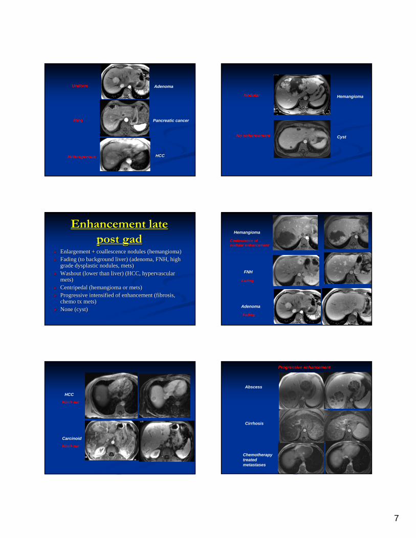
Enlargement + Enlargement + coallescencecoallescence nodules (nodules (hemangiomahemangioma))Fading (to background liver) (adenoma, FNH, high Fading (to background liver) (adenoma, FNH, high grade grade dysplasticdysplastic nodules, nodules, metsmets) ) Washout (lower than liver) (HCC, Washout (lower than liver) (HCC, hypervascularhypervascularmetsmets))CentripedalCentripedal ((hemangiomahemangioma or or metsmets) ) Progressive intensified of enhancement (fibrosis, Progressive intensified of enhancement (fibrosis, chemo chemo txtx metsmets))None (cyst)None (cyst)
Hemangioma
FNH
Adenoma
Coalescence ofnodular enhancement
Fading
Fading
Carcinoid
Wash out
HCC
Wash out
Progressive enhancement
Abscess
Cirrhosis
Chemotherapytreated metastases
8
HemangiomaHemangioma always bright on T2always bright on T2HCC never bright on T2HCC never bright on T2Background fatty liver: FNH, Background fatty liver: FNH, metsmetsFatty lesions: focal fat, adenoma, HCCFatty lesions: focal fat, adenoma, HCCCirrhotic liver, never describe FNH or Cirrhotic liver, never describe FNH or adenoma, describe RN/DN/HCCadenoma, describe RN/DN/HCCCould this liver be cirrhotic? Could this liver be cirrhotic?
Fibrosis on short TE T1W imagesFibrosis on short TE T1W imagesEarly negligible, late progressive Early negligible, late progressive
enhancementenhancement
ImagingImaging Pearls:Pearls: Carcinoid metastases
Background fatty liver
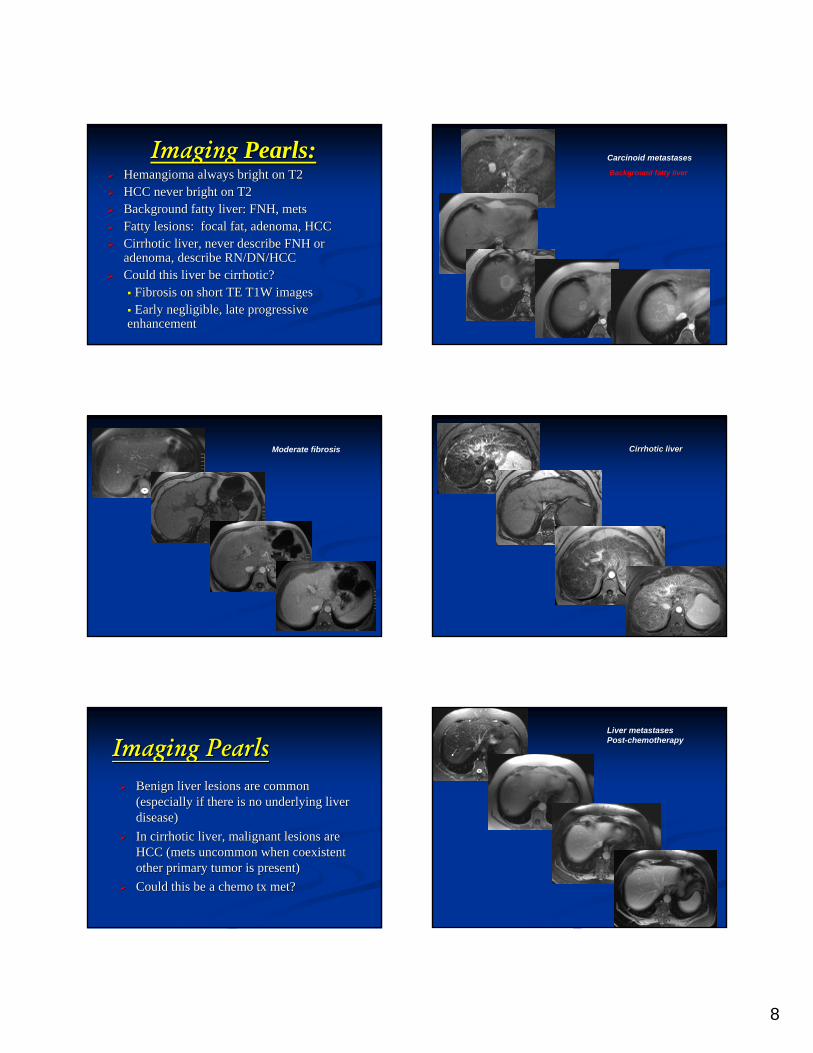
Moderate fibrosis Cirrhotic liverCirrhotic liver
Imaging PearlsImaging PearlsBenign liver lesions are common Benign liver lesions are common (especially if there is no underlying liver (especially if there is no underlying liver disease)disease)In cirrhotic liver, malignant lesions are In cirrhotic liver, malignant lesions are HCC (HCC (metsmets uncommon when coexistent uncommon when coexistent other primary tumor is present)other primary tumor is present)Could this be a chemo Could this be a chemo txtx met?met?
Liver metastases Post-chemotherapy
9
Imaging Pearls:Imaging Pearls:Distinguish hemorrhage/protein from Distinguish hemorrhage/protein from enhancement (enhancement (noncontrastnoncontrast T1)T1)Distinguish fat effects from washout Distinguish fat effects from washout (look at all non suppressed and fat (look at all non suppressed and fat suppressed images)suppressed images)Confluent fibrosis or Confluent fibrosis or fibroticfibrotic lesions, lesions, minimal early enhancement with minimal early enhancement with progressive increased enhancementprogressive increased enhancement
Hemorrhage secondary to RFA
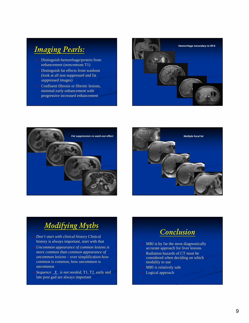
Fat suppression vs wash-out effect Multiple focal fat
DonDon’’t start with clinical historyt start with clinical history Clinical Clinical history is always important, start with thathistory is always important, start with thatUncommon appearance of common lesions is Uncommon appearance of common lesions is more common than common appearance of more common than common appearance of uncommon lesionsuncommon lesions –– over simplificationover simplification--how how common is common, how uncommon is common is common, how uncommon is uncommonuncommonSequence Sequence X X is not neededis not needed, T1, T2, early and , T1, T2, early and late post gad are always importantlate post gad are always important
Modifying MythsModifying MythsConclusionConclusion
MRI is by far the most diagnostically MRI is by far the most diagnostically accurate approach for liver lesions accurate approach for liver lesions Radiation hazards of CT must be Radiation hazards of CT must be considered when deciding on which considered when deciding on which modality to usemodality to useMRI is relatively safeMRI is relatively safeLogical approachLogical approach
10
ConclusionConclusionLogical approachLogical approach
Clinical historyClinical historyLesion appearance: T1, T2, Lesion appearance: T1, T2, Early and late post Early and late post GdGdBenign lesions commonBenign lesions commonCould this be a chemoCould this be a chemo--txtx metmetCould this be a cirrhotic liver Could this be a cirrhotic liver