Embed Size (px)
Citation preview

Approach to patients with Approach to patients with colorectal symptomscolorectal symptoms
Dr. Janet Lee
Team 3 Surgery
10 JUL 2003
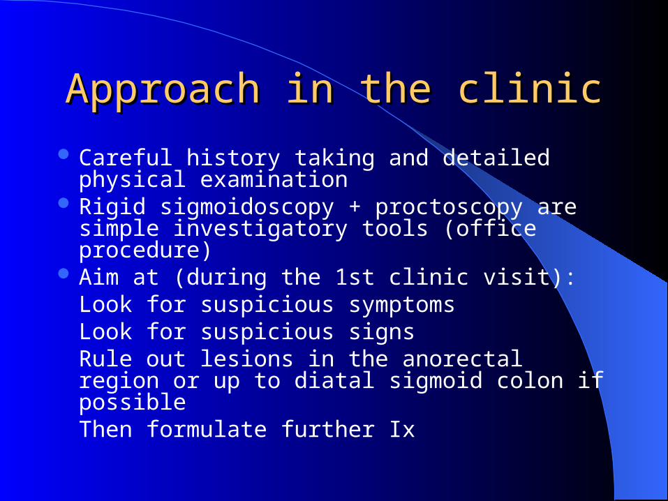
Approach in the clinicApproach in the clinic
Careful history taking and detailed physical examination
Rigid sigmoidoscopy + proctoscopy are simple investigatory tools (office procedure)
Aim at (during the 1st clinic visit):Look for suspicious symptomsLook for suspicious signsRule out lesions in the anorectal region or up to diatal sigmoid colon if possibleThen formulate further Ix

How common is PRB?How common is PRB?
PWH data (since year 2000)76% patients seen in colorectal clinic
presented with PRB either as the Chief complaint or associated symptoms or other colorectal complaints
30% patients presented with isolated PRB

Common presenting Common presenting symptoms symptoms
in our colorectal clinicin our colorectal clinic
Fresh per rectal bleedingChange in bowel habitAbdominal painMucus in stoolTenesmus

ColonoscopyColonoscopy
Introduction in the late 60sNow being the first line gold standard
investigatory tool for colorectal pathology
Both diagnostic and therapeutic

Polyps: the commonest Polyps: the commonest lesions identified during lesions identified during
colonoscopycolonoscopy

Yield for cancers / polyps Yield for cancers / polyps ((1cm)1cm)
Highest for patients with PRBNeugut Am J Gastroenterol 1993; 88: 1179-83
Brenna Scan J Gastroenterol 1990; 25: 81-8
Berkowitz S Afr Med J 1993; 83: 245-8

AdenomasAdenomas
Adenoma-to-carcinoma sequenceJass World J Surg 1989; 13: 4551
Cannon-Albright N Engl J Med 1988; 319: 533-7
Time progression:
small adenoma to carcinoma is 10-15 years
big adenoma (1cm) to carcinoma is 5 yearsMuto Cancer 1975; 36: 2251-70

Complications of colonoscopyComplications of colonoscopy

IncidenceIncidence
Range from 1-20 per 1000 proceduresGastrointest Endosc 2001; 54: 302-9
Retrospective review of 6066 colonoscopies
1979-1995 in 1 Swedish county
Morbidity 0.4% (Diagnostic 0.2% vs therapeutic 1.2%)
Higher rate reported in therapeutic endoscopy (polypectomy, laser application)

Sometimes complications arise before the procedure
e.g. at the time of bowel preparation
Bowel preparationBowel preparation
Purge method
Lavage method

Purge methodPurge method
LaxativesStimulant type
Abdominal pain, excessive mucus secretion, fluid loss
Osmotically active type(e.g. sodium phosphate, Mg citrate, Mannitol)
Produce net loss of water, electrolyte disturbance, explosion

Lavage methodLavage method
Saline / electrolyte solutions
Net gain in fluid and electrolyteSodium sulfate/PEG solutionSulfate-free PEG solution
Problem: ingest at a rate of 1L/hr, nausea and vomiting (5-15% patients cannot tolerate)

Bowel preparationBowel preparation
Fluid (dehydration or fluid overload)Electrolyte imbalances
Hypokalemia
Hyperphosphatemia (phosphate soda)
Hypocalcemia (phosphate soda)Intestinal obstructionToxic megacolon (severe active IBD)

At risk groupAt risk group
Renal or cardiac diseasePatients who take drugs that affect fluid
and electrolyte balancePEG-based gut lavage is designed to
avoid fluid and electrolytes shifts (Gastroenterol 1990; 98: 11-6)
Obstructing lesion in the large bowel

Problems with poor bowel Problems with poor bowel preparationpreparation
Missing a significant lesion Fecal peritonitis following a perforation of a
poorly prepared bowel Risk of explosion from ignition of combustible
gases (methane) by electrocautery
Beware of these possibilities if one persists examinating a poorly prepared bowel

InfectionInfection

Antibiotic ProphylaxisAntibiotic Prophylaxis
Transient bacteremia (both aerobic and anaerobic organisms)
4.7% (Shorvon Gut 1983; 24: 1078-93)
Clinical relevance is unclear (as this may occur in normal activities like bowel movement and tooth brushing)
E. coli is the commonest bacteremic organism (endocarditis due to this is rare)

Risk of bacteremia does not increase with biopsy or polypectomy procedures(Low Dig Dis Sci 1987; 32:1239-43)
Increased risk (20%) in laser treatment for neoplasia (Kohler Gastrointest Endosc 1988; 34: 73-4)
Antibiotic ProphylaxisAntibiotic Prophylaxis

Rare infective complicationsRare infective complications
Gram –ve septicemiaPeritonitis
cirrhotic patients
chronic renal failure patients Thornton Gut 1991; 32: 450-1
Shrake Am J Gastroenterol 1989; 84: 453-4
Petersen Am J Gastroenterol 1987; 82: 171-2

High risk patientsHigh risk patients
Ascites Peritoneal dialysis Prosthetic heart valves Previous bacterial endocarditis Most congenital heart malformations Rheumatic or acquired valvular diseases Hypertrophic cardiomyopathy Mitral valve prolapse with regurgitation Artificial joints or vascular prosthesis (<1 yr)

IV Ampicillin 2g + Gentamicin (1.5mg/kg) given 30 mins before and 8 hr after colonoscopy
Single oral amoxicillin 3g followed by 1.5g orally 6 hr after the procedure
Perforation and Perforation and HaemorrhageHaemorrhage

Perforation:Diagnostic 0.14-0.26%Therapeutic 0.11-0.42%
Haemorrhage:Diagnostic 0.008-0.07%Therapeutic 0.7-2.24%
Death:Diagnostic 0-0.03%Therapeutic 0-0.1%

Serosal tear / mesenteric haematomaIncidence is unknown (likely to be more
frequent than frank perforation)
Lesser form of bowel damage

HaemorrhageHaemorrhage
Rare in diagnostic colonoscopy(Traumatization of an existing lesion, self-limiting)
Usually caused by insufficient coagulation before polyp removal
Clinically significant bleeding at Bx site is rare Immediate or delayed (up to 2 weeks later) Risk increased by aspirin, NSAID or anti-
coagulant therapy

Extraluminal bleedingExtraluminal bleeding
Seromuscular tearRupture of liver / spleenMesenteric tear

Refrain from NSAID use 7-10 days before therapeutic colonoscopy (Gastrointest Endosc Clin N Am 1996; 6: 265-75)
and stopped for 7 days after polypectomy
Warfarin should be stopped for 5 days before procedure and change over to heparin or LMWH
Preventive measuresPreventive measures

ManagementManagement
Generally conservative (Surg Endosc 1994; 8: 672-6)
Adrenaline (1:10,000) injection or Electrocauterization with heat probe or laser ( Am J Gastroenterol 1992; 87: 1681-2)
Endoscopic haemostasis can be achieved in 75% cases (Surg Endosc 1994; 8: 672-6)
Selective arterial vasoconstrictive agent instillation or embolization

Perforation (Diagnostic)Perforation (Diagnostic)
Primarily located at rectosigmoid junction or the sigmoid loop or in regions of post-op adhesions (J Am Coll Surg 1994; 179: 333-7)
Longitudinal tears at the antimesenteric border Direct exaggerated pressure of the tip Overinflation in diverticulosis or colonic
stenosis(Gut 1983; 24:76-83)
Confusion between colic lumen and a diverticular ostium

Perforation (Therapeutic)Perforation (Therapeutic)
0.1-1%Primarily occur after polypectomy Site of perforation corresponds to site of
pathology

Preventive measuresPreventive measures
Well-prepared bowelProperly functioning equipmentAvoid over inflationAvoid over sedationSound clinical judgment and skillExtreme patience

Farley Mayo Clin Proc 1997; 72: 729-33
57028 colonoscopies (1980-1995)43 perforations (1 in 1333)
93% treated with emergency laparotomy2/3 - 10 repair or limited resection + Anastomosis1/3 - stoma
Post-op complications related to:Old ageSize of perforationPrior hospitalization

Polypectomy coagulation Polypectomy coagulation syndromesyndrome
Diffuse or localized abdominal painLow grade feverStarting 6-8 hr after the procedurePneumoperitoneum is absentEtiology is a thermal transmural injury of
the colonic wall

Cardiac and RespiratoryCardiac and Respiratory

Cardiac and RespiratoryCardiac and Respiratory
Oversedation (0.06-0.54%) (Endoscopy 1994; 26:231-4)
Vasovagal reactions: sustained bradycardia, hypotension and diaphoresis (16%)Only 1/3 required minimal intervention mainly in the form of IV fluid (Gastrointest Endosc 1993; 39: 388-91)
Severe vasovagal reaction is a reason to abort the procedure

Though the incidence of significant cardiopulmonary complications is very low
Careful surveillance during the procedure is still highly recommended with antidote (for narcotics and benzodiazepines) readily available

PainPain Tension in the colonic mesentery
9% patients suffer from severe pain while 27% were pain free (Surg Endosc 1994; 8: 784-7)
Loop formation is the commonest cause Patient with short, tight mesenteries are more
prone to pain Air insufflation promotes discomfort by
accentuating loops and elongating the bowel, thus increasing the mesenteric tension
Operator dependent

Contrast enemaContrast enema
Three types of contrast enemas
Double contrast barium enema
Single contrast barium enema
Water soluble contrast enema

Double contrast barium Double contrast barium enemaenema
Considered the standard radiological examination of the colon
Administering high-density low-viscosity barium into the colon followed by air insufflation
In well-prepped patients:90% accurate in identifying index polyps in patients with >1 polyps 5mm in size97% accurate in identifying index polyps in patients with >1 polyps 10mm in size

ContraindicationsContraindications
Partial obstructionAcute diverticulitisFulminating colitisToxic megacolonLesion with suspected localized perforationIncontinent patients

Water-soluble contrastWater-soluble contrast
To “rule-in” or “rule-out” many acute or subacute conditions such as:
Obstruction
Anastomotic leakDo not require detailed mucosal
examination

Pros and Cons of water soluble Pros and Cons of water soluble contrast enemacontrast enema
Advantages
Readily absorbed from peritoneal cavity in the event of perforation
Clear liquidsDisadvantages
High osmolarity
Irritant effect on the mucosa (inflammation)

CT and MR colonographyCT and MR colonography
Both based on acquisition of high spatial resolution 3D data sets that encompass the entire colon
Advanced imaging processing allows analysis of the colon in a multiplanar and virtual endoscopic format

CT colonographyCT colonography
Prepped and air insufflated colon Helical CT with specialized 3D imaging software Supine + Prone position
Helps to ddx mobile stool from fixed pathology
Allows more even distension of the colon
Improves visualization of segments obscured by intraluminal fluid
Total study time ~20 mins

How good is CT How good is CT colonography?colonography?
Radiology 1996; 200: 49-54
70 consecutive patients
Polyps size >10mm 5-10mm <5mm
Sensitivity 75% 66% 45%
Specificity 90% 63% 80%

MR colonographyMR colonography
Standard bowel prepFluid enema (3L water with 60ml 0.5mol/L
paramegnetic contrast material)Instillate in prone position3D spoiled gradient-echo & 2D single-shot
fast spin-echo pulse sequencesProne + Supine positionEntire study ~20 mins

How good is MR How good is MR colonography?colonography?
For lesions >10mm
Sensitivity: 93%Specificity: 99%

Clinical application of CT/MR Clinical application of CT/MR colonographycolonography
Incomplete colonoscopy ~5-7% (assess the remaining segment not covered by the reach of the colonoscope)
Pre-op localization of primary tumor Occlusive lesion e.g. stenotic tumor (assess the
proximal segment) Obtain extraluminal information (local invasion /
metastases e.g. liver 20, enlarged LNs)

? Screening tool ? Screening tool (CT/MR colonography)(CT/MR colonography)
Satisfactory results for clinically significant sized lesions (>10mm)
The clinical significance of missing small sized lesions (<5mm) is negligible
Missing medium sized lesions (6-9mm) is a real concern


![DIGEST DIAGNOSIS THROUGH INVESTIGATION OF GASTROESOPHAGEAL SYMPTOMS COLORECTAL CANCER LEARNING PROGRAM [COPY] Colorectal Cancer [LOGO] RXD Takeda](https://img.pdfslide.net/doc/110x75/5a4d1bad7f8b9ab0599cb502/digest-diagnosis-through-investigation-of-gastroesophageal-symptoms-colorectal-cancer.jpg)


![Efficacy and safety of the starting position during colonoscopy: a … · colorectal cancer screening, polyp surveillance, and diagnosis of lower gastrointestinal symptoms [1]. Colonoscopy](https://img.pdfslide.net/doc/110x75/6099afc227b38f0f5f01fd5d/efficacy-and-safety-of-the-starting-position-during-colonoscopy-a-colorectal-cancer.jpg)