Embed Size (px)
Citation preview
Page 1/21
Modi�cation of Breast Cancer Milieu withChemotherapy Plus Dendritic Cell Vaccines: AnApproach to Select Best Therapeutic StrategiesLuis Daniel Mejias Sosa ( [email protected] )
Hospital Universitario Rey Juan Carlos https://orcid.org/0000-0003-0927-6997Álvaro López-Janeiro
Hospital Universitario La PazAlicia Córdoba
Complejo Hospitalario de NavarraPablo Sala
Clínica Universidad de NavarraBelén P Solans
Universidad de Navarra Facultad de Farmacia: Universidad de Navarra Facultad de Farmacia y NutricionSusana Inogés
Clínica Universidad de NavarraAscensión López‐Díaz de Cerio
Clínica Universidad de NavarraLaura Hato
Clínica Universidad de NavarraFrancisco Guillén-Grima
Clínica Univseridad de NavarraÓscar Fernández-Hidalgo
Clínica Universidad de NavarraSusana De La Cruz
Complejo Hospitalario de NavarraMaría Dolores Lozano
Clínica Universidad de NavarraMiguel A Idoate
Clínica Universidad de NavarraMarta Santisteban
Clínica Universidad de Navarra
Research Article

Page 2/21
Keywords: Breast cancer, dendritic cell vaccines, TILs, neoadjuvant scenario, CD8 and triple negative.
Posted Date: November 1st, 2021
DOI: https://doi.org/10.21203/rs.3.rs-643452/v3
License: This work is licensed under a Creative Commons Attribution 4.0 International License. Read Full License
Page 3/21
AbstractPurpose: The addition of dendritic cell vaccines (DCV) to neoadjuvant chemotherapy (NAC) could induceimmune biomarker changes in those patients with residual disease (RD) by transforming tumormicroenvironment.
Methods: Core diagnostic biopsies and surgical specimens from 80 patients (38 in the Vaccinated Groupplus NAC (VG) and 42 in the Control Group (CG) treated only with NAC) were selected. We quantify TILs(CD8, CD4 and CD45RO) using Immunohistochemistry (IHC) and the Automated Cellular Imaging System(ACIS III) in the core-diagnostic biopsies and in the surgical specimens, to compare the amount of TILs ineach group.
Results: A CD8 rise in TNBC samples was observed after NAC plus DCV, changing from 4.48% in thebiopsy to 6.70% in the surgical specimen, not reaching statistically signi�cant differences (p = 0.11).TNBC patients in the CG showed a TILs drop from 2.71% in the biopsy to 0.18% in the surgical specimen(p = 0.5). We also found that 66.7% (4/6) of TNBC patients from VG registered an increase in TILs aftertreatment as compared to 20% (1/5) of TNBC patients in the CG (p=0.24). This phenomenon is notobserved in the other biologic subtypes. An association between before NAC CD8 TILs (4% cut-off point )and pathological complete response in the VG was found in univariate and multivariate analysis(OR=1.41, IC95% 1.05-1.90; p=0.02, and OR=2.0, IC95% 1.05-3.9; p=0.03, respectively).
Conclusion: Our �ndings suggest that patients with TNBC especially bene�t from the stimulation of theantitumor immune system by using DCV pulsed with tumor antigens.
BackgroundImmune in�ltration in breast cancer (BC) milieu has driven to the achievement of a better outcome ofthese patients with standard chemotherapy, but also has enhanced the incorporation of immunotherapyas a therapeutic strategy. Thus, higher levels of tumor in�ltrating lymphocytes (TILs) on the corediagnostic biopsy have been related to increased pathological complete responses (pCR) withneoadjuvant chemotherapy (NAC) and longer event-free survival (EFS) and overall survival (OS) in themore aggressive BC subtypes [1–5]. However, the presence of residual disease (RD) after NAC impliesfurther adjuvant chemotherapy that could be avoided if pCR would have been reached. Additionally, therise of TILs in RD after NAC has been linked to a better outcome and it could be considered a surrogatemarker for long-term treatment e�cacy in triple negative breast cancer (TNBC) patients in the absence ofpCR [5–7].
Aside from these data, the development of new immune strategies that convert cold into hot immuneenriched tumors are mandatory. The study of biological changes in BC patients with RD after NAC opensa translational window in breast tumors and its milieu in this population with a worse prognosis. Immunecheckpoint inhibitors (CPI) have been evaluated in BC with outstanding results in combination withchemotherapy in the neoadjuvant arena [8–14] although with an increased toxicity pro�le, and without
Page 4/21
information regarding biological impact on tumor milieu. Dendritic cells, as the main directors of theimmune system, have been studied for their role in the antigenic cross presentation, a key step for Tcytotoxic responses. Active cell therapy with dendritic vaccines (DCV) has shown tumor growth inhibitionand T cell memory activation in preclinical models [15]as well as clinical improvement in BC patientswithout further toxicity in different scenarios [16, 17]. So far, the implementation of DCV together withNAC in naïve BC patients could be a big challenge by increasing host immunity and immunogenic celldeath in tumoral cells and generating potential modi�cations in tumor milieu. Moreover, our center hasalready demonstrated prior experience with active DCV in other solid malignancies[18, 19]
The aim of this study is to evaluate if the addition of DCV to NAC could induce immune biomarkerchanges in those patients with RD by transforming tumor microenvironment, in order to identify whichgroup of patients could bene�t from this strategy and to select the best therapy in the maintenancescenario in upcoming studies.
Materials And MethodsPatients
Patients in the vaccinated group (VG) were recruited from 2011 to 2015 in the phase II non-randomizedmulticentric clinical trial NCT01431196 and as compassionate use for DCV. All these patients weretreated with the same NAC schedule consisting of 4 cycles of dose-dense epirubicin pluscyclophosphamide with G-CSF support sequenced to 4 cycles of docetaxel each 21 days according tostandard protocols and with the addition of DCV during taxane therapy. The control group (CG) wasobtained from an historic cohort (2008-2015) treated at our center in the same way but without vaccines.Demographic features were well balanced among both groups[20]. No adjuvant chemotherapy wasprescribed. Surgical management was performed after NAC and was followed by radiation therapy ±endocrine therapy if needed. In the adjuvant setting, patients in the VG also received intradermal DCVloaded with autologous tumor lysate as described in our previous work [17, 20].
Samples and criteria for analysis
Core-diagnostic biopsies and surgical specimens from 80 patients (38 in the VG and 42 in the CG wereselected. Pathologists did not know if the samples belong to the VG or CG when they performed thequanti�cation of TILs (blinded-study).
Among the CG, two patients had multifocal/multicentric tumors in the breast. We were able to quantifyTILs according to the characteristics of the core-diagnostic biopsies in the CG as follows: CD8 (37samples), CD4 (39 samples) and CD45RO (36 samples). With respect to the surgical specimen, westudied 35 specimens for each marker. Regarding VG; 35, 34 and 34 core-diagnostic biopsies wereavailable for CD8, CD4 and CD45ro biomarker, respectively. Surgical specimens were 27, 28 and 26 forCD8, CD4 and CD45RO markers, respectively. Samples were classi�ed by biological subtype according tothe 14th St Gallen International Breast Cancer Conference [21]. Total pCR was considered as no in�ltrating
Page 5/21
residual tumor (ypT0/Tis ypN0) in both breast surgical specimen and lymph nodes according toAmerican Joint Committee on Cancer 8th Edition. Recommendations from the International Immuno-oncology Biomarker Working Group on BC were used in order to evaluate TILs in the stromalcompartment related to tumoral cells. The denominator used to determine the percentage of stromal TILswas the area of stromal tissue occupied by lymphocytes, not the number of stromal cells [22]. Moreover,these recommendations were applied to immunohistochemistry to be able to measure CD8, CD4 andCD45RO lymphocytes in the stromal tumor area. After NAC markers were only measured on the sampleswith RD. Pathologists have not quanti�ed TILs in the surgical specimen from patients that reached pCRbecause it is not accurate to know if TILs were epitelial or stromal in this scenario. All the quanti�cationsare in the stromal compoment, epitehlial quanti�cations are not shown. No data for tertiary lymphoidstructures were included in the study.
Immunohistochemistry
Immunohistochemistry (IHC) was performed using formaldehyde-�xed and para�n-embedded tissuesections from 3 to 4 mm thick. The tissue sections were stained by Autostainer Link48 using amonoclonal Mouse Anti-Human CD8 (Dako® Clone C8/144B), CD4 (Dako® Clone 4B12) and CD45RO(Dako® Clon UCHL1). Antigen retrieval was performed by PT link in high pH at 98 degrees for 5 minutes.
Immunohistochemistry measurement and scoring
IHC was quanti�ed using Automated Cellular Imaging System (ACIS III) in both the diagnostic and thesurgical samples [23, 24]. TILs quanti�cation according to the international working group in HE sampleswas obtained by three pathologist (LM, AC, MI) from two different services with variability. These resultsare not shown in the manuscript.
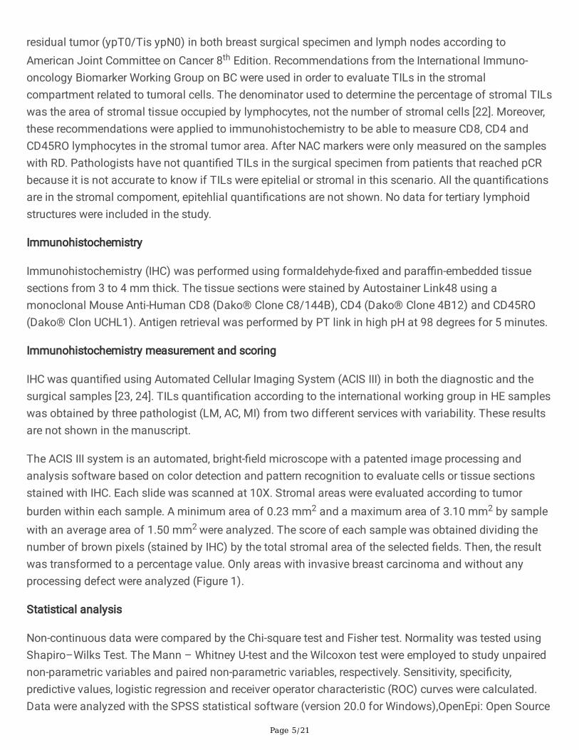
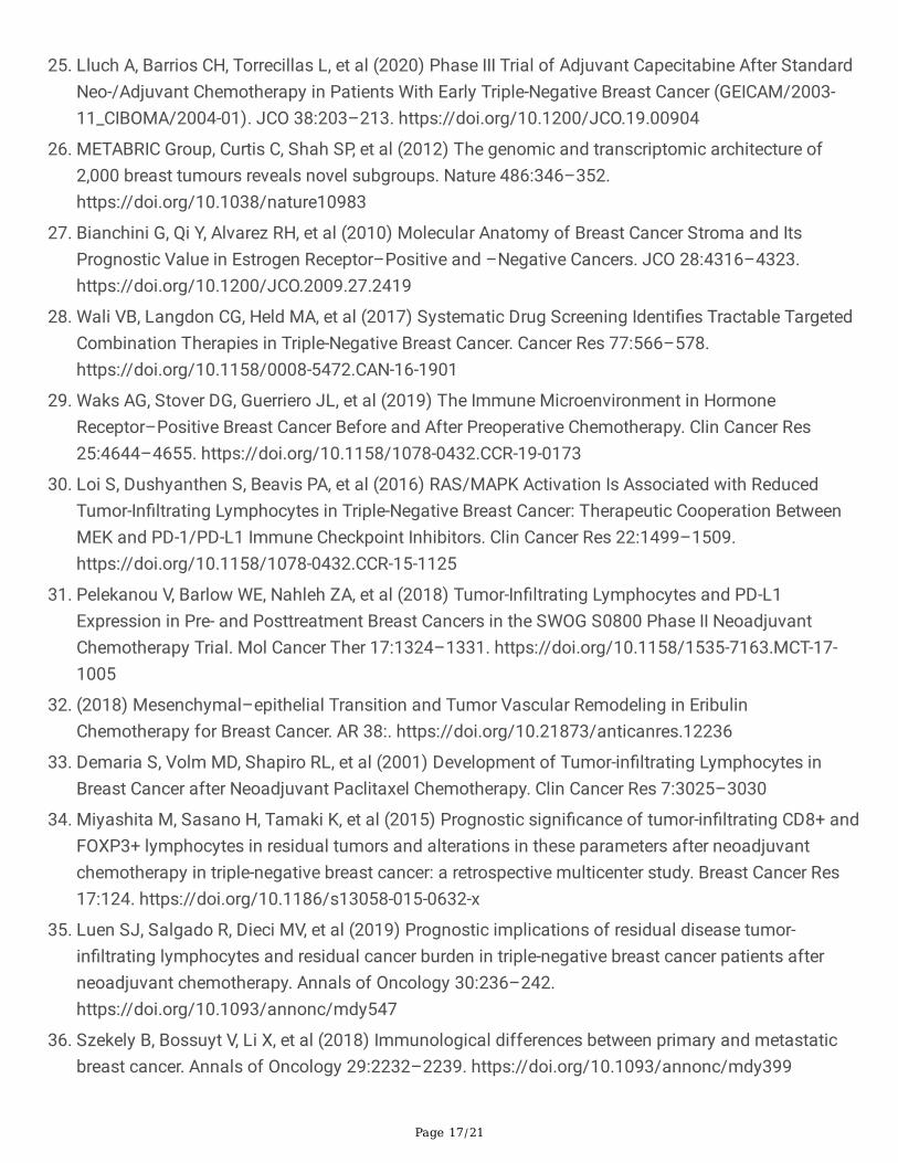
The ACIS III system is an automated, bright-�eld microscope with a patented image processing andanalysis software based on color detection and pattern recognition to evaluate cells or tissue sectionsstained with IHC. Each slide was scanned at 10X. Stromal areas were evaluated according to tumorburden within each sample. A minimum area of 0.23 mm2 and a maximum area of 3.10 mm2 by samplewith an average area of 1.50 mm2 were analyzed. The score of each sample was obtained dividing thenumber of brown pixels (stained by IHC) by the total stromal area of the selected �elds. Then, the resultwas transformed to a percentage value. Only areas with invasive breast carcinoma and without anyprocessing defect were analyzed (Figure 1).
Statistical analysis
Non-continuous data were compared by the Chi-square test and Fisher test. Normality was tested usingShapiro–Wilks Test. The Mann – Whitney U-test and the Wilcoxon test were employed to study unpairednon-parametric variables and paired non-parametric variables, respectively. Sensitivity, speci�city,predictive values, logistic regression and receiver operator characteristic (ROC) curves were calculated.Data were analyzed with the SPSS statistical software (version 20.0 for Windows),OpenEpi: Open Source
Page 6/21
Epidemiologic Statistics for Public Health, Version 3.01 and R software Version 4.0.1. P-values ≤0.05were considered signi�cant, and 95% con�dence intervals were calculated.
Results
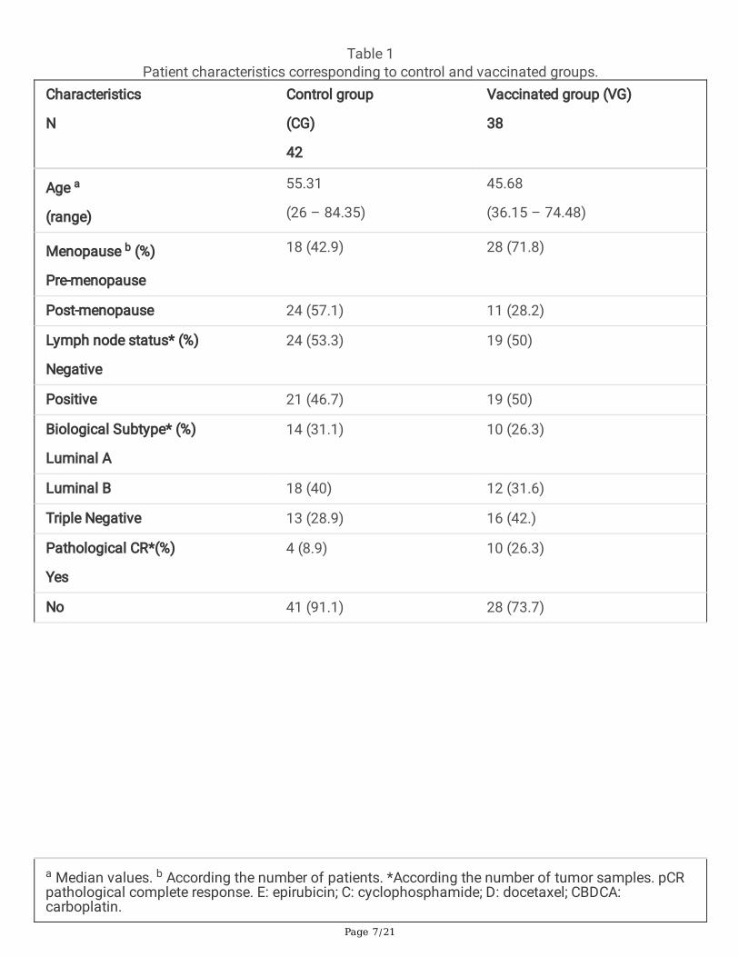
PatientsPatients features, therapy as well as pCR are described in Table 1. Pathological CR was ~three timeshigher in the VG (p=0.03). No differences were found by biologic subtypes (not shown).
Page 7/21
Table 1Patient characteristics corresponding to control and vaccinated groups.
Characteristics
N
Control group
(CG)
42
Vaccinated group (VG)
38
Age a
(range)
55.31
(26 – 84.35)
45.68
(36.15 – 74.48)
Menopause b (%)
Pre-menopause
18 (42.9) 28 (71.8)
Post-menopause 24 (57.1) 11 (28.2)
Lymph node status* (%)
Negative
24 (53.3) 19 (50)
Positive 21 (46.7) 19 (50)
Biological Subtype* (%)
Luminal A
14 (31.1) 10 (26.3)
Luminal B 18 (40) 12 (31.6)
Triple Negative 13 (28.9) 16 (42.)
Pathological CR*(%)
Yes
4 (8.9) 10 (26.3)
No 41 (91.1) 28 (73.7)
a Median values. b According the number of patients. *According the number of tumor samples. pCRpathological complete response. E: epirubicin; C: cyclophosphamide; D: docetaxel; CBDCA:carboplatin.
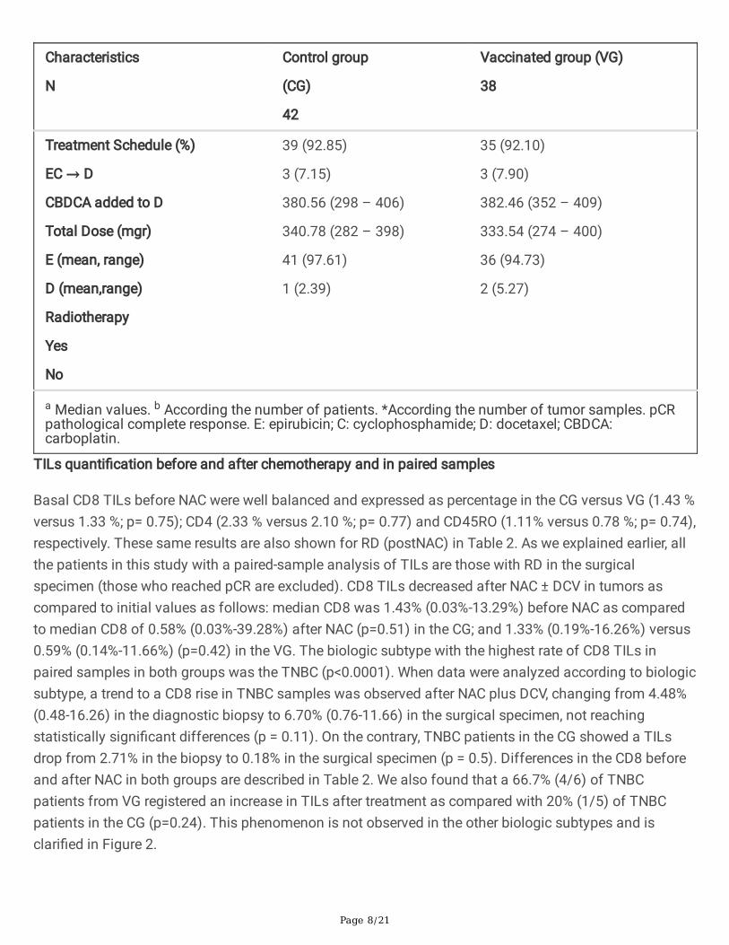
Page 8/21
Characteristics
N
Control group
(CG)
42
Vaccinated group (VG)
38
Treatment Schedule (%)
EC → D
CBDCA added to D
Total Dose (mgr)
E (mean, range)
D (mean,range)
Radiotherapy
Yes
No
39 (92.85)
3 (7.15)
380.56 (298 – 406)
340.78 (282 – 398)
41 (97.61)
1 (2.39)
35 (92.10)
3 (7.90)
382.46 (352 – 409)
333.54 (274 – 400)
36 (94.73)
2 (5.27)
a Median values. b According the number of patients. *According the number of tumor samples. pCRpathological complete response. E: epirubicin; C: cyclophosphamide; D: docetaxel; CBDCA:carboplatin.
TILs quanti�cation before and after chemotherapy and in paired samples
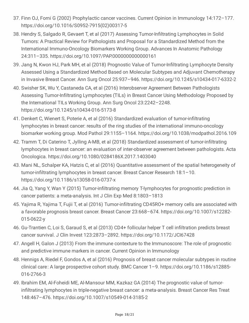
Basal CD8 TILs before NAC were well balanced and expressed as percentage in the CG versus VG (1.43 %versus 1.33 %; p= 0.75); CD4 (2.33 % versus 2.10 %; p= 0.77) and CD45RO (1.11% versus 0.78 %; p= 0.74),respectively. These same results are also shown for RD (postNAC) in Table 2. As we explained earlier, allthe patients in this study with a paired-sample analysis of TILs are those with RD in the surgicalspecimen (those who reached pCR are excluded). CD8 TILs decreased after NAC ± DCV in tumors ascompared to initial values as follows: median CD8 was 1.43% (0.03%-13.29%) before NAC as comparedto median CD8 of 0.58% (0.03%-39.28%) after NAC (p=0.51) in the CG; and 1.33% (0.19%-16.26%) versus0.59% (0.14%-11.66%) (p=0.42) in the VG. The biologic subtype with the highest rate of CD8 TILs inpaired samples in both groups was the TNBC (p<0.0001). When data were analyzed according to biologicsubtype, a trend to a CD8 rise in TNBC samples was observed after NAC plus DCV, changing from 4.48%(0.48-16.26) in the diagnostic biopsy to 6.70% (0.76-11.66) in the surgical specimen, not reachingstatistically signi�cant differences (p = 0.11). On the contrary, TNBC patients in the CG showed a TILsdrop from 2.71% in the biopsy to 0.18% in the surgical specimen (p = 0.5). Differences in the CD8 beforeand after NAC in both groups are described in Table 2. We also found that a 66.7% (4/6) of TNBCpatients from VG registered an increase in TILs after treatment as compared with 20% (1/5) of TNBCpatients in the CG (p=0.24). This phenomenon is not observed in the other biologic subtypes and isclari�ed in Figure 2.
Page 9/21
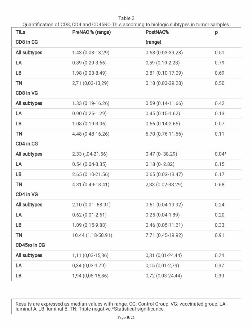
Table 2Quanti�cation of CD8, CD4 and CD45RO TILs according to biologic subtypes in tumor samples.
TILs
CD8 in CG
PreNAC % (range) PostNAC%
(range)
p
All subtypes 1.43 (0.03-13.29) 0.58 (0.03-39.28) 0.51
LA 0.89 (0.29-3.66) 0,59 (0.19-2.23) 0.79
LB 1.98 (0.03-8.49) 0.81 (0.10-17.09) 0.69
TN
CD8 in VG
2,71 (0,03-13,29) 0.18 (0.03-39.28) 0.50
All subtypes 1.33 (0.19-16.26) 0.59 (0.14-11.66) 0.42
LA 0.90 (0.25-1.29) 0.45 (0.15-1.62) 0.13
LB 1.08 (0.19-3.06) 0.56 (0.14-2.65) 0.07
TN
CD4 in CG
4.48 (0.48-16.26) 6.70 (0.76-11.66) 0.11
All subtypes 2.33 (.,04-21.56) 0.47 (0- 38.29) 0.04*
LA 0.54 (0.04-3.35) 0.18 (0- 2.82) 0.15
LB 2.65 (0.10-21.56) 0.65 (0.03-13.47) 0.17
TN
CD4 in VG
4.31 (0.49-18.41) 2,33 (0.02-38.29) 0.68
All subtypes 2.10 (0.01- 58.91) 0.61 (0.04-19.92) 0.24
LA 0.62 (0.01-2.61) 0.25 (0.04-1,89) 0.20
LB 1.09 (0.15-9.88) 0.46 (0.05-11.21) 0.33
TN
CD45ro in CG
10.44 (1.18-58.91) 7.71 (0.45-19.92) 0.91
All subtypes 1,11 (0,03-15,86) 0,31 (0,01-24,44) 0,24
LA 0,34 (0,03-1,79) 0,15 (0,01-2,79) 0,37
LB 1,94 (0,05-15,86) 0,72 (0,03-24,44) 0,30
Results are expressed as median values with range. CG: Control Group; VG: vaccinated group; LA:luminal A, LB: luminal B, TN: Triple negative.*Statistical signi�cance.
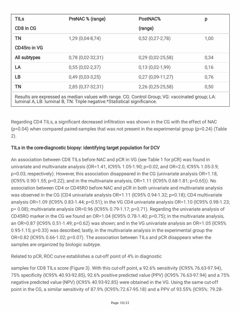
Page 10/21
TILs
CD8 in CG
PreNAC % (range) PostNAC%
(range)
p
TN
CD45ro in VG
1,29 (0,04-8,74) 0,52 (0,27-2,78) 1,00
All subtypes 0,78 (0,02-32,31) 0,29 (0,02-25,58) 0,34
LA 0,55 (0,02-2,37) 0,13 (0,02-1,99) 0,16
LB 0,49 (0,03-3,25) 0,27 (0,09-11,27) 0,76
TN 2,85 (0,37-32,31) 2,26 (0,25-25,58) 0,50
Results are expressed as median values with range. CG: Control Group; VG: vaccinated group; LA:luminal A, LB: luminal B, TN: Triple negative.*Statistical signi�cance.
Regarding CD4 TILs, a signi�cant decreased in�ltration was shown in the CG with the effect of NAC(p=0.04) when compared paired-samples that was not present in the experimental group (p=0.24) (Table2).
TILs in the core-diagnostic biopsy: identifying target population for DCV
An association between CD8 TILs before NAC and pCR in VG (see Table 1 for pCR) was found inunivariate and multivariate analysis (OR=1.41, IC95% 1.05-1.90; p=0.02, and OR=2.0, IC95% 1.05-3.9;p=0.03, respectively). However, this association disappeared in the CG (univariate analysis OR=1.18,(IC95% 0.90-1.55; p=0.22); and in the multivariate analysis, OR=1.11 (IC95% 0.68-1.81; p=0.65)). Noassociation between CD4 or CD45RO before NAC and pCR in both univariate and multivariate analysiswas observed in the CG (CD4 univariate analysis OR=1.11 (IC95% 0.94-1.32; p=0.18); CD4 multivariateanalysis OR=1.09 (IC95% 0.83-1.44; p=0.51); in the VG CD4 univariate analysis OR=1.10 (IC95% 0.98-1.23;p= 0.08); multivariate analysis OR=0.96 (IC95% 0.79-1.17; p=0.71). Regarding the univariate analysis ofCD45RO marker in the CG we found an OR=1.04 (IC95% 0.78-1.40; p=0.75); in the multivariate analysis,an OR=0.87 (IC95% 0.51-1.49; p=0.62) was shown; and in the VG univariate analysis an OR=1.05 (IC95%0.95-1.15; p=0.33) was described; lastly, in the multivariate analysis in the experimental group theOR=0.82 (IC95% 0.66-1.02; p=0.07). The association between TILs and pCR disappears when thesamples are organized by biologic subtype.
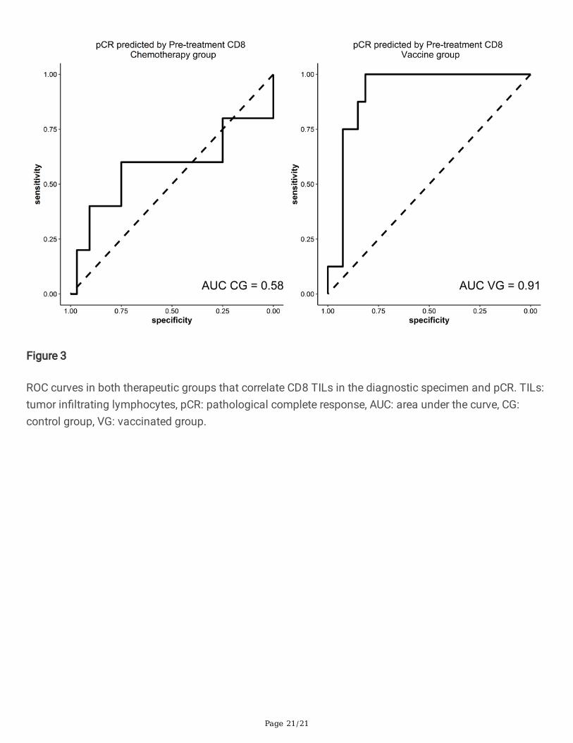
Related to pCR, ROC curve establishes a cut-off point of 4% in diagnostic
samples for CD8 TILs score (Figure 3). With this cut-off point, a 92.6% sensitivity (IC95% 76.63-97.94),75% speci�city (IC95% 40.93-92.85), 92.6% positive predicted value (PPV) (IC95% 76.63-97.94) and a 75%negative predicted value (NPV) (IC95% 40.93-92.85) were obtained in the VG. Using the same cut-offpoint in the CG, a similar sensitivity of 87.9% (IC95%:72.67-95.18) and a PPV of 93.55% (IC95%: 79.28-
Page 11/21
98.21) was reached, although the speci�city and NPV were 50% (IC95%: 15-85) and 33.33% (IC95%: 9.6-70), respectively. Differences between both groups were not statistically signi�cant (p=0.20).
Regarding outcome, we have calculated the percentage of patients that relapse regarding percentage ofTILs at diagnosis with a median follow-up of 8-years. With the established 4% TILs cut-off in thediagnostic specimen, 12.5% (1/8) patients with ≥4% CD8 TILs in the VG and 16.6% (1/6) in the CG havedisease progression; among those patients with <4% TILs, 7.4% (2/27) patients in the VG and 19% (8/42)in the CG relapsed. There is a trend in the VG to decrease relapse but due to the small sample size there isnot enough statistical power to get signi�cance (Fisher test, p=0.30).
No comparable results were found with CD4 and CD45RO TILs.
Residual DiseaseWe also correlate quanti�cation of TILs in residual samples with the level of pathological responsecategorized by Miller & Payne (3 and 4 M&P grades). Although sample size was pretty limited, we havenot seen correlation among both variables (Spearman´s rank correlation P=0.77), although 23 vaccinatedpatients with residual disease reached moderate to high pathological responses (3 and 4 M&P grades)compared to 4 patients with poorer pathological responses (1 and 2 M&P grades).
We have not established a cut-off point in the RD related to outcome as we have performed withindiagnostic biopsy, because our simple size is pretty limited. In fact, we have only 4 and 3 patients in theVG and CG respectively with ≥ 4% TILs. Results should not be reliable under these conditions.
DiscussionTo our knowledge, this is the �rst report that identi�es good responders to DCV in combination with NACrelated to CD8 expression in the needle core biopsy with the established 4% cut-off point. Moreover, this isthe �rst time that biomarker changes in the tumor and its milieu are reported in RD in naïve BC patientstreated with standard NAC ± active cell based-immunotherapy, by showing maintained CD4 levels and atrend to a rise in CD8 in�ltration (only in TNBC). We have also appreciate a trend to a better outcomewhen TILs were higher in RD, with less relapse events in the VG. Clinical impact of these �ndings could berelevant to improve selection of: 1) patients that could bene�t from the addition of DCV to NAC; and 2)the best maintenance therapy in TNBC subtypes with RD based on the expression of CD8 and CD4markers to choose standard capecitabine [8, 25] versus immunotherapy. Modi�cations on the tumorniche that re�ects immune activation after DCV helps to be open wide to less toxic and more speci�cimmunotherapy.
Regarding TILs, TNBC patients reach higher levels than the luminal subtypes in our study, as describedbefore [5]. This could be in part due to speci�c mutational signatures, copy number variations, stromalmetagenes and clonal heterogeneity of TNBC subtype [26–28].
Page 12/21
We have shown a non-signi�cant increase in stromal TILs after combined therapies with NAC plus DCV inup to 66% of TNBC patients, but not in the luminal subtypes. Probably our results are not signi�cant dueto the small sample size, being one limitation, and conferring the study an exploratory role that should bevalidated in other cohorts of patients.
Increased TILS could illustrate a reinforcement of the immune niche produced by DCV in BC patients. Itcan be suggested that vaccines could produce an increase in CD8 T cells post-treatment in TNBC. Wakset al. found that luminal BC are less enriched in CD8 TILs than other BC subtypes and this cell populationdecreased after NAC [29]. Nowadays, there is not enough knowledge about how immunotherapy couldchange BC microenvironment. Results of the studies that have worked with paired samples (before andafter conventional NAC and no immunotherapy) remain controversial, suggesting different roles ofimmune cell populations in carcinogenesis, response to therapies, tumor progression and a crosstalkamong the tumor and the microenvironment. While some reports described a reduction of CD8 TILs incancer milieu after NAC [29–32], others shown an increase on TILs count [6, 33, 34] or an inversion on theCD4/CD8 ratio [4]. Luen et al. described an increase in TILs level in 48% of the patients and a decreasedin 47% of the patients in RD after NAC [35].
The input of immunotherapy in BC patients in the neoadjuvant scenario looks for a global improvementwithin the tumor (higher pCR), the niche (hottest tumors) and the systemic immune surveillance. Dendriticcell-based adjuvant immunotherapy has already shown a gain in CD8 T cells in peripheral blood in non-luminal BC with an encouraging PFS improvement, suggesting bene�ts in the systemic immunity [16].Vaccination is more effective in the prevention of tumor growth, and probably the clinical advantagecould be more relevant in patients with a small tumor burden and with a preserved immune system (naïveof therapies) than in large tumor burden patients, including metastatic scenario, and with an exhaustedimmune system. Thus, immune cell pro�le of primary versus metastases of BC patients is different [36,37].
Current efforts have been made to standardize the quanti�cation of TILs and to produce reliable results[38–41]. Some authors pointed out that the reproducibility of TILs evaluation improves when thecategories are simpli�ed to low versus high TILs, based on the concept of lymphocyte-predominantbreast cancer phenotype [42] but the standard cut-off points need to be de�ned. The incorporation ofdigital analysis (ACIS III) provided a better quanti�cation of the stromal TILs avoiding interobserver bias.Additionally, other aspect to be considered is the selection of the area to be evaluated and how many�elds should be selected. Recent works support the fact that the average lymphocyte score from a singlebiopsy of a tumor is reasonably representative of the whole cancer [43]. Our results showed that a cut-offpoint of 4% CD8 in the diagnostic specimen could predict a better pathologic response when DCV areadded to conventional NAC, with a lower relapse (not signi�cant). As it can be observed in Figure 1, thiscut-off could be considered a relatively high density of CD8 cells in the stromal tumor. This opticalmicroscope immunohistochemical image could be used as a reference of what we consider as theminimal density of cells which correlates with response to vaccines.
Page 13/21
Qingzhu et al., found in their meta-analysis that memory T lymphocyte in�ltration of a tumor site couldserve as a indicator for OS and DFS prediction in patients with malignant tumours [44]. Also, Yajima et al.,�nd a relationship between high CD45RO (i.e memory T lymphocyte and marker of activated T cells)expression and a lower pathological stage in BC patients [45]. The importance of CD4 TILs is not clear inBC scenario. No association between CD4 and pCR was found in our study, although in the CG CD4 TILssigni�cantly decreased in patients after NAC (p=0.04) as compared to those who received theexperimental therapy which remained stable (p=0.24) (Table 2). However, most of the studies correlateCD4 or CD4/CD8 ratio with an increased pCR and better survival [34, 5], and this fact strengths the role ofDCV.
We have not look for tertiary lymphoid structures in this study although they are seen occasionally in thesamples. Its neogenesis is not speci�c for tumors and immune therapies applied in oncology, so theyalso appear in autoimmune and infectious diseases, trasplanted solid organs, in�ammatory disorders aswell as after preventive vaccines (eg; HPV).
Although more studies are needed to establish the bene�t of DCV addition to chemotherapy, our resultssuggest that we are moving in the right direction. Identi�cation of predictive and prognostic biomarkers toselect patients that could bene�t from the addition of immunotherapy to the standard systemicchemotherapy is key to develop more e�cient therapeutic strategies [47]. The relationship betweenlymphocyte-predominant breast cancer patients in the case of the TNBC [48] and the pCR, PFS and OS isclearly established [49–51]. Nonetheless, an outstanding selection of good responders to immunotherapyis complex [52] and the study of biomarkers in RD after NAC plus immunotherapy as well as in peripheralblood is mandatory to improve this limitation.
Evaluation of tumor responses to immune strategies by imaging techniques has become tricky becausechanges in tumor burden need new response criteria based on special guidelines (iRECIST) [53]. In thisway, biologic markers in the blood, the tumor and its milieu could contribute to a more speci�cinformation than imaging markers regarding patient selection for immune strategies in the early BCarena.
In conclusion, a 4% cut-off point of CD8 TILs in a TNBC subtype could help to establish which patientscan bene�t from DCV added to NAC. In the same way, a maintained expression of CD4 as well as anincreased in�ltration of stromal CD8 after DCV therapy could improve responses to further therapies, asseen in other solid tumors after relapse to IMT strategy when patients were treated with second linechemotherapy in the advanced scenario [6, 7]. A trend to a lower relapse in diagnostic biopsies enrichedwith TILs has been also shown. Our �ndings suggest that patients with TNBC especially bene�t from thestimulation of the antitumor immune system by using DCV pulsed with tumor antigens. Deeper studiesbased on immune pro�ling genomic panels in the tumor and immune cell populations on peripheralblood are still ongoing within our patients and could help us to elucidate the biological behavior of BC aswell as the bene�ts from adding immunotherapy to conventional NAC. Promising combined immune
Page 14/21
approaches potentiating immune system with DCV added to the blockade of immune checkpointstogether with NAC should be tested in clinical trials in this selected population.
DeclarationsFunding
This work is supported by the Ministerio de Sanidad y Política Social in the section of AdvancedTherapies [TRA-005] and by the Ministerio de Ciencia e Innovación [PI16/01245], Gobierno de España.
Competing interest
M. Santisteban has received honoraria from Roche, P�zer and Novartis and travel
support from Roche, P�zer and Miltenyi. A. López-Díaz de Cerio and S. Inogés have travel support fromMiltenyi. No other disclosures were reported. All remaining authors have declared no con�ict of interest.
Availability of data and materials
The datasets generated and analysed during the current study are available from the corresponding autoron reasonable request.
Code availability Not applicable.
Authors´ contributions
L.M collected data, built the dataset, performed the analysis and helped write and revise the manuscript.A. L.J collected data and helped revise the manuscript. A. C collected data and helped revise themanuscript. P. S collected data and helped revise the manuscript. B.P.S helped to perform the statisticalanalysis and revise the manuscript. S.I made the dendritic cell vaccines and helped revise the manuscript.A.L.D.C. made the dendritic cell vaccines and helped revise the manuscript. F.G helped to perform thestatistical analysis. O. F.H helped recruit patients and collect data. S.D.L.C helped recruit patients. L.H.collect data. MD. L collect data. M.I designed the samples assessment, collect data and helped write andrevise the manuscript. M.S. designed the clinical trial, collected data and helped write and revise themanuscript.
Ethics approval and consent to participate
This study was approved by the Navarra University Ethics Committee
Consent for publication Not applicable.
Acknowledgements
We acknowledge Jaione García for her support and technical expertise in Immunohistochemistry.
Page 15/21
References1. Savas P, Salgado R, Denkert C, et al (2016) Clinical relevance of host immunity in breast cancer: from
TILs to the clinic. Nat Rev Clin Oncol 13:228–241. https://doi.org/10.1038/nrclinonc.2015.215
2. Ali HR, Provenzano E, Dawson S-J, et al (2014) Association between CD8+ T-cell in�ltration andbreast cancer survival in 12 439 patients. Annals of Oncology 25:1536–1543.https://doi.org/10.1093/annonc/mdu191
3. Wang K, Xu J, Zhang T, Xue D (2016) Tumor-in�ltrating lymphocytes in breast cancer predict theresponse to chemotherapy and survival outcome: A meta-analysis. Oncotarget 7:44288–44298.https://doi.org/10.18632/oncotarget.9988
4. García-Martínez E, Gil GL, Benito AC, et al (2014) Tumor-in�ltrating immune cell pro�les and theirchange after neoadjuvant chemotherapy predict response and prognosis of breast cancer. BreastCancer Res 16:488. https://doi.org/10.1186/s13058-014-0488-5
5. Denkert C, von Minckwitz G, Darb-Esfahani S, et al (2018) Tumour-in�ltrating lymphocytes andprognosis in different subtypes of breast cancer: a pooled analysis of 3771 patients treated withneoadjuvant therapy. The Lancet Oncology 19:40–50. https://doi.org/10.1016/S1470-2045(17)30904-X
�. Dieci MV, Criscitiello C, Goubar A, et al (2014) Prognostic value of tumor-in�ltrating lymphocytes onresidual disease after primary chemotherapy for triple-negative breast cancer: a retrospectivemulticenter study. Annals of Oncology 25:611–618. https://doi.org/10.1093/annonc/mdt556
7. Hwang HW, Jung H, Hyeon J, et al (2019) A nomogram to predict pathologic complete response(pCR) and the value of tumor-in�ltrating lymphocytes (TILs) for prediction of response toneoadjuvant chemotherapy (NAC) in breast cancer patients. Breast Cancer Res Treat 173:255–266.https://doi.org/10.1007/s10549-018-4981-x
�. Masuda N, Lee S-J, Ohtani S, et al (2017) Adjuvant Capecitabine for Breast Cancer after PreoperativeChemotherapy. N Engl J Med 376:2147–2159. https://doi.org/10.1056/NEJMoa1612645
9. Schmid P, Cortes J, Pusztai L, et al (2020) Pembrolizumab for Early Triple-Negative Breast Cancer. NEngl J Med 382:810–821. https://doi.org/10.1056/NEJMoa1910549
10. Nanda R, Liu MC, Yau C, et al (2020) Effect of Pembrolizumab Plus Neoadjuvant Chemotherapy onPathologic Complete Response in Women With Early-Stage Breast Cancer: An Analysis of theOngoing Phase 2 Adaptively Randomized I-SPY2 Trial. JAMA Oncol 6:676.https://doi.org/10.1001/jamaoncol.2019.6650
11. Mittendorf EA, Zhang H, Barrios CH, et al (2020) Neoadjuvant atezolizumab in combination withsequential nab-paclitaxel and anthracycline-based chemotherapy versus placebo and chemotherapyin patients with early-stage triple-negative breast cancer (IMpassion031): a randomised, double-blind,phase 3 trial. The Lancet 396:1090–1100. https://doi.org/10.1016/S0140-6736(20)31953-X
12. Loibl S, Untch M, Burchardi N, et al (2019) A randomised phase II study investigating durvalumab inaddition to an anthracycline taxane-based neoadjuvant therapy in early triple-negative breast cancer:
Page 16/21
clinical results and biomarker analysis of GeparNuevo study. Annals of Oncology 30:1279–1288.https://doi.org/10.1093/annonc/mdz158
13. Martin-Romano P, Ammari S, El-Dakdoukti Y, et al (2020) Chemotherapy beyond immune checkpointinhibitors in patients with metastatic colorectal cancer. European Journal of Cancer 137:117–126.https://doi.org/10.1016/j.ejca.2020.06.030
14. Dwary AD, Master S, Patel A, et al (2017) Excellent response to chemotherapy post immunotherapy.Oncotarget 8:91795–91802. https://doi.org/10.18632/oncotarget.20030
15. Fields RC, Shimizu K, Mulé JJ (1998) Murine dendritic cells pulsed with whole tumor lysates mediatepotent antitumor immune responses in vitro and in vivo. PNAS 95:9482–9487.https://doi.org/10.1073/pnas.95.16.9482
1�. Qi C-J, Ning Y-L, Han Y-S, et al (2012) Autologous dendritic cell vaccine for estrogen receptor(ER)/progestin receptor (PR) double-negative breast cancer. Cancer Immunology, Immunotherapy61:1415–1424. https://doi.org/10.1007/s00262-011-1192-2
17. Trial With Autologous Dendritic Cell Vaccination in Patients With Stage II-III HER2 Negative BreastCancer - Full Text View - ClinicalTrials.gov
1�. Rodriguez J, Castañón E, Perez-Gracia JL, et al (2018) A randomized phase II clinical trial of dendriticcell vaccination following complete resection of colon cancer liver metastasis. j immunotherapycancer 6:96. https://doi.org/10.1186/s40425-018-0405-z
19. Inogés S, Tejada S, de Cerio AL-D, et al (2017) A phase II trial of autologous dendritic cell vaccinationand radiochemotherapy following �uorescence-guided surgery in newly diagnosed glioblastomapatients. J Transl Med 15:104. https://doi.org/10.1186/s12967-017-1202-z
20. Solans BP, López‐Díaz de Cerio A, Elizalde A, et al (2019) Assessing the impact of the addition ofdendritic cell vaccination to neoadjuvant chemotherapy in breast cancer patients: A model‐basedcharacterization approach. Br J Clin Pharmacol 85:1670–1683. https://doi.org/10.1111/bcp.13947
21. Coates AS, Winer EP, Goldhirsch A, et al (2015) Tailoring therapies—improving the management ofearly breast cancer: St Gallen International Expert Consensus on the Primary Therapy of Early BreastCancer 2015. Annals of Oncology 26:1533–1546. https://doi.org/10.1093/annonc/mdv221
22. Salgado R, Denkert C, Demaria S, et al (2015) The evaluation of tumor-in�ltrating lymphocytes (TILS)in breast cancer: Recommendations by an International TILS Working Group 2014. Annals ofOncology
23. Rojo MG, Bueno G, Slodkowska J (2010) Review of imaging solutions for integrated quantitativeimmunohistochemistry in the Pathology daily practice. Folia Histochem Cytobiol 47:349–354.https://doi.org/10.2478/v10042-008-0114-4
24. Słodkowska J, Filas V, Buszkiewicz E, et al (2010) Study on breast carcinoma Her2/neu andhormonal receptors status assessed by automated images analysis systems: ACIS III (Dako) andScanScope (Aperio). Folia Histochem Cytobiol 48:19–25. https://doi.org/10.2478/v10042-010-0015-1
Page 17/21
25. Lluch A, Barrios CH, Torrecillas L, et al (2020) Phase III Trial of Adjuvant Capecitabine After StandardNeo-/Adjuvant Chemotherapy in Patients With Early Triple-Negative Breast Cancer (GEICAM/2003-11_CIBOMA/2004-01). JCO 38:203–213. https://doi.org/10.1200/JCO.19.00904
2�. METABRIC Group, Curtis C, Shah SP, et al (2012) The genomic and transcriptomic architecture of2,000 breast tumours reveals novel subgroups. Nature 486:346–352.https://doi.org/10.1038/nature10983
27. Bianchini G, Qi Y, Alvarez RH, et al (2010) Molecular Anatomy of Breast Cancer Stroma and ItsPrognostic Value in Estrogen Receptor–Positive and –Negative Cancers. JCO 28:4316–4323.https://doi.org/10.1200/JCO.2009.27.2419
2�. Wali VB, Langdon CG, Held MA, et al (2017) Systematic Drug Screening Identi�es Tractable TargetedCombination Therapies in Triple-Negative Breast Cancer. Cancer Res 77:566–578.https://doi.org/10.1158/0008-5472.CAN-16-1901
29. Waks AG, Stover DG, Guerriero JL, et al (2019) The Immune Microenvironment in HormoneReceptor–Positive Breast Cancer Before and After Preoperative Chemotherapy. Clin Cancer Res25:4644–4655. https://doi.org/10.1158/1078-0432.CCR-19-0173
30. Loi S, Dushyanthen S, Beavis PA, et al (2016) RAS/MAPK Activation Is Associated with ReducedTumor-In�ltrating Lymphocytes in Triple-Negative Breast Cancer: Therapeutic Cooperation BetweenMEK and PD-1/PD-L1 Immune Checkpoint Inhibitors. Clin Cancer Res 22:1499–1509.https://doi.org/10.1158/1078-0432.CCR-15-1125
31. Pelekanou V, Barlow WE, Nahleh ZA, et al (2018) Tumor-In�ltrating Lymphocytes and PD-L1Expression in Pre- and Posttreatment Breast Cancers in the SWOG S0800 Phase II NeoadjuvantChemotherapy Trial. Mol Cancer Ther 17:1324–1331. https://doi.org/10.1158/1535-7163.MCT-17-1005
32. (2018) Mesenchymal–epithelial Transition and Tumor Vascular Remodeling in EribulinChemotherapy for Breast Cancer. AR 38:. https://doi.org/10.21873/anticanres.12236
33. Demaria S, Volm MD, Shapiro RL, et al (2001) Development of Tumor-in�ltrating Lymphocytes inBreast Cancer after Neoadjuvant Paclitaxel Chemotherapy. Clin Cancer Res 7:3025–3030
34. Miyashita M, Sasano H, Tamaki K, et al (2015) Prognostic signi�cance of tumor-in�ltrating CD8+ andFOXP3+ lymphocytes in residual tumors and alterations in these parameters after neoadjuvantchemotherapy in triple-negative breast cancer: a retrospective multicenter study. Breast Cancer Res17:124. https://doi.org/10.1186/s13058-015-0632-x
35. Luen SJ, Salgado R, Dieci MV, et al (2019) Prognostic implications of residual disease tumor-in�ltrating lymphocytes and residual cancer burden in triple-negative breast cancer patients afterneoadjuvant chemotherapy. Annals of Oncology 30:236–242.https://doi.org/10.1093/annonc/mdy547
3�. Szekely B, Bossuyt V, Li X, et al (2018) Immunological differences between primary and metastaticbreast cancer. Annals of Oncology 29:2232–2239. https://doi.org/10.1093/annonc/mdy399
Page 18/21
37. Finn OJ, Forni G (2002) Prophylactic cancer vaccines. Current Opinion in Immunology 14:172–177.https://doi.org/10.1016/S0952-7915(02)00317-5
3�. Hendry S, Salgado R, Gevaert T, et al (2017) Assessing Tumor-In�ltrating Lymphocytes in SolidTumors: A Practical Review for Pathologists and Proposal for a Standardized Method from theInternational Immuno-Oncology Biomarkers Working Group. Advances In Anatomic Pathology24:311–335. https://doi.org/10.1097/PAP.0000000000000161
39. Jang N, Kwon HJ, Park MH, et al (2018) Prognostic Value of Tumor-In�ltrating Lymphocyte DensityAssessed Using a Standardized Method Based on Molecular Subtypes and Adjuvant Chemotherapyin Invasive Breast Cancer. Ann Surg Oncol 25:937–946. https://doi.org/10.1245/s10434-017-6332-2
40. Swisher SK, Wu Y, Castaneda CA, et al (2016) Interobserver Agreement Between PathologistsAssessing Tumor-In�ltrating Lymphocytes (TILs) in Breast Cancer Using Methodology Proposed bythe International TILs Working Group. Ann Surg Oncol 23:2242–2248.https://doi.org/10.1245/s10434-016-5173-8
41. Denkert C, Wienert S, Poterie A, et al (2016) Standardized evaluation of tumor-in�ltratinglymphocytes in breast cancer: results of the ring studies of the international immuno-oncologybiomarker working group. Mod Pathol 29:1155–1164. https://doi.org/10.1038/modpathol.2016.109
42. Tramm T, Di Caterino T, Jylling A-MB, et al (2018) Standardized assessment of tumor-in�ltratinglymphocytes in breast cancer: an evaluation of inter-observer agreement between pathologists. ActaOncologica. https://doi.org/10.1080/0284186X.2017.1403040
43. Mani NL, Schalper KA, Hatzis C, et al (2016) Quantitative assessment of the spatial heterogeneity oftumor-in�ltrating lymphocytes in breast cancer. Breast Cancer Research 18:1–10.https://doi.org/10.1186/s13058-016-0737-x
44. Jia Q, Yang Y, Wan Y (2015) Tumor-in�ltrating memory T-lymphocytes for prognostic prediction incancer patients: a meta-analysis. Int J Clin Exp Med 8:1803–1813
45. Yajima R, Yajima T, Fujii T, et al (2016) Tumor-in�ltrating CD45RO+ memory cells are associated witha favorable prognosis breast cancer. Breast Cancer 23:668–674. https://doi.org/10.1007/s12282-015-0622-y
4�. Gu-Trantien C, Loi S, Garaud S, et al (2013) CD4+ follicular helper T cell in�ltration predicts breastcancer survival. J Clin Invest 123:2873–2892. https://doi.org/10.1172/JCI67428
47. Angell H, Galon J (2013) From the immune contexture to the Immunoscore: The role of prognosticand predictive immune markers in cancer. Current Opinion in Immunology
4�. Hennigs A, Riedel F, Gondos A, et al (2016) Prognosis of breast cancer molecular subtypes in routineclinical care : A large prospective cohort study. BMC Cancer 1–9. https://doi.org/10.1186/s12885-016-2766-3
49. Ibrahim EM, Al-Foheidi ME, Al-Mansour MM, Kazkaz GA (2014) The prognostic value of tumor-in�ltrating lymphocytes in triple-negative breast cancer: a meta-analysis. Breast Cancer Res Treat148:467–476. https://doi.org/10.1007/s10549-014-3185-2
Page 19/21
50. Fridman WH, Pagès F, Sautès-Fridman C, Galon J (2012) The immune contexture in human tumours:impact on clinical outcome. Nat Rev Cancer 12:298–306. https://doi.org/10.1038/nrc3245
51. Loi S, Sirtaine N, Piette F, et al (2013) Prognostic and Predictive Value of Tumor-In�ltratingLymphocytes in a Phase III Randomized Adjuvant Breast Cancer Trial in Node-Positive Breast CancerComparing the Addition of Docetaxel to Doxorubicin With Doxorubicin-Based Chemotherapy: BIG 02-98. JCO 31:860–867. https://doi.org/10.1200/JCO.2011.41.0902
52. Emens LA (2008) Cancer vaccines: on the threshold of success. Expert Opinion on Emerging Drugs13:295–308. https://doi.org/10.1517/14728214.13.2.295
53. Seymour L, Bogaerts J, Perrone A, et al (2017) iRECIST: guidelines for response criteria for use intrials testing immunotherapeutics. The Lancet Oncology 18:e143–e152.https://doi.org/10.1016/S1470-2045(17)30074-8
Figures
Figure 1
Immunohistochemistry measurement and scoring with ACIS III in a breast cancer sample. A) Imageanalysis by ACIS III of a biopsy, two stromal areas selected (0.43mm2). B) TILs quanti�cation by ACIS III,
Page 20/21
in red CD8 lymphocytes 5%. Scanned at 10X. Magni�cation of 50%.
Figure 2
Boxplots diagram with the distribution of CD8 TILs before and after neoadjuvant chemotherapyaccording to biological subtype and group of treatment. Each dot represents the value for each patient. Y-axis is shown in logarithmic scale. TILs: tumor in�ltrating lymphocytes, LA: luminal A, LB: luminal B, TN:triple negative BC, CG: control group, VG: vaccinated group.
Page 21/21
Figure 3
ROC curves in both therapeutic groups that correlate CD8 TILs in the diagnostic specimen and pCR. TILs:tumor in�ltrating lymphocytes, pCR: pathological complete response, AUC: area under the curve, CG:control group, VG: vaccinated group.