Embed Size (px)
Citation preview

OR I G I N A L A R T I C L E
Appropriate use criteria in dermatopathology: Initialrecommendations from the American Society ofDermatopathology
Task Force / Committee Members:
Claudia I. Vidal1 | Eric A. Armbrect2 | Aleodor A. Andea3 | Angela K. Bohlke4 |
Nneka I. Comfere5 | Sarah R. Hughes6 | Jinah Kim7 | Jessica A. Kozel8 | Jason B. Lee9 |
Konstantinos Linos10 | Brandon R. Litzner11,12 | Tricia A. Missall1 | Roberto A. Novoa7 |
Uma Sundram13 | Brian L. Swick14 | Maria Yadira Hurley (Chair)1 |
Rating Panel:
Murad Alam15 | Zsolt Argenyi16 | Lyn M. Duncan17 | Dirk M. Elston18 |
Patrick O. Emanuel19 | Tammie Ferringer20 | Maxwell A. Fung21 | Gregory A. Hosler22 |
Alexander J. Lazar23 | Lori Lowe3 | Jose A. Plaza24 | Victor G. Prieto23 |
June K. Robinson25 | Andras Schaffer26 | Antonio Subtil27 | Wei-Lien Wang28
1Departments of Dermatology and Pathology, Saint Louis University School of Medicine, St. Louis, Missouri
2Center for Health Outcomes Research, Saint Louis University, St. Louis, Missouri
3Departments of Dermatology and Pathology, University of Michigan Medical Center, Ann Arbor, Michigan
4Silver Falls Dermatology, Salem, Oregon
5Department of Dermatology and Laboratory Medicine and Pathology, Mayo Clinic, Rochester, Minnesota
6Department of Pathology, Gundersen Health System, La Crosse, Wisconsin
7Departments of Dermatology and Pathology, Stanford University School of Medicine, Stanford, California
8Midwest Pathology Associates, LLC, Overland Park, Kansas
9Departments of Dermatology and Cutaneous Biology, Sidney Kimmel Medical College at Thomas Jefferson University, Philadelphia, Pennsylvania
10Department of Pathology and Laboratory Medicine, Dartmouth-Hitchcock Medical Center and Geisel School of Medicine at Dartmouth Lebanon, Hanover,
New Hampshire
The authors Dr. C.I. Vidal, Dr. E.A. Armbrect, Dr. A.A. Andea, Dr. A.K. Bohlke, Dr. N.I. Comfere, Dr. S.R. Hughes, Dr. J. Kim, Dr. J.A. Kozel, Dr. J.B. Lee, Dr. K. Linos,
Dr. B.R. Litzner, Dr. T.A. Missall, Dr. R.A. Novoa, Dr. U. Sundram, Dr. B.L. Swick, and Dr. M. Yadira Hurley are members of the Appropriate Use Criteria (AUC)
Committee.
The authors Dr. M. Alam, Dr. Z. Argenyi, Dr. L.M. Duncan, Dr. D.M. Elston, Dr. P.O. Emanuel, Dr. T. Ferringer, Dr. M.A. Fung, Dr. G.A. Hosler, Dr. A.J. Lazar, Dr. L.
Lowe, Dr. J.A. Plaza, Dr. V.G. Prieto, Dr. J.K. Robinson, Dr. A. Schaffer, and Dr. A. Subtil, Dr. W.-L. Wang are members of the Rating Panel.
Dr. M. Yadira Hurley, as designated, was the Chair of AUC Committee and is the senior author.
The authors Dr. J. Kim, Dr. J.A. Kozel, and Dr. B.R. Litzner, U. Sundram are AUC subgroup lead.
The authors Dr. Z. Argenyi, Dr. L.M. Duncan, Dr. P.O. Emanuel, Dr. T. Ferringer, Dr. M.A. Fung, Dr. G.A. Hosler, Dr. L. Lowe, Dr. J.A. Plaza, Dr. V.G. Prieto, Dr. A.
Subtil, and Dr. W.-L. Wang are ASDP representative.
Dr. D.M. Elston was the ASDP President during time of AUC development.
Dr. M. Alam and Dr. J.K. Robinson, are representative of American Academy of Dermatology (AAD).
The authors Dr. A.J. Lazar and Dr. A. Schaffer, are representative of College of American Pathologist (CAP).
Please see related paper on pages [561–562] of this issue.
Received: 3 November 2017 Revised: 21 February 2018 Accepted: 13 March 2018
DOI: 10.1111/cup.13142
© 2018 John Wiley & Sons A/S. Published by John Wiley & Sons Ltd
J Cutan Pathol. 2018;45:563–580. wileyonlinelibrary.com/journal/cup 563

11Departments of Dermatology and Pathology, Via Christi Clinic, Ascension Medical Group, Wichita, Kansas
12Department of Family Medicine, University of Kansas Medical Center-Wichita, Wichita, Kansas
13Department of Anatomic Pathology, Oakland University William Beaumont School of Medicine and Beaumont Health Systems, Royal Oak, Michigan
14Departments of Dermatology and Pathology, University of Iowa, Iowa City, Iowa
15Departments of Dermatology Otolaryngology, and Surgery, Feinberg School of Medicine, Northwestern University, Chicago, Illinois
16Department of Pathology, University of Washington, Seattle, Washington
17Pathology Service and Dermatopathology Unit, Massachusetts General Hospital and Harvard Medical School, Boston, Massachusetts
18Department of Dermatology, Dermatologic Surgery, Medical University of SC, Charleston, South Carolina
19Department of Pathology and Molecular Medicine, University of Auckland, Auckland, New Zealand
20Departments of Dermatology and Laboratory Medicine, Geisinger Medical Center, Danville, Pennsylvania
21Departments of Dermatology and Pathology and Laboratory Medicine, University of California, Davis School of Medicine, Sacramento, California
22ProPath; Departments of Dermatology and Pathology, University of Texas Southwestern Medical Center, Dallas, Texas
23Departments of Pathology, Dermatology, & Genomic Medicine, The University of Texas MD Anderson Cancer Center, Houston, Texas
24Miraca Life Sciences, Irving, Texas
25Department of Dermatology, Northwestern University, Chicago, Illinois
26Bey Dermatology and Cosmetic Surgery, Spring Hill, Florida
27Department of Dermatology, Yale School of Medicine, New Haven, Connecticut
28Departments of Pathology and Translational Molecular Pathology, Division of Pathology and Laboratory Medicine, The University of Texas
MD Anderson Cancer Center, Houston, Texas
Correspondence
Claudia I. Vidal, MD, PhD, Departments of
Dermatology and Pathology, Saint Louis
University School of Medicine, 1755 South
Grand Blvd. St. Louis, MO 63104.
Email: [email protected]
Disclaimer The recommendations presented in
this study were developed using the RAND
Corp methodology (Santa Monica, California)/
University of California-Los Angeles (RAND/
UCLA appropriateness method).
Appropriateness ratings represent the best
interpretation of the literature combined with
expert judgment at the time of their
development. The selection of a test
ultimately lies with the physician and the
assessment of multiple factors associated with
the individual patient. The clinical scenarios
used should not be considered inclusive of all
situations in which a test/study should or can
be performed. Future literature may require
changes to the recommendations based on
additional information.
Background: Appropriate use criteria (AUC) provide physicians guidance in test selection, and
can affect health care delivery, reimbursement policy and physician decision-making.
Objectives: The American Society of Dermatopathology, with input from the American Acad-
emy of Dermatology and the College of American Pathologists, sought to develop AUC in
dermatopathology.
Methods: The RAND/UCLA appropriateness methodology, which combines evidence-based medi-
cine, clinical experience and expert judgment, was used to develop AUC in dermatopathology.
Results: With the number of ratings predetermined at 3, AUC were developed for 211 clinical
scenarios involving 12 ancillary studies. Consensus was reached for 188 (89%) clinical scenarios,
with 93 (44%) considered “usually appropriate,” 52 (25%) “rarely appropriate” and 43 (20%)
“uncertain appropriateness.”
Limitations: The methodology requires a focus on appropriateness without comparison
between tests and irrespective of cost.
Conclusions: The ultimate decision of when to order specific test rests with the physician and
is one where the expected benefit exceeds the negative consequences. This publication outlines
the recommendations of appropriateness—AUC for 12 tests used in dermatopathology. Impor-
tantly, these recommendations may change considering new evidence. Results deemed “uncer-
tain appropriateness” and where consensus was not reached may benefit from further research.
KEYWORDS
ancillary studies, appropriate use criteria, dermatopathology, evidence-based medicine, expert
rating
1 | INTRODUCTION
Medical leaders and consumers are calling for a safer, more efficient
and effective health care system. In recent years, there has been an
exponential increase in the number of diagnostic tests. Given the
increase in cost from new technologies, physicians need tools to
help them make decisions about health care, especially in appropri-
ateness of care, that achieve value, increase quality and control
costs.1
Appropriate use criteria (AUC) combine the best scientific evi-
dence available with the collective judgment of experts to yield a
statement of the appropriateness for performing a test in specific
clinical scenarios encountered in everyday practice. Qualifying appro-
priateness is the first step in addressing cost-effectiveness as studies
have shown good correlation between the two.2
In 2015, the American Society of Dermatopathology (ASDP) cre-
ated the AUC Task Force to help guide dermatopathologists in their
use of ancillary studies. Four subgroups were established and each
564 VIDAL ET AL.
group chose 2 to 3 ancillary studies for which to develop AUC. The
subgroups were divided into 4 broad categories: lymphoproliferative,
melanocytic, soft tissue and other.
This report provides a synopsis of the AUC for the ancillary
studies chosen by each of the subgroups and developed using the
RAND/UCLA appropriateness method.3 The goal in a health system
is for inappropriate care to be reduced while necessary and appropri-
ate care is increased or maintained. It is imperative to understand
that the ancillary studies and clinical scenarios chosen are not
exhaustive and that this publication is not a comparison of the differ-
ent tests as each ancillary study was reviewed independently for each
clinical scenario. In addition, as literature emerges updates to the
AUC will need to be made and are already planned by the ASDP.
2 | MATERIALS AND METHODS
The AUC process combines evidence-based medicine with clinical sce-
narios and expert judgment by engaging a rating panel in a modified
Delphi exercise based on the validated appropriateness method of
RAND/UCLA to yield a statement regarding the appropriateness of
performing a test or procedure in a specific patient scenario. The pro-
cess begins by selection of tests or procedures for which AUC will be
created. In general, AUC focus on tests that are widely and frequently
used, consume significant resources or have wide variations in their
use. The process overview taken by the ASDP is outlined in (Figure 1).
In total, 12 dermatopathology ancillary studies underwent the
AUC process. These include 3 topics for the lymphoproliferative
group: T-cell receptor (TCR) clonality assay for the beta chain by
polymerase chain reaction (PCR), TCR clonality assay for the gamma
chain by PCR and B-cell receptor immunoglobulin heavy chain (IgH)
clonality assay by PCR. The melanocytic group also examined 3 topics:
fluorescence in situ hybridization (FISH), comparative genomic
hybridization (CGH) and gene expression profiling by quantitative
reverse transcription polymerase chain reaction (qRT-PCR) for mela-
nocytic lesions. The other group explored 4 topics: human papilloma-
virus (HPV) in situ hybridization (ISH; HPV subtypes 6, 11, 16, 18,
31 and 33), HPV immunohistochemistry (IHC; Abcam HPV subtypes
1, 6, 11, 16, 18, 31/Dako HPV subtypes 6, 11, 16, 18, 31, 33, 42, 51,
52, 56 and 58) and mismatch repair (MMR) protein IHC 4 antibody
panel (MSH2, MLH1, MSH6 and PMS2) and MMR IHC 2 antibody
panel (MSH2 and MLH1) in the screening for Muir-Torre syndrome
(MTS). Finally, the soft tissue group explored 2 topics: t(17;22)
COL1A1-PDGFB FISH assay in the diagnosis of dermatofibrosarcoma
protuberans (DFSP) and dual-color break-apart EWSR1 FISH assay in
differentiating melanocytic tumors and clear cell sarcoma (CSS).
2.1 | Development of definitions and clinicalindications
Each of the 4 subgroups developed a set of definitions to clearly
explain the meaning of assigned terms and histologic diagnoses as well
as developed clinical scenarios (“indications”) to simulate situations
most likely to be encountered in clinical practice. A total of 211 clinical
scenarios were produced and then reviewed independently for
conciseness and completeness by 12 clinical indication reviewers com-
posed of dermatopathologists from across the country with expertise
in various areas. They were then modified accordingly such that they
comprised the most often encountered situations in dermatopathology
practice. The clinical scenarios were not intended to be exhaustive, but
to represent at least 85% of anticipated scenarios. They were based
on information that is readily available to dermatopathologists during
routine practice (age, body site, histomorphology, etc.). Further specific
information regarding definitions and clinical scenarios for each sub-
group is summarized in Tables 1–7.4–9
2.2 | Evidence
The development of AUC is founded on combining evidence review
and analysis with expert judgment that is provided by the panel
raters. A detailed literature review was performed by the AUC Task
Force to provide the best available evidence on each ancillary
study. The 4 subgroups received general guidelines for evidence
review including: journal articles written in English, search years
beginning in 1940 to 2016, overlapping studies removed and case
series with n > 3 included only if no other evidence was available.
In total, 239 articles were identified and summarized for the
development of the literature review tables that were provided to
the panel raters for use during rating. Each subgroup added additional
parameters if deemed necessary. Synopses of the best scientific
evidence for each of the ancillary studies chosen are separately
published in the Journal of Cutaneous Pathology.10–14
2.3 | Rating process
Seventeen panel raters were carefully selected for balance, expertise
in a field, and breadth of knowledge. Attempt to avoid selection of
panel raters with any financial conflict of interest was made. Twelve
panel raters (3 per topic) representing a cross section of academi-
cians and private practice physicians were chosen for their expertise
in each of the 4 subgroups and then approved by the Chair of the
AUC committee. Additionally, there were 2 representatives nomi-
nated by the American Academy of Dermatology to incorporate the
dermatologists' perspective (both were dermatologists, non-derma-
topathologists), 2 representatives nominated by the College of
American Pathologists (both were pathologists and dermatopatholo-
gists) to incorporate the broader pathology perspective, and a medi-
cal director from a regional Medicare carrier. The number of rating
rounds was predetermined at 3. Panel raters received the literature
review tables for all of the ancillary studies that included a general
summary by test/procedure, concise individual article summaries and
the exact citations. They were also provided with clinical scenario
booklets. All panel raters rated all of the ancillary studies and all rat-
ings were done independently by each panel rater, with the over-
arching objective being to converge in consensus. They were
instructed to rate the appropriateness of each clinical scenario using
their own best expert clinical judgment and the available literature.
They were specifically instructed to not consider cost during rating
and to rate each test/procedure independently, such that each test/
procedure was rated on its own merits. During each round, panelists
VIDAL ET AL. 565

were asked to rate each clinical scenario on a 9-point scale (shown
below). A score of 9 to 7 indicated the test/procedure belonged to a
category of “usually appropriate” where higher scores indicate
greater agreement within the category. A score of 1 to 3 indicated
the test/procedure is “rarely appropriate” in that specific clinical sce-
nario while acknowledging clinician discretion may be suitable for
ordering the test under selected circumstances. A lower score within
the range would indicate strength in conviction of the test being less
appropriate. The category nomenclature was chosen to reflect that
the ultimate decision to perform a test lies with the physician and
takes into account not only the clinical scenario, but also the
individual patient. Scores in the rage of 4 to 6 were used to indicate
“uncertain appropriateness” for ordering the test/procedure in that
clinical scenario. Scores in this midrange generally indicated panel
rater’s assessment that there was lack of scientific evidence for
the test/procedure in general or for that individual clinical
scenario. Insufficient scientific evidence could be due to the data
being considered as emergent or underdeveloped.
The first-round rating was done individually without interaction
with other panel raters or AUC Task Force members. Ratings were ana-
lyzed by 2 research team members who identified clinical scenarios
where there was no apparent consensus. After the initial rating, an in-
Selection of tests/ancillary studies
Literature review and summary tablesClinical scenarios / indications
Clinial indicationreviewers
Rating of indications (rating panel)Predetermined 3 rounds:
1st round no interaction
2nd round in-person meeting followed by independentrating
3rd round conference call (2) followed by independentrating
Rarely appropriateScore 1-3
Uncertain appropriatenessScore 4-6
Usually appropriateScore 7-9
FIGURE 1 Process overview taken by the American Society of Dermatopathology and appropriate use criteria (AUC) Task Force in the
development of the dermatopathology AUC
566 VIDAL ET AL.
person moderated meeting of the panel raters occurred during the 51st
annual ASDP meeting (Chicago, 2016). During this meeting, there was a
discussion of clinical scenarios where consensus had not been achieved.
The discussion was preceded by a literature review summary by an
AUC Task Force member for each ancillary study for which AUC were
being developed. The goal was to discuss the literature, draw from other
experts in the field while also being mindful of not requesting ratings or
influencing the panel to seek consensus. After the in-person meeting,
the second-round rating was done individually and submitted to the
research team within 2 weeks. Prior to the third-round rating, there were
2 moderated teleconference sessions, which focused on clinical scenarios
that were close to consensus. Panel raters explored wording of clinical
TABLE 1 Lymphoproliferative definitions and clinical scenarios for T-cell receptor clonality
Definitions:
Specific clinical entities in B-cell and T-cell subgroups categorized according to the 2008 WHO Classification were further examined.4
• “Diagnostic for” mycosis fungoides:○ Presence of nearly all typical histopathologic diagnostic features of mycosis fungoides (atypical lymphocytes with hyperchromatic,
cerebriform nuclei surrounded by clear haloes, epidermotropism of solitary lymphocytes or clusters of atypical lymphocytes in the absenceof spongiosis, epidermal lymphocytes larger than dermal lymphocytes)
○ Loss of 1 or more important T-cell marker (CD2, CD5 and/or CD7) within the neoplastic T-cell infiltrate along the dermoepidermal junctionand/or in the epidermis
○ Nearly all neoplastic cells express CD4 or CD8 (CD4 or CD8 significant predominance)• “Consistent with” mycosis fungoides:
○ Histopathologic diagnostic criteria of mycosis fungoides are present○ Epidermotropic atypical lymphocytes:
• Predominantly immunoreactive for CD2, CD3, CD4, CD5 and CD7 (partial)• Predominantly immunoreactive for CD4 or CD8• Loss of 1 or more mature T-cell markers (CD2, CD3, CD5 and CD7)
• “Concerning for,” “suspicious of” or “suggestive of” mycosis fungoides:○ Presence of 1 or more typical histopathologic diagnostic features of mycosis fungoides
• Atypical lymphocytes with hyperchromatic, cerebriform nuclei surrounded by clear haloes• Epidermotropism of solitary lymphocytes or clusters of atypical lymphocytes in the absence of spongiosis• Epidermal lymphocytes larger than dermal lymphocytes• Perivascular distribution of atypical lymphocytes (“bare underbelly” sign)• Papillary dermal fibrosis
○ Normal immunophenotypical features: T-cell lymphoid infiltrate along the dermoepidermal junction and/or in the epidermis that isimmunoreactive for CD2, CD3, CD5 and CD7 (partial or no loss) with a normal CD4:CD8 ratio
• "Not diagnostic for" mycosis fungoides:○ Limited/minimal/scant T-cell lymphoid infiltrate along the dermoepidermal junction and/or within the superficial dermal perivascular space○ Absence of lymphocyte epidermotropism or folliculotropism○ Absence of lymphocyte atypia○ Absence of papillary dermal fibrosis○ Normal immunophenotypical features: T-cell lymphoid infiltrate along the dermoepidermal junction and/or in the epidermis that is
immunoreactive for CD2, CD3, CD5 and CD7 (partial or no loss) with a normal CD4:CD8 ratio• Lymphomatoid papulosis (LyP):
○ Wedge-shaped mixed infiltrate of small and large lymphocytes with eosinophils and neutrophils, numerous CD30 positive large lymphocytes○ Or, scant to moderate mixed infiltrate with small and large lymphocytes with epidermotropism○ Or, dense diffuse infiltrate of large atypical CD30 positive lymphocytes
• Pityriasis lichenoides (PL):○ Mixed lichenoid and spongiotic dermatitis with mounds of parakeratosis, extravasated erythrocytes; large cells present○ Or, wedge-shaped superficial and deep dermal lymphocytic infiltrate with extravasated erythrocytes (lymphocytic vasculitis), epidermal
necrosis, parakeratosis, lichenoid reaction pattern; large cells present
Clinical scenarios:
1. Solitary or generalized scaly patches/plaques that are clinically concerning for mycosis fungoides (clinical impression: rule out mycosis fungoidesor cutaneous T-cell lymphoma) and that are histologically and immunophenotypically “concerning for”, “suspicious of” or “suggestive of” mycosisfungoides.”
2. Clinical presentation of erythroderma with clinical impression of rule out mycosis fungoides, cutaneous T-cell lymphoma or Sézary syndrome andthat is "not diagnostic for" mycosis fungoides.
3. Clinical presentation of dermatitis with clinical impression of rule out mycosis fungoides or cutaneous T-cell lymphoma and that is "not diagnosticfor" mycosis fungoides.
4. Inflammatory/reactive papular or papulonecrotic eruption (solitary, regional or generalized) with clinical impression of LyP or PL, rule out mycosisfungoides or cutaneous T-cell lymphoma and histopathologic and immunophenotypic features typical for LyP or PL.
5. The development of T-cell cutaneous infiltrate that is "not diagnostic for" mycosis fungoides but is present in a patient with a history of mycosisfungoides with a known T-cell clone (comparison of past and present clones).
6. The development of a T-cell cutaneous infiltrate in a patient with a history of systemic T-cell lymphoma.7. A cutaneous T-cell infiltrate with a folliculotropic rather than epidermotropic T-cell infiltrate.8. Pigmented purpuric patches (solitary, regional or generalized) and clinical impression of rule out mycosis fungoides or cutaneous T-cell lymphoma
and histopathologic and immunophenotypic features that are "not diagnostic for" mycosis fungoides.9. Clinically reactive entities (see references for individual diagnoses) with histologically and immunophenotypically “concerning for,” “suspicious of”
or “suggestive of” mycosis fungoides.10. Preexisting diagnosis of mycosis fungoides and new or evolving lesions similar to original lesions with clinical impression of rule out mycosis
fungoides in setting of preexisting mycosis fungoides and histopathologic and immunophenotypic features “consistent with” mycosis fungoides.11. Development of nodules in a patient with mycosis fungoides which are histologically “concerning for,” “suspicious of” or “suggestive of” large cell
transformation with CD30 positivity.12. Development of nodules in a patient with mycosis which are histologically “concerning for,” “suspicious of” or “suggestive of” large cell
transformation without CD30 positivity.
Abbreviations: PL, pityriasis lichenoides; LyP, lymphomatoid papulosis; WHO, World Health Organization.
VIDAL ET AL. 567

scenarios or definitional understandings that needed clarification. Panel
raters were also provided the statistical analysis based on results from
the first and second rounds. The third and final ratings were completed
individually, again within about 2 weeks of the teleconference sessions.
One panelist withdrew from the project after the first round; thus, the
complete data for all 3 rounds were provided by 16 panel raters.
To facilitate the panel rater discussion and support categoriza-
tion for each clinical scenario the mean of ratings was calculated;
the mean was adjusted by filtering/removing 2 scores—the highest
and lowest—to minimize the impact of outlying raters (mean’). A
mean’ of ≥7.0 was classified as “usually appropriate.” A mean’ of
≤3.0 was classified as “rarely appropriate.” clinical scenarios with a
mean’ between 3.1 and 6.9 that had a standard deviation (SD) ≥2.0
were designated as not having reached consensus. It was deter-
mined by the research team that a SD ≥2.0 on the 9-point scale
captured wide variation in rater scores. The clinical scenarios with
a mean’ of ≥4.0 and ≤ 6.0 with a SD <2.0 were classified as having
reached consensus of “uncertain appropriateness.” Clinical scenar-
ios with a SD <2.0 with a mean’ between 6.1 and 6.9 were classi-
fied as “majority usually appropriate” (usually appropriate to
uncertain) while those with a mean’ between 3.1 and 3.9 were
TABLE 2 Lymphoproliferative definitions and clinical scenarios for B-cell receptor (IgH) clonality
Definitions:
Specific clinical entities in B-cell and T-cell subgroups categorized according to the 2008 WHO Classification were further examined.4
• “Consistent with” cutaneous marginal zone lymphoma or FCL:○ Histopathologic diagnostic criteria of cutaneous marginal zone lymphoma or FCL are present○ Predominance of B-cells○ B-cells cannot be explained by normal architecture (ie, confined to lymphoid follicles)○ No light chain restriction is present by protein immunohistochemistry (kappa and lambda) or mRNA chromogenic ISH (kappa and lambda)
• “Concerning for,” “suspicious of” or “suggestive of” cutaneous marginal zone lymphoma:○ Presence of 1 or more typical histopathologic features of cutaneous marginal zone lymphoma (Grenz zone, predominance of plasma cells,
“bottom heavy” infiltrate, superficial and deep perivascular and periadnexal infiltrate, nodular infiltrate with periphery of plasma cells andnumerous “monocytoid” B-cells, diffuse infiltrate of monotonous lymphocytes)
○ Normal immunophenotypical features (mixed B- and T-cell infiltrate)• “Concerning for,” “suspicious of” or “suggestive of” FCL:
○ Presence of 1 or more typical histopathologic features of FCL (Grenz zone, predominance of cleaved cells (centrocytes) and/or largenoncleaved cells (centroblasts), nodular infiltrate composed of disorganized follicles, “bottom heavy” infiltrate, follicle-like structures withouttingible body macrophages, diffuse infiltrate of monotonous small cleaved or large noncleaved lymphocytes)
○ Normal immunophenotypical features (mixed B- and T-cell infiltrate, B-cells confined to follicles, high Ki67 proliferative rate within follicles,lack of Bcl-6+, CD10+ B-cells outside of follicles)
• “Not diagnostic for" cutaneous B-cell lymphoma (cutaneous marginal zone lymphoma or FCL):○ Grenz zone is absent and there is epidermal involvement by lymphocytes○ Scant (less than 200 lymphoid cells) infiltrate○ Minimal number of B-cells within a nodular or diffuse infiltrate○ No light chain restriction as measured by protein immunohistochemistry (kappa and lambda); no light chain restriction as measured by
mRNA chromogenic ISH (kappa and lambda)• “Concerning for,” “suspicious of” or “suggestive of” cutaneous diffuse large B-cell lymphoma, leg type:
○ Presence of 1 or more typical histopathologic features of large B-cell lymphoma, leg type• Grenz zone, predominance of large immunoblastic cells• Diffuse infiltrate, necrosis and easily observable mitotic activity in neoplastic appearing cells
○ Predominance of B-cells on immunohistochemistry
Clinical scenarios:
1. Solitary or multiple erythematous nodules that are clinically concerning for cutaneous B-cell lymphoma (clinical impression—rule out B-celllymphoma) and that are histologically and immunophenotypically “concerning for,” “suspicious of” or “suggestive of” cutaneous marginal zonelymphoma.
2. Solitary or multiple erythematous nodules that are clinically concerning for cutaneous B-cell lymphoma (clinical impression—rule out B-celllymphoma) and that are histologically and immunophenotypically “concerning for,” “suspicious of” or “suggestive of” FCL.
3. Clinical presentation of solitary or multiple nodules with clinical impression of cutaneous lymphoid hyperplasia and that are histologically andimmunophenotypically “concerning for,” “suspicious of” or “suggestive of” cutaneous marginal zone lymphoma.
4. Clinical presentation of solitary or multiple nodules with clinical impression of cutaneous lymphoid hyperplasia and that are histologically andimmunophenotypically “concerning for,” “suspicious of” or “suggestive of” FCL.
5. Clinical presentation of solitary or multiple nodules with clinical impression of rule out cutaneous B-cell lymphoma (cutaneous marginal zone orFCL) and that is "not diagnostic for" cutaneous B-cell lymphoma.
6. Clinical presentation of a solitary lesion, suggestive of a nonneoplastic process clinically, that has a diffuse infiltrate of lymphocytes and has apredominance of B-cells immunophenotypically.
7. Clinical presentation of a dermatitis, suggestive of a nonneoplastic process clinically, that has a diffuse infiltrate of lymphocytes and has apredominance of B-cells immunophenotypically.
8. Unknown history, but histopathologic and immunophenotypic features “consistent with” cutaneous marginal zone lymphoma or FCL.9. Preexisting diagnosis of cutaneous B-cell lymphoma (cutaneous marginal zone lymphoma or FCL) and new or evolving lesions similar to original
lesions with clinical impression of rule out cutaneous B-cell lymphoma and histopathologic and immunophenotypic features “consistent with”cutaneous marginal zone lymphoma or FCL.
10. Solitary or multiple erythematous nodules that are clinically concerning for an aggressive B-cell lymphoma (clinical impression—rule out B-celllymphoma, leg type) and that are histologically and immunophenotypically “concerning for,” “suspicious of” or “suggestive of” cutaneous diffuselarge B-cell lymphoma, leg type.
11. The development of a B-cell cutaneous infiltrate that is not diagnostic for cutaneous B-cell lymphoma in a patient with a history of cutaneousB-cell lymphoma with a known B-cell clone (comparison of past and present clones).
12. The development of a B-cell cutaneous infiltrate in a patient with a history of any systemic B-cell lymphoma.13. Other more aggressive cutaneous B-cell lymphomas other than cutaneous diffuse large B-cell lymphoma, leg type, such as intravascular large
B-cell lymphoma or cutaneous plasmablastic lymphoma.
Abbreviations: FCL, follicle center lymphoma; ISH, in situ hybridization; WHO, World Health Organization.
568 VIDAL ET AL.
TABLE 3 Melanocytic definitions and clinical scenarios
Definitions:
• Nevoid melanoma: lesion of malignant melanocytes with some histologic features which closely mimic architectural and cytologic features of abenign compound or intradermal nevus
• Nevoid cutaneous metastatic melanoma: lesion of metastatic malignant melanoma with some histologic features which closely mimic architecturaland cytologic features of a benign compound or intradermal nevus
• Benign melanocytic nevus: lesion of benign melanocytes with either a compound or intradermal configuration• Atypical blue nevus: lesion of spindled melanocytes with or without an admixed epithelioid component which have any of the following:
pronounced cytologic atypia or hyperchromasia, necrosis, increased mitotic rate or dysmaturation• Blue nevus-like cutaneous metastatic melanoma: lesion of metastatic malignant melanoma composed of spindled and pigmented melanocytes which
closely mimic architectural and cytologic features of a benign blue nevus or blue nevus subtype• Blue nevus-like melanoma (malignant blue nevus): lesion of malignant melanocytes which closely mimic architectural and cytologic features of
benign blue nevus or arises within a histologically recognizable benign blue nevus remnant• Benign blue nevus: lesion of benign spindled melanocytes occurring within a fibrotic stroma, subtypes include cellular, deep penetrating and
epithelioid• Congenital nevus with proliferative nodule: nodular lesion of atypical epithelioid or spindled melanocytes occurring within a preexisting congenital
nevus• Atypical Spitz tumor: lesion of Spitzoid melanocytes which have any of the following: marked architectural asymmetry, dysmaturation, ulceration,
increased mitotic rate or increased and/or atypical mitoses in the deep portion of the lesion, marked cytologic atypia• Incompletely sampled unclassified Spitz tumor: lesion of Spitzoid melanocytes which is partially sampled to the degree it is not able to be
subclassified and with atypical features• Spitzoid melanoma: lesion of malignant melanocytes with some histologic features which closely mimic architectural and cytologic features of a
benign Spitz nevus• Sclerosing (desmoplastic) nevus: lesion of benign melanocytes which may be ovoid, dendritic or Spitzoid occurring within a distinctive eosinophilic
stroma with overall architectural symmetry and without significant cytologic atypia or mitotic activity• Desmoplastic melanoma: lesion of malignant melanocytes with a predominantly spindled shaped, prominent desmoplasia and frequent neurotropism• Pathology suggestive of/suspicious for melanoma = atypical melanocytic proliferation• Pediatric patient is <18 years of age• Adult patient is ≥18 years of age• Fluorescence in-situ hybridization panel includes:
� RREB1 (6p25)� MYC (8q24)� CDKN2A p16 (9p21)� CCND1 (11q13)
• The 23 genes included in qRT-PCR testing are:� PRAME a single gene involved in cell differentiation� S100A7, S100A8, S100A9, S100A12 and PI3, a group of genes involved in multiple cell signaling pathways� CCL5, CD38, CXCL10, CXCL9, IRF1, LCP2, PTPRC and SELL involved in tumor immune response signaling� Nine housekeeping genes that are measured to normalize RNA expression for analysis
Clinical scenarios:
1. Adult patient with pathology definitive for melanoma.2. Adult patient with pathology suggestive or suspicious for melanoma: nevoid melanoma vs benign melanocytic nevus.3. Adult patient with pathology suggestive or suspicious for melanoma: nevoid cutaneous metastatic melanoma vs benign melanocytic nevus.4. Adult patient with pathology suggestive or suspicious for melanoma: melanoma arising within a nevus/dysplastic nevus.5. Adult patient with pathology suggestive or suspicious for melanoma: atypical blue nevus vs benign blue nevus.6. Adult patient pathology suggestive or suspicious for melanoma: blue nevus-like cutaneous metastatic melanoma vs benign blue nevus.7. Adult patient with pathology suggestive or suspicious for melanoma: blue nevus-like melanoma (malignant blue nevus) vs benign blue nevus.8. Adult with pathology suggestive or suspicious for melanoma: congenital nevus with proliferative nodule vs melanoma.9. Adult patient with pathology suggestive or suspicious for melanoma: atypical Spitz tumor vs Spitzoid melanoma.10. Adult patient with pathology suggestive or suspicious for melanoma: incompletely sampled unclassified Spitz tumor vs Spitzoid melanoma11. Adult patient with pathology suggestive or suspicious for melanoma: sclerosing (desmoplastic) nevus incompletely sampled vs desmoplastic
melanoma.12. Adult patient with pathology suggestive or suspicious for melanoma: severely atypical compound melanocytic proliferation vs melanoma on
cosmetically sensitive areas and special sites, including digits, acral, genital, ears and scalp13. Adult patient with pathology definitive for nevus.14. Distinction of nevus from primary melanoma in an adult patient when the morphologic findings are ambiguous by light microscopic parameters.15. Distinction of nevus from primary melanoma in an adult patient when the partial nature of the biopsy precludes optimal assessment by light
microscopic parameters.16. Distinction of nevus from metastatic melanoma in an adult patient when the morphologic findings are ambiguous by light microscopic
parameters.17. Distinction of nevus from metastatic melanoma in an adult patient when the partial nature of the biopsy precludes optimal assessment by light
microscopic parameters.18. Pediatric patient with pathology definitive for melanoma.19. Pediatric patient with pathology suggestive or suspicious for melanoma: nevoid melanoma vs benign melanocytic nevus.20. Pediatric patient with pathology suggestive or suspicious for melanoma: nevoid cutaneous metastatic melanoma vs benign melanocytic nevus.21. Pediatric patient with pathology suggestive or suspicious for melanoma: melanoma arising within a nevus/dysplastic nevus.22. Pediatric patient with pathology suggestive or suspicious for melanoma: atypical blue nevus vs benign blue nevus.23. Pediatric patient with pathology suggestive or suspicious for melanoma: blue nevus-like cutaneous metastatic melanoma vs benign blue nevus.24. Pediatric patient with pathology suggestive or suspicious for melanoma: blue nevus-like melanoma (malignant blue nevus) vs benign blue nevus.25. Pediatric patient with pathology suggestive or suspicious for melanoma: congenital nevus with proliferative nodule vs melanoma.26. Pediatric patient with pathology suggestive or suspicious for melanoma: atypical Spitz tumor vs Spitzoid melanoma.27. Pediatric with pathology suggestive or suspicious for melanoma: incompletely sampled unclassified Spitz tumor vs Spitzoid melanoma.28. Pediatric with pathology suggestive or suspicious for melanoma: sclerosing (desmoplastic) nevus incompletely sampled vs desmoplastic melanoma.29. Pediatric patient with pathology suggestive or suspicious for melanoma: severely atypical compound melanocytic proliferation vs melanoma on
cosmetically sensitive areas and special sites, including digits, acral, genital, ears and scalp.30. Pediatric patient with pathology definitive for nevus.
VIDAL ET AL. 569

classified as “majority rarely appropriate” (rarely appropriate to
uncertain).
During the in-person meeting, panel raters requested 2 addi-
tional options be allowed during the rating process: Unqualified
(UQ), which was to be used if “as a dermatopathologist I do not
have the expertise to decide if this is appropriate” and OUT, which
was not an acronym, but rather an indication that “assessment of
appropriateness of test cannot be made without direct communica-
tion with the clinician and furthermore the appropriateness will
change on a case by case basis depending on the clinical informa-
tion provided.” Panel raters were instructed that these 2 options
should be used sparingly.
3 | RESULTS
A total of 211 clinical scenarios were rated. Consensus was reached for
188 (89%) scenarios while no consensus was reached for 23 (11%) sce-
narios. A consensus of “usually appropriate” was reached in 78 (37%)
scenarios with an additional 15 (7%) scenarios where the majority of rat-
ings were usually appropriate ("majority usually appropriate"), consensus
of “rarely appropriated” was reached in 45 (21%) scenarios with an addi-
tional 7 (3%) scenarios where the majority of ratings were rarely appro-
priate ("majority rarely appropriate"), while consensus for “uncertain
appropriateness” was reached in 43 (20%) scenarios.
Number of times raters used the options “OUT” and “UQ” was
recorded in detail during the third round. Important to note, all panel
raters felt they had the expertise to rate all clinical scenarios as “UQ”
was never used. The use of the “OUT” rating, indicating that consul-
tation with the clinician may be necessary to determine the appropri-
ateness of ordering the ancillary studies, was considered meaningful
if ≥3 panel raters used it and only occurred in a total of 9 clinical
scenarios. Scenarios that were rated more than once for separate
ancillary tests had complementary “OUT” numbers.
Tables 8–14 summarize appropriateness ratings for each ancillary
study by group.
4 | DISCUSSION
4.1 | Lymphoproliferative group
Additional testing is commonly considered when dealing with a cuta-
neous lymphoid infiltrate. In examining the literature, evidence for
the use of T-cell clonality assays was generally more extensive than
that for the B-cell clonality assays.
4.1.1 | TCR clonality assays
Evidence supports the use of both beta and gamma clonality assays,
and is reflected in the results with panel raters ranking the appropri-
ateness of beta and gamma clonality similarly for each scenario. T-cell
clonality is recommended as a confirmatory test in cases where the
histology and immunophenotype are “concerning,” “suspicious” or
“suggestive of” MF, if a folliculotropic infiltrate is encountered, and
for clone comparison. Interestingly, despite the lack of robust litera-
ture, experts still ranked the scenario dealing with a T-cell infiltrate in
a patient with a history of T-cell lymphoma as "majority usually
appropriate." This may be reflective of the knowledge that in some
cases of systemic T-cell lymphomas (ie, angioimmunoblastic T-cell
lymphoma), secondary cutaneous infiltrates are often not histologi-
cally atypical in appearance. In addition, some specialized immunohis-
tochemical stains (ie, PD-1) are not uniformly available in all
laboratories. In these cases, TCR clonality assays may be a rapid and
inexpensive way to confirm the diagnosis of secondary cutaneous
involvement by systemic T-cell lymphoma. Testing would also be a
good approach to cases in which the systemic T-cell lymphoma has
the TCR in the germline configuration or if the patient has synchro-
nous primary lymphomas.
Congruent with current scientific evidence, testing is “rarely appro-
priate” in cases of dermatitis or pigmented purpuric patches with a non-
diagnostic histology given the inherent limitations in sensitivity and
specificity of clonality tests to reliably distinguish between early presen-
tations of T-cell lymphoproliferative disorders and benign inflammatory
dermatoses, such as lymphomatoid drug eruptions, lichen sclerosus, enti-
ties within the pityriasis lichenoides disease group, and pigmented pur-
puric eruptions. The high rate of false positives with clonality testing is
reflected in the “rarely appropriate” recommendation for the clinical sce-
narios in which a diagnosis of lymphomatoid papulosis or pityriasis liche-
noides is made histologically and the “uncertain appropriateness”
recommendation for clinical reactive entities displaying histology and
IHC “concerning,” “suspicious” or “suggestive of” MF. Not surprisingly,
panel raters felt it was “rarely appropriate” to perform this assay in cases
of new nodules in a patient with a known diagnosis of MF “concerning,”
“suspicious” or “suggestive of” large cell transformation, regardless of
CD30 positivity. Surprisingly, there was “no consensus” to perform clon-
ality studies in the scenario of a new or evolving lesion in a patient with
a history of MF where the histology and immunophenotype is “consis-
tent with” MF. It may be inferred that in this clinical scenario, it would
be more appropriate to compare clones between the current biopsy and
the patients' previous biopsies. Ratings also yielded a recommendation
of “no consensus” in the scenario of an erythrodermic patient with
TABLE 3 (Continued)
31. Distinction of nevus from primary melanoma in a pediatric patient when the morphologic findings are ambiguous by light microscopicparameters.
32. Distinction of nevus from primary melanoma in a pediatric patient when the partial nature of the biopsy precludes optimal assessment by lightmicroscopic parameters.
33. Distinction of nevus from metastatic melanoma in a pediatric patient when the morphologic findings are ambiguous by light microscopicparameters.
34. Distinction of nevus from metastatic melanoma in a pediatric patient when the partial nature of the biopsy precludes optimal assessment by lightmicroscopic parameters.
570 VIDAL ET AL.
nondiagnostic histology, which may in general reflect poor global experi-
ence with early Sézary syndrome.
Although there was 1 clinical scenario where panel raters utilized
the “OUT” option during the rating process for both the beta and
gamma clonality assays, this was considered to not be significant as
rating was completed by 88% of panel raters.
4.1.2 | IgH clonality assay
In looking at the results for B-cell clonality assays, there were 6 clini-
cal scenarios where testing for the rearrangement of the B-cell recep-
tor (IgH) by PCR was “usually appropriate.” It is not surprising that
testing was found to be “usually appropriate” for scenarios when the
histology and immunophenotype of the infiltrate was “concerning
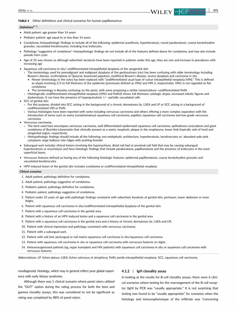
TABLE 4 Other definitions and clinical scenarios for human papillomavirus
Definitions5–7:
• Adult patient: age greater than 14 years
• Pediatric patient: age equal to or less than 14 years
• Condyloma: histopathologic findings to include all of the following: epidermal acanthosis, hyperkeratosis, round parakeratosis, coarse keratohyalinegranules, vacuolated keratinocytes, including true koilocytes
• Pathology "suggestive of condyloma": histopathologic findings do not include all of the features defined above for condyloma, and may also includepseudo horn cysts
• Age of 25 was chosen as although seborrheic keratosis have been reported in patients under this age, they are rare and increase in prevalence withincreasing age
• Squamous cell carcinoma in situ/ undifferentiated intraepithelial dysplasia of the anogenital skin� The terminology used for premalignant and malignant dysplasia of the genitourinary tract has been confusing with older terminology including
Bowen's disease, erythroplasia of Queyrat, bowenoid papulosis, multifocal Bowen's disease, severe dysplasia and carcinoma in situ• Newer terminology in the vulva has been replaced with "undifferentiated usual type of vulvar intraepithelial neoplasia (VIN)." This is defined
as atypia involving 2/3 to full thickness of the epidermis (previously defined as VIN2 and VIN 3, respectively). VIN1 is not regarded as flatcondyloma
• The terminology is likewise confusing on the penis, with some proposing a similar nomenclature—undifferentiated PeIN� Histologically undifferentiated intraepithelial neoplasia (VIN3 and PeIN3) shows full thickness cytologic atypia, increased mitotic figures and
dyskeratosis. It can have the presence of hypergranulosis +/− partially vacuolated cells• SCC of genital skin
� For this purpose, divided into SCC arising in the background of a chronic dermatoses (ie, LSEA and LP or SCC arising in a background ofundifferentiated VIN or PeIN
� Various histologies have been reported with some including verrucous carcinoma and others offering a more complex separation with theintroduction of terms such as warty (condylomatous) squamous cell carcinoma, papillary squamous cell carcinoma and low-grade verrucouscarcinoma
• Verrucous carcinoma:� The term used here encompass verrucous carcinoma, well differentiated epidermoid squamous cell carcinoma, epithelioma cuniculatum and giant
condyloma of Buschke-Löwenstein that clinically present as a warty, exophytic plaque in the oropharynx, lower limb (typically sole of foot) andanogenital region, respectively.
� Histopathologic findings should include all the following: exo-endophytic architecture, hyperkeratosis, keratinocytes w/ abundant pale pinkcytoplasm, large bulbous rete ridges with pushing boarder
• Subungual wart includes clinical lesions involving the hyponychium, distal nail bed or proximal nail fold that may be causing subungualhyperkeratosis or onycholysis and have histologic findings that include parakeratosis, papillomatosis and the presence of koilocytes in the mostsuperficial layers.
• Verrucous features defined as having any of the following histologic features: epidermal papillomatosis, coarse keratohyaline granules andvacuolated keratinocytes
• HPV-induced lesion of the genital skin includes condyloma or undifferentiated intraepithelial neoplasia
Clinical scenarios:
1. Adult patient, pathology definitive for condyloma.
2. Adult patient, pathology suggestive of condyloma.
3. Pediatric patient, pathology definitive for condyloma.
4. Pediatric patient, pathology suggestive of condyloma.
5. Patient under 25 years of age with pathologic findings consistent with seborrheic keratosis of genital skin, perineum, lower abdomen or innerthighs.
6. Patient with squamous cell carcinoma in situ/undifferentiated intraepithelial dysplasia of the genital skin.
7. Patient with a squamous cell carcinoma in the genital area.
8. Patient with a history of an HPV-induced lesion and a squamous cell carcinoma in the genital area
9. Patient with a squamous cell carcinoma in the genital area and a history of chronic dermatoses (ie, LSEA and LP).
10. Patient with clinical impression and pathology consistent with verrucous carcinoma.
11. Patient with a subungual wart.
12. Patient with nail bed, periungual or nail matrix squamous cell carcinoma in situ/squamous cell carcinoma
13. Patient with squamous cell carcinoma in situ or squamous cell carcinoma with verrucous features on digits.
14. Immunosuppressed patients (eg, organ transplant and HIV patients) with squamous cell carcinoma in situ or squamous cell carcinoma withverrucous features.
Abbreviations: LP, lichen planus; LSEA, lichen sclerosus et atrophicus; PeIN, penile intraepithelial neoplasia; SCC, squamous cell carcinoma.
VIDAL ET AL. 571

for,” “suspicious of” or “suggestive of” either primary cutaneous mar-
ginal zone lymphoma (PCMZL) or follicle center lymphoma (FCL).
These entities tend to be difficult to diagnose based primarily on his-
tology or immunohistochemistry. In PCMZL, definitive diagnosis
often relies on detection of light chain restriction which can be diffi-
cult unless plasma cells are abundant. In FCL, the typical histologic
features relied on by hematopathologists such as back-to-back follicle
formation or bcl-2 expression are often absent even in grade 2 FCL.
While in FCL with a diffuse pattern, the presence of sheets of B-cells
is concerning for lymphoma, again, lack of typical follicular lymphoma
markers such as expression of CD10 and bcl-2 can lead to confusion
even among experienced dermatopathologists. In these scenarios,
testing with clonality assays can confirm the diagnosis.9 As expected,
testing was “usually appropriate” in cases where the clonality assay
was being used for clone comparison. Testing was recommended by
the majority of panel raters in cases where the clinical impression
was of a single lesion suggestive of a nonneoplastic process or of der-
matitis, but the histology showed a B-cell predominant infiltrate. Con-
versely, there were 2 clinical scenarios ranked “rarely appropriate”:
when single or multiple nodules are found and the clinical impression is
rule out B-cell lymphoma (PCMZL or FCL), but the histology and immu-
nohistochemistry results are “not diagnostic” for cutaneous B-cell lym-
phoma; and in patients with a preexisting diagnosis of cutaneous B-cell
lymphoma (either PCMZL or FCL) and when a diagnosis of PCMZL or
FCL can be made on histologic grounds. There was “no consensus” for
2 scenarios, which included cases where the history is unknown, but
the histology and immunophenotype of the infiltrate are “consistent
with” with PCMZL or FCL and when other more aggressive cutaneous
B-cell lymphomas other than primary cutaneous large B-cell lymphoma,
leg type (PCLBCL-LT) is considered in the diagnosis. The latter may be
related to the lack of clarity among some panel raters for this scenario
and the scarcity of literature pertaining to the use of clonality assays for
more aggressive and rarer lymphomas.
4.2 | Melanocytic group
4.2.1 | Fluorescence in situ hybridization and comparativegenomic hybridization
Regarding melanocytic lesions, ratings indicate that in most scenarios
where the diagnosis of melanoma is in question it is reasonable to
use FISH or CGH as an ancillary test. In general, the results of expert
panel ratings for FISH and CGH were similar. Results were also
TABLE 5 Other definitions and clinical scenarios for Muir-Torre
syndrome
Definitions8,9:
• Age 60: there are some articles that suggest age 50 instead of 60 asa cut off, this may be because sebaceous neoplasms present at amean age of 53
• MTS-associated sebaceous neoplasm: sebaceous adenoma,sebaceoma, sebaceous epithelioma and sebaceous carcinoma
• MTS-associated neoplasm: MTS-associated sebaceous neoplasms,cystic sebaceous neoplasm, basal cell carcinoma with sebaceousdifferentiation and keratoacanthoma with sebaceous differentiation
• MTS-associated visceral malignancy: colorectal adenocarcinoma(most common), genitourinary carcinoma (second most common),breast, hematologic and endometrial and gastric carcinoma (lesscommon)
Clinical scenarios:
1. A patient over the age of 60 with a periocular sebaceouscarcinoma.
2. A patient over the age of 60 with a single sebaceous tumor on thehead and neck.
3. A patient over the age of 60 with a single sebaceous tumor on asite other than the head and neck.
4. A patient over the age of 60 with multiple (greater than or equalto 2) sebaceous tumors.
5. A patient over the age of 60 with a basal cell carcinoma withsebaceous differentiation.
6. A patient over the age of 60 with a keratoacanthoma withsebaceous differentiation.
7. A patient over the age of 60 with a cystic sebaceous neoplasm.8. A patient over the age of 60 with an MTS-associated neoplasm
and/or a personal history of an MTS-associated visceralmalignancy.
TABLE 6 Definitions and clinical scenarios for dermatofibrosarcoma
protuberans
Definitions:
• Typical histomorphology of DFSP: monotonous spindled cells in astoriform pattern with “honeycombing” or entrapment of adnexalstructures and/or adipocytes and extension into the subcutis
• Nontypical histomorphology of DFSP refers to varianthistomorphology such as fibrosarcomatous, giant cell fibroblastoma,myxoid, epithelioid or nonspecific spindled cell histomorphology.
Clinical scenarios:
1. Tissue with sampling down to subcutis with typicalhistomorphology of dermatofibrosarcoma protuberans and CD34+by immunohistochemistry.
2. Tissue with sampling down to subcutis with typicalhistomorphology of dermatofibrosarcoma protuberans and CD34immunohistochemistry not uniformly reactive.
3. Tissue with sampling down to subcutis with nontypicalhistomorphology of dermatofibrosarcoma protuberans and CD34+by immunohistochemistry.
4. Superficial, CD34+ tumor with typical histomorphology ofdermatofibrosarcoma protuberans except that good honeycombingof fat is not seen due to superficial sampling.
5. Superficial, CD34+ tumor with nontypical histomorphology fordermatofibrosarcoma protuberans. (SD5)
6. Superficial, CD34+ tumor with scant tumor sampling as to limitcytologic and/or architectural evaluation.
7. High grade spindle cell tumor (“fibrosarcomatous transformation”)and no areas of typical histomorphology of dermatofibrosarcomaprotuberans.
8. Metastatic tumor with histomorphology similar to previouslydiagnosed primary dermatofibrosarcoma protuberans.
9. Metastatic tumor with histomorphology distinct from previouslydiagnosed primary dermatofibrosarcoma protuberans.
10. Patient with locally recurrent dermatofibrosarcoma protuberansin which testing for translocation by another establishedmolecular technique (RT-PCR, FISH and cytogenetics) waspreviously positive.
11. Patient with metastatic dermatofibrosarcoma protuberans inwhich testing for translocation by another established moleculartechnique (RT-PCR, FISH and cytogenetics) was previouslypositive in the primary tumor.
12. Patients for which tyrosine kinase therapy is being considered inthe treatment plan.
13. Patient with tissue that has been decalcified or processed withfixative other than 10% formalin.
14. Patient with a pathologic diagnosis of dermatofibrosarcomaprotuberans by hematoxylin and eosin with CD34+immunohistochemistry but where the treating physician isrequesting molecular studies (RT-PCR, FISH and cytogenetics) tobe performed to further confirm the diagnosis.
572 VIDAL ET AL.
similar across age groups (adult vs pediatric). In most scenarios,
except for those where the pathology is definitive for melanoma or
melanocytic nevus, expert rating found that it is “usually appropriate”
to perform FISH or CGH on melanocytic lesions when the diagnosis
is in question. In those cases where the pathology is definitive for
either a melanoma or melanocytic nevus, testing with FISH and CGH
is “rarely appropriate.” This was not surprising as histology is consid-
ered the “gold standard” in the diagnosis of melanocytic lesions. Of
note, inclusion of these clinical scenarios may be considered a proof
of concept that the rounds of ratings yielded meaningful results.
Interestingly, the results also indicate that currently CGH is the only
test ranked “usually appropriate” when it comes to distinguishing
benign blue nevi from more worrisome dermal melanocytoses. The
TABLE 7 Soft tissue clinical scenarios and definitions for clear cell
sarcoma
Definitions:
• Melanocytic markers: S100, Melan-A/MART-1, HMB45, MiTF andSOX10
• Typical histologic features of clear cell sarcoma: relatively uniform(nonpleomorphic) nuclei, large central nucleoli, nested appearancedivided by fibrous septations, scattered osteoclast-like giant cells,little or no conspicuous melanin and no epidermal component
Clinical scenarios:
1. Patient less than 50 years of age with acral tumor with typicalhistologic features of clear cell sarcoma, expressing melanocyticmarkers and involving deep dermis, subcutis or aponeurosis. Nopast history of melanoma.
2. Patient less than 50 years of age with acral tumor WITHOUTtypical histologic features of clear cell sarcoma, expressingmelanocytic markers and involving deep dermis, subcutis oraponeurosis. No past history of melanoma.
3. Patient greater than or equal to 50 years of age with acral tumorwith typical histologic features of clear cell sarcoma, expressingmelanocytic markers and involving deep dermis, subcutis oraponeurosis. No past history of melanoma.
4. Patient greater than or equal 50 years of age with acral tumorWITHOUT typical histologic features of clear cell sarcoma,expressing melanocytic markers and involving deep dermis,subcutis or aponeurosis. No past history of melanoma.
5. Patient less than 50 years of age with NON-acral site tumorexpressing melanocytic markers, WITHOUT typical histologicfeatures of clear cell sarcoma and involving deep dermis, subcutisor aponeurosis. No past history of melanoma but with whatappears to be a cutaneous metastasis of melanoma from anunknown primary.
6. Patient greater than or equal to 50 years of age with NON-acralsite tumor expressing melanocytic markers, WITHOUT typicalhistologic features of clear cell sarcoma and involving deep dermis,subcutis or aponeurosis. No past history of melanoma but withwhat appears to be a cutaneous metastasis of melanoma from anunknown primary.
7. Patient with dermal-based tumor expressing melanocytic markersand demonstrating typical histological features of clear cellsarcoma. Patient has past history of invasive melanoma at anotheranatomic site.
8. Patient with an acral tumor in the dermis/subcutis that not onlyhas typical histologic features of clear cell sarcoma and expressesmelanocytic markers, but also has an overlying intraepidermal insitu component.
9. Patient with a non-acral tumor in the dermis/subcutis that not onlyhas typical histologic features of clear cell sarcoma and expressesmelanocytic markers but also has an overlying intraepidermal insitu component.
10. Patient with metastatic tumor with histomorphology similar topreviously diagnosed primary clear cell sarcoma.
11. Patient with metastatic tumor with histomorphology distinct frompreviously diagnosed primary clear cell sarcoma.
12. Patient with recurrent or metastatic clear cell sarcoma in whichtesting for translocation by another established technique (RT-PCR, FISH and cytogenetics) was previously positive.
13. Patient with primary or metastatic tumor expressing melanocyticmarkers in which BRAF or NRAS mutation has been detected.
14. Patient with tissue that has been decalcified or processed withfixative other than 10% formalin.
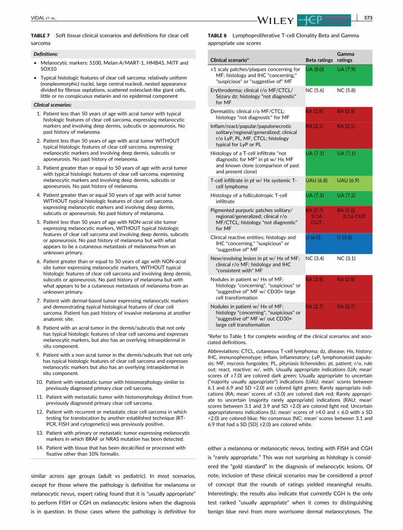
TABLE 8 Lymphoproliferative T-cell Clonality Beta and Gamma
appropriate use scores
Clinical scenario* Beta ratingsGammaratings
≥1 scaly patches/plaques concerning forMF; histology and IHC "concerning,""suspicious" or "suggestive of" MF
UA (8.0) UA (7.9)
Erythroderma; clinical r/o MF/CTCL/Sézary dz; histology "not diagnostic"for MF
NC (5.6) NC (5.8)
Dermatitis; clinical r/o MF/CTCL;histology "not diagnostic" for MF
RA (2.8) RA (2.8)
Inflam/react/papular/papulonecroticsolitary/regional/generalized; clinicalr/o LyP, PL, MF, CTCL; histologytypical for LyP or PL
RA (2.1) RA (2.1)
Histology of a T-cell infiltrate "notdiagnostic for MF" in pt w/ Hx MFand known clone (comparison of pastand present clone)
UA (7.1) UA (7.1)
T-cell infiltrate in pt w/ Hx systemic T-cell lymphoma
UAU (6.8) UAU (6.9)
Histology of a folliculotropic T-cellinfiltrate
UA (7.1) UA (7.2)
Pigmented purpuric patches solitary/regional/generalized; clinical r/oMF/CTCL; histology "not diagnostic"for MF
RA (2.7)2/16OUT
RA (2.6)2/16 OUT
Clinical reactive entities; histology andIHC "concerning," "suspicious" or"suggestive of" MF
U (6.0) U (3.6)
New/evolving lesion in pt w/ Hx of MF;clinical r/o MF; histology and IHC"consistent with" MF
NC (3.4) NC (3.1)
Nodules in patient w/ Hx of MF;histology "concerning", "suspicious" or"suggestive of" MF w/ CD30+ largecell transformation
RA (2.8) RA (2.8)
Nodules in patient w/ Hx of MF;histology "concerning", "suspicious" or"suggestive of" MF w/ out CD30+large cell transformation
RA (2.7) RA (2.7)
*Refer to Table 1 for complete wording of the clinical scenarios and asso-ciated definitions.
Abbreviations: CTCL, cutaneous T-cell lymphoma; dz, disease; Hx, history;IHC, immunophenotype; inflam, inflammatory; LyP, lymphomatoid papulo-sis; MF, mycosis fungoides; PL, pityriasis lichenoides; pt, patient; r/o, ruleout; react, reactive; w/, with. Usually appropriate indications (UA; mean’scores of ≥7.0) are colored dark green; Usually appropriate to uncertain(“majority usually appropriate”) indications (UAU; mean’ scores between6.1 and 6.9 and SD <2.0) are colored light green; Rarely appropriate indi-cations (RA; mean’ scores of ≤3.0) are colored dark red; Rarely appropri-ate to uncertain (majority rarely appropriate) indications (RAU; mean’scores between 3.1 and 3.9 and SD <2.0) are colored light red; Uncertainappropriateness indications (U; mean’ scores of ≥4.0 and ≤ 6.0 with a SD<2.0) are colored blue; No consensus (NC; mean’ scores between 3.1 and6.9 that had a SD [SD] ≥2.0) are colored white.
VIDAL ET AL. 573
consensus rating for FISH in the same clinical scenario was of uncer-
tain appropriateness. Pouryazdanparast et al described the utility in
epithelioid blue nevi and cutaneous melanoma metastases simulating
blue nevi15 and Gammon et al explored FISH in distinguishing cellular
blue nevi from blue nevus-like melanoma showing 100% sensitivity
and specificity.16 While these studies utilized a FISH probe set differ-
ent from the one defined by the group in this analysis, there was
overlap of at least of 2 of the probes used—the RREB1 and 6p25
probes. There was “no consensus” on the value of FISH for situations
where the pathology is suggestive or suspicious for melanoma where
the differential diagnosis is between sclerosing desmoplastic nevus
and desmoplastic melanoma, in partially sampled lesions. However, in
this specific scenario, CGH was rated “usually appropriate.” This may
relate to Gerami et al in 2011, which showed a low sensitivity but
high specificity in this subset with FISH.17
The “OUT” rating was used once in 3 clinical scenarios by the
panel raters when rating FISH and CGH, with 94% of panel raters
participating. Scenarios rated for FISH and CGH independently
showed the same number of “OUT” ratings and when they were con-
sidered in pediatric vs adult patients the use of “OUT” was similar.
4.2.2 | Quantitative reverse transcription polymerasechain reaction
Consensus ratings in most of the clinical scenarios using qRT-PCR
were of “appropriateness uncertain” with the exception being those
cases where a diagnosis can be made on histologic grounds. While
validation studies and studies exploring unequivocal cases had been
published when the AUC process began,18,19 only one study was
available exploring the test in ambiguous lesions20 at the time of
rating. In addition, the possibility of limited clinical experience with
the test may have played a role in the rating result. Since the com-
pletion of the AUC process, additional studies have been reported
in the literature, including one dealing with diagnostically challeng-
ing cases21 and another that correlates with clinical outcome.22
Thus, recommendations for the appropriateness of qRT-PCR in the
studied clinical scenarios are expected to change as the AUC are
subsequently and expectedly updated.
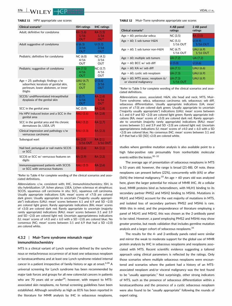
4.3 | Other groups
4.3.1 | Human papillomavirus, in situ hybridization andimmunohistochemistry
Use of HPV, ISH and IHC shows wide variability and these tests are cur-
rently frequently performed and often at the request of clinicians.
Although there are many commercially available type-specific probes
and “cocktails” for the detection of HPV by ISH, type-specific probes for
HPV 6, 11, 16, 18, 31 and 33 are the most commonly utilized by derma-
topathologists. The availability of commercially available antibodies tar-
geting HPV is much more limited, with only 2 currently available.11
While most of the literature for detection of HPV centers on use
of ISH in condylomas or lesions histologically concerning for condylo-
mas in adults, consensus ratings found testing by ISH to be “rarely
appropriate” to "majority rarely appropriate" for many scenarios
ranked. Only in pediatric cases where pathology is suggestive of con-
dyloma, did experts feel testing by ISH was “usually appropriate.” Lit-
erature on this topic suggests that sensitivities for detection of HPV
by ISH in the pediatric population ranges from 60% to 100%,23–26
which may be the reason for the recommendation. However, there
was “no consensus” in a similar scenario of a pediatric patient, but
with histology definitive for condyloma. This rating may be because
HPV 2, which is not typically detected by ISH, is the most common
subtype of HPV found in this age group.27
Most scenarios were ranked as “rarely appropriate” for the use of
IHC in the detection of HPV. These ratings probably reflect the
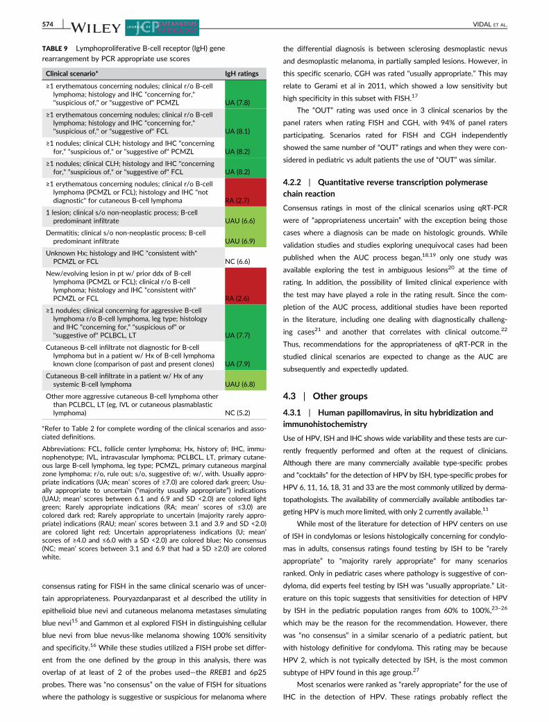
TABLE 9 Lymphoproliferative B-cell receptor (IgH) gene
rearrangement by PCR appropriate use scores
Clinical scenario* IgH ratings
≥1 erythematous concerning nodules; clinical r/o B-celllymphoma; histology and IHC "concerning for,""suspicious of," or "suggestive of" PCMZL UA (7.8)
≥1 erythematous concerning nodules; clinical r/o B-celllymphoma; histology and IHC "concerning for,""suspicious of," or "suggestive of" FCL UA (8.1)
≥1 nodules; clinical CLH; histology and IHC "concerningfor," "suspicious of," or "suggestive of" PCMZL UA (8.2)
≥1 nodules; clinical CLH; histology and IHC "concerningfor," "suspicious of," or "suggestive of" FCL UA (8.2)
≥1 erythematous concerning nodules; clinical r/o B-celllymphoma (PCMZL or FCL); histology and IHC "notdiagnostic" for cutaneous B-cell lymphoma RA (2.7)
1 lesion; clinical s/o non-neoplastic process; B-cellpredominant infiltrate UAU (6.6)
Dermatitis; clinical s/o non-neoplastic process; B-cellpredominant infiltrate UAU (6.9)
Unknown Hx; histology and IHC "consistent with"PCMZL or FCL NC (6.6)
New/evolving lesion in pt w/ prior ddx of B-celllymphoma (PCMZL or FCL); clinical r/o B-celllymphoma; histology and IHC "consistent with"PCMZL or FCL RA (2.6)
≥1 nodules; clinical concerning for aggressive B-celllymphoma r/o B-cell lymphoma, leg type; histologyand IHC "concerning for," "suspicious of" or"suggestive of" PCLBCL, LT UA (7.7)
Cutaneous B-cell infiltrate not diagnostic for B-celllymphoma but in a patient w/ Hx of B-cell lymphomaknown clone (comparison of past and present clones) UA (7.9)
Cutaneous B-cell infiltrate in a patient w/ Hx of anysystemic B-cell lymphoma UAU (6.8)
Other more aggressive cutaneous B-cell lymphoma otherthan PCLBCL, LT (eg, IVL or cutaneous plasmablasticlymphoma) NC (5.2)
*Refer to Table 2 for complete wording of the clinical scenarios and asso-ciated definitions.
Abbreviations: FCL, follicle center lymphoma; Hx, history of; IHC, immu-nophenotype; IVL, intravascular lymphoma; PCLBCL, LT, primary cutane-ous large B-cell lymphoma, leg type; PCMZL, primary cutaneous marginalzone lymphoma; r/o, rule out; s/o, suggestive of; w/, with. Usually appro-priate indications (UA; mean’ scores of ≥7.0) are colored dark green; Usu-ally appropriate to uncertain (“majority usually appropriate”) indications(UAU; mean’ scores between 6.1 and 6.9 and SD <2.0) are colored lightgreen; Rarely appropriate indications (RA; mean’ scores of ≤3.0) arecolored dark red; Rarely appropriate to uncertain (majority rarely appro-priate) indications (RAU; mean’ scores between 3.1 and 3.9 and SD <2.0)are colored light red; Uncertain appropriateness indications (U; mean’scores of ≥4.0 and ≤6.0 with a SD <2.0) are colored blue; No consensus(NC; mean’ scores between 3.1 and 6.9 that had a SD ≥2.0) are coloredwhite.
574 VIDAL ET AL.
presence of only 2 articles exploring the use of IHC for detection
of HPV.
A significant number of panel raters utilized the “OUT” rating in
scenarios dealing with the use of ISH and IHC for the detection of
HPV. The scenarios with a significant number of “OUT” ratings were
those where the pathology is “suggestive of” a condyloma in an adult,
in situations when the pathology is definitive or “suggestive of” a
condyloma in the pediatric population, and in cases where the pathol-
ogy is “consistent with” a seborrheic keratosis of the genital skin, per-
ineum, lower abdomen or inner thighs. This likely reflects the
psychosocial implications surrounding a diagnosis of HPV, especially
in the genital area and in children, emphasizing the importance of
direct communication between dermatopathologist and clinician
before performing these tests.
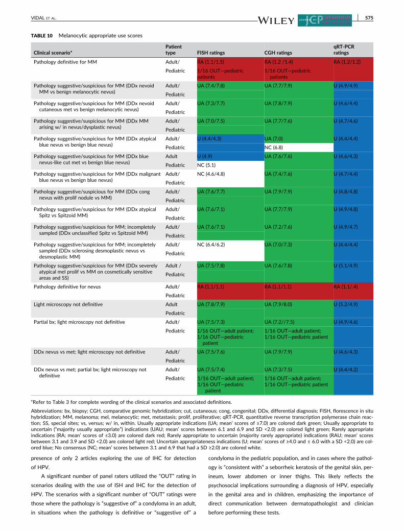
TABLE 10 Melanocytic appropriate use scores
Clinical scenario*Patienttype FISH ratings CGH ratings
qRT-PCRratings
Pathology definitive for MM Adult/ RA (1.1/1.5) RA (1.2 /1.4) RA (1.2/1.2)
Pediatric 1/16 OUT—pediatricpatients
1/16 OUT—pediatricpatients
Pathology suggestive/suspicious for MM (DDx nevoidMM vs benign melanocytic nevus)
Adult/ UA (7.4/7.8) UA (7.7/7.9) U (4.9/4.9)
Pediatric
Pathology suggestive/suspicious for MM (DDx nevoidcutaneous met vs benign melanocytic nevus)
Adult/ UA (7.3/7.7) UA (7.8/7.9) U (4.6/4.4)
Pediatric
Pathology suggestive/suspicious for MM (DDx MMarising w/ in nevus/dysplastic nevus)
Adult/ UA (7.0/7.5) UA (7.7/7.6) U (4.7/4.6)
Pediatric
Pathology suggestive/suspicious for MM (DDx atypicalblue nevus vs benign blue nevus)
Adult/ U (4.4/4.3) UA (7.0) U (4.4/4.4)
Pediatric NC (6.8)
Pathology suggestive/suspicious for MM (DDx bluenevus-like cut met vs benign blue nevus)
Adult U (4.9) UA (7.6/7.6) U (4.6/4.3)
Pediatric NC (5.1)
Pathology suggestive/suspicious for MM (DDx malignantblue nevus vs benign blue nevus)
Adult/ NC (4.6/4.8) UA (7.4/7.6) U (4.7/4.4)
Pediatric
Pathology suggestive/suspicious for MM (DDx congnevus with prolif nodule vs MM)
Adult/ UA (7.6/7.7) UA (7.9/7.9) U (4.8/4.8)
Pediatric
Pathology suggestive/suspicious for MM (DDx atypicalSpitz vs Spitzoid MM)
Adult/ UA (7.6/7.1) UA (7.7/7.9) U (4.9/4.8)
Pediatric
Pathology suggestive/suspicious for MM; incompletelysampled (DDx unclassified Spitz vs Spitzoid MM)
Adult/ UA (7.6/7.1) UA (7.2/7.6) U (4.9/4.7)
Pediatric
Pathology suggestive/suspicious for MM; incompletelysampled (DDx sclerosing desmoplastic nevus vsdesmoplastic MM)
Adult/ NC (6.4/6.2) UA (7.0/7.3) U (4.4/4.4)
Pediatric
Pathology suggestive/suspicious for MM (DDx severelyatypical mel prolif vs MM on cosmetically sensitiveareas and SS)
Adult / UA (7.5/7.8) UA (7.6/7.8) U (5.1/4.9)
Pediatric
Pathology definitive for nevus Adult/ RA (1.1/1.1) RA (1.1/1.1) RA (1.1/.4)
Pediatric
Light microscopy not definitive Adult UA (7.8/7.9) UA (7.9/8.0) U (5.2/4.9)
Pediatric
Partial bx; light microscopy not definitive Adult/ UA (7.5/7.3) UA (7.2//7.5) U (4.9/4.6)
Pediatric 1/16 OUT—adult patient;1/16 OUT—pediatric
patient
1/16 OUT—adult patient;1/16 OUT—pediatric patient
DDx nevus vs met; light microscopy not definitive Adult/ UA (7.5/7.6) UA (7.9/7.9) U (4.6/4.3)
Pediatric
DDx nevus vs met; partial bx; light microscopy notdefinitive
Adult/ UA (7.5/7.4) UA (7.3/7.5) U (4.4/4.2)
1/16 OUT—adult patient;1/16 OUT—pediatric
patient
1/16 OUT—adult patient;1/16 OUT—pediatric patient
Pediatric
*Refer to Table 3 for complete wording of the clinical scenarios and associated definitions.
Abbreviations: bx, biopsy; CGH, comparative genomic hybridization; cut, cutaneous; cong, congenital; DDx, differential diagnosis; FISH, florescence in situhybridization; MM, melanoma; mel, melanocytic; met, metastasis; prolif, proliferative; qRT-PCR, quantitative reverse transcription polymerase chain reac-tion; SS, special sites; vs, versus; w/ in, within. Usually appropriate indications (UA; mean’ scores of ≥7.0) are colored dark green; Usually appropriate touncertain (“majority usually appropriate”) indications (UAU; mean’ scores between 6.1 and 6.9 and SD <2.0) are colored light green; Rarely appropriateindications (RA; mean’ scores of ≤3.0) are colored dark red; Rarely appropriate to uncertain (majority rarely appropriate) indications (RAU; mean’ scoresbetween 3.1 and 3.9 and SD <2.0) are colored light red; Uncertain appropriateness indications (U; mean’ scores of ≥4.0 and ≤ 6.0 with a SD <2.0) are col-ored blue; No consensus (NC; mean’ scores between 3.1 and 6.9 that had a SD ≥2.0) are colored white.
VIDAL ET AL. 575
4.3.2 | Muir-Torre syndrome mismatch repairimmunohistochemistry
MTS is a clinical variant of Lynch syndrome defined by the synchro-
nous or metachronous occurrence of at least one sebaceous neoplasm
or keratoacanthoma and at least one Lynch syndrome-related internal
cancer in a patient irrespective of family history or age at onset.6,28 A
universal screening for Lynch syndrome has been recommended by
major task forces and groups for all new colorectal cancers in patients
who are 70 years old or under29; however, with respect to MTS-
associated skin neoplasms, no formal screening guidelines have been
established. Although sensitivity as high as 81% has been reported in
the literature for MMR analysis by IHC in sebaceous neoplasms,
studies where germline mutation analysis is also available point to a
high false-positive rate presumably from nonheritable molecular
events within the lesion.30–32
The average age of presentation of sebaceous neoplasms in MTS
is 53 years old; however, the range is broad (21-88). Of note, these
neoplasms can present before (22%), concurrently with (6%) or after
(56%) the internal malignancy.33 An age > 60 years old was analyzed
here, given the larger potential for misuse of MMR IHC. At a cellular
level, MMR proteins bind as heterodimers, with MLH1 binding to its
secondary partner PMS2 and MSH2 binding to MSH6. Mutations in
MLH1 and MSH2 account for the vast majority of mutations in MTS,
and isolated loss of secondary partners PMS2 and MSH6 is rare.
With this in mind, and the preponderance of literature employing a
panel of MLH1 and MSH2, this was chosen as the 2-antibody panel
to be rated. However, a panel employing PMS2 and MSH6 may show
greater promise, but needs validation that includes germline mutation
analysis and a larger cohort of sebaceous neoplasms.34
The results for the 4- and 2-antibody panels rated were similar
and mirror the weak to moderate support for the global use of MMR
protein analysis by IHC in sebaceous neoplasms and neoplasms asso-
ciated with MTS. Recent scientific evidence suggesting a tailored
approach using clinical parameters is reflected by the ratings. Only
those scenarios where multiple sebaceous neoplasms were encoun-
tered and scenarios where the patient had a history of an MTS-
associated neoplasm and/or visceral malignancy was the test found
to be “usually appropriate.” Not surprisingly, other strong indicators
of MTS, such as the presence of sebaceous differentiation within a
keratoacanthoma and the presence of a cystic sebaceous neoplasm
were also found to be “usually appropriate” following the rounds of
expert rating.
TABLE 11 HPV appropriate use scores
Clinical scenario* ISH ratings IHC ratings
Adult; definitive for condyloma RA (1.6)1/16OUT
RA (1.5)1/16OUT
Adult suggestive of condyloma U (6.3)3/16OUT
U (5.2)2/16OUT
Pediatric; definitive for condyloma NC (6.0)4/16OUT
NC (4.1)3/16OUT
Pediatric; suggestive of condyloma UA (7.5)4/16OUT
NC (5.7)4/16OUT
Age < 25; pathologic findings c/wseborrheic keratosis of genital skin,perineum, lower abdomen, or innerthighs
UAU (6.7)4/16OUT
U (3.6)3/16OUT
SCCIS/ undifferentiated intraepithelialdysplasia of the genital skin
U (3.7)1/16OUT
RA (2.8)1/16OUT
SCC in the genital area NC (3.9) RA (2.8)
Hx HPV induced lesion and a SCC in thegenital area
RAU (3.6) RA (2.8)
SCC in the genital area and Hx chronicdermatoses (ie, LSEA, LP)
RAU (3.2) RA (2.7)
Clinical impression and pathology c/wverrucous carcinoma
RAU (3.3) RA (2.3)
Subungual wart RA (2.0)1/16 OUT
RA (2.1)1/16 OUT
Nail bed, periungual or nail matrix SCCISor SCC
RA (2.6) RA (2.2)
SCCIS or SCC w/ verrucous features ondigits
RA (2.9) RA (2.2)
Immunosuppressed patients with SCCISor SCC with verrucous features
RAU (3.3) RA (2.6)
*Refer to Table 4 for complete wording of the clinical scenarios and asso-ciated definitions.
Abbreviations: c/w, consistent with; IHC, immunohistochemistry; ISH, insitu hybridization; LP, lichen planus; LSEA, Lichen sclerosus et atrophicus;SCCIS, squamous cell carcinoma in situ; SCC, squamous cell carcinoma.Usually appropriate indications (UA; mean’ scores of ≥7.0) are coloreddark green; Usually appropriate to uncertain (“majority usually appropri-ate”) indications (UAU; mean’ scores between 6.1 and 6.9 and SD <2.0)are colored light green; Rarely appropriate indications (RA; mean’ scoresof ≤3.0) are colored dark red; Rarely appropriate to uncertain (majorityrarely appropriate) indications (RAU; mean’ scores between 3.1 and 3.9and SD <2.0) are colored light red; Uncertain appropriateness indications(U; mean’ scores of ≥4.0 and ≤ 6.0 with a SD <2.0) are colored blue; Noconsensus (NC; mean’ scores between 3.1 and 6.9 that had a SD ≥2.0)are colored white.
TABLE 12 Muir-Torre syndrome appropriate use scores
Clinical scenario*4 AB panelratings
2 AB panelratings
Age > 60; periocular sebca NC (3.5) RA (3.0)
Age > 60; 1 seb tumor H&N NC (5.1)1/16 OUT
U (4.9)1/16 OUT
Age > 60; 1 seb tumor non-H&N NC (6.7)1/16 OUT
UAU (6.9)1/16 OUT
Age > 60; multiple seb tumors UA (7.2) UA (7.2)
Age > 60; BCC w/ seb diff U (5.0) U (4.6)
Age > 60; KA w/ seb diff UA (7.1) UAU (6.6)
Age > 60; cystic seb neoplasm UA (7.3) UAU (6.9)
Age > 60; MTS assoc. neoplasm &/or visceral malignancy
UA (7.3)1/16 OUT
UAU (6.9)
*Refer to Table 5 for complete wording of the clinical scenarios and asso-ciated definitions.
Abbreviations: assoc, associated; H&N, site head and neck; MTS, Muir-Torre syndrome; sebca, sebaceous carcinoma; seb, sebaceous; seb diff,sebaceous differentiation. Usually appropriate indications (UA; mean’scores of ≥7.0) are colored dark green; Usually appropriate to uncertain(“majority usually appropriate”) indications (UAU; mean’ scores between6.1 and 6.9 and SD <2.0) are colored light green; Rarely appropriate indi-cations (RA; mean’ scores of ≤3.0) are colored dark red; Rarely appropri-ate to uncertain (majority rarely appropriate) indications (RAU; mean’scores between 3.1 and 3.9 and SD <2.0) are colored light red; Uncertainappropriateness indications (U; mean’ scores of ≥4.0 and ≤ 6.0 with a SD<2.0) are colored blue; No consensus (NC; mean’ scores between 3.1 and6.9 that had a SD [SD] ≥2.0) are colored white.
576 VIDAL ET AL.

Interestingly, the “OUT” option was not used frequently in the
rating of these clinical scenarios.
4.4 | Soft tissue group
4.4.1 | t(17;22) FISH assay for DFSP
Cytogenetically, DFSP is characterized by a balanced or unbalanced
translocation, t(17;22)(q22;q13) or a supernumerary ring chromo-
some, resulting in the fusion of exon 2 of PDGFB gene encoding the
platelet-derived growth factor beta with various exons (from 6-47) of
COL1A1 gene encoding the alpha chain type 1 collagen. Multiple
modalities of FISH can be utilized to detect the translocation. These
include: dual-fusion COL1A1/PDGFB FISH, PDGFB break-apart FISH
or COL1A1 break-apart FISH.35–40 The overall sensitivity of the dual-
fusion FISH test in the literature is 94.3% (range 86%-100%). This was
similar for PDGFB break-apart FISH that has an overall sensitivity of
95% (range 91%-100%). The sensitivity of the COL1A1 break-apart
probe is probably in the same range; however, there is only one study
that explicitly mentioned this probe being identified.41–44 Given the
high sensitivity of FISH and the therapy potential if the translocation
is detected, it is not surprising that the two scenarios where the test
TABLE 13 t(17;22) in dermatofibrosarcoma protuberans appropriate
use scores
Clinical scenario*t(17:22)ratings
Histology typical for DFSP; CD34+ RA (1.4)
Histology typical for DFSP; CD34 not uniformly reactive NC (3.2)
Histology not typical for DFSP; CD34+ UA (7.2)
Histology typical for DFSP; CD34+; but SQ notvisualized
NC (4.7)
Histology not typical for DFSP; CD34+; SQ notvisualized
UAU (6.5)
Limited cytological and / or architectural histologyevaluation; CD34+
NC (6.0)7/16 OUT
Fibrosarcoma-like (high grade) histology; no histologytypical for DFSP
UAU (6.9)
Met lesion with histology similar to prior DFSP RA (2.9)1/16 OUT
Met lesion with histology different from prior DFSP U (6.5)
Locally recurrent DFSP; + translocation testing by othermolecular test
RA (1.6)
Met DFSP; + translocation testing by other moleculartest
RA (1.7)
Tyrosine kinase therapy is being considered UA (7.2)1/16 OUT
Tissue that has been decalcified or processed w/ fixativeother than 10% formalin
UAU (6.6)
Histology typical for DFSP; CD34+; treating MDrequesting cytogenetics to confirm diagnosis
UAU (6.3)3/16 OUT
*Refer to Table 6 for complete wording of the clinical scenarios and asso-ciated definitions.
Abbreviations: DFSP, dermatofibrosarcoma protuberans; histology typical,monotonous spindled cells in a storiform pattern with “honeycombing” orentrapment of adnexal structures and/or adipocytes and extension intothe subcutis; ; Met, metastatic; SQ, subcutis; w/, with. Usually appropriateindications (UA; mean’ scores of ≥7.0) are colored dark green; Usuallyappropriate to uncertain (“majority usually appropriate”) indications (UAU;mean’ scores between 6.1 and 6.9 and SD <2.0) are colored light green;Rarely appropriate indications (RA; mean’ scores of ≤3.0) are colored darkred; Rarely appropriate to uncertain (majority rarely appropriate) indica-tions (RAU; mean’ scores between 3.1 and 3.9 and SD <2.0) are coloredlight red; Uncertain appropriateness indications (U; mean’ scores of ≥4.0and ≤6.0 with a SD <2.0) are colored blue; No consensus (NC; mean’scores between 3.1 and 6.9 that had an SD ≥2.0) are colored white.
TABLE 14 EWSR1 FISH clear cell sarcoma appropriate use scores
Clinical scenario*EWSR1 FISHratings
Age < 50; typical location; tumor w/ histology typicalfor CCS, expressing melanocytic markers, involvingdeep dermis, SQ or aponeurosis. No Hx ofmelanoma
UA (8.3)
Age < 50; typical location; tumor w/ non-typicalhistology for CCS expressing melanocytic markers,involving deep dermis, SQ or aponeurosis. No Hx ofmelanoma
NC (6.3)
Age ≥ 50; typical location; tumor w/ histology typicalfor CCS, expressing melanocytic markers, involvingdeep dermis, SQ or aponeurosis. No Hx ofmelanoma
UA (8.1)
Age ≥ 50; typical location; tumor w/ non-typicalhistology for CCS expressing melanocytic markers,involving deep dermis, SQ or aponeurosis. No Hx ofmelanoma
RAU (3.4)
Age < 50; non-typical location; tumor expressingmelanocytic markers; w/ non-typical histology forCCS, involving deep dermis, SQ or aponeurosis. NoHx of melanoma but w/ what appears to be cutmet of MM from unknown primary
RAU (3.2)
Age ≥ 50; non-typical location; tumor expressingmelanocytic markers; w/ non-typical histology forCCS, involving deep dermis, SQ or aponeurosis. NoHx of melanoma but w/ what appears to be cutmet of MM from unknown primary
RA (2.2)
Dermal tumor expressing melanocytic markers anddemonstrating typical histology of CCS. Pt has Hxinvasive MM at another site.
UA (7.9)
Typical location; tumor in dermis/subcutis; histologytypical for CCS, expressing melanocytic markers,but also has an intraepidermal in situ component
UA (7.5)
Non-typical location; tumor in dermis/subcutis;histology typical for CCS expressing melanocyticmarkers, but also has an intraepidermal in situcomponent
UA (7.4)
Met tumor w/ histology similar to previous CCS RAU (3.2)
Met tumor w/ histology different from previous CCS UA (7.3)
Recurrent / met CCS w/ translocation testing byother method positive
RA (1.9)
Primary or met tumor expressing melanocyticmarkers; BRAF or NRAS mutation detected
UAU (6.3)
Tissue that has been decalcified or processed w/fixative other than 10% formalin
UA (7.0)
*Refer to Table 7 for complete wording of the clinical scenarios and asso-ciated definitions.
Abbreviations: CCS, clear cell sarcoma; Hx, history; MM, malignant mela-noma; Met, metastatic; SQ, subcutis; typical histology, tumor with a dis-tinctly nested growth pattern that is divided by fibrous septations. Cellshave a relatively uniform nucleus and large central nucleoli. Scatteredosteoclast-like giant cells can be seen. There is little to no melanin appre-ciated; typical location, deep soft tissue of tendon, aponeuroses and fas-cial structures of the distal extremities; w/, with. Usually appropriateindications (UA; mean’ scores of ≥7.0) are colored dark green; Usuallyappropriate to uncertain (“majority usually appropriate”) indications (UAU;mean’ scores between 6.1 and 6.9 and SD <2.0) are colored light green;Rarely appropriate indications (RA; mean’ scores of ≤3.0) are colored darkred; Rarely appropriate to uncertain (majority rarely appropriate) indica-tions (RAU; mean’ scores between 3.1 and 3.9 and SD <2.0) are coloredlight red; Uncertain appropriateness indications (U; mean’ scores of ≥4.0and ≤ 6.0 with a SD <2.0) are colored blue; No consensus (NC; mean’scores between 3.1 and 6.9 that had a SD ≥2.0) are colored white.
VIDAL ET AL. 577
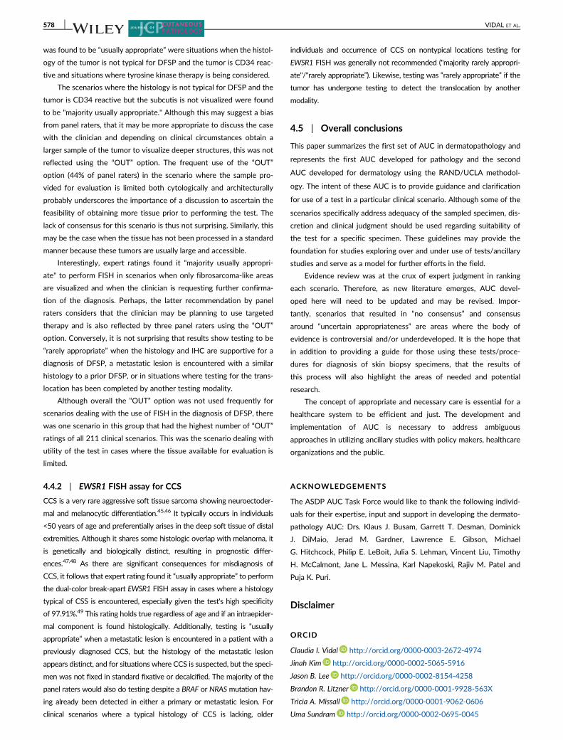
was found to be “usually appropriate” were situations when the histol-
ogy of the tumor is not typical for DFSP and the tumor is CD34 reac-
tive and situations where tyrosine kinase therapy is being considered.
The scenarios where the histology is not typical for DFSP and the
tumor is CD34 reactive but the subcutis is not visualized were found
to be "majority usually appropriate." Although this may suggest a bias
from panel raters, that it may be more appropriate to discuss the case
with the clinician and depending on clinical circumstances obtain a
larger sample of the tumor to visualize deeper structures, this was not
reflected using the “OUT” option. The frequent use of the “OUT”
option (44% of panel raters) in the scenario where the sample pro-
vided for evaluation is limited both cytologically and architecturally
probably underscores the importance of a discussion to ascertain the
feasibility of obtaining more tissue prior to performing the test. The
lack of consensus for this scenario is thus not surprising. Similarly, this
may be the case when the tissue has not been processed in a standard
manner because these tumors are usually large and accessible.
Interestingly, expert ratings found it "majority usually appropri-
ate" to perform FISH in scenarios when only fibrosarcoma-like areas
are visualized and when the clinician is requesting further confirma-
tion of the diagnosis. Perhaps, the latter recommendation by panel
raters considers that the clinician may be planning to use targeted
therapy and is also reflected by three panel raters using the “OUT”
option. Conversely, it is not surprising that results show testing to be
“rarely appropriate” when the histology and IHC are supportive for a
diagnosis of DFSP, a metastatic lesion is encountered with a similar
histology to a prior DFSP, or in situations where testing for the trans-
location has been completed by another testing modality.
Although overall the “OUT” option was not used frequently for
scenarios dealing with the use of FISH in the diagnosis of DFSP, there
was one scenario in this group that had the highest number of “OUT”
ratings of all 211 clinical scenarios. This was the scenario dealing with
utility of the test in cases where the tissue available for evaluation is
limited.
4.4.2 | EWSR1 FISH assay for CCS
CCS is a very rare aggressive soft tissue sarcoma showing neuroectoder-
mal and melanocytic differentiation.45,46 It typically occurs in individuals
<50 years of age and preferentially arises in the deep soft tissue of distal
extremities. Although it shares some histologic overlap with melanoma, it
is genetically and biologically distinct, resulting in prognostic differ-
ences.47,48 As there are significant consequences for misdiagnosis of
CCS, it follows that expert rating found it “usually appropriate” to perform
the dual-color break-apart EWSR1 FISH assay in cases where a histology
typical of CSS is encountered, especially given the test's high specificity
of 97.91%.49 This rating holds true regardless of age and if an intraepider-
mal component is found histologically. Additionally, testing is “usually
appropriate” when a metastatic lesion is encountered in a patient with a
previously diagnosed CCS, but the histology of the metastatic lesion
appears distinct, and for situations where CCS is suspected, but the speci-
men was not fixed in standard fixative or decalcified. The majority of the
panel raters would also do testing despite a BRAF or NRAS mutation hav-
ing already been detected in either a primary or metastatic lesion. For
clinical scenarios where a typical histology of CCS is lacking, older
individuals and occurrence of CCS on nontypical locations testing for
EWSR1 FISH was generally not recommended ("majority rarely appropri-
ate"/"rarely appropriate”). Likewise, testing was “rarely appropriate” if the
tumor has undergone testing to detect the translocation by another
modality.
4.5 | Overall conclusions
This paper summarizes the first set of AUC in dermatopathology and
represents the first AUC developed for pathology and the second
AUC developed for dermatology using the RAND/UCLA methodol-
ogy. The intent of these AUC is to provide guidance and clarification
for use of a test in a particular clinical scenario. Although some of the
scenarios specifically address adequacy of the sampled specimen, dis-
cretion and clinical judgment should be used regarding suitability of
the test for a specific specimen. These guidelines may provide the
foundation for studies exploring over and under use of tests/ancillary
studies and serve as a model for further efforts in the field.
Evidence review was at the crux of expert judgment in ranking
each scenario. Therefore, as new literature emerges, AUC devel-
oped here will need to be updated and may be revised. Impor-
tantly, scenarios that resulted in “no consensus” and consensus
around “uncertain appropriateness” are areas where the body of
evidence is controversial and/or underdeveloped. It is the hope that
in addition to providing a guide for those using these tests/proce-
dures for diagnosis of skin biopsy specimens, that the results of
this process will also highlight the areas of needed and potential
research.
The concept of appropriate and necessary care is essential for a
healthcare system to be efficient and just. The development and
implementation of AUC is necessary to address ambiguous
approaches in utilizing ancillary studies with policy makers, healthcare
organizations and the public.
ACKNOWLEDGEMENTS
The ASDP AUC Task Force would like to thank the following individ-
uals for their expertise, input and support in developing the dermato-
pathology AUC: Drs. Klaus J. Busam, Garrett T. Desman, Dominick
J. DiMaio, Jerad M. Gardner, Lawrence E. Gibson, Michael
G. Hitchcock, Philip E. LeBoit, Julia S. Lehman, Vincent Liu, Timothy
H. McCalmont, Jane L. Messina, Karl Napekoski, Rajiv M. Patel and
Puja K. Puri.
Disclaimer
ORCID
Claudia I. Vidal http://orcid.org/0000-0003-2672-4974
Jinah Kim http://orcid.org/0000-0002-5065-5916
Jason B. Lee http://orcid.org/0000-0002-8154-4258
Brandon R. Litzner http://orcid.org/0000-0001-9928-563X
Tricia A. Missall http://orcid.org/0000-0001-9062-0606
Uma Sundram http://orcid.org/0000-0002-0695-0045
578 VIDAL ET AL.

Maria Yadira Hurley https://orcid.org/0000-0002-1162-7250
Patrick O. Emanuel http://orcid.org/0000-0003-1068-6969
Maxwell A. Fung http://orcid.org/0000-0003-1771-8257
Gregory A. Hosler http://orcid.org/0000-0002-6281-0565
Alexander J. Lazar http://orcid.org/0000-0002-6395-4499
REFERENCES
1. Brook RH, Vaiana ME. Using the knowledge base of health services
research to redefine health care systems. J Gen Intern Med. 2015;
30(10):1547-1556.2. Kuntz KM, Tsevat J, Weinstein MC, Goldman L. Expert panel vs
decision-analysis recommendations for postdischarge coronary angi-
ography after myocardial infarction. JAMA. 1999;282(23):2246-2251.3. Fitch K, Bernstein S, Aguilar MS, et al. The RAND/UCLA Appropriate-
ness Method User's Manual 2001. No. MR-1269-DG-XII/RE:126. Santa
Monica, CA: RAND Corp.; 2001.4. Berg G, Jaffe ES, Kempf W, et al. WHO/EORTC classification of cuta-
neous lymphomas. In: LeBoit PE, Burg G, Weedon D, Sarasin A, eds.
WHO Classification of Skin Tumors. 4th ed. Lyon, France: IARC Press;
2006:165-228.5. Martinez G, Copen CE, Abma JC. Teenagers in the United States: sex-
ual activity, contraceptive use, and childbearing, 2006–2010 National
Survey of family growth. National Center for Health Statistics. Vital
Health Stat. 2011;23(31):1–35.6. Calonje E, Brenn T, Lazar A, McKee PH, eds. McKee’s Pathology of the
Skin. 4th ed. China: Elsevier/Sanders; 2012.7. Gill D, Dorevitch A, Marks R. The prevalence of seborrheic keratoses
in people age 15 to 30 years: is the term senile keratosis redundant?
Arch Dermatol. 2000;136(6):759-762.8. John AM, Schwartz RA. Muir-Torre syndrome (MTS): an update and
approach to diagnosis and management. J Am Acad Dermatol. 2016;
74(3):558-566.9. Bhaijee F, Brown AS. Muir-Torre syndrome. Arch Pathol Lab Med.
2014;138(12):1685-1689.10. Comfere NI, Sundram U, Hurley MY, Swick BL. Views of dermato-
pathologists about clonality assays in the diagnosis of cutaneous T
cell and B cell lymphoproliferative disorders. J Cutan Pathol. 2018;
45(1):39-47.11. Emanuel PO, Andea AA, Vidal CI, et al. Review of the medical litera-
ture and assessment of current utilization patterns regarding molecu-
lar testing for the diagnosis of cutaneous melanoma. J Cutan Pathol.
[Submitted].12. Litzner BR, Lee JB, Vidal CI. Review of the current medical literature
and assessment of current utilization patterns regarding human papil-
lomavirus in situ hybridization and immunohistochemistry in dermato-
pathology. J Cutan Pathol. 2017;44(11):938-943.13. Lee JB, Litzner BR, Vidal CI. Review of the current medical literature
and assessment of current utilization patterns regarding mismatch
repair protein immunohistochemistry in cutaneous Muir-Torre
syndrome-associated neoplasms. J Cutan Pathol. 2017;44(11):931-937.14. Konstantinos L, Kozel JA, Hurley MY, Andea AA. Review of the medi-
cal literature and assessment of current utilization patterns regarding
the use of two common fluorescence in situ hybridization assays for
the diagnosis of dermatofibrosarcoma protuberans and clear cell
sarcoma. J Cutan Pathol. [Submitted].15. Pouryazdanparast P, Newman M, Mafee M, Haghighat Z, Guitart J,
Gerami P. Distinguishing epithelioid blue nevus from blue nevus-like
cutaneous melanoma metastasis using fluorescence in situ hybridiza-
tion. Am J Surg Pathol. 2009;33(9):1396-1400.16. Gammon B, Beilfuss B, Guitart J, Busam KJ, Gerami P. Fluorescence
in situ hybridization for distinguishing cellular blue nevi from blue
nevus-like melanoma. J Cutan Pathol. 2011;38(4):335-341.17. Gerami P, Beilfuss B, Haghighat Z, Fang Y, Jhanwar S, Busam KJ.
Fluorescence in situ hybridization as an ancillary method for the
distinction of desmoplastic melanomas from sclerosing melanocytic
nevi. J Cutan Pathol. 2011;38(4):329-334.
18. Clarke LE, Warf MB. Flake DD 2nd, et al.. Clinical validation of a gene
expression signature that differentiates benign nevi from malignant
melanoma. J Cutan Pathol. 2015;42(4):244-252.19. Clarke LE, Flake DD 2nd, Busam K, et al. An independent validation
of a gene expression signature to differentiate malignant melanoma
from benign melanocytic nevi. Cancer 2017;123(4):617–628.20. Minca EC, Al-Rohil RN, Wang M, et al. Comparison between mela-
noma gene expression score and fluorescence in situ hybridization for
the classification of melanocytic lesions. Mod Pathol. 2016;29(8):
832-843.21. Cockerell C, Tschen J, Billings SD, et al. The influence of a gene-
expression signature on the treatment of diagnostically challenging
melanocytic lesions. Per Med. 2017;14(2):123-130.22. Ko JS, Matharoo-Ball B, Billings SD, et al. Diagnostic distinction of
malignant melanoma and benign nevi by a gene expression signature
and correlation to clinical outcomes. Cancer Epidemiol Biomarkers Prev.
2017;26(7):1107-1113.23. Aguilera-Barrantes I, Magro C, Nuovo GJ. Verruca vulgaris of the
vulva in children and adults: a nonvenereal type of vulvar wart.
Am J Surg Pathol. 2007;31:529-535.24. Nuovo GJ, Lastarria DA, Smith S, Lerner J, Comité SL, Eliezri YD.
Human papillomavirus segregation patterns in genital and nongenital
warts in prepubertal children and adults. Am J Clin Pathol. 1991;95:
467-474.25. Yun K, Joblin L. Presence of human papillomavirus DNA in condylo-
mata acuminata in children and adolescents. Pathology. 1993;25:1-3.26. Padel AF, Venning VA, Evans MF, Quantrill AM, Fleming KA. Human
papillomaviruses in anogenital warts in children: typing by in situ
hybridisation. BMJ. 1990;300:1491-1494.27. Handley J, Hanks E, Armstrong K, et al. Common association of HPV
2 with anogenital warts in prepubertal children. Pediatr Dermatol.
1997;14:339-343.28. Schwartz RA, Torre DP. The Muir–Torre syndrome: a 25-year retro-
spect. J Am Acad Dermatol. 1995;33:90-104.29. Giardiello FM, Allen JI, Axilbund JE, et al. Guidelines on genetic evalu-
ation and management of lynch syndrome: a consensus statement by
the US multi-society task force on colorectal cancer.
Am J Gastroenterol. 2014;109:1159-1179.30. Everett JN, Raymond VM, Dandapani M, et al. Screening for germline
mismatch repair mutations following diagnosis of sebaceous neo-
plasm. JAMA Dermatol. 2014;150:1315-1321.31. Roberts ME, Riegert-Johnson DL, Thomas BC, et al. A clinical scoring
system to identify patients with sebaceous neoplasms at risk for the
Muir–Torre variant of Lynch syndrome. Genet Med. 2014;16:
711-716.32. Plocharczyk EF, Frankel WL, Hampel H, Peters SB. Mismatch repair
protein deficiency is common in sebaceous neoplasms and suggests
the importance of screening for lynch syndrome. Am J Dermatopathol.
2013;35:191-195.33. Dores GM, Curtis RE, Toro JR, Devesa SS, Fraumeni JF Jr. Incidence
of cutaneous sebaceous carcinoma and risk of associated neoplasms:
insight into Muir-Torre syndrome. Cancer 2008. 113(12):3372–81.34. Mojtahed A, Schrijver I, Ford JM, Longacre TA, Pai RK. A two-
antibody mismatch repair protein immunohistochemistry screening
approach for colorectal carcinomas, skin sebaceous tumors, and gyne-
cologic tract carcinomas. Mod Pathol. 2011;24(7):1004-1014.35. Naeem R, Lux ML, Huang SF, Naber SP, Corson JM, Fletcher JA. Ring
chromosomes in dermatofibrosarcoma protuberans are composed of
interspersed sequences from chromosomes 17 and 22. Am J Pathol.
1995;147(6):1553-1558.36. Poland KS, Shardy DL, Azim M, et al. Overexpression of ZNF342 by
juxtaposition with MPO promoter/enhancer in the novel transloca-
tion t(17;19)(q23;q13.32) in pediatric acute myeloid leukemia and
analysis of ZNF342 expression in leukemia. Genes Chromosomes
Cancer. 2009;48(6):480-489.37. Simon MP, Pedeutour F, Sirvent N, et al. Deregulation of the
platelet-derived growth factor B-chain gene via fusion with collagen
gene COL1A1 in dermatofibrosarcoma protuberans and giant-cell
fibroblastoma. Nat Genet. 1997;15(1):95-98.
VIDAL ET AL. 579

38. Sirvent N, Maire G, Pedeutour F. Genetics of dermatofibrosarcomaprotuberans family of tumors: from ring chromosomes to tyrosine kinaseinhibitor treatment. Genes Chromosomes Cancer. 2003;37(1):1-19.
39. Rutkowski P, Wozniak A, Switaj T. Advances in molecular characteri-zation and targeted therapy in dermatofibrosarcoma protuberans.Sarcoma. 2011;2011:959132.
40. Rutkowski P, Van Glabbeke M, Rankin CJ, et al. Imatinib mesylate inadvanced dermatofibrosarcoma protuberans: pooled analysis of twophase II clinical trials. J Clin Oncol. 2010;28(10):1772-1779.
41. Salgado R, Llombart B, M Pujol R, et al. Molecular diagnosis ofdermatofibrosarcoma protuberans: a comparison between reversetranscriptase-polymerase chain reaction and fluorescence in situ hybridi-zation methodologies. Genes Chromosomes Cancer. 2011;50(7):510-517.
42. Papp G, Mihály D, Sápi Z. Unusual signal patterns of break-apart FISHprobes used in the diagnosis of soft tissue sarcomas. Pathol OncolRes. 2017;23(4):863-871.
43. Ha SY, Lee SE, Kwon MJ, et al. PDGFB rearrangement in dermatofi-brosarcoma protuberans: correlation with clinicopathologic character-istics and clinical implications. Hum Pathol. 2013;44(7):1300-1309.
44. Karanian M, Perot G, Coindre JM, Chibon F, Pedeutour F, Neuville A.Fluorescence in situ hybridization analysis is a helpful test for thediagnosis of dermatofibrosarcoma protuberans. Mod Pathol. 2015;28(2):230-237.
45. Enzinger FM. Clear-cell sarcoma of tendons and aponeuroses. Ananalysis of 21 cases. Cancer. 1965;18:1163-1174.
46. Chung EB, Enzinger FM. Malignant melanoma of soft parts. A reas-sessment of clear cell sarcoma. Am J Surg Pathol. 1983;7(5):405-413.
47. Graadt van Roggen JF, Mooi WJ, Hogendoorn PC. Clear cell sarcomaof tendons and aponeuroses (malignant melanoma of soft parts) andcutaneous melanoma: exploring the histogenetic relationship betweenthese two clinicopathological entities. J Pathol. 1998;186(1):3-7.
48. Takahira T, Oda Y, Tamiya S, et al. Alterations of the p16INK4a/p14ARFpathway in clear cell sarcoma. Cancer Sci. 2004;95(8):651-655.
49. Segal NH, Pavlidis P, Noble WS, et al. Classification of clear-cellsarcoma as a subtype of melanoma by genomic profiling. J Clin Oncol.2003;21(9):1775-1781.
How to cite this article: Vidal CI, Armbrect EA, Andea AA,
et al. Appropriate use criteria in dermatopathology: Initial
recommendations from the American Society of Dermato-
pathology. J Cutan Pathol. 2018;45:563–580. https://doi.
org/10.1111/cup.13142
580 VIDAL ET AL.