Embed Size (px)
Citation preview
MEDICAL & BIOLOGICAL LABORATORIES CO., LTD. URL http://ruo.mbl.co.jp
e-mail [email protected], TEL 052-238-1904
Printed February 15, 2015
Version 1.0
Autophagy Watch
CODE No. 8486
For Autophagy Flux Assay and LC3 Immunocytochemistry
For Research Use Only. Not for use in diagnostic procedures.

-1-
Description
Autophagy has been suggested to have an important role in development, differentiation,
neurodegeneration, infection and cancer. Autophagy is a process of intracellular bulk degradation
in which cytoplasmic components including organelles are sequestered within double-membrane
vesicles. As shown in Fig. 1, a flat vesicle called an isolate membrane appears in the cytoplasm (1).
Subsequently, the membrane extends while taking in the cytoplasm (2), its edges fusing to
compose an AP (Autophagosome) (3). Mitochondria and other large organelles are also contained
within the AP. When the AP fuses with a lysosome (4), its contents are degraded (5).
Autophagosomes deliver the contents to the lysosome/vacuole for degradation. This process aids
in cell survival during starvation periods by eliminating nutrients being needed for unnecessary
cellular processes. .
Detecting LC3 protein by using western blot application is widely used for monitoring
autophagic activity. LC3 is most actively studied and frequently used as a mammalian autophagy
marker. Shortly after translation (proLC3), LC3 is processed at the C-terminus into LC3-I. Upon
induction of autophagy, LC3-I is conjugated to the substrate phosphatidylethanolamine (PE). The
PE-LC3-I conjugate is referred to as LC3-II. Despite having a higher molecular weight than LC3-I,
LC3-II is more hydrophobic and shows a higher mobility in sodium dodecyl
sulfate-polyacrylamide gel electrophoresis (SDS-PAGE). Although the amount of LC3-II is
correlated with the number of autophagosomes, it can be difficult to interpret LC3 immunoblot
analysis.
Autophagy Watch is a combination of antibodies and lysosomal inhibitors that can detect
autophagy flux in Western blotting, and visualize autophagosomes in Immunocytochemistry.
Autophagy Watch is ready to use for the rapid and quantitative monitoring of autophagic activity.
Fig. 1. Autophagy pathway and lysosomal inhibitors
-2-
Features of Autophagy Watch
1. Anti-LC3 antibody and lysosomal inhibitors that detect autophagy flux in western blots are
included.
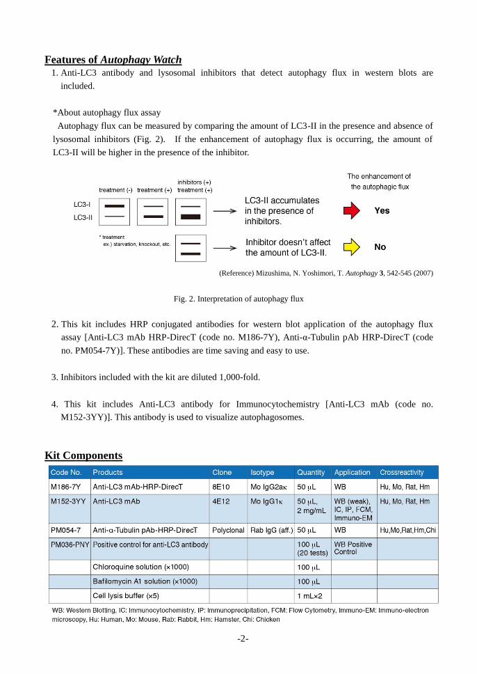
*About autophagy flux assay
Autophagy flux can be measured by comparing the amount of LC3-II in the presence and absence of
lysosomal inhibitors (Fig. 2). If the enhancement of autophagy flux is occurring, the amount of
LC3-II will be higher in the presence of the inhibitor.
(Reference) Mizushima, N. Yoshimori, T. Autophagy 3, 542-545 (2007)
Fig. 2. Interpretation of autophagy flux
2. This kit includes HRP conjugated antibodies for western blot application of the autophagy flux
assay [Anti-LC3 mAb HRP-DirecT (code no. M186-7Y), Anti-α-Tubulin pAb HRP-DirecT (code
no. PM054-7Y)]. These antibodies are time saving and easy to use.
3. Inhibitors included with the kit are diluted 1,000-fold.
4. This kit includes Anti-LC3 antibody for Immunocytochemistry [Anti-LC3 mAb (code no.
M152-3YY)]. This antibody is used to visualize autophagosomes.
Kit Components
-3-
Formulation Antibodies: PBS containing 50% Glycerol (pH 7.2). No preservative is contained.
PM036-PNY: human LC3B overexpressed 293T 1×10⁵ cells were suspended with 100 L of
Laemmli sample buffer. This product contains 5% 2-mercaptoethanol. Handle with
care.
Chloroquine solution (×1000): 50 mM Chloroquine/PBS
Bafilomycin A1 solution (×1000): 100 μM Bafilomycin A1/DMSO
Cell lysis buffer (×5): 0.25 M Tris-HCl (pH 7.5), 0.75 M NaCl, 0.25% Nonidet P-40
Storage
This kit is stable for one year from the date of purchase when stored at -20°C.
Example of protocols using this kit
1. Autophagy flux assay by Western blot
The following protocol is an example of LC3 autophagy flux assay detected by western blot under
starvation conditions of mammalian cells (MEF cells).
Materials required but not provided
・Nutrient medium [RPMI-1640 (Sigma; code no. R8787), 10% FCS, 100 U/mL Penicillin, 50
μg/mL Streptomycin]
・Starvation medium [HBSS- Hanks’ Balanced Salt solution (Sigma; code no. H9269)]
・0.25% trypsin/1 mM EDTA solution (Wako; code no. 201-16945)
・PBS
・Laemmli sample buffer
・Protein concentration assay kit (e.g,.Bio-Rad; code no. 500-001JA)
・Skimmed milk
・PBS-T (0.05% Tween-20/PBS)
・Chemiluminescent substrate (GE Healthcare; code no. RPN2106)
・Culture dish (90 mm) (Thermo Scientific; code no. 172931)
・Cell scraper (Sumitomo Bakelite; code no. MS-93170)
・PVDF membrane (Merck Millipore; code no. IPVH00010)
Method
a) Preparation of loading samples
Preparation of reagents
Cell lysis buffer
Dilute Cell lysis buffer (×5) with 5 times its volume with distilled water. Store at 4°C.
(e.g., dilute 1 mL of Cell lysis buffer (×5) with 4 mL of distilled water.)
1) (First day)
Maintain MEF cells in a 90 mm culture dish in nutrient medium (10% FCS/RPMI-1640) until
70~80% confluence at 37°C with 5% CO2.
-4-
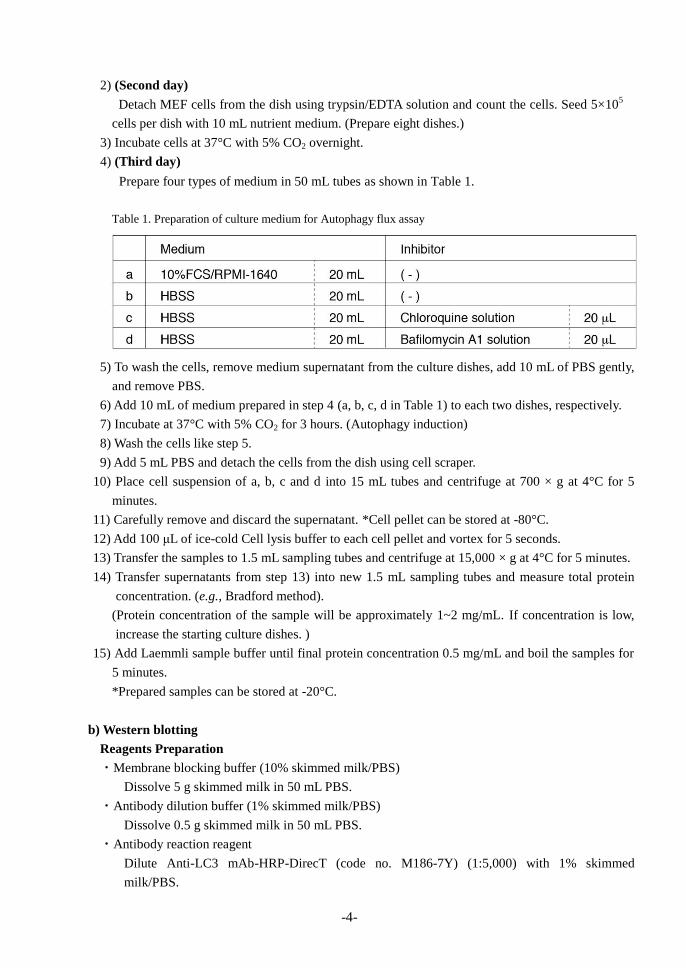
2) (Second day)
Detach MEF cells from the dish using trypsin/EDTA solution and count the cells. Seed 5×10⁵
cells per dish with 10 mL nutrient medium. (Prepare eight dishes.)
3) Incubate cells at 37°C with 5% CO2 overnight.
4) (Third day)
Prepare four types of medium in 50 mL tubes as shown in Table 1.
Table 1. Preparation of culture medium for Autophagy flux assay
5) To wash the cells, remove medium supernatant from the culture dishes, add 10 mL of PBS gently,
and remove PBS.
6) Add 10 mL of medium prepared in step 4 (a, b, c, d in Table 1) to each two dishes, respectively.
7) Incubate at 37°C with 5% CO2 for 3 hours. (Autophagy induction)
8) Wash the cells like step 5.
9) Add 5 mL PBS and detach the cells from the dish using cell scraper.
10) Place cell suspension of a, b, c and d into 15 mL tubes and centrifuge at 700 × g at 4°C for 5
minutes.
11) Carefully remove and discard the supernatant. *Cell pellet can be stored at -80°C.
12) Add 100 μL of ice-cold Cell lysis buffer to each cell pellet and vortex for 5 seconds.
13) Transfer the samples to 1.5 mL sampling tubes and centrifuge at 15,000 × g at 4°C for 5 minutes.
14) Transfer supernatants from step 13) into new 1.5 mL sampling tubes and measure total protein
concentration. (e.g., Bradford method).
(Protein concentration of the sample will be approximately 1~2 mg/mL. If concentration is low,
increase the starting culture dishes. )
15) Add Laemmli sample buffer until final protein concentration 0.5 mg/mL and boil the samples for
5 minutes.
*Prepared samples can be stored at -20°C.
b) Western blotting
Reagents Preparation
・Membrane blocking buffer (10% skimmed milk/PBS)
Dissolve 5 g skimmed milk in 50 mL PBS.
・Antibody dilution buffer (1% skimmed milk/PBS)
Dissolve 0.5 g skimmed milk in 50 mL PBS.
・Antibody reaction reagent
Dilute Anti-LC3 mAb-HRP-DirecT (code no. M186-7Y) (1:5,000) with 1% skimmed
milk/PBS.
-5-
(e.g., dilute 1 L antibody with 5 mL 1% skimmed milk/PBS.)
*In the case of detecting loading control, dilute Anti-α-Tubulin pAb-HRP-DirecT (code no.
PM054-7) (1:5,000) with 1% skimmed milk/PBS.
1) Load 20 μL of the sample per lane in 15% SDS-polyacrylamide gel.
*In the case of using positive control, load 5 μL of the PM036-PNY per lane.
2) Run the gel electrophoresis at 15 mA for 2 hours.
3) Blot the protein to a PVDF membrane at 1 mA/cm2 for 1 hour in a semi-dry transfer system. (See
the manufacturer’s manual for precise transfer procedure.)
4) To reduce nonspecific binding, soak the membrane in 50 mL of 10% skimmed milk/PBS
overnight at 4°C.
5) Wash the membrane with PBS-T (0.05% Tween-20/PBS) for 5minutes. Repeat 3 times.
6) Incubate the membrane with antibody diluted with 1% skimmed milk for 1 hour at room
temperature. *Do not allow the membrane to dry during incubation.
7) Wash the membrane as described in step 5.
8) Detect LC3 band using chemiluminescence substrate.
To detect loading control using PM054-7 (Anti-α-Tubulin pAb-HRP-DirecT), soak the used
membrane in 50 mL 10% skimmed milk/PBS for 1 hour at room temperature or overnight at 4°C
and repeat procedures step 5 to step 8.
-6-
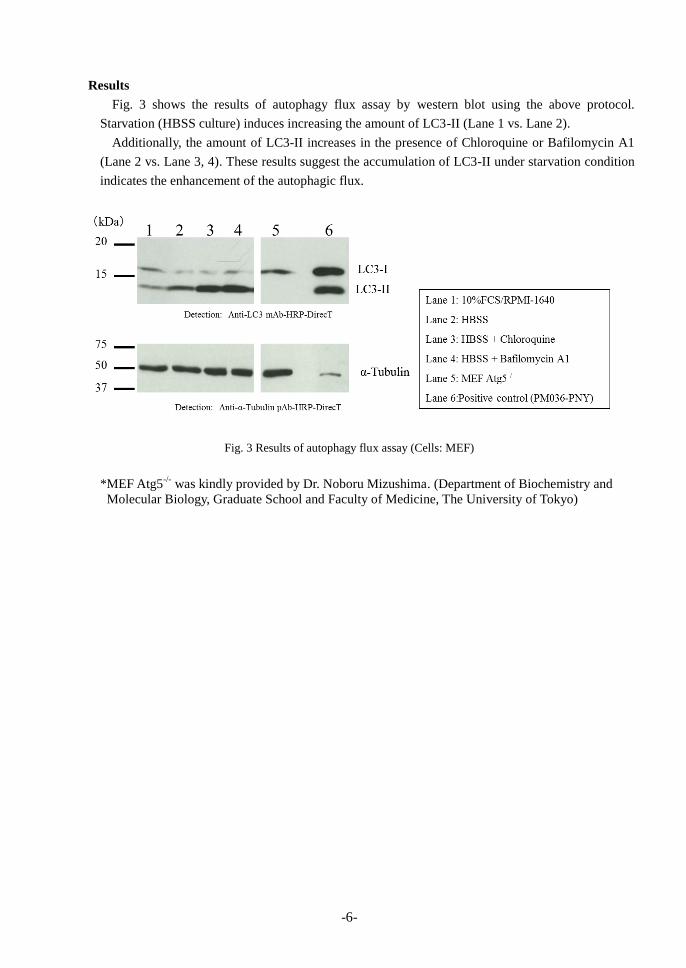
Results
Fig. 3 shows the results of autophagy flux assay by western blot using the above protocol.
Starvation (HBSS culture) induces increasing the amount of LC3-II (Lane 1 vs. Lane 2).
Additionally, the amount of LC3-II increases in the presence of Chloroquine or Bafilomycin A1
(Lane 2 vs. Lane 3, 4). These results suggest the accumulation of LC3-II under starvation condition
indicates the enhancement of the autophagic flux.
Fig. 3 Results of autophagy flux assay (Cells: MEF)
*MEF Atg5-/-
was kindly provided by Dr. Noboru Mizushima. (Department of Biochemistry and
Molecular Biology, Graduate School and Faculty of Medicine, The University of Tokyo)
-7-
2. Immunocytochemistry The following protocol is the example of observing autophagosomes by LC3 Immunocytochemistry
under starvation conditions of mammalian cells (MEF cells).
Materials required but not provided
・Nutrient medium
[RPMI-1640 (Sigma; code no. R8787), 10% FCS, Penicillin 100 U/mL, Streptomycin 50 μg/mL]
・Starvation medium [HBSS- Hanks’ Balanced Salt solution (Sigma; code no. H9269)]
・0.25% trypsin/1 mM EDTA solution (Wako; code no. 201-16945)
・PBS
・4% Paraformaldehyde (PFA)/PB Solution (Wako; code no.163-20145)
・Digitonin (e.g. Sigma; code no. D141-100MG)
・DAPI solution (e.g. Dojindo; code no. 340-07971 diluting 1000-fold in PBS)
・Mounting medium (Life Technologies; code no. P36930)
・Secondary antibody [Anti-Mouse IgG Alexa Fluor488 conjugate (Life Technologies; code no.
A11001)]
・8 wells chamber slide (Thermo Scientific; code no. 154941)
Preparation of reagents
・Cell permeabilization solution
Prepare 100 μg/mL Digitonin/PBS just before use.
(e.g., dissolve 1 mg Digitonin in 10 mL PBS.)
・Primary antibody reaction solution
Dilute anti-LC3 mAb (code no. M152-3YY) (1:50) with PBS.
(e.g., dilute 20 μL antibody in 1 mL PBS.)
・Secondary antibody reaction solution
Dilute anti-Mouse IgG Alexa Fluor488 conjugate (1:500) with PBS.
(e.g., dilute 2 L antibody in 1 mL PBS.)
Method
1) (First day)
Maintain MEF cells in a 90 mm culture dish in nutrient medium (10% FCS/RPMI-1640) until
70-80% confluence at 37°C with 5% CO2.
2) (Second day )
Detach MEF cells from the dish using trypsin/EDTA solution and count the cells.
Seed 5×10³ cells per wells with 200 L nutrient medium.
3) Incubate at 37°C with 5% CO2 for overnight.
4) (Third day)
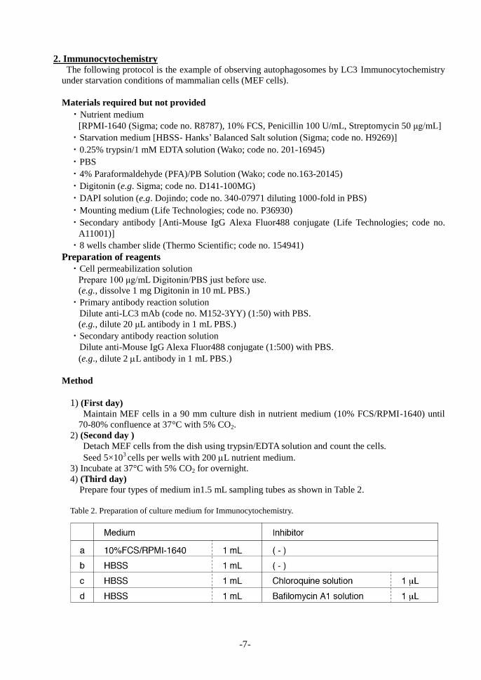
Prepare four types of medium in1.5 mL sampling tubes as shown in Table 2.
Table 2. Preparation of culture medium for Immunocytochemistry.
-8-
5) Remove the culture supernatant by careful aspiration. Do not allow the slides to dry during steps
5-17.
6) To wash the cells on slides, add 200 μL HBSS in each well and remove HBSS.
7) Add 200 L medium prepared in step 4 (a, b, c, d in Table 2) to each well, respectively.
8) Incubate at 37°C with 5% CO2 for 3 hours.
9) Remove supernatant and wash gently the cells with PBS. Repeat 2 times.
10) Fix the cells adding 200 μL of 4% PFA/PB solution to each well for 10 minutes at room
temperature.
11) Wash the cells as in the step 9.
12) Permeabilize the cells with 200 μL of 100 μg/mL Digitonin/PBS for 10 minutes at room
temperature.
13) Wash the cells as described in the step 9.
12) Add 200 μL of primary antibody reaction reagent for 1 hour at room temperature.
14) Wash the cells as described in the step 9.
15) Add 200 μL of secondary antibody reaction reagent and incubate for 30 minutes at room
temperature. Keep out light.
16) Add a drop of DAPI solution and incubate for 5 minutes at room temperature.
17) Wash the cells as described in the step 9.
18) Add mounting medium onto the slide, then put a cover slip on it and image the slide with
fluorescence microscope.
Results
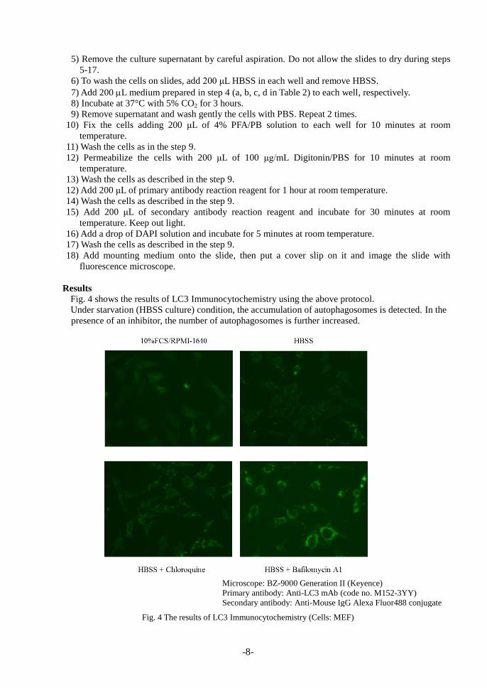
Fig. 4 shows the results of LC3 Immunocytochemistry using the above protocol.
Under starvation (HBSS culture) condition, the accumulation of autophagosomes is detected. In the
presence of an inhibitor, the number of autophagosomes is further increased.
Fig. 4 The results of LC3 Immunocytochemistry (Cells: MEF) Microscope:BZ-9000 Generation II(Keyence)
Primary antibody: Anti-LC3 mAb (code:M152-3YY)
Secondary antibody: Anti-Mouse IgG Alexa Fluor488 conjugate
Microscope: BZ-9000 Generation II (Keyence)
Primary antibody: Anti-LC3 mAb (code no. M152-3YY)
Secondary antibody: Anti-Mouse IgG Alexa Fluor488 conjugate
-9-
Troubleshooting 1. It is difficult to separate the bands of LC3-I and LC3-II using western blot.
LC3 are detected as two bands at 16 kDa and 14 kDa in various cell lysates, respectively. We
recommend using 15% SDS-polyacrylamide gel to obtain optimal performance.
2. LC3-I and LC3-II band cannot be detected.
Consider increasing the volume of loading sample or increasing the concentration of anti-LC3
antibody. In addition, we recommend using Western blotting control for anti-LC3 antibody (code no.
PM036-PNY).
3. During the reaction of immunocytochemistry, the cells come off from the slide wells.
Never allow the slide to dry out.
Related products Antibodies
PM036 Anti-LC3 pAb [WB, IP, IC, IHC, FCM]
M152-3 Anti-LC3 mAb (4E12) [WB, IP, IC, FCM, EM]
M186-3 Anti-LC3 mAb (8E10) [WB]
M186-7 Anti-LC3 mAb-HRP-DirecT (8E10)
PD015 Anti-LC3 pAb [IC]
PM045 Anti-p62 (SQSTM1) pAb
PM066 Anti-p62 C-terminal pAb
PM066-7 Anti-p62 C-terminal pAb
M162-3 Anti-p62 (SQSTM1) (Human) mAb (5F2) PM074 Anti-Phospho-p62 (SQSTM1) (Ser351) pAb M217-3 Anti-Phospho-p62 (SQSTM1) (Ser351) mAb
D343-3 Anti-Phospho-p62 (SQSTM1) (Ser403) mAb
(4F6)
D344-3 Anti-Phospho-p62 (SQSTM1) (Ser403) mAb
(4C8)
PD017 Anti-Beclin 1 pAb
PM037 Anti-GABARAP pAb
M135-3 Anti-GABARAP mAb (1F4)
PM038 Anti-GATE-16 pAb
PD041 Anti-Atg2A pAb
PM034 Anti-Atg3 pAb
M133-3 Anti-Atg3 mAb (3E8)
M134-3 Anti-Atg4B mAb (9H5)
PM050 Anti-Atg5 pAb
M153-3 Anti-Atg5 mAb (4D3)
PM039 Anti-Atg7 (Human) pAb
PD042 Anti-Atg9A pAb
M151-3 Anti-Atg10 (Human) mAb (5A7)
M154-3 Anti-Atg12 (Human) mAb (6E5)
PD036 Anti-Atg13 (Human) pAb
M183-3 Anti-Atg13 mAb (5G4)
PD026 Anti-Atg14 pAb
M184-3 Anti-Atg14 (Human) mAb (4H8)
PM040 Anti-Atg16L pAb
M150-3 Anti-Atg16L mAb (1F12)
M160-3 Anti-UVRAG mAb (1H4)
PD027 Anti-Rubicon (Human) pAb
M170-3 Anti-Rubicon (Human) mAb (1H6)
PD037 Anti-Tel2 pAb
PM069 Anti-NRF2 pAb
M200-3 Anti-NRF2 mAb (1F2)
PM072 Anti-VMP1 pAb
PM076 Anti-Syntaxin-17 (Human) pAb
M212-3 Anti-Syntaxin-17 (Human) mAb (2F8)
PM054-7 Anti-α-Tubulin pAb-HRP-DirecT
PM053-7 Anti-β-actin pAb-HRP-DirecT
Kits
8485 Autophagy Ab Sampler Set
PM036-PN Positive control for anti-LC3 antibody
WB: Western blotting
IP: Immunoprecipitation
IC: Immunocytochemistry
IHC: Immunohistochemistry
FCM: Flow cytometry
EM: Immuno-electron microscopy
Other related antibodies and kits are also available.
Please visit our website at http://ruo.mbl.co.jp/

-10-
はじめに
オートファジーは細胞内の不要なタンパク質やオルガネラ、侵入した細菌を基質として分解し、細胞内から除去する機能で、細胞の飢餓応答時に強く誘導されます。図 1 のように、隔離膜の形
成後に「隔離膜による基質の取り込み」→「オートファゴソームの形成」→「リソソームと融合」
といった経路を通り、基質が分解されます。近年、オートファジーはがんや神経変性疾患、糖尿
病などの疾患や発生・分化などの生理機能に重要な働きをしていることが明らかとなり、幅広い
分野での研究が行われています。
オートファジーの生化学的な解析には一般的にオートファゴソームに局在する LC3 タンパク
質を抗体で検出するウエスタンブロットで行われています。LC3 は細胞質中に存在している
LC3-I 型が、オートファジーの誘導によりリン脂質(PE; Phosphatidylethanolamine)と共有結合し
LC3-II型となり、隔離膜やオートファゴソームに局在します。LC3は LC3-Iと LC3-IIは SDS-PAGE
での移動度が異なるため、それぞれを単一のバンドとして検出することが可能です。LC3-II の量
はオートファゴソーム数と相関するといわれていますが、「LC3-I と LC3-II のバンドが検出でき
ないといったトラブル」や「オートファジーの誘導を LC3-II の増減だけでは単純に示すことがで
きない」など検出や結果の解釈が難しい面がありました。
Autophagy Watch はオートファジー解析を始めようとされているお客様に簡便で迅速な実験結
果を得られることを目的とし、オートファジー経路の阻害剤と、オートファジー活性評価に必須
な抗 LC3 抗体をセットにした初めてのキットです。
図 1. オートファジーの経路と阻害剤
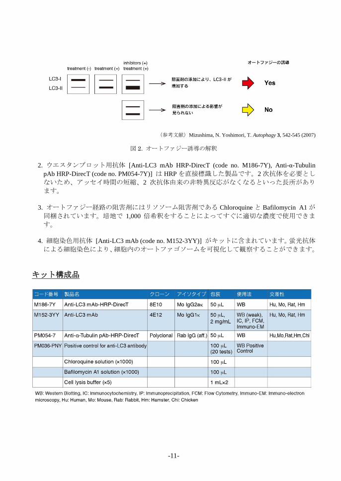
Autophagy Watch の特徴
Autophagy Watchは以下のような特徴を持ったキットです。
1. 抗 LC3 抗体とオートファジー経路の阻害剤がセットで同梱されています。これらを用
いてウエスタンブロットを行うことでオートファジー誘導の有無を調べるオートファジ
ーフラックスアッセイができます。
*オートファジーフラックスアッセイとは
LC3-IIは隔離膜やオートファゴソーム膜に局在していますが、単純に LC3-IIのバンドの
増加のみでオートファジー誘導の有無を決定することはできません。リソソーム阻害剤の
処理を行ったサンプルと比較することで図 2 のようにオートファジーの誘導を判断でき
るようになります。
-11-
(参考文献)Mizushima, N. Yoshimori, T. Autophagy 3, 542-545 (2007)
図 2. オートファジー誘導の解釈
2. ウエスタンブロット用抗体 [Anti-LC3 mAb HRP-DirecT (code no. M186-7Y), Anti-α-Tubulin
pAb HRP-DirecT (code no. PM054-7Y)] は HRP を直接標識した製品です。2次抗体を必要とし
ないため、アッセイ時間の短縮、2 次抗体由来の非特異反応がなくなるといった長所があり
ます。
3. オートファジー経路の阻害剤にはリソソーム阻害剤である Chloroquine と Bafilomycin A1 が
同梱されています。培地で 1,000 倍希釈をすることによってすぐに適切な濃度で使用できま
す。
4. 細胞染色用抗体 [Anti-LC3 mAb (code no. M152-3YY)] がキットに含まれています。蛍光抗体
による細胞染色により、細胞内のオートファゴソームを可視化して観察することができます。
キット構成品
-12-
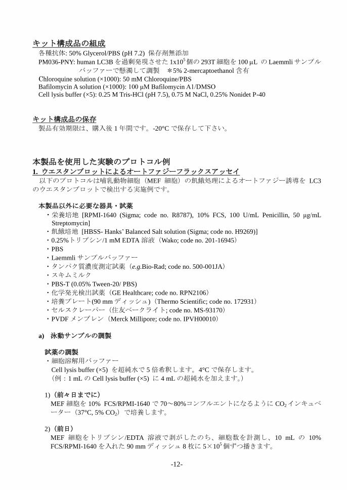
キット構成品の組成
各種抗体: 50% Glycerol/PBS (pH 7.2) 保存剤無添加
PM036-PNY: human LC3Bを過剰発現させた 1x10⁵個の 293T細胞を 100 L の Laemmliサンプル
バッファーで懸濁して調製 *5% 2-mercaptoethanol 含有
Chloroquine solution (×1000): 50 mM Chloroquine/PBS
Bafilomycin A solution (×1000): 100 μM Bafilomycin A1/DMSO
Cell lysis buffer (×5): 0.25 M Tris-HCl (pH 7.5), 0.75 M NaCl, 0.25% Nonidet P-40
キット構成品の保存
製品有効期限は、購入後 1年間です。-20°Cで保存して下さい。
本製品を使用した実験のプロトコル例
1. ウエスタンブロットによるオートファジーフラックスアッセイ
以下のプロトコルは哺乳動物細胞(MEF 細胞)の飢餓処理によるオートファジー誘導を LC3
のウエスタンブロットで検出する実施例です。
本製品以外に必要な器具・試薬
・栄養培地 [RPMI-1640 (Sigma; code no. R8787), 10% FCS, 100 U/mL Penicillin, 50 μg/mL
Streptomycin]
・飢餓培地 [HBSS- Hanks’ Balanced Salt solution (Sigma; code no. H9269)]
・0.25%トリプシン/1 mM EDTA溶液(Wako; code no. 201-16945)
・PBS
・Laemmli サンプルバッファー
・タンパク質濃度測定試薬(e.g.Bio-Rad; code no. 500-001JA)
・スキムミルク
・PBS-T (0.05% Tween-20/ PBS)
・化学発光検出試薬(GE Healthcare; code no. RPN2106)
・培養プレート(90 mmディッシュ)(Thermo Scientific; code no. 172931)
・セルスクレーパー(住友ベークライト; code no. MS-93170)
・PVDF メンブレン(Merck Millipore; code no. IPVH00010)
a) 泳動サンプルの調製
試薬の調製
・細胞溶解用バッファー
Cell lysis buffer (×5) を超純水で 5 倍希釈します。4°Cで保存します。
(例:1 mLの Cell lysis buffer (×5) に 4 mLの超純水を加えます。)
1)(前々日までに)
MEF 細胞を 10% FCS/RPMI-1640 で 70~80%コンフルエントになるように CO2インキュベ
ーター(37°C, 5% CO2)で培養します。
2)(前日)
MEF 細胞をトリプシン/EDTA 溶液で剥がしたのち、細胞数を計測し、10 mL の 10%
FCS/RPMI-1640 を入れた 90 mmディッシュ 8枚に 5×10⁵個ずつ播きます。
-13-
3) CO2インキュベーター(37°C, 5% CO2)で一晩培養します。
4)(オートファジー誘導 当日)
下記の表のように 4 種類の培地を 50 mL チューブに用意します。(90 mm ディッシュ各 2
枚分)
表 1. オートファジーフラックスアッセイのための培地の準備
5) 前日用意した MEF 細胞のディッシュの上清を除き、各 10 mLの PBS を細胞が剥がれない
ようにゆっくりと加え、細胞を洗浄したのち、PBS を除きます。
6) 4)で用意した a)、b)、c)、d)の培地をディッシュに各 10 mLずつ添加します。(各 2 枚)
7) CO2インキュベーター(37°C, 5% CO2)で 3 時間培養します。(オートファジーの誘導)
8) 5)と同様に PBSで細胞を 1回洗浄します。
9) 各ディッシュに PBS を新たに 5 mL加え、セルスクレーパーで細胞を培養プレートから分
離させます。
10) a)、b)、c)、d)で処理した細胞懸濁液各 2枚分を 15 mLチューブ各1本に移し、4°C, 700 × g
で 5分間遠心します。
11) 上清を取り除き a)、b)、c)、d) の細胞ペレットを得ます。*細胞ペレットは-80°C で保存
することができます。
12) 各細胞ペレットに氷冷した細胞溶解用バッファーを 100 μL加え、5秒間ボルテックスしま
す。
13) 細胞溶液を 1.5 mL マイクロチューブに移し、4°C, 15,000 × gで 5分間遠心します。
14) 上清を別のマイクロチューブに移し、Bradford法等で上清のタンパク質濃度を計測します。
(およそ 1~2 mg/mL のタンパク質濃度のサンプルが得られます。得られたサンプルの濃度
が薄い場合は細胞を培養するディッシュの数を増やしてください。)
15) 最終濃度0.5 mg/mLになるように Laemmliサンプルバッファーを加え 5分間煮沸します。
b) ウエスタンブロット
試薬の調製
・メンブレンブロッキング液
スキムミルク 5 gを PBS 50 mL に溶かし、10%スキムミルク/PBS を作製します。
・抗体希釈液
スキムミルク 0.5 g を PBS 50 mLに溶かし、1%スキムミルク/PBS を作製します。
・1次抗体反応液
Anti-LC3 mAb-HRP-DirecT (code no. M186-7Y)を 1%スキムミルク/PBSで 5,000倍希釈します。
(例:5 mLの 1% スキムミルク/PBSに 1 Lの抗体を加えます。
*ローディングコントロールの検出の場合は Anti-α-Tubulin pAb-HRP-DirecT(code no.
PM054-7)を 1% スキムミルク/PBSで 5,000 倍希釈します。

-14-
1) 準備したサンプルを 1レーンあたり 20 μL (10 g/lane) 、15% SDS-ポリアクリルアミドゲ
ルへアプライします。
*ウエスタンブロットのポジティブコントロール(code no. PM036-PNY)は 5 μLアプライ
します。
2) 15 mAで 2 時間泳動します。
3) メンブレンにタンパク質を転写します。(セミドライ法による 1 mA/cm2で 1時間の転写を
行います。詳細な転写条件はご使用の転写装置のマニュアルを参照してください。)
4) メンブレンを 50 mLのブロッキング液に浸し、4°Cで一晩ブロッキングします。
5) メンブレンを PBS-T で 5 分間振とうして洗浄します。この操作を 3回繰り返します。
6) 希釈した1次抗体反応液をラップ上においたメンブレンを覆うように添加し、抗体希釈液
が乾燥しないようプラスチックケースなどをかぶせ、室温で 1 時間静置します。
7) 5)と同様に PBS-T でメンブレンを洗浄します。
8) 化学発光の検出試薬を用いて LC3 のバンドを検出します。
PM054-7(Anti-α-Tubulin pAb-HRP-DirecT)を用いてローディングコントロールデータを取る場
合は、使用したメンブレンを再度 10%スキムミルク/PBS に浸して室温で 1 時間振とうまたは
4°Cで一晩ブロッキングし、5)~8)と同様の操作を行います。
結果
図 3は上記のプロトコルで行った、ウエスタンブロットによるオートファジーフラックスアッ
セイの結果です。
栄養培地(10% FCS/RPMI-1640)で培養したサンプル(Lane 1)と飢餓培地(HBSS)で培養し
たサンプル(Lane 2)を比較すると Lane 2 の LC3-II のバンドのシグナルが増加していることがわ
かります。また、Lane 2のサンプルと阻害剤を添加したサンプル(Lane 3, 4)を比較するとさ
らに LC3-II のバンドのシグナルが増加しています。これらの結果により、飢餓条件下の培養によ
って、MEF 細胞のオートファジーが誘導されていると考えることができます。
図 3. MEF 細胞におけるオートファジーフラックスアッセイ結果
MEF Atg5-/-
:東京大学大学院医学系研究科水島昇先生ご提供

-15-
2. 細胞染色によるオートファゴソームの観察
哺乳動物細胞(MEF細胞)の飢餓処理によるオートファジー誘導を LC3の細胞染色で検出する
実施例です。
本製品以外に必要な器具・試薬
・栄養培地 [RPMI-1640 (Sigma; code no. R8787), 10% FCS, 100 U/mL Penicillin, 50 μg/mL
Streptomycin]
・飢餓培地 [HBSS- Hanks’ Balanced Salt solution (Sigma; code no. H9269)]
・0.25%トリプシン/1 mM EDTA溶液(Wako; code no. 201-16945)
・PBS
・4% Paraformaldehyde (PFA)/PB Solution(Wako; code no. 163-20145)
・Digitonin (e.g. Sigma; code no. D141-100MG)
・DAPI solution(e.g. 同仁化学; code no. 340-07971 を PBSで 1,000倍希釈して使用)
・マウント剤(Life Technologies; code no. P36930)
・2次抗体 [Anti-Mouse IgG Alexa Fluor488 conjugate (Life Technologies; code no. A11001)
・8穴チャンバースライド(Thermo Scientific; code no. 154941)
試薬の調製
・細胞透過処理液
100 μg/mL Digitonin/PBS を調製します。*用事調製
(例:1 mgの Digitonin を 10 mLの PBS に溶かします。)
・1次抗体反応液
Anti-LC3 mAb (code:M152-3YY)を PBS で 50倍希釈します。
(例:1 mLの PBSに 20 L の抗体を加えます。)
・2次抗体反応液
Anti-Mouse IgG Alexa Fluor488 conjugate を PBSで 500倍希釈します。
(例:1 mLの PBSに 2 L の抗体を加えます。)
1)(前々日までに)
MEF 細胞を 10% FCS/RPMI-1640 で 70~80%コンフルエントになるように CO2インキュベ
ーター(37°C, 5% CO2)で培養します。
2) (前日)
MEF 細胞をトリプシン/EDTA溶液で剥がしたのち、細胞数を計測し、10% FCS/RPMI-1640
で 2.5×10⁴個/mLに細胞数を調製し、8ウェルチャンバースライドに 200 Lずつ播きます。
3) CO2インキュベーター(37°C, 5% CO2)で一晩培養します。
4) (オートファジー誘導 当日)
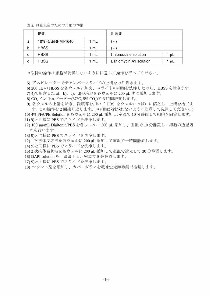
表 2のように 4 種類の培地を 1.5 mLチューブに用意します。
-16-
表 2. 細胞染色のための培地の準備
*以降の操作は細胞が乾燥しないように注意して操作を行ってください。
5) アスピレーターでチャンバースライドの上清を取り除きます。
6) 200 μLの HBSSを各ウェルに加え、スライドの細胞を洗浄したのち、HBSSを除きます。
7) 4)で用意した a)、b)、c)、d)の培地を各ウェルに 200 μLずつ添加します。
8) CO2インキュベーター(37°C, 5% CO2)で 3 時間培養します。
9) 各ウェルの上清を除き、洗瓶等を用いて PBS をウェルいっぱいに満たし、上清を捨てま
す。この操作を 2 回繰り返します。(*細胞が剥がれないように注意して洗浄しください。)
10) 4% PFA/PB Solutionを各ウェルに 200 μL添加し、室温で 10分静置して細胞を固定します。
11) 9)と同様に PBS でスライドを洗浄します。
12) 100 μg/mL Digitonin/PBS を各ウェルに 200 μL 添加し、室温で 10分静置し、細胞の透過処
理を行います。
13) 9)と同様に PBS でスライドを洗浄します。
12) 1 次抗体反応液を各ウェルに 200 μL添加して室温で一時間静置します。
14) 9)と同様に PBS でスライドを洗浄します。
15) 2 次抗体希釈液を各ウェルに 200 μL添加して室温で遮光して 30分静置します。
16) DAPI solution を一滴滴下し、室温で 5 分静置します。
17) 9)と同様に PBS でスライドを洗浄します。
18) マウント剤を添加し、カバーガラスを載せ蛍光顕微鏡で検鏡します。
-17-
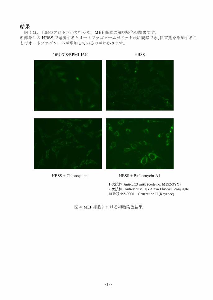
結果 図 4は、上記のプロトコルで行った、MEF細胞の細胞染色の結果です。
飢餓条件の HBSSで培養するとオートファゴソームがドット状に観察でき、阻害剤を添加するこ
とでオートファゴソームが増加しているのがわかります。
図 4. MEF細胞における細胞染色結果
1次抗体:Anti-LC3 mAb (code no. M152-3YY)
2次抗体: Anti-Mouse IgG Alexa Fluor488 conjugate
顕微鏡:BZ-9000 Generation II (Keyence)
-18-
トラブルシューティング 1. ウエスタンブロットで LC3-I と LC3-II のバンドが分離できません。
LC3-I と LC3-II はそれぞれ 16 kDa, 14k Da 付近にバンドが検出します。15%アクリルアミドゲ
ルの使用をお勧めします。グラジエントゲルを用いても LC3-I と LC3-II を分離できますが、
15%アクリルアミドゲルを用いた場合の方がシャープなバンドを検出することができます。
2. ウエスタンブロットで LC3-I と LC3-II のバンドが検出できません。
細胞種により、LC3 の発現量は異なります。泳動時のサンプルアプライ量を増やすか、抗体
の希釈倍率を下げて検討を行ってください。また、キットに同梱のポジティブコントロール
(Positive control for anti-LC3 antibody; PM036-PNY)も併せてご利用ください。
3. 細胞染色の操作でスライドから細胞がはがれてしまいます。
細胞を固定化するまでの間に細胞が乾燥してしまうとスライドからはがれやすくなってしま
います。細胞を乾燥させないように素早く操作を行ってください。

-19-
関連製品 Antibodies
PM036 Anti-LC3 pAb [WB, IP, IC, IHC, FCM]
M152-3 Anti-LC3 mAb (4E12) [WB, IP, IC, FCM, EM]
M186-3 Anti-LC3 mAb (8E10) [WB]
M186-7 Anti-LC3 mAb-HRP-DirecT (8E10)
PD015 Anti-LC3 pAb [IC]
PM045 Anti-p62 (SQSTM1) pAb
PM066 Anti-p62 C-terminal pAb
PM066-7 Anti-p62 C-terminal pAb
M162-3 Anti-p62 (SQSTM1) (Human) mAb (5F2) PM074 Anti-Phospho-p62 (SQSTM1) (Ser351) pAb M217-3 Anti-Phospho-p62 (SQSTM1) (Ser351) mAb
D343-3 Anti-Phospho-p62 (SQSTM1) (Ser403) mAb
(4F6)
D344-3 Anti-Phospho-p62 (SQSTM1) (Ser403) mAb
(4C8)
PD017 Anti-Beclin 1 pAb
PM037 Anti-GABARAP pAb
M135-3 Anti-GABARAP mAb (1F4)
PM038 Anti-GATE-16 pAb
PD041 Anti-Atg2A pAb
PM034 Anti-Atg3 pAb
M133-3 Anti-Atg3 mAb (3E8)
M134-3 Anti-Atg4B mAb (9H5)
PM050 Anti-Atg5 pAb
M153-3 Anti-Atg5 mAb (4D3)
PM039 Anti-Atg7 (Human) pAb
PD042 Anti-Atg9A pAb
M151-3 Anti-Atg10 (Human) mAb (5A7)
M154-3 Anti-Atg12 (Human) mAb (6E5)
PD036 Anti-Atg13 (Human) pAb
M183-3 Anti-Atg13 mAb (5G4)
PD026 Anti-Atg14 pAb
M184-3 Anti-Atg14 (Human) mAb (4H8)
PM040 Anti-Atg16L pAb
M150-3 Anti-Atg16L mAb (1F12)
M160-3 Anti-UVRAG mAb (1H4)
PD027 Anti-Rubicon (Human) pAb
M170-3 Anti-Rubicon (Human) mAb (1H6)
PD037 Anti-Tel2 pAb
PM069 Anti-NRF2 pAb
M200-3 Anti-NRF2 mAb (1F2)
PM072 Anti-VMP1 pAb
PM076 Anti-Syntaxin-17 (Human) pAb
M212-3 Anti-Syntaxin-17 (Human) mAb (2F8)
PM054-7 Anti-α-Tubulin pAb-HRP-DirecT
PM053-7 Anti-β-actin pAb-HRP-DirecT
Kits
8485 Autophagy Ab Sampler Set
PM036-PN Positive control for anti-LC3 antibody
WB: Western blotting
IP: Immunoprecipitation
IC: Immunocytochemistry
IHC: Immunohistochemistry
FCM: Flow cytometry
EM: Immuno-electron microscopy
Other related antibodies and kits are also available.
Please visit our website at http://ruo.mbl.co.jp/
発売元
技術資料や関連する情報は、ホームページ(http://ruo.mbl.co.jp)から利用できます。
最新の情報をご利用ください。
株式会社 医学生物学研究所
URL http://ruo.mbl.co.jp
e-mail [email protected]
TEL 052-238-1904