Embed Size (px)
Citation preview

1
1
Dr. Eman Al-Kateeb
Aseel Olaimat
5
15-4-2013
Sheet April – 15
th
- 2013
5
Aseel Olaimat

2
2
Hope this sheet is useful .. Especially for Absent students of 15/4
Physiology lecture :P
Diagnosing of abnormalities
Review for last slide of previous lecture:
If we have stimulated a radial nerve by stimulating electrodes we'll record a compound
action potential .. and according to the strength of our stimuli we'll get the following
results :
A) minimal stimuli (0.1 mv for example ) .. we will record nothing Because the stimuli is
subthreshold ( very small stimuli ) and that means that no electrical activity happened
B) increasing the stimulus to 0.5 mv -For example - we'll Start recording a small electrical
activity which means that only the superficial nerve fibers are stimulated and signaling
their AP ( closer to the skin )
Q : WHY the signals are displayed in this manner (as it was graded potential ?)
Because the machine has the capability to summate all these Action potentials together (
This is not physiological this is something technical ) and this summation will tell us how
many nerve fibers have been discharged .
c) Increasing the electrical stimulation -for example- to 5 mv … electricity is going deeper
and stimulating more nerve fibers , here we'll be able to reach the maximum amplitude of
AP . ( why it is maximum ? because at this point all nerve fibers are stimulated even the
deeper and smaller ones under the effect of this powerful stimuli so they will be able to
reach the threshold and fire an action potential )
Q : How Can we prove that the AP is all or non ?! i.e it is not graded ?
Giving even higher stimuli for example 3-5 v ( here we used Voltes ! Because we need a
strong stimuli ;) (supra maximum) .. the amplitude of the AP will never change this
indicates that the AP has a fixed amplitude ( all nerve fibers had been stimulated by
maximum stimulus so by the supra maximum nothing will change )

3
3
*If the maximum stimuli is 5 mv .. we have to give the patient 20-30 % stronger stimuli
above the maximum , to make sure 100% that all nerve fibers have been stimulated.
Q : How we will know that there is a problem in the nerve we are diagnosing ?
Suppose that we're diagnosing the radial N and I expect to have an amplitude of 3 cm but I
only got 1 cm instead .. this indicates a problem .
Other criteria in Compound AP :
Increasing stimuli strength will lead to multiple peaks of the compound AP and this is very
clear if we are recording from an area far away from the stimuli ( stimulating from fingers
and recording from the wrist these peaks won’t be very clear but recording from the elbow
this will give a clear multiple peaks ) Why is that ?!
Explanation : In Myelinated large conductive N.fibers .. electrical Activity will reach first ..
{ A-Alpha,A-Beta (responsible for equilibrium and balance ) .. While In unmyelinated
N.fibers will be the last to arrive . So Compound AP depends on the conduction velocity .

4
4
Compound AP Has these criteria : looks like graded potential until reaching the Maximum
stimuli and having multiple peaks because each nerve is composed of multiple nerve
fibers some are fast others are slow in conduction .
Diagnosing of abnormalities :
We Have two types of nerve fibers :
Sensory N fibers that are usually ascending and Motor N fibers that are usually descending.
So if we want to diagnose a sensory N , Ex : Median \ ulnar or Radial N the normal
physiological transporting of sensory AP is from peripheral to CNS (brain).
There are 2 types of recordings :
a) Orthodromic (physiological, follows the sensation direction ): For ex: stimulating the
ulnar N from the little finger and recording from the wrist .
BUT .. physiologists found that following the physiological pathway stimuli will record
a small activity and easily mixed with external noise ( background noise ) SO they
decided to use another type of recordings .

5
5
b) Antidromic (against physiological pathway ) : stimulating (proximally) from the wrist for
example and recording (distally) from the fingers , here physiologists found that the
electrical activity recorded is clearer and larger
Notice that in both Recording types the latent period is the same ( If we have a constant
distance from fingers to wrist ) but we get a better record ( bigger amplitude ) in the
antidromic way .
WHY Antidromic gave a better recording ?!
Because the Nerves in fingers are usually superficial and we can easily record the
normalities and abnormaleties but as they get deeper while moving away from the fingers
the record become weaker .
Remember we don't insert a needle to record we only record from the skin !
If we want to record Motor AP from N.fiber we'll record from the muscles supplied by that
motor nerve ( Median N Thenar MS , Ulnar N Hypothenar Ms )
(Go) Refers to
earthing ;)

6
6
And we put the active ( recording ) electrode on the muscle's belly facing the stimulating
electrode and the reference non active electrode is on the tendon of that muscle .
*Sensory Nerve AP "SNAP" Slide :
Its Orthodromic type ..( Not Favorable )Stimulating electrode on the finger , Recording
electrode at wrist .

7
7
*Compound Motor action potential recording CMAP :
Here we have two electrodes a fixed recording surface electrode on the muscle and a
movable stimulating electrode .
Ex : If we want to examine the Median N
here we stimulate twice ! once from the wrist and the other from the elbow and Record
from The muscle (Thenar muscles)
Notice that the Record obtained from the wrist stimulation has a smaller Latent period
than the one obtained from the elbow . WHY ?? because the distance is larger.
How to measure the conduction velocity of the median Nerve ?
Velocity = distance\time (m\s)
Distance : distance between the wrist and the elbow (cm) m .
Time : difference in latent periods between the two recordings ( latent period for the
elbow's record – Latent period for the Wrist's record ) in milliseconds seconds
Check the slides ^_^

8
8
Why do we stimulate twice and do this complicated process ?!
*In the muscle there is neuromuscular junction that cause an extensive delay for the CMAP
so if we took the whole record from the finger to the elbow for example the
neuromuscular synapse and the spread of AP in the muscle will be taken into consideration
in our calculations ( and these lead to a delay \ Slower conduction velocity ) so we need an
area where the median nerve is pure without synapses or junctions !
Keep in mind that for a Stimulus to reach the CNS as fast as possible it has to pass through
a minimum # of Synapses
WHY ?!
Because In synapse there is a releasing of chemicals (neurotransmitters) that bind to the
receptors and open the channels … and those all take time ! .. and that will make the
Latent period
9
9
measurement incorrect (because we are calculating the conduction velocity + delay
caused by the junctions )
Next slide
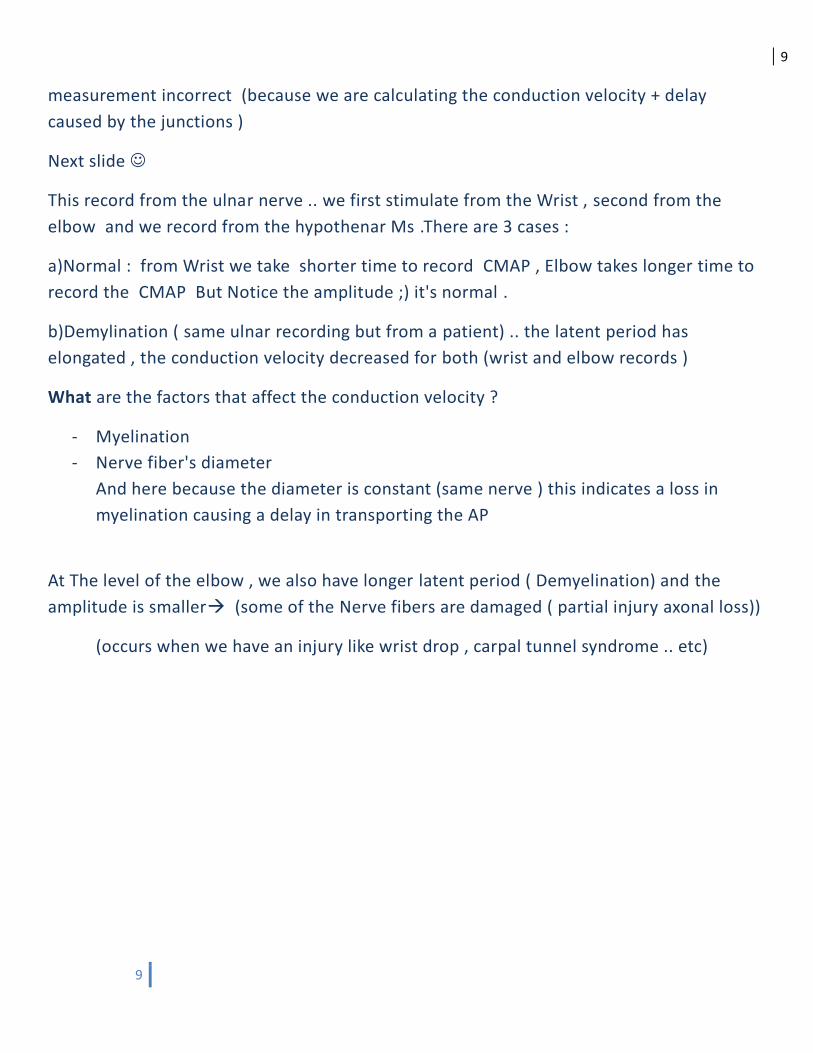
This record from the ulnar nerve .. we first stimulate from the Wrist , second from the
elbow and we record from the hypothenar Ms .There are 3 cases :
a)Normal : from Wrist we take shorter time to record CMAP , Elbow takes longer time to
record the CMAP But Notice the amplitude ;) it's normal .
b)Demylination ( same ulnar recording but from a patient) .. the latent period has
elongated , the conduction velocity decreased for both (wrist and elbow records )
What are the factors that affect the conduction velocity ?
- Myelination
- Nerve fiber's diameter
And here because the diameter is constant (same nerve ) this indicates a loss in
myelination causing a delay in transporting the AP
At The level of the elbow , we also have longer latent period ( Demyelination) and the
amplitude is smaller (some of the Nerve fibers are damaged ( partial injury axonal loss))
(occurs when we have an injury like wrist drop , carpal tunnel syndrome .. etc)

10
10
ENG shows us: Conduction velocity, Myelination status and If the axons are healthy or
abnormal (pathology).
*Major site of Median Nerve injury is (Wrist) carpal tunnel syndrome, Major site of ulnar
N injury is medial epicondyle of humerus (so it's typically injured at the elbow because
Ulnar N is superficial there ),Major site of Radial N injury is radial groove at the shaft of the
humerus and at axilla (people who use crutches , we advice them to put a cushion).
c) Axonal (other patient): The latent periods are the same but the amplitude is much more
less .. Indicates a partial injury in the nerve ( axonal degradation\loss ) involving the wrist
area until the elbow.
What Are the major Health problems in JORDAN ?
- Diabetes (33% of Jordan's population got Diabetes )
- Hypercholesterolemia\ Hyperlipidemia
- Vitamin B12 deficiency
A
B
C

11
11
What causes Demyelination? ( Very common case )
Diabetes: One of the Disorders of demyelination .
A problem in glucose entrance to the cell caused by the resistance of the cells to
glucose ( insulin resistance Diabetes ) Or No enough amounts of insulin are secreted by the
pancreas .
Eating food.. Having Glucose in our blood ..glucose has to enter the cells(as a source of
energy) and in order to enter the cells it needs Insulin … But Diabetes patient doesn’t have
the required amounts of Insulin .. so the Glucose cannot enter the cells ( No energy source
for the cells ) it stays in the blood causing Hyperglycemia ( the patients are always
thirsty and drink a lot amounts of water and large amounts of glucose are present in the
patient's urine ) but the cells are deprived from their nutrients .. so they will use Amino
Acids and fatty acid as nutrients ( Diabetes is a metabolic disorder ) , a lot of toxic
substance ( Ketone bodies and acetests ) will accumulate In the blood and affect the
peripheral nerves leading to "peripheral neuropathy " not healthy peripheral nerves \
( اعتلال الاعصاب المحيطية ) usually diabetes patients lose sensations in their Hands and legs
after 10-15years )
-Also diabetes might cause a vasculitis
After Demyelination we got Axonal loss ( early stages of neural pathology demyelination
then as an advanced result we get Axonal loss )
Next Pic Shows …
Ulnar Nerve recording from hypothenar muscles. stimulating from :*wrist …*below elbow
…*above elbow…*from the arm.
2 Reorders from right and left arm to compare .
The report says : "Normal left ulnar N recording . Abnormal right ulnar N recording at the
wrist , below and above the elbow with an indication of axonal loss \ partial injury of ulnar
N "

12
12
And this is typical presentation of children who fall and broke their bone around t he elbow.
Correction team notes
-anatomy :
Sheet #28: Page 3: It has Apex: within the lumen or cavity of the atrium >> from the apex there are
like fibrous cords called (chordate tendineae), they are like parachute man. .... The cavity is -tendon
left ventricle.the
-physiology :
45.-90 to -iman: page 3 … the resting potential ranges from -Sheet #2 dr
40 mV and the large one -Page 5 … the narrow nerve fiber will having resting membrane potential
70 mV.-will have it as
that diazepam is a common anti anxiety drug.iman: page 5... Add -Sheet #4 dr
Page 6 … it’s never fibers not nerve that is larger than neurons.
Page 7 … graded potential amplitude is 50 mV not threshold.
Page 10 … it’s ENG machine not EMG.
increase, which means becomes less negative).15MV decrease not -sheet#2 also page 3, (10MV
sheet#4 the last page, increasing the voltage to 1MV not 1V.