Embed Size (px)
Citation preview

4376
www.advmat.dewww.MaterialsViews.com
CO
MM
UN
ICATI
ON
Li Chen , Xueli Liu , Bin Su , Jing Li , Lei Jiang , Dong Han ,* and Shutao Wang *
Aptamer-Mediated Effi cient Capture and Release of T Lymphocytes on Nanostructured Surfaces
T lymphocytes have fundamental functions in immune responses to many diseases, such as pathogenic infections, malignancies, and autoimmune diseases. Detections of tar-geted T lymphocytes provide important information in clinical diagnosis. [ 1 ] For example, human immunodefi ciency virus (HIV) depletes CD4 + T lymphocytes in peripheral blood [ 2 ] and other lymphoid tissues. [ 3 ] As a result, the absolute counts of CD4 + T-cells and the ratio of CD4 + /CD8 + T lymphocytes are used as indicators of the onset of autoimmune defi ciency syn-drome (AIDS) and as benchmarks for the beginning of antiviral therapy. [ 4 ] Because of the low specifi city and low effi ciency of traditional approaches based on size, density, and fl ow cytom-etry, cell counts, cell phenotype assessments, and genomic/mRNA analysis of target cells require the emergence of new effi cient capture and release technologies. Many functional molecules, such as antibodies, [ 5 ] peptides, [ 6 ] and DNA [ 7 ] are immobilized onto various surfaces such as magnetic beads, [ 8 ] 2D microarrays, [ 9 , 10 ] and microfl uidic channels [ 11 , 12 ] for specifi c recognition and capture of targeted cells. The introduction of microfl uidic techniques [ 13 ] has improved the capture effi ciency of targeted cells to a certain extent by optimization of channel dimension, mixing fashion, and fl ow rate. [ 9 , 12 , 14 ] Since capture and release takes place at the interface between cells and sub-strates, can we realize the effi cient capture and release only by engineering the surface chemistry and topography without the external fl uidic force? Recently, knowledge about cell–nano-structure interactions in biological and artifi cial cell micro-environments has shown that nanometer-scale topography infl uences diverse cell behaviors, including cell adhesion, cell orientation, and cell motility. [ 15 ] In addition, use of size and shape-matched nanometer-scale topography can enhance interactions between the substrate and target cells. [ 16 , 17 ] These fi ndings inspired us to achieve effi cient capture and release of targeted T lymphocytes by directly controlling cell–substrate interactions.
© 2011 WILEY-VCH Verlag Gwileyonlinelibrary.com
Dr. L. Chen , Dr. X. Liu , Dr. B. Su , Prof. L. Jiang , Prof. S. T. Wang Beijing National Laboratory for Molecular Sciences (BNLMS) Institute of Chemistry Chinese Academy of Sciences Beijing, 100190, P. R. China E-mail: [email protected] Dr. J. Li , Prof. D. Han National Centre for NanoScience and Technology Beijing, 100190, P. R. China E-mail: [email protected] Dr. L. Chen , Dr. X. Liu , Dr. J. Li Graduate School of Chinese Academy of Sciences Beijing, 100864, P. R. China
DOI: 10.1002/adma.201102435
Herein, we report a nanostructured platform that combines cell affi nitive DNA aptamers and cell-preferred nanometer-scale topography, where two orders of enhanced capture effi -ciency compared to planar surfaces and ≈ 97% cell release were obtained for T lymphocytes. We believe this nanostructured surface, in comparison with 2D surfaces, has the following cru-cial advantages: i) it bridges the nanogap between cells and cell affi nity molecules, ii) it substantially increases the local ratio of cell affi nity molecules to cells, and iii) it creates a 3D mode for cell contact, [ 18 ] as described in Figure 1 . This effi cient nanoplat-form provides a novel strategy to fulfi ll the demands of capture and release of T lymphocytes, which have great potential in effi -cient, sensitive, immune cell-based disease diagnosis.
In our work, we selected DNA aptamers as the capture and release mediator molecules since they can easily be synthesized to bind with specifi c molecular or cell targets and can be decom-posed by exonuclease without cell damage. [ 7 , 19 ] Compared with antibodies, aptamers are more easily acquired and have shown signifi cant applications in protein detection, [ 20 ] targeted drug delivery, [ 21 ] and cancer cell capture. [ 12 , 22 ] To obtain a cell-pre-ferred nanometer-scale topography, we used silicon nanowire arrays (SiNWAs) as the substrate material. SiNWAs have been widely studied for their novel optic and electric properties. [ 23 , 24 ] Their biomedical applications have also been explored in bio-sensors, gene transduction, scaffolds, and antifouling mate-rials. [ 25 ] As a substrate with novel nanometer-scale topography, it enhances interactions with cells, since the tips of the SiNWs are well matched with the sub-micrometer structures on cell surfaces. [ 16 , 26 ]
The T lymphocyte we used was the CCRF-CEM cell line, which is a human acute lymphoblastic leukemia T cell line (Figure S1a, Supporting Information; diameter: 10.9 ± 1.9 μ m). Correspondingly, we synthesized a DNA aptamer selected from the previously reported aptamer library [ 22 ] which showed high specifi c affi nity for CCRF-CEM cells (see Experimental Section in the Supporting Information) and a random DNA aptamer for control. [ 14 ] The calculated equilibrium dissociation constant is reported to be in the nanomolar-to-picomolar range. [ 22 ] The fabrication and immobilization procedures of DNA aptamer-modifi ed SiNWAs (DNA-SiNWAs) are wet chemical etching of the silicon wafer and sequential immobilization of 3-mercap-topropyl trimethoxysilane, N -maleimidobutyryloxy succinimide ester (GMBS), and DNA aptamers onto the silicon surface. To investigate the effi ciency of the DNA aptamer immobilization procedures, we designed a complementary DNA strand modi-fi ed with a fl uorescein isothiocyanate (FITC) functional group on its 3´ terminal. After treating with the complementary DNA strand and thoroughly rinsing, the DNA-SiNWA surface and DNA-Si planar surface both showed increased fl uorescence signal (Figure S2,S3, Supporting Information). Notably, the
mbH & Co. KGaA, Weinheim Adv. Mater. 2011, 23, 4376–4380
www.advmat.dewww.MaterialsViews.com
CO
MM
UN
ICATIO
N
Figure 1 . Schematic (a) and SEM (b) of the nanostructured surface for enhanced T lym-phocyte capture through combining cell affi nitive molecules and cell-preferred nanometer-scale topography.
increase of fl uorescence intensity on the DNA-SiNWA surface is much higher than that on the DNA aptamer-modifi ed silicon wafer (DNA-Si) planar surface, which indicates a higher DNA-aptamer density on the former in the same projective area. Thus, the nanostructured surface will provide a higher local ratio of cell affi nitive DNA-aptamers to targeted cells than the planar surface in the following cell capture process.
The SiNWAs fabricated by the chemical etching method [ 24 ] possess a unique nanometer-scale topography compared to planar silicon wafer surfaces. As shown in Figure 2 a, most nanowires are perpendicular to the silicon wafer substrate
© 2011 WILEY-VCH Verlag GmbH & Co. KGaA, WeinhAdv. Mater. 2011, 23, 4376–4380
Figure 2 . Topography of SiNWAs and results of T lymphocyte capture. a) SEM top and side (inset) view images of SiNWAs. b) Densities of captured CCRF-CEM cells (positive) and Ramos cells (negative) on DNA-Si and DNA-SiNWA surfaces. DNA-SiNWAs show specifi city and high capture effi ciency for CCRF-CEM cells. Each column and error bar represents a mean ± standard deviation from three repeats. DAPI fl uorescence microscopy images of captured cells on c) DNA-SiNWA and d) DNA-Si surface. Insets are magnifi ed ESEM images.
and have uniform diameters in the range of 50 to 100 nm (average value 70 ± 4 nm). The length of the silicon nanowires increases with longer etching time, thus enabling control of the topography of the SiNWA substrates. In addition, many silicon nanoclusters, formed by several nanowires combining together, can be observed from the top scanning elec-tron microscopy (SEM) view. After modifi ca-tion with DNA aptamer, the SiNWA surface can provide a much higher capture effi ciency compared to DNA-Si surfaces, as evidenced by 4 ′ ,6-diamidino-2-phenylindole (DAPI)-staining fl uorescence images (Figure 2 c,d). Importantly, T lymphocytes also exhibited striking differences in morphology on a
DNA-SiNWA and DNA-Si surfaces as observed by environ-mental scanning electronic microscopy (ESEM). From the inset of Figure 2 c, a T lymphocyte with fully extended pseudopodia attached to the tips and sidewalls of the silicon nanowires can clearly be observed. The diameters of the pseudopodia and the silicon nanowires are well matched to form effi cient bonds to maintain T lymphocyte adherence to the DNA-SiNWA surface. Conversely, T lymphocytes on the DNA-Si surface are rare, and exhibit a rounded conformation with few to no extended pseu-dopodia (inset of Figure 2 d). These morphologic differences between T lymphocytes on a DNA-SiNWA and DNA-Si surfaces
suggest that DNA-SiNWAs may achieve more effi cient capture by enhanced cell–substrate interactions.
In comparison, detailed investigations were conducted by employing a DNA aptamer-negative human B lymphocyte line, Ramos [ 12 , 22 ] (Figure S1b, Supporting Infor-mation, diameter: 9.9 ± 0.5 μ m). As shown in Figure 2 b, the densities of Ramos cells on DNA-Si and DNA-SiNWA surfaces are 160 ± 40 and 3059 ± 1753 cm − 2 , while for captured CCRF-CEM cells, the densities are 549 ± 148 and 64 821 ± 11739 cm − 2 , respectively. On planar DNA-Si surfaces, an approximagely three times greater cap-ture ratio of CCRF-CEM cells compared to Ramos cells demonstrates the basic spe-cifi city of the DNA-aptamer for CCRF-CEM cells. On DNA-SiNWA surfaces, the corre-sponding ratio increases to ≈ 21 times, indi-cating a seven times enhanced specifi city of the DNA-SiNWA surfaces. Moreover, the density of CCRF-CEM cells on the DNA-SiNWA surface is about two orders of that on the DNA-Si surface, exhibiting signifi cantly enhanced capture of the CCRF-CEM cells on the DNA-SiNWAs substrate.
To verify the specifi c recognition and cap-ture of DNA-SiNWAs to targeted cells, we employed a random DNA aptamer that is negative to CCRF-CEM cells, as a control. [ 14 ]
4377eim wileyonlinelibrary.com
4378
www.advmat.dewww.MaterialsViews.com
CO
MM
UN
ICATI
ON
The random DNA aptamer is modifi ed onto the SiNWAs withthe same protocols as the targeted DNA-SiNWAs. As shown in Figure S4, Supporting Information, CCRF-CEM and Ramos cells show a small non-specifi c interaction with the random DNA-SiNWAs, and their numbers are similar to Ramos cells on DNA-SiNWA surfaces but signifi cantly less than that of the captured CCRF-CEM cells. We also conducted the cell capture on bare SiNWAs, which show slightly higher cell numbers than on random DNA-SiNWAs for both CCRF-CEM and Ramos cells, and on DNA-SiNWAs for Ramos cells (in Figure S4, Supporting Information). These results together give solid evidence that only the specifi c aptamers to targeted cells can yield largely enhanced capture effi ciency through modifi cation onto SiNWAs and non-targeted capture of cells may originate from non-specifi c interac-tion between SiNWAs and cells. This specifi c recognition and capture capability of nanostructured surfaces may be very useful for applications in cell capture, detection, and diagnosis.
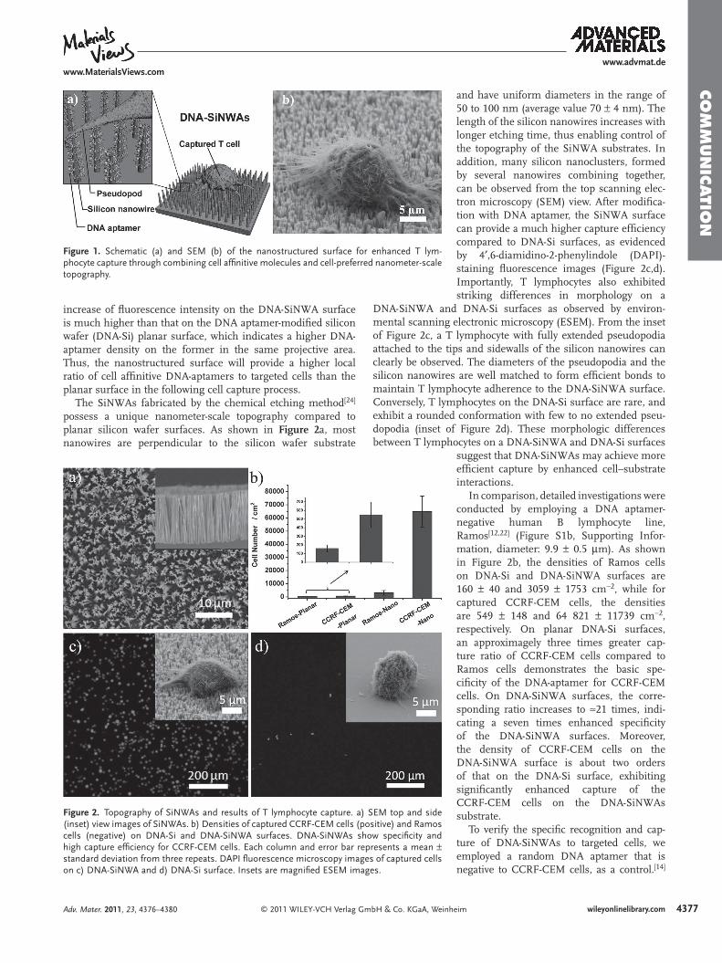
While exploring the cell capture procedures, we found two other factors that were signifi cant for achieving high cap-ture effi ciency: incubation time and length of the SiNWAs. Figure 3 a describes the correlation between incubation time and the number of captured CCRF-CEM cells on DNA-SiNWA sub-strates with the same length of ≈ 7 μ m. The number of captured cells increases with longer incubation time initially, and plateaus at a maximum value at 60 min. This period of time is longer than that in our previous work capturing circulating tumor cells, probably because of the non-adherent characteristic of T lymphocytes. We also changed the length of DNA-SiNWAs by varying the SiNWA etching time (Figure S5, Supporting Infor-mation). We thus fabricated DNA-SiNWAs with lengths ranging from 0 (planar) to 22 μ m and investigated the correlation between length and the number of captured cells, as shown in Figure 3 b. The results show that DNA-SiNWAs with a length longer than ≈ 7 μ m provide the optimal effi ciency for cell capture.
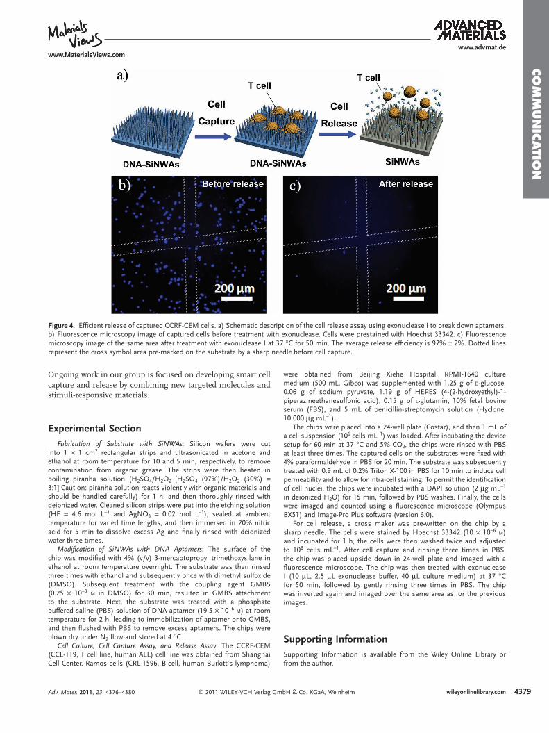
For subsequent cellular and molecular analysis, such as cell phenotype assessments and genomic/mRNA analysis, [ 7 ] it is necessary to effi ciently release the captured T lymphocytes with minimum infl uence on their physiological states. We designed a simple and effi cient way using a biochemical method to break down the bonds between the aptamers and substrate and release the captured cells, as shown in Figure 4 a. Hoechst 33342 is used here because of its better fl uorescent staining
© 2011 WILEY-VCH Verlag wileyonlinelibrary.com
Figure 3 . Quantitative evaluation of capture yields for CCRF-CEM cells: aSiNWA lengths ranging from 0 to 22 μ m. Each plot and error bar represen
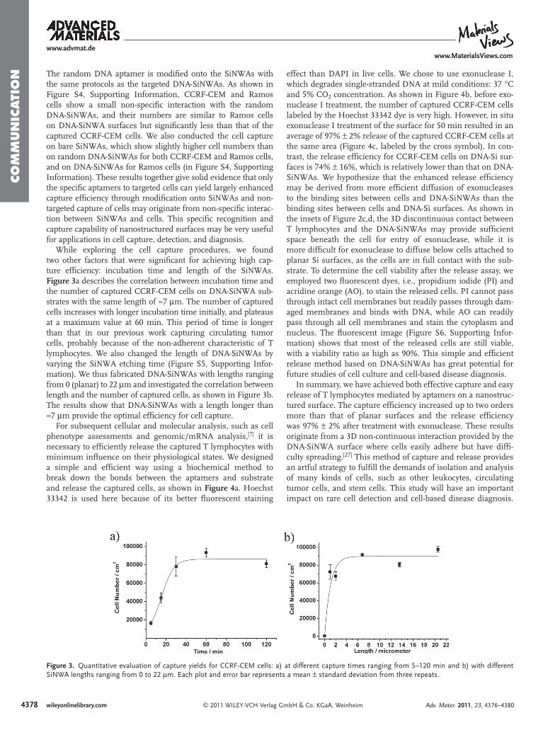
effect than DAPI in live cells. We chose to use exonuclease I, which degrades single-stranded DNA at mild conditions: 37 ° C and 5% CO 2 concentration. As shown in Figure 4 b, before exo-nuclease I treatment, the number of captured CCRF-CEM cells labeled by the Hoechst 33342 dye is very high. However, in situ exonuclease I treatment of the surface for 50 min resulted in an average of 97% ± 2% release of the captured CCRF-CEM cells at the same area (Figure 4 c, labeled by the cross symbol). In con-trast, the release effi ciency for CCRF-CEM cells on DNA-Si sur-faces is 74% ± 16%, which is relatively lower than that on DNA-SiNWAs. We hypothesize that the enhanced release effi ciency may be derived from more effi cient diffusion of exonucleases to the binding sites between cells and DNA-SiNWAs than the binding sites between cells and DNA-Si surfaces. As shown in the insets of Figure 2 c,d, the 3D discontinuous contact between T lymphocytes and the DNA-SiNWAs may provide suffi cient space beneath the cell for entry of exonuclease, while it is more diffi cult for exonuclease to diffuse below cells attached to planar Si surfaces, as the cells are in full contact with the sub-strate. To determine the cell viability after the release assay, we employed two fl uorescent dyes, i.e., propidium iodide (PI) and acridine orange (AO), to stain the released cells. PI cannot pass through intact cell membranes but readily passes through dam-aged membranes and binds with DNA, while AO can readily pass through all cell membranes and stain the cytoplasm and nucleus. The fl uorescent image (Figure S6, Supporting Infor-mation) shows that most of the released cells are still viable, with a viability ratio as high as 90%. This simple and effi cient release method based on DNA-SiNWAs has great potential for future studies of cell culture and cell-based disease diagnosis.
In summary, we have achieved both effective capture and easy release of T lymphocytes mediated by aptamers on a nanostruc-tured surface. The capture effi ciency increased up to two orders more than that of planar surfaces and the release effi ciency was 97% ± 2% after treatment with exonuclease. These results originate from a 3D non-continuous interaction provided by the DNA-SiNWA surface where cells easily adhere but have diffi -culty spreading. [ 27 ] This method of capture and release provides an artful strategy to fulfi ll the demands of isolation and analysis of many kinds of cells, such as other leukocytes, circulating tumor cells, and stem cells. This study will have an important impact on rare cell detection and cell-based disease diagnosis.
GmbH & Co. KGaA, Weinheim Adv. Mater. 2011, 23, 4376–4380
) at different capture times ranging from 5–120 min and b) with different ts a mean ± standard deviation from three repeats.
www.advmat.dewww.MaterialsViews.com
CO
MM
UN
ICATIO
N
Figure 4 . Effi cient release of captured CCRF-CEM cells. a) Schematic description of the cell release assay using exonuclease I to break down aptamers. b) Fluorescence microscopy image of captured cells before treatment with exonuclease. Cells were prestained with Hoechst 33342. c) Fluorescence microscopy image of the same area after treatment with exonuclease I at 37 ° C for 50 min. The average release effi ciency is 97% ± 2%. Dotted lines represent the cross symbol area pre-marked on the substrate by a sharp needle before cell capture.
Ongoing work in our group is focused on developing smart cell capture and release by combining new targeted molecules and stimuli-responsive materials.
Experimental Section Fabrication of Substrate with SiNWAs: Silicon wafers were cut
into 1 × 1 cm 2 rectangular strips and ultrasonicated in acetone and ethanol at room temperature for 10 and 5 min, respectively, to remove contamination from organic grease. The strips were then heated in boiling piranha solution (H 2 SO 4 /H 2 O 2 [H 2 SO 4 (97%)/H 2 O 2 (30%) = 3:1] Caution: piranha solution reacts violently with organic materials and should be handled carefully) for 1 h, and then thoroughly rinsed with deionized water. Cleaned silicon strips were put into the etching solution (HF = 4.6 mol L − 1 and AgNO 3 = 0.02 mol L − 1 ), sealed at ambient temperature for varied time lengths, and then immersed in 20% nitric acid for 5 min to dissolve excess Ag and fi nally rinsed with deionized water three times.
Modifi cation of SiNWAs with DNA Aptamers: The surface of the chip was modifi ed with 4% (v/v) 3-mercaptopropyl trimethoxysilane in ethanol at room temperature overnight. The substrate was then rinsed three times with ethanol and subsequently once with dimethyl sulfoxide (DMSO). Subsequent treatment with the coupling agent GMBS (0.25 × 10 − 3 M in DMSO) for 30 min, resulted in GMBS attachment to the substrate. Next, the substrate was treated with a phosphate buffered saline (PBS) solution of DNA aptamer (19.5 × 10 − 6 M ) at room temperature for 2 h, leading to immobilization of aptamer onto GMBS, and then fl ushed with PBS to remove excess aptamers. The chips were blown dry under N 2 fl ow and stored at 4 ° C.
Cell Culture, Cell Capture Assay, and Release Assay: The CCRF-CEM (CCL-119, T cell line, human ALL) cell line was obtained from Shanghai Cell Center. Ramos cells (CRL-1596, B-cell, human Burkitt’s lymphoma)
© 2011 WILEY-VCH Verlag GAdv. Mater. 2011, 23, 4376–4380
were obtained from Beijing Xiehe Hospital. RPMI-1640 culture medium (500 mL, Gibco) was supplemented with 1.25 g of D -glucose, 0.06 g of sodium pyruvate, 1.19 g of HEPES (4-(2-hydroxyethyl)-1-piperazineethanesulfonic acid), 0.15 g of L -glutamin, 10% fetal bovine serum (FBS), and 5 mL of penicillin-streptomycin solution (Hyclone, 10 000 μ g mL − 1 ).
The chips were placed into a 24-well plate (Costar), and then 1 mL of a cell suspension (10 6 cells mL − 1 ) was loaded. After incubating the device setup for 60 min at 37 ° C and 5% CO 2 , the chips were rinsed with PBS at least three times. The captured cells on the substrates were fi xed with 4% paraformaldehyde in PBS for 20 min. The substrate was subsequently treated with 0.9 mL of 0.2% Triton X-100 in PBS for 10 min to induce cell permeability and to allow for intra-cell staining. To permit the identifi cation of cell nuclei, the chips were incubated with a DAPI solution (2 μ g mL − 1 in deionized H 2 O) for 15 min, followed by PBS washes. Finally, the cells were imaged and counted using a fl uorescence microscope (Olympus BX51) and Image-Pro Plus software (version 6.0).
For cell release, a cross maker was pre-written on the chip by a sharp needle. The cells were stained by Hoechst 33342 (10 × 10 − 6 M ) and incubated for 1 h, the cells were then washed twice and adjusted to 10 6 cells mL − 1 . After cell capture and rinsing three times in PBS, the chip was placed upside down in 24-well plate and imaged with a fl uorescence microscope. The chip was then treated with exonuclease I (10 μ L, 2.5 μ L exonuclease buffer, 40 μ L culture medium) at 37 ° C for 50 min, followed by gently rinsing three times in PBS. The chip was inverted again and imaged over the same area as for the previous images.
Supporting Information Supporting Information is available from the Wiley Online Library or from the author.
4379mbH & Co. KGaA, Weinheim wileyonlinelibrary.com
4380
www.advmat.dewww.MaterialsViews.com
CO
MM
UN
ICATI
ON
AcknowledgementsThe authors acknowledge fi nancial support from the National Research Fund for Fundamental Key Projects (2010CB934700, 2009CB930404, 2007CB936403), and the National Natural Science Foundation (20920102036, 20974113). The authors also thank Prof. Fulong Liao, Prof. Dongsheng Liu, Jiayi Xie, Quanmei Sun, Dr. Bo Yuan, Dr. Ke Zhang, Dr. Ligeng Xu, Dr. Ling Lin, Dr. Zhongxin Xue, and Lin Jin for technical support and helpful discussions. Gwen Owens from the David Geffen School of Medicine at UCLA is greatly appreciated for support and helpful suggestions.
Received: June 27, 2011 Revised: July 15, 2011
Published online: August 22, 2011
[ 1 ] a) E. V. Barsov , H. Andersen , V. J. Coalter , M. Carrington , J. D. Lifson , D. E. Ott , Immunol. Lett. 2006 , 105 , 26 ; b) H. Kim , R. E. Cohen , P. T. Hammond , D. J. Irvine , Adv. Funct. Mater. 2006 , 16 , 1313 .
[ 2 ] R. F. Siliciano , T. Lawton , C. Knall , R. W. Karr , P. Berman , T. Gregory , E. L. Reinherz , Cell 1988 , 54 , 561 .
[ 3 ] a) Z. Smit-McBride , J. J. Mattapallil , M. McChesney , D. Ferrick , S. Dandekar , J. Virol. 1998 , 72 , 6646 ; b) R. S. Veazey , M. DeMaria , L. V. Chalifoux , D. E. Shvetz , D. R. Pauley , H. L. Knight , M. Rosenzweig , R. P. Johnson , R. C. Desrosiers , A. A. Lackner , Sci-ence 1998 , 280 , 427 ; c) R. S. Veazey , P. A. Marx , A. A. Lackner , Trends Immunol. 2001 , 22 , 626 .
[ 4 ] a) J. Guarner , P. Montoya , C. delRio , G. HernandezTepichin , Cytom-etry 1997 , 30 , 178 ; b) G. G. Sherman , J. S. Galpin , J. M. Patel , B. V. Mendelow , D. K. Glencross , J. Immunol. Methods 1999 , 222 , 209 .
[ 5 ] a) I. K. Ko , K. Kato , H. Iwata , Biomaterials 2005 , 26 , 687 ; b) A. Hoff , T. Andre , T. E. Schaffer , G. Jung , K. H. Wiesmuller , R. Brock , ChemBio-Chem 2002 , 3 , 1183 ; c) L. Belov , O. de la Vega , C. G. dos Remedios , S. P. Mulligan , R. I. Christopherson , Cancer Res. 2001 , 61 , 4483 .
[ 6 ] a) J. R. Falsey , M. Renil , S. Park , S. J. Li , K. S. Lam , Bioconjugate Chem. 2001 , 12 , 346 ; b) A. Hoff , T. Andre , R. Fischer , S. Voss , M. Hulko , U. Marquardt , K. H. Wiesmuller , R. Brock , Mol. Divers. 2004 , 8 , 311 .
[ 7 ] G. A. Kwong , C. G. Radu , K. Hwang , C. J. Y. Shu , C. Ma , R. C. Koya , B. Comin-Anduix , S. R. Hadrup , R. C. Bailey , O. N. Witte , T. N. Schumacher , A. Ribas , J. R. Heath , J. Am. Chem. Soc. 2009 , 131 , 9695 .
[ 8 ] V. Zieglschmid , C. Hollmann , O. Bocher , Crit. Rev. Clin. Lab. Sci. 2005 , 42 , 155 .
[ 9 ] D. S. Chen , Y. Soen , T. B. Stuge , P. P. Lee , J. S. Weber , P. O. Brown , M. M. Davis , PloS Med. 2005 , 2 , 1018 .
[ 10 ] B. Angres , Expert Rev. Mol. Diagn. 2005 , 5 , 769 . [ 11 ] a) Z. A. K. Wang , S. Y. Chin , C. D. Chin , J. Sarik , M. Harper ,
J. Justman , S. K. Sia , Anal. Chem. 2010 , 82 , 36 ; b) S. Nagrath , L. V. Sequist , S. Maheswaran , D. W. Bell , D. Irimia , L. Ulkus , M. R. Smith , E. L. Kwak , S. Digumarthy , A. Muzikansky , P. Ryan , U. J. Balis , R. G. Tompkins , D. A. Haber , M. Toner , Nature 2007 , 450 , 1235 ; c) A. A. Adams , P. I. Okagbare , J. Feng , M. L. Hupert ,
© 2011 WILEY-VCH Verlag wileyonlinelibrary.com
D. Patterson , J. Gottert , R. L. McCarley , D. Nikitopoulos , M. C. Murphy , S. A. Soper , J. Am. Chem. Soc. 2008 , 130 , 8633 .
[ 12 ] Y. Xu , J. A. Phillips , J. L. Yan , Q. G. Li , Z. H. Fan , W. H. Tan , Anal. Chem. 2009 , 81 , 7436 .
[ 13 ] S. Hou , S. T. Wang , Z. T. F. Yu , N. Q. M. Zhu , K. Liu , J. Sun , W.-Y. Lin , C. K. F. Shen , X. Fang , H.-R. Tseng , Angew. Chem. Int. Ed. 2008 , 47 , 1072 .
[ 14 ] J. A. Phillips , Y. Xu , Z. Xia , Z. H. Fan , W. H. Tan , Anal. Chem. 2009 , 81 , 1033 .
[ 15 ] a) M. M. Stevens , J. H. George , Science 2005 , 310 , 1135 ; b) C. J. Bettinger , R. Langer , J. T. Borenstein , Angew. Chem. Int. Ed. 2009 , 48 , 5406 ; c) H. S. Gupta , J. Seto , W. Wagermaier , P. Zaslansky , P. Boesecke , P. Fratzl , Proc. Natl. Acad. Sci. USA 2006 , 103 , 17741 ; d) T. L. Sun , D. Han , K. Riehemann , L. F. Chi , H. Fuchs , J. Am. Chem. Soc. 2007 , 129 , 1496 .
[ 16 ] K. E. Fischer , B. J. Aleman , S. L. Tao , R. H. Daniels , E. M. Li , M. D. Bunger , G. Nagaraj , P. Singh , A. Zettl , T. A. Desai , Nano Lett. 2009 , 9 , 716 .
[ 17 ] A. Ranella , M. Barberoglou , S. Bakogianni , C. Fotakis , E. Stratakis , Acta Biomater. 2010 , 6 , 2711 .
[ 18 ] L. Chen , D. Han , L. Jiang , Colloids Surf. B 2011 , 85 , 2 . [ 19 ] J. K. Herr , J. E. Smith , C. D. Medley , D. H. Shangguan , W. H. Tan ,
Anal. Chem. 2006 , 78 , 2918 . [ 20 ] a) K. J. Cash , F. Ricci , K. W. Plaxco , J. Am. Chem. Soc. 2009 , 131 , 6955 ;
b) M. Famulok , J. S. Hartig , G. Mayer , Chem. Rev. 2007 , 107 , 3715 . [ 21 ] X. H. Fang , W. H. Tan , Acc. Chem. Res. 2010 , 43 , 48 . [ 22 ] D. Shangguan , Y. Li , Z. W. Tang , Z. H. C. Cao , H. W. Chen ,
P. Mallikaratchy , K. Sefah , C. Y. J. Yang , W. H. Tan , Proc. Natl. Acad. Sci. USA 2006 , 103 , 11838 .
[ 23 ] a) A. I. Hochbaum , R. K. Chen , R. D. Delgado , W. J. Liang , E. C. Garnett , M. Najarian , A. Majumdar , P. D. Yang , Nature 2008 , 451 , 163 ; b) A. I. Hochbaum , P. D. Yang , Chem. Rev. 2010 , 110 , 527 .
[ 24 ] K. Q. Peng , Y. Wu , H. Fang , X. Y. Zhong , Y. Xu , J. Zhu , Angew. Chem. Int. Ed. 2005 , 44 , 2737 .
[ 25 ] a) L. Chen , M. Liu , H. Bai , P. Chen , F. Xia , D. Han , L. Jiang , J. Am. Chem. Soc. 2009 , 131 , 10467 ; b) Z. Q. Gao , A. Agarwal , A. D. Trigg , N. Singh , C. Fang , C. H. Tung , Y. Fan , K. D. Buddharaju , J. M. Kong , Anal. Chem. 2007 , 79 , 3291 ; c) M. W. Shao , Y. Y. Shan , N. B. Wong , S. T. Lee , Adv. Funct. Mater. 2005 , 15 , 1478 ; d) W. Kim , J. K. Ng , M. E. Kunitake , B. R. Conklin , P. D. Yang , J. Am. Chem. Soc. 2007 , 129 , 7228 ; e) S. J. Qi , C. Q. Yi , S. L. Ji , C. C. Fong , M. S. Yang , ACS Appl. Mater. Interfaces 2009 , 1 , 30 ; f) A. K. Shalek , J. T. Robinson , E. S. Karp , J. S. Lee , D. R. Ahn , M. H. Yoon , A. Sutton , M. Jorgolli , R. S. Gertner , T. S. Gujral , G. MacBeath , E. G. Yang , H. Park , Proc. Natl. Acad. Sci. USA 2010 , 107 , 1870 .
[ 26 ] a) S. T. Wang , H. Wang , J. Jiao , K. J. Chen , G. E. Owens , K. I. Kamei , J. Sun , D. J. Sherman , C. P. Behrenbruch , H. Wu , H. R. Tseng , Angew. Chem. Int. Ed. 2009 , 48 , 8970 ; b) S. T. Kim , D.-J. Kim , T.-J. Kim , D.-W. Seo , T.-H. Kim , S.-Y. Lee , K. Kim , K.-M. Lee , S.-K. Lee , Nano Lett. 2010 , 10 , 2877 ; c) S. T. Wang , K. Liu , J. A. Liu , Z. T. F. Yu , X. W. Xu , L. B. Zhao , T. Lee , E. K. Lee , J. Reiss , Y. K. Lee , L. W. K. Chung , J. T. Huang , M. Rettig , D. Seligson , K. N. Duraiswamy , C. K. F. Shen , H. R. Tseng , Angew. Chem. Int. Ed. 2011 , 50 , 3084 .
[ 27 ] S. Qi , C. Yi , S. Ji , C.-C. Fong , M. Yang , ACS Appl. Mater. Interfaces 2008 , 1 , 30 .
GmbH & Co. KGaA, Weinheim Adv. Mater. 2011, 23, 4376–4380