Embed Size (px)
Citation preview
ARABIDOPSIS DEHISCENCE ZONE POLYGALACTURONASE1(ADPG1), ADPG2, and QUARTET2 Are PolygalacturonasesRequired for Cell Separation during ReproductiveDevelopment in Arabidopsis W
Mikihiro Ogawa,a Pippa Kay,a,b Sarah Wilson,c and Stephen M. Swaina,1
a CSIRO Plant Industry, Private Mail bag, Merbein, Victoria 3505, Australiab La Trobe University, Bundoora, Victoria 3086, Australiac University of Melbourne, Melbourne, Victoria 3500, Australia
Cell separation is thought to involve degradation of pectin by several hydrolytic enzymes, particularly polygalacturonase
(PG). Here, we characterize an activation tagging line with reduced growth and male sterility caused by increased
expression of a PG encoded by QUARTET2 (QRT2). QRT2 is essential for pollen grain separation and is part of a small family
of three closely related endo-PGs in the Arabidopsis thaliana proteome, including ARABIDOPSIS DEHISCENCE ZONE
POLYGALACTURONASE1 (ADPG1) and ADPG2. Functional assays and complementation experiments confirm that ADPG1,
ADPG2, and QRT2 are PGs. Genetic analysis demonstrates that ADPG1 and ADPG2 are essential for silique dehiscence. In
addition, ADPG2 and QRT2 contribute to floral organ abscission, while all three genes contribute to anther dehiscence.
Expression analysis is consistent with the observed mutant phenotypes. INDEHISCENT (IND) encodes a putative basic
helix-loop-helix required for silique dehiscence, and we demonstrate that the closely related HECATE3 (HEC3) gene is
required for normal seed abscission and show that IND and HEC3 are required for normal expression of ADPG1 in the silique
dehiscence zone and seed abscission zone, respectively. We also show that jasmonic acid and ethylene act together with
abscisic acid to regulate floral organ abscission, in part by promoting QRT2 expression. These results demonstrate that
multiple cell separation events, including both abscission and dehiscence, require closely related PG genes.
INTRODUCTION
Cell separation events that lead to organ abscission or dehis-
cence play important roles in plant development, particularly
during reproductive processes. Examples include the abscission
of leaves and outer floral organs and several processes related to
pollination and seed set, fruit maturation, and seed dispersal
(Lewis et al., 2006). Depending on individual plant species, entire
flowers can abscise in the absence of fertilization and seed set or
in unfavorable conditions. Abscission generally occurs at the
completion of fruit maturation, often as an aid to seed dispersal,
but can also occur during the early stages of fruit development
even if seed set is successful. Undesirable abscission events are
a major issue in the commercial production of many crops,
including fruit loss from premature abscission (e.g., in many
perennial horticultural crops) and seed loss from field crops (e.g.,
canola [Brassica napus]).
Pod shatter is a specialized type of fruit dehiscence event in
which the fruit breaks into parts and, because it can be studied in
Arabidopsis thaliana, represents one of the best understood
types of cell separation. In the family Brassicaceae, which
includes Arabidopsis and canola, cell separation generally oc-
curs along the sites of fusion between carpels that compose the
fruit. A second separation event then occurs to allow the seed to
detach from the maternal plant. Dehiscence of the Arabidopsis
silique is very similar to the process in canola (Spence et al.,
1996) and has been used as an effective model in which to study
pod shatter. During ovary and fruit development, cell fate spec-
ification must occur to form the dehiscence zone (DZ), a spe-
cialized layer in which cell separation occurs to allow the silique
to open. In Arabidopsis, the DZ consists of a few cell layers
separating the replum from the edges of the two fused carpels
(Spence et al., 1996). Genetic approaches have revealed that
several genes encoding transcription factors are required for DZ
differentiation (Ferrandiz et al., 2000; Liljegren et al., 2000; Rajani
and Sundaresan, 2001; Liljegren et al., 2004). Once the DZ is
correctly specified and established, other essential processes
include secondary wall formation at the valve margins (Mitsuda
and Ohme-Takagi, 2008) and degradation of cell walls, including
the middle lamella, in the separation layer. This is thought to
occur by the action of proteins involved in cell wall loosening,
including polygalacturonases (PGs), b-1,4-glucanase, and ex-
pansin (Bonghi et al., 1993; Taylor et al., 1993, 1994; Lashbrook
et al., 1994; del Campillo and Bennett, 1996; Cho and Cosgrove,
2000), although this hypothesis is supported by only limited
direct evidence.
1 Address correspondence to [email protected] author responsible for distribution of materials integral to thefindings presented in this article in accordance with the policy describedin the Instructions for Authors (www.plantcell.org/) is Stephen M. Swain([email protected]).WOnline version contains Web-only data.www.plantcell.org/cgi/doi/10.1105/tpc.108.063768
The Plant Cell, Vol. 21: 216–233, January 2009, www.plantcell.org ã 2009 American Society of Plant Biologists
Other important cell separation events occur in male flower
organs during flower development. During pollen development, a
separation event is required after meiosis of the pollen mother
cell to separate the four microspores. The three quartet (qrt)
mutants (qrt1, qrt2, and qrt3) of Arabidopsis are defective in this
process and produce tetrad pollen in which microspores fail to
separate during pollen development (Preuss et al., 1994). Im-
munohistochemical analyses suggest that QRT1 and QRT2 are
required for pectin degradation of the cell wall surrounding
the pollen mother cell during pollen development (Rhee and
Somerville, 1998). Recent molecular studies have revealed that
QRT1 and QRT3 encode a pectin methylesterase (PME) and an
atypical PG, respectively (Rhee et al., 2003; Francis et al., 2006).
Although QRT2 has been mapped to the top of chromosome 3
(Preuss et al., 1994), the affected gene has not yet been identified.
Pollen function also requires a second cell separation event in
which the mature anthers dehisce to release functional pollen
grains at anthesis. Anther dehiscence requires breakdown of the
stomium, specialized cells that keep the anther locules closed,
and genetic analysis has shown that the plant hormone jasmonic
acid (JA) is required for this process. For example, plants lacking
ALLENE OXIDE SYNTHASE (AOS) are JA deficient and do not
shed pollen (Park et al., 2002; von Malek et al., 2002). At the
cellular level, anther dehiscence is similar to silique dehiscence
and, like microspore separation, is thought to involve similar cell
wall degrading enzymes (Roberts et al., 2002). Later in Arabi-
dopsis flower development, additional cell separation events
occur in the floral tissues of the outer three whorls. Several days
after anthesis, the sepals, petals, and stamens detach from the
flower base to reveal either an unfertilized ovary or a developing
silique containing immature seeds.
The cell separation events described above are all thought to
involve the degradation of pectin by PGs, although this hypoth-
esis has not been confirmed by genetic evidence. It is also not
clear to what extent, if any, the same PGs function in different
abscission/dehiscence events, and this uncertainty has contrib-
uted to the complex naming system, based on expression in
different abscision zones (AZs) andDZs, sometimes used for PGs.
Homogalacturonan-rich pectin is commonly found in the middle
lamella region of the cell wall where two adjacent cells abut and
pectin integrity is important for cell adhesion (MacDougall et al.,
1996; Ridley et al., 2001). Endopolygalacturonases (endo-PGs)
catalyze random hydrolysis of a-1,4-glycosidic linkages in poly-
galacturonic acid (GalUA), a polymer that constitutes the main
chain of the homogalacturonan region of pectin (Biely et al., 1996).
Although there is only limited direct genetic evidence for the
physiological importanceof individual PGs, correlations have been
reported between increasing PG activity and cell separation in fruit
ripening and in the shedding of leaves, flowers, and fruit (Taylor
et al., 1993; Kalaitzis et al., 1995; Brown, 1997; Kalaitzis et al.,
1997). More recently, silencing of tomato (Solanum lycopersicum)
abscission-related PGs was shown to increase the break strength
of the leaf abscission zoneanddelayabscission inexplants treated
with ethylene (Jiang et al., 2008), and a putative Arabidopsis PG
has been shown to promote floral organ abscission (Gonzalez-
Carranza et al., 2007). The importance of PG is also illustrated by
the ‘Flavr savr’ tomato (Hadfield and Bennett, 1998) and peach
(Prunus persica) lacking a functionalMelting flesh/Freestone locus
(Peace et al., 2005), both of which have reduced expression of a
fruit ripening-associated PG and delayed fruit softening.
The best-characterized (fungal) endo-PGenzyme requires four
to five consecutive runs of unesterified GalUA residues for
cleavage (Benen et al., 1999; Parenicova et al., 2000). However,
little is known about the enzyme activity and substrate specificity
of most plant PGs. There are at least 69 and 59 predicted PGs in
the Arabidopsis and rice (Oryza sativa) genomes, respectively
(Kim et al., 2006; Gonzalez-Carranza et al., 2007), and it has been
suggested that one group of related PGs tend to be expressed in
flowers and flower buds, while PGs expressed in vegetative
tissues generally belong to other groups (Torki et al., 2000; Kim
et al., 2006). The implication is that the diverse potential phys-
iological roles of PGs may be a consequence of differential
expression in specific tissues rather than or in addition to
differences in enzyme substrate specificity. These questions
have been investigated to a limited extent using transgenic
plants with altered expression of endo-PGs. For example, over-
expression of an apple (Malus domestica) endo-PG in transgenic
apples resulted in silvery colored leaves and premature leaf
shedding due to reduced cell adhesion in leaves and in the leaf
abscission zone (Atkinson et al., 2002). By contrast, ectopic
expression of a tomato fruit-specific endo-PG (pTOM6) in to-
bacco (Nicotiana tabacum) revealed no leaf phenotype or de-
tectable alterations in cell wall pectins, although the pTOM6
protein was properly processed and localized in the cell wall of
tobacco leaves (Osteryoung et al., 1990). While these results
suggest that individual PGs may have different substrate spec-
ificities or requirements for activity, it is not clear how overall PG
activity is regulated in different plant tissues in vivo.
Here,we report on the characterization of an activation tagging
line with reduced growth and male sterility caused by increased
expression of a PG encoded by At3g07970. This gene has been
previously named QRT2 based on tetrad pollen production in
mutant qrt2 plants (Preuss et al., 1994). QRT2 is part of a small
family of three closely related endo-PGs in the Arabidopsis
proteome, and our genetic analysis demonstrates that two of
these PGs are required for silique dehiscence (ARABIDOPSIS
DEHISCENCE ZONE POLYGALACTURONASE1 [ADPG1] and
ADPG2), two are required for normal floral organ abscission
(ADPG2 and QRT2), and all three are required for normal anther
dehiscence. Expression analysis based on transcriptional
b-glucuronidase (GUS) constructs is consistent with the ob-
servedmutant phenotypes.Basedon the transcriptional regulation
of all three genes by JA, and of ADPG2 by ethylene, we show that
JA and ethylene act together with abscisic acid (ABA) to regulate
floral organ abscission, in part by promoting QRT2 expression.
RESULTS
Activation Tagging of the At3g07970 Gene
From a population of several hundred independent activation
tagging lines, we identified one line, designated activation tag-
ging line no. 3 (AT3), in which most anthers failed to shed pollen
and the majority of flowers did not set seeds (see Supplemental
Figure 1 online). The selectable marker (the bar gene)
Polygalacturonases and Cell Separation 217
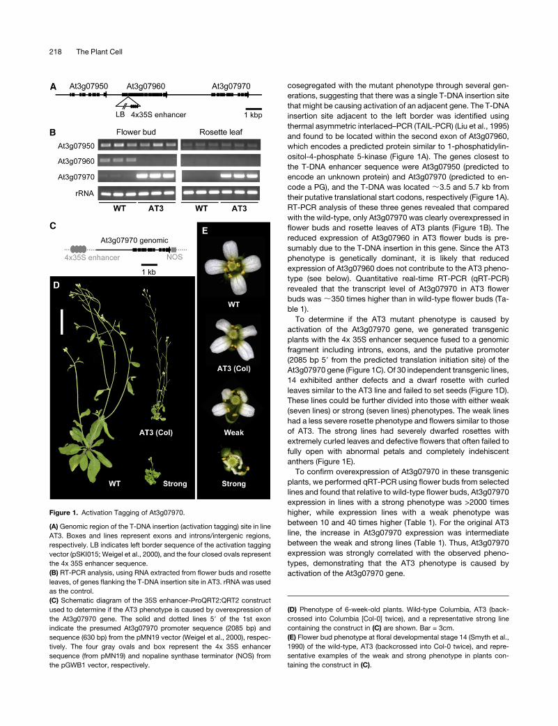
cosegregated with the mutant phenotype through several gen-
erations, suggesting that there was a single T-DNA insertion site
that might be causing activation of an adjacent gene. The T-DNA
insertion site adjacent to the left border was identified using
thermal asymmetric interlaced–PCR (TAIL-PCR) (Liu et al., 1995)
and found to be located within the second exon of At3g07960,
which encodes a predicted protein similar to 1-phosphatidylin-
ositol-4-phosphate 5-kinase (Figure 1A). The genes closest to
the T-DNA enhancer sequence were At3g07950 (predicted to
encode an unknown protein) and At3g07970 (predicted to en-
code a PG), and the T-DNA was located ;3.5 and 5.7 kb from
their putative translational start codons, respectively (Figure 1A).
RT-PCR analysis of these three genes revealed that compared
with the wild-type, only At3g07970 was clearly overexpressed in
flower buds and rosette leaves of AT3 plants (Figure 1B). The
reduced expression of At3g07960 in AT3 flower buds is pre-
sumably due to the T-DNA insertion in this gene. Since the AT3
phenotype is genetically dominant, it is likely that reduced
expression of At3g07960 does not contribute to the AT3 pheno-
type (see below). Quantitative real-time RT-PCR (qRT-PCR)
revealed that the transcript level of At3g07970 in AT3 flower
buds was;350 times higher than in wild-type flower buds (Ta-
ble 1).
To determine if the AT3 mutant phenotype is caused by
activation of the At3g07970 gene, we generated transgenic
plants with the 4x 35S enhancer sequence fused to a genomic
fragment including introns, exons, and the putative promoter
(2085 bp 59 from the predicted translation initiation site) of the
At3g07970 gene (Figure 1C). Of 30 independent transgenic lines,
14 exhibited anther defects and a dwarf rosette with curled
leaves similar to the AT3 line and failed to set seeds (Figure 1D).
These lines could be further divided into those with either weak
(seven lines) or strong (seven lines) phenotypes. The weak lines
had a less severe rosette phenotype and flowers similar to those
of AT3. The strong lines had severely dwarfed rosettes with
extremely curled leaves and defective flowers that often failed to
fully open with abnormal petals and completely indehiscent
anthers (Figure 1E).
To confirm overexpression of At3g07970 in these transgenic
plants, we performed qRT-PCR using flower buds from selected
lines and found that relative to wild-type flower buds, At3g07970
expression in lines with a strong phenotype was >2000 times
higher, while expression lines with a weak phenotype was
between 10 and 40 times higher (Table 1). For the original AT3
line, the increase in At3g07970 expression was intermediate
between the weak and strong lines (Table 1). Thus, At3g07970
expression was strongly correlated with the observed pheno-
types, demonstrating that the AT3 phenotype is caused by
activation of the At3g07970 gene.
Figure 1. Activation Tagging of At3g07970.
(A) Genomic region of the T-DNA insertion (activation tagging) site in line
AT3. Boxes and lines represent exons and introns/intergenic regions,
respectively. LB indicates left border sequence of the activation tagging
vector (pSKI015; Weigel et al., 2000), and the four closed ovals represent
the 4x 35S enhancer sequence.
(B) RT-PCR analysis, using RNA extracted from flower buds and rosette
leaves, of genes flanking the T-DNA insertion site in AT3. rRNA was used
as the control.
(C) Schematic diagram of the 35S enhancer-ProQRT2:QRT2 construct
used to determine if the AT3 phenotype is caused by overexpression of
the At3g07970 gene. The solid and dotted lines 59 of the 1st exon
indicate the presumed At3g07970 promoter sequence (2085 bp) and
sequence (630 bp) from the pMN19 vector (Weigel et al., 2000), respec-
tively. The four gray ovals and box represent the 4x 35S enhancer
sequence (from pMN19) and nopaline synthase terminator (NOS) from
the pGWB1 vector, respectively.
(D) Phenotype of 6-week-old plants. Wild-type Columbia, AT3 (back-
crossed into Columbia [Col-0] twice), and a representative strong line
containing the construct in (C) are shown. Bar = 3cm.
(E) Flower bud phenotype at floral developmental stage 14 (Smyth et al.,
1990) of the wild-type, AT3 (backcrossed into Col-0 twice), and repre-
sentative examples of the weak and strong phenotype in plants con-
taining the construct in (C).
218 The Plant Cell
At3g07970 IsQRT2
To examine the biological role of At3g07970, we identified loss-
of-function alleles of At3g07970 using T-DNA insertion lines
obtained from the ABRC (Figure 2A). These two mutant lines are
likely to represent null or near-null alleles because expression
could not be detected by qRT-PCR using 40 cycles and gene-
specific primers on either side of the T-DNA insertion sites (see
Supplemental Figures 2 and 3 online). Plants homozygous for
either of the two independentmutant alleles of At3g07970 had no
apparent phenotype compared with the wild-type, with the
exception that mature pollen grains were arranged in a tetrad
similar to the phenotype of the qrt mutants (Preuss et al., 1994).
Although the QRT2 gene was not identified, previous mapping
experiments revealed that this locus is linked to GAPC at the top
of chromosome 3 (Preuss et al., 1994), in the same region as
At3g07970. In addition, microscopy observations using an anti-
body that recognizes pectin indicated that QRT2 may be re-
quired for cell type–specific pectin degradation to separate
microspores (Rhee and Somerville, 1998). As these results are
consistent with At3g07970 encoding QRT2, we performed allel-
ism tests by crossing the original qrt2-1 allele with the two
At3g07970 T-DNA insertion mutants (hereafter called qrt2-2 and
qrt2-3). Pollen grains of all F1 progeny exhibited the tetrad
phenotype, confirming that At3g07970 is QRT2. Consistent with
this result, F1 progeny from control crosses between qrt2-2 and
the wild-type or between qrt2-3 and the wild-type produced only
monad pollen.
The qrt2-1 allele was generated using ethyl methanesulfonate
(Preuss et al., 1994). To identify the mutation present in qrt2-1, a
2735-bp genomic fragment, corresponding to the QRT2 open
reading frame with 359 bp upstream of the translational start
codon and 10 bp downstream of the stop codon, was cloned
from qrt2-1 and wild-type Landsberg erecta (Ler) plants and
sequenced. A single nucleotidemutationwas found in qrt2-1 that
changed the predicted amino acid sequence from Val (GTG) to
Ala (GCG) at position 372 (Figure 2A; seeSupplemental Figure 2A
online). Val is a hydrophobic amino acid, and sequence analysis
revealed that a hydrophobic amino acid (Val, Leu, or Ile) in this
position is highly conserved in PGs from plants and fungi
(Markovic and Janecek, 2001; Kim et al., 2006). qRT-PCR from
flower buds revealed no detectable change in QRT2 expression
level between wild-type Ler and qrt2-1 (see Supplemental Figure
2B online), consistent with the qrt2-1 mutant phenotype being
caused by a single amino acid substitution that decreases
protein activity.
Loss of PG Function Prevents Pod Shatter
Phylogenetic analysis of predicted Arabidopsis PGs revealed
that there are two PGs closely related to QRT2, encoded by
At3g57510 and At2g41850 (Kim et al., 2006). The putative PG
encoded by At3g57510 has previously been given several
names, including ADPG1, PGA9, pga1;6, and SAC70 (Jenkins
et al., 1999; Sander et al., 2001). Although the putative PG
encoded by At2g41850 has been referred to as PGAZAT
(Gonzalez-Carranza et al., 2002), we refer to this protein as
ADPG2 based on its expression pattern and biological role (see
below).
The similarity between QRT2 and ADPG1 is potentially reveal-
ing because the latter has previously been identified as a putative
ortholog of RAPESEED DEHISCENCE ZONE POLYGALACTU-
RONASE1 (RDPG1) from canola. RDPG1, also called SAC66,
has been proposed to be involved in breakdown of the middle
lamella of the separation zone during pod shatter, although
functional analysis of ADPG1 or RDPG1 in planta has not been
reported (Jenkins et al., 1996, 1999; Petersen et al., 1996; Sander
et al., 2001). Based on the similarities between the predicted
proteins and the observation thatADPG1 is also expressed in the
silique DZ, a similar role has been proposed for ADPG1 in
Arabidopsis (Gonzalez-Carranza et al., 2007). Consistent with
possible roles in fruit abscission processes, expression data also
revealed that ADPG1 and ADPG2 are expressed predominantly
in floral tissues (Grennan, 2006; Kim et al., 2006; Gonzalez-
Carranza et al., 2007; see below).
To examine the biological role of ADPG1 and ADPG2, we
identified two independent loss-of-function T-DNA insertion
mutant alleles for each gene obtained from ABRC (SALK lines)
and the Max-Planck Institute (GABI line) (Figure 2A). With the
possible exception of adpg1-1, these lines are likely to represent
null or near-null alleles because expression could not be de-
tected by qRT-PCR using 40 cycles and gene-specific primers
on either side of the T-DNA insertion sites (see Supplemental
Figure 3 online). For adpg1-1, the presence of the T-DNA inserted
into the third intron did not completely prevent expression based
on a similar analysis. However, qRT-PCR revealed that the
transcript level in adpg1-1 was 18 times less than the wild-type
(see Supplemental Figure 4A online). In addition, sequence
analysis of RT-PCR products revealed that the entire 4th exon,
consisting of 21 nucleotides and encoding seven amino acids,
was missing from the adpgp1-1mRNA (Figure 2A, missing exon
in adpg1-1 in red; see Supplemental Figure 4B online). Since this
sequence is still present in genomic DNA of adpg1-1, it is likely
that the T-DNA insertion in the third intron causes incorrect
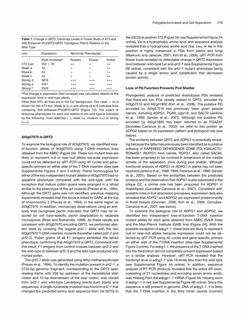
Table 1. Change in QRT2 Transcript Levels in Flower Buds of AT3 and
35S Enhancer-ProQRT2:QRT2 Transgenic Plants Relative to the
Wild-Type
Line Expression Abnormal Phenotypes
(Fold Increase)a Rosette Flower Anther
AT3 (Ler) 352 6 35 ++ + ++
Weak a 10 + + ++
Weak b 24 + + ++
Weak c 42 + + ++
Strong d 3818 +++ +++ +++
Strong e 2451 +++ +++ +++
Strong f 3526 +++ +++ +++
aThe change in expression (fold increase) was calculated relative to the
expression level in wild-type plants.
Other than AT3, all lines are in the Col background. The mean 6 SE is
shown for the AT3 line. Weak (a to c) and strong (d to f) indicates lines
containing 35S enhancer-ProQRT2:QRT2 (Figure 1C). The extent of
abnormal phenotypes for each line relative to the wild-type is indicated
by the following: none detected (�), weak (+), medium (++) or strong
(+++).
Polygalacturonases and Cell Separation 219
splicing which results in exon 3 being joined directly to exon 5.
The effect, if any, that this has on protein activity is not known.
Plants homozygous for mutant alleles adpg1-1 or adpg1-2
were indistinguishable from the wild-type, including the release
of monad pollen, with the exception of impaired pod shatter due
to failure of the valve to properly detach from the central part of
the silique (Figure 2B). However, pod shatter did occur when
mature, dry adpg1 siliques were gently compressed to increase
pressure on the valve DZs. Once opened in this way, seed
abscission appeared normal. By contrast, under normal growing
conditions, single adpg2 mutants appeared similar to wild-type
plants in terms of pod shatter and also produced monad pollen.
However, reduced pod shatter was observed in adpg2-1 and
adpg2-2 plants (and in double mutants with qrt2) when watering
was ceased before overall plant senescence was complete (see
Supplemental Table 1 online). To determine if the ADPG1,
ADPG2, and QRT2 genes are functionally redundant, double
and triple mutants were generated by crossing. Siliques of the
doublemutants adpg1-1 adpg2-1 and adpg1-2 adpg2-2exhibited
a more severe phenotype than did those of the adpg1 single
mutants and failed to dehisce even if compressed.When siliques
were cut open, seed abscission again appeared normal, as did
pollen grain separation. Triple mutants were also constructed
with the two qrt2 T-DNA alleles. In terms of pod shatter and seed
abscission, adpg2 qrt2 double mutants were similar to adpg2
single mutants, double mutants lacking both ADPG1 and QRT2
appeared identical to adpg1 single mutants, and the adpg1-1
adpg2-1 qrt2-2 and adpg1-2 adpg2-2 qrt2-3 triple mutants re-
sembled the adpg1 adpg2 double mutants (see Supplemental
Table 1 online). These results suggest that ADPG1 and ADPG2
have partially redundant roles in Arabidopsis pod shatter. As
expected, all plants homozygous for qrt2 produced tetrad pollen
(Figure 2E).
As pod shatter represents a well-characterized cell separation
event (Roberts et al., 2002; Lewis et al., 2006), the silique DZwas
used to investigate pectin levels associated with reduced PG
function. Consistent with a role for ADPG1 and ADPG2 in cell
separation in the final stages of pod shatter, transverse sections
Figure 2. adpg1, adpg2, and qrt2 Loss-of-Function Phenotypes.
(A) Position of the T-DNA insertions in At3g07970 (QRT2) and the related genes, ADPG1 and ADPG2. Boxes and lines represent exons and introns,
respectively. Triangles indicate positions of T-DNA insertions, and the position of the point mutation in qrt2-1 is shown. Open boxes represent predicted
59 and 39 untranslated regions. There is no information available regarding the 59 untranslated region of QRT2. The exon shown in red is missing in
transcript from the adpg1-1 allele.
(B) Silique indehiscence phenotype of the adpg1-1 and adpg1-2 mutants. Fully matured wild-type siliques break easily, while those of the adpg1
mutants require external mechanical stress
(C) Transverse sections of wild-type and adpg1 adpg2 qrt2 triple mutant siliques (stage 17) stained with Toluidine Blue. The red boxes indicate where
the DZs form in the wild-type and fail to form in the mutant. V, silique valve; S, septum. Bar = 50 mm.
(D) Flowers of the wild-type (stage 14: pollination) and the adpg1 adpg2 qrt2 triple mutant with delayed anther dehiscence.
(E) Scanning electron microscopy image of a mature pollen tetrad from the adpg1 adpg2 qrt2 triple mutant.
(F) Transverse sections of wild-type and adpg1 adpg2 qrt2 triple mutant anthers (stage 13) stained with Toluidine Blue. Arrowheads indicate where
stomium separation has occurred in the wild-type anther and has not yet occurred in the adpg1 adpg2 qrt2 anther.
220 The Plant Cell
of maturing siliques just prior to pod shatter (stage 17b; Roeder
and Yanofsky, 2006) revealed that adpg1 adpg2 qrt2 triple
mutants were morphologically similar to the wild-type (Figure
2C). At stage 18, when silique dehiscence normally takes place
(Roeder and Yanofsky, 2006), cell separation occurred in wild-
type siliques but not in adpg1 adpg2 qrt2 triple mutant siliques
(Figure 3). Although it has been reported previously that cell
separation occurs between cells without cell rupture (Spence
et al., 1996), we observed separation between intact cells in
addition to breaking of nearby cells (Figure 3B) in the DZ of wild-
type siliques. Pectic polysaccharides, including potential PG
substrates, can be visualized using monoclonal antibodies that
recognize unesterified (JIM5) and esterified (JIM7) regions of
pectin (e.g., Francis et al., 2006). For example, JIM5 has been
used previously to show that, in contrast with the wild-type, the
primary cell wall that surrounds the microspores is not degraded
in qrt2-1 anthers (Rhee and Somerville, 1998). If ADPG1 and
ADPG2 degrade the majority of pectin present in the silique DZ,
clear differences would be expected in the pectin levels of wild-
type and mutant siliques. However, comparison between trans-
mission electron microscopy (TEM) sections of wild-type and
adpg1 adpg2 qrt2 triple mutant siliques at stage 18 did not reveal
any obvious differences in the overall levels of pectin (Figures 3D
and 3E), suggesting that the degradation of only a small propor-
tion of the total JIM5-recognized pectin is required for pod
shatter.
Loss of PG Function Delays Anther Dehiscence
In contrast with the single and double mutants, the adpg1 adpg2
qrt2 triple mutants consistently exhibited delayed anther dehis-
cence in early flowers (Figure 2D) due to failure of the stomium to
separate at stage 13 (Figure 2F; Sanders et al., 1999). The anther
phenotype progressively became weaker as the plant continued
to grow and produced more flowers. The delay in pollen release
and pollination in early flowers did not prevent seed set in the
triple mutants, although it appeared that a small proportion of
flowers were not pollinated. These results suggest that functional
redundancy exists betweenADPG1,ADPG2, andQRT2 in anther
dehiscence. Furthermore, it is clear that a commonPG-dependent
mechanism is involved in cell separation events in the anther and
silique DZ and during microspore development.
Loss of PG Function Delays Floral Organ Abscission
After investigating several methods to assess floral organ ab-
scission, firmly pressing each flower on intact plants grown
under standard conditions was chosen as the most reliable and
consistent (see Supplemental Figure 5 online). There was a clear
delineation between younger flowers with floral organs that did
not detach when pressed and older flowers that lost their floral
organs easily. In particular, this approach resolved the issue of
floral organs being lost during plant growth and handling. Anal-
ysis of the adpg1 adpg2 and qrt2 single mutants in this manner
revealed that ADPG2 and QRT2 promote floral organ abscission
(Figure 4), consistent with previous reports that ADPG2 pro-
motes this cell separation event (Gonzalez-Carranza et al., 2007).
The adpg2 qrt2 double mutant exhibited a slightly greater delay
than either single mutant, while the adpg1 adpg2 qrt2 triple
mutant appeared similar to the adpg2 qrt2 double mutant,
consistent with no role for ADPG1 in this process.
ADPG1, ADPG2, and At1g48100 Encode PGs
Based on the similarity of their predicted amino acid sequence to
plant PGs for which functional assays have been reported
(Hadfield et al., 1998; Degan et al., 2001), ADPG1, ADPG2,
QRT2, and a less closely related putative PG encoded by
At1g48100 are expected to encode PGs. However, as there is
no direct evidence of PG activity for these proteins, we attempted
to directly measure PG activity of heterologous proteins in vitro.
Truncated versions of these four proteins, lacking the N-terminal
hydrophobic region predicted to function as a signal peptide,
were expressed in Escherichia coli as His-tagged recombinant
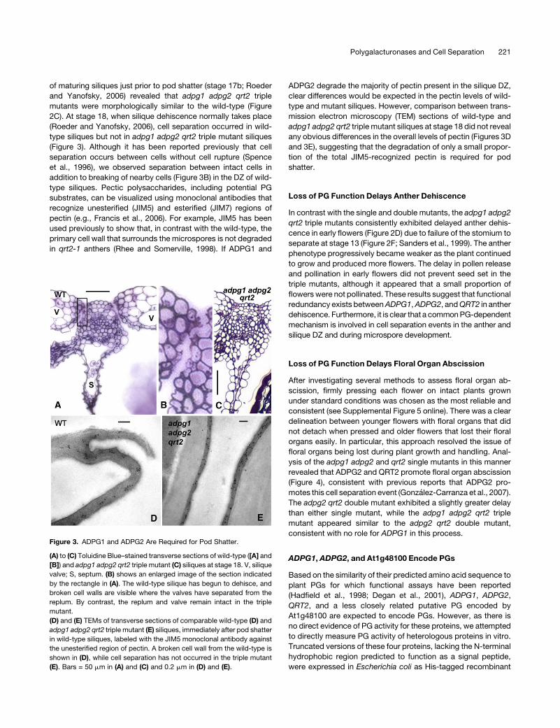
Figure 3. ADPG1 and ADPG2 Are Required for Pod Shatter.
(A) to (C) Toluidine Blue–stained transverse sections of wild-type ([A] and
[B]) and adpg1 adpg2 qrt2 triple mutant (C) siliques at stage 18. V, silique
valve; S, septum. (B) shows an enlarged image of the section indicated
by the rectangle in (A). The wild-type silique has begun to dehisce, and
broken cell walls are visible where the valves have separated from the
replum. By contrast, the replum and valve remain intact in the triple
mutant.
(D) and (E) TEMs of transverse sections of comparable wild-type (D) and
adpg1 adpg2 qrt2 triple mutant (E) siliques, immediately after pod shatter
in wild-type siliques, labeled with the JIM5 monoclonal antibody against
the unesterified region of pectin. A broken cell wall from the wild-type is
shown in (D), while cell separation has not occurred in the triple mutant
(E). Bars = 50 mm in (A) and (C) and 0.2 mm in (D) and (E).
Polygalacturonases and Cell Separation 221
fusion proteins. His-GFP (green fluorescent protein), His-
ADPG1, His-ADPG2, and His-At1g48100 proteins of the ex-
pected size were successfully expressed in E. coli based
on SDS-PAGE followed by Coomassie blue staining. These
His-tagged recombinant proteins were purified using a nickel-
nitrilotriacetic acid agarose (Ni-NTA) column and their activity
assayed using polygalacturonic acid as the substrate (Table 2).
All three proteins possessed PG activity, and the specific activity
of His-ADPG2 was ;30 times higher than that of His-ADPG1,
whereas activity of His-GFP could not be detected. The activity
of the His-At1g48100 recombinant protein was 40 and 100 times
higher than that of His-ADPG2 and His-ADPG1, respectively.
Expression of ADPG1, ADPG2, andQRT2
Previous work has investigated the expression pattern of both
ADPG1 and ADPG2. In Arabidopsis, ADPG1 expression has
previously been localized to the DZ of anthers and maturing
siliques by immuno-electron microscopy with an anti-RDPG1
antibody, by comparison with an RDPG1:GUS reporter (Sander
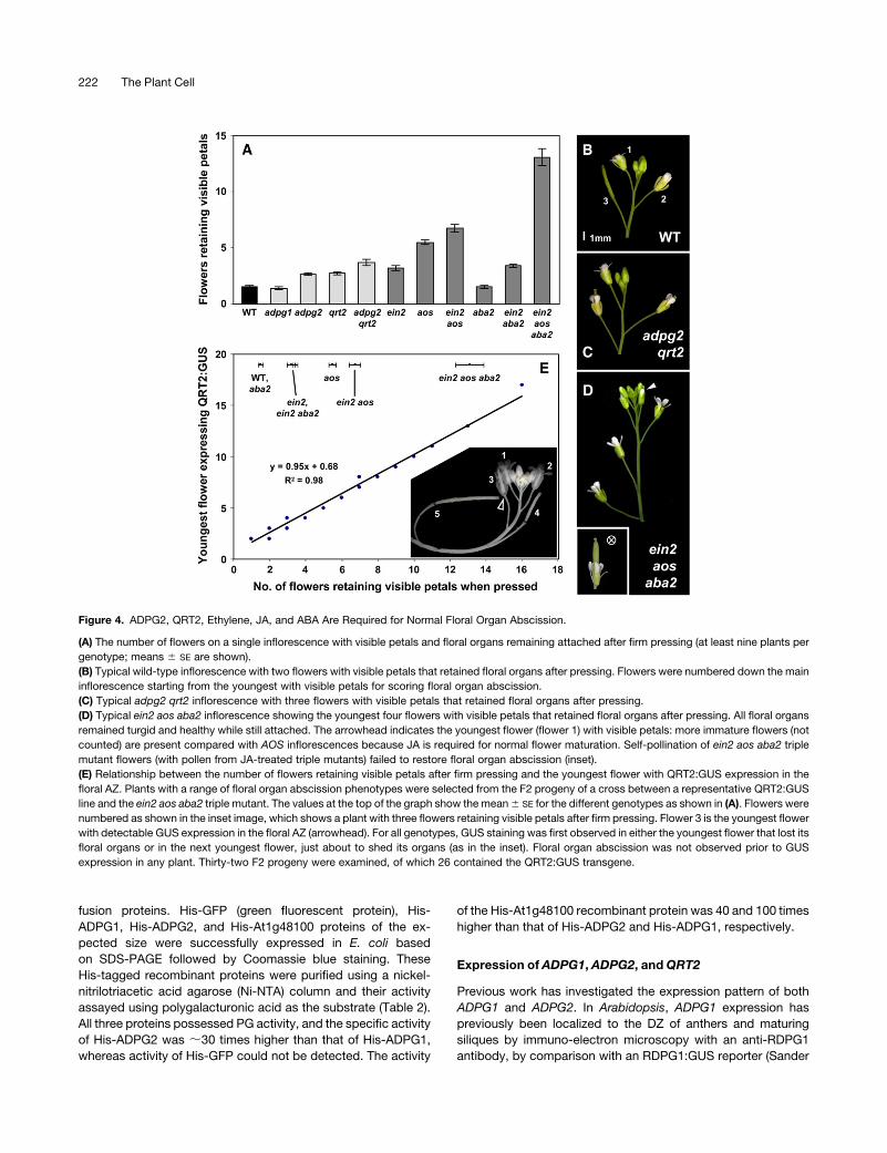
Figure 4. ADPG2, QRT2, Ethylene, JA, and ABA Are Required for Normal Floral Organ Abscission.
(A) The number of flowers on a single inflorescence with visible petals and floral organs remaining attached after firm pressing (at least nine plants per
genotype; means 6 SE are shown).
(B) Typical wild-type inflorescence with two flowers with visible petals that retained floral organs after pressing. Flowers were numbered down the main
inflorescence starting from the youngest with visible petals for scoring floral organ abscission.
(C) Typical adpg2 qrt2 inflorescence with three flowers with visible petals that retained floral organs after pressing.
(D) Typical ein2 aos aba2 inflorescence showing the youngest four flowers with visible petals that retained floral organs after pressing. All floral organs
remained turgid and healthy while still attached. The arrowhead indicates the youngest flower (flower 1) with visible petals: more immature flowers (not
counted) are present compared with AOS inflorescences because JA is required for normal flower maturation. Self-pollination of ein2 aos aba2 triple
mutant flowers (with pollen from JA-treated triple mutants) failed to restore floral organ abscission (inset).
(E) Relationship between the number of flowers retaining visible petals after firm pressing and the youngest flower with QRT2:GUS expression in the
floral AZ. Plants with a range of floral organ abscission phenotypes were selected from the F2 progeny of a cross between a representative QRT2:GUS
line and the ein2 aos aba2 triple mutant. The values at the top of the graph show the mean6 SE for the different genotypes as shown in (A). Flowers were
numbered as shown in the inset image, which shows a plant with three flowers retaining visible petals after firm pressing. Flower 3 is the youngest flower
with detectable GUS expression in the floral AZ (arrowhead). For all genotypes, GUS staining was first observed in either the youngest flower that lost its
floral organs or in the next youngest flower, just about to shed its organs (as in the inset). Floral organ abscission was not observed prior to GUS
expression in any plant. Thirty-two F2 progeny were examined, of which 26 contained the QRT2:GUS transgene.
222 The Plant Cell
et al., 2001) and by using an ADPG1:GUS reporter (Gonzalez-
Carranza et al., 2007). The expression domain for ADPG2 was
examined using transcriptional constructs in which either GUS or
GFP was under the control of the presumed ADPG2 promoter
(Gonzalez-Carranza et al., 2002, 2007). Based on this analysis,
ADPG2 has been reported to be expressed in roots and in the
abscission zone of the sepals, petals, and stamens of flowers
and is upregulated by ethylene in these tissues (Gonzalez-
Carranza et al., 2007). ADPG2 expression has not been reported
in anthers or silique DZs. Previous attempts to monitor
At3g07970 (QRT2) expression using GUS were not successful
(Gonzalez-Carranza et al., 2007), although this gene is expressed
in flowers undergoing floral organ abscission (Kim andPatterson,
2006).
Expression of ADPG1, ADPG2, and QRT2 was initially exam-
ined using qRT-PCRwith RNA fromdifferent plant tissues (Figure
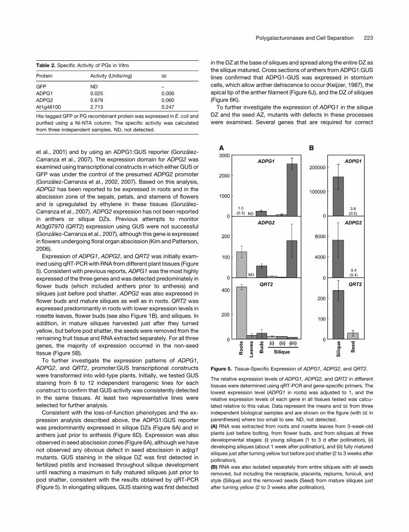
5). Consistent with previous reports, ADPG1was themost highly
expressed of the three genes andwas detected predominately in
flower buds (which included anthers prior to anthesis) and
siliques just before pod shatter. ADPG2 was also expressed in
flower buds and mature siliques as well as in roots. QRT2 was
expressed predominantly in roots with lower expression levels in
rosette leaves, flower buds (see also Figure 1B), and siliques. In
addition, in mature siliques harvested just after they turned
yellow, but before pod shatter, the seeds were removed from the
remaining fruit tissue and RNA extracted separately. For all three
genes, the majority of expression occurred in the non-seed
tissue (Figure 5B).
To further investigate the expression patterns of ADPG1,
ADPG2, and QRT2, promoter:GUS transcriptional constructs
were transformed into wild-type plants. Initially, we tested GUS
staining from 8 to 12 independent transgenic lines for each
construct to confirm that GUS activity was consistently detected
in the same tissues. At least two representative lines were
selected for further analysis.
Consistent with the loss-of-function phenotypes and the ex-
pression analysis described above, the ADPG1:GUS reporter
was predominantly expressed in silique DZs (Figure 6A) and in
anthers just prior to anthesis (Figure 6D). Expression was also
observed in seed abscission zones (Figure 6A), althoughwe have
not observed any obvious defect in seed abscission in adpg1
mutants. GUS staining in the silique DZ was first detected in
fertilized pistils and increased throughout silique development
until reaching a maximum in fully matured siliques just prior to
pod shatter, consistent with the results obtained by qRT-PCR
(Figure 5). In elongating siliques, GUS staining was first detected
in theDZ at the base of siliques and spread along the entire DZ as
the silique matured. Cross sections of anthers from ADPG1:GUS
lines confirmed that ADPG1-GUS was expressed in stomium
cells, which allow anther dehiscence to occur (Keijzer, 1987), the
apical tip of the anther filament (Figure 6J), and the DZ of siliques
(Figure 6K).
To further investigate the expression of ADPG1 in the silique
DZ and the seed AZ, mutants with defects in these processes
were examined. Several genes that are required for correct
Table 2. Specific Activity of PGs in Vitro
Protein Activity (Units/mg) SE
GFP ND –
ADPG1 0.025 0.006
ADPG2 0.679 0.060
At1g48100 2.713 0.247
His-tagged GFP or PG recombinant protein was expressed in E. coli and
purified using a Ni-NTA column. The specific activity was calculated
from three independent samples. ND, not detected.
Figure 5. Tissue-Specific Expression of ADPG1, ADPG2, and QRT2.
The relative expression levels of ADPG1, ADPG2, and QRT2 in different
tissues were determined using qRT-PCR and gene-specific primers. The
lowest expression level (ADPG1 in roots) was adjusted to 1, and the
relative expression levels of each gene in all tissues tested was calcu-
lated relative to this value. Data represent the means and SE from three
independent biological samples and are shown on the figure (with SE in
parentheses) where too small to see. ND, not detected.
(A) RNA was extracted from roots and rosette leaves from 3-week-old
plants just before bolting, from flower buds, and from siliques at three
developmental stages: (i) young siliques (1 to 3 d after pollination), (ii)
developing siliques (about 1 week after pollination), and (iii) fully matured
siliques just after turning yellow but before pod shatter (2 to 3 weeks after
pollination).
(B) RNA was also isolated separately from entire siliques with all seeds
removed, but including the receptacle, placenta, replums, funiculi, and
style (Silique) and the removed seeds (Seed) from mature siliques just
after turning yellow (2 to 3 weeks after pollination).
Polygalacturonases and Cell Separation 223
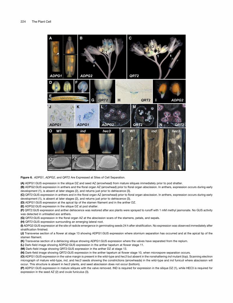
Figure 6. ADPG1, ADPG2, and QRT2 Are Expressed at Sites of Cell Separation.
(A) ADPG1:GUS expression in the silique DZ and seed AZ (arrowhead) from mature siliques immediately prior to pod shatter.
(B) ADPG2:GUS expression in anthers and the floral organ AZ (arrowhead) prior to floral organ abscission. In anthers, expression occurs during early
development (1), is absent at later stages (2), and returns just prior to dehiscence (3).
(C) QRT2:GUS expression in anthers and in the floral organ AZ (arrowhead) prior to floral organ abscission. In anthers, expression occurs during early
development (1), is absent at later stages (2), and returns just prior to dehiscence (3).
(D) ADPG1:GUS expression at the apical tip of the stamen filament and in the anther DZ.
(E) ADPG2:GUS expression in the silique DZ at pod shatter.
(F) QRT2:GUS expression and anther dehiscence was restored after aos plants were sprayed to runoff with 1 mM methyl jasmonate. No GUS activity
was detected in untreated aos anthers.
(G) QRT2:GUS expression in the floral organ AZ at the abscission scars of the stamens, petals, and sepals.
(H) QRT2:GUS expression surrounding an emerging lateral root.
(I) ADPG2:GUS expression at the site of radicle emergence in germinating seeds 24 h after stratification. No expression was observed immediately after
stratification finished.
(J) Transverse section of a flower at stage 13 showing ADPG1:GUS expression where stomium separation has occurred and at the apical tip of the
stamen filament.
(K) Transverse section of a dehiscing silique showing ADPG1:GUS expression where the valves have separated from the replum.
(L) Dark-field image showing ADPG2:GUS expression in the anther tapetum at flower stage 11.
(M) Dark-field image showing QRT2:GUS expression in the anther DZ at stage 13.
(N) Dark-field image showing QRT2:GUS expression in the anther tapetum at flower stage 10, when microspore separation occurs.
(O) ADPG1:GUS expression in the valve margin is present in the wild-type and hec3 but absent in the nonshattering indmutant (top). Scanning electron
micrograph of mature wild-type, ind, and hec3 seeds showing the constrictions (arrowheads) in the wild-type and ind funiculi where abscission will
occur. This structure is absent in hec3 plants, and seed abscission does not occur (bottom).
(P) ADPG1:GUS expression in mature siliques with the valve removed. IND is required for expression in the silique DZ (1), while HEC3 is required for
expression in the seed AZ (2) and ovule funiculus (3).
224 The Plant Cell
formation of the silique DZ have been identified, including
INDEHISCENT (IND). IND encodes a putative basic helix-loop-
helix transcription factor and forms part of a large gene family in
Arabidopsis. Siliques on ind mutants do not shatter due to a
failure to develop specialized cells at the junction between the
carpels and the replum that later form the DZ (Liljegren et al.,
2004). Compared with wild-type siliques, ADPG1:GUS expres-
sion was not detectable at the junction between a carpel and
replum, where the DZ fails to form in this mutant (Figures 6O and
6P). By contrast, ADPG1:GUS expression was still present at the
site of the seed AZ, consistent with normal seed abscission in the
ind mutant. Unfertilized ovules also expressed ADPG1:GUS in
the funiculus, despite the fact that only fertilized seeds abscise.
HECATE3 (HEC3) encodes a protein closely related to IND that
is required for normal functioning of female reproductive tissues
(Gremski et al., 2007). In addition to these phenotypes, hec3
mutants also fail to forma seedAZ, and seed abscission does not
occur (Figure 6O). Consistent with a role for ADPG1 in seed
abscission, ADPG1:GUS was not expressed in the funiculus of
fertilized seeds or unfertilized ovules in hec3 plants (Figure 6P).
However, pod shatter occurs normally in hec3 plants, and
ADPG1:GUS was expressed in the DZ of hec3 siliques (Figures
6O and 6P). These results suggest that IND and HEC3 are
required for normal expression of ADPG1 in the silique DZ and
seed AZ, respectively.
In agreement with previous reports and qRT-PCR analysis
(Figure 5), ADPG2:GUS was expressed in the AZs of sepals,
petals, and stamens in flowers just prior to floral organ abscission
(Figure 6B) and at the site of lateral root emergence (Kim and
Patterson, 2006; Gonzalez-Carranza et al., 2007). ADPG2:GUS
was also expressed early in anther development, at around the
time of microspore separation (Figure 6L), later in the anther DZ
just prior to dehiscence (Figure 6B), and in the DZ of maturing
siliques (Figure 6E). These observations, obtained using less-
stringent GUS staining conditions to increase sensitivity, are
consistent with the observed role ofADPG2 in anther dehiscence
and pod shatter and the qRT-PCR analysis (Figure 5) described
above but are inconsistent with previously published results
(Gonzalez-Carranza et al., 2007). A likely explanation for this
discrepancy is differences in the presumed promoter sequence
used. We used 2177 bp upstream from the predicted translation
initiation site, whereas Gonzalez-Carranza et al. (2007) used
1476 bp of the ADPG2 promoter which lacked the 25 bp of 59untranslated ADPG2 sequence. Based on GUS analysis, ADPG2
was also expressed in germinating seeds, at the point at which
the radicle broke through the seed coat during germination
(Figure 6I), a process that involves cell separation (Roberts et al.,
2002). No defect in seed germination was observed, presumably
because other PGs are also expressed in this tissue (Gonzalez-
Carranza et al., 2007).
The QRT2:GUS reporter was predominantly expressed in the
abscission zones of sepals, petals, and stamens in flowers just
prior to floral organ abscission (Figure 6C), similar to the reported
expression pattern of ADPG2 and consistent with previous
results obtained from a time course of floral organ abscission
(Kim and Patterson, 2006). A previous attempt to drive GUS
expression by the presumedAt3g07970 (QRT2) promoter did not
result in detectable GUS activity (Gonzalez-Carranza et al.,
2007), presumably because this construct used 1492 bp, while
we used 2085 bp of 59 sequence. QRT2:GUS was also ex-
pressed early in anther development, at around the time of
microspore separation (Figure 6N), and later just prior to anther
dehiscence (Figure 6M). GUS staining was also observed in
mature siliques at pod shatter, consistent with qRT-PCR analysis
(Figure 5). In contrast with ADPG1 and ADPG2 expression in
mature siliques, and consistent with no detectable role forQRT2
in pod shatter, QRT2:GUS was expressed only in the vascular
bundles in the replumand at the central region of the valve. These
data are consistent with the roles for QRT2 in microspore
separation, anther dehiscence, and floral organ abscission de-
scribed above.
Although expression of both ADPG2 (Gonzalez-Carranza
et al., 2007) and QRT2 (Figure 6H) was detected in at the site
of lateral root emergence, where cell separation occurs (Roberts
et al., 2002), no obvious root phenotype was observed in either
single mutant or in the adpg2 qrt2 double mutant. Finally, the
expression of QRT2 throughout the plant, albeit in very localized
regions, is consistent with the defects observed in AT3 flowers
and vegetative tissues (Figure 1; see Supplemental Figure 1 on-
line).
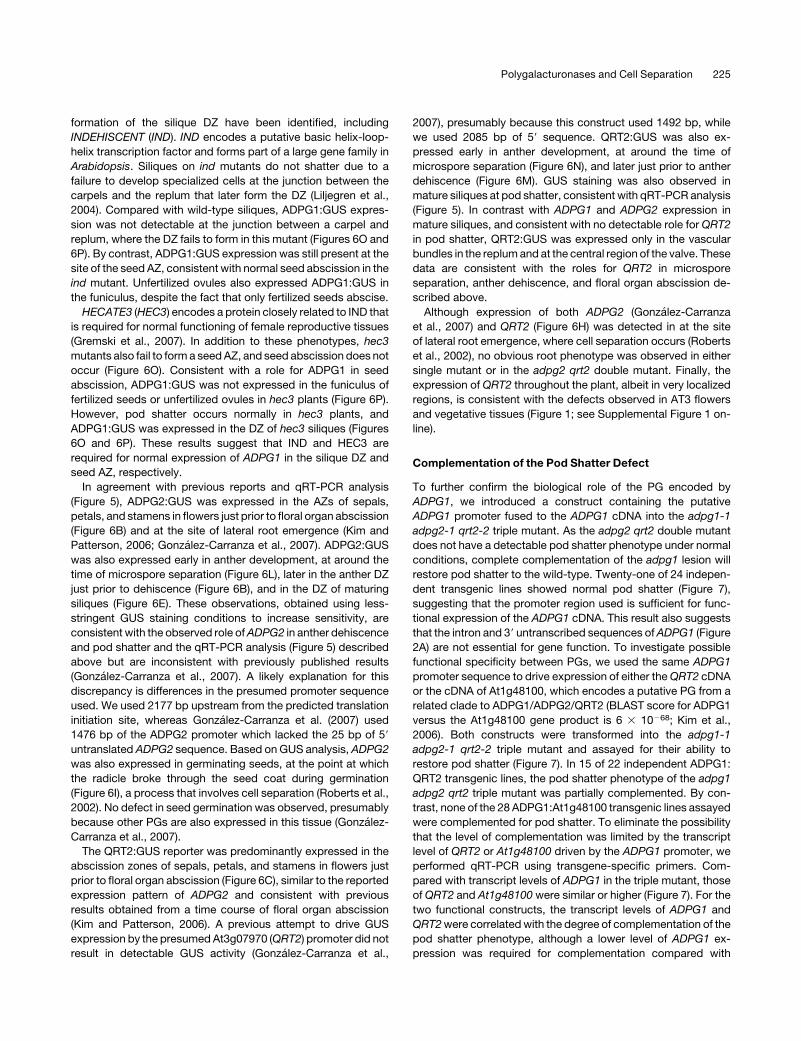
Complementation of the Pod Shatter Defect
To further confirm the biological role of the PG encoded by
ADPG1, we introduced a construct containing the putative
ADPG1 promoter fused to the ADPG1 cDNA into the adpg1-1
adpg2-1 qrt2-2 triple mutant. As the adpg2 qrt2 double mutant
does not have a detectable pod shatter phenotype under normal
conditions, complete complementation of the adpg1 lesion will
restore pod shatter to the wild-type. Twenty-one of 24 indepen-
dent transgenic lines showed normal pod shatter (Figure 7),
suggesting that the promoter region used is sufficient for func-
tional expression of the ADPG1 cDNA. This result also suggests
that the intron and 39 untranscribed sequences ofADPG1 (Figure
2A) are not essential for gene function. To investigate possible
functional specificity between PGs, we used the same ADPG1
promoter sequence to drive expression of either theQRT2 cDNA
or the cDNA of At1g48100, which encodes a putative PG from a
related clade to ADPG1/ADPG2/QRT2 (BLAST score for ADPG1
versus the At1g48100 gene product is 6 3 10268; Kim et al.,
2006). Both constructs were transformed into the adpg1-1
adpg2-1 qrt2-2 triple mutant and assayed for their ability to
restore pod shatter (Figure 7). In 15 of 22 independent ADPG1:
QRT2 transgenic lines, the pod shatter phenotype of the adpg1
adpg2 qrt2 triple mutant was partially complemented. By con-
trast, none of the 28 ADPG1:At1g48100 transgenic lines assayed
were complemented for pod shatter. To eliminate the possibility
that the level of complementation was limited by the transcript
level of QRT2 or At1g48100 driven by the ADPG1 promoter, we
performed qRT-PCR using transgene-specific primers. Com-
pared with transcript levels of ADPG1 in the triple mutant, those
of QRT2 and At1g48100 were similar or higher (Figure 7). For the
two functional constructs, the transcript levels of ADPG1 and
QRT2were correlatedwith the degree of complementation of the
pod shatter phenotype, although a lower level of ADPG1 ex-
pression was required for complementation compared with
Polygalacturonases and Cell Separation 225
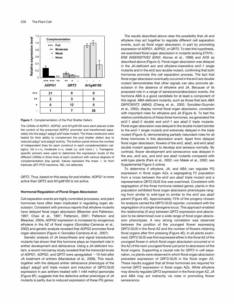
QRT2. Thus, based on this assay for pod shatter, ADPG1 is more
active than QRT2 and At1g48100 is not active.
Hormonal Regulation of Floral Organ Abscission
Cell separation events are highly controlled processes, and plant
hormones have often been implicated in regulating organ ab-
scission. Consistent with previous reports that ethylene mutants
have delayed floral organ abscission (Bleecker and Patterson,
1997; Chao et al., 1997; Patterson, 2001; Patterson and
Bleecker, 2004), ADPG2 expression is increased by exogenous
ethylene in the AZ of floral organs (Gonzalez-Carranza et al.,
2002) and genetic analysis revealed that ADPG2 promotes floral
organ abscission (Figure 4; Gonzalez-Carranza et al., 2007).
Genetic analysis of JA biosynthesis and signal transduction
mutants has shown that this hormone plays an important role in
anther development and dehiscence. Using a JA-deficient mu-
tant, a recent microarray study revealed that the transcript levels
of ADPG1, ADPG2, and QRT2 were upregulated ;10-fold after
JA treatment of anthers (Mandaokar et al., 2006). This result,
together with the delayed anther dehiscence phenotype of the
adpg1 adpg2 qrt2 triple mutant and induction of QRT2:GUS
expression in aos anthers treated with 1 mM methyl jasmonate
(Figure 6F), suggests that the defective anther phenotype of JA
mutants is partly due to reduced expression of these PG genes.
The results described above raise the possibility that JA and
ethylene may act together to regulate different cell separation
events, such as floral organ abscission, in part by promoting
expression of ADPG1, ADPG2, orQRT2. To test this hypothesis,
we examined floral organ abscission in mutants lacking ETHYL-
ENE INSENSITIVE2 (EIN2; Alonso et al., 1999) and AOS as
described above (Figure 4). Floral organ abscission was delayed
in the JA-deficient aos and ethylene-insensitive ein2-1 single
mutants and in the ein2 aos double mutant, confirming that both
hormones promote this cell separation process. The fact that
floral organ abscission eventually occurred in the ein2 aosdouble
mutant demonstrates that other signals can also promote ab-
scission in the absence of ethylene and JA. Because of its
proposed role in a range of senescence/abscission events, the
hormone ABA is a good candidate for at least a component of
this signal. ABA-deficient mutants, such as those that lack ABA
DEFICIENT2 (ABA2) (Cheng et al., 2002; Gonzalez-Guzman
et al., 2002), display normal floral organ abscission, consistent
with important roles for ethylene and JA (Figure 4). To test the
relative contributions of these three hormones, we generated the
ein2-1 aba2-2 double and ein2-1 aos aba2-2 triple mutants.
Floral organ abscissionwas delayed in the doublemutant (similar
to the ein2-1 single mutant) and extremely delayed in the triple
mutant (Figure 4), demonstrating partially redundant roles for all
three hormones in this abscission event. Apart from delayed
floral organ abscission, flowers of the ein2, aba2, and ein2 aba2
double mutant appeared to develop and senesce normally. By
contrast, flower development and senescence was delayed in
the aos, ein2 aos, and ein2 aos aba2 mutants compared with
wild-type plants (Park et al., 2002; von Malek et al., 2002; see
Supplemental Figure 5 online).
To determine if ethylene, JA, and ABA can regulate PG
expression in floral organ AZs, a segregating F2 population
from a cross between the ein2 aos aba2 triple mutant and a
representative QRT2:GUS line was examined. Consistent with
segregation of the three hormone-related genes, plants in this
population exhibited floral organ abscission phenotypes rang-
ing from similar to wild-type to similar to the ein2 aos aba2
parent (Figure 4E). Approximately 75% of the progeny chosen
for analysis carried the QRT2:GUS reporter, consistent with the
segregation of a single transgene locus. This approach enabled
the relationship (if any) between QRT2 expression and abscis-
sion to be determined over a wide range of floral organ abscis-
sion phenotypes. A very strong correlation was observed
between the position of the youngest flower expressing
QRT2:GUS in the floral AZ and the number of flowers retaining
floral organs after firm pressing (Figure 4E). In all plants exam-
ined, QRT2:GUSwas first expressed either in the floral AZ of the
youngest flower in which floral organ abscission occurred or in
the AZ of the next-youngest flower just prior to abscission of the
floral organs. Supporting a causal role for QRT2 in cell sepa-
ration, no plants were observed in which floral organ abscission
preceded expression of QRT2:GUS in the floral organ AZ.
These results suggest that all three hormones are required for
normal QRT2 expression in floral organ AZs. While ethylene
may directly regulateQRT2 expression in the floral organ AZ, JA
and ABA may act indirectly via roles in promoting flower
senescence.
Figure 7. Complementation of the Pod Shatter Defect.
The cDNAs of ADPG1, ADPG2, and At1g48100 were each placed under
the control of the presumed ADPG1 promoter and transformed sepa-
rately into the adpg1 adpg2 qrt2 triple mutant. The three constructs were
tested for their ability to complement the pod shatter defect due to
reduced adpg1 and adpg2 activity. The bottom panel shows the number
of independent lines for each construct in each complementation cat-
egory: full (+++), moderate (++), weak (+), and none (�). Transgene-
specific primers were used to determine the expression levels of the
different cDNAs in three lines of each construct with various degrees of
complementation (top panel). Values represent the mean 6 SE from
triplicate qRT-PCR reactions. ND, not detected.
226 The Plant Cell
DISCUSSION
PGs have been suggested to play critical roles in cell separation
in plants during a number of physiological processes involving
abscission or dehiscence of plant organs and tissues. For
example, an increase in the activity and protein level of PGs
can be detected in the cell separation zone just prior to pod
dehiscence (Sander et al., 2001). However, despite a range of
indirect evidence, relatively little experimental data using loss-of-
function alleles of genes known to encode PGs have been
reported (Lewis et al., 2006). In addition to ‘Flavr Savr’ tomato,
which has reduced PG expression and delayed fruit softening
(Dellapenna et al., 1986; Sheehy et al., 1988; Smith et al., 1990),
QRT3 has been shown to encode a PG required for separation of
microspores during pollen development (Rhee et al., 2003).
Based on a single loss-of-function allele, Gonzalez-Carranza
et al. (2007) have also recently reported that ADPG2 promotes
floral organ abscission, although these authors did not confirm
PG activity for ADPG2.
Using recombinant proteins, we have demonstrated that
ADPG1, ADPG2, and At1g48100 encode functional PGs. The
ability of an ADPG1:QRT2 construct to partially complement the
adpg1 adpg2 qrt2 pod shatter phenotype also confirms that
QRT2 is a PG, consistent with its proposed role in pectin
degradation (Rhee and Somerville, 1998). Supporting an impor-
tant role for pectin in plant growth, overexpression of QRT2
causes dwarfism and male sterility. The simplest explanation for
this result is that increased QRT2 expression leads to abnormal
cell–cell adhesion, which inhibits growth and flower function.
As overexpression studies provide only limited information on
normal gene function, we have analyzed loss-of-function alleles
for ADPG1, ADPG2, and QRT2 and identified important physi-
ological roles for these genes in several cell separation events
during reproductive development (Table 3). In addition, Jiang
et al. (2008) have recently shown that PGs are also involved in leaf
abscission in tomato. Taken together, these results support
the conclusion that multiple, and possibly all, cell separation/
abscission/dehiscence events in plants use a common PG-
dependent mechanism. InArabidopsis, combinations of ADPG1,
ADPG2, and QRT2 appear to be involved in all cell separation
processes associated with reproductive development so far
examined.
PGs Are Required for Microspore Separation
Previous genetic analyses have shown that likeQRT1 andQRT3,
QRT2 is required during microsporogenesis for the separation of
the developing pollen grains (Preuss et al., 1994; Rhee and
Somerville, 1998), and we have confirmed this result with two
additional loss-of-function qrt2 alleles. QRT1 and QRT3 encode
a PME and a PG relatively distantly related to QRT2, respectively
(Rhee et al., 2003; Francis et al., 2006). Pectins are synthesized in
Golgi bodies and secreted from cells in a highly methylesterified
form (Micheli, 2001; Schols and Voragen, 2002). PME catalyzes
pectin demethylesterification and appears to increase the ability
of PG to cleave pectin (Tucker and Seymour, 2002). Conse-
quently, an attractive model for microspore separation is that
pectin is first demethylated by QRT1 and subsequently de-
graded by PGs, such as QRT2 and QRT3. Although both QRT2
and QRT3 encode PGs, they are not functionally redundant
because loss of either gene prevents microspore separation.
Structural comparison of pollen grain tetrads using scanning
electron microscopy of the qrt mutants has revealed that qrt1
and qrt2 are structurally similar to the wild-type, with the excep-
tion of the pollen fusion phenotype. By contrast, qrt3 pollen
grains frequently had a layer of material deposited on the surface
of the distal region of the pollen grain tetrads (Rhee et al., 2003),
suggesting that QRT2 andQRT3 have distinct roles during pollen
development. Since the structure of qrt1 pollen is similar to that
of qrt2, it is possible that pectin demethylesterified by QRT1may
be used as a substrate by QRT2 rather than by QRT3.
The qrt2-1 allele is predicted to encode a mutant protein with a
single amino acid substitution (Val to Ala) at position 372 com-
pared with the wild-type QRT2 protein, suggesting that this amino
acid is important for enzyme activity. Val is a highly hydrophobic
and aliphatic amino acid, as are Leu and Ile. Phylogenetic analysis
reveals that Val, Leu, or Ile at the position corresponding to the
qrt2-1 lesion is highly conserved in PGs from plants and fungi,
supporting an important role for this residue in enzyme function.
Although the three-dimensional structure of PGII from the phyto-
pathogenic fungus Aspergillus niger has been determined, and
site-directed mutagenesis used to identify several critical amino
acid residues required for activity and substrate binding (van
Santen et al., 1999; Armand et al., 2000; Pages et al., 2000), the
role of this Val/Leu/Ile residue has not yet been clarified.
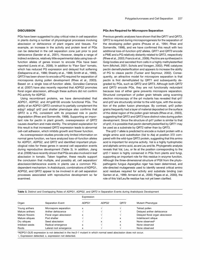
Table 3. Distinct and Overlapping Roles of ADPG1, ADPG2, and QRT2 in Separation Events during Arabidopsis Development
Organ Separation Event
Expression
Mutant PhenotypeADPG1 ADPG2 QRT2
Young anthers Microspore separation x � � Tetrad pollen
Mature anthers Anther dehiscence � � � Delayed anther dehiscence
Mature flowers Floral organ abscission x � � Delayed floral organ abscission
Mature siliques Pod shatter � � x Indehiscent silique
Dry siliques Seed abscission � x x None observeda
Imbibed seeds Radical emergence x � x None observed
Roots Lateral root emergence x � � None observed
aADPG1:GUS expression is not detected in the hec3-1 mutant in which normal seed abscission does not occur.
�, Expression detected; x, expression not detected.
Polygalacturonases and Cell Separation 227
PGs Are Required for Pod Shatter and Normal
Anther Dehiscence
The two Arabidopsis proteins most closely related to QRT2 are
encoded by the duplicate genes ADPG1 and ADPG2 (Kim et al.,
2006). Based on its expression pattern in the DZ of maturing
siliques, ADPG1 has previously been suggested to be involved in
pod shatter (Gonzalez-Carranza et al., 2007). Genetic analysis
revealed that both ADPG1 and ADPG2 are partially functionally
redundant for silique dehiscence (Figures 2 and 3), a conclusion
supported by the observation that ADPG2 is also expressed in
the silique DZ prior to pod shatter (Figure 6E). A role forADPG1 in
pod shatter is also supported by the absence of detectable
expression of this gene at the valve margin of the nonshattering
ind mutant. In addition, although no defect in seed abscission
was observed in the adpg1 adpg2 qrt2 triplemutant,ADPG1was
not detected in funiculi of the seed abscission-defective hec3
mutant. This result suggests that ADPG1, along with PGs other
than ADPG2 and QRT2, are required for seed abscission.
Functional redundancy between related PGs is also observed
in anther dehiscence such that the adpg1 adpg2 qrt2 triple
mutant has delayed pollen release, while single and double
mutants have no detectable anther defect. The fact that anthers
of the triple mutant do eventually dehisce suggests that other
PGs may also contribute to this process. Based on phylogenetic
and expression analyses (Grennan, 2006; Kim et al., 2006;
Gonzalez-Carranza et al., 2007), At1g80170 is a good candidate
to encode an additional PG involved in anther dehiscence.
PGs Are Required for Normal Floral Organ Abscission
Floral organ abscission was delayed, but not completely pre-
vented, in the adpg2 and qrt2 single mutants and in the adpg2
qrt2 double mutant (Figure 4), consistent with the expression of
both genes in the floral organ AZ (Figure 6). By contrast, ADPG1
is not expressed in this tissue and does not appear to be required
for this abscission process. Based on expression analysis,
At2g43890 and At2g43880 (Kim et al., 2006) may also be
involved in floral organ abscission together with ADPG2 and
QRT2.
Hormones and Floral Organ Abscission
Plant hormones have a number of important roles during flower
development, fertilization, and seed development and through-
out fruit set, growth, and senescence. Based on available data
for the regulation of ADPG1/ADPG2/QRT2 by JA and ethylene,
we investigated the role of these two hormones in organ abscis-
sion using the ethylene-insensitive ein2 and JA-deficient aos
mutants. Analysis of the ein2 aos double mutant revealed that
these plant hormones act in a partially redundant manner to
promote floral organ abscission. This conclusion is consistent
with the expression of ADPG2 (Figure 6; Gonzalez-Carranza
et al., 2002, 2007) andQRT2 (Figure 6; Kim and Patterson, 2006)
in the floral organ abscission zone, delayed flower organ abscis-
sion in plants with reduced ADPG2 expression (Figure 4;
Gonzalez-Carranza et al., 2007), and the known role of ethylene
in promoting floral organ senescence and detachment (Bleecker
and Patterson, 1997; Chao et al., 1997; Patterson, 2001). ABA
also promotes floral organ abscission in combinationwith JA and
ethylene as this process was further delayed in an ein2 aos aba2
triple mutant (Figure 4). As floral organ abscission eventually
occurred in the ein2 aos aba2 triple mutant, and other cell
separation events appeared normal in these plants, additional
signals that promote cell separation must also exist. Auxin is a
good candidate for this signal as it has been suggested to be
involved in many, and perhaps all, abscission events. For exam-
ple, application of auxin or auxin-like molecules can inhibit pod
shatter in canola (Chauvaux et al., 1997), and auxin analogs have
been shown to inhibit RDPG1 activity by blocking its secretion
into the cell wall (Degan et al., 2001). Finally, auxin mutants have
been reported to have delayed floral organ abscission and silique
dehiscence in Arabidopsis (Ellis et al., 2005; Okushima et al.,
2005). Thus, floral organ abscission is controlled by the com-
bined action of at least four different plant hormones that act in
part by regulating PG expression.
Functional Specificity of Arabidopsis PGs
Many genes encoding putative PGs have been identified in a
number of species, including Arabidopsis, tomato, and rice
(Hadfield et al., 1998; Kim et al., 2006; Gonzalez-Carranza
et al., 2007). Other cell wall proteins, such as expansin and
xyloglucan endotransglycosylase, also consist of multiple gene
families in plants (Rose et al., 2002; Sampedro and Cosgrove,
2005). However, in the absence of comprehensive biochemical
analyses, it is not known whether members of these gene
families, which often display different expression patterns,
possess different substrate specificities (Rose et al., 2002;
Sampedro and Cosgrove, 2005). For example, cytochrome
P450 monooxygenases are also encoded by multiple gene
families, with individual genes encoding enzymes with strict
substrate specificity (Schuler and Werck-Reichhart, 2003). Our
cDNA swapping experiments using the ADPG1 promoter in the
adpg1 adpg2 qrt2 triple mutant background suggest that closely
relatedPGs can also exhibit substrate specificity (Figure 7).While
the ADPG1:ADPG1 construct was able to fully restore pod
shatter, ADPG1:QRT2 (BLAST score for ADPG1 versus the
QRT2 gene product is 102104) only partially restored pod shatter
in adpg1 adpg2 qrt2 plants. Furthermore, when the same pro-
moter was used to drive expression of the At1g48100 cDNA
(BLAST score for ADPG1 versus the At1g48100 gene product is
63 10268), no restoration of pod shatter was observed. Although
we cannot formally exclude defects in translation or posttrans-
lational processing in these experiments, these results suggest
the existence of strict substrate specificity or substrate acces-
sibility for closely related PGs. Consistent with limited pectin
substrates for ADPG1 and ADPG2 in planta, it appears that the
majority of JIM5-recognized pectin in cell walls of the silique DZ
is not degraded during pod shatter (Figure 3).
Agricultural Importance of Abscission Events
The roles for PGs and plant hormones described here have
important practical implications for agriculture and horticulture.
For example, preventing or delaying floral organ abscission is of
228 The Plant Cell
great potential interest to the cut flower industry. More generally,
abscission/dehiscence associated with the harvest of seeds and/
or fruit is also a major production issue in a wide range of crops.
One of the best known examples is pod shatter, which can cause
seed loss prior to harvest and is an important problem for several
crops, particularly canola (oil seed rape). Reducing the activity of
canola genes closely related toADPG1 andADPG2 (Sander et al.,
2001; Gonzalez-Carranza et al., 2002) may lead to siliques that do
not shatter as readily as normal siliques, reducing seed losses.
METHODS
Plant Materials and Growth Conditions
Arabidopsis thaliana ecotypes Col-0 or Ler were used in this study. The
aos (accession number CS6149) and ein2-1 (CS3071) mutant seeds were
obtained from the ABRC (http://www.biosci.ohio-state.edu/~plantbio/
Facilities/abrc/abrchome.htm). The aba2-2 seeds were obtained from Eiji
Nambara (University of Toronto, Canada). Plants were grown under long-
day conditions (16 h light/8 h dark) in a 228C growth room either on soil
(equal volumes of seed raising mix [Debco] and perlite) with standard
Arabidopsis nutrient solution (http://www.biosci.ohio-state.edu/pcmb/
Facilities/abrc/handling.htm), on Growool (Swain et al., 2004), or on
agar medium consisting of 0.53 MS salts, 1% (w/v) sucrose, and 0.8%
agar for selection of transgenic plants or 0.6% phytagel (Sigma-Aldrich)
for detection of GUS staining in roots.
Activation Tagging Mutant Screening and TAIL-PCR
We generated several hundred activation tagging lines in the Ler back-
ground using Agrobacterium tumefaciens–mediated transformation and
the vacuum infiltration method (Bechtold et al., 1993) with the activation
tagging vector pSKI015 (Weigel et al., 2000). Seeds from infiltrated plants
were sown on Growool and sprayed with Basta (Swain et al., 2004) to
identify transgenic seedlings that were screened for novel phenotypes.
To identify the T-DNA insertion site, TAIL-PCR was performed according
to Liu et al. (1995) using genomic DNA extracted from AT3 inflorescences
with the PhytoPure plant DNA extraction kit (Amersham Life Sciences)
and primers SKIL1 (59-ACGACGGATCGTAATTTGTCG-39), SKIL2 (59-TTC-
ATTTTATAATAACGCTGCGG-39), or SKIL3 (59-CTTTCTTTTCCTCCAT-
ATTGACC-39) for the pSKI015 left border sequence and the AD2
degenerate primer (Liu et al., 1995).
Identification of T-DNA Insertion Mutants
T-DNA insertion mutants were obtained from the ABRC for SALK lines
(Alonso et al., 2003) or the Max-Planck institute for the GABI-Kat line
(Rosso et al., 2003). Alleles were named as follows: adpg1-1
(SALK_034714), adpg1-2 (SALK_057704), adpg2-1 (SALK_035098),
adpg2-2 (GABI_289C07), qrt2-2 (SALK_132478), and qrt2-3
(SALK_031337). Homozygous T-DNA insertion mutants were identified
using PCR with gene-specific primers (see Supplemental Table 2 online).
The primer complementary to the T-DNA was SALK LBa1 (59-TGG-
TTCACGTAGTGGGCCATCG-39) for SALK lines and GABI LB (59-CCC-
ATTTGGACGTGAATGTAGACAC-39) for the GABI-Kat line. Genomic
DNA was isolated by standard methods (Neff et al., 1998). PCR was per-
formed using rTaq DNA polymerase (Invitrogen).
RNA Extraction and Analysis
RNA was extracted using the phenol/SDS method (Naito et al., 1994).
Total RNA (1 mg) was treated with DNase I (Promega) to eliminate
genomic DNA contamination. After inactivation of DNA polymerase I
activity by adding EDTA (2.5 mM final concentration) and heating at 658C
for 10min, first-strand cDNAwas synthesized fromDNase I (Amplification
Grade; Invitrogen) treated total RNA with random hexamers using a
SuperScript III reverse transcriptase according to the manufacturer’s
instructions (Invitrogen). For RT-PCR, PCR was performed using first-
strand cDNA as a template with gene-specific primers (see Supplemental
Tables 3 and 4 online) followed by agarose gel electrophoresis to quantify
the amount of PCR product. qRT-PCRwith the SYBRGreen I dyemethod
was performed using the first-strand cDNA as a template with gene-
specific primers (see Supplemental Tables 3 and 4 online) on a sequence
detector system (My iQ single color real-time PCR detection system
Model: MyiQ Optical Module; Bio-Rad). The mean value of three biolog-
ical replicates was normalized using an 18S rRNA as the internal control
with primers 18S rRNA For (59-GGGACAGTCGGGGGCATTCG-39) and
18S rRNA Rev (59-TCCGCTGATCCCTGGTCGGC-39).
Plasmid Construction and Transformation
We used the Gateway system (Invitrogen) for plasmid construction, and
the sequences of all plasmids used in this study were examined to ensure
that no mutations were introduced during plasmid construction.
TheQRT2 cDNA and ADPG1 cDNAwere cloned into the pENTR vector
(Invitrogen) to create pENTR-QRT2 and pENTR-ADPG1, respectively.
The 35S enhancer sequence was fused upstream of the QRT2 genomic
fragment consisting of 2085 bp of QRT2 promoter sequence from the
predicted translational initiation site and the QRT2 gene with introns and
exons to create pENTR-35S enhancer-ProQRT2:QRT2. The ADPG1 pro-
moter, 2433bp upstream from thepredicted translational initiation site,was
fused upstream of the ADPG1 cDNA,QRT2 cDNA, and At1g48100 cDNA
to create pENTR-ProADPG1:ADPG1, pENTR-ProADPG1:QRT2, and
pENTR-ProADPG1:At1g48100, respectively. The GUS coding region
was fused downstream of the ADPG1 promoter (2433 bp upstream
from the translation initiation site), the ADPG2 promoter (2177 bp up-
stream from the translation initiation site), and the QRT2 promoter (2085
bp upstream from the predicted translation initiation site) to create
pENTR-ProADPG1:GUS, pENTR-ProADPG2:GUS, and pENTR-ProQRT2:
GUS, respectively. Precise procedures for constructing these plasmids
are described in the Supplemental Methods online.
To generate binary vectors for plant transformation, an LR reaction was
performed with the binary vector for the Gateway system, pGWB1, in
which a target gene from pENTR can be introduced before the nopaline
synthase terminator. The resulting binary vectors, 35S enhancer-
ProQRT2:QRT2, ADPG1:GUS, ADPG2:GUS, QRT2:GUS, ADPG1:
ADPG1, ADPG1:QRT2, and ADPG1:At1g48100, were transformed into
wild-type Col-0 or the adpg1-1 adpg2-1 qrt2-2 mutant using the Agro-
bacterium-mediated floral dip method (Clough and Bent, 1998). Plants
carrying a T-DNA insertion were selected on agar plates containing 20
mg/L kanamycin (Sigma-Aldrich), 20 mg/L hygromycin (Sigma-Aldrich),
and 50 mg/L carbenicillin (Sigma-Aldrich).
Scanning Electron Microscopy of Pollen Tetrad
Pollen from the adpg1 adpg2 qrt2 triple mutant was mounted on double-
sided carbon tabs, coated with ;20 nm gold using an Emitech K550X
sputter coater, and viewed at 15 kV accelerating voltage in a Zeiss EVO
LS15 scanning electron microscope.
Light Microscopy
Tissuewas fixed in 2%glutaraldehyde in 25mMsodiumphosphate buffer
at pH 6.8, vacuum infiltrated, and incubated at 48C overnight. Tissue was
briefly rinsed in 25mM sodium phosphate buffer, pH 6.8, and dehydrated
Polygalacturonases and Cell Separation 229
in an ethanol series (25, 50, 70, 95, and 100%), with 2 h in each solution.
Tissue was run through an LR white resin/ethanol mix (25:75, 50:50,
75:25, 100:0 32) and finally embedded in LR white resin. Using a
microtome, 2-mm cross sections were cut and placed on glass slides.
These were stained with 0.3% Toluidine Blue in 1% sodium tetraborate,
pH 9.0, for 30 s or in 0.02% ruthenium red, then water mounted and
covered with a cover slip for observation by light microscopy.
For GUS assays, plant material was gently fixed by incubation in 90%
acetone on ice for 15 min, and the tissue washed in sodium phosphate
buffer, pH 7.2. The tissue was then incubated in a 3 mM GUS staining
solution consisting of 3 mMK3Fe(CN)6, 3 mMK4Fe(CN)6, 2 mM 5-bromo-
4-chloro-3-indolyl-b-D-glucronic acid, and 50 mM NaPO4, pH 7.2, vacuum
infiltrated, and incubated at 378C for 16 h. For detection of ADPG2:GUSand
QRT2:GUS in anthers and siliques, 0 mM K3Fe(CN)6/0 mM K4Fe(CN)6 was
used to increase sensitivity. After staining, tissues were fixed in 50%
ethanol, 5% acetic acid, and 3.7% formaldehyde and then washed several
times with 70% and then 85% ethanol. For making cross sections after
staining, the tissues were processed through an ethanol series (30, 50, 70,
85, 95,and100%),with the50%step incorporatinga1-hfixativewash (50%
ethanol, 5%acetic acid, and 3.7% formaldehyde). Tissuewas run through a
Lemosol/ethanol gradient (25:75, 50:50, 75:25, 100:0 32) and finally em-
bedded in Paraffin. Using a microtome, 7-mm cross sections were cut and
placed on glass slides. The tissue was examined using a microscope.
Preparation of Siliques for Light and Electron Microscopy
Arabidopsis siliques were fixed in 2.5% glutaraldehyde (EM Grade) in
PBS, pH 7.2, for 2 h at room temperature and stored overnight at 48C.
Siliqueswerewashed three times in distilledwater and then dehydrated in
a graded ethanol series. Over several days, the samples were infiltrated
with LR White resin, and then individual siliques were placed in gelatin
capsules, filledwith fresh resin, and polymerized in a 558Coven overnight.
Sectioning and Toluidine Blue Staining of Dehiscing Siliques
Individual siliques in resin blocks were sectioned (1 mm) transversely on a
Leica Ultracut R microtome. The sections were collected and dried onto
glass microscope slides and stained with a 2% (w/v) aqueous Toluidine
Blue solution for 30 s, washed, and dried on a hotplate.
Immunolabeling with Gold-Labeled Pectin Antibodies for TEM
Ultrathin sections (80 nm) were cut on a Leica Ultracut Rmicrotome using
a diamond knife and collected on 100 mesh formvar-coated gold grids.
The grids were preincubated in a blocking buffer (1% BSA in PBS) for 30
min at room temperature to prevent nonspecific antibody binding. Two
monoclonal antibodies raised against pectic polysaccharides, JIM5 and
JIM7 (Knox et al., 1990; Plant Probes) were diluted 1:50 in blocking buffer.
The grids were incubated in drops of the diluted primary antibody for 1 h
at room temperature and then overnight at 48C. The grids were washed
three times in PBS and two times in blocking buffer and then incubated in
a 1:20 dilution of an anti-rat secondary antibody conjugated to 18-nm
colloidal gold (Jackson ImmunoResearch). Grids were washed in PBS
and several times in water before poststaining in 2% (w/v) aqueous uranyl
acetate. The sections were viewed on a Philips BioTwin transmission
electron microscope and images captured on a Gatan multiscan digital
camera. In control experiments, the primary antibody was omitted and no
labeling was observed.
Functional Assay of PG Proteins
Truncated versions of the PG proteins, lacking the hydrophobic
N-terminal region encoding a signal peptide predicted by the SOSUI
program (Hirokawa et al., 1998; http://bp.nuap.nagoya-u.ac.jp/sosui/)
were expressed as His-tagged fusion proteins in Escherichia coli. GFP
and N-terminal truncated versions of ADPG1, ADPG2, QRT2, and
At1g48100 were cloned into the pENTR vector (Invitrogen) to create
pENTR-GFP, pENTR-tADPG1, pENTR-tADPG2, pENTR-tQRT2, and
pENTR-tAt1g48100, respectively. GFP was used as a negative control.
Details of these plasmid constructions are provided in the Supplement
Methods online. A Gateway LR reaction was performed using pENTR-
GFP, pENTR-tADPG1, pENTR-tADPG2, pENTR-tQRT2, and pENTR-
tAt1g48100 with pDEST17, which is Gateway E. coli expression vector
that provides a 6x Histidine tag at the N terminus of the expressed target
gene driven by the T7 promoter (Invitrogen). The resulting plasmids,
pDEST17-His-GFP, pDEST17-His-tADPG1, pDEST17-His-tADPG2,
pDEST17-His-tQRT2, and pDEST17-His-tAt1g48100, were transformed
into E. coli strain origami B (DE3) (Novagen) that is able to provide a
soluble and active enzyme with proper disulfide bond formation (Shimizu
et al., 2005). Soluble PG protein was purified using a Ni-NTA column (Ni-
NTA agarose; Qiagen) according to the manufacturer’s protocol. Purified
PG solution was dialyzed against a 150 mM sodium chloride/50 mM
sodium acetate, pH 5.0, solution. The standard curve was determined by
the use of galacturonic acid (Sigma-Aldrich), and PG activity was mea-
sured according to Gross (1982) using polygalacturonic acid from citrus
fruit (Sigma-Aldrich) as the substrate. One unit of the endo-PG activity
was defined as the amount of enzyme that liberated 1 mmol of reducing
end groups per 1 min at pH 5.0 and 308C. Specific activity was calculated
from three independent samples.
Accession Numbers
Sequence data for the genes described in this article can be found in the
Arabidopsis Genome Initiative under the following accession numbers:
ADPG1 (At3g57510),ADPG2 (At2g41850),QRT2 (At3g07970), andGAPC
(At3g04120).
Supplemental Data
The following materials are available in the online version of this article.
Supplemental Figure 1. Phenotype of the AT3 Line.
Supplemental Figure 2. qrt2-1 Causes a Single Amino Acid Substi-
tution in the Predicted QRT2 Protein without Altering the Transcript
Level.
Supplemental Figure 3. Reduced Transcript Levels of ADPG1,
ADPG2, and QRT2 in the adpg1 adpg2 qrt2 Triple Mutants.
Supplemental Figure 4. adpg1-1 Reduces Transcript Levels and
Causes an In-Frame Deletion.
Supplemental Figure 5. Delayed Abscission of Floral Organs in PG
and Hormone Mutants.
Supplemental Table 1. Phenotypes of adpg1, adpg2, and qrt2
Single, Double, and Triple Mutants.
Supplemental Table 2. Primers for Genotyping T-DNA Insertion
Mutants.
Supplemental Table 3. Primers for RT-PCR to Examine the Tran-
script Level in T-DNA Insertion Mutants.
Supplemental Table 4. Primers for RT-PCR to Examine Target Gene
Expression.
Supplemental Methods. Plasmid Construction.
ACKNOWLEDGMENTS
We thank ABRC and the Max-Planck institute for providing the Arabi-
dopsis T-DNA insertion mutants, Tsuyoshi Nakagawa (Shimane
230 The Plant Cell
University) for providing the Gateway destination binary vector
(pGWB1), and Michael Groszmann and Mark Talbot for help with
sectioning for GUS visualization and scanning electron microscopy
analysis. M.O. was supported by a CSIRO Office of the Chief Executive
(OCE) Fellowship. P.K. was supported by a Riverlink PhD studentship
from La Trobe University and CSIRO. S.W. was supported by the Grains
Research and Development Corporation, Australia.
Received October 12, 2008; revised December 21, 2008; accepted
January 6, 2009; published January 23, 2009.
REFERENCES
Alonso, J.M., Hirayama, T., Roman, G., Nourizadeh, S., and Ecker,
J.R. (1999). EIN2, a bifunctional transducer of ethylene and stress
responses in Arabidopsis. Science 284: 2148–2152.
Alonso, J.M., et al. (2003). Genome-wide insertional mutagenesis of
Arabidopsis thaliana. Science 301: 653–657.
Armand, S., Wagemaker, M.J.M., Sanchez-Torres, P., Kester, H.C.,
van Santen, Y., Dijkstra, B.W., Visser, J., and Benen, J.A. (2000).
The active site topology of Aspergillus niger endopolygalacturonase II
as studied by site-directed mutagenesis. J. Biol. Chem. 275: 691–696.
Atkinson, R.G., Schroder, R., Hallett, I.C., Cohen, D., and MacRae,
E.A. (2002). Overexpression of polygalacturonase in transgenic apple
trees leads to a range of novel phenotypes involving changes in cell
adhesion. Plant Physiol. 129: 122–133.
Bechtold, N., Ellis, J., and Pelletier, G. (1993). In planta Agrobacterium
mediated gene transfer by infiltration of adult Arabidopsis thaliana
plants. C. R. Acad. Sci. Paris Life Sci. 316: 1194–1199.
Benen, J.A., Kester, H.C., and Visser, J. (1999). Kinetic characteriza-
tion of Aspergillus niger N400 endopolygalacturonases I, II and C. Eur.
J. Biochem. 259: 577–585.
Biely, P., Benen, J., Heinrichova, K., Kester, H.C.M., and Visser, J.
(1996). Inversion of configuration during hydrolysis of a-1,4-galact-
uronidic linkage by three Aspergillus polygalacturonases. FEBS Lett.
382: 249–255.
Bleecker, A.B., and Patterson, S.E. (1997). Last exit: Senescence,
abscission, and meristem arrest in Arabidopsis. Plant Cell 9: 1169–
1179.
Bonghi, Z.C., Casadoro, G., Ramina, A., and Rascio, N. (1993).
Abscission in leaf and fruit explants of Prunus persica (L.) Batsch. New
Phytol. 123: 555–565.
Brown, K.M. (1997). Ethylene and abscission. Physiol. Plant. 100:
567–576.
Chao, Q., Rothenberg, M., Solano, R., Roman, G., Terzaghi, W., and
Ecker, J.R. (1997). Activation of the ethylene gas response pathway in
Arabidopsis by the nuclear protein ETHYLENE-INSENSITIVE3 and
related proteins. Cell 89: 1133–1144.
Chauvaux, N., Child, R., John, K., Ulvskov, P., Borkhardt, B.,
Prinsen, E., and Van Onckelen, H.A. (1997). The role of auxin in
cell separation in the dehiscence zone of oilseed rape pods. J. Exp.
Bot. 48: 1423–1429.
Cheng, W.-H., Endo, A., Zhou, L., Penney, J., Chen, H.-C., Arroyo, A.,
Leon, P., Nambara, E., Asami, T., Seo, M., Koshiba, T., and Sheen,
J. (2002). A unique short-chain dehydrogenase/reductase in Arabi-
dopsis glucose signaling and abscisic acid biosynthesis and func-
tions. Plant Cell 14: 2723–2743.
Cho, H.-T., and Cosgrove, D.J. (2000). Altered expression of expansin
modulates leaf growth and pedicel abscission in Arabidopsis thaliana.
Proc. Natl. Acad. Sci. USA 97: 9783–9788.
Clough, S.J., and Bent, A.F. (1998). Floral dip: A simplified method for
Agrobacterium-mediated transformation of Arabidopsis thaliana. Plant
J. 16: 735–743.
Degan, F.D., Child, R., Svendsen, I., and Ulvskov, P. (2001). The
cleavable N-terminal domain of plant endopolygalacturonases from
clade B may be involved in a regulated secretion mechanism. J. Biol.
Chem. 276: 35297–35304.
del Campillo, E., and Bennett, A.B. (1996). Pedicel breakstrength and
cellulase gene expression during tomato flower abscission. Plant
Physiol. 111: 813–820.
Dellapenna, D., Alexander, D.C., and Bennett, A.B. (1986). Molecular
cloning of tomato fruit polygalacturonase: Analysis of polygalacturo-
nase mRNA levels during ripening. Proc. Natl. Acad. Sci. USA 83:
6420–6424.
Ellis, C.M., Nagpal, P., Young, J.C., Hagen, G., Guilfoyle, T.J., and
Reed, J.W. (2005). AUXIN RESPONSE FACTOR1 and AUXIN RE-
SPONSE FACTOR2 regulate senescence and floral organ abscission
in Arabidopsis thaliana. Development 132: 4563–4574.
Ferrandiz, C., Liljegren, S.J., and Yanofsky, M.F. (2000). Negative
regulation of the SHATTERPROOF genes by FRUITFULL during
Arabidopsis fruit development. Science 289: 436–438.
Francis, K.E., Lam, S.Y., and Copenhaver, G.P. (2006). Separation of
Arabidopsis pollen tetrads is regulated by QUARTET1, a pectin
methylesterase gene. Plant Physiol. 142: 1004–1013.
Gonzalez-Carranza, Z.H., Elliott, K.A., and Roberts, J.A. (2007).
Expression of polygalacturonases and evidence to support their role
during cell separation processes in Arabidopsis thaliana. J. Exp. Bot.
58: 3719–3730.
Gonzalez-Carranza, Z.H., Whitelaw, C.A., Swarup, R., and Roberts,
J.A. (2002). Temporal and spatial expression of a polygalacturonase
during leaf and flower abscission in oilseed rape and Arabidopsis.
Plant Physiol. 128: 534–543.
Gonzalez-Guzman, M., Apostolova, N., Belles, J.M., Barrero, J.M.,
Piqueras, P., Ponce, M.R., Micol, J.L., Serrano, R., and Rodriguez,
P.L. (2002). The short-chain alcohol dehydrogenase ABA2 catalyzes the
conversion of xanthoxin to abscisic aldehyde. Plant Cell 14: 1833–1846.
Gremski, K., Ditta, G., and Yanofsky, M.F. (2007). The HECATE genes
regulate female reproductive tract development in Arabidopsis thali-
ana. Development 134: 3593–3601.
Grennan, A.K. (2006). Genevestigator. Facilitating web-based gene-
expression analysis. Plant Physiol. 141: 1164–1166.
Gross, K.C. (1982). A rapid and sensitive spectrophotometric method
for assaying polygalacturonase using 2-cyanoacetamide. HortScience
17: 933–934.
Hadfield, K.A., and Bennett, A.B. (1998). Polygalacturonases: Many
genes in search of a function. Plant Physiol. 117: 337–343.
Hadfield, K.A., Rose, J.K., Yaver, D.S., Berka, R.M., and Bennett,
A.B. (1998). Polygalacturonase gene expression in ripe melon fruit
supports a role for polygalacturonase in ripening-associated pectin
disassembly. Plant Physiol. 117: 363–373.
Hirokawa, T., Boon-Chieng, S., and Mitaku, S. (1998). SOSUI: Clas-
sification and secondary structure prediction system for membrane
proteins. Bioinformatics 14: 378–379.
Jenkins, E.S., Paul, W., Coupe, S.A., Bell, S.J., Davies, E.C., and
Roberts, J.A. (1996). Characterization of an mRNA encoding a
polygalacturonase expressed during pod development in oilseed
rape (Brassica napus L.). J. Exp. Bot. 47: 111–115.
Jenkins, E.S., Paul, W., Craze, M., Whitelaw, C.A., Weigand, A., and
Roberts, J.A. (1999). Dehiscence-related expression of an Arabidop-
sis thaliana gene encoding a polygalacturonase in transgenic plants of
Brassica napus. Plant Cell Environ. 22: 159–167.
Jiang, C.Z., Lu, F., Imsabai, W., Meir, S., and Reid, M.S. (2008).
Silencing polygalacturonase expression inhibits tomato petiole ab-
scission. J. Exp. Bot. 59: 973–979.
Polygalacturonases and Cell Separation 231
Kalaitzis, P., Koehler, S.M., and Tucker, M.L. (1995). Cloning of a
tomato polygalacturonase expressed in abscission. Plant Mol. Biol.
28: 647–656.
Kalaitzis, P., Solomos, T., and Tucker, M.L. (1997). Three different
polygalacturonases are expressed in tomato leaf and flower abscis-
sion, each with a different temporal expression pattern. Plant Physiol.
113: 1303–1308.
Keijzer, C.J. (1987). The process of anther dehiscence and pollen
dispersal. I. The opening mechanism of longitudinally dehiscing
anthers. New Phytol. 105: 487–498.
Kim, J., and Patterson, S.E. (2006). Expression divergence and func-
tional redundancy of polygalacturonases in floral organ abscission.
Plant Signal. Behav. 1: 281–283.
Kim, J., Shiu, S.-H., Thoma, S., Li, W.-H., and Patterson, S.E. (2006).
Patterns of expansion and expression divergence in the plant poly-
galacturonase gene family. Genome Biol. 7: R87.
Knox, J.P., Linstead, P.J., King, J., Cooper, C., and Roberts, K.
(1990). Pectin esterification is spatially regulated both within cell walls
and between developing tissues of root apices. Planta 181: 512–521.
Lashbrook, C.C., Gonzalez-Bosch, C., and Bennett, A.B. (1994). Two
divergent endo-b-1,4-glucanase genes exhibit overlapping expres-
sion in ripening fruits and abscising flowers. Plant Cell 6: 1485–1493.
Lewis, M.W., Leslie, M.E., and Liljegren, S.J. (2006). Plant separation:
50 ways to leave your mother. Curr. Opin. Plant Biol. 9: 59–65.
Liljegren, S.J., Ditta, G.S., Eshed, Y., Savidge, B., Bowman, J.L., and
Yanofsky, M.F. (2000). SHATTERPROOF MADS-box genes control
seed dispersal in Arabidopsis. Nature 404: 766–770.
Liljegren, S.J., Roeder, A.H.K., Kempin, S.A., Gremski, K., Østergaard,
L., Guimil, S., Reyes, D.K., and Yanofsky, M.F. (2004). Control of
fruit patterning in Arabidopsis by INDEHISCENT. Cell 116: 843–853.
Liu, Y.-G., Mitsukawa, N., Oosumi, T., and Whittier, R.F. (1995).
Efficient isolation and mapping of Arabidopsis thaliana T-DNA insert
junctions by thermal asymmetric interlaced PCR. Plant J. 8: 457–463.
MacDougall, A.J., Needs, P.W., Rigby, N.M., and Ring, S.G. (1996).
Calcium gelation of pectic polysaccharides isolated from unripe
tomato fruit. Carbohydr. Res. 293: 235–249.
Mandaokar, A., Thines, B., Shin, B., Lange, B.M., Choi, G., Koo, Y.J.,
Yoo, Y.J., Choi, Y.D., Choi, G., and Browse, J. (2006). Transcrip-
tional regulators of stamen development in Arabidopsis identified by
transcriptional profiling. Plant J. 46: 984–1008.
Markovic, O., and Janecek, S. (2001). Pectin degrading glycoside
hydrolases of family 28: Sequence-structural features, specificities
and evolution. Protein Eng. 14: 615–631.
Micheli, F. (2001). Pectin methylesterases: Cell wall enzymes with
important roles in plant physiology. Trends Plant Sci. 6: 414–419.
Mitsuda, N., and Ohme-Takagi, M. (2008). NAC transcription factors
NST1 and NST3 regulate pod shattering in a partially redundant
manner by promoting secondary wall formation after the establish-
ment of tissue identity. Plant J. 56: 768–778.
Naito, S., Hirai, M.Y., Chino, M., and Komeda, Y. (1994). Expression of
a soybean (Glycine max [L.] Merr.) seed storage protein gene in
transgenic Arabidopsis thaliana and its response to nutritional stress
and to abscisic acid mutations. Plant Physiol. 104: 497–503.
Neff, M.M., Neff, J.D., Chory, J., and Pepper, A.E. (1998). dCAPS, a
simple technique for the genetic analysis of single nucleotide poly-
morphisms: Experimental applications in Arabidopsis thaliana genet-
ics. Plant J. 14: 387–392.
Okushima, Y., Mitina, I., Quach, H.L., and Theologis, A. (2005).
AUXIN RESPONSE FACTOR 2 (ARF2): A pleiotropic developmental
regulator. Plant J. 43: 29–46.
Osteryoung, K.W., Toenjes, K., Hall, B., Winkler, V., and Bennett,
A.B. (1990). Analysis of tomato polygalacturonase expression in
transgenic tobacco. Plant Cell 2: 1239–1248.
Pages, S., Heijne, W.H.M., Kester, H.C.M., Visser, J., and Benen,
J.A. (2000). Subsite mapping of Aspergillus niger endopolygalactur-
onase II by site-directed mutagenesis. J. Biol. Chem. 275: 29348–
29353.
Parenicova, L., Kester, H.C., Benen, J.A., and Visser, J. (2000).
Characterization of a novel endopolygalacturonase from Aspergillus
niger with unique kinetic properties. FEBS Lett. 467: 333–336.
Park, J.-H., Halitschke, R., Kim, H.B., Baldwin, I.T., Feldmann, K.A.,
and Feyereisen, R. (2002). A knock-out mutation in allene oxide
synthase results in male sterility and defective wound signal trans-
duction in Arabidopsis due to a block in jasmonic acid biosynthesis.
Plant J. 31: 1–12.
Patterson, S.E. (2001). Cutting loose. Abscission and dehiscence in
Arabidopsis. Plant Physiol. 126: 494–500.
Patterson, S.E., and Bleecker, A.B. (2004). Ethylene-dependent and
-independent processes associated with floral organ abscission in
Arabidopsis. Plant Physiol. 134: 194–203.
Peace, C.P., Crisosto, C.H., and Gradziel, T.M. (2005). Endopoly-
galacturonase: A candidate gene for Freestone and Melting flesh in
peach. Mol. Breed. 16: 21–31.
Petersen, M., Sander, L., Child, R., van Onckelen, H., Ulvskov, P.,
and Borkhardt, B. (1996). Isolation and characterization of a pod
dehiscence zone-specific polygalacturonase from Brassica napus.
Plant Mol. Biol. 31: 517–527.
Preuss, D., Rhee, S.Y., and Davis, R.W. (1994). Tetrad analysis
possible in Arabidopsis with mutation of the QUARTET (QRT) genes.
Science 264: 1458–1460.
Rajani, S., and Sundaresan, V. (2001). The Arabidopsis myc/bHLH
gene ALCATRAZ enables cell separation in fruit dehiscence. Curr.
Biol. 11: 1914–1922.
Rhee, S.Y., Osborne, E., Poindexter, P.D., and Somerville, C.R.
(2003). Microspore separation in the quartet3 mutants of Arabidopsis
is impaired by a defect in a developmentally regulated polygalact-
uronase required for pollen mother cell wall degradation. Plant
Physiol. 133: 1170–1180.
Rhee, S.Y., and Somerville, C.R. (1998). Tetrad pollen formation in
quartet mutants of Arabidopsis thaliana is associated with persistence
of pectic polysaccharides of the pollen mother cell wall. Plant J. 15:
79–88.
Ridley, B.L., O’Neill, M.A., and Mohnen, D. (2001). Pectins: Structure,
biosynthesis, and oligogalacturonide-related signaling. Phytochemis-
try 57: 929–967.
Roberts, J.A., Elliott, K.A., and Gonzalez-Carranza, Z.H. (2002).
Abscission, dehiscence, and other cell separation processes. Annu.
Rev. Plant Biol. 53: 131–158.
Roeder, A.H.K., and Yanofsky, M.F. (February 22, 2006). Fruit
development in Arabidopsis. In The Arabidopsis Book, C.R.
Somerville and E.M. Meyerowitz, eds (Rockville, MD: American So-
ciety of Plant Biologists), doi/10.1199/tab.0075, http://www.aspb.org/
publications/arabidopsis/.
Rose, J.K., Braam, J., Fry, S.C., and Nishitani, K. (2002). The XTH
family of enzymes involved in xyloglucan endotransglucosylation and
endohydrolysis: Current perspectives and a new unifying nomencla-
ture. Plant Cell Physiol. 43: 1421–1435.
Rosso, M.G., Li, Y., Strizhov, N., Reiss, B., Dekker, K., and
Weisshaar, B. (2003). An Arabidopsis thaliana T-DNA mutagenized
population (GABI-Kat) for flanking sequence tag-based reverse ge-
netics. Plant Mol. Biol. 53: 247–259.
Sampedro, J., and Cosgrove, D.J. (2005). The expansin superfamily.
Genome Biol. 6: 242.
Sander, L., Child, R., Ulvskov, P., Albrechtsen, M., and Borkhardt, B.
(2001). Analysis of a dehiscence zone endo-polygalacturonase in
oilseed rape (Brassica napus) and Arabidopsis thaliana: Evidence for
232 The Plant Cell
roles in cell separation in dehiscence and abscission zones, and in
stylar tissues during pollen tube growth. Plant Mol. Biol. 46: 469–479.
Sanders, P.M., Bui, A.Q., Weterings, K., McIntire, K.N., Hsu, Y.-C.,
Lee, P.Y., Truong, M.T., Beals, T.P., and Goldberg, R.B. (1999).
Anther developmental defects in Arabidopsis thaliana male-sterile
mutants. Sex. Plant Reprod. 11: 297–322.
Schols, H.A., and Voragen, A.G.J. (2002). The chemical structure of
pectins. In Pectins and Their Manipulation, G.B. Seymour and P.G.
Knox, eds (Boca Raton, FL: CRC Press), pp. 1–29.
Schuler, M.A., and Werck-Reichhart, D. (2003). Functional genomics
of P450s. Annu. Rev. Plant Biol. 54: 629–667.
Sheehy, R.E., Kramer, M., and Hiatt, W.R. (1988). Reduction of
polygalacturonase activity in tomato fruit by antisense RNA. Proc.
Natl. Acad. Sci. USA 85: 8805–8809.
Shimizu, T., Shibata, H., Araya, T., Nakatsu, T., Miyairi, K., Okuno, T.,
and Kato, H. (2005). Expression, purification, and crystallization of
endopolygalacturonase from a pathogenic fungus, Stereum purpur-
eum, in Escherichia coli. Protein Expr. Purif. 44: 130–135.
Smith, C.J.S., Watson, C.F., Morris, P.C., Bird, C.R., Seymour, G.B.,
Gray, J.E., Arnold, C., Tucker, G.A., Schuch, W., Harding, S. and
Grierson, D. (1990). Inheritance and effect on ripening of antisense
polygalacturonase genes in transgenic tomatoes. Plant Mol. Biol. 14:
369–379.
Smyth, D.R., Bowman, J.L., and Meyerowitz, E.M. (1990). Early flower
development in Arabidopsis. Plant Cell 2: 755–767.
Spence, J., Vercher, Y., Gates, P., and Harris, N. (1996). Pod shatter in
Arabidopsis thaliana, Brassica napus and B. juncea. J. Microsc. 181:
195–203.
Swain, S.M., Muller, A.D., and Singh, D.P. (2004). The gar2 and rga
alleles increase the growth of gibberellin-deficient pollen tubes in
Arabidopsis. Plant Physiol. 134: 694–705.
Taylor, J.E., Coupe, S.A., Picton, S., and Roberts, J.A. (1994).
Characterization and accumulation pattern of an mRNA encoding an
abscission-related b-1,4-glucanase from leaflets of Sambucus nigra.
Plant Mol. Biol. 24: 961–964.
Taylor, J.E., Webb, S.T.J., Coupe, S.A., Tucker, G.A., and Roberts,
J.A. (1993). Changes in polygalacturonase activity and solubility of
polyuronides during ethylene-stimulated leaf abscission in Sambucus
nigra. J. Exp. Bot. 44: 93–98.
Torki, M., Mandaron, P., Mache, R. and Falconet, D. (2000). Char-
acterization of a ubiquitous expressed gene family encoding polyga-
lacturonase in Arabidopsis thaliana. Gene 242: 427–436.
Tucker, G.A., and Seymour, G.B. (2002). Modification and degradation
of pectins. In Pectins and Their Manipulation, G.B. Seymour and P.G.
Knox, eds (Boca Raton, FL: CRC Press), pp. 150–173.
van Santen, Y., Benen, J.A.E., Schroter, K.-H., Kalk, K.H., Armand,
S., Visser, J., and Dijkstra, B.W. (1999). 1.68-A crystal structure of
endopolygalacturonase II from Aspergillus niger and identification of
active site residues by site-directed mutagenesis. J. Biol. Chem. 274:
30474–30480.
von Malek, B., van der Graaff, E., Schneitz, K., and Keller, B. (2002).
The Arabidopsis male-sterile mutant dde2–2 is defective in the
ALLENE OXIDE SYNTHASE gene encoding one of the key enzymes
of the jasmonic acid biosynthesis pathway. Planta 216: 187–192.
Weigel, D., et al. (2000). Activation tagging in Arabidopsis. Plant
Physiol. 122: 1003–1014.
Polygalacturonases and Cell Separation 233
DOI 10.1105/tpc.108.063768; originally published online January 23, 2009; 2009;21;216-233Plant Cell
Mikihiro Ogawa, Pippa Kay, Sarah Wilson and Stephen M. SwainArabidopsisDevelopment in
QUARTET2 Are Polygalacturonases Required for Cell Separation during Reproductive ARABIDOPSIS DEHISCENCE ZONE POLYGALACTURONASE1 (ADPG1), ADPG2, and
This information is current as of March 3, 2020
Supplemental Data /content/suppl/2009/01/23/tpc.108.063768.DC1.html /content/suppl/2009/01/24/tpc.108.063768.DC2.html
References /content/21/1/216.full.html#ref-list-1
This article cites 84 articles, 33 of which can be accessed free at:
Permissions https://www.copyright.com/ccc/openurl.do?sid=pd_hw1532298X&issn=1532298X&WT.mc_id=pd_hw1532298X
eTOCs http://www.plantcell.org/cgi/alerts/ctmain
Sign up for eTOCs at:
CiteTrack Alerts http://www.plantcell.org/cgi/alerts/ctmain
Sign up for CiteTrack Alerts at:
Subscription Information http://www.aspb.org/publications/subscriptions.cfm
is available at:Plant Physiology and The Plant CellSubscription Information for
ADVANCING THE SCIENCE OF PLANT BIOLOGY © American Society of Plant Biologists