Embed Size (px)
Citation preview
Arabidopsis Photorespiratory SerineHydroxymethyltransferase Activity Requires the MitochondrialAccumulation of Ferredoxin-Dependent Glutamate Synthase W
Aziz Jamai,a Patrice A. Salome,a,1 Stephen H. Schilling,a,2 Andreas P.M. Weber,b and C. Robertson McClunga,3
a Department of Biological Sciences, Dartmouth College, Hanover, New Hampshire 03755b Heinrich-Heine-Universitat, Institut fur Biochemie der Pflanzen, 40225 Dusseldorf, Germany
The dual affinity of ribulose-1,5-bisphosphate carboxylase/oxygenase for O2 and CO2 results in the net loss of fixed carbon
and energy in a process termed photorespiration. The photorespiratory cycle is complex and occurs in three organelles,
chloroplasts, peroxisomes, and mitochondria, which necessitates multiple steps to transport metabolic intermediates.
Genetic analysis has identified a number of mutants exhibiting photorespiratory chlorosis at ambient CO2, including several
with defects in mitochondrial serine hydroxymethyltransferase (SHMT) activity. One class of mutants deficient in SHMT1
activity affects SHM1, which encodes the mitochondrial SHMT required for photorespiration. In this work, we describe a
second class of SHMT1-deficient mutants defective in a distinct gene, GLU1, which encodes Ferredoxin-dependent
Glutamate Synthase (Fd-GOGAT). Fd-GOGAT is a chloroplastic enzyme responsible for the reassimilation of photorespira-
tory ammonia as well as for primary nitrogen assimilation. We show that Fd-GOGAT is dual targeted to the mitochondria and
the chloroplasts. In the mitochondria, Fd-GOGAT interacts physically with SHMT1, and this interaction is necessary for
photorespiratory SHMT activity. The requirement of protein–protein interactions and complex formation for photorespira-
tory SHMT activity demonstrates more complicated regulation of this crucial high flux pathway than anticipated.
INTRODUCTION
Ribulose-1,5-bisphosphate carboxylase/oxygenase (Rubisco)
initiates the Calvin (C3) cycle with the carboxylation of ribulose
1,5 bisphosphate to yield two molecules of 3-phosphoglycerate
(3PGA). However, the dual affinity of Rubisco for O2 and CO2
means that Rubisco also catalyzes the oxygenation of ribulose
1,5 bisphosphate to yield one molecule of 3PGA and one
molecule of 2-phosphoglycolate (2PG), thereby initiating the
photorespiratory (C2) cycle (Bowes et al., 1971; Ogren and
Bowes, 1971). The photorespiratory cycle regenerates 3PGA
from 2PG in a complex series of reactions involving at least 16
enzymes and occurring in the cytosol (Timm et al., 2008) and in
three organelles (chloroplasts, peroxisomes, and mitochondria),
which necessitates the involvement of 14 to 18 transport steps
(Leegood et al., 1995; Douce and Neuburger, 1999; Reumann
and Weber, 2006). In the mitochondria, the glycine decarbox-
ylase complex (GDC) and serine hydroxymethyltransferase
(SHMT) catalyze the photorespiratory conversion of the Gly,
derived from 2PG via the activities of phosphoglycolate phos-
phatase, glycolate oxidase, and an aminotransferase, into Ser,
with the concomitant evolution of CO2 and ammonia (Douce
et al., 2001; Bauwe and Kolukisaoglu, 2003). Thus, photorespi-
ration lowers photosynthetic efficiency in that the CO2 and
ammonia must be reassimilated in the chloroplast by Rubisco
and the glutamine synthetase (GS)/Ferredoxin-dependent glu-
tamate synthase (Fd-GOGAT) system, respectively, with the
concomitant consumption of both ATP and reducing power
(Leegood et al., 1995; Douce and Neuburger, 1999; Zhu et al.,
2008). This energetic inefficiency means that photorespiration
protects against photoinhibition, especially under stress condi-
tions in which CO2 assimilation is lessened. The generation of
CO2 by photorespiration continues to drive the C3 cycle, and the
combined C2 and C3 cycles consume ATP and reducing equiv-
alents, limiting the diversion of light energy into the production of
active oxygen species that cause photoinhibition (Kozaki and
Takeba, 1996; Wingler et al., 2000).
Forward genetic analysis has been important in the elucidation
of the photorespiratory pathway (Somerville, 1986, 2001). Re-
cent supplementation with reverse genetics has allowed the
characterization of the last known enzyme of the photorespira-
tory cycle (Boldt et al., 2005). Although it is likely that some
transporters remain incompletely characterized (Weber, 2004;
Linka and Weber, 2005), it is tempting to conclude that the
photorespiratory cycle is fully characterized. However, the as-
sembly of a parts list does not necessarily constitute a detailed
understanding and, by definition, excludes components whose
function has not yet been identified.
1Current address: Max Planck Institute for Developmental Biology,Department of Molecular Biology, Spemannstrasse 37-39, D-72076Tubingen, Germany.2 Current address: Department of Pharmacology and Cancer Biology,Duke University Medical Center, Box 3813, Durham, NC 27710.3 Address correspondence to [email protected] author responsible for distribution of materials integral to thefindings presented in this article in accordance with the policy describedin the Instructions for Authors (www.plantcell.org) is: C. RobertsonMcClung ([email protected]).WOnline version contains Web-only data.www.plantcell.org/cgi/doi/10.1105/tpc.108.063289
The Plant Cell, Vol. 21: 595–606, February 2009, www.plantcell.org ã 2009 American Society of Plant Biologists
Nitrogen metabolism is crucial in photorespiration and the flux
through this pathway is ;10-fold greater than the amount of
nitrogen assimilated from the soil (Keys et al., 1978). For many
years it has been accepted that the reassimilation of photo-
respiratory ammonia is catalyzed by GS/Fd-GOGAT in the chlo-
roplasts (Leegood et al., 1995; Douce and Neuburger, 1999).
The Arabidopsis thaliana genome includes two genes encoding
distinct Fd-GOGAT isozymes; the photorespiratory function
is associated exclusively with FERREDOXIN-DEPENDENT
GLUTAMATE SYNTHASE1 (GLU1) (Somerville and Ogren,
1980; Coschigano et al., 1998). Similarly, the completion of the
Arabidopsis genome sequence indicates that SHMT in Arabi-
dopsis is encoded by seven SHM genes, two of which encode
mitochondrial isoforms (McClung et al., 2000; Bauwe and
Kolukisaoglu, 2003). However, only SHM1 is necessary and
sufficient to specify photorespiratory SHMT activity (Voll et al.,
2006).
In this work, we uncover an additional level of complexity that
further modifies our understanding of photorespiration. We show
an unanticipated physical interaction between two of the first
photorespiratory pathway components to be identified, SHMT
(Somerville and Ogren, 1981) and Fd-GOGAT (Somerville and
Ogren, 1980). This interaction was unexpected because photo-
respiratory SHMT activity is mitochondrial, whereas photores-
piratory Fd-GOGAT activity is chloroplastic (Leegood et al.,
1995; Douce and Neuburger, 1999). Such spatial separation
would seem to preclude a physical interaction. Nonetheless, we
provide genetic evidence thatGLU1 is required for mitochondrial
SHMT activity through the characterization of a novel GLU1
allele, glu1-201, that retains wild-type Fd-GOGAT activity but is
deficient in photorespiratory SHMT activity. Microscopy imaging
of green fluorescent protein (GFP) fusions and immunological
analysis of biochemically purified organelles show that GLU1-
encoded Fd-GOGAT is dual targeted to both chloroplasts and
mitochondria. We further show that Fd-GOGAT and SHMT1
physically interact in vivo through coimmunoprecipitation and
bimolecular fluorescence complementation (BiFC). Thus, we
conclude that GLU1-encoded Fd-GOGAT associated with the
mitochondria is necessary for photorespiratory SHMT activity.
RESULTS
Characterization of a Novel Photorespiratory Mutant
Defective in Mitochondrial SHMT Activity
Homozygous shm1-1 (originally called stm; Somerville and
Ogren, 1981) mutants exhibit a severe photorespiratory pheno-
type of lethal chlorosis under low CO2, and the requirement for
supplementary CO2 is absolute (Voll et al., 2006) (compare Figure
1A, the glabra1 [gl1] mutant that serves as the isogenic wild type
to shm1-1 in Figure 1B; see Supplemental Figures 1A and 1B for
seedlings grown at elevated CO2). By contrast, a second stm
allele, herein called glu1-201, that confers a similar reduction in
SHMT activity (Table 1) will growwithout added CO2 in dim (;50
to 75 mmol·m22·s21) light, although the plants are chlorotic and
considerably smaller than the wild type (Figure 1C; see Supple-
mental Figure 1C online). Although we initially attributed this less
severe phenotype to a partial loss of SHM1 function, we failed to
identify any nucleotide lesion in the coding sequence and pro-
moter region of SHM1 in the mutant. Unexpectedly, the F1
progeny from crosses of this second allele with shm1-1were not
chlorotic in low CO2 (Figure 1F; see Supplemental Figure 1F
online) and showed wild-type levels of SHMT activity (Table 1).
This genetic complementation suggests that this mutation, al-
though previously thought to be allelic with shm1-1, is a mutation
in a distinct gene.
We established that this second mutation maps to a position
near the top of chromosome V between the markers CTR1.2 and
NGA151, which is distinct from the positions of any of the seven
SHM genes (Figure 2A). Further analysis using 300 F2 chlorotic
plants positioned the mutation close to GLU1, which encodes
photorespiratory Fd-GOGAT (Coschigano et al., 1998). We
therefore tested the hypothesis that our mutation was a novel
allele ofGLU1 by genetic complementation. Indeed, introduction
into the mutant background of the wild-type GLU1 gene driven
by either the 35S promoter or the endogenous GLU1 promoter
both rescued the photorespiratory phenotype of chlorosis at low
CO2 (Figures 1G and 1I; see Supplemental Figures 1G and 1I
online) and restored wild-type levels of SHMT activity (Table 1).
Accordingly, we conclude that this SHMT-defective photores-
piratory phenotype is conferred by a novel allele of GLU1
designated glu1-201. Earlier authors called glu1 mutants gluS
(Somerville and Ogren, 1980), gltS (Suzuki and Rothstein, 1997),
or gls (Coschigano et al., 1998), but we propose to revise the
mutant designation to be consistent with the accepted gene
name.
Consistent with the identification ofGLU1 as the gene respon-
sible for the photorespiratory phenotype and loss of SHMT
activity in glu1-201, there is a single nucleotide change (C6410T)
between the wild type andmutant that changes amino acid 1270
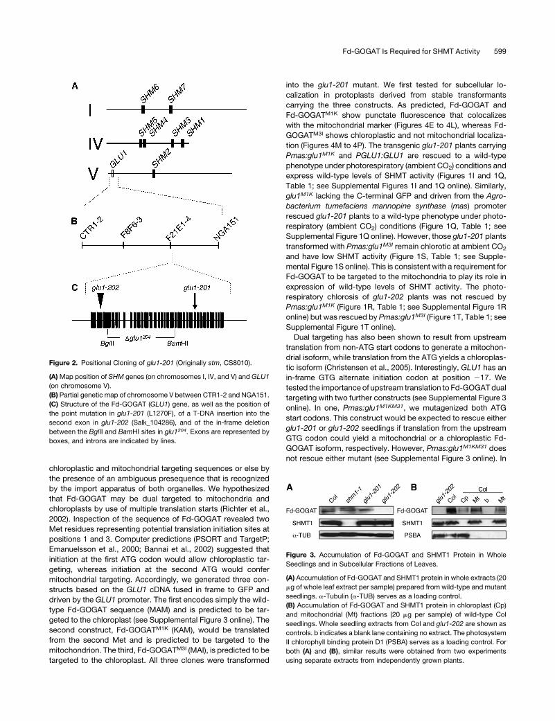
from Leu to Phe. Protein gel blot analysis indicates that Fd-
GOGAT accumulates to approximately normal levels in glu1-201
seedlings, consistent with a point mutation that does not disrupt
protein accumulation (Figure 3A). SHMT1 protein levels are
unaffected in the glu1-201 mutant, although SHMT1 protein is
below detectable levels in the shm1-1 mutant (Figure 3A). As
indicated above, introduction of the wild-type GLU1 gene driven
by either its endogenous promoter or the 35S promoter into glu1-
201 rescues the photorespiratory chlorosis phenotype and re-
stores wild-type levels of SHMT activity (Figures 1G and 1I, Table
1; see Supplemental Figures 1G and 1I online). However, intro-
duction of a modifiedGLU1 carrying the L1270F mutation driven
by the 35S promoter into glu1-201 fails to rescue chlorosis or to
restore wild-type levels of SHMT activity (Figure 1L, Table 1; see
Supplemental Figure 1L online) in plants growing at ambient CO2
levels, consistent with this mutation conferring the photorespi-
ratory phenotype.
Loss-of-function mutations in GLU1 had been previously
demonstrated to exhibit photorespiratory chlorosis and loss of
Fd-GOGAT activity (Somerville and Ogren, 1980; Suzuki and
Rothstein, 1997; Coschigano et al., 1998). We confirmed that a
T-DNA insertion mutant (Salk_104286, termed glu1-202), in
which the T-DNA has inserted into the 2nd exon of GLU1, lacks
Fd-GOGAT protein (Figure 3A) and exhibits reduced Fd-GOGAT
activity (Table 1) and photorespiratory chlorosis (compare Figure
596 The Plant Cell
1D with the isogenic wild type, Columbia-0 [Col-0], in Figure 1E
and Supplemental Figures 1D and 1E online). This allele also
confers a reduction in SHMT activity (Table 1). The glu1-202
mutant is fully rescued by expression of wild-type GLU1 but not
by glu1L1270F under control of the 35S promoter (Figures 1H, 1J,
and 1M, Table 1; see Supplemental Figures 1H, 1J, and 1M
online). The F1 plants resulting from a cross between glu1-201
and glu1-202 display photorespiratory chlorosis and reduced
SHMT activity, indicating that the two mutations are allelic
(Figure 1K, Table 1; see Supplemental Figure 1K online).
glu1-201 has reduced SHMT activity but retains wild-type
levels of Fd-GOGAT activity, suggesting that the positive effect
ofGLU1 on SHMT activity is independent of Fd-GOGAT catalytic
activity. To test this, we generated a new allele ofGLU1, glu1204,
in which residues 299 to 1007, including the central domain and
parts of the N-terminal aminotransferase domain and the FMN
binding domain, are deleted in frame (Figure 2C). The glu1204
allele fails to rescue either photorespiratory chlorosis or Fd-
GOGAT activity when introduced into glu1-202 (Figure 1O, Table
1; see Supplemental Figure 1O online), consistent with its
predicted lack of Fd-GOGAT catalytic activity. However, the
glu1204 allele fully rescues photorespiratory chlorosis and SHMT
activity when introduced into the glu1-201 mutant (Figure 1N,
Table 1; see Supplemental Figure 1N online). Thus, we conclude
that wild-type levels of photorespiratory SHMT activity require
Fd-GOGAT expression (although not catalytic activity) and that a
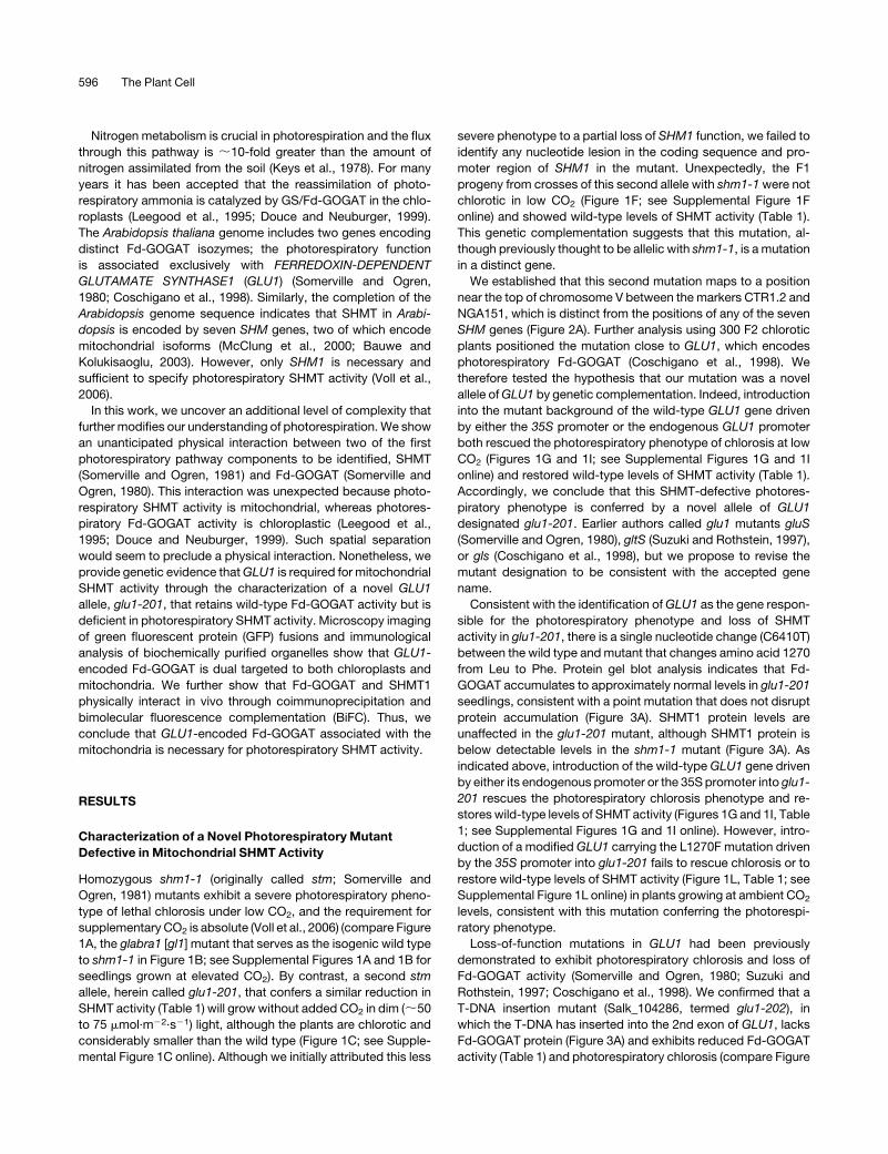
Figure 1. Phenotypic Analysis of Photorespiratory Mutants at Ambient CO2.
Seedlings were grown at elevated (3%) CO2 levels for 10 d at 228C in long days (16 h light at 100 mmol·m�2·s�1: 8 h dark) and then transferred to ambient
CO2 for 5 d.
(A) glabra1 (Col), the isogenic parent to shm1-1 and glu1-201; (B) shm1-1; (C) glu1-201; (D) glu1-202; (E) Col-0, the isogenic parent to glu1-202; (F) F1:
shm1-1 3 glu1-201; (G) glu1-201/p35S:GLU1; (H) glu1-202/P35S:GLU1; (I) glu1-201/PGLU1:GLU1; (J) glu1-202/PGLU1:GLU1; (K) F1: glu1-201 3
glu1-202; (L) glu1-201/P35S:glu1L1270F; (M) glu1-202/P35S:glu1L1270F; (N) glu1-201/P35S:glu1204; (O) glu1-202/P35S:glu1204; (P) glu1-201 / Pmas:
GLU1; (Q) glu1-201/Pmas:glu1M1K; (R) glu1-202/Pmas:glu1M1K; (S) glu1-201/Pmas:glu1M3I; (T) glu1-202/Pmas:glu1M3I.
Fd-GOGAT Is Required for SHMT Activity 597
mutation of L1270F results in a glu1 species that is unable to
sustain wild-type SHMT activity, even though it retains wild-type
Fd-GOGAT activity.
DualTargetingofFd-GOGATto theMitochondria inAddition
to the Chloroplasts
It is surprising and puzzling that the wild-type GLU1 gene, which
encodes a chloroplastic Fd-GOGAT (Suzuki and Rothstein,
1997; Coschigano et al., 1998; Lee et al., 2008), rescues a
mutant lacking mitochondrial SHMT activity (Somerville and
Ogren, 1981). Because Fd-GOGAT catalytic activity is not re-
quired for this rescue, we speculated that perhaps a physical
interaction between SHMT and Fd-GOGAT might be necessary
for full SHMT activity. Such a physical interaction would require
common localization of Fd-GOGAT and SHMT1, which seemed
unlikely as photorespiratory SHMT activity is known to be mito-
chondrial (Somerville and Ogren, 1981), whereas photorespira-
tory Fd-GOGAT activity is chloroplastic (Somerville and Ogren,
1980). At this time, the Subcellular Proteomics Database does
not provide proteomic data to support a mitochondrial localiza-
tion of Fd-GOGAT (Heazlewood et al., 2007). Therefore, to verify
these localizations, we performed microscopy analyses and
biochemical fractionation experiments.
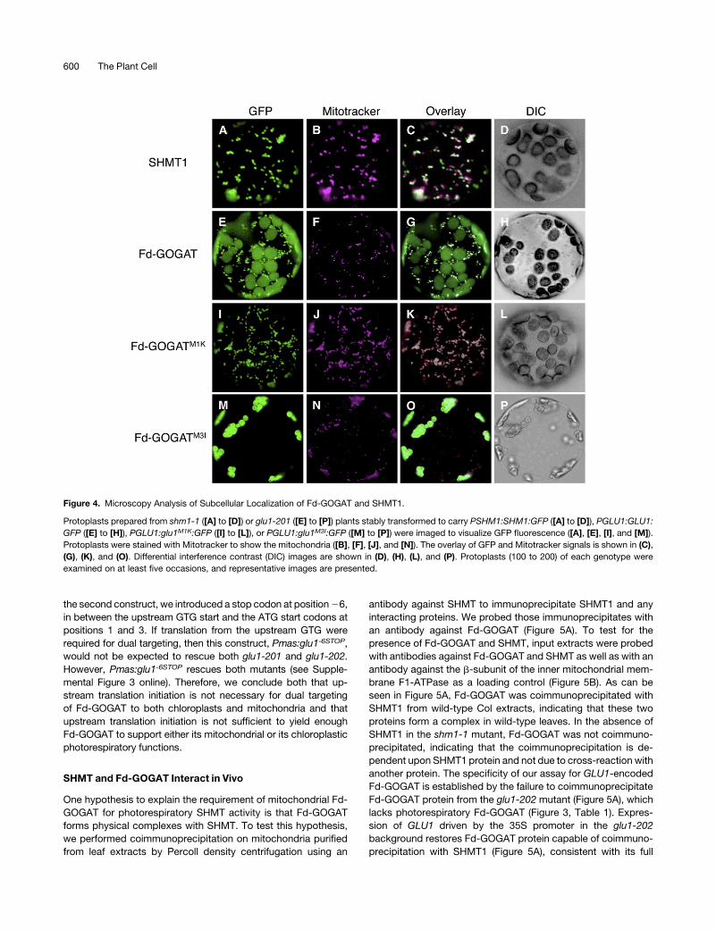
First, we made C-terminal GFP fusions to SHMT1 and to Fd-
GOGAT in constructs driven by the endogenous SHM1 and
GLU1 promoters, respectively. These transgenes were intro-
duced into shm1-1 and glu1-201 seedlings to yield stably
transformed lines. Each GFP fusion construct rescued the
photorespiratory defect of the corresponding loss-of-function
mutant. Protoplasts were generated from leaves of these res-
cued lines and analyzed by fluorescence microscopy. SHMT1-
GFP showed green punctate fluorescence (Figure 4A) that
colocalized with fluorescence from the MitoTracker Red mito-
chondrial marker (Figures 4B and 4C), confirming that SHMT1 is
targeted to the mitochondria. As expected, Fd-GOGAT-GFP
showed chloroplastic localization (cf. the GFP fluorescence
Figure 4E with the differential interference contrast image of
Figure 4H). Unexpectedly, Fd-GOGAT-GFP also showed green
punctate fluorescence (Figure 4E) that colocalized with the
MitoTracker Red mitochondrial marker (Figures 4F and 4G).
This result indicates that Fd-GOGAT is targeted to both the
chloroplasts and the mitochondria.
Second, we performed biochemical fractionation experiments
to confirm our microscopy observations. Fd-GOGAT was de-
tected by protein gel blot analysis of wild-type Col seedlings in
both chloroplastic and mitochondrial fractions (Figure 3B). By
contrast, SHMT1 was detected in mitochondrial but not in
chloroplastic fractions. As expected, the photosystem II chloro-
phyll binding protein D1 PSBA was detected in chloroplastic but
not in mitochondrial fractions (Figure 3B). These data are con-
sistent with our microscopy localizations of GFP fusion proteins.
Therefore, we conclude that Fd-GOGAT is dual targeted to both
chloroplasts and mitochondria.
An increasing number of examples have been described in
which proteins are dual targeted to both the chloroplast and
mitochondrion (Small and Peeters, 2001;Mackenzie, 2005). Dual
targeting is typically accomplished by the presence of tandem
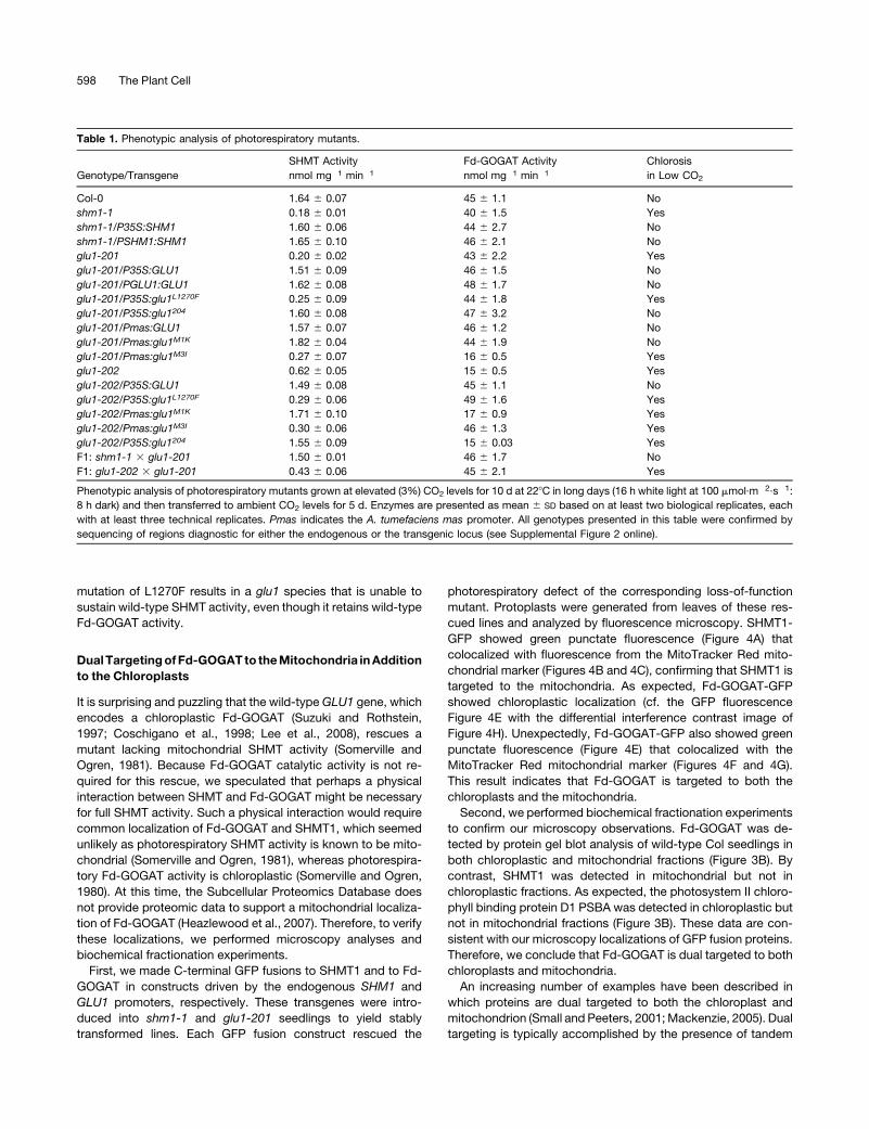
Table 1. Phenotypic analysis of photorespiratory mutants.
Genotype/Transgene
SHMT Activity
nmol mg�1 min�1
Fd-GOGAT Activity
nmol mg�1 min�1
Chlorosis
in Low CO2
Col-0 1.64 6 0.07 45 6 1.1 No
shm1-1 0.18 6 0.01 40 6 1.5 Yes
shm1-1/P35S:SHM1 1.60 6 0.06 44 6 2.7 No
shm1-1/PSHM1:SHM1 1.65 6 0.10 46 6 2.1 No
glu1-201 0.20 6 0.02 43 6 2.2 Yes
glu1-201/P35S:GLU1 1.51 6 0.09 46 6 1.5 No
glu1-201/PGLU1:GLU1 1.62 6 0.08 48 6 1.7 No
glu1-201/P35S:glu1L1270F 0.25 6 0.09 44 6 1.8 Yes
glu1-201/P35S:glu1204 1.60 6 0.08 47 6 3.2 No
glu1-201/Pmas:GLU1 1.57 6 0.07 46 6 1.2 No
glu1-201/Pmas:glu1M1K 1.82 6 0.04 44 6 1.9 No
glu1-201/Pmas:glu1M3I 0.27 6 0.07 16 6 0.5 Yes
glu1-202 0.62 6 0.05 15 6 0.5 Yes
glu1-202/P35S:GLU1 1.49 6 0.08 45 6 1.1 No
glu1-202/P35S:glu1L1270F 0.29 6 0.06 49 6 1.6 Yes
glu1-202/Pmas:glu1M1K 1.71 6 0.10 17 6 0.9 Yes
glu1-202/Pmas:glu1M3I 0.30 6 0.06 46 6 1.3 Yes
glu1-202/P35S:glu1204 1.55 6 0.09 15 6 0.03 Yes
F1: shm1-1 3 glu1-201 1.50 6 0.01 46 6 1.7 No
F1: glu1-202 3 glu1-201 0.43 6 0.06 45 6 2.1 Yes
Phenotypic analysis of photorespiratory mutants grown at elevated (3%) CO2 levels for 10 d at 228C in long days (16 h white light at 100 mmol·m�2·s�1:
8 h dark) and then transferred to ambient CO2 levels for 5 d. Enzymes are presented as mean 6 SD based on at least two biological replicates, each
with at least three technical replicates. Pmas indicates the A. tumefaciens mas promoter. All genotypes presented in this table were confirmed by
sequencing of regions diagnostic for either the endogenous or the transgenic locus (see Supplemental Figure 2 online).
598 The Plant Cell
chloroplastic and mitochondrial targeting sequences or else by
the presence of an ambiguous presequence that is recognized
by the import apparatus of both organelles. We hypothesized
that Fd-GOGAT may be dual targeted to mitochondria and
chloroplasts by use of multiple translation starts (Richter et al.,
2002). Inspection of the sequence of Fd-GOGAT revealed two
Met residues representing potential translation initiation sites at
positions 1 and 3. Computer predictions (PSORT and TargetP;
Emanuelsson et al., 2000; Bannai et al., 2002) suggested that
initiation at the first ATG codon would allow chloroplastic tar-
geting, whereas initiation at the second ATG would confer
mitochondrial targeting. Accordingly, we generated three con-
structs based on the GLU1 cDNA fused in frame to GFP and
driven by the GLU1 promoter. The first encodes simply the wild-
type Fd-GOGAT sequence (MAM) and is predicted to be tar-
geted to the chloroplast (see Supplemental Figure 3 online). The
second construct, Fd-GOGATM1K (KAM), would be translated
from the second Met and is predicted to be targeted to the
mitochondrion. The third, Fd-GOGATM3I (MAI), is predicted to be
targeted to the chloroplast. All three clones were transformed
into the glu1-201 mutant. We first tested for subcellular lo-
calization in protoplasts derived from stable transformants
carrying the three constructs. As predicted, Fd-GOGAT and
Fd-GOGATM1K show punctate fluorescence that colocalizes
with the mitochondrial marker (Figures 4E to 4L), whereas Fd-
GOGATM3I shows chloroplastic and not mitochondrial localiza-
tion (Figures 4M to 4P). The transgenic glu1-201 plants carrying
Pmas:glu1M1K and PGLU1:GLU1 are rescued to a wild-type
phenotype under photorespiratory (ambient CO2) conditions and
express wild-type levels of SHMT activity (Figures 1I and 1Q,
Table 1; see Supplemental Figures 1I and 1Q online). Similarly,
glu1M1K lacking the C-terminal GFP and driven from the Agro-
bacterium tumefaciens mannopine synthase (mas) promoter
rescued glu1-201 plants to a wild-type phenotype under photo-
respiratory (ambient CO2) conditions (Figure 1Q, Table 1; see
Supplemental Figure 1Q online). However, those glu1-201 plants
transformed with Pmas:glu1M3I remain chlorotic at ambient CO2
and have low SHMT activity (Figure 1S, Table 1; see Supple-
mental Figure 1S online). This is consistent with a requirement for
Fd-GOGAT to be targeted to the mitochondria to play its role in
expression of wild-type levels of SHMT activity. The photo-
respiratory chlorosis of glu1-202 plants was not rescued by
Pmas:glu1M1K (Figure 1R, Table 1; see Supplemental Figure 1R
online) but was rescued by Pmas:glu1M3I (Figure 1T, Table 1; see
Supplemental Figure 1T online).
Dual targeting has also been shown to result from upstream
translation from non-ATG start codons to generate a mitochon-
drial isoform, while translation from the ATG yields a chloroplas-
tic isoform (Christensen et al., 2005). Interestingly, GLU1 has an
in-frame GTG alternate initiation codon at position 217. We
tested the importance of upstream translation to Fd-GOGATdual
targeting with two further constructs (see Supplemental Figure 3
online). In one, Pmas:glu1M1KM31, we mutagenized both ATG
start codons. This construct would be expected to rescue either
glu1-201 or glu1-202 seedlings if translation from the upstream
GTG codon could yield a mitochondrial or a chloroplastic Fd-
GOGAT isoform, respectively. However, Pmas:glu1M1KM31 does
not rescue either mutant (see Supplemental Figure 3 online). In
Figure 2. Positional Cloning of glu1-201 (Originally stm, CS8010).
(A)Map position of SHM genes (on chromosomes I, IV, and V) and GLU1
(on chromosome V).
(B) Partial genetic map of chromosome V between CTR1-2 and NGA151.
(C) Structure of the Fd-GOGAT (GLU1) gene, as well as the position of
the point mutation in glu1-201 (L1270F), of a T-DNA insertion into the
second exon in glu1-202 (Salk_104286), and of the in-frame deletion
between the BglII and BamHI sites in glu1204. Exons are represented by
boxes, and introns are indicated by lines.
Figure 3. Accumulation of Fd-GOGAT and SHMT1 Protein in Whole
Seedlings and in Subcellular Fractions of Leaves.
(A) Accumulation of Fd-GOGAT and SHMT1 protein in whole extracts (20
mg of whole leaf extract per sample) prepared from wild-type and mutant
seedlings. a-Tubulin (a-TUB) serves as a loading control.
(B) Accumulation of Fd-GOGAT and SHMT1 protein in chloroplast (Cp)
and mitochondrial (Mt) fractions (20 mg per sample) of wild-type Col
seedlings. Whole seedling extracts from Col and glu1-202 are shown as
controls. b indicates a blank lane containing no extract. The photosystem
II chlorophyll binding protein D1 (PSBA) serves as a loading control. For
both (A) and (B), similar results were obtained from two experiments
using separate extracts from independently grown plants.
Fd-GOGAT Is Required for SHMT Activity 599
the second construct, we introduced a stop codon at position26,
in between the upstream GTG start and the ATG start codons at
positions 1 and 3. If translation from the upstream GTG were
required for dual targeting, then this construct, Pmas:glu1-6STOP,
would not be expected to rescue both glu1-201 and glu1-202.
However, Pmas:glu1-6STOP rescues both mutants (see Supple-
mental Figure 3 online). Therefore, we conclude both that up-
stream translation initiation is not necessary for dual targeting
of Fd-GOGAT to both chloroplasts and mitochondria and that
upstream translation initiation is not sufficient to yield enough
Fd-GOGAT to support either its mitochondrial or its chloroplastic
photorespiratory functions.
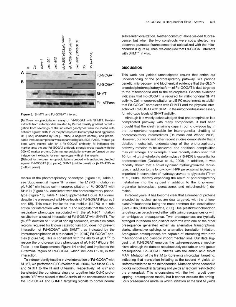
SHMT and Fd-GOGAT Interact in Vivo
One hypothesis to explain the requirement of mitochondrial Fd-
GOGAT for photorespiratory SHMT activity is that Fd-GOGAT
forms physical complexes with SHMT. To test this hypothesis,
we performed coimmunoprecipitation on mitochondria purified
from leaf extracts by Percoll density centrifugation using an
antibody against SHMT to immunoprecipitate SHMT1 and any
interacting proteins. We probed those immunoprecipitates with
an antibody against Fd-GOGAT (Figure 5A). To test for the
presence of Fd-GOGAT and SHMT, input extracts were probed
with antibodies against Fd-GOGAT and SHMT as well as with an
antibody against the b-subunit of the inner mitochondrial mem-
brane F1-ATPase as a loading control (Figure 5B). As can be
seen in Figure 5A, Fd-GOGAT was coimmunoprecipitated with
SHMT1 from wild-type Col extracts, indicating that these two
proteins form a complex in wild-type leaves. In the absence of
SHMT1 in the shm1-1 mutant, Fd-GOGAT was not coimmuno-
precipitated, indicating that the coimmunoprecipitation is de-
pendent upon SHMT1 protein and not due to cross-reaction with
another protein. The specificity of our assay for GLU1-encoded
Fd-GOGAT is established by the failure to coimmunoprecipitate
Fd-GOGAT protein from the glu1-202 mutant (Figure 5A), which
lacks photorespiratory Fd-GOGAT (Figure 3, Table 1). Expres-
sion of GLU1 driven by the 35S promoter in the glu1-202
background restores Fd-GOGAT protein capable of coimmuno-
precipitation with SHMT1 (Figure 5A), consistent with its full
Figure 4. Microscopy Analysis of Subcellular Localization of Fd-GOGAT and SHMT1.
Protoplasts prepared from shm1-1 ([A] to [D]) or glu1-201 ([E] to [P]) plants stably transformed to carry PSHM1:SHM1:GFP ([A] to [D]), PGLU1:GLU1:
GFP ([E] to [H]), PGLU1:glu1M1K:GFP ([I] to [L]), or PGLU1:glu1M3I:GFP ([M] to [P]) were imaged to visualize GFP fluorescence ([A], [E], [I], and [M]).
Protoplasts were stained with Mitotracker to show the mitochondria ([B], [F], [J], and [N]). The overlay of GFP and Mitotracker signals is shown in (C),
(G), (K), and (O). Differential interference contrast (DIC) images are shown in (D), (H), (L), and (P). Protoplasts (100 to 200) of each genotype were
examined on at least five occasions, and representative images are presented.
600 The Plant Cell
rescue of the photorespiratory phenotype (Figure 1H, Table 1;
see Supplemental Figure 1H online). The L1270F mutation in
glu1-201 eliminates coimmunoprecipitation of Fd-GOGAT with
SHMT1 (Figure 5A), consistent with the photorespiratory pheno-
type (Figure 1C, Table 1; see Supplemental Figure 1C online),
despite the presence of wild-type levels of Fd-GOGAT (Figures 3
and 5B). This result implicates this residue (L1270) in a role
critical for interaction with SHMT1 and suggests that the photo-
respiratory phenotype associated with the glu1-201 mutation
results from a loss of interaction of Fd-GOGAT with SHMT1. The
glu1204 deletion of;2 kb of coding sequence, which eliminates
regions required for known catalytic function, does not prevent
interaction of Fd-GOGAT with SHMT1, as indicated by the
immunoprecipitation of a truncated (;100 kD) Fd-GOGAT spe-
cies (Figure 5A). This is consistent with the ability of glu1204 to
rescue the photorespiratory phenotype of glu1-201 (Figure 1N,
Table 1; see Supplemental Figure 1N online) and implicates the
C-terminal region of Fd-GOGAT, which includes L1270, in that
interaction.
To independently test the in vivo interaction of Fd-GOGATwith
SHMT, we performed BiFC (Walter et al., 2004). We fused GLU1
and SHM1 to the N and C termini, respectively, of YFP and
transfected the constructs singly or together into Col-0 proto-
plasts. YFP was placed at the C termini of the constructs to allow
the Fd-GOGAT and SHMT1 targeting signals to confer normal
subcellular localization. Neither construct alone yielded fluores-
cence, but when the two constructs were cotransfected, we
observed punctate fluorescence that colocalized with the mito-
chondria (Figure 6). Thus, we conclude that Fd-GOGAT interacts
with SHMT1 in vivo.
DISCUSSION
This work has yielded unanticipated results that enrich our
understanding of the photorespiratory pathway. We provide
genetic, microscopy, and biochemical evidence that the GLU1-
encoded photorespiratory isoform of Fd-GOGAT is dual targeted
to the mitochondria and to the chloroplasts. Genetic evidence
indicates that Fd-GOGAT is required for mitochondrial SHMT
activity. Coimmunoprecipitation and BiFC experiments establish
that Fd-GOGAT complexes with SHMT1 and the physical inter-
action of Fd-GOGATwith SHMT in themitochondria is necessary
for wild-type levels of SHMT activity.
Although it is widely acknowledged that photorespiration is a
complicated pathway with many components, it had been
thought that the chief remaining gaps in our knowledge lay in
the transporters responsible for interorganellar shuttling of
photorespiratory intermediates (Reumann and Weber, 2006).
However, our work and other recent studies demonstrate that a
detailed mechanistic understanding of the photorespiratory
pathway remains to be achieved, and additional complexities
may yet emerge. For example, it was recently established that
10-formyl tetrahydrofolate deformylase (10-FDF) is essential for
photorespiration (Collakova et al., 2008). In addition, it was
recently shown that a novel cytosolic hydroxypyruvate reduc-
tase, in addition to the long-known HPR peroxisomal isoform, is
important in conversion of hydroxypyruvate to glycerate (Timm
et al., 2008), thereby expanding the realm of photorespiratory
metabolism into the cytosol in addition to the long-known
organellar (chloroplast, peroxisome, and mitochondrion) do-
mains.
In recent years, it has become clear that a number of proteins
encoded by nuclear genes are dual targeted, with the chloro-
plasts/mitochondria being the most common dual destinations
(Silva-Filho, 2003;Mackenzie, 2005). Dual plastid/mitochondrion
targeting can be achieved either with twin presequences or with
an ambiguous presequence. Twin presequences are typically
arranged in tandem and distinct isoforms with one or the other
presequence generated either via alternative transcriptional
starts, alternative splicing, or alternative translation initiation.
Ambiguous presequences are capable of interacting with both
mitochondrial and plastidic import mechanisms. Our data sug-
gest that Fd-GOGAT employs the twin-presequence mecha-
nism, although the data do not absolutely exclude an ambiguous
presequence. Fd-GOGAT initiates with the amino acid triplet
MAM. Mutation of the first M to K prevents chloroplast targeting,
indicating that translation initiating at the second M yields an
isoform restricted to the mitochondria. Mutation of the secondM
blocksmitochondrial targeting and yields an isoform restricted to
the chloroplast. This is consistent with the twin, albeit over-
lapping, presequence model, but it cannot exclude the ambig-
uous presequence model in which initiation at the first M yields
Figure 5. SHMT1 and Fd-GOGAT Interact.
(A) Coimmunoprecipitation assay of Fd-GOGAT with SHMT1. Protein
extracts from mitochondria isolated by Percoll density gradient centrifu-
gation from seedlings of the indicated genotypes were incubated with
antisera against SHMT1 or the photosystem II chlorophyll binding protein
D1 (PsbA) (indicated by Col [a-PsbA], a negative control), and precip-
itated immunocomplexes were separated by 8% SDS-PAGE. Protein gel
blots were stained with an a-Fd-GOGAT antibody. M indicates the
marker lane; the anti-Fd-GOGAT antibody strongly cross-reacts with the
200-kD marker protein. Coimmunoprecipitations were performed on two
independent extracts for each genotype with similar results.
(B) Input for the coimmunoprecipitations probed with antibodies directed
against Fd-GOGAT (top panel), SHMT (middle panel), or b- F1-ATPase
(bottom panel).
Fd-GOGAT Is Required for SHMT Activity 601
an isoform that interacts with both targeting machineries. In this
model, mutation of the second M would disrupt the mitochon-
drial targeting sequence but leave the chloroplast targeting
sequence intact. Translation would not normally occur at the
secondM, but mutation of the first Mwould yield a transcript that
could be translated from the second M. The distinction between
the twin and ambiguous presequence models is that translation
from the secondMwould be integral to the former and only occur
as an artifact of our experimental mutation of the first M in the
latter. Our data do not allow us to distinguish between these two
alternatives.
Why might the mitochondrial fraction of Fd-GOGAT have
escaped detection for so long? Fd-GOGAT is well established
as a chloroplastic enzyme (Lea and Miflin, 1974), and it is highly
abundant, representing;1%of total leaf protein (Marquez et al.,
1988; Pajuelo et al., 1997). Thus, it would have been easy to
attribute any Fd-GOGAT identified in other compartments, in-
cluding the mitochondria, to contamination from the chloroplast
fraction. In this regard, it is interesting to note that while pea
(Pisum sativum) leaf mitochondrial SHMT purifies as a 220-kD
homotetramer (Bourguignon et al., 1988; Turner et al., 1992), in
less pure fractions, SHMT activity can be resolved into two
distinct fractions (Turner et al., 1992). Thus, it is possible that a
complex of SHMT and Fd-GOGAT might have been seen in
earlier preparations but that the Fd-GOGAT/SHMT1 complex
was lost in purification.
Intriguingly, GLN2-encoded GS-2 was recently suggested to
be dual targeted to the mitochondria in addition to the chloro-
plasts (Taira et al., 2005). This raises the possibility that ammo-
nia, which is generated by GDC activity in the mitochondria
(Douce et al., 2001; Bauwe and Kolukisaoglu, 2003), may be
reassimilated in the mitochondria (Linka and Weber, 2005; Taira
et al., 2005). However, GS is depleted from mitochondrial frac-
tions highly purified by free-flow electrophoresis (FFE) relative to
pre-FFE mitochondrial fractions, suggesting that it was primarily
detected as a plastidic contaminant of incompletely purified
mitochondrial fractions (Eubel et al., 2007). FFE provides a
second dimension of organellar resolution based on surface
charge, thus augmenting conventional separations based on
centrifugation to resolve by size and density. Although we
detected Fd-GOGAT in mitochondria purified by Percoll density
gradient centrifugation (Figure 3B), FFE-purified Col-0 shoot
mitochondria and Landsberg erecta tissue culture mitochondria
do not contain sufficient amounts of Fd-GOGAT to be detected
by mass spectrometry or by protein gel blots using Fd-GOGAT
antibodies (H. Millar and C.P. Lee, personal communication).
One interpretation of this failure to find Fd-GOGAT by proteomic
techniques is that Fd-GOGAT in the mitochondrial fraction
results fromplastidic contamination. However, this interpretation
is inconsistent with our evidence supporting a mitochondrial
fraction of Fd-GOGAT based on multiple independent experi-
mental approaches (genetic, biochemical, and microscopy ev-
idence). We offer two possible explanations to reconcile these
different observations. One possibility is that mitochondrial Fd-
GOGAT is scarce, which would be consistent with a catalytic
rather than a stoichiometric role of Fd-GOGAT inmitochondria. A
second possibility is that Fd-GOGAT is present in a subset of
a heterogeneous mitochondrial population and that this Fd-
GOGAT-containing subset is lost during FFE purification. For
example, distinct yeast mitochondrial populations originating
from different biological conditions or experimental manipula-
tions can be resolved by FFE and as little as deletion of a single
outer-membrane protein can yield a mitochondrial fraction of
distinct mobility in FFE (Zischka et al., 2006).
The ability of glu1204, an internally deleted Fd-GOGAT isoform
lacking the catalytic region of the protein, to rescue the photo-
respiratory SHMT activity of a glu-201 mutant (Figure 1N, Table
1; see Supplemental Figure 1N online) demonstrates that the
mitochondrial role does not require Fd-GOGAT catalytic activity.
Fd-GOGAT is a large four-domain protein, although no function
has been attributed to the C-terminal domain (Binda et al., 2000;
van den Heuvel et al., 2002). The glu1-201 mutation (L1270F)
confers the loss of photorespiratory SHMT activity (Figure 1C,
Table 1; see Supplemental Figure 1C online), presumably by
preventing the interaction of Fd-GOGAT with SHMT1. This result
assigns an important photorespiratory function, the mediation
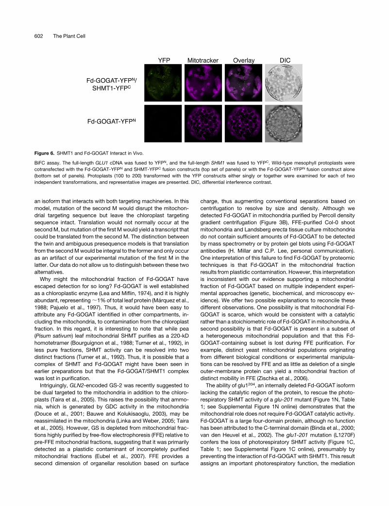
Figure 6. SHMT1 and Fd-GOGAT Interact in Vivo.
BiFC assay. The full-length GLU1 cDNA was fused to YFPN, and the full-length SHM1 was fused to YFPC. Wild-type mesophyll protoplasts were
cotransfected with the Fd-GOGAT-YFPN and SHMT-YFPC fusion constructs (top set of panels) or with the Fd-GOGAT-YFPN fusion construct alone
(bottom set of panels). Protoplasts (100 to 200) transformed with the YFP constructs either singly or together were examined for each of two
independent transformations, and representative images are presented. DIC, differential interference contrast.
602 The Plant Cell
of the interaction between Fd-GOGAT and SHMT1, to the
C-terminal domain of Fd-GOGAT.
The mechanistic function of the SHMT-Fd-GOGAT interaction
remains speculative. The binding of Fd-GOGAT is not necessary
to stabilize SHMT1protein becauseSHMT1protein levels are not
reduced in the glu1-201 or glu1-202 mutants (Table 1). In vitro
SHMT activity is not dependent on Fd-GOGAT, so this require-
ment seems to be restricted to the in vivo situation. One model
for a noncatalytic role of Fd-GOGAT in a complex with SHMT is
as an in vivo regulatory subunit whose function is independent of
Fd-GOGAT activity. O-acetylserine(thiol)-lyase has been shown
to play such a regulatory role in the Cys synthase complex with
serine acetyltransferase (SAT). Although it was proposed that
this complex promotes metabolic channeling (Winkel, 2004), it
was demonstrated using recombinantly produced enzymes that
O-acetylserine(thiol)-lyase binds to SAT as a catalytically inactive
enhancer of SAT activity (Ruffet et al., 1994; Droux et al., 1998).
There is precedent for Fd-GOGAT playing a role independent of
catalytic activity. Fd-GOGAT participates in a large multimeric
complex with UDP-sulfoquinovose synthase (SQD1) (Shimojima
et al., 2005). This role apparently does not require glutamate
synthase catalytic activity of Fd-GOGAT but is thought to rely on
an FMN cofactor that binds sulfite and may channel it to SGD1.
Nonetheless, we do not favor this model because the low
abundance of Fd-GOGAT in the mitochondrion suggests that
only a small fraction of the SHMT is likely to be complexed with
Fd-GOGAT.
Wepropose a catalytic role for Fd-GOGAT in themitochondria,
which would be consistent with an essential function for an
inabundant protein. What catalytic role, independent of GOGAT
activity, might Fd-GOGAT bound to SHMT1 play? It has been
known for some years that SHMT plays a second catalytic role,
the irreversible hydrolysis of 5,10-methenyl tetrahydrofolate
(5,10-CH=THF) to 5-formyl tetrahydrofolate (5-CHO-THF), which
is a slow tight binding inhibitor of SHMT (Stover and Schirch,
1993). It has been recently established that two 10-FDF oppose
the accumulation of 5-CHO-THF, and a double loss-of-function
mutant confers a photorespiratory phenotype via inhibition of
SHMT and GDC activities, confirming the importance of this
inhibitor in vivo (Collakova et al., 2008). 5-CHO-THF is metabo-
lized back to 5,10-CH=THF by 5-formyltetrahydrofolate cyclo-
ligase, encoded in Arabidopsis by a single 5-FCL gene (Roje
et al., 2002). However, the loss of 5-FCL activity does not
dramatically reduce plant growth under photorespiratory condi-
tions, despite the accumulation of 5-CHO-THF (Goyer et al.,
2005). No compensatory increase in mitochondrial SHMT activ-
ity could be detected in the 5-FCL mutants, suggesting some
other mechanism to tolerate elevated 5-CHO-THF (Goyer et al.,
2005).We speculate that the role of Fd-GOGAT bound to SHMT1
may be to reduce the sensitivity of SHMT activity to 5-CHO-THF
or to oppose the accumulation of 5-CHO-THF, possibly by
reducing the hydrolysis of of 5,10-CH=THF by SHMT. This would
be consistent with the loss of SHMT activity in glu1-201 despite
the retention of wild-type levels of SHMT protein.
While all primary enzymes and several of the transporters
required for a functional C2 carbon oxidative cycle have now
been identified, the resulting parts list apparently does not yet
permit one to draw a complete picture of this crucial high flux
pathway in photosynthetic plant cells. As we and others have
shown, a functional pathway requires more than the sum of its
parts and, in addition, more parts than previously anticipated
(e.g., Collakova et al., 2008). Protein–protein interactions and
complex formation are required for mitochondrial SHMT activity,
and this requires mitochondrial targeting of an enzyme, Fd-
GOGAT, that was hitherto believed to be confined to chloro-
plasts. We conclude that this crucial high flux pathway is more
complicated than anticipated and remains incompletely under-
stood.
METHODS
Plant Growth
Seeds of the glu1-201 mutant (CS8010) and of the Salk T-DNA-Insertion
line Salk_104286, glu1-202, were obtained from the ABRC (Ohio State
University, Columbus, OH). Seeds were grown on soil in high (3%) CO2
growth chambers (Biochambers) at 228C in long days (16 h white light at
100 mmol·m22·s21: 8 h dark). For mapping, seeds were vapor sterilized
(Clough and Bent, 1998) and germinated at room temperature in a 12-h-
light/12-h-dark cycle on half-strength Murashige and Skoog medium
(Murashige and Skoog, 1962) in 3% CO2 in white light at a photon flux
density of;100mmol·m22·s21. Plantlets were transferred to soil after the
first four primary leaves had emerged and grown at 228C in long days (16 h
white light at 100 mmol·m22·s21: 8 h dark) at ambient CO2 to score
photorespiratory chlorosis. The plants were returned to the 3% CO2
growth chamber and the growth cycle was completed.
Mapping of the glu1-201 Locus
The homozygous recessive mutant glu1-201 (Col gl1) was crossed to
Landsberg erecta. The F1 progenywere allowed to self-fertilize, and in the
F2 generation cosegregation of the photorespiratory (chlorotic at ambient
CO2) phenotype with molecular markers was determined as described
(Lukowitz et al., 2000; Jander et al., 2002).
Assays of Enzyme Activity
Crude extracts for measuring SHMT activity were prepared by grinding
400 mg fresh leaf tissue at 48C in 300 mL of extraction buffer (50 mM
phosphate buffer, pH 7.5, 1 mM 2-mercaptoethanol, and 2.5 mM EDTA).
Extracts for measuring Fd-GOGAT activity were prepared by grinding 2 g
fresh leaves at 48C in 1mL of extraction buffer (50 mMHEPES, pH 7.5, 15
mM KCl, 1 mM EDTA, 1 mM DTT, and 1 mM PMSF). The extracts were
cleared by centrifugation at 20,000g for 10 min. SHMT activity was
assayed by the incorporation of radioactivity from L-[3-3H] serine into
5,10-methylene-tetrahydrofolate (Geller and Kotb, 1989) as modified by
Voll et al. (2006). Fd-GOGAT activity was determined spectrophotomet-
rically by following the glutamine-dependent oxidation of NADPH at 340
nm (Meers et al., 1970; Misra and Oaks, 1986). The reaction mixture
contained 50 mM HEPES buffer, pH 8.5, 1% (v/v) 2-mercaptoethanol,
3.65 mM glutamine, 3 mM 2-oxoglutarate, 0.2 mM NADPH, 4 mM
ferredoxin, and 0.2 mL plant extract in a final volume of 1 mL.
Plasmid Construction
To generate full-length cDNA clones, RNA was extracted from leaves of
Col-0 and the glu1-201 mutant using the RNeasy Plant Mini Kit (Qiagen).
Amplification of GLU1 and glu1-201 cDNAs was performed using high-
fidelity Taq polymerase (Promega) in three separate fragments with
specific primers. We exploited restriction sites in the genomic sequence.
Fd-GOGAT Is Required for SHMT Activity 603
The primers GOGAT start (59-ATGGCGATGCAATCTCTTT-39) and
Rev1XhoI (59-TACCAAATGGACGAATTT-39) were used to amplify an 1120-
bp fragment including the XhoI site at the position 1055. The second
fragment between the XhoI and BamHI restriction sites was amplified
using For1XhoI (59-GAGGTTTCTTGGACATAACG-39) and Rev2BamHI
(59-GTAGTGTTGGTGAATATCCA-39). The third fragment was amplified
using For2BamHI (59-GGAATGTCACTTGGTGCTAT-39) and Rev3
(59-CTAAGCCGATTGAAATGTGA-39).PCR-amplifiedproductswerecloned
in the TA-cloning system (Promega). After appropriate restriction endo-
nuclease digestion, the three resulting fragments, EcoRI-XhoI, XhoI-
BamHI, and BamHI-NotI were isolated from agarose gels using the Gel
Extraction Kit (Qiagen) and cloned into the appropriate sites in pENTR1A
(Invitrogen). All clones were completely sequenced to ensure that no
mutations had been introduced.
A 10-kb fragment ofGLU1 genomicDNA,which includes 1505bp of the
promoter region, was amplified from BAC clone F21E1 using a two-step
PCR reaction and TaKaRa EX Taq polymerase (PanVera). The first
product was generated with the specific primers GLU1B1 (59-AAAAAG-
CAGGCTcccgatgcatgcatgtttatctt-39) and GLU1B2 (59-AGAAAGCTGGGT-
ctgagcgatatgaagtg-39) containing 12 nucleotides of attB sites (capitals)
and gene-specific nucleotides (lower case). The resulting product was
subjected to the second PCR step with attB adapter primers attB1
(59-GGGGACAAGTTTGTACAAAAAAGCAGGCT-39) andattB2 (59-GGGGA-
CCACTTTGTA CAAGAAAGCTGGGT-39). The 10-kb fragment was cloned
in pDONR207 by BP clonase (Invitrogen) and then transferred to the
destination vector using LR Clonase.
To generate C-terminal fusion proteins, the GLU1 and SHM1 genomic
sequences lacking stop codons were amplified and recombined into
pDONR207 (Invitrogen) and then into pMDC110 to fuse with GFP (Curtis
and Grossniklaus, 2003). This clone was sequenced to confirm that the
fusions were in frame.
To generate overexpression lines, theGLU1 gene was transferred from
pENTR1A, using LR Clonase (Invitrogen), to the modified binary vector
pB7WG2D (Karimi et al., 2002) in which the 1060-bp 35S promoter was
replaced by an ;800-bp 35S promoter from pMDC32 (Curtis and
Grossniklaus, 2003) or by the mas promoter from 35SpBARN (LeClere
and Bartel, 2001).
The glu1204 allele, which encodes a truncated Fd-GOGAT protein
lacking the catalytic region, was made by deleting ;2 kb of coding
sequence between BglII and BamHI restriction sites, the resulting plas-
mid was moved to destination vector pB7WG2D.
Site-directed mutagenesis was used to create glu1M3I, encoding a
chloroplast-targeted Fd-GOGAT, and glu1M1K, encoding amitochondrion-
targeted Fd-GOGAT. A GLU1 promoter fragment including the first
12 amino acids of the coding sequence and the HindIII site was PCR
amplified (HindIII I M3I: 59-GAGAAGCTTAGGAACAGGGGAAAGAGA-
TTGGATCGCCAT-39 andGLU1P SalI:59-GAGGTCGACGCGTAAATTCA-
CATATT-39) and (HindIII M1K: 59-GAGAAGCTTAGGAACAGGGGAAA-
GAGATTGCATCGCCTT-39 and GLU1P SalI) to introduce the mutations
(underlined in the primers) and appropriate restriction sites and then
cloned in pENTR1A:GLU1 using the appropriate restriction enzymes.
For plant transformation, plasmidswere introduced intoAgrobacterium
tumefaciens strain AGL1 and then into wild-type, glu1-201, or glu1-202
mutant plants by vacuum infiltration (Clough and Bent, 1998). Transform-
ants were selected on agar plates containing either 30 mg/mL Basta or 15
mg/mL hygromycin. Resistant seedlings were allowed to self, and T2
seeds were collected. Several lines (at least four) for each construct and
genetic background were analyzed.
Protein Gel Blot Analysis
Total leaf protein was extracted from ;300 mg of ground frozen tissue
mixed with 400 mL of extraction buffer containing 4 M urea, 2.5% SDS,
20% glycerol, 20 mM Tris, pH 6.8, 1 mM EDTA, 1 mM PMSF, and 0.2%
Halt Protease Inhibitor (Pierce). Mitochondria and chloroplasts were
isolated and purified by Percoll density gradient centrifugation (Bergman
et al., 1980). Samples (20 mg protein per lane) were separated by SDS-
PAGE (Laemmli, 1970) and transferred to PVDF membrane (Millipore).
Membranes were blocked for 2 h at room temperature with blocking
buffer (5% powdered milk, 1% PBS, pH 7.4, and 0.05% Tween 20).
Membranes were incubated for at least 4 h with primary rabbit polyclonal
antibodies against SHMT (Agrisera), Fd-GOGAT (Agrisera), or PSBA
(Agrisera) or mouse monoclonal antibodies against a-tubulin (Sigma-
Aldrich) or the b-subunit of maize (Zea mays) F1-ATPase (Luethy et al.,
1993). Excess antibodies were removed by washing the blot twice for 15
min in 1% milk, 13 PBS, and 0.1% Tween 20 and twice for 15 min in 1%
PBST. Membranes were incubated with secondary antibody, either goat
anti-rabbit IgG conjugated to horseradish peroxidase (Santa Cruz Bio-
technology) or goat anti-mouse IgG conjugated to horseradish peroxi-
dase (Bio-Rad) followed by washing as described above. Signals were
detected using SuperSignal West Pico Chemiluminescent Substrate
(Pierce) according to the manufacturer’s instructions.
Coimmunoprecipitation
Protein extracts were prepared by grinding 2 g fresh leaves at 48C in 1mL
of lysis buffer (50 mM Tris, pH 7.5, 150 mM KCl, 1 mM EDTA, 1 mM DTT,
0.1% Triton X-100, 1 mM PMSF, and Halt Protease Inhibitor Cocktail
[Pierce]) (Shao et al., 2003). Extracts were pretreated with protein A
agarose suspension (Calbiochem), cleared by centrifugation, and then
incubated with anti-SHMT at 48C for 2 h. Immunocomplexes were
incubated with prewashed protein A agarose for 1 h at 48C, collected
by centrifugation, washed six times with 1 mL lysis buffer, suspended in
50 mL of 13 SDS loading buffer and boiled for 5 min. Twenty-five
microliters of this solution was subjected to 8% SDS-PAGE electropho-
resis. Twenty-five micrograms of total Col lysate was loaded on the same
gel. Immunoblots to PVDF membrane were probed with anti-Fd-GOGAT
as described above.
BiFC
Full-length GLU1 and SHM1 cDNAs were fused in frame to the N and C
termini, respectively, of YFP using vectors pSPYNE-35S/pUC-SPYNE
and pSPYCE-35S/pUC-SPYCE (Walter et al., 2004). For BiFC, the fusion
constructs were transfected together or singly by the polyethylene glycol
method (Yoo et al., 2007) into wild-type Col protoplasts (Jamai et al.,
1996). Briefly, leaves were cut into fine strips and incubated for 90 min at
288C in 10 mL of protoplast medium containing 500 mM sorbitol, 1 mM
CaCl2, 10mMMES, pH 5.5, 0.5 mM polyvinylpolypyrrolidone, 0.1%BSA,
1.5% cellulase R10 (Yakult Honsha), and 0.1% macerozyme R10 (Yakult
Honsha). Protoplasts were harvested by centrifugation at 100g for 2 min
in protoplast medium without enzymes. For fluorescence detection,
excitation was at 488 nm, and the fluorescence emission signal was
collected between 498 and 561 nm. To visualize mitochondria, proto-
plasts were stained with 40 mMMitoTracker Red (Molecular Probes) with
excitation at 568 nm and emission signal collected at 579 to 687 nm.
Subcellular Localization of SHMT1 and Fd-GOGAT
Protoplasts from 2-week-old seedlings from stable transgenic lines were
isolated as described (Jamai et al., 1996). Subcellular localization of the
fusion proteins was determined by fluorescence microscopy as de-
scribed above.
Accession Numbers
Sequence data from this article can be found in the Arabidopsis Genome
Initiative or GenBank/EMBL databases under the following accession
604 The Plant Cell
numbers: 5-FCL (At5g13050), 10-FDF (At4g17360 and At5g47435),
GL1 (At3g279920), GLN2 (At5g35630), GLU1 (At5g04140.1), SHM1
(At4g37930), and SQD1 (At4g33030).
Supplemental Data
The following materials are available in the online version of this article.
Supplemental Figure 1. Phenotypic Analysis of Photorespiratory
Mutants at Ambient CO2.
Supplemental Figure 2. Genotyping of Mutants and Transgenic
Complementation Lines.
Supplemental Figure 3. Phenotypic Analysis of Ability of Constructs
Bearing Various glu1Mutations to Rescue Growth of Photorespiratory
Mutants, glu1-201 and glu1-202, at Ambient CO2.
ACKNOWLEDGMENTS
We acknowledge the ABRC (Ohio State University, Columbus, OH) for
seed stocks and DNA clones and SIGnAL (Salk Institute, La Jolla, CA)
for providing the sequence-indexed Arabidopsis T-DNA insertion mu-
tants. We thank Ann M. Lavanway and Joohyun Lee for their help with
the microscopy. This work was supported by an American Society of
Plant Biologists Summer Undergraduate Research Fellowship to S.H.S.
and by National Science Foundation grants to C.R.M.
Received September 15, 2008; revised January 26, 2009; accepted
February 3, 2009; published February 17, 2009.
REFERENCES
Bannai, H., Tamada, Y., Maruyama, O., Nakai, K., and Miyano, S.
(2002). Extensive feature detection of N-terminal protein sorting
signals. Bioinformatics 18: 298–305.
Bauwe, H., and Kolukisaoglu, U. (2003). Genetic manipulation of
glycine decarboxylation. J. Exp. Bot. 54: 1523–1535.
Bergman, A., Gardestrom, P., and Ericson, I. (1980). Method to obtain
a chlorophyll-free preparation of intact mitochondria from spinach
leaves. Plant Physiol. 66: 442–445.
Binda, C., Bossi, R.T., Wakatsuki, S., Arzt, S., Coda, A., Curti, B.,
Vanoni, M.A., and Mattevi, A. (2000). Cross-talk and ammonia
channeling between active centers in the unexpected domain ar-
rangement of glutamate synthase. Structure 8: 1299–1308.
Boldt, R., Edner, C., Kolukisaoglu, U., Hageman, M., Weckworth, W.,
Wienkoop, S., Morgenthal, K., and Bauwe, H. (2005). D-Glycerate
3-kinase, the last unknown enzyme in the photorespiratory cycle in
Arabidopsis, belongs to a novel kinase family. Plant Cell 17: 2413–
2420.
Bourguignon, J., Neuburger, M., and Douce, R. (1988). Resolution
and characterization of the glycine-cleavage reaction in pea leaf
mitochondria. Biochem. J. 255: 169–178.
Bowes, G., Ogren, W.L., and Hageman, R.H. (1971). Phosphoglyco-
late production catalyzed by ribulose diphosphate carboxylase. Bio-
chem. Biophys. Res. Commun. 45: 716–722.
Christensen, A.C., Lyznik, A., Mohammed, S., Elowsky, C.G., Elo, A.,
Yule, R., and Mackenzie, S.A. (2005). Dual-domain, dual-targeting
organellar protein presequences in Arabidopsis can use non-AUG
start codons. Plant Cell 17: 2805–2816.
Clough, S.J., and Bent, A.F. (1998). Floral dip: A simplified method
for Agrobacterium-mediated transformation of Arabidopsis thaliana.
Plant J. 16: 735–743.
Collakova, E., Goyer, A., Naponelli, V., Krassovskaya, I., Gregory,
J.F.I., Hanson, A.D., and Shachar-Hill, Y. (2008). Arabidopsis 10-
formyl tetrahydrofolate deformylases are essential for photorespira-
tion. Plant Cell 20: 1818–1832.
Coschigano, K.T., Melo-Oliveira, R., Lim, J., and Coruzzi, G.M.
(1998). Arabidopsis gls mutants and distinct Fd-GOGAT genes: im-
plications for photorespiration and primary nitrogen assimilation. Plant
Cell 10: 741–752.
Curtis, M.D., and Grossniklaus, U. (2003). A Gateway cloning vector
set for high-throughput functional analysis of genes in planta. Plant
Physiol. 133: 462–469.
Douce, R., Bourguignon, J., Neuburger, M., and Rebeille, F. (2001).
The glycine decarboxylase system: A fascinating complex. Trends
Plant Sci. 6: 167–176.
Douce, R., and Neuburger, M. (1999). Biochemical dissection of
photorespiration. Curr. Opin. Plant Biol. 2: 214–222.
Droux, M., Ruffet, M.L., Douce, R., and Job, D. (1998). Interactions
between serine acetyl-transferase and O-acetylserine (thiol) lyase in
higher plants. Structural and kinetic properties of the free and
boundzymes. Eur. J. Biochem. 255: 235–245.
Emanuelsson, O., Nielsen, H., Brunak, S., and von Heijne, G. (2000).
Predicting subcellular localization of proteins based on their N-terminal
amino acid sequence. J. Mol. Biol. 300: 1005–1016.
Eubel, H., Lee, C.P., Kuo, J., Meyer, E.H., Taylor, N.L., and Millar,
A.H. (2007). Free-flow electrophoresis for purification of plant mito-
chondria by surface charge. Plant J. 52: 583–594.
Geller, A.M., and Kotb, M.Y. (1989). A binding assay for serine
hydroxymethyltransferase. Anal. Biochem. 180: 120–125.
Goyer, A., Collakova, E., de la Garza, R.D., Quinlivan, E.P.,
Williamson, J., Gregory, J.F., Shachar-Hill, Y., and Hanson, A.D.
(2005). 5-Formyltetrahydrofolate is an inhibitory but well tolerated
metabolite in Arabidopsis leaves. J. Biol. Chem. 280: 26137–26142.
Heazlewood, J.L., Verboom, R.E., Tonti-Filippini, J., Small, I., and
Millar, A.H. (2007). SUBA: The Arabidopsis Subcellular Database.
Nucleic Acids Res. 35: D213–D218.
Jamai, A., Tommasini, R., Martinoia, E., and Delrot, S. (1996).
Characterization of glutathione uptake in broad bean leaf protoplasts.
Plant Physiol. 111: 1145–1152.
Jander, G., Norris, S.R., Rounsley, S.D., Bush, D.F., Levin, I.M., and
Last, R.L. (2002). Arabidopsis map-based cloning in the post-genome
era. Plant Physiol. 129: 440–450.
Karimi, M., Inze, D., and Depicker, A. (2002). GATEWAY vectors for
Agrobacterium-mediated plant transformation. Trends Plant Sci. 7:
193–195.
Keys, A.J., Bird, I.F., Cornelius, M.J., Lea, P.J., Wallsgrove, R.M., and
Miflin, B.J. (1978). Photorespiratory nitrogen cycle. Nature 275: 741–743.
Kozaki, A., and Takeba, G. (1996). Photorespiration protects C3 plants
from photooxidation. Nature 384: 557–560.
Laemmli, U.K. (1970). Cleavage of structural proteins during the as-
sembly of the head of bacteriophage T4. Nature 227: 680–685.
Lea, P.J., and Miflin, B.J. (1974). An alternative route for nitrogen
assimilation in higher plants. Nature 251: 614–616.
LeClere, S., and Bartel, B. (2001). A library of Arabidopsis 35S-cDNA
lines for identifying novel mutants. Plant Mol. Biol. 46: 695–703.
Lee, D.W., Kim, J.K., Lee, S., Choi, S., Kim, S., and Hwang, I. (2008).
Arabidopsis nuclear-encoded plastid transit peptides contain multiple
sequence subgroups with distinctive chloroplast-targeting sequence
motifs. Plant Cell 20: 1603–1622.
Leegood, R.C., Lea, P.J., Adcock, M.D., and Hausler, R.E. (1995). The
regulation and control of photorespiration. J. Exp. Bot. 46: 1397–1414.
Linka, M., and Weber, A.P.M. (2005). Shuffling ammonia between
mitochondria and plastids during photorespiration. Trends Plant Sci.
10: 461–465.
Fd-GOGAT Is Required for SHMT Activity 605
Luethy, M.H., Horak, A., and Elthon, T.E. (1993). Monoclonal anti-
bodies to the a- and b-subunits of the plant mitochondrial F1-ATPase.
Plant Physiol. 101: 931–937.
Lukowitz, W., Gillmor, C.S., and Scheible, W.-R. (2000). Positional
cloning in Arabidopsis. Why it feels good to have a genome initiative
working for you. Plant Physiol. 123: 795–806.
Mackenzie, S.A. (2005). Plant organellar protein targeting: a traffic plan
still under construction. Trends Cell Biol. 15: 548–554.
Marquez, A.J., Avila, C., Forde, B.G., and Wallsgrove, R.M. (1988).
Ferredoxin-glutamate synthase from barley leaves: Rapid purification
and partial characterization. Plant Physiol. Biochem. 43: 645–651.
McClung, C.R., Hsu, M., Painter, J.E., Gagne, J.M., Karlsberg, S.D.,
and Salome, P.A. (2000). Integrated temporal regulation of the
photorespiratory pathway: Circadian regulation of two Arabidopsis
genes encoding serine hydroxymethyltransferase. Plant Physiol. 123:
381–392.
Meers, J.L., Tempest, D.W., and Brown, C.M. (1970). Glutamine
(amide): 2-Oxoglutarate amino transferase oxidoreductase (NADP), an
enzyme involved in the synthesis of glutamate by some bacteria.
J. Gen. Microbiol. 64: 187–194.
Misra, S., and Oaks, A. (1986). Ferredoxin and pyridine nucleotide-
dependent glutamate synthase activities in maize endosperm. Plant
Sci. 39: 1–5.
Murashige, T.R., and Skoog, F. (1962). A revised medium for rapid
growth and bioassays with tobacco tissue culture. Physiol. Plant. 15:
473–497.
Ogren, W.L., and Bowes, G. (1971). Ribulose diphosphate carboxylase
regulates soybean photorespiration. Nat. New Biol. 230: 159–160.
Pajuelo, P., Pajuelo, E., Forde, B.G., and Marquez, A.J. (1997).
Regulation of the expression of ferredoxin-glutamate synthase in
barley. Planta 203: 517–525.
Reumann, S., and Weber, A.P.M. (2006). Plant peroxisomes respire in
the light: Some gaps of the photorespiratory C2 cycle have become
filled—Others remain. Biochim. Biophys. Acta 1763: 1496–1510.
Richter, U., Kiessling, J., Hedtke, B., Decker, E., Reski, R., Borner,
T., and Weihe, A. (2002). Two RpoT genes of Physcomitrella patens
encode phage-type RNA polymerases with dual targeting to mito-
chondria and plastids. Gene 290: 95–105.
Roje, S., Janave, M.T., Ziemak, M.J., and Hanson, A.D. (2002).
Cloning and characterization of mitochondrial 5-formyltetrahydrofo-
late cycloligase from higher plants. J. Biol. Chem. 277: 42748–42754.
Ruffet, M.L., Droux, M., and Douce, R. (1994). Purification and kinetic
properties of serine acetyltransferase free of O-acetyl-serine(thiol)
lyase from spinach chloroplasts. Plant Physiol. 104: 597–604.
Shao, F., Golstein, C., Ade, J., Stoutemyer, M., Dixon, J.E., and
Innes, R.W. (2003). Cleavage of Arabidopsis PBS1 by a bacterial type
III effector. Science 301: 1230–1233.
Shimojima, M., Hoffmann-Benning, S., Garavito, R.M., and Benning,
C. (2005). Ferredoxin-dependent glutamate synthase moonlights in
plant sulfolipid biosynthesis by forming a complex with SQD1. Arch.
Biochem. Biophys. 436: 206–214.
Silva-Filho, M.C. (2003). One ticket for multiple destinations: Dual
targeting of proteins to distinct subcellular locations. Curr. Opin. Plant
Biol. 6: 589–595.
Small, I., and Peeters, N. (2001). Dual targeting to mitochondria and
chloroplasts. Biochim. Biophys. Acta 1541: 54–63.
Somerville, C.R. (1986). Analysis of photosynthesis with mutants of
higher plants and algae. Annu. Rev. Plant Physiol. 37: 467–507.
Somerville, C.R. (2001). An early Arabidopsis demonstration. Resolving
a few issues concerning photorespiration. Plant Physiol. 125: 20–24.
Somerville, C.R., and Ogren, W.L. (1980). Inhibition of photosynthesis
in Arabidopsis mutants lacking leaf glutamate synthase activity.
Nature 286: 257–259.
Somerville, C.R., and Ogren, W.L. (1981). Photorespiration-deficient
mutants of Arabidopsis thaliana lacking mitochondrial serine trans-
hydroxymethylase activity. Plant Physiol. 67: 666–671.
Stover, P., and Schirch, V. (1993). The metabolic role of leucovorin.
Trends Biochem. Sci. 18: 102–106.
Suzuki, A., and Rothstein, S. (1997). Structure and regulation of
ferredoxin-dependent glutamate synthase from Arabidopsis thaliana:
Cloning of cDNA, expression in different tissues of wild-type and gltS
mutant strains, and light induction. Eur. J. Biochem. 243: 708–718.
Taira, M., Valtersson, U., Burkhardt, B., and Ludwig, R.A. (2005).
Arabidopsis thaliana GLN2-encoded glutamine synthetase is dual
targeted to leaf mitochondria and chloroplasts. Plant Cell 16: 2048–
2058.
Timm, S., Nunes-Nesi, A., Parnik, T., Morgenthal, K., Wienkoop, S.,
Keerberg, O., Weckwerth, W., Kleczkowski, L.A., Fernie, A.R., and
Bauwe, H. (2008). A cytosolic pathway for the conversion of hydroxy-
pyruvate to glycerate during photorespiration in Arabidopsis. Plant
Cell 20: 2848–2859.
Turner, S.R., Ireland, R., Morgan, C., and Rawsthorne, S. (1992).
Identification and localization of multiple forms of serine hydroxy-
methyltransferase in pea (Pisum sativum) and characterization of a cDNA
encoding a mitochondrial isoform. J. Biol. Chem. 267: 13528–13534.
van den Heuvel, R.H.H., Ferrari, D., Bossi, R.T., Ravasio, S., Curti, B.,
Vanoni, M.A., Florencio, F.J., and Mattevi, A. (2002). Structural
studies on the synchronization of catalytic centers in glutamate
synthase. J. Biol. Chem. 277: 24579–24583.
Voll, L.M., Jamai, A., Renne, P., Voll, H., McClung, C.R., and Weber,
A.P.M. (2006). The photorespiratory Arabidopsis thaliana shm1 mu-
tant is deficient in SHM1. Plant Physiol. 140: 59–66.
Walter, M., Chaban, C., Schutze, K., Batistic, O., Weckermann, K.,
Nake, C., Blazevic, D., Grefen, C., Schumacher, K., Oecking, C.,
Harter, K., and Kudla, J. (2004). Visualization of protein interactions
in living plant cells using bimolecular fluorescence complementation.
Plant J. 40: 428–438.
Weber, A.P.M. (2004). Solute transporters as connecting ele-
ments between cytosol and plastid stroma. Curr. Opin. Plant Biol. 7:
247–253.
Wingler, A., Lea, P.J., Quick, W.P., and Leegood, R.C. (2000).
Photorespiration: Metabolic pathways and their role in stress protec-
tion. Philos. Trans. R. Soc. Lond. B Biol. Sci. 355: 1517–1529.
Winkel, B.S.J. (2004). Metabolic channeling in plants. Annu. Rev. Plant
Biol. 55: 85–107.
Yoo, S.-D., Cho, Y.-H., and Sheen, J. (2007). Arabidopsis mesophyll
protoplasts: A versatile cell system for transient gene expression
analysis. Nat. Protocols 2: 1565–1572.
Zhu, X.-G., Long, S.P., and Ort, D.R. (2008). What is the maximum
efficiency with which photosynthesis can convert solar energy into
biomass? Curr. Opin. Biotechnol. 19: 153–159.
Zischka, H., Braun, R.J., Marantidis, E.P., Buringer, D., Bornhovd,
C., Hauck, S.M., Demmer, O., Gloeckner, C.J., Reichert, A.S.,
Madeo, F., and Ueffing, M. (2006). Differential analysis of Saccha-
romyces cerevisiae mitochondria by free flow electrophoresis. Mol.
Cell. Proteomics 5: 2185–2200.
606 The Plant Cell
DOI 10.1105/tpc.108.063289; originally published online February 17, 2009; 2009;21;595-606Plant Cell
Aziz Jamai, Patrice A. Salomé, Stephen H. Schilling, Andreas P.M. Weber and C. Robertson McClungMitochondrial Accumulation of Ferredoxin-Dependent Glutamate Synthase
Photorespiratory Serine Hydroxymethyltransferase Activity Requires theArabidopsis
This information is current as of August 12, 2019
Supplemental Data /content/suppl/2009/02/12/tpc.108.063289.DC1.html
References /content/21/2/595.full.html#ref-list-1
This article cites 67 articles, 24 of which can be accessed free at:
Permissions https://www.copyright.com/ccc/openurl.do?sid=pd_hw1532298X&issn=1532298X&WT.mc_id=pd_hw1532298X
eTOCs http://www.plantcell.org/cgi/alerts/ctmain
Sign up for eTOCs at:
CiteTrack Alerts http://www.plantcell.org/cgi/alerts/ctmain
Sign up for CiteTrack Alerts at:
Subscription Information http://www.aspb.org/publications/subscriptions.cfm
is available at:Plant Physiology and The Plant CellSubscription Information for
ADVANCING THE SCIENCE OF PLANT BIOLOGY © American Society of Plant Biologists


![PBL13 Is a Serine/Threonine Protein Kinase That Negatively ......PBL13 Is a Serine/Threonine Protein Kinase That Negatively Regulates Arabidopsis Immune Responses1[OPEN] Zuh-Jyh Daniel](https://img.pdfslide.net/doc/110x75/60d76136c8bc2d5ade4d6ea2/pbl13-is-a-serinethreonine-protein-kinase-that-negatively-pbl13-is-a-serinethreonine.jpg)
![The Benefits of Photorespiratory Bypasses: How CanThe Benefits of Photorespiratory Bypasses: How Can They Work?1[OPEN] Chang-Peng Xin, Danny Tholen, Vincent Devloo, and Xin-Guang](https://img.pdfslide.net/doc/110x75/5fe8611358ad3761a9466165/the-beneits-of-photorespiratory-bypasses-how-the-beneits-of-photorespiratory.jpg)