Embed Size (px)
Citation preview

ARCHITECT AFP (3P36) Package Insert Page 1 of 25
ARCHITECT AFP (3P36) PACKAGE INSERT WARNING: The concentration of alpha-fetoprotein (AFP) in a given specimen, determined with assays from different manufacturers, can vary due to differences in assay methods and reagent specificity. The results reported by the laboratory to the physician must include the identity of the AFP assay used. Values obtained with different assay methods cannot be used interchangeably. If, in the course of monitoring a patient, the assay method used for determining AFP levels serially is changed, additional sequential testing should be carried out. Prior to changing assays, the laboratory MUST:
1. For Cancer Management - Confirm baseline values for patients being serially monitored. 2. For Prenatal Testing - Establish a range of expected values for the new assay based on serum or plasma
and amniotic fluid from pregnant women with confirmed gestational age. NAME ARCHITECT AFP INTENDED USE The ARCHITECT AFP assay is a chemiluminescent microparticle immunoassay (CMIA) for the quantitative determination of alpha-fetoprotein (AFP) in:
1. Human serum or plasma to aid in monitoring disease progression during the course of disease and treatment of patients with nonseminomatous testicular cancer.
2. Human serum, plasma, and amniotic fluid at 15 to 21 weeks gestation to aid in the detection of fetal open neural tube defects (NTD). Test results when used in conjunction with ultrasonography or amniography are a safe and effective aid in the detection of fetal open NTD.
SUMMARY AND EXPLANATION OF TEST The discovery of alpha-fetoprotein (AFP) in fetal serum was first recorded by Bergstrand and Czar in 1956.1
Alpha-fetoprotein is a single polypeptide chain glycoprotein with a molecular weight of approximately 70,000 daltons. The physicochemical properties and amino acid composition are similar to those of albumin.2,3
Synthesis of AFP occurs primarily in the liver and yolk sac of the fetus. It is secreted into fetal serum, reaching a peak at about 13 weeks gestation and gradually declining thereafter. Elevated serum AFP levels subsequently reappear during pregnancy and in conjunction with several malignant diseases. Cancer Management Alpha-fetoprotein (AFP) was first described as a human tumor-associated protein in 1964 by Tatarinov.4 Since then, it has been shown that elevation of serum AFP above values typically found in healthy individuals occurs in several malignant diseases,5-8 most notably nonseminomatous testicular cancer and primary hepatocellular carcinoma. In the case of nonseminomatous testicular cancer, a direct relationship has been observed between the incidence of elevated AFP levels and the stage of disease.9,10 Elevated AFP levels have also been observed in patients diagnosed as having seminoma with nonseminomatous elements but have not been observed in patients with pure seminoma.9,11,12 Human chorionic gonadotropin (hCG) and AFP are also important prognostic indicators of survival rate among patients with advanced nonseminomatous germ cell testicular tumors.13 The usefulness of AFP measurements in the management of patients with nonseminomatous testicular cancers has been well documented.7,11,14 For patients in clinical remission following treatment, AFP levels generally decrease.11 Post-operative AFP values which fail to return to normal strongly suggest the presence of residual tumor.6,7,11 Tumor recurrence is often accompanied by a rise in AFP before progressive disease is clinically evident.7,9 Greater than 70% of patients with primary hepatocellular carcinoma have been reported to have elevated levels of serum AFP.5,6,15 Elevated AFP levels have occasionally been found in association with gastrointestinal tract cancers with and without liver metastases16 and only rarely in other malignancies.5,6 Serum AFP has been found to be elevated during pregnancy, in diseases such as ataxia telangiectasia, hereditary tyrosinemia, teratocarcinoma, and in benign hepatic conditions such as acute viral hepatitis, chronic active hepatitis and cirrhosis.6,15,17 Elevation of serum AFP in benign hepatic diseases is usually transient.5

ARCHITECT AFP (3P36) Package Insert Page 2 of 25
AFP testing is not recommended as a screening procedure to detect cancer in the general population. Prenatal Testing Many studies have confirmed the utility of AFP in the early detection of fetal open neural tube defects (NTD).18-20 In the US, NTD, primarily anencephaly and spina bifida, occur at the rate of between 1 and 2 per 1000 live births and are among the most common major congenital malformations.21,31 The incidence of NTD varies geographically and across racial groups.22-26 Anencephaly is incompatible with life and accounts for one-third to one-half of all NTD. Open spina bifida can vary widely in severity. Reports from the scientific literature suggest additional factors to be considered when assessing the risk of an NTD being present.22-28 One is the effect of maternal weight. Maternal blood volume, as reflected by maternal weight, has been reported to affect maternal serum AFP (MSAFP) concentration in maternal circulation; the higher the maternal weight, the lower the MSAFP concentration.26-29 Another factor to consider is maternal diabetes. Insulin dependent diabetic women reportedly have MSAFP levels significantly lower than non-diabetic women, and an increased incidence of NTD.27,28,30 Maternal serum AFP levels in the black population average about 10% higher than MSAFP values in the non-black population. An adjustment factor or use of an appropriate normative data base have been suggested in the literature.25,26 Amniotic fluid AFP (AFAFP) levels peak at about 13 weeks gestation after which they rapidly decline until about 22 weeks gestation and then gradually decline until term. Transfer of AFP into maternal circulation is accomplished primarily through diffusion across the placenta.31 If the fetus has an open neural tube defect, AFP is thought to leak directly into the amniotic fluid (AF) causing unexpectedly high levels of AFAFP. Subsequently, the AFAFP reaches the maternal circulation, thus producing abnormally elevated levels of MSAFP. Certain fetal abnormalities such as congenital renal disease and esophageal atresia also show AFAFP elevations.32,33 Other fetal distress situations such as omphalocele or gastroschisis, defective kidneys, threatened abortion, prematurity, and sometimes fetal demise34-37 may exhibit abnormally high levels of MSAFP. Increased MSAFP values are also seen in multiple pregnancies38 and in normal singleton pregnancies in which the gestational age has been underestimated. Low MSAFP values have been associated with molar pregnancy, missed abortion, pseudocyesis, overestimated gestational age, and Down Syndrome.29,39
In a report on over 18,000 pregnancies, the U.K. Collaborative Study has established multiples of the median (MoM) as the preferred way to express AFP results.18 The median AFP value for each gestational week is first determined; then individual AFP levels are reported as multiples of this value. This method of expression facilitates comparison of AFP test results across gestational weeks and between laboratories. AFP testing during pregnancy is recommended as an effective way to determine those women potentially at risk of carrying a fetus affected with an open NTD. Used in conjunction with other confirmatory procedures such as ultrasonography or amniography, measurement of AFP serves as an important tool in the care and management of these patients. BIOLOGICAL PRINCIPLES OF THE PROCEDURE The ARCHITECT AFP assay is a two-step immunoassay for the quantitative measurement of AFP in human serum, plasma, and amniotic fluid using CMIA technology, with flexible assay protocols, referred to as Chemiflex. In the first step, sample and anti-AFP coated paramagnetic microparticles are combined. AFP present in the sample binds to the anti-AFP coated microparticles. After washing, anti-AFP acridinium-labeled conjugate is added to create a reaction mixture in the second step. Following another wash cycle, pre-trigger and trigger solutions are added to the reaction mixture. The resulting chemiluminescent reaction is measured as relative light units (RLUs). A direct relationship exists between the amount of AFP in the sample and the RLUs detected by the ARCHITECT i System optics. For additional information on system and assay technology, refer to the ARCHITECT System Operations Manual, Section 3.

ARCHITECT AFP (3P36) Package Insert Page 3 of 25
REAGENTS Reagent Kit, 100 Tests/500 Tests NOTE: Some kit sizes are not available in all countries or for use on all ARCHITECT i Systems. Please contact your local distributor. ARCHITECT AFP Reagent Kit (3P36)
MICROPARTICLES 1 Bottle (6.6 mL/27.0 mL) Anti-AFP (mouse, monoclonal) coated microparticles in MES buffer with protein (bovine) stabilizer. Minimum concentration: 0.1% solids. Preservative: ProClin 300.
CONJUGATE 1 Bottle (5.9 mL/26.3 mL) Anti-AFP (mouse, monoclonal) acridinium-labeled conjugate in MES buffer with protein (bovine) stabilizer. Minimum concentration: 400 ng/mL. Preservatives: antimicrobial agents and sodium azide.
Assay Diluent ARCHITECT i Multi-Assay Manual Diluent (7D82-50)
MULTI-ASSAY MANUAL DILUENT 1 Bottle (100 mL) ARCHITECT i Multi-Assay Manual Diluent containing phosphate buffered saline solution. Preservative: antimicrobial agent.
Other Reagents ARCHITECT i Pre-Trigger Solution
PRE-TRIGGER SOLUTION Pre-trigger solution containing 1.32% (w/v) hydrogen peroxide. ARCHITECT i Trigger Solution
TRIGGER SOLUTION Trigger solution containing 0.35 N sodium hydroxide. ARCHITECT i Wash Buffer
WASH BUFFER Wash buffer containing phosphate buffered saline solution. Preservatives: antimicrobial agents.
WARNINGS AND PRECAUTIONS IVD For In Vitro Diagnostic Use Package insert instructions must be carefully followed. Reliability of assay results cannot be guaranteed
if there are any deviations from the instructions in this package insert. Safety Precautions
CAUTION: This product requires the handling of human specimens. It is recommended that all human sourced materials be considered potentially infectious and be handled in accordance with the OSHA Standard on Bloodborne Pathogens40. Biosafety Level 241 or other appropriate biosafety practices42,43 should be used for materials that contain or are suspected of containing infectious agents.
The conjugate contains sodium azide. Contact with acids liberates very toxic gas. This material and its container must be disposed of in a safe way.

ARCHITECT AFP (3P36) Package Insert Page 4 of 25
The following warnings and precautions apply to these components: Microparticles
WARNING: Contains methylisothiazolones
H317 May cause an allergic skin reaction.
Prevention
P261 Avoid breathing mist/vapours/spray.
P272 Contaminated work clothing should not be allowed out of the workplace.
P280 Wear protective gloves/protective clothing/eye protection.
Response
P302+P352 IF ON SKIN: Wash with plenty of water.
P333+P313 If skin irritation or rash occurs: Get medical advice/attention.
P363 Wash contaminated clothing before reuse.
This material and its container must be disposed of in a safe way. Safety Data Sheets are available on www.abbottdiagnostics.com or contact your local representative. For information on the safe disposal of sodium azide and a detailed discussion of safety precautions
during system operation, refer to the ARCHITECT System Operations Manual, Section 8. Handling Precautions
Do not use reagent kits beyond the expiration date. Do not pool reagents within a kit or between reagent kits. Before loading the ARCHITECT AFP Reagent Kit on the system for the first time, the microparticle
bottle requires mixing to resuspend microparticles that may have settled during shipment. For microparticle mixing instructions, refer to the PROCEDURE, Assay Procedure section of this package insert.
Septums MUST be used to prevent reagent evaporation and contamination and to ensure reagent integrity. Reliability of assay results cannot be guaranteed if septums are not used according to the instructions in this package insert. To avoid contamination, wear clean gloves when placing a septum on an uncapped reagent bottle. Once a septum has been placed on the reagent bottle, do not invert the bottle as this will result in reagent leakage and may compromise assay results. Over time, residual liquids may dry on the septum surface. These are typically dried salts, and have no effect on assay efficacy.
For a detailed discussion of handling precautions during system operation, refer to the ARCHITECT System Operations Manual, Section 7.

ARCHITECT AFP (3P36) Package Insert Page 5 of 25
Storage Instructions The ARCHITECT AFP Reagent Kit must be stored at 2-8°C in an upright position and may be used
immediately after removal from 2-8°C storage. When stored and handled as directed, the reagents are stable until the expiration date. The ARCHITECT AFP Reagent Kit may be stored on board the ARCHITECT i System for a maximum
of 30 days. After 30 days, the reagent kit must be discarded. For information on tracking onboard time, refer to the ARCHITECT System Operations Manual, Section 5.
Reagents may be stored on or off the ARCHITECT i System. If reagents are removed from the system, store them at 2-8°C (with septums and replacement caps) in an upright position. For reagents stored off the system, it is recommended that they be stored in their original trays and boxes to ensure they remain upright. If the microparticle bottle does not remain upright (with a septum installed) while in refrigerated storage off the system, the reagent kit must be discarded. For information on unloading reagents, refer to the ARCHITECT System Operations Manual, Section 5.
Indications of Reagent Deterioration When a control value is out of the specified range, it may indicate deterioration of the reagents or errors in technique. Associated test results are invalid and samples must be retested. Assay recalibration may be necessary. For troubleshooting information, refer to the ARCHITECT System Operations Manual, Section 10. INSTRUMENT PROCEDURE
The ARCHITECT AFP assay is designed for use on the ARCHITECT i System. The ARCHITECT AFP assay file (assay number 003) must be installed on the ARCHITECT i System
before performing the assay. For detailed information on assay file installation and viewing and editing assay parameters, refer to the
ARCHITECT System Operations Manual, Section 2. For information on printing assay parameters, refer to the ARCHITECT System Operations Manual,
Section 5. For a detailed description of system procedures, refer to the ARCHITECT System Operations Manual. The default result unit for the ARCHITECT AFP assay is ng/mL. An alternate result unit, IU/mL, may
be selected for reporting results by editing assay parameter “Result concentration units” to IU/mL. The conversion factor used by the system is 0.83 as follows: (Concentration in ng/mL) x (0.83) = IU/mL
SPECIMEN COLLECTION AND PREPARATION FOR ANALYSIS Specimen Types
Serum, plasma, or amniotic fluid specimens may be used with the ARCHITECT AFP assay. The specimen collection tubes listed below were verified for use for serum and plasma with the
ARCHITECT AFP assay. Other specimen collection tubes have not been tested with this assay. Human serum, plastic (including serum collected in plastic serum separator tubes). Human plasma collected in:
sodium heparin, plastic dipotassium EDTA, plastic
lithium heparin, plastic sodium EDTA, glass
Serum or plasma specimens should be collected aseptically in such a way as to avoid hemolysis. For maternal serum or plasma analysis, the blood specimen should be collected prior to the initiation of
amniocentesis. It has been demonstrated that increased levels of AFP may occur in maternal serum or plasma following amniocentesis.44
When serial specimens are being evaluated, the same type of specimen should be used.

ARCHITECT AFP (3P36) Package Insert Page 6 of 25
Amniotic fluid should be collected aseptically with appropriate precautions relative to both fetal and maternal safety by appropriately trained personnel. Visibly bloodstained specimens should be examined for the presence of fetal blood cells by using the Kleihauer-Betke technique and/or fetal hemoglobin by electrophoresis, immunoelectrophoresis, or other available techniques. Amniotic fluid specimens contaminated with fetal blood may exhibit abnormally high AFP values which may lead to misinterpretation of test results.
Performance has not been established for the use of cadaveric specimens or body fluids other than human serum, plasma, or amniotic fluid.
The ARCHITECT i System does not provide the capability to verify specimen type. It is the responsibility of the operator to verify that the correct specimen types are used in the ARCHITECT AFP assay.
Specimen Conditions Do not use specimens with the following conditions:
heat-inactivated pooled grossly hemolyzed obvious microbial contamination
For accurate results, serum and plasma specimens should be free of fibrin, red blood cells, and other particulate matter. Serum specimens from patients receiving anticoagulant or thrombolytic therapy may contain fibrin due to incomplete clot formation.
Use caution when handling patient specimens to prevent cross contamination. Use of disposable pipettes or pipette tips is recommended.
For optimal results, inspect all specimens for bubbles. Remove bubbles with an applicator stick before analysis. Use a new applicator stick for each specimen to prevent cross contamination.
Preparation for Analysis Follow the tube manufacturer’s processing instructions for serum and plasma collection tubes. Gravity
separation is not sufficient for specimen preparation. Prepare frozen specimens as follows:
Frozen specimens must be completely thawed before mixing. Mix thawed specimens thoroughly by inverting 10 times or by low speed vortexing. Visually inspect
the specimens. If layering or stratification is observed, continue mixing until specimens are visibly homogeneous. If samples are not mixed thoroughly, inconsistent results may be obtained.
Centrifuge mixed specimens as described below. To ensure consistency in results, specimens must be transferred to a centrifuge tube and centrifuged at
≥10,000 RCF (Relative Centrifugal Force) for 10 minutes before testing if they contain fibrin, red blood cells, or other particulate matter or they were frozen and thawed.
Centrifuged specimens with a lipid layer on the top must be transferred to a sample cup or secondary tube. Care must be taken to transfer only the clarified specimen without the lipemic material.
Transfer clarified specimen to a sample cup or secondary tube for testing. Storage Serum or Plasma
Specimens may be stored on or off the clot, red blood cells, or separator gel for up to 3 days at room temperature or up to 7 days at 2-8°C.
If testing will be delayed more than 7 days, remove serum or plasma from the clot, red blood cells, or separator gel and store at -20°C or colder.
Avoid more than 5 freeze/thaw cycles.

ARCHITECT AFP (3P36) Package Insert Page 7 of 25
Amniotic Fluid Specimens may be stored for
up to 2 days at room temperature or up to 5 days at 2-8°C.
If testing is delayed more than 5 days, store at -20°C or colder. Avoid more than 3 freeze/thaw cycles.
Shipping Before shipping specimens, it is recommended that specimens be removed from the clot, red blood cells,
or separator gel. When shipping specimens, package and label specimens in compliance with applicable state, federal,
and international regulations covering the transport of clinical specimens and infectious substances. Specimens may be shipped ambient, at 2-8°C (wet ice), or frozen (dry ice). Do not exceed the storage
time limitations listed above. PROCEDURE Materials Provided
3P36 ARCHITECT AFP Reagent Kit Materials Required but not Provided
ARCHITECT i System ARCHITECT AFP assay file, obtained from the
ARCHITECT i System e-Assay CD-ROM obtained from www.abbottdiagnostics.com or
ARCHITECT i System Assay CD-ROM 3P36-01 ARCHITECT AFP Calibrators 3P36-10 ARCHITECT AFP Controls or other control material 7D82-50 ARCHITECT i Multi-Assay Manual Diluent ARCHITECT i PRE-TRIGGER SOLUTION
ARCHITECT i TRIGGER SOLUTION
ARCHITECT i WASH BUFFER
ARCHITECT i REACTION VESSELS
ARCHITECT i SAMPLE CUPS
ARCHITECT i SEPTUM
ARCHITECT i REPLACEMENT CAPS Pipettes or pipette tips (optional) to deliver the specified volumes.
For information on materials required for maintenance procedures, refer to the ARCHITECT System Operations Manual, Section 9. Assay Procedure
Before loading the ARCHITECT AFP Reagent Kit on the system for the first time, the microparticle bottle requires mixing to resuspend microparticles that may have settled during shipment. After the first time the microparticles have been loaded, no further mixing is required. Invert the microparticle bottle 30 times. Visually inspect the bottle to ensure microparticles are resuspended. If microparticles are still
adhered to the bottle, continue to invert the bottle until the microparticles have been completely resuspended.
If the microparticles do not resuspend, DO NOT USE. Contact your Abbott representative. Once the microparticles have been resuspended, place a septum on the bottle. For instructions about
placing septums on bottles, refer to the Handling Precautions section of this package insert.

ARCHITECT AFP (3P36) Package Insert Page 8 of 25
Load the ARCHITECT AFP Reagent Kit on the ARCHITECT i System. Verify that all necessary reagents are present. Ensure that septums are present on all reagent bottles.
Order calibration, if necessary. For information on ordering calibrations, refer to the ARCHITECT System Operations Manual,
Section 6. Order tests.
For information on ordering patient specimens and controls and for general operating procedures, refer to the ARCHITECT System Operations Manual, Section 5.
The minimum sample cup volume is calculated by the system and is printed on the Orderlist report. No more than 10 replicates may be sampled from the same sample cup. To minimize the effects of evaporation, verify adequate sample cup volume is present before running the test. Priority: 75 µL for the first AFP test plus 25 µL for each additional AFP test from the same sample
cup. ≤ 3 hours on-board: 150 µL for the first AFP test plus 25 µL for each additional AFP test from the
same sample cup. > 3 hours on-board: replace with a fresh sample (patient specimens, controls, and calibrators). If using primary or aliquot tubes, use the sample gauge to ensure sufficient patient specimen is
present. Prepare calibrators and controls.
Mix the ARCHITECT AFP Calibrators and Controls by gentle inversion before use. To obtain the recommended volume requirements for the ARCHITECT AFP Calibrators and
Controls, hold the bottles vertically, and dispense 4 drops of each calibrator or control into each respective sample cup.
Load samples For information on loading samples, refer to the ARCHITECT System Operations Manual,
Section 5. Press RUN. For additional information on principles of operation, refer to the ARCHITECT System Operations
Manual, Section 3. For optimal performance, it is important to perform routine maintenance as described in the
ARCHITECT System Operations Manual, Section 9. Perform maintenance more frequently when required by laboratory procedures.
Specimen Dilution Procedures Specimens with a AFP concentration greater than 2000 ng/mL will be flagged as “> 2000.00 ng/mL”
and may be diluted using either the Automated Dilution Protocol or the Manual Dilution Procedure. Automated Dilution Protocol for Serum or Plasma Specimens
If the Automated Dilution Protocol is chosen, use the 1:10 dilution for serum or plasma. The system automatically calculates the concentration of the sample before dilution and reports the result.
Dilutions other than the automated 1:10 serum or plasma dilution should be done manually. Automated Dilution Protocol for Amniotic Fluid Specimens NOTE: Amniotic fluid specimens must be diluted.
If the Automated Dilution Protocol is chosen, amniotic fluid MUST ONLY USE the 1:40 dilution. The system automatically calculates the concentration of the sample before dilution and reports the result.
Dilutions other than the automated 1:40 amniotic fluid dilution should be done manually.

ARCHITECT AFP (3P36) Package Insert Page 9 of 25
Manual Dilution Procedure for All Specimen Types NOTE: The ARCHITECT i Multi-Assay Manual Diluent (7D82-50) must be used when performing the manual dilution procedure.
For a 1:20 dilution, add 50 μL of the patient specimen to 950 μL of the ARCHITECT i Multi-Assay Manual Diluent (7D82-50). For a 1:101 dilution, add 10 μL of the patient specimen to 1 mL of the ARCHITECT i Multi-Assay Manual Diluent (7D82-50).
The operator must enter the dilution factor in the Patient or Control order screen. The system will use this dilution factor to automatically calculate the concentration of the sample before dilution.
For detailed information on ordering dilutions, refer to the ARCHITECT System Operations Manual, Section 5.
Calibration To perform an ARCHITECT AFP calibration, test calibrators A, B, C, D, E, and F in replicates of 2. The
calibrators should be priority loaded. Calibration Range: 0 to 2000 ng/mL. A single sample of each control level must be tested to evaluate the assay calibration.
Order controls as described in the Assay Procedure section. Ensure that assay control values are within the ranges specified in the control package insert.
Once an ARCHITECT AFP calibration is accepted and stored, all subsequent samples may be tested without further calibration unless: A reagent kit with a new lot number is used. Controls are out of range.
For detailed information on how to perform an assay calibration, refer to the ARCHITECT System Operations Manual, Section 6.
QUALITY CONTROL PROCEDURES The recommended control requirement for the ARCHITECT AFP assay is that a single sample of each control be tested once every 24 hours each day of use. If your laboratory quality control procedures require more frequent use of controls to verify test results, follow those procedures. Additional controls may be tested in conformance with local, state, and/or federal regulations or accreditation requirements and your laboratory’s quality control policy. Each laboratory should establish control means and ranges to monitor the acceptable performance of the assay. If a control is out of its specified range, the associated test results are invalid and samples must be retested. Recalibration may be indicated. After the median AFP values have been established for maternal serum/plasma and amniotic fluid, the control means should remain within acceptable limits set by the laboratory. The acceptability of each calibration should be closely monitored by the controls using guidance from CLSI I/LA25-A2,45 the National Academy of Clinical Biochemistry (NACB),46 and/or the laboratories internal operating procedures to detect any shifts which may require assay recalibration or re-evaluation of the maternal serum/plasma and amniotic fluid medians. Verification of Assay Claims For protocols to verify package insert claims, refer to the ARCHITECT System Operations Manual, Appendix B. The ARCHITECT AFP assay belongs to method group 1. RESULTS The ARCHITECT AFP assay uses a 4 Parameter Logistic Curve fit (4PLC, Y-weighted) data reduction method to generate a calibration curve.

ARCHITECT AFP (3P36) Package Insert Page 10 of 25
Alternate Result Unit The default result unit for the ARCHITECT AFP assay is ng/mL. When the alternate result unit, IU/mL,
is selected, the conversion factor used by the system is 0.83. Conversion Formula: (Concentration in ng/mL) x (0.83) = IU/mL
To convert amniotic fluid values to μg/mL, divide the reported AFP concentration (ng/mL) by 1000, as this calculation is not performed automatically.
Flags Some results may contain information in the Flags field. For a description of the flags that may appear in this field, refer to the ARCHITECT System Operations Manual, Section 5. Measuring Interval (Reportable Range) Measuring interval is defined as the range of values in ng/mL which meets the limits of acceptable performance for both imprecision and bias for an undiluted sample. For the studies described in this package insert, the range was 2.00 ng/mL (Limit of Quantitation - LoQ) to 2000.00 ng/mL. When using the 1:10 automated dilution protocol, the assay can report values up to 20,000.00 ng/mL. When using the 1:40 automated dilution protocol, the assay can report values up to 80,000.00 ng/mL. LIMITATIONS OF THE PROCEDURE
If the AFP results are inconsistent with clinical evidence, additional testing is suggested to confirm the result.
For diagnostic purposes, results should be used in conjunction with other data; e.g., symptoms, results of other tests, clinical impressions, etc.
Specimens from patients who have received preparations of mouse monoclonal antibodies for diagnosis or therapy may contain human anti-mouse antibodies (HAMA).47,48 Specimens containing HAMA may produce anomalous values when tested with assay kits such as ARCHITECT AFP that employ mouse monoclonal antibodies. 47
Heterophilic antibodies in human serum can react with reagent immunoglobulins, interfering with in vitro immunoassays.49 Patients routinely exposed to animals or to animal serum products can be prone to this interference and anomalous results may be observed. Additional information may be required for diagnosis.
Although the ARCHITECT AFP assay is specifically designed to minimize the effects of HAMA and heterophilic antibodies, assay results that are not consistent with other clinical observations may require additional information for diagnosis.
The ARCHITECT AFP assay is a valuable aid in the management of nonseminomatous testicular cancer patients when used in conjunction with information available from the clinical evaluation and other diagnostic procedures. Increased serum AFP concentrations have also been observed in ataxia telangiectasia, hereditary tyrosinemia, primary hepatocellular carcinoma, teratocarcinoma, gastrointestinal tract cancers with and without liver metastases, and in benign hepatic conditions such as acute viral hepatitis, chronic active hepatitis, and cirrhosis.
The ARCHITECT AFP assay should not be used as a cancer screening test. Valid measurements of AFP in maternal serum or plasma CANNOT be made after amniocentesis;
therefore, maternal serum or plasma specimens MUST be drawn PRIOR to amniocentesis. For further information, refer to the SPECIMEN COLLECTION AND PREPARATION FOR ANALYSIS section in this package insert.
Amniotic fluid specimens contaminated with fetal blood may exhibit abnormally high AFP values which may lead to misinterpretation of test results. Visibly bloodstained specimens should be examined for the presence of fetal blood cells by using the Kleihauer-Betke technique and/or fetal hemoglobin by electrophoresis, immunoelectrophoresis, or other available techniques. For further information, refer to the SPECIMEN COLLECTION AND PREPARATION FOR ANALYSIS section in this package insert.

ARCHITECT AFP (3P36) Package Insert Page 11 of 25
The reliability of MSAFP evaluation in prenatal testing is dependent upon the accurate determination of gestational age. An inaccurate estimation of gestational age may result in an inaccurate estimation of risk of NTD. When gestational age is uncertain, a reliable ultrasound examination is important.
While elevated levels of MSAFP indicate increased risk of NTD, they are not diagnostic. Increased serum AFP concentrations have been seen in some cancers and some nonmalignant diseases as described above and, thus, may be indicative of maternal conditions. Other conditions including placental malformations, open fetal malformations such as omphalocele or gastroschisis (ventral wall defects), fetal kidney abnormalities, threatened or imminent abortion, and fetal demise are associated with elevated levels of MSAFP. Elevated MSAFP levels have also been associated with premature deliveries and low birth weights and have been seen in multiple births. Rarely, singleton, viable, and unaffected pregnancies may exhibit elevated MSAFP levels. Confirmatory testing, such as amniocentesis for AFAFP evaluation, high resolution ultrasonography or amniography is an essential part of the AFP testing process.
Refer to the SPECIMEN COLLECTION AND PREPARATION FOR ANALYSIS section of this package insert for specimen limitations.
EXPECTED VALUES Data in the EXPECTED VALUES section were generated using the ARCHITECT i 2000/i 2000SR Systems. The distribution of ARCHITECT AFP values was determined in 400 specimens from apparently healthy individuals (200 males and 200 females), in 238 patients with nonmalignant diseases, and in 224 patients diagnosed with malignant diseases. The data are summarized in the following tables.
Distribution of ARCHITECT AFP Values
Distribution of Values (%) by AFP Concentration Range in ng/mL
Group/Category n 0 -
8.78 >8.78 - 15.00
>15 - 200
>200 - 500
>500 - 1000
>1000 - 2000 >2000
Apparently Healthy Subjects 400 97.5 2.0 0.5 a 0.0 0.0 0.0 0.0 a These 2 samples had AFP concentrations of 25.16 and 27.81 ng/mL.

ARCHITECT AFP (3P36) Package Insert Page 12 of 25
The observed nonparametric central 95% of the 400 apparently healthy individuals ranged from 0.89 to 8.78 ng/mL. It is recommended that each laboratory establish its own expected reference range for the population of interest.
Distribution of ARCHITECT AFP Values
Distribution of Values (%) by AFP Concentration Range in ng/mL
Group/Category n 0 -
8.78 >8.78 - 15.00
>15 - 200
>200 - 500
>500 - 1000
>1000 - 2000 >2000
Nonmalignant Disease
Cirrhosis 49 98.0 2.0 0.0 0.0 0.0 0.0 0.0
Genitourinary 26 100.0 0.0 0.0 0.0 0.0 0.0 0.0
Hepatitis 149 90.6 6.7 2.0 0.0 0.0 0.0 0.7
Pancreatitis 14 92.9 7.1 0.0 0.0 0.0 0.0 0.0
Malignant Disease a
Gastrointestinal 64 98.4 1.6 0.0 0.0 0.0 0.0 0.0
Hepatocellular 29 69.0 0.0 17.2 0.0 0.0 0.0 13.8
Pancreatic 34 85.3 0.0 5.9 2.9 0.0 2.9 2.9
Nonseminoma Testicular 72 87.5 1.4 9.7 1.4 0.0 0.0 0.0
Seminoma Testicular 25 100.0 0.0 0.0 0.0 0.0 0.0 0.0 a The nonseminoma testicular samples were from treated patients. The disease status was unknown for
specimens from the other malignant diseases. AFP Values in Maternal Serum and Amniotic Fluid Due to variations in populations at different locations, it is important for each laboratory to establish its own medians for each gestational week for maternal serum and amniotic fluid. AFP values should be expressed as multiples of the median (MoM) as shown in the calculation below:
Specimen AFP Concentration MoM =
Median AFP Concentration for Gestational Week
Each laboratory should attempt to gather approximately 100 or more specimens for each gestational week in order to arrive at median values50, then utilize a cutoff value (MoM) which most closely suits its needs for specificity and sensitivity.

ARCHITECT AFP (3P36) Package Insert Page 13 of 25
A total of 685 maternal serum and 687 amniotic fluid specimens from unaffected or low-risk singleton pregnancies were evaluated with the ARCHITECT AFP assay. The AFP values expressed as the regressed medians and multiples of the regressed medians (MoM) for gestational weeks 15 to 21 are shown in the following tables.
Maternal Serum AFP
Multiples of Regressed Medians (ng/mL) Gestational
Week n
Regressed Medians a (ng/mL) 2.0 2.5 3.0
15 101 32.17 64.35 80.44 96.52
16 95 36.86 73.73 92.16 110.59
17 102 42.24 84.48 105.60 126.72
18 103 48.40 96.79 120.99 145.19
19 101 55.45 110.90 138.63 166.35
20 106 63.53 127.07 158.84 190.60
21 77 72.80 145.59 181.99 218.39 a The regressed median values were determined using a weighted log-linear regression analysis.18
Amniotic Fluid AFP
Multiples of Regressed Medians (µg/mL) Gestational
Week n
Regressed Medians a (µg/mL) 2.0 2.5 3.0
15 104 16.41 32.82 41.02 49.22
16 108 13.38 26.76 33.45 40.14
17 105 10.91 21.82 27.27 32.72
18 109 8.89 17.79 22.23 26.68
19 102 7.25 14.50 18.13 21.75
20 97 5.91 11.83 14.78 17.74
21 62 4.82 9.64 12.05 14.46 a The regressed median values were determined using a weighted log-linear regression analysis.18 Note: AFP values were assigned on the basis of completed gestational weeks. For example, a specimen obtained on gestational day 132 (week 18, day 6) was assigned week 18, because the gestation had only completed 18 gestational weeks, plus 6 days.

ARCHITECT AFP (3P36) Package Insert Page 14 of 25
Clinical Specificity and Sensitivity The specificity and sensitivity estimates (and associated 95% confidence intervals) of the ARCHITECT AFP assay were determined for maternal serum and amniotic fluid at various multiples of the median (MoM). As defined here, specificity is the probability that the test will be negative in the absence of open NTD and sensitivity is the probability that the test will be positive in the presence of open NTD. The specificity table represents data gathered on unaffected singleton pregnancies from 15 to 21 weeks gestation using the ARCHITECT AFP assay. The data are summarized in the following table.
Specificity (95% Confidence Interval) by Multiples of the Median (MoM)
Specimen Type n 2.0 2.5 3.0
Maternal Serum 682 95.45%
(93.61%, 96.89%) 98.24%
(96.95%, 99.09%) 99.71%
(98.94%, 99.96%)
Amniotic Fluid 222 98.65%
(96.10%, 99.72%) 99.10%
(96.78%, 99.89%) 99.55%
(97.52%, 99.99%)
The sensitivity table represents data gathered on confirmed affected, singleton pregnancies using the ARCHITECT AFP assay. The data are summarized in the following table.
Sensitivity (95% Confidence Interval) by Multiples of the Median (MoM)
Specimen Type n 2.0 2.5 3.0
Maternal Serum 21 95.24%
(76.18%, 99.88%) 80.95%
(58.09%, 94.55%) 71.43%
(47.82%, 88.72%)
Amniotic Fluid 19 100.00%
(82.35%, 100.00%) 94.74%
(73.97%, 99.87%) 94.74%
(73.97%, 99.87%)
AFP Serial Monitoring Performance In conjunction with physical examination, histology/pathology, and other clinical evaluation procedures, changes observed in serial AFP assay values should be evaluated when monitoring nonseminomatous testicular cancer. The reference change value (RCV) was used to determine if a significant change in AFP occurred.56 For this calculation, the RCV for each assay (ARCHITECT AFP and the comparator) was derived by taking into account the published biological variation for AFP57 and the total imprecision of the specific assay. The RCV for the ARCHITECT AFP method was calculated to be 39.22% and that of the comparator to be 39.98%. A minimum of 3 serial samples were obtained from each of 72 subjects and were analyzed to determine the change in AFP concentration per sequential pair (n=207). The data are summarized in the following tables.

ARCHITECT AFP (3P36) Package Insert Page 15 of 25
Change in Disease Status
% Change in AFP R
n (%) S
n (%) NED n (%)
P n (%)
Total n (%)
> RCV Increase 7
(3.38) 3
(1.45) 9
(4.35) 8
(3.86) 27
(13.04)
No Significant Change 20
(9.66) 38
(18.36)70
(33.82)18
(8.70) 146
(70.53)
> RCV Decrease 8
(3.86) 12
(5.80) 5
(2.42) 9
(4.35) 34
(16.43)
Total 35
(16.91) 53
(25.60)84
(40.58)35
(16.91)207
(100.00)
R = Responding; S = Stable; NED = No Evidence of Disease; P = Progressing.
Change in Disease Status
% Change in AFP Progression No Progression Total
> 39.22% Increase 8 (A) 19 (B) 27 (A+B)
≤ 39.22% Increase 27 (C) 153 (D) 180 (C+D)
Total 35 (A+C) 172 (B+D) 207 (A+B+C+D)
Specificity = D / (B+D) × 100% = 88.95%; 95% CI = 84.35% to 93.55% Sensitivity = A / (A+C) × 100% = 22.86%; 95% CI = 9.38% to 40.00% Negative Predictive Value = D / (C+D) × 100% = 85.00%; 95% CI = 78.34% to 90.68% Positive Predictive Value = A / (A+B) × 100% = 29.63%; 95% Cl = 12.00% to 52.17% In addition, samples were analyzed on a per subject basis. Efficacy is demonstrated when the sum of sensitivity and specificity are greater than one. In this study, the RCV efficacy for monitoring testicular cancer was determined to be 1.12 with a 95% CI of 0.98 to 1.25. The change in AFP concentration results available on both the ARCHITECT AFP assay and the comparator AFP assay were analyzed for agreement using their respective RCV.

ARCHITECT AFP (3P36) Package Insert Page 16 of 25
Comparator AFP
ARCHITECT AFP > 39.98% Increase
≤ 39.98% Increase Total
> 39.22% Increase 18 (A) 9 (B) 27 (A+B)
≤ 39.22% Increase 12 (C) 166 (D) 178 (C+D)
Total 30 (A+C) 175 (B+D) 205 (A+B+C+D)
Overall Agreement = (A+D) / (A+B+C+D) × 100% = 89.76%; 95% CI = 84.77% to 93.55% Positive Agreement = A / (A+C) × 100% = 60.00%; 95% CI = 40.60% to 77.34% Negative Agreement = D / (B+D) × 100% = 94.86%; 95% CI = 90.46% to 97.62% SPECIFIC PERFORMANCE CHARACTERISTICS All performance studies were conducted using the ARCHITECT i 2000/i 2000SR Systems. Assay results obtained in individual laboratories may vary from data presented. Precision The ARCHITECT AFP assay is designed to have an imprecision of ≤ 7.5% within-laboratory (Total) %CV for samples between 10 and 2000 ng/mL and an SD of ≤ 0.75 for samples less than 10 ng/mL down to the LoQ (i.e., 2.0 ng/mL).
System Reproducibility A 5-day precision study was performed for the ARCHITECT AFP assay based on guidance from the National Committee for Clinical Laboratory Standards (NCCLS) document EP5-A251 and the Clinical and Laboratory Standards Institute (CLSI) document EP15-A2.52 Testing was conducted at 3 clinical sites using 3 lots each of ARCHITECT AFP Reagents, Calibrators, and Controls and 1 ARCHITECT i 2000/i 2000SR instrument per site. Three controls and 5 human serum panels were assayed in replicates of 4 at 2 separate times of day for 5 days. The results are summarized in the following table.
Within-Run Within-Day
Within-Laboratory
Precision (Total)a
Precision with Additional
Component of Between-Site
Precision with
Additional Component of Between-
Lot
Precision with Additional
Components of Site and Lot (Overall)b
Sample n
Grand Mean
(ng/mL) SD %CV SD %CV SD %CV SD %CV SD %CV SD %CV
Low Control
360 19.46 0.685 3.5 0.747 3.8 0.747 3.8 0.797 4.1 0.813 4.2 0.830 4.3
Medium Control
360 203.65 7.502 3.7 7.987 3.9 7.987 3.9 13.984 6.9 17.293 8.5 17.293 8.5
High Control
360 973.10 45.461 4.7 46.393 4.8 46.450 4.8 63.066 6.5 65.166 6.7 65.891 6.8
Panel 1 360 2.95 0.114 3.9 0.127 4.3 0.128 4.4 0.239 8.1 0.278 9.4 0.278 9.4
Panel 2 360 9.47 0.355 3.7 0.359 3.8 0.377 4.0 0.506 5.3 0.486 5.1 0.527 5.6
Panel 3 360 591.26 25.303 4.3 25.620 4.3 26.503 4.5 42.717 7.2 50.677 8.6 50.677 8.6
Panel 4 360 1511.80 70.406 4.7 75.992 5.0 81.050 5.4 90.629 6.0 90.629 6.0 90.629 6.0
Panel 5 360 1743.35 79.509 4.6 86.121 4.9 89.956 5.2 100.632 5.8 100.632 5.8 100.632 5.8 a Within-Laboratory (Total) variability contains within-run, within-day, and between-day variance components. b Overall variability contains within-run, within-day, between-day, between-lot, between-site and lot-site
interaction variance components.

ARCHITECT AFP (3P36) Package Insert Page 17 of 25
Within-Laboratory Precision A study was performed based on guidance from the NCCLS document EP5-A2.51 Testing was conducted using 3 lots of ARCHITECT AFP Reagents and Calibrators, 1 lot of ARCHITECT AFP Controls, and 4 instruments. Three controls and 5 human serum panels were assayed in a minimum of 2 replicates at 2 separate times per day for 20 different days. Each reagent lot used a single calibration curve throughout the study. The data are summarized in the following table.
Within-Run Within-Laboratory Precision (Total)a
Instrument Reagent
Lot Sample n Mean
(ng/mL) SD %CV SD %CV
Low Control 120 19.81 0.317 1.6 0.327 1.6
Medium Control 120 199.11 3.165 1.6 3.263 1.6 1
High Control 120 950.53 16.411 1.7 17.200 1.8
Low Control 120 20.02 0.349 1.7 0.349 1.7
Medium Control 120 195.29 2.725 1.4 3.043 1.6 2
High Control 120 928.69 16.340 1.8 17.628 1.9
Low Control 120 20.23 0.248 1.2 0.286 1.4
Medium Control 120 198.45 2.743 1.4 3.058 1.5
i 2000SR (1)
3
High Control 120 955.96 17.389 1.8 17.389 1.8
Panel 1 120 3.01 0.070 2.3 0.082 2.7
Panel 2 120 9.54 0.191 2.0 0.201 2.1
Panel 3 120 577.58 13.137 2.3 13.977 2.4
Panel 4 120 1514.74 41.437 2.7 47.765 3.2
1
Panel 5 120 1763.53 43.353 2.5 51.115 2.9
Panel 1 120 3.10 0.060 1.9 0.065 2.1
Panel 2 120 9.67 0.188 1.9 0.202 2.1
Panel 3 120 564.10 13.445 2.4 14.358 2.5
Panel 4 120 1489.93 43.567 2.9 44.077 3.0
2
Panel 5 120 1729.19 50.297 2.9 54.344 3.1
Panel 1 120 3.15 0.061 1.9 0.068 2.2
Panel 2 120 9.73 0.190 2.0 0.197 2.0
Panel 3 120 559.72 12.053 2.2 12.053 2.2
Panel 4 120 1490.94 43.967 2.9 45.619 3.1
i 2000SR (2)
3
Panel 5 120 1743.06 53.149 3.0 55.158 3.2
Low Control 120 19.53 0.403 2.1 0.419 2.1
Medium Control 120 192.55 3.896 2.0 4.161 2.2 1
High Control 120 925.64 20.138 2.2 22.571 2.4
i 2000 (1)
2 Low Control 120 19.60 0.460 2.3 0.476 2.4
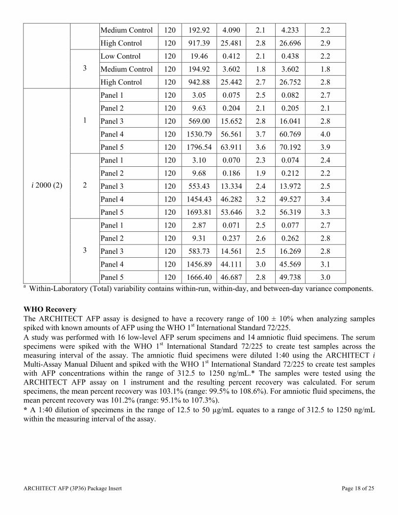
ARCHITECT AFP (3P36) Package Insert Page 18 of 25
Medium Control 120 192.92 4.090 2.1 4.233 2.2
High Control 120 917.39 25.481 2.8 26.696 2.9
Low Control 120 19.46 0.412 2.1 0.438 2.2
Medium Control 120 194.92 3.602 1.8 3.602 1.8 3
High Control 120 942.88 25.442 2.7 26.752 2.8
Panel 1 120 3.05 0.075 2.5 0.082 2.7
Panel 2 120 9.63 0.204 2.1 0.205 2.1
Panel 3 120 569.00 15.652 2.8 16.041 2.8
Panel 4 120 1530.79 56.561 3.7 60.769 4.0
1
Panel 5 120 1796.54 63.911 3.6 70.192 3.9
Panel 1 120 3.10 0.070 2.3 0.074 2.4
Panel 2 120 9.68 0.186 1.9 0.212 2.2
Panel 3 120 553.43 13.334 2.4 13.972 2.5
Panel 4 120 1454.43 46.282 3.2 49.527 3.4
2
Panel 5 120 1693.81 53.646 3.2 56.319 3.3
Panel 1 120 2.87 0.071 2.5 0.077 2.7
Panel 2 120 9.31 0.237 2.6 0.262 2.8
Panel 3 120 583.73 14.561 2.5 16.269 2.8
Panel 4 120 1456.89 44.111 3.0 45.569 3.1
i 2000 (2)
3
Panel 5 120 1666.40 46.687 2.8 49.738 3.0 a Within-Laboratory (Total) variability contains within-run, within-day, and between-day variance components. WHO Recovery The ARCHITECT AFP assay is designed to have a recovery range of 100 ± 10% when analyzing samples spiked with known amounts of AFP using the WHO 1st International Standard 72/225. A study was performed with 16 low-level AFP serum specimens and 14 amniotic fluid specimens. The serum specimens were spiked with the WHO 1st International Standard 72/225 to create test samples across the measuring interval of the assay. The amniotic fluid specimens were diluted 1:40 using the ARCHITECT i Multi-Assay Manual Diluent and spiked with the WHO 1st International Standard 72/225 to create test samples with AFP concentrations within the range of 312.5 to 1250 ng/mL.* The samples were tested using the ARCHITECT AFP assay on 1 instrument and the resulting percent recovery was calculated. For serum specimens, the mean percent recovery was 103.1% (range: 99.5% to 108.6%). For amniotic fluid specimens, the mean percent recovery was 101.2% (range: 95.1% to 107.3%). * A 1:40 dilution of specimens in the range of 12.5 to 50 µg/mL equates to a range of 312.5 to 1250 ng/mL within the measuring interval of the assay.

ARCHITECT AFP (3P36) Package Insert Page 19 of 25
Linearity The ARCHITECT AFP assay is designed to have a deviation from linearity within ± 1 ng/mL for samples less than 10 ng/mL, and within ± 10% for samples between 10 ng/mL and 2000 ng/mL. A study was performed based on guidance from the NCCLS document EP6-A.53 Three dilution series were prepared as follows: a high AFP sample (> 2000 ng/mL) was combined in specific ratios with a low AFP sample (< 2.0 ng/mL). The 3 dilution series, including the low-level and high-level samples, were tested using the ARCHITECT AFP assay. The ARCHITECT AFP assay demonstrated linearity from 0.91 ng/mL to 2487.76 ng/mL. Sensitivity Limit of Detection and Limit of Quantitation The ARCHITECT AFP assay is designed to have a Limit of Detection (LoD) of ≤ 1.0 ng/mL and a Limit of Quantitation (LoQ) of ≤ 2.0 ng/mL. The LoQ is defined as the lowest amount of analyte in a sample that can be accurately quantitated with a total analytical error of ≤ 2.5 ng/mL. Based on guidance from the NCCLS document EP17-A,54 a study was performed with 4 zero-level samples (Calibrator A) and 8 low-level AFP samples (2 samples at each of 4 unique target concentration levels of approximately 0.50, 1.00, 1.50, and 2.00 ng/mL). These samples were tested in 5 separate runs over a minimum of 3 days using 3 reagent lots and 2 instruments. The observed LoD was 0.04 ng/mL and the observed LoQ was 0.5 ng/mL. Limit of Blank In the same study, the Limit of Blank (LoB) was determined to be 0.0 ng/mL. Interference Potentially Interfering Endogenous Substances The ARCHITECT AFP assay is designed to have a difference in AFP concentration within or equal to ± 10% when comparing samples containing elevated levels of endogenous substances to reference samples. A study was performed based on guidance from the CLSI document EP7-A2.55 Potentially interfering endogenous substances were evaluated to determine whether AFP concentrations were affected when using the ARCHITECT AFP assay. The endogenous substances listed below were spiked into samples with 2 levels of AFP (approximately 10 and 1000 ng/mL). The samples were assayed (n = 20), and the AFP concentrations of the spiked samples were compared to reference samples. The data are summarized in the following table.
% Interferencea Potentially Interfering Endogenous Substance
High Test Level 10 ng/mL 1000 ng/mL
Bilirubin (Unconjugated) 20 mg/dL -0.5 0.3
Bilirubin (Conjugated) 20 mg/dL -0.9 -0.8
Hemoglobin 500 mg/dL -0.2 -1.3
Total Protein 12 g/dL 2.9 -0.2
Triglycerides 3000 mg/dL -1.0 -1.9
Mean/Median Test Result - Mean/Median Reference Result a % Interference = Mean/Median Reference Result
x 100

ARCHITECT AFP (3P36) Package Insert Page 20 of 25
Potentially Interfering Substances The ARCHITECT AFP assay is designed to have a mean % recovery of 100% ± 10% when analyzing rheumatoid Factor (RF) and Human Anti-Mouse Antibodies (HAMA) samples spiked with known amounts of AFP. A study was performed based on guidance from the CLSI document EP7-A2.55 Potentially interfering substances were evaluated to determine whether AFP concentrations were affected when using the ARCHITECT AFP assay. Specimens from individuals with the substances listed below were divided into 3 samples. Two of the samples were spiked to 2 levels of AFP (approximately 10 and 1000 ng/mL). The samples were assayed, and the AFP concentrations of the spiked samples were compared to the samples that were not spiked with AFP. The data are summarized in the following table.
% Recoverya
Potentially Interfering Substances n 10 ng/mL 1000 ng/mL
Human Anti-Mouse Antibodies 13 104.7 105.9
Rheumatoid Factor 13 104.6 102.3
Mean/Median Spiked Result – Mean/Median Unspiked Result a % Recovery = Mean/Median Amount AFP Added
x 100
Analytical Specificity The ARCHITECT AFP assay is designed to have a difference in AFP concentration within or equal to ± 10% when comparing samples containing potential interferents to reference samples. A study was performed based on guidance from the CLSI document EP7-A2.55 Potential interferents were evaluated to determine whether AFP concentrations were affected when using the ARCHITECT AFP assay. The potential interferents were spiked into samples with 2 levels of AFP (approximately 10 and 1000 ng/mL). The samples were assayed, and the AFP concentrations of the spiked samples were compared to the reference samples. The data are summarized in the following table.

ARCHITECT AFP (3P36) Package Insert Page 21 of 25
% Interferencea
Potential Interferent High Test Level 10 ng/mL 1000 ng/mL
5-Fluorouracil 3 mmol/L -0.4 0.3
Acetaminophen 6.5 mg/mL -3.1 -3.1
Albumin 160 mg/mL 2.4 -4.1
Alpha-1-Acid Glycoprotein 2 mg/mL 0.2 -1.1
Alpha-1-Antitrypsin 5 mg/mL 8.1 0.3
Alpha-2-Macroglobulin 9 mg/mL 0.1 -0.1
Aspirin 10 mg/mL -4.7 -4.9
Bleomycin 1000 μU/mL -2.5 -4.3
Carboplatin 0.432 mg/mL 0.3 1.3
Ceruloplasmin 2.5 mg/mL -0.3 -0.6
Chorionic Gonadotropin 1000 IU/mL -1.1 -2.2
Cisplatin 1000 μg/mL -0.6 -1.2
Cyclophosphamide 1437 μmol/L 0.3 -0.7
Etoposide 30 μg/mL -0.8 0.3
Gamma-Globulins 30 mg/mL -2.7 -2.4
Haptoglobin 6 mg/mL 0.7 -1.0
Ifosfamide 249 μg/mL -3.1 -2.7
Methotrexate 2 mmol/L -0.6 -0.5
Placental Lactogen 100 μg/mL -3.3 -3.6
Prolactin 500 ng/mL -4.8 -5.0
Transferrin 25 mg/mL -1.6 -3.4
Vinblastine 500 μg/mL -3.4 -3.5
Vincristine 1000 ng/mL -3.1 -4.4
Mean/Median Test Result - Mean/Median Reference Result a % Interference = Mean/Median Reference Result
x 100

ARCHITECT AFP (3P36) Package Insert Page 22 of 25
Autodilution Verification The ARCHITECT AFP assay is designed to have a mean difference in concentration within ± 10% when comparing the autodilution method to the manual dilution method for samples with values > 2000 ng/mL. Twenty-one serum samples were evaluated with the 1:10 autodilution method versus a 1:10 manual dilution method. Fifteen amniotic fluid samples were evaluated with the 1:40 autodilution method versus a 1:40 manual dilution method. The manually diluted samples and the undiluted samples designated for autodilution were assayed in replicates of 2 using the ARCHITECT AFP assay. For serum samples, the mean percent difference was 2.9% (range: -5.8% to 10.9%) and for amniotic fluid samples, the mean percent difference was 4.6% (range: -1.1% to 11.2%). High Dose Hook High dose hook is a phenomenon whereby very high level specimens may read within the measuring interval of the assay. For the ARCHITECT AFP assay, no high dose hook effect was observed when samples containing up to 10,000,000 ng/mL of AFP were assayed. BIBLIOGRAPHY 1. Bergstrand CG, Czar B. Demonstration of a new protein fraction in serum from the human fetus. Scand J
Clin Lab Invest 1956;8:174. 2. Ruoslahti E, Engvall E, Kessler MJ. Chemical properties of alpha-fetoprotein. In: Herberman RB, McIntire
KR, eds. Immunodiagnosis of Cancer. New York: Marcel Dekker, Inc.,1979:101-17. 3. Ruoslahti E, Seppälä M. Studies of carcino-fetal proteins: physical and chemical properties of human alpha-
fetoprotein. Int J Cancer 1971;7:218-25. 4. Tatarinov YS. Detection of embryo-specific alpha-globulin in the blood serum of patients with primary liver
tumors. Vopr Med Khim 1964;10:90-1. 5. Silver HKB, Gold P, Feder S, et al. Radioimmunoassay for human alpha-fetoprotein. Proc Natl Acad Sci
USA 1973;70(2):526-30. 6. Waldmann TA, McIntire KR. The use of a radioimmunoassay for alpha-fetoprotein in the diagnosis of
malignancy. Cancer 1974;34:1510-5. 7. Kohn J, Orr AH, McElwain TJ, et al. Serum-alpha-fetoprotein in patients with testicular tumours. Lancet
1976;2:433-6. 8. Abelev GI. Alpha-fetoprotein as a model for studying reexpression of embryonic antigens in neoplasia. In:
Herberman RB, McIntire KR, eds. Immunodiagnosis of Cancer. New York: Marcel Dekker, Inc., 1979:76-101.
9. Scardino PT, Cox HD, Waldmann TA, et al. The value of serum tumor markers in the staging and prognosis of germ cell tumors of the testis. J Urol 1977;118:994-9.
10. Bosl GJ, Lange PH, Fraley EE, et al. Human chorionic gonadotropin and alphafetoprotein in the staging of nonseminomatous testicular cancer. Cancer 1981;47:328-32.
11. Lange PH, McIntire KR, Waldmann TA, et al. Serum alpha fetoprotein and human chorionic gonadotropin in the diagnosis and management of nonseminomatous germ-cell testicular cancer. N Engl J Med 1976;295(22):1237-40.
12. Javadpour N, McIntire KR, Waldmann TA. Human chorionic gonadotropin (hCG) and alpha-fetoprotein (AFP) in sera and tumor cells of patients with testicular seminoma. Cancer 1978;42:2768-72.
13. Report from the Medical Research Council Working Party on Testicular Tumours. Prognostic factors in advanced non-seminomatous germ-cell testicular tumours: results of a multicentre study. Lancet 1985:8-11.
14. Perlin E, Engeler JE, Edson M, et al. The value of serial measurement of both human chorionic gonadotropin and alpha-fetoprotein for monitoring germinal cell tumors. Cancer 1976;37:215–9.

ARCHITECT AFP (3P36) Package Insert Page 23 of 25
15. Wepsic HT. Alpha-fetoprotein: its quantitation and relationship to neoplastic disease. In: Kirkpatrick AM, Nakamura RM, eds. Alpha-fetoprotein, laboratory procedures and clinical applications. New York: Masson Publishing USA Inc.,1981:115-29.
16. McIntire KR, Waldmann TA, Moertel CG, et al. Serum alpha-fetoprotein in patients with neoplasms of the gastrointestinal tract. Cancer Res 1975;35:991-6.
17. Chen DS, Sung JL. Relationship of hepatitis B surface antigen to serum alpha-fetoprotein in nonmalignant diseases of the liver. Cancer 1979;44:984-92.
18. Report of U.K. Collaborative Study on Alpha-fetoprotein in Relation to Neural-tube Defects. Maternal serum-alpha-fetoprotein measurement in antenatal screening for anencephaly and spina bifida in early pregnancy. Lancet 1977:1323-32.
19. Second Report of the U.K. Collaborative Study on Alpha-fetoprotein in Relation to Neural-tube Defects. Amniotic-fluid alpha-fetoprotein measurement in antenatal diagnosis of anencephaly and open spina bifida in early pregnancy. Lancet 1979:651-62.
20. Haddow JE, Kloza EM, Smith DE, et al. Data from an alpha-fetoprotein pilot screening program in Maine. Obstet Gynecol 1983;62(5):556-60.
21. Brock DJH. The prenatal diagnosis of neural tube defects. Obstet Gynecol Surv 1976;31(1):32-40. 22. Main DM, Mennuti MT. Neural tube defects: issues in prenatal diagnosis and counselling. Obstet Gynecol
1986;67(1):1-16. 23. Adams MJ, Windham GC, James LM, et al. Clinical interpretation of maternal serum alpha-fetoprotein
concentrations. Am J Obstet Gynecol 1984;148(3):241-54. 24. American Society of Human Genetics policy statement for maternal serum alpha-fetoprotein screening
programs and quality control for laboratories performing maternal serum and amniotic fluid alpha-fetoprotein assays. Am J Hum Genet 1987;40:75-82.
25. Cuckle HS, Nanchahal K, Wald NJ. Maternal serum alpha-fetoprotein and ethnic origin. Br J Obstet Gynaecol 1987;94:1111-2.
26. Crandall BF, Lebherz TB, Schroth PC, et al. Alpha-fetoprotein concentrations in maternal serum: relation to race and body weight. Clin Chem 1983;29(3):531-3.
27. Milunsky A, Alpert E, Kitzmiller JL, et al. Prenatal diagnosis of neural tube defects. VIII. The importance of serum alpha-fetoprotein screening in diabetic pregnant women. Am J Obstet Gynecol 1982;142:1030-2.
28. Baumgarten A, Robinson J. Prospective study of an inverse relationship between maternal glycosylated hemoglobin and serum alpha-fetoprotein concentrations in pregnant women with diabetes. Am J Obstet Gynecol 1988;159(1):77-81.
29. Palomaki GE, Knight GJ, Kloza EM, et al. Maternal weight adjustment and low serum alpha-fetoprotein values. Lancet 1985:468.
30. Wald NJ, Cuckle H, Boreham J, et al. Maternal serum alpha-fetoprotein and diabetes mellitus. Br J Obstet Gynaecol 1979;86:101-5.
31. Crandall BF. Second trimester maternal serum screening to identify neural tube defects. In: Kirkpatrick AM, Nakamura RM, eds. Alpha-fetoprotein, laboratory procedures and clinical applications. New York: Masson Publishing USA Inc., 1981:93-105.
32. Seppälä M, Rapola J, Huttunen NP, et al. Congenital nephrotic syndrome: prenatal diagnosis and genetic counselling by estimation of amniotic fluid and maternal serum alpha-fetoprotein. Lancet 1976:123-5.
33. Seppälä M. Increased alpha fetoprotein in amniotic fluid associated with a congenital esophageal atresia of the fetus. Obstet Gynecol 1973;42(4):613-4.
34. Palomaki GE, Hill LE, Knight GJ, et al. Second-trimester maternal serum alpha-fetoprotein levels in pregnancies associated with gastroschisis and omphalocele. Obstet Gynecol 1988;71(6):906-9.
35. Wald N, Barker S, Cuckle H, et al. Maternal serum alpha-fetoprotein and spontaneous abortion. Br J Obstet Gynaecol 1977;84:357-62.
36. Brock DJH, Barron L, Duncan P, et al. Significance of elevated mid-trimester maternal plasma-alpha-fetoprotein values. Lancet 1979:1281-2.

ARCHITECT AFP (3P36) Package Insert Page 24 of 25
37. Nelson LH, Bensen J, Burton BK. Outcomes in patients with unusually high maternal serum alpha-fetoprotein levels. Am J Obstet Gynecol 1987;157(3):572-6.
38. Redford DHA, Whitfield CR. Maternal serum alpha-fetoprotein in twin pregnancies uncomplicated by neural tube defect. Am J Obstet Gynecol 1985;152(5):550-3.
39. Davenport DM, Macri JN. The clinical significance of low maternal serum alpha-fetoprotein. Am J Obstet Gynecol 1983;146(6):657-61.
40. US Department of Labor, Occupational Safety and Health Administration, 29 CFR Part 1910.1030, Bloodborne pathogens.
41. US Department of Health and Human Services. Biosafety in Microbiological and Biomedical Laboratories. 5th ed. Washington, DC: US Government Printing Office; December 2009.
42. World Health Organization. Laboratory Biosafety Manual 3rd ed. Geneva: World Health Organization; 2004.
43. Clinical and Laboratory Standards Institute. Protection of Laboratory Workers from Occupationally Acquired Infections: Approved Guideline—Third Edition. CLSI Document M29-A3. Wayne, PA: Clinical and Laboratory Standards Institute; 2005.
44. Horacek I, Pepperell RJ, Hay DL, et al. Detection of fetomaternal hæmorrhage by measurement of maternal serum-alpha-fetoprotein. Lancet 1976:200.
45. Clinical and Laboratory Standards Institute. Maternal Serum Screening; Approved Standard – Second Edition. CLSI document I/LA25-A2. Wayne, PA: CLSI; 2011.
46. Sherwin JE ed. National Academy of Clinical Biochemistry: Laboratory Medicine Practice Guidelines. Maternal-fetal risk assessment and reference values in pregnancy. Washington, DC: AACC Press, 2006:24.
47. Primus FJ, Kelley EA, Hansen HJ, et al. “Sandwich”-type immunoassay of carcinoembryonic antigen in patients receiving murine monoclonal antibodies for diagnosis and therapy. Clin Chem 1988;34(2):261-4.
48. Schroff RW, Foon KA, Beatty SM, et al. Human anti-murine immunoglobulin responses in patients receiving monoclonal antibody therapy. Cancer Res 1985;45:879-85.
49. Boscato LM, Stuart MC. Heterophilic antibodies: a problem for all immunoassays. Clin Chem 1988;34(1):27-33.
50. Bradley LA, Palomaki GE, and McDowell GA. ACMG Standards and Guidelines: technical standards and guidelines: prenatal screening for open neural tube defects. Genetics in Medicine 2005;7(5):355-69.
51. National Committee for Clinical Laboratory Standards (NCCLS). Evaluation of Precision Performance of Quantitative Measurement Methods; Approved Guideline–Second Edition. NCCLS Document EP5-A2. Wayne, PA: NCCLS; 2004.
52. Clinical and Laboratory Standards Institute. User Verification of Performance for Precision and Trueness; Approved Guideline – Second Edition. CLSI document EP15-A2. Wayne, PA: CLSI; 2005.
53. National Committee for Clinical Laboratory Standards (NCCLS). Evaluation of the Linearity of Quantitative Measurement Procedures: A Statistical Approach; Approved Guideline. NCCLS Document EP6-A. Wayne, PA: NCCLS; 2003.
54. National Committee for Clinical Laboratory Standards (NCCLS). Protocols for Determination of Limits of Detection and Limits of Quantitation; Approved Guideline. NCCLS Document EP17-A. Wayne, PA: NCCLS; 2004.
55. Clinical and Laboratory Standards Institute (CLSI). Interference Testing in Clinical Chemistry; Approved Guideline–Second Edition. CLSI Document EP7-A2. Wayne, PA: CLSI: 2005.
56. Fraser CG. Biological Variation: From Principles to Practice. Washington, DC: AACC Press, 2001:71-8. 57. Trapé J, Botargues JM, Porta F, et al. Reference change value for alpha-fetoprotein and its application in
early detection of hepatocellular carcinoma in patients with hepatic disease. Clin Chem 2003;49:1209-11.

ARCHITECT AFP (3P36) Package Insert Page 25 of 25
The following U.S. Patents are relevant to the ARCHITECT System or its components. There are other such patents and patent applications in the United States and worldwide. 5 468 646 5 543 524 5 545 739 5 565 570 5 669 819 5 783 699 ARCHITECT and Chemiflex are trademarks of Abbott Laboratories in various jurisdictions. ProClin is property of its respective owner. Manufactured by Abbott Ireland Diagnostics Division Finisklin Business Park Sligo, Ireland +353-71-9171712 Distributed by Abbott Laboratories Abbott Park, IL 60064 USA and ABBOTT 65205 Wiesbaden, Germany April 2012 ©2012 Abbott Laboratories