Embed Size (px)
Citation preview

Review
Architectural Representation of Valence in the Limbic System
Praneeth Namburi1,2, Ream Al-Hasani3,4,5, Gwendolyn G Calhoon1, Michael R Bruchas*,3,4,5,6 and Kay M Tye*,1
1Department of Brain and Cognitive Sciences, The Picower Institute for Learning and Memory, Massachusetts Institute of Technology, Cambridge,MA, USA; 2Neuroscience Graduate Program, Massachusetts Institute of Technology, Cambridge, MA, USA; 3Division of Basic Research,Department of Anesthesiology, Washington University School of Medicine, St Louis, MO, USA; 4Washington University Pain Center, WashingtonUniversity School of Medicine, St Louis, MO, USA; 5Department of Neuroscience, Washington University School of Medicine, St Louis, MO, USA;6Division of Biology and Biomedical Sciences, Washington University School of Medicine, St Louis, MO, USA
In order to thrive, animals must be able to recognize aversive and appetitive stimuli within the environment and subsequently initiateappropriate behavioral responses. This assignment of positive or negative valence to a stimulus is a key feature of emotional processing, theneural substrates of which have been a topic of study for several decades. Until recently, the result of this work has been the identificationof specific brain regions, such as the basolateral amygdala (BLA) and nucleus accumbens (NAc), as important to valence encoding. The adventof modern tools in neuroscience has allowed further dissection of these regions to identify specific populations of neurons signaling thevalence of environmental stimuli. In this review, we focus upon recent work examining the mechanisms of valence encoding, and provide amodel for the systematic investigation of valence within anatomically-, genetically-, and functionally defined populations of neurons.Neuropsychopharmacology advance online publication, 3 February 2016; doi:10.1038/npp.2015.358
��������������������������������������
INTRODUCTION
For more than a century following William James’ originalthesis on emotion (James, 1884), psychologists haveattempted to determine whether the diverse range of humanaffect can be understood using few independent factors. Onthe basis of self-reported emotional states, early theoristscharted emotions in two or more dimensions (Nowlis andNowlis, 1956; Russell, 1980; Schlosberg, 1954). Popular amongthese models is the two-dimensional circumplex model ofemotion (Russell, 1980), wherein emotions arise from theinteraction between two neuropsychological systems—onerepresenting the degree of pleasantness, ranging from aversiveto appetitive (valence), and the other representing alertness(arousal; Posner et al, 2005). Identifying and understandingthe neurobiological substrates underlying these features ofemotion is an active area of neuroscience research.The idea that anatomically localized regions in the brain
drive emotion and emotional behaviors was initiallysuggested by the finding that lesions to the temporal lobeand amygdala cause affective deficits (Klüver and Bucy,1939). Following this early work, animal models forstudying affect have been instrumental in advancing our
understanding of the neurobiological basis of emotion.Although the subjective aspect of emotions cannot bedirectly tested in animal models, the behavioral andphysiological responses elicited by emotionally relevantstimuli can be objectively assessed.Arousal is commonly studied in relation to consciousness,
sleep, attention, sex, and emotion. Emotional arousal is animportant aspect of emotion that is known to enhanceemotional memory, either positive or negative. For a detailedreview of the neural representation of arousal, refer to(Adolphs et al, 1999; Harris and Aston-Jones, 2006; Langet al, 1998; McGaugh, 2000, 2004; McIntyre and Roozendaal,2007).Monitoring neural activity evoked by emotionally salient
stimuli in model organisms, such as non-human primatesand rodents, has proved to be an invaluable method toinvestigate the neurobiological basis of valence. A stimulusthat is inherently appetitive or pleasant is said to carrypositive valence, whereas a stimulus that is inherentlyaversive is said to carry negative valence. These stimuli aresufficient to evoke appetitive or aversive responses, and aretherefore designated positive or negative unconditionedstimuli (US), respectively. When a previously neutralstimulus (known as a conditioned stimulus or CS), such asa tone, odor, or image, predicts a positive or negative US, itacquires valence. Pavlovian conditioning (Pavlov, 1927;Rescorla, 1988), in which the CS and US are repeatedlypaired, is a common behavioral paradigm for teaching ananimal a CS–US association. After the acquisition of asuccessful CS–US pairing, a positive CS is sufficient toevoke appetitive behaviors such as approach toward a fooddispenser, and a negative CS is sufficient to evoke fear- or
*Correspondence: Professor KM Tye, Department of Brain andCognitive Sciences, The Picower Institute for Learning and Memory,Massachusetts Institute of Technology, 77 Massachusetts Avenue,46-6263, Cambridge, MA 02139, USA, Tel: +1 617 324 8133 orProfessor MR Bruchas, Washington University in St Louis, Departments ofAnesthesiology and Neuroscience, 660 South Euclid Box 8054, St Louis,MO 63108, USA, E-mail: [email protected] or [email protected] 4 September 2015; revised 4 December 2015; accepted 5December 2015; accepted article preview online 9 December 2015
Neuropsychopharmacology (2016), 1–19© 2016 American College of Neuropsychopharmacology. All rights reserved 0893-133X/16
www.neuropsychopharmacology.org

avoidance-related behaviors such as freezing in response to aCS predicting a foot shock.In several regions of the brain, neural responses to the CS
change after the acquisition of CS–US pairing. Moreover,these changes are different depending on whether the CS ispositive or negative. A neuron recorded during a behavioralparadigm such as Pavlovian conditioning or retrieval is saidto represent valence if its output is differentially modulatedby the positive and negative natures of the stimuli,independent of all other features. Some brain regions, suchas the basolateral amygdala (BLA), contain a greaterproportion of neurons signaling valence (~40%) comparedwith others, such as the hippocampus (~25%; Fuster andUyeda, 1971). Valence-encoding neurons represent only asubset of neurons within each of these brain regions. Evenamong the valence-representing subset in a given region,there are some neurons signaling positive valence and someneurons signaling negative valence. Much effort is beingdevoted to isolate a common property within neuronssignaling one valence vs neurons signaling the oppositevalence. Projection target or genetic markers are promisingcandidates for properties that could distinguish neuronsselectively signaling positive or negative valence.In the next few years, we predict the characterization of
several new valence-signaling populations identified by theirprojection target and/or genetic markers, and distributed in awide network throughout the brain. Comparing andcontrasting the extent of valence representation within andbetween these populations will be essential in directing thefield’s efforts to understand the neurobiological basis ofvalence. Here we propose a model that will facilitatecomparison and contrast between the multitudes of candi-date populations signaling valence.
STRATEGIES TO ASSAY REWARD AND AVERSION INRODENT MODELS
The field is now utilizing modern approaches to dissect thebasis of valence in mammalian neural circuits, and, as aresult, there have been a variety of classical and novelbehavioral models employed to capture valence respondingin rodents. Historically, many of these behavioral paradigmshave relied on Pavlovian conditioning approaches.These models date back to early work in primates with bothdrugs of abuse and natural rewards (Spragg, 1940). In allexamples, a CS is typically paired with a US, and the timeanimals spend in a given CS–US paired context is measured.Place preference models in rodents were then adopted fromthese early methods and are now widely used to assessreward and aversion, ranging from natural rewards (ie,sucrose) and drugs to aversive stimuli (Rossi and Reid, 1976).When a US is repeatedly paired with a neutral environmentalstimulus, the motivational properties of the neutral stimuluschange, and, over the course of the conditioning period, theneutral stimulus becomes a CS. Subsequent to training, theCS can elicit either approach or withdrawal from theenvironment, depending on whether the US is rewardingor aversive (Tzschentke, 2007). Alongside these efforts,Skinner (1938) developed the term ‘operant conditioning’whereby behavioral manipulations can be achieved throughthe use of reinforcement, which is given after the desired
behavioral response. Skinner organized potential outcomesinto three basic categories: neutral, reinforcing, or punishing.Operant training has been expanded to include an intracra-nial self-stimulation (ICSS) paradigm (Carlezon andChartoff, 2007). Rodents learn to deliver brief electricalpulses into the medial forebrain bundle (Carlezon andChartoff, 2007), and more recently into other specific brainregions hypothesized to mediate both natural and ICSSrewards. These basic models have evolved over the last75 years to include elaborate measures of valence respondingduring pharmacological, electrical, and genetic modifica-tions. They are now commonly used for dissecting thecontributions of individual brain regions and circuits inreward and avoidance.In the last decade, the rise of optogenetic and chemoge-
netic approaches has greatly influenced these traditionalbehavioral paradigms in order to take advantage of theunique spatiotemporal features of these tools that facilitatediscrete control over neural circuits. Most commonly, cre-recombinase/loxP technology is utilized to gain cell-type-selective expression and thus precise excitation, inhibition, ormodulation of specific circuits in vivo (Atasoy et al, 2008;Tsai et al, 2009). In a seminal paper by Tsai et al (2009),it was demonstrated that closed-loop control of behavioralresponding was possible with in vivo optogenetics, such thatthe animal’s presence in a certain context triggered phasicphotostimulation of dopamine (DA) neurons to elicit placepreference. Since then numerous adaptations of closed loop,‘real-time’ in vivo optogenetic engagement of neural circuitshave been used with behavioral models in Pavlovian,operant, and acute measures of valence. The increasedspatiotemporal control over neural circuits afforded by thesemodern approaches has rapidly advanced our understandingof the role of the limbic system in reward and aversion (Al-Hasani et al, 2015; Nieh et al, 2013; Tye and Deisseroth,2012). We have compiled several of these studies utilizingadapted behavioral models into Table 1 to summarize themost recent findings, the behavioral models employed, andthe brain regions examined (for a comprehensive review ofanxiety, see Calhoon and Tye, 2015). This provides abackground reference for the remainder of the review, whichwill break down these related findings into a conceptualframework of valence in the limbic system. It is anticipatedthat further advances in optogenetic tools, hardware, in vivoimaging, and mouse genetic models over the next decade willfurther expand the types of behavioral measures that assessvalence in the limbic system.
NEURAL REPRESENTATION OF REWARD ANDAVERSION
Where is Valence Represented in the Brain?
Certain anatomically localized populations of neurons showdifferential responses to reward or aversion-associated cues.These regions have been considered to represent valence, andinclude the BLA (Fuster and Uyeda, 1971; Paton et al, 2006;Shabel and Janak, 2009), nucleus accumbens (NAc; Roitmanet al, 2005), ventral tegmental area (VTA; Bromberg-Martinet al, 2010; Cohen et al, 2012; Matsumoto and Hikosaka,2009), orbitofrontal cortex (Schoenbaum et al, 1999), lateralhypothalamus (LH; Fukuda et al, 1990; Li et al, 2013;
Architectural representation of valenceP Namburi et al
2
Neuropsychopharmacology

Table 1 A Summary of Studies Investigating the Causal Relationship Between Neuronal Populations and Valence within the VTA, NAcand BLA
4102,lategnohZevitageNnoiteled5KdcNSF,ecnereferpesorcuSATVVTA (DA neurons) 4102,lategnohZevitageNnoiteled5Kdc
cdK5NSF,ecnereferpesorcuS
,lategnohZ)noisserpedsesrever(evitisoP)sG(sDDAERD+noiteledNSF,ecnereferpesorcuS)snoruenAD(ATV 2014VTA (DA neurons) Sucrose preference, Tail suspension test NpHR Nega�ve Tye et al, 2013VTA (DA neurons) Sucrose preference, Tail suspension test ChR2 Posi�ve (reverses depression) Tye et al, 2013VTA (DA neurons) CPP ChR2 Posi�ve Tsai et al, 2009VTA (DA neurons) CPP 3102,latemiKevitisoP2RhCVTA (DA neurons) CPA Arch 4102,lateojnaDevitageNVTA (DA neurons) ICSS 3102,latemiKevitisoP2RhCVTA (DA neurons) ICSS ChR2 Posi�ve Wi�en et al, 2011VTA (DA neurons) ICSS ChR2 Posi�ve Ilango et al, 2014VTA (DA neurons) tnaR2D/tnaR1D+2RhCSSCI A�enua�on of posi�ve Steinberg et al, 2014VTA (GABA neurons) CPA ChR2 Nega�ve Tan et al, 2012VTA (GABA neurons) RTPT Opto-MOR Posi�ve Siuda et al, 2015VTA (GABA neurons) RTPT NpHR Posi�ve Jennings et al, 2013VTA (GABA neurons) Self inhibi�on NpHR Posi�ve Jennings et al, 2013VTA (GABA neurons) Elevated plus maze NpHR Posi�ve Jennings et al, 2013VTA (GABA neurons) Reward consump�on (licks) ChR2 Disrup�on of posi�ve van Zessen et al, 2012VTA (DA neurons) - NAc 2RhCSSCI Posi�ve Steinberg et al, 2014VTA (DA neurons) - NAc Operant self-administra�on DREADDs (Gq) + CAV Posi�ve Boender et al, 2014VTA (DA neurons) - NAcSh RTPT ChR2 Posi�ve Jeong et al, 2015VTA (DA neurons) - NAc (D2R) CPA Arch + shRNA knockdown in NAc (D2R) A�enua�on of nega�ve Danjo et al, 2014VTA (GABA neurons) - NAc Reward consump�on (licks) ChR2 No effect van Zessen et al, 2012VTA (GABA neurons) - NAc Fear condi�oning ChR2 Enhancement of nega�ve Brown et al, 2012LDT - VTA surivseibaR,2RhCPPC Posi�ve Lammel et al, 2012LHb - VTA surivseibaR,2RhCPPC Nega�ve Lammel et al, 2012LHb - VTA RTPT ChR2 Nega�ve Stamatakis and Stuber, 2012
2RhCSSCIATV-HL Posi�ve Kempadoo et al, 2013)tna1RSTN(29684RS+2RhCSSCIATV-HL A�enua�on of posi�ve Kempadoo et al, 2013
2RhCgnikeesesorcusevislupmoCATV-HL Enhancement of posi�ve Nieh et al, 2015RHpNgnikeesesorcusevislupmoCATV-HL A�enua�on of posi�ve Nieh et al, 20152RhCgnideeFATV-HL Posi�ve Nieh et al, 2015
BNST (Glu neurons) - VTA RTPT ChR2 Nega�ve Jennings et al, 2013BNST (Glu neurons) - VTA Open field ChR2 Nega�ve Jennings et al, 2013BNST (GABA neurons) - VTA RTPT ChR2 Posi�ve Jennings et al, 2013BNST (GABA neurons) - VTA ICSS ChR2 Posi�ve Jennings et al, 2013BNST (GABA neurons) - VTA Elevated plus maze ChR2 Posi�ve Jennings et al, 2013BNST (GABA neurons) - VTA Fear condi�oning ChR2 A�enua�on of nega�ve Jennings et al, 2013
BLA 2RhCSSCI Variable results Stuber et al, 2011BLA (shock responsive cells) Fear condi�oning, CPP cFOS + ChR2 Nega�ve Gore et al, 2015
2RhC+SOFcTPTR)sllecevisnopserkcohs(ALB Nega�ve Redondo et al, 2014BLA (CS responsive cells upon fear condi�oning) Fear condi�oning cFOS + ChR2 Nega�ve Gore et al, 2015BLA (nico�ne responsive cells) CPP, ICSS cFOS + ChR2 Posi�ve Gore et al, 2015
2RhC+SOFcTPTR)selamnisllecevisnopseresuomelamef(ALB Posi�ve Redondo et al, 2014)SUgnirud(2RhCgninoitidnocraeF)snoruenVP(ALB A�enua�on of nega�ve Wolff et al, 2014
)SUgnirud(hcrAgninoitidnocraeF)snoruenVP(ALB Enhancement of nega�ve Wolff et al, 2014)SCgnirud(2RhCgninoitidnocraeF)snoruenVP(ALB Enhancement of nega�ve Wolff et al, 2014
)SCgnirud(hcrAgninoitidnocraeF)snoruenVP(ALB A�enua�on of nega�ve Wolff et al, 2014)SCgnirud(2RhCgninoitidnocraeF)snoruenMOS(ALB A�enua�on of nega�ve Wolff et al, 2014
)SCgnirud(hcrAgninoitidnocraeF)snoruenMOS(ALB Enhancement of nega�ve Wolff et al, 2014BLA - NAc ICSS ChR2 Posi�ve Stuber et al, 2011BLA - NAc Reward condi�oning NpHR A�enua�on of posi�ve Stuber et al, 2011BLA - NAc ICSS ChR2, Rabies Virus Posi�ve Namburi et al, 2015BLA - NAc (Medial shell) ICSS ChR2 Posi�ve Bri� et al, 2012BLA - CeM RTPT ChR2, Rabies Virus Nega�ve Namburi et al, 2015BLA - CeM Fear condi�oning NpHR, CAV A�enua�on of nega�ve Namburi et al, 2015BLA - CeM Reward condi�oning NpHR, CAV Enhancement of posi�ve Namburi et al, 2015BLA - IL Fear condi�oning ChR2 + HSV/CAV A�enua�on of nega�ve (rel. to NpHR Senn et al, 2014BLA - ILBLA - PL Fear condi�oning ChR2 + HSV/CAV Senn et al, 2014BLA - PL Fear condi�oning NpHR + HSV/CAV Senn et al, 2014
NAc CPP Opto-α1 Posi�ve Airan et al, 2009NAc (GABA neurons) CPP ChR2 + cocaine Transient a�enua�on of posi�ve Wang et al, 2014NAc (D1R MSN) eniacoc+2RhCPPC Posi�ve Lobo et al, 2010NAc (D1R MSN) Chronic social defeat stress ChETA Posi�ve (promotes resilience) Francis et al, 2015NAc (D1R MSN) Chronic social defeat stress DREADDs (Gi) Nega�ve (pro-depressive) Francis et al, 2015NAc (D2R MSN) eniacoc+2RhCPPC A�enua�on of posi�ve Lobo et al, 2010NAc (D2R MSN) Chronic social defeat stress ChETA No effect Francis et al, 2015NAc (D2R MSN) Chronic social defeat stress DREADDs (Gi) No effect Francis et al, 2015NAc (Substance P expressing neurons) Cocaine-induced locomo�on Reversible neurotransmission block A�enua�on of posi�ve (cocaine effect) Hikida et al, 2010NAc (Substance P expressing neurons) CPP Reversible neurotransmission block A�enua�on of posi�ve Hikida et al, 2010NAc (Substance P expressing neurons) Inhibitory avoidance Reversible neurotransmission block No effect Hikida et al, 2010NAc (Enkephalin expressing neurons) Cocaine-induced locomo�on Reversible neurotransmission block No effect Hikida et al, 2010NAc (Enkephalin expressing neurons) CPP Reversible neurotransmission block No effect Hikida et al, 2010NAc (Enkephalin expressing neurons) Inhibitory avoidance Reversible neurotransmission block A�enua�on of nega�ve Hikida et al, 2010NAc (ChAT neurons) CPP NpHR No effect Wi�en et al, 2010NAc (ChAT neurons) CPP ChR2 No effect Wi�en et al, 2010NAc (ChAT neurons) CPP Cocaine + NpHR A�enua�on of posi�ve Wi�en et al, 2010NAc (ChAT neurons) Fear condi�oning NpHR Enhancement of nega�ve Wi�en et al, 2010
ecnelaV)s(looTksaT)epyTlleC(tiucriC/noigeR Reference
Fear condi�oning NpHR + HSV/CAV Enhancement of nega�ve (rel. to ChR2) Senn et al, 2014Enhancement of nega�ve (rel. to NpHR)A�enua�on of nega�ve (rel. to ChR2)
Architectural representation of valenceP Namburi et al
3
Neuropsychopharmacology

Nakamura et al, 1987; Ono et al, 1986; Yamamoto et al,1989), dorsal raphe nucleus (DRN; Cohen et al, 2015), locuscoeruleus (Bouret and Richmond, 2015; Hofmeister andSterpenich, 2015; McCall et al, 2015), subthalamic nucleus(Sieger et al, 2015), and hippocampus (Fuster and Uyeda,1971). Within many of these regions, there is markedheterogeneity among neural populations, such that neuronsrepresenting positive valence reside side-by-side with thoserepresenting negative valence.Techniques that reveal two populations of neurons in the
same brain—one active in response to a rewarding stimulusand the other active in response to an aversive stimulus—willbe immensely useful for gaining a comprehensive under-standing of brain regions containing topographicallyintermingled populations of neurons representing valence.These emergent strategies are already proving their utility;a recent study (Xiu et al, 2014) used a technique knownas tyramide-amplified-immunohistochemistry–fluorescencein situ hybridization (TAI–FISH) to simultaneously labelpopulations of neurons that respond to a rewarding stimulussuch as cocaine, and an aversive stimulus, such as foot shock.TAI-FISH exploits the difference between the time courses ofc-fos mRNA and protein expression to simultaneously labeltwo populations of neurons, each active in response to adifferent stimulus. The authors found several brain regionscontaining intermingled populations of neurons activated bymorphine and foot shock, including the ventral division ofthe lateral septum, dorsomedial shell of the NAc, and thefusiform nucleus of the bed nucleus of stria terminalis.Therefore, valence is represented across several brain
regions in distributed networks, and, more importantly, all ofthe brain regions discussed above represent both positiveand negative valences. Hence, we can posit that severalbrain regions contain either largely distinct or overlappingpopulations of neurons representing positive and negativevalences. An important challenge is to identify a uniquefeature shared by neurons representing only positive or only
negative valence. Projection target (downstream target ofaxons) and genetic markers are two major classes of possiblefeatures shared by neurons selectively representing a singlevalence, and recent findings in valence representation withinVTA neurons clearly illustrate this point.
Distinct Neural Populations Representing Reward andAversion: the VTA as an Example
Recent evidence has identified separable populations of VTAneurons that differentially encode positive and negativevalence. These populations can be segregated by geneticmarkers; for instance, phasic activation of VTA DA neuronscauses place preference (Tsai et al, 2009), whereas activationof VTA GABA neurons causes place aversion (Kim et al,2013; Tan et al, 2012). VTA DA and GABA neurons alsoshow differences in their response properties during fear andreward conditioning (Cohen et al, 2012).Projection target can also be used to disentangle VTA
neurons encoding positive and negative valences. VTA DAneurons are a heterogeneous population in the context ofvalence, and dissecting VTA DA neurons by their projectiontarget has elucidated their differential role in reward andaversion. Synaptic transmission onto largely nonoverlappingVTA DA neurons projecting to the medial shell of NAc andthe medial prefrontal cortex (mPFC) can be modulated by arewarding and an aversive experience, respectively (Lammelet al, 2011). VTA DA neurons projecting to NAc lateral shellare enriched in the lateral VTA and receive a relatively largerinput from the laterodorsal tegmentum (LDT), and VTA DAneurons projecting to mPFC are enriched in the medial VTAand receive stronger input from the lateral habenula(LHb; Lammel et al, 2012). Activating LDT inputs to VTAis sufficient to evoke place preference, whereas activatingLHb inputs to VTA is sufficient to evoke place aversion(Lammel et al, 2012; Stamatakis and Stuber, 2012). There-fore, the medial–lateral axis of the VTA has parallel pathways
Table 1 Continued
NAc (Core) Cocaine reinstatement NpHR/Arch A�enua�on of posi�ve Stefanik et al, 2013aNAc (Core, astrocytes) ICSS DREADDs (Gq) Posi�ve Bull et al, 2014NAc (Shell) Forced swim test ChR2 Enhancement of nega�ve Larson et al, 2015NAc (Ventral shell) RTPT, CPP, ICSS ChR2 Nega�ve Al-Hasani et al, 2015NAc (Dorsal shell) RTPT, CPP, ICSS ChR2 Posi�ve Al-Hasani et al, 2015NAc (Core) - dlVP Cocaine reinstatement Arch A�enua�on of posi�ve Stefanik et al, 2013bNAc (Core) - SN Cocaine reinstatement Arch No effect Stefanik et al, 2013bNAc (Shell) - LH Forced swim test ChR2 Enhancement of nega�ve Larson et al, 2015NAc (Shell) - LH Cocaine self-administra�on ChR2 Enhancement of posi�ve Larson et al, 2015vHippocampus - NAc (Medial shell) Cocaine-induced locomo�on NpHR A�enua�on of posi�ve (cocaine effect) Bri� et al, 2012vHippocampus - NAc (Medial shell) Cocaine-induced locomo�on ChR2 Enhancement of posi�ve (cocaine effect) Bri� et al, 2012vHippocampus - NAc (Medial shell) CPP 2102 ,la te ttirBevitisoP2RhCvHippocampus - NAc (Medial shell) ICSS 2102 ,la te ttirBevitisoP2RhCPL - NAc (Core) Cocaine reinstatement NpHR/Arch A�enua�on of posi�ve Stefanik et al, 2013adlVP - NAc (Core) Cocaine reinstatement Arch No effect Stefanik et al, 2013bVTA - NAc (Core) Cocaine reinstatement Arch A�enua�on of posi�ve Stefanik et al, 2013bmPFC - NAc (Medial shell) ICSS ChR2 Posi�ve Bri� et al, 2012mPFC - NAc ICSS ChR2 No effect Stuber et al, 2011mPFC (GABA neurons) - NAc RTPT ChR2 Nega�ve Lee et al, 2014
ecnelaV)s(looTksaT)epyT lleC( tiucriC/noigeR Reference
Abbreviations: Arch, archaerhodopsin; BLA, basolateral amygdala; BNST, bed nucleus of the stria terminalis; CeA, central nucleus of the amygdala; ChR2,channelrhodopsin-2; CPA, conditioned place aversion; CPP, conditioned place preference; ICSS, intracranial self-stimulation; IL, infralimbic; LDT, laterodorsal tegmentum;LHb, lateral habenula; mPFC, medial prefrontal cortex; MSN, medium spiny neurons; NAc, nucleus accumbens; NpHR, halorhodopsin; NSF, novelty suppressed feeding;PL, prelimbic; PV, parvalbumin; RTPT, real-time place testing; SOM, somatostatin; Vgat, vesicular GABA transporter; Vglut, vesicular glutamate transporter; VTA, ventraltegmental area. Positive and negative refer to the valence induced by the manipulation.
Non-specific popula�on
Gene�cally-defined popula�on
Anatomically-defined popula�on (projec�on-target)
Anatomically-defined popula�on (sub-region)
Anatomically-defined popula�on (afferents)Func�onally-defined popula�on
Architectural representation of valenceP Namburi et al
4
Neuropsychopharmacology

running alongside each other that process reward andaversion (Lammel et al, 2014a). The VTA provides but oneexample highlighting the importance of studying geneticallydefined (DA vs GABA VTA neurons) and projection-target-defined (mPFC vs NAc projecting VTA DA neurons)populations. For more in-depth reviews on these circuits,see (Lammel et al, 2014a, b).In the following two sections, we will highlight valence
representation in projection-target-, genetically and func-tionally defined populations of neurons (Figure 1), as well asthe relationships between these populations of neurons in theBLA and the NAc. Although initial studies on the BLA werefocused on its role in processing both appetitive and aversivestimuli (Fuster and Uyeda, 1971; Machne and Segundo,1956), more than three decades of BLA research has focusedprimarily on the role of BLA in fear conditioning(Davis, 1992; LeDoux, 2000; LeDoux et al, 1990; Maren,2001). Conversely, the NAc has been studied primarily in thecontext of reward learning (Carlezon and Thomas, 2009;Kelley, 2004; Robbins and Everitt, 1996; Salamone et al, 2005;Wise, 2004). Recent research has started to focus on the roleof both the BLA and the NAc in representing valence.
REPRESENTATION OF REWARD AND AVERSION INTHE BLA
Neural populations in the BLA have been dissected functionally,anatomically, and genetically in the context of fear andreward. In this section, we discuss these neural populationswithin the BLA, and the relationships between them.
Diversity of BLA Neural Populations
The BLA complex, consisting of the lateral (LA) and thebasal (BA) subdivisions, contains topographically inter-mingled populations of neurons related to a variety ofbehaviors (Zhang et al, 2013) including fear conditioning,reward conditioning, anxiety, feeding, and social interaction(Janak and Tye, 2015). Here we will focus on the functionalpopulations of BLA neurons related to fear and rewardbehaviors. Advances in activity-dependent regulation oftransgene expression is fueling dissection of such function-ally defined BLA neural populations (Denny et al, 2014; Goreet al, 2015; Liu et al, 2012; Redondo et al, 2014; Reijmerset al, 2007). Optogenetic advances have allowed us to dissectBLA neurons based on criteria other than a functionallydefined population, including projection target (Felix-Ortizand Tye 2014; Felix-Ortiz et al, 2013, 2015; Namburi et al,2015; Senn et al, 2014; Stuber et al, 2011; Tye et al, 2011) andgenetic markers (Wolff et al, 2014). The field is currentlyemploying a substantial amount of effort toward under-standing the relationship between populations of BLAneurons defined by the following three criteria: functional,anatomical (projection target), and genetic identity.The idea that multimodal information converges onto
single neurons in the BLA was established at the dawn ofsingle-unit recordings in the amygdala (Machne andSegundo, 1956), and increased firing of BLA neurons tobehaviorally relevant stimuli was observed shortly thereafter(Sawa and Dalgado, 1963). Almost a decade later, 37% ofneurons in the amygdala were reported to be selectively
responsive to motivationally significant stimuli—a higherproportion compared with neurons in the hippocampus andpiriform cortex (Fuster and Uyeda, 1971). Some BLAneurons modulated their firing rate when the affective valueof the outcome was reversed, establishing that BLA neuronswere tracking the valence of the stimulus (Nishijo et al,1988), and this reversal, or valence-tracking property, waspresent in about half of the BLA neurons selectively
Anatomical
BLA
NAc CeM
GeneticFunctional
Forward approach
Reverse approach
Stimulus
VTA DA VTA GABA
NAc mPFC
Figure 1 Three common ways to define subpopulations of neurons,based on functional (activity-tagged), genetic, and anatomical (eg, projectiontarget) criteria. (a) Recent attempts to establish the identity of positive(represented in the figure by a +) and negative (represented in the figure bya − ) valence-encoding populations of topographically overlapping neuronscan be broadly classified into forward and reverse approaches. In a forwardapproach, properties of functionally defined populations of neurons areexamined, whereas in the reverse approach the functional role of eitheranatomically- or genetically-defined populations of neurons is examined.An example of a forward approach in the basolateral amygdala (BLA) isactivity-dependent labeling of BLA neurons activated by a positive and/or anegative stimulus (Gore et al, 2015; Nonaka et al, 2014; Redondo et al,2014; Han et al, 2009), followed by the study of the properties of eachsubpopulation to understand their necessity, sufficiency, experience-dependent plasticity, and molecular identity. An example of a reverseapproach in the BLA is examining the functional role of projection-target-defined BLA neurons (Namburi et al, 2015; Senn et al, 2014). With theexplosion of tools recently made available for circuit dissection, including theability to tag and manipulate neural populations with a common geneticmarker, or a common projection target, we can now move at an acceleratedpace toward understanding the relationship between functionally defined,genetically-defined and projection-target-defined neural populations. (b) TheBLA is an example where stimulation of distinct projection-target-definedpopulations of neurons, those projecting to the NAc and those projecting tothe CeM evoke positive or negative behaviors (Namburi et al, 2015).(c) The VTA is an example where genetically defined and projection-target(anatomical)-defined populations of neurons are known to play differentialroles in positive and negative behaviors (Cohen et al, 2012; Lammelet al, 2011). CeM, medial division of the central amygdala; mPFC, medialprefrontal cortex; NAc, nucleus accumbens; VTA, ventral tegmental area.
Architectural representation of valenceP Namburi et al
5
Neuropsychopharmacology

responsive to affective stimuli (Schoenbaum et al, 1999).Furthermore, valence-tracking BLA neurons reversed theirresponse properties before behavioral reversal in the animal(Belova et al, 2007; Schoenbaum et al, 1999). Taken together,we should expect about a fifth of all randomly sampledBLA neurons to track valence. Consistent with single-unitrecording data (Zhang et al, 2013), the complementaryapproach of activity-dependent labeling of BLA neuronsusing nicotine or a conspecific of the opposite sex as apositive US and foot shock as a negative US in thesame animal reveal two largely nonoverlapping, buttopographically intermingled, populations in the BLA(Gore et al, 2015; Redondo et al, 2014).Populations of BLA neurons can also be classified into
nonoverlapping sets based on the primary neurotransmitterthey carry—glutamate or GABA (Sah et al, 2003). Thereare multiple partially overlapping subpopulations amongthe GABAergic population that can be distinguished basedon their immunoreactivity to various proteins, such asparvalbumin (PV), somatostatin (SOM), and calbindin andcalretnin (Capogna, 2014; Kemppainen and Pitkänen, 2000;McDonald and Mascagni, 2002).Although the functional role of PV and SOM interneurons
in the BLA during fear conditioning has been identifiedrecently (Wolff et al, 2014), the functional role of BLAinterneurons in reward learning has not yet been explored.PV cells primarily contact the soma of principal neurons(Muller et al, 2006) and inhibit SOM neurons. SOM neuronsprimarily contact the distal dendrites of principal neurons(Muller et al, 2007). PV neurons are active during anauditory CS, and inhibit SOM neurons, thereby disinhibitingdistal principal neuron dendrites. Both PV and SOMneurons are inhibited by the US, thereby disinhibiting theprincipal neuron (Wolff et al, 2014). The authors also find apopulation of PV neurons that is inhibited by the CS.Perhaps distinct subpopulations of PV interneurons mightdisinhibit BLA principal neurons in the BLA differentiallyresponsive to fear or reward cues (Janak and Tye, 2015).BLA projection neurons have diverse targets in the brain,
notably including the NAc (McDonald, 1991a), the lateraldivision of the central nucleus of the amygdala, medialdivision of the central nucleus of the amygdala (CeM;Pitkänen et al, 1997), ventral hippocampus (Pikkarainenet al, 1999), and the pre-limbic (PL) and infralimbic (IL)subdivisions of the mPFC (McDonald, 1991b). Although theexact extent of overlap between various projection-target-defined subpopulations of neurons remains to be elucidated,retrograde tracing using two tracers suggests that some of theprojection-target-defined populations of BLA neurons arelargely nonoverlapping, such as vHPC vs mPFC-projectingBLA neurons (Senn et al, 2014), whereas other projection-target-defined BLA neurons are largely overlapping, espe-cially striatal and prefrontal cortex-projecting BLA neurons(McDonald, 1991b; Shinonaga et al, 1994).The broad relationship between projection-target- and
neurotransmitter-defined populations of BLA neurons isrelatively straight forward. About 70–90% of the projectionneurons in the BLA are glutamatergic (McDonald andAugustine, 1993; Millhouse and DeOlmos, 1983; Washburnand Moises, 1992), with the exception of some SOM+GABAergic neurons that project to the basal forebrain(McDonald et al, 2012), entorhinal cortex (McDonald and
Zaric, 2015), and preoptic–hypothalamic region (McDonald,1987). However, by exploiting modern transcriptomictechniques such as RNA-seq, we are able to appreciatemore subtle gene expression profile differences betweenprojection-target-defined subpopulations of glutamatergicBLA projection neurons. The first of such attempts inthe BLA shows several differentially expressed genesbetween NAc- and CeM-projecting BLA principal neurons,including some membrane-bound receptors (Namburiet al, 2015).Recent studies are starting to shed light on the relationship
between functionally defined and projection-target-definedBLA neurons. Activation of either nicotine-US (positive)-labeled subpopulation of BLA neurons (Gore et al, 2015) orNAc-projecting BLA neurons (Namburi et al, 2015) issufficient to induce ICSS. Moreover, activation of either footshock-US-labeled subpopulation of BLA neurons (Redondoet al, 2014) or CeM-projecting BLA neurons (Namburi et al,2015) is sufficient to support place avoidance. Although thesedata suggest a general relationship between functionallydefined and projection-target-defined populations of BLAneurons, the extent of overlap between functionally-definedand projection-target-defined populations of BLA neuronsremains to be quantified.It is also important to consider the interplay among
different subpopulations of BLA neurons. Different popula-tions of GABAergic interneurons target different cellcompartments of glutamatergic BLA principal neurons(Capogna, 2014). Since projection-target-defined BLA neu-rons are being shown to have opposing functional roles(Namburi et al, 2015; Senn et al, 2014), there may beinterpopulation inhibition within the BLA (Janak and Tye,2015). The first evidence for functional opposition amongprojection-target-defined BLA populations came from theanxiolytic properties of BLA projections to the lateralsubdivision of the CeA (Tye et al, 2011) and the anxiogenicproperties of BLA projections to the ventral hippocampus(Felix-Ortiz et al, 2013). In the context of conditioned asso-ciations, there is also evidence for projection-target-definedfunctional opposition as seen by an inversely correlatedpattern of FOS expression between PL- and IL-projectingBLA neurons (Senn et al, 2014). Further work is required toestablish an understanding of the interplay between BLAneurons that process positive and negative valences.
Learning-Induced Plasticity in the BLA
Acquisition of an association between a CS and either anaversive or appetitive outcome leads to an increase inAMPAR/NDMAR ratio (a proxy for synaptic strength) ofinternal capsule inputs to BLA neurons (Clem and Huganir,2010; Rumpel et al, 2005; Tye et al, 2008).Inputs onto BLA neurons undergo plastic changes upon
fear and reward learning (McKernan and Shinnick-Gallagher,1997; Rogan et al, 1997; Tye et al, 2008). This long-termplasticity is mediated via AMPA receptor trafficking to thepostsynaptic membrane (Clem and Huganir, 2010; Rumpelet al, 2005; Tye et al, 2008). Emerging evidence suggests thatthese plastic changes can vary by cell type and projection-target-defined populations of BLA neurons.As BLA neural populations mediate a diverse set of
behaviors, some of which are opposing, experience-depen-
Architectural representation of valenceP Namburi et al
6
Neuropsychopharmacology
dent plasticity in the BLA is perhaps a function of the neuralpopulation sampled. A recent study examined experience-dependent changes in AMPAR/NMDAR ratios in NAc- andCeM-projecting BLA neurons, wherein photoactivationdrives positive reinforcement or punishment, respectively(Namburi et al, 2015). AMPAR/NMDAR ratios in theinternal capsule inputs to NAc- and CeM-projecting BLAneurons underwent opposing changes after fear and rewardconditioning—AMPAR/NMDAR ratios onto NAc-projectingBLA neurons decreased after fear conditioning and increasedafter reward conditioning. Conversely, AMPAR/NMDARratios onto CeM-projecting BLA neurons increased after fearconditioning and decreased after reward conditioning.Another study (Nonaka et al, 2014) expressed a fluor-
ophore dVenus under the control of an Arc promoter(Eguchi and Yamaguchi, 2009) during a fear-conditioningparadigm, thus labeling the functional population of neuronsinvolved in fear conditioning. The authors contrasted inputsynaptic transmission between dVenus-expressing and -non-expressing LA neurons after a cued fear-conditioningparadigm (Nonaka et al, 2014). The authors demonstratedan increase in synaptic transmission selectively onto dVenus-positive cells (ie, labeled by fear conditioning). This increasewas at least in part due to an increase in probability of releasefrom the presynaptic terminals of the cortical inputs. It isinteresting to note that the authors did not see a difference insynaptic transmission from internal capsule inputs onto LAneurons with (dVenus-expressing) and without (dVenus-negative) activity-dependent labeling. Taken together withstudies showing increased post-synaptic transmission frominternal capsule inputs onto nonspecific LA neurons afterfear conditioning (Clem and Huganir, 2010; Rumpel et al,2005), perhaps changes in internal capsule synaptic transmis-sion represent a more generalized sum of aversive experiences,and changes in cortical pathway synaptic transmission signalchanges specific to one aversive experience. A reward-conditioning analog of this study has yet to be conducted.
Flexibility of Valence Representation in the BLA
Does a BLA neuron have the flexibility to encode eitherpositive or negative valence, or is the valence encoded by aBLA neuron indelible? Might there be distinct populationsthat are flexible or indelible? If so, might the indeliblepopulation(s) be anatomically hard-wired?The existence of valence-tracking BLA neurons (satisfying
the reversal criterion) in the BLA (Paton et al, 2006;Schoenbaum et al, 1999) poses a strong argument in favorof indelible valence encoding on the timescale of multiplehours within the BLA. It is prudent to remember that only afraction (about a fifth) of BLA neurons exhibit this property.Further work, such as monitoring valence-tracking neuronsover multiple days would be required to determine whetherthese neurons represent the same valence over longertimescales. Using optogenetic advances to manipulate func-tionally defined or projection-target-defined populations ofBLA neurons, recent studies have advanced our understandingregarding the flexibility of valence representation in the BLA.Activity-dependent tagging in the BLA using foot shock
and nicotine label two largely nonoverlapping BLA neuralpopulations, thus supporting the idea of valence-codingneurons in the BLA (Gore et al, 2015). A more direct test of a
BLA neuron’s flexibility in encoding valence was performedby attempting to reverse the valence encoded by afunctionally defined population of BLA neurons, taggedunder the control of a c-fos promoter (Redondo et al, 2014).The authors showed that activating foot shock-labeled BLAneurons after stimulating these neurons during a rewardingstimulus (female mouse) did not elicit place preference.Conversely, activating reward-labeled BLA neurons afterassociating these neurons with foot shocks did not elicitplace avoidance. Whereas the authors could not reverse thevalence-coding properties of activity-labeled BLA neurons,they were able to reverse the association of activity-labeledcells in the dentate gyrus.A complementary approach to determine the flexibility of
valence representation in the BLA is offered by efforts todetermine the criteria for recruiting a cell into a memorytrace. Neurons with elevated CREB expression are recruitedinto a fear memory trace (Han et al, 2009). Elevating CREBexpression (Zhou et al, 2009) or neural excitability (Yiu et al,2014) increases the probability of recruiting that neuron intothe memory trace. It remains to be seen whether elevatingCREB/neural excitability increases the probability of recruit-ing the neuron into a memory trace of any valence orone specific valence. The former outcome would suggest thatthe memory trace is flexible, whereas the latter would suggestthat it is indelible. However, the outcome may depend on thesubpopulation of BLA neurons under investigation, whichwould imply BLA neurons to contain both flexible andindelible subpopulations.Finally, activating projection-target-defined populations of
BLA neurons was sufficient to evoke either positive ornegative behaviors (Namburi et al, 2015). Stimulating NAc-projecting BLA neurons is sufficient to support ICSS,whereas stimulating CeM-projecting BLA neurons is suffi-cient to cause place aversion, suggesting that BLA neuronswith innate valence representations are anatomically hard-wired. Therefore, some BLA neurons have indelible valencerepresentations, and some valence representing BLA neuronsare anatomically hard-wired.
REPRESENTATION OF REWARD AND AVERSION INTHE NAc
As is the case in the BLA, specific ensembles of neuronswithin the NAc have been identified based on theirfunctional, anatomical, and genetic characteristics, and thesepopulations of cells have been found to differentially impactappetitive and aversive behaviors. Here we discuss thesetypes of populations in the NAc, including cells functionallydefined as responsive to rewarding or aversive events,genetically defined groups of cells such as D1 receptor(D1R) and D2 receptor (D2R) populations, and groups ofcells anatomically divided into NAc core vs shell, dorsal vsventral, and rostral vs caudal placements. We go on todiscuss how these populations may interact, and each of theircontributions to valence-coding in the NAc.
Diversity of NAc Neural Populations
The NAc has been widely identified as a key mediator ofreward behaviors. However, as activity within this structure
Architectural representation of valenceP Namburi et al
7
Neuropsychopharmacology

has been linked to processing both rewarding and aversiveevents (Reynolds and Berridge, 2002; Roitman et al, 2005;Salamone et al, 2005), it is also appropriately described as avalence-encoding system. Located in the ventromedial aspectof the striatum, the NAc can be roughly divided into coreand shell subregions (although these two subregions havebeen further divided into anatomically defined sections; seebelow). Both the core and shell regions receive and integrateinputs from numerous afferent structures, including themPFC, hippocampus, BLA, thalamus, and midbrain DAneurons in the VTA. They subsequently project downstreamto basal ganglia nuclei, as well as interact directly with eachother; however, there are known afferents from the core tothe shell but very few from the shell to the core (van Dongenet al, 2005, 2008; Saddoris et al, 2013; Wenzel et al, 2015).Single-unit recordings in awake, behaving animals have
revealed that rewarding and aversive events are encoded bylargely distinct populations of NAc medial shell neurons.Moreover, primary rewards such as sucrose preferentiallyresult in inhibition among reward-responsive NAc neurons,whereas aversive stimuli such as quinine primarily driveexcitatory responses (Roitman et al, 2005). Among therelatively small populations of cells responsive to bothquinine and sucrose, responses to the two stimuli tend tobe opposing (Roitman et al, 2005). Compellingly, directelectrical stimulation of NAc neurons observed to beinhibited during sucrose consumption results in the inter-ruption of licking behavior (Krause et al, 2010). This findingindicates the necessity of inhibition in the NAc for theexecution of appetitive behaviors.These functionally defined populations of reward- and
aversion-selective neurons have been identified in both thecore and the shell; however, these regions appear to separatelymodulate reward learning and valence encoding, respectively.For example, although presentation of rewarding stimuliproduces transient increases in extracellular DA in the coreand shell (Roitman et al, 2004, 2008), the roles of DA releasein these two regions in motivated behavior are distinct. DArelease in the core of the NAc is important for acquisition ofreinforced behavior, insomuch as it is necessary for animalsto learn instrumental behaviors such as reward-seekingfollowing presentation of a cue (Abercrombie et al, 1989;Bassareo et al, 2002; Young, 2004). By contrast, DA releaseinto the NAc shell is necessary for hedonic and aversiveresponses to natural rewards and punishments (Aragonaet al, 2008; Bassareo et al, 2002; Di Chiara and Bassareo, 2007;Goto et al, 2007; Stuber et al, 2005).Consistent with the distinct impacts of DA in the core and
shell, a large body of evidence indicates that the hedonicvalue of stimuli is preferentially encoded within the medialshell (Berridge and Kringelbach, 2015). Distinct populationswithin this region have been functionally defined based upontheir sensitivity to opioids, which have been shown to evokepositive hedonic responses independent of DA signaling(Bardo, 1998; Berridge and Robinson, 1998; Cannon andPalmiter, 2003; Hyman et al, 2006; Pettit et al, 1984;Robinson et al, 2005). A specialized opioid ‘hotspot’ in therostrodorsal quadrant of the medial NAc shell is composedof a population of neurons mediating hedonic responsesfollowing mu opioid receptor (MOR) activation. Conversely,another population of neurons in the caudal half of the shell—a so-called ‘coldspot’—reduces hedonic responses to
sucrose when stimulated with MOR, delta opioid receptor(DOR), or kappa opioid receptor (KOR) agonists (Castroand Berridge, 2014), suggesting an opposing role for thesepopulations in valence encoding.The discrete anatomical placement of these populations
has led to further mapping of an affective gradient within theNAc medial shell. Microinjections of the GABAA agonist,muscimol, at different rostro/caudal sites along the medialshell of the NAc result in distinct responses depending upontheir placement; rostral injections evoke increased feedingbehavior along with enhanced hedonic orofacial ‘liking’responses to sucrose, whereas caudal injections insteadpromote fearful and defensive behaviors, and elicit aversive‘disliking’ reactions to sucrose and quinine (Faure et al, 2010;Reynolds and Berridge, 2002). AMPA receptor antagonismwithin equivalent sites has an impact on motivatedbehaviors, increasing appetitive behaviors in rostral sitesand increasing fear-related behaviors in caudal sites, withoutcorrespondingly affecting hedonic responses (Faure et al,2010). By contrast, antagonism of metabotropic glutamatesignaling in the medial shell of NAc shifts both motivatedbehaviors and affect from positive to negative valencehomogenously throughout the entire shell (Richard andBerridge, 2011). These data underscore the interplay betweenanatomically defined populations of cells and the array ofneurochemical signals in the NAc in the calculation of thevalence of environmental stimuli.Genetically defined populations of NAc neurons have also
been shown to differentially contribute to motivatedbehaviors. The NAc primarily comprises GABAergic med-ium spiny neurons (MSNs), which make up 495% of theregion’s neurons (Gerfen, 1992; Kita and Kitai, 1988); theremaining proportion is largely represented by GABAergicand cholinergic interneurons (Tepper and Bolam, 2004).Among MSNs, there are two partially overlapping subpopu-lations defined by their projection targets and DA receptorexpression. Direct pathway MSNs, which express D1Rs(Gs-coupled), project to the midbrain, whereas indirectpathway MSNs express D2Rs (Gi-coupled) and project to theventral pallidum (Spanagel and Weiss, 1999; Swanson, 1982).These populations of MSNs are not entirely segregated,however; recent evidence suggests that NAc projections tothe ventral pallidum do not conform to the traditionallyaccepted model of D1-direct and D2-indirect neuronalcircuitry (Kupchik et al, 2015).Although activation of both D1Rs and D2Rs is involved in
motivated behavior, the specific roles of the two populationsof MSNs are not yet well understood. However, modern toolsin neuroscience such as optogenetics are allowing fordissection of the discrete roles of the direct and indirectpathways in valence processing. For example, targetedactivation of D2R MSNs was demonstrated to attenuate thebehavioral response to cocaine, whereas activation ofD1R MSNs enhanced cocaine’s reinforcing effects(Lobo et al, 2010). Similarly, direct optogenetic activation ofD1R-expressing MSNs has been shown to induce persistentreinforcement, whereas activation of D2R-expressing MSNsis transiently punishing (Kravitz et al, 2012). Distinct rolesfor D1R- and D2R-expressing MSNs in appetitive andaversive behaviors have also been evaluated using pathway-specific blockade of NAc transmission; pharmacologicalactivation of D1Rs in the direct pathway has demonstrated
Architectural representation of valenceP Namburi et al
8
Neuropsychopharmacology

the necessity of this pathway for reward-based learning,whereas inactivation of D2Rs in the indirect pathwaylikewise demonstrated their necessity for aversive learning(Hikida et al, 2013). Taken together, these results illustratethe divergent roles of genetically and anatomically definedNAc MSNs, and emphasize the importance of investigatingthese populations individually (Kupchik et al, 2015).Recent work is beginning to examine the interplay between
anatomically-, genetically-, and functionally defined popula-tions of NAc neurons in valence encoding. For example, arecent report demonstrated that photostimulation withindiscrete subregions of the NAc shell of cells expressing bothdynorphin (the endogenous peptide ligand for KOR) andD1Rs drives opposing motivational behavioral states(Al-Hasani et al, 2015). In this study, it was found thatphotostimulation of these D1R/dynorphin-positive cells inthe ventral shell drives aversion, whereas photostimulation inthe dorsal shell drives preference (Al-Hasani et al, 2015).D1R expression patterns with dynorphin-containing cells didnot differ in either region that produced the opposingbehavior, suggesting that these cells are genetically similar;however, additional work to further define their geneticmarkers now that these subregions have been identifiedis warranted. Together, these findings suggest thatanatomical specificity gates the valence of endogenous opioidsignaling in genetically defined populations of NAc MSNs.This effect is likely modulated by functionally distinct inputsto the dorsal and ventral shell, as has been demonstrated to bethe case in VTA neurons encoding opposite valence (Lammelet al, 2011, 2012), or potentially through divergent outputsfrom these two regions back to the VTA or other basalganglia loci.Together, this body of evidence suggests that valence
encoding is dependent on multiple parallel circuits withindiscrete subregions of the NAc. Further work is required toclarify how different populations of valence-sensitive neu-rons within the NAc may interact, or, in some cases, overlap.For instance, the relationship between D1R- and D2R-expressing MSNs and the reward- and aversion-selective cellsidentified in the NAc via single-unit recording remains to beconclusively determined, particularly in light of recentevidence that these genetically identified populations donot perfectly map onto the anatomically distinct direct andindirect pathways (Kupchik et al, 2015). Determining howthe diverse populations of NAc neurons interact to shapevalence encoding will be instrumental in clarifying rewardand aversion learning in the brain.
Experience-Dependent Plasticity in the NAc
As in the BLA, experience of aversive or appetitive eventsleads to lasting changes in the NAc. Because of the centralrole of NAc in driving motivated behavior, plastic changes inNAc structure and activity following exposure to drugs ofabuse have received a great deal of attention, and arereviewed in depth elsewhere (Britt and Bonci, 2013; Gipsonet al, 2014; Grueter et al, 2012; van Huijstee and Mansvelder,2014; Lüscher and Malenka, 2011; Morales and Pickel, 2012).Much of this work has pointed toward the hypothesis thataddiction results from plasticity within the same NAccircuits that drive motivated behaviors for natural rewards.Fewer studies have investigated experience-dependent
plasticity in the NAc following exposure to natural rewardsor punishments; however, these studies indicate that thisregion undergoes lasting changes in response to valencedstimuli.For example, using extracellular recordings in awake,
behaving rats, Roitman et al (2005) identified a population ofNAc neurons that develop responses to cues predictingsucrose or quinine across training. The activity of theseneurons tracks with behavioral evidence of learning,suggesting that their emergent activity is central tothe association of the CS to a positive or negative outcome.In order for responses to emerge to the CS during training,either the threshold of these NAc neurons for responding tothe CS must decrease, or upstream inputs to the NAc mustthemselves be amplified. Changes in AMPA/NMDA ratiosand morphological evidence of plasticity support the formerpossibility, and indicate that the NAc undergoes experience-dependent plasticity after positive and negative experiences.Exposure to both appetitive and aversive experiences
shapes structure and activity in the NAc. Following thenatural reward of sexual experience in male rats, along-lasting reduction in AMPA/NMDA ratio is apparentwithin a day and persists for at least a month. This reductionin AMPAR/NMDAR results in part from an increase insurface and intracellular NMDARs (Pitchers et al, 2012).Moreover, sexual experience in males results in increasednumbers of dendrites and spines in MSNs in both NAc coreand shell (Pitchers et al, 2010). Similarly, housing inenriched environments leads to increased dendritic arbor-ization and spine density in NAc MSNs (Kolb et al, 2003).Whereas chronic sucrose consumption leads to an increasein vesicular glutamate transporters in the NAc, indicating anincrease in glutamatergic input to the structure, chronicpain resulting from spared nerve injury decreases levels ofthese transporters in the NAc (Tukey et al, 2013). Socialdefeat, a powerfully aversive stimulus, evokes lastingchanges in the NAc—namely that even 4 weeksfollowing the stress, BDNF levels are elevated in the NAc(Berton et al, 2006).The specific populations of NAc neurons that undergo
these plastic changes following emotionally charged experi-ences remains undefined and requires further study.
Flexibility of Valence Representation in the NAc
Although certain populations of NAc neurons appear toindelibly encode positive or negative valence (for example, therostrodorsal tip of the NAc shell reliably encodes positivehedonic value), other populations in the NAc more flexiblyencode valence. This flexibility is in large part based upon themotivational state of the animal. For example, the majority(77%) of sucrose-responsive NAc neurons exhibit a decreasein firing rate when sucrose is consumed. However, followingconditioned taste aversion to sucrose using lithium chloride,rats evidence behavioral aversion to sucrose, which isaccompanied by a remarkable shift in activity among thesucrose-responsive NAc neurons. In cases in which sucrosehad been rendered aversive, the majority (69%) of sucrose-responsive neurons increase their firing rate during sucroseconsumption (Roitman et al, 2010). This pattern of respond-ing suggests that activity in NAc neurons tracks the hedonicvalue of a stimulus per se, although it is still unclear in this
Architectural representation of valenceP Namburi et al
9
Neuropsychopharmacology

case whether a single population of neurons responded tosucrose when it was rewarding vs when it was aversive.Complementary to these findings, NAc responses to
environmental stimuli shift with shifting motivationalstates of the animal. Whereas a concentrated salt solutionis aversive in most cases, a salt-depleted animal is highlymotivated to seek out that solution, which is appetitive, giventhe homeostatic needs of the animal. In the NAc shell, salt-responsive neurons increase activity when salt is non-preferred (sodium replete); however, these cells decreaseactivity in response to sodium when it is preferred (sodium-deplete animals). In the NAc core, neurons were onlyresponsive to sodium after the sodium balance was restoredin the sodium-depleted animals (Loriaux et al, 2011).Together, these data suggest that the shell flexibly encodesthe stimulus value based upon internal drives andmotivational state.Further evidence for the flexibility of valence encoding in
the NAc is offered by the Berridge group, who have carefullymapped the ‘affective keyboard’ in the NAc medial shell.Whereas under normal conditions the medial shell is roughlydivided into rostral and caudal portions, which signalpositive and negative hedonic values, respectively, the layoutof this map is sensitive to a variety of factors, including thestress level of the rodent. When animals are in familiarenvironments such as the home cage, the majority of themedial shell is retuned to encode positive hedonic value. Bycontrast, stressful environments rife with bright lights andloud music cause a rapid reorganization of the affectivekeyboard, such that a greater proportion of the NAc shellencodes negative valence and only the rostral-most edge ofthe shell persists in driving appetitive behaviors (Reynoldsand Berridge, 2008; Richard and Berridge, 2011; Richardet al, 2013). Taken together, it appears as if valence encodingin the NAc is more flexible than valence encoding inthe BLA.
ADVANCES IN TARGETING SPECIFIC SUBPOPULATIONSOF NEURONS
Recent advances in using viral vectors to target and expressgenes in specific neural populations have facilitated inasserting their role, necessity, and sufficiency in valence-learning. Here we summarize some of the modern toolsavailable for targeting projection-target-defined and geneti-cally defined neural populations. We will also summarizesome of the tools available to selectively express genes inpopulations of neurons active during a specific time window,which we hope will evolve to target even more specificfunctional populations of neurons.Retrograde viruses have been immensely useful for projec-
tion specific targeting—including the herpes simplex virus(HSV; Lima et al, 2009), canine adenovirus (CAV; Kremeret al, 2000), and rabies virus (RV; Wickersham et al, 2007). Adual virus recombination approach can be used to drive geneexpression selectively in a projection-target-defined popula-tion of neurons. In this approach, a cre-dependent constructintroduced nonspecifically into a brain region is unlocked inspecific cells with a retrograde virus carrying a construct toexpress Cre-recombinase (Hnasko et al, 2006; Lima et al, 2009;Namburi et al, 2015; Nieh et al, 2015; Senn et al, 2014).
Genetically defined populations can be targeted either usingmouse lines expressing cre/flp recombinase in specific cellpopulations (Gong et al, 2007; Taniguchi et al, 2011) or viraldelivery of constructs expressing genes under the control of apromoter that is active only in certain populations of neurons.There are also tools available to drive gene expression inpopulations complementary to those expressing Cre (Cre-out;Cai et al, 2014; McDevitt et al, 2014), or, more generally, inpopulations specified by multiple cell-type features, such asDA neurons in the VTA that do not project to mPFC (Fennoet al, 2014). Targeting populations of neurons that project to aspecific subpopulation in a downstream region can beachieved using monosynaptic tracing technology employingreplication-incompetent RV (Callaway and Luo, 2015; Ogawaet al, 2014; Pollak Dorocic et al, 2014; Watabe-Uchida et al,2012; Wickersham et al, 2010).In addition to viral approaches, the last few years
have seen the development and use of μ-ILED devicesimplanted in the brain to target discrete subpopulations ofcells (Jeong et al, 2015; Kim et al, 2013; McCall et al, 2013).These devices are tailor-made to be implanted andtarget a specific subset of cells with photostimulation.Al-Hasani et al (2015) show that two subpopulations ofD1-dynorphin cells can be controlled independently usingμ-ILED devices to drive opposing motivational behaviors.Current activity-dependent tagging techniques involve
gene expression under the control of an immediate earlygene promoter, such as cFOS and/or Arc (Denny et al, 2014;Eguchi and Yamaguchi, 2009; Garner et al, 2012; Guenthneret al, 2013; Liu et al, 2012; Reijmers and Mayford, 2009;Reijmers et al, 2007). These are limited to labeling neuronswhose activity is above a certain threshold within a timewindow that is in the order of hours. The time window fortagging is dictated either by a pharmacological agent(Guenthner et al, 2013), life time of protein degradation(Eguchi and Yamaguchi, 2009), or more recently, by light(Fosque et al, 2015). CaMPARI detects the coincidencebetween calcium levels in a cell (neural activity) and thepresence of light (time window; Fosque et al, 2015). Currenttechniques are not able to label neurons inhibited by astimulus. From the populations of neurons designated a–iin Figure 2, they are only able to selectively label (c+f+i) or(a+b+c). The ability to tag each individual population ina–i would be a considerable addition to the arsenal of toolsavailable for circuit-based drug discovery. Their role can beappreciated in a two-step approach. First, the ability tocontrol a precisely defined functional population of neurons(eg, reward-selective neurons, d+f) will help us determinetheir necessity and sufficiency during a particular behavior.Second, if the population is either determined to be necessaryfor a desirable behavior, or sufficient to cause an undesirablebehavior, genetic dissection of this population has the abilityto reveal potential drug targets as a means to the end ofselectively turning a population of neurons on or off.In addition, the advent of optical tools that confer
spatiotemporal specificity of signaling will provide anadditional layer of resolution. Recent efforts to utilize opto-XR receptors (modified G-protein-coupled receptors, GPCRs)to mimic endogenous neurotransmission through peptide andmonoamine receptors (Airan et al, 2009; Gunaydin et al, 2014;Siuda et al, 2015) will further extend the possible selectivetargets for intervention in therapeutic realms. These receptors
Architectural representation of valenceP Namburi et al
10
Neuropsychopharmacology

couple endogenous receptor-signaling domains to class Arhodopsin GPCRs, and upon photostimulation allow for rapidtime-locked engagement of excitatory or inhibitory signalingin vivo in closed-loop behavioral models (Table 1). Furtheradvances in other optically sensitive protein–protein interac-tions using CIB1/CRY domains (Konermann et al, 2013;Schindler et al, 2015; Taslimi et al, 2014; Tucker et al, 2014)for in vivo manipulations are now possible, and provide the
ability to directly target native pathways with unprecedentedprecision and will prove useful in studies of the mechanismsof plasticity within defined neuronal populations.These tools will accelerate the discovery of valence-
signaling populations distributed throughout the brain,and will thus demand a comprehensive strategy forcharacterizing these newly identified populations. Compar-ing the extent of valence representation in these populations
f
c
Response to rewarding CS
Res
pons
e to
ave
rsiv
e C
SN
o re
sp.
Inhi
bitio
nE
xcita
tion
Excitation
d
g
a
No resp.
e
h
b
Inhibition
i
Response to rewarding CS
Res
pons
e to
ave
rsiv
e C
SN
o re
sp.
Inhi
bitio
nE
xcita
tion
Excitation
a = 0
No resp.Inhibition
f = 22
c = 15
d = 3
g = 1
e = 2
h = 4
b = 1
i = 2
Example: DRN (5-HT)
a,b,c,d,f,g,h,i e
Task response index
(a+b+c+d+f+g+h+i)
Task responsive Non-responsive
(a+b+c+d+f+g+h+i) + (e)f
c
d
g
a
e
h
b
i
22
15
3
1
0
2
4
1
2
(48)= 0.96
(48) + (2)
48 2
Task response index
Distinct responsesto aversive / rewarding CS
Similar responsesto aversive / rewarding CS
a,b,d,f,h,i c,g
Valence index(a+b+d+f+h+i)
(a+b+d+f+h+i) + (c+g)f
c
d
g
a
h
b
i
22
15
3
1
0
4
1
2
(32)= 0.67
(32) + (16)
32 16
Valence index
Responses torewarding CS only
Responses toaversive CS only
d,f b,h
Valence selectivity(d+f)
(d+f) + (b+h)fd
h
b
223
4
1(25)
= 0.83(25) + (5)
25 5
Valence selectivity
f,h,i a,b,d
Valence bias(f+h+i)
(f+h+i) + (a+b+d)fd
a
h
b
i
223
0
4
1
2
Excited to rewarding CSand/or inhibited to aversive CS
Inhibited to rewarding CSand/or excited to aversive CS
(28)= 0.88
(28) + (4)
28 4
Valence bias
Figure 2 A model for quantifying valence representation. The above figure illustrates the process for computing metrics from single-unit recording studiesto examine valence representation. The right column illustrates the process of applying the model to a population of neurons. In this case, we apply the modelto serotonin neurons in the dorsal raphe nucleus (DRN), based on the results from Cohen et al (2015). A neuron can be qualitatively classified into non-responsive, excited, or inhibited to a conditioned stimulus (CS). In a typical valence-conditioning paradigm, there is a positive CS, predictive of an appetitiveoutcome, and a negative CS, predictive of an aversive outcome. Given these definitions, there are nine possible disjoint base classes for each neuron, asillustrated above (a–i). To compare the valence representation between brain regions, we define the following classes of neurons, based on the nine baseclasses. Task response index is the proportion of task-responsive neurons. Valence index is the proportion of differentially responsive neurons (neurons thateither have opposite responses to each valence or a selective response to one valence), among the task-responsive neurons. Valence selectivity is the number ofneurons selectively responsive to reward (d,f), relative to the number of neurons selectively responsive to either valence. Valence bias is the ratio of number ofneurons excited by a positive CS and/or inhibited by a negative CS to the number of neurons having distinct responses to each valence. Each computedparameter in the model is color-coded for ease of visualization, and the meaning of each color is presented in the left column.
Architectural representation of valenceP Namburi et al
11
Neuropsychopharmacology
and contrasting valence representation between populationswill be of particular value in shaping the direction ofresearch. To this end, we propose a model containing metricseasily quantifiable from a single-unit recording study thatwill facilitate comparison and contrast between the multi-tudes of candidate populations signaling valence.
MODEL FOR VALENCE REPRESENTATION
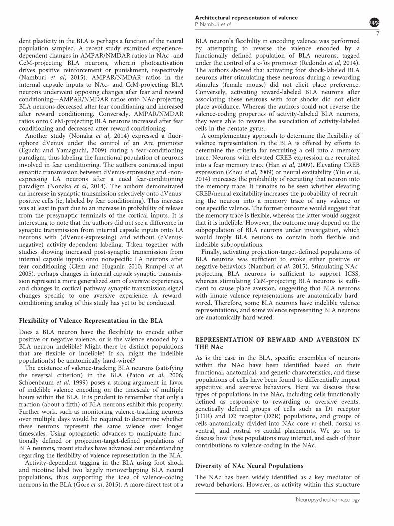
In this section, we will develop a model to parametrize theneural responses from a population of neurons in responseto cues predicting positive and negative outcomes. Thismodel summarizes neural responses into four parameters,which can then be used to contrast valence representationbetween populations of neurons either within one brainregion, or across brain regions. Applying this model toneural activity recorded from various brain regions in thecontext of valence illustrates the heterogeneity amonganatomically localized populations of neurons (Figure 3a).Applying this model to either genetically or projection-target-defined subpopulations of neurons within a brainregion (Figure 4b) illustrates that examining specificsubpopulations can help reduce heterogeneity in theirvalence-signaling properties. Therefore, this model can beused to describe valence coding neural populations.
A Neurobiological Definition of Valence
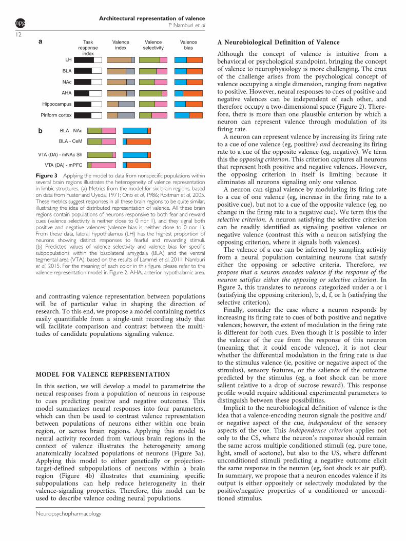
Although the concept of valence is intuitive from abehavioral or psychological standpoint, bringing the conceptof valence to neurophysiology is more challenging. The cruxof the challenge arises from the psychological concept ofvalence occupying a single dimension, ranging from negativeto positive. However, neural responses to cues of positive andnegative valences can be independent of each other, andtherefore occupy a two-dimensional space (Figure 2). There-fore, there is more than one plausible criterion by which aneuron can represent valence through modulation of itsfiring rate.A neuron can represent valence by increasing its firing rate
to a cue of one valence (eg, positive) and decreasing its firingrate to a cue of the opposite valence (eg, negative). We termthis the opposing criterion. This criterion captures all neuronsthat represent both positive and negative valences. However,the opposing criterion in itself is limiting because iteliminates all neurons signaling only one valence.A neuron can signal valence by modulating its firing rate
to a cue of one valence (eg, increase in the firing rate to apositive cue), but not to a cue of the opposite valence (eg, nochange in the firing rate to a negative cue). We term this theselective criterion. A neuron satisfying the selective criterioncan be readily identified as signaling positive valence ornegative valence (contrast this with a neuron satisfying theopposing criterion, where it signals both valences).The valence of a cue can be inferred by sampling activity
from a neural population containing neurons that satisfyeither the opposing or selective criteria. Therefore, wepropose that a neuron encodes valence if the response of theneuron satisfies either the opposing or selective criterion. InFigure 2, this translates to neurons categorized under a or i(satisfying the opposing criterion), b, d, f, or h (satisfying theselective criterion).Finally, consider the case where a neuron responds by
increasing its firing rate to cues of both positive and negativevalences; however, the extent of modulation in the firing rateis different for both cues. Even though it is possible to inferthe valence of the cue from the response of this neuron(meaning that it could encode valence), it is not clearwhether the differential modulation in the firing rate is dueto the stimulus valence (ie, positive or negative aspect of thestimulus), sensory features, or the salience of the outcomepredicted by the stimulus (eg, a foot shock can be moresalient relative to a drop of sucrose reward). This responseprofile would require additional experimental parameters todistinguish between these possibilities.Implicit to the neurobiological definition of valence is the
idea that a valence-encoding neuron signals the positive and/or negative aspect of the cue, independent of the sensoryaspects of the cue. This independence criterion applies notonly to the CS, where the neuron’s response should remainthe same across multiple conditioned stimuli (eg, pure tone,light, smell of acetone), but also to the US, where differentunconditioned stimuli predicting a negative outcome elicitthe same response in the neuron (eg, foot shock vs air puff).In summary, we propose that a neuron encodes valence if itsoutput is either oppositely or selectively modulated by thepositive/negative properties of a conditioned or uncondi-tioned stimulus.
Valenceindex
Taskresponse
index
Valenceselectivity
Valencebias
Piriform cortex
BLA
NAc
BLA - NAc
BLA - CeM
VTA (DA) - mNAc Sh
VTA (DA) - mPFC
Hippocampus
LH
AHA
Figure 3 Applying the model to data from nonspecific populations withinseveral brain regions illustrates the heterogeneity of valence representationin limbic structures. (a) Metrics from the model for six brain regions, basedon data from Fuster and Uyeda, 1971; Ono et al, 1986; Roitman et al, 2005.These metrics suggest responses in all these brain regions to be quite similar,illustrating the idea of distributed representation of valence. All these brainregions contain populations of neurons responsive to both fear and rewardcues (valence selectivity is neither close to 0 nor 1), and they signal bothpositive and negative valences (valence bias is neither close to 0 nor 1).From these data, lateral hypothalamus (LH) has the highest proportion ofneurons showing distinct responses to fearful and rewarding stimuli.(b) Predicted values of valence selectivity and valence bias for specificsubpopulations within the basolateral amygdala (BLA) and the ventraltegmental area (VTA), based on the results of Lammel et al, 2011; Namburiet al, 2015. For the meaning of each color in this figure, please refer to thevalence representation model in Figure 2. AHA, anterior hypothalamic area.
Architectural representation of valenceP Namburi et al
12
Neuropsychopharmacology

A Model for Investigating Valence Representation
On the basis of the neurobiological definition of valence coding,we will now define four terms to facilitate the understanding ofvalence representation in different brain regions or within apopulation of interest (eg, dopaminergic neurons of the VTA,NAc-projecting neurons in the BLA, or neurons in the LH).First, we begin by categorizing each neuron into one of ninepossible categories (Figure 2) based on the neuron’s response topositive and negative conditioned stimuli. Once we categorizean entire population, we use the number of neurons in eachcategory to compute the model parameters. The meaning ofeach parameter is described below, and the guide to computeeach metric is presented in Figure 2.Task response index quantifies the fraction of task-
responsive neurons. Valence index quantifies the proportionof task-responsive cells within a population that encodevalence. A high or low valence selectivity informs whether apopulation is primarily responsive to rewarding stimuli oraversive stimuli, respectively. A high valence bias suggests thepopulation to have a net excitatory output to a positivestimulus and/or inhibitory output to a negative stimulus.The use of these proposed metrics carries multiple
advantages, as described below.
Generalizability. The proposed metrics can be used toobjectively compare populations of neurons within andbetween brain regions in the context of valence learning(Figures 3 and 4).
Reward vs aversion. A population of neurons primarilyrepresenting reward will have a valence selectivity close to 1,and a population of neurons primarily representing aversion
will have a valence selectivity close to 0. The valenceselectivity of the BLA is close to 0.6, which supports the ideathat there are representations of both reward and aversion inthis region. On the basis of the results from (Xiu et al, 2014),most of the neurons in the medial amygdala and amygdala–striatal transition zone respond to foot shock, not tomorphine or chocolate rewards, and, therefore, they mayhave a valence selectivity close to 0. In contrast, neurons inthe oval nucleus of the bed nucleus of stria terminalis andlateral subdivision of the central amygdala respond torewards and not to foot shocks, and, therefore, may have avalence selectivity close to 1. Single-unit recordings areneeded to confirm this observation.
Candidate populations for studying mechanisms of valenceacquisition. A population with a high probability offinding a neuron representing valence is a good candidatefor studying valence. This probability is simply the productof the task response index and valence index. If we multiplythese two numbers, we get the probability of discovering aneuron representing valence within the given population.From this definition, the probability of finding a neuronrepresenting valence in the BLA is ~ 0.4, LH is ~ 0.6, andpiriform cortex is ~ 0.2 (Fuster and Uyeda, 1971; Ono et al,1986).
Power analysis. The objective measures provided in thisstudy can be used to inform the minimum number ofneurons needed to find desired subpopulations within apopulation. Consider the BLA for example—the probabilitythat a neuron randomly sampled from the BLA representspositive valence is at best: task response index × valence
Task response index
Val
ence
inde
xHippocampus
Piriform cortex
LH VTA (Type 1)
VTA (Type 3)
VTA (DA)
VTA (GABA)
VTA (Type 2)
VTA (Type 1/2),VTA (DA/GABA)
VTA (Type 3)
AHADRN
BLA
NAc
Candidate populations for identifying drug targets
Valence selectivity
Direct evidence
Hypothetical position from indirect evidence
Val
ence
bia
s
BLA
DRN (5-HT)
DRN (un-identified)
DRN
NAc
VTA
VTA(DA)-mPFC
VTA (DA)-mNAc Sh
BLA-CeM
BLA-NAc
Figure 4 Plotting valence representation in multiple neural populations. (a) Plotting brain regions according to their task response index and valence indexprovides an alternative visualization to contrast them. Both brain regions and specific populations of neurons can be included in this plot. (b) Contrasting neuralpopulations using valence selectivity and valence bias reveals a zone (blue) where one can find good candidate populations for circuit-based drug discoverybecause populations within this zone are relatively more selective and homogeneous in their response to positively and negatively valenced stimuli. Axes inboth (a) and (b) are between 0 and 1. Axes in (b) cross at 0.5. Data for BLA, hippocampus and piriform cortex are from Fuster and Uyeda, 1971. Data for LHand AHA are from Ono et al, 1986. Data for NAc are from Roitman et al, 2005. Data for DRN, DRN 5-HT and DRN un-identified populations are fromCohen et al, 2015. Positions of BLA-NAc and BLA-CeM are based on data from Namburi et al, 2015. Positions of VTA (DA)-mNAc Sh and VTA (DA)-mPFCare based on data from Lammel et al, 2011. Data for VTA (Type 1/2/3) and VTA (DA/GABA) populations are from Cohen et al, 2012, where the authorsdefine VTA (Type 1) as neurons that show reward prediction error like responses, VTA (Type 2) and VTA (Type 3) populations as neurons whose activitybetween CS and US was modulated positively and negatively by the value of the outcome.
Architectural representation of valenceP Namburi et al
13
Neuropsychopharmacology

index × valence bias= 0.16. Therefore, if we would like tosample neural activity from 10 positive valence-encodingneurons in the BLA (perhaps for assessing their role in adifferent behavioral paradigm), we would record from ~63neurons (10/0.16).
Parallel vs opposing pathways for reward and aversion.Valence selectivity and valence bias, taken together, caninform us whether a subpopulation is differentially con-tributing to reward or aversion, or is having an opposing rolein reward and aversion. If two subpopulations are contribut-ing differentially to reward or fear, then valence selectivitywill be close to 1 or 0, respectively, eg, based on the resultsfrom Lammel et al (2011), we may expect VTA-NAc medialshell to have a valence selectivity and valence bias close to 1and VTA-mPFC projectors to have a valence selectivity andvalence bias close to 0. If two subpopulations have opposingroles in fear and reward, then valence selectivity will be closeto 0.5, but valence bias will be close to 0 or 1. Based on theresults from Namburi et al, 2015, we can expect a mixture ofBLA-NAc and BLA-CeM neurons to have a valenceselectivity close to 0.5, with BLA-NAc having a valence biasclose to 1, and BLA-CeM neurons having a valence bias closeto 0 (Figure 3b).
Although the model introduced here has several advan-tages, it only synthesizes part of the spectrum of valencerepresentation found in the brain. For example, DRNneurons represent valence over multiple timescales (Cohenet al, 2015) and valence representations between thehippocampus and BLA vary in their degree of flexibility(Redondo et al, 2014). Neither the timescale of valencerepresentation, nor the flexibility of valence processing arecurrently captured by our model.
A circuit-based approach to identify novel drug targets.Identifying functional roles for specific circuit componentsoffers subpopulations of neurons in which to search fornovel drug targets. Within specific subpopulations ofneurons that have well-characterized roles in modulatingdisease-relevant behaviors, we can examine the transcrip-tomic profiles of these cells to reveal surface receptors thatcould be targeted for therapeutic interventions. While eachligand would need empirical testing following the develop-ment of a novel target ‘short list,’ this could enhance theselection of promising, mechanistically novel drugs fortreating neuropsychiatric disease.
The neurobiological basis of emotion has long been thefocus of intense study, and early efforts succeeded inidentifying the key nuclei that shape emotion throughencoding valence. With the advent of modern techniquesin neuroscience, we are now moving beyond evaluating thenet effect of entire brain regions upon emotional behaviors,and instead beginning to parse regions into the specificpopulations of neurons responsible for valence processing.As reviewed here, these populations can be segregated fromneighboring cells based upon anatomical, genetic, and/orfunctional categorizations. Because populations of cellsdefined by these categories can now be readily targeted andmanipulated, we anticipate the imminent discovery ofmultiple valence-encoding populations of neurons distrib-uted throughout the brain. These discoveries will require a
unified model to serve as the basis of comparison amongspecific populations, to clearly delineate their uniquecontributions to valence coding. To meet this need, we offerhere one such model, which we advance in an effort tocompare and contrast the neural substrates of valence-processing and emotion distributed in the brain.
FUNDING AND DISCLOSURE
KMT is a New York Stem Cell Foundation—RobertsonInvestigator and McKnight Scholar, and this work wassupported by funding from the JPB Foundation, PIIF,PNDRF, JFDP, Whitehall Foundation, Klingenstein Founda-tion, NARSAD Young Investigator Award, Alfred P SloanFoundation, New York Stem Cell Foundation, McKnightFoundation, Whitehead Career Development Chair, NIHR01-MH102441-01 (NIMH) and NIH Director’s NewInnovator Award DP2-DK-102256-01 (NIDDK). PN wassupported by the Whitaker fellowship. GGC is supported bythe JFDP Postdoctoral Fellowship from the JPB Foundation.This work is supported by NIDA R01 DA033396 (MRB),EUREKA grant R01DA037152 (MRB) and NIDA K99/R00Pathway to Independence Award DA038725 (RA-H). Theremaining authors declare no conflict of interest.
ACKNOWLEDGMENTS
We thank A Beyeler and the entire Tye Laboratory forhelpful discussion. We thank J Cohen for sharing quantita-tive information from previously published work for use inFigures 2 and 4.
REFERENCES
Abercrombie ED, Keefe KA, DiFrischia DS, Zigmond MJ (1989).Differential effect of stress on in vivo dopamine release instriatum, nucleus accumbens, and medial frontal cortex.J Neurochem 52: 1655–1658.
Adolphs R, Russell JA, Tranel D (1999). A role for the humanamygdala in recognizing emotional arousal from unpleasantstimuli. Psychol Sci 10: 167–171.
Airan RD, Thompson KR, Fenno LE, Bernstein H, Deisseroth K(2009). Temporally precise in vivo control of intracellularsignalling. Nature 458: 1025–1029.
Al-Hasani R, McCall JG, Shin G, Gomez AM, Schmitz GP,Bernardi JM et al (2015). Distinct subpopulations of nucleusaccumbens dynorphin neurons drive aversion and reward.Neuron 87: 1063–1077.
Aragona BJ, Cleaveland NA, Stuber GD, Day JJ, Carelli RM,Wightman RM (2008). Preferential enhancement of dopaminetransmission within the nucleus accumbens shell by cocaine isattributable to a direct increase in phasic dopamine release events.J Neurosci Off J Soc Neurosci 28: 8821–8831.
Atasoy D, Aponte Y, Su HH, Sternson SM (2008). A FLEX switchtargets channelrhodopsin-2 to multiple cell types for imaging andlong-range circuit mapping. J Neurosci 28: 7025–7030.
Bardo MT (1998). Neuropharmacological mechanisms of drugreward: beyond dopamine in the nucleus accumbens. Crit RevNeurobiol 12: 37–67.
Bassareo V, De Luca MA, Di Chiara G (2002). Differentialexpression of motivational stimulus properties by dopamine innucleus accumbens shell versus core and prefrontal cortex.J Neurosci Off J Soc Neurosci 22: 4709–4719.
Architectural representation of valenceP Namburi et al
14
Neuropsychopharmacology

Belova MA, Paton JJ, Morrison SE, Salzman CD (2007). Expectationmodulates neural responses to pleasant and aversive stimuli inprimate amygdala. Neuron 55: 970–984.
Berridge KC, Kringelbach ML (2015). Pleasure systems in the brain.Neuron 86: 646–664.
Berridge KC, Robinson TE (1998). What is the role of dopamine inreward: hedonic impact, reward learning, or incentive salience?Brain Res Brain Res Rev 28: 309–369.
Berton O, McClung CA, Dileone RJ, Krishnan V, Renthal W,Russo SJ et al (2006). Essential role of BDNF in the mesolimbicdopamine pathway in social defeat stress. Science 311: 864–868.
Boender AJ, de Jong JW, Boekhoudt L, Luijendijk MCM, van derPlasse G, Adan RAH (2014). Combined use of the canineadenovirus-2 and DREADD-technology to activate specific neuralpathways in vivo. PLoS ONE 9: e95392.
Bouret S, Richmond BJ (2015). Sensitivity of locus ceruleus neuronsto reward value for goal-directed actions. J Neurosci 35: 4005–4014.
Britt JP, Benaliouad F, McDevitt RA, Stuber GD, Wise RA, Bonci A(2012). Synaptic and behavioral profile of multiple glutamatergicinputs to the nucleus accumbens. Neuron 76: 790–803.
Britt JP, Bonci A (2013). Optogenetic interrogations of theneural circuits underlying addiction. Curr Opin Neurobiol 23:539–545.
Bromberg-Martin ES, Matsumoto M, Hikosaka O (2010). Dopa-mine in motivational control: rewarding, aversive, and alerting.Neuron 68: 815–834.
Brown MTC, Tan KR, O’Connor EC, Nikonenko I, Muller D,Lüscher C (2012). Ventral tegmental area GABA projectionspause accumbal cholinergic interneurons to enhance associativelearning. Nature 492: 452–456.
Bull C, Freitas KC, Zou S, Poland RS, Syed WA, Urban DJ et al(2014). Rat nucleus accumbens core astrocytes modulate rewardand the motivation to self-administer ethanol after abstinence.Neuropsychopharmacology 39: 2835–2845.
Cai H, Haubensak W, Anthony TE, Anderson DJ (2014). Centralamygdala PKC-δ+ neurons mediate the influence of multipleanorexigenic signals. Nat Neurosci 17: 1240–1248.
Calhoon GG, Tye KM (2015). Resolving the neural circuits ofanxiety. Nat Neurosci 18: 1394–1404.
Callaway EM, Luo L (2015). Monosynaptic circuit tracing withglycoprotein-deleted rabies viruses. J Neurosci 35: 8979–8985.
Cannon CM, Palmiter RD (2003). Reward without dopamine.J Neurosci Off J Soc Neurosci 23: 10827–10831.
Capogna M (2014). GABAergic cell type diversity in the basolateralamygdala. Curr Opin Neurobiol 26: 110–116.
Carlezon WA, Chartoff EH (2007). Intracranial self-stimulation(ICSS) in rodents to study the neurobiology of motivation. NatProtoc 2: 2987–2995.
Carlezon WA, Thomas MJ (2009). Biological substrates of rewardand aversion: a nucleus accumbens activity hypothesis. Neuro-pharmacology 56(Suppl 1): 122–132.
Cassataro D, Bergfeldt D, Malekian C, Van Snellenberg JX, Thanos PK,Fishell G et al (2014). Reverse pharmacogenetic modulation of thenucleus accumbens reduces ethanol consumption in a limited accessparadigm. Neuropsychopharmacology 39: 283–290.
Castro DC, Berridge KC (2014). Opioid hedonic hotspot in nucleusaccumbens shell: mu, delta, and kappa maps for enhancement ofsweetness ‘liking’ and ‘wanting.’. J Neurosci Off J Soc Neurosci 34:4239–4250.
Chaudhury D, Walsh JJ, Friedman AK, Juarez B, Ku SM,Koo JW et al (2013). Rapid regulation of depression-relatedbehaviours by control of midbrain dopamine neurons. Nature493: 532–536.
Clem RL, Huganir RL (2010). Calcium-permeable AMPA receptordynamics mediate fear memory erasure. Science 330: 1108–1112.
Cohen JY, Amoroso MW, Uchida N (2015). Serotonergic neuronssignal reward and punishment on multiple timescales. eLife 4:e06346.
Cohen JY, Haesler S, Vong L, Lowell BB, Uchida N (2012). Neuron-type-specific signals for reward and punishment in the ventraltegmental area. Nature 482: 85–88.
Danjo T, Yoshimi K, Funabiki K, Yawata S, Nakanishi S (2014).Aversive behavior induced by optogenetic inactivation of ventraltegmental area dopamine neurons is mediated by dopamine D2receptors in the nucleus accumbens. Proc Natl Acad Sci 111:6455–6460.
Davis M (1992). The role of the amygdala in fear and anxiety. AnnuRev Neurosci 15: 353–375.
Denny CA, Kheirbek MA, Alba EL, Tanaka KF, Brachman RA,Laughman KB et al (2014). Hippocampal memory traces aredifferentially modulated by experience, time, and adult neurogen-esis. Neuron 83: 189–201.
Di Chiara G, Bassareo V (2007). Reward system and addiction: whatdopamine does and doesn’t do. Curr Opin Pharmacol 7: 69–76.
Eguchi M, Yamaguchi S (2009). In vivo and in vitro visualization ofgene expression dynamics over extensive areas of the brain.NeuroImage 44: 1274–1283.
Faure A, Richard JM, Berridge KC (2010). Desire and dread fromthe nucleus accumbens: cortical glutamate and subcortical GABAdifferentially generate motivation and hedonic impact in the rat.PLoS ONE 5: e11223.
Felix-Ortiz AC, Beyeler A, Seo C, Leppla CA, Wildes CP, Tye KM(2013). BLA to vHPC inputs modulate anxiety-related behaviors.Neuron 79: 658–664.
Felix-Ortiz AC, Burgos-Robles A, Bhagat ND, Leppla CA, Tye KM(2015). Bidirectional modulation of anxiety-related and socialbehaviors by amygdala projections to the medial prefrontal cortex.Neuroscience. doi:10.1016/j.neuroscience.2015.07.041 (e-pub aheadof print).
Felix-Ortiz AC, Tye KM (2014). Amygdala inputs to the ventralhippocampus bidirectionally modulate social behavior. J NeurosciOff J Soc Neurosci 34: 586–595.
Fenno LE, Mattis J, Ramakrishnan C, Hyun M, Lee SY, He M et al(2014). Targeting cells with single vectors using multiple-featureBoolean logic. Nat Methods 11: 763–772.
Fosque BF, Sun Y, Dana H, Yang C-T, Ohyama T, Tadross MR et al(2015). Labeling of active neural circuits in vivo with designedcalcium integrators. Science 347: 755–760.
Francis TC, Chandra R, Friend DM, Finkel E, Dayrit G, Miranda Jet al (2015). Nucleus accumbens medium spiny neuron subtypesmediate depression-related outcomes to social defeat stress. BiolPsychiatry 77: 212–222.
Fukuda M, Ono T, Nakamura K, Tamura R (1990). Dopamine andACH involvement in plastic learning by hypothalamic neuronsin rats. Brain Res Bull 25: 109–114.
Fuster JM, Uyeda AA (1971). Reactivity of limbic neurons of themonkey to appetitive and aversive signals. Electroencephalogr ClinNeurophysiol 30: 281–293.
Garner AR, Rowland DC, Hwang SY, Baumgaertel K, Roth BL,Kentros C et al (2012). Generation of a synthetic memory trace.Science 335: 1513–1516.
Gerfen CR (1992). The neostriatal mosaic: multiple levels ofcompartmental organization. Trends Neurosci 15: 133–139.
Gipson CD, Kupchik YM, Kalivas PW (2014). Rapid, transientsynaptic plasticity in addiction. Neuropharmacology 76 Pt B:276–286.
Gong S, Doughty M, Harbaugh CR, Cummins A, Hatten ME,Heintz N et al (2007). Targeting Cre recombinase to specificneuron populations with bacterial artificial chromosome con-structs. J Neurosci 27: 9817–9823.
Gore F, Schwartz EC, Brangers BC, Aladi S, Stujenske JM, Likhtik Eet al (2015). Neural representations of unconditioned stimuli inbasolateral amygdala mediate innate and learned responses. Cell162: 134–145.
Goto Y, Otani S, Grace AA (2007). The Yin and Yang of dopaminerelease: a new perspective. Neuropharmacology 53: 583–587.
Architectural representation of valenceP Namburi et al
15
Neuropsychopharmacology

Grueter BA, Rothwell PE, Malenka RC (2012). Integrating synapticplasticity and striatal circuit function in addiction. Curr OpinNeurobiol 22: 545–551.
Guenthner CJ, Miyamichi K, Yang HH, Heller HC, Luo L (2013).Permanent genetic access to transiently active neurons via TRAP:targeted recombination in active populations. Neuron 78: 773–784.
Gunaydin LA, Grosenick L, Finkelstein JC, Kauvar IV, Fenno LE,Adhikari A et al (2014). Natural neural projection dynamicsunderlying social behavior. Cell 157: 1535–1551.
Han J-H, Kushner SA, Yiu AP, Hsiang H.-L.L., Buch T, Waisman Aet al (2009). Selective erasure of a fear memory. Science 323:1492–1496.
Harris GC, Aston-Jones G (2006). Arousal and reward: a dichotomyin orexin function. Trends Neurosci 29: 571–577.
Hikida T, Kimura K, Wada N, Funabiki K, Nakanishi S (2010).Distinct roles of synaptic transmission in direct and indirectstriatal pathways to reward and aversive behavior. Neuron 66:896–907.
Hikida T, Yawata S, Yamaguchi T, Danjo T, Sasaoka T, Wang Yet al (2013). Pathway-specific modulation of nucleus accumbensin reward and aversive behavior via selective transmitterreceptors. Proc Natl Acad Sci USA 110: 342–347.
Hnasko TS, Perez FA, Scouras AD, Stoll EA, Gale SD, Luquet S et al(2006). Cre recombinase-mediated restoration of nigrostriataldopamine in dopamine-deficient mice reverses hypophagia andbradykinesia. Proc Natl Acad Sci USA 103: 8858–8863.
Hofmeister J, Sterpenich V (2015). A role for the locus ceruleus inreward processing: encoding behavioral energy required for goal-directed actions. J Neurosci 35: 10387–10389.
Hyman SE, Malenka RC, Nestler EJ (2006). Neural mechanisms ofaddiction: the role of reward-related learning and memory. AnnuRev Neurosci 29: 565–598.
Ilango A, Kesner AJ, Keller KL, Stuber GD, Bonci A, Ikemoto S(2014). Similar roles of substantia nigra and ventral tegmentaldopamine neurons in reward and aversion. J Neurosci Off J SocNeurosci 34: 817–822.
James W (1884). What is an emotion? Mind 9: 188–205.Janak PH, Tye KM (2015). From circuits to behaviour in theamygdala. Nature 517: 284–292.
Jennings JH, Sparta DR, Stamatakis AM, Ung RL, Pleil KE, Kash TLet al (2013). Distinct extended amygdala circuits for divergentmotivational states. Nature 496: 224–228.
Jeong J-W, McCall JG, Shin G, Zhang Y, Al-Hasani R, Kim M et al(2015). Wireless optofluidic systems for programmable in vivopharmacology and optogenetics. Cell 162: 662–674.
Kelley AE (2004). Ventral striatal control of appetitive motivation:role in ingestive behavior and reward-related learning. NeurosciBiobehav Rev 27: 765–776.
Kempadoo KA, Tourino C, Cho SL, Magnani F, Leinninger G-M,Stuber GD et al (2013). Hypothalamic neurotensin projectionspromote reward by enhancing glutamate transmission inthe VTA. J Neurosci Off J Soc Neurosci 33: 7618–7626.
Kemppainen S, Pitkänen A (2000). Distribution of parvalbumin,calretinin, and calbindin-D28k immunoreactivity in the ratamygdaloid complex and colocalization with γ-aminobuty-ric acid. J Comp Neurol 426: 441–467.
Kim T, McCall JG, Jung YH, Huang X, Siuda ER, Li Y et al (2013).Injectable, cellular-scale optoelectronics with applications forwireless optogenetics. Science 340: 211–216.
Kita H, Kitai ST (1988). Glutamate decarboxylase immunoreactiveneurons in rat neostriatum: their morphological types andpopulations. Brain Res 447: 346–352.
Klüver H, Bucy P (1939). Preliminary analysis of functions of thetemporal lobes in monkeys. Arch Neurol Psychiatry 9: 606–620.
Kolb B, Gorny G, Söderpalm AHV, Robinson TE (2003).Environmental complexity has different effects on the structureof neurons in the prefrontal cortex versus the parietal cortex ornucleus accumbens. Synap N Y N 48: 149–153.
Konermann S, Brigham MD, Trevino AE, Hsu PD, Heidenreich M,Cong L et al (2013). Optical control of mammalian endogenoustranscription and epigenetic states. Nature 500: 472–476.
Krause M, German PW, Taha SA, Fields HL (2010). A pause innucleus accumbens neuron firing is required to initiate andmaintain feeding. J Neurosci Off J Soc Neurosci 30: 4746–4756.
Kravitz AV, Tye LD, Kreitzer AC (2012). Distinct roles for directand indirect pathway striatal neurons in reinforcement. NatNeurosci 15: 816–818.
Kremer EJ, Boutin S, Chillon M, Danos O (2000). Canineadenovirus vectors: an alternative for adenovirus-mediated genetransfer. J Virol 74: 505–512.
Kupchik YM, Brown RM, Heinsbroek JA, Lobo MK, Schwartz DJ,Kalivas PW (2015). Coding the direct/indirect pathways by D1and D2 receptors is not valid for accumbens projections. NatNeurosci 18: 1230–1232.
Lammel S, Ion DI, Roeper J, Malenka RC (2011). Projection-specificmodulation of dopamine neuron synapses by aversive andrewarding stimuli. Neuron 70: 855–862.
Lammel S, Lim BK, Malenka RC (2014a). Reward and aversion in aheterogeneous midbrain dopamine system. Neuropharmacology76 Pt B: 351–359.
Lammel S, Lim BK, Ran C, Huang KW, Betley MJ, Tye KM et al(2012). Input-specific control of reward and aversion in theventral tegmental area. Nature 491: 212–217.
Lammel S, Tye KM, Warden MR (2014b). Progress in under-standing mood disorders: optogenetic dissection of neuralcircuits. Genes Brain Behav 13: 38–51.
Lang PJ, Bradley MM, Fitzsimmons JR, Cuthbert BN, Scott JD,Moulder B et al (1998). Emotional arousal and activation of thevisual cortex: an fMRI analysis. Psychophysiology 35: 199–210.
Larson EB, Wissman AM, Loriaux AL, Kourrich S, Self DW (2015).Optogenetic stimulation of accumbens shell or shell projections tolateral hypothalamus produce differential effects on the motiva-tion for cocaine. J Neurosci 35: 3537–3543.
LeDoux JE (2000). Emotion circuits in the brain. Annu Rev Neurosci23: 155–184.
LeDoux JE, Cicchetti P, Xagoraris A, Romanski LM (1990). Thelateral amygdaloid nucleus: sensory interface of the amygdala infear conditioning. J Neurosci 10: 1062–1069.
Lee AT, Vogt D, Rubenstein JL, Sohal VS (2014). A class ofGABAergic neurons in the prefrontal cortex sends long-rangeprojections to the nucleus accumbens and elicits acute avoidancebehavior. J Neurosci Off J Soc Neurosci 34: 11519–11525.
Li JX, Yoshida T, Monk KJ, Katz DB (2013). Lateral hypothalamuscontains two types of palatability-related taste responses withdistinct dynamics. J Neurosci 33: 9462–9473.
Lima SQ, Hromádka T, Znamenskiy P, Zador AM (2009). PINP: anew method of tagging neuronal populations for identificationduring in vivo electrophysiological recording. PLoS ONE 4: e6099.
Liu X, Ramirez S, Pang PT, Puryear CB, Govindarajan A, Deisseroth Ket al (2012). Optogenetic stimulation of a hippocampal engramactivates fear memory recall. Nature 484: 381–385.
Lobo MK, Covington HE, Chaudhury D, Friedman AK, Sun H,Damez-Werno D et al (2010). Cell type-specific loss of BDNFsignaling mimics optogenetic control of cocaine reward. Science330: 385–390.
Loriaux AL, Roitman JD, Roitman MF (2011). Nucleus accumbensshell, but not core, tracks motivational value of salt.J Neurophysiol 106: 1537–1544.
Lüscher C, Malenka RC (2011). Drug-evoked synaptic plasticity inaddiction: from molecular changes to circuit remodeling. Neuron69: 650–663.
MacAskill AF, Cassel JM, Carter AG (2014). Cocaine exposurereorganizes cell type- and input-specific connectivity in thenucleus accumbens. Nat Neurosci 17: 1198–1207.
Machne X, Segundo JP (1956). Unitary responses to afferent volleysin amygdaloid complex. J Neurophysiol 19: 232–240.
Architectural representation of valenceP Namburi et al
16
Neuropsychopharmacology

Marchant NJ, Whitaker LR, Bossert JM, Harvey BK, Hope BT,Kaganovsky K et al (2016). Behavioral and physiological effects ofa novel kappa-opioid receptor-based DREADD in rats. Neurop-sychopharmacology 41: 402–409.
Maren S (2001). Neurobiology of pavlovian fear conditioning. AnnuRev Neurosci 24: 897–931.
Matsumoto M, Hikosaka O (2009). Two types of dopamine neurondistinctly convey positive and negative motivational signals.Nature 459: 837–841.
McCall JG, Al-Hasani R, Siuda ER, Hong DY, Norris AJ, Ford CPet al (2015). CRH engagement of the locus coeruleus noradre-nergic system mediates stress-induced anxiety. Neuron 87:605–620.
McCall JG, Kim T, Shin G, Huang X, Jung YH, Al-Hasani R et al(2013). Fabrication and application of flexible, multimodal light-emitting devices for wireless optogenetics. Nat Protoc 8: 2413–2428.
McDevitt RA, Tiran-Cappello A, Shen H, Balderas I, Britt JP,Marino RA et al (2014). Serotonergic versus non-serotonergicdorsal raphe projection neurons: differential participation inreward circuitry. Cell Rep 8: 1857–1869.
McDonald AJ (1987). Somatostatinergic projections from theamygdala to the bed nucleus of the stria terminalis and medialpreoptic-hypothalamic region. Neurosci Lett 75: 271–277.
McDonald AJ (1991a). Topographical organization of amygdaloidprojections to the caudatoputamen, nucleus accumbens, andrelated striatal-like areas of the rat brain. Neuroscience 44: 15–33.
McDonald AJ (1991b). Organization of amygdaloid projections tothe prefrontal cortex and associated striatum in the rat.Neuroscience 44: 1–14.
McDonald AJ, Augustine JR (1993). Localization of GABA-likeimmunoreactivity in the monkey amygdala. Neuroscience 52:281–294.
McDonald AJ, Mascagni F (2002). Immunohistochemical charac-terization of somatostatin containing interneurons in the ratbasolateral amygdala. Brain Res 943: 237–244.
McDonald AJ, Mascagni F, Zaric V (2012). Subpopulations ofsomatostatin-immunoreactive non-pyramidal neurons in theamygdala and adjacent external capsule project to the basalforebrain: evidence for the existence of GABAergic projectionneurons in the cortical nuclei and basolateral nuclear complex.Front Neural Circuits 6: 46.
McDonald AJ, Zaric V (2015). Gabaergic somatostatin-immunoreactive neurons in the amygdala project to theentorhinal cortex. Neuroscience 290: 227–242.
McGaugh JL (2000). Memory–a century of consolidation. Science287: 248–251.
McGaugh JL (2004). The amygdala modulates the consolidation ofmemories of emotionally arousing experiences. Annu RevNeurosci 27: 1–28.
McIntyre CK, Roozendaal B (2007)Adrenal Stress hormonesand enhanced memory for emotionally arousing experiences.In: Bermúdez-Rattoni F(ed) Neural Plasticity and Memory: FromGenes to Brain Imaging. CRC Press: Boca Raton, FL, USA.
McKernan MG, Shinnick-Gallagher P (1997). Fear conditioninginduces a lasting potentiation of synaptic currents in vitro. Nature390: 607–611.
Millhouse OE, DeOlmos J (1983). Neuronal configurations in lateraland basolateral amygdala. Neuroscience 10: 1269–1300.
Morales M, Pickel VM (2012). Insights to drug addiction derivedfrom ultrastructural views of the mesocorticolimbic system. AnnN Y Acad Sci 1248: 71–88.
Muller JF, Mascagni F, McDonald AJ (2006). Pyramidal cells of therat basolateral amygdala: synaptology and innervation byparvalbumin-immunoreactive interneurons. J Comp Neurol 494:635–650.
Muller JF, Mascagni F, McDonald AJ (2007). Postsynaptic targets ofsomatostatin-containing interneurons in the rat basolateralamygdala. J Comp Neurol 500: 513–529.
Nakamura K, Ono T, Tamura R (1987). Central sites involved inlateral hypothalamus conditioned neural responses to acousticcues in the rat. J Neurophysiol 58: 1123–1148.
Namburi P, Beyeler A, Yorozu S, Calhoon GG, Halbert SA,Wichmann R et al (2015). A circuit mechanism for differentiatingpositive and negative associations. Nature 520: 675–678.
Nieh EH, Kim S-Y, Namburi P, Tye KM (2013). Optogeneticdissection of neural circuits underlying emotional valence andmotivated behaviors. Brain Res 1511: 73–92.
Nieh EH, Matthews GA, Allsop SA, Presbrey KN, Leppla CA,Wichmann R et al (2015). Decoding neural circuits that controlcompulsive sucrose seeking. Cell 160: 528–541.
Nishijo H, Ono T, Nishino H (1988). Single neuron responses inamygdala of alert monkey during complex sensory stimulationwith affective significance. J Neurosci Off J Soc Neurosci 8:3570–3583.
Nonaka A, Toyoda T, Miura Y, Hitora-Imamura N, Naka M, Eguchi Met al (2014). Synaptic plasticity associated with a memory engram inthe basolateral amygdala. J Neurosci 34: 9305–9309.
Nowlis V, Nowlis HH (1956). The description and analysisof mood. Ann N Y Acad Sci 65: 345–355.
Ogawa SK, Cohen JY, Hwang D, Uchida N, Watabe-Uchida M(2014). Organization of monosynaptic inputs to the serotonin anddopamine neuromodulatory systems. Cell Rep 8: 1105–1118.
Ono T, Nakamura K, Nishijo H, Fukuda M (1986). Hypothalamicneuron involvement in integration of reward, aversion, and cuesignals. J Neurophysiol 56: 63–79.
Paton JJ, Belova MA, Morrison SE, Salzman CD (2006). Theprimate amygdala represents the positive and negative value ofvisual stimuli during learning. Nature 439: 865–870.
Pavlov IP (1927). Conditioned Reflexes: An Investigation of thePhysiological Activity of the Cerebral Cortex. Oxford UniversityPress: Humphrey Milford.
Pettit HO, Ettenberg A, Bloom FE, Koob GF (1984). Destruction ofdopamine in the nucleus accumbens selectively attenuates cocainebut not heroin self-administration in rats. Psychopharmacology(Berl) 84: 167–173.
Pikkarainen M, Rönkkö S, Savander V, Insausti R, Pitkänen A(1999). Projections from the lateral, basal, and accessory basalnuclei of the amygdala to the hippocampal formation in rat.J Comp Neurol 403: 229–260.
Pitchers KK, Balfour ME, Lehman MN, Richtand NM, Yu L,Coolen LM (2010). Neuroplasticity in the mesolimbic systeminduced by natural reward and subsequent reward abstinence.Biol Psychiatry 67: 872–879.
Pitchers KK, Schmid S, Di Sebastiano AR, Wang X, Laviolette SR,Lehman MN et al (2012). Natural reward experience alters AMPAand NMDA receptor distribution and function in the nucleusaccumbens. PLoS ONE 7: e34700.
Pitkänen A, Savander V, LeDoux JE (1997). Organization of intra-amygdaloid circuitries in the rat: an emerging framework forunderstanding functions of the amygdala. Trends Neurosci 20:517–523.
Pollak Dorocic I, Fürth D, Xuan Y, Johansson Y, Pozzi L, Silberberg Get al (2014). A whole-brain atlas of inputs to serotonergic neurons ofthe dorsal and median raphe nuclei. Neuron 83: 663–678.
Posner J, Russell JA, Peterson BS (2005). The circumplex model ofaffect: an integrative approach to affective neuroscience, cognitivedevelopment, and psychopathology. Dev Psychopathol 17:715–734.
Redondo RL, Kim J, Arons AL, Ramirez S, Liu X, Tonegawa S(2014). Bidirectional switch of the valence associated with ahippocampal contextual memory engram. Nature 513: 426–430.
Reijmers L, Mayford M (2009). Genetic control of active neuralcircuits. Front Mol Neurosci 2: 27.
Reijmers LG, Perkins BL, Matsuo N, Mayford M (2007). Localiza-tion of a stable neural correlate of associative memory. Science317: 1230–1233.
Architectural representation of valenceP Namburi et al
17
Neuropsychopharmacology

Rescorla RA (1988). Behavioral studies of pavlovian conditioning.Annu Rev Neurosci 11: 329–352.
Reynolds SM, Berridge KC (2002). Positive and negative motivationin nucleus accumbens shell: bivalent rostrocaudal gradients forGABA-elicited eating, taste ‘liking’/’disliking’ reactions, placepreference/avoidance, and fear. J Neurosci Off J Soc Neurosci 22:7308–7320.
Reynolds SM, Berridge KC (2008). Emotional environments retunethe valence of appetitive versus fearful functions in nucleusaccumbens. Nat Neurosci 11: 423–425.
Richard JM, Berridge KC (2011). Metabotropic glutamate receptorblockade in nucleus accumbens shell shifts affective valencetowards fear and disgust. Eur J Neurosci 33: 736–747.
Richard JM, Plawecki AM, Berridge KC (2013). Nucleus accumbensGABAergic inhibition generates intense eating and fear thatresists environmental retuning and needs no local dopamine. EurJ Neurosci 37: 1789–1802.
Robbins TW, Everitt BJ (1996). Neurobehavioural mechanisms ofreward and motivation. Curr Opin Neurobiol 6: 228–236.
Robinson S, Sandstrom SM, Denenberg VH, Palmiter RD (2005).Distinguishing whether dopamine regulates liking, wanting,and/or learning about rewards. Behav Neurosci 119: 5–15.
Rogan MT, Stäubli UV, LeDoux JE (1997). Fear conditioninginduces associative long-term potentiation in the amygdala.Nature 390: 604–607.
Roitman MF, Stuber GD, Phillips PEM, Wightman RM, Carelli RM(2004). Dopamine operates as a subsecond modulator of foodseeking. J Neurosci Off J Soc Neurosci 24: 1265–1271.
Roitman MF, Wheeler RA, Carelli RM (2005). Nucleus accumbensneurons are innately tuned for rewarding and aversive tastestimuli, encode their predictors, and are linked to motor output.Neuron 45: 587–597.
Roitman MF, Wheeler RA, Tiesinga PHE, Roitman JD, Carelli RM(2010). Hedonic and nucleus accumbens neural responses to anatural reward are regulated by aversive conditioning. Learn MemCold Spring Harb N 17: 539–546.
Roitman MF, Wheeler RA, Wightman RM, Carelli RM (2008). Real-time chemical responses in the nucleus accumbens differentiaterewarding and aversive stimuli. Nat Neurosci 11: 1376–1377.
Rossi NA, Reid LD (1976). Affective states associated withmorphine injections. Physiol Psychol 4: 269–274.
Rumpel S, LeDoux J, Zador A, Malinow R (2005). Postsynapticreceptor trafficking underlying a form of associative learning.Science 308: 83–88.
Russell JA (1980). A circumplex model of affect. J Pers Soc Psychol39: 1161–1178.
Saddoris MP, Sugam JA, Cacciapaglia F, Carelli RM (2013). Rapiddopamine dynamics in the accumbens core and shell: learningand action. Front Biosci Elite Ed 5: 273–288.
Sah P, Faber ESL, Lopez De Armentia M, Power J (2003). Theamygdaloid complex: anatomy and physiology. Physiol Rev 83:803–834.
Salamone J, Correa M, Mingote S, Weber S (2005). Beyond thereward hypothesis: alternative functions of nucleus accumbensdopamine. Curr Opin Pharmacol 5: 34–41.
Sawa M, Dalgado JR (1963). Amygdala unitary activity in the unre-strained cat. Electroencephalogr Clin Neurophysiol 15: 637–650.
Schindler SE, McCall JG, Yan P, Hyrc KL, Li M, Tucker CL et al(2015). Photo-activatable Cre recombinase regulates gene expres-sion in vivo. Sci Rep 5: 13627.
Schlosberg H (1954). Three dimensions of emotion. Psychol Rev 61:81–88.
Schoenbaum G, Chiba AA, Gallagher M (1999). Neural encoding inorbitofrontal cortex and basolateral amygdala during olfactorydiscrimination learning. J Neurosci 19: 1876–1884.
Senn V, Wolff SBE, Herry C, Grenier F, Ehrlich I, Gründemann Jet al (2014). Long-range connectivity defines behavioral specifi-city of amygdala neurons. Neuron 81: 428–437.
Shabel SJ, Janak PH (2009). Substantial similarity in amygdalaneuronal activity during conditioned appetitive and aversiveemotional arousal. Proc Natl Acad Sci USA 106: 15031–15036.
Shinonaga Y, Takada M, Mizuno N (1994). Topographic organiza-tion of collateral projections from the basolateral amygdaloidnucleus to both the prefrontal cortex and nucleus accumbens inthe rat. Neuroscience 58: 389–397.
Sieger T, Serranová T, Růžička F, Vostatek P, Wild J, Šťastná D et al(2015). Distinct populations of neurons respond to emotionalvalence and arousal in the human subthalamic nucleus. Proc NatlAcad Sci USA 112: 3116–3121.
Siuda ER, Copits BA, Schmidt MJ, Baird MA, Al-Hasani R,Planer WJ et al (2015). Spatiotemporal control of opioid signalingand behavior. Neuron 86: 923–935.
Skinner BF. The Behavior of Organisms: an Experimental Analysis.Appleton-Century-Crofts: New York, 1938, 457p.
Spanagel R, Weiss F (1999). The dopamine hypothesis of reward:past and current status. Trends Neurosci 22: 521–527.
Spragg SD (1940). Morphine addiction in chimpanzees. CompPsychol Monogr 15: 132.
Stamatakis AM, Stuber GD (2012). Activation of lateral habenulainputs to the ventral midbrain promotes behavioral avoidance.Nat Neurosci 15: 1105–1107.
Stefanik MT, Kupchik YM, Brown RM, Kalivas PW (2013b).Optogenetic evidence that pallidal projections, not nigral projec-tions, from the nucleus accumbens core are necessary forreinstating cocaine seeking. J Neurosci Off J Soc Neurosci 33:13654–13662.
Stefanik MT, Moussawi K, Kupchik YM, Smith KC, Miller RL, HuffML et al (2013a). Optogenetic inhibition of cocaine seekingin rats. Addict Biol 18: 50–53.
Steinberg EE, Boivin JR, Saunders BT, Witten IB, Deisseroth K,Janak PH (2014). Positive reinforcement mediated by midbraindopamine neurons requires D1 and D2 receptor activation in thenucleus accumbens. PLoS ONE 9: e94771.
Stuber GD, Sparta DR, Stamatakis AM, van Leeuwen WA,Hardjoprajitno JE, Cho S et al (2011). Excitatory transmissionfrom the amygdala to nucleus accumbens facilitates rewardseeking. Nature 475: 377–380.
Stuber GD, Wightman RM, Carelli RM (2005). Extinction ofcocaine self-administration reveals functionally and temporallydistinct dopaminergic signals in the nucleus accumbens. Neuron46: 661–669.
Swanson LW (1982). The projections of the ventral tegmental areaand adjacent regions: a combined fluorescent retrograde tracerand immunofluorescence study in the rat. Brain Res Bull 9:321–353.
Tan KR, Yvon C, Turiault M, Mirzabekov JJ, Doehner J,Labouèbe G et al (2012). GABA neurons of the VTA driveconditioned place aversion. Neuron 73: 1173–1183.
Taniguchi H, He M, Wu P, Kim S, Paik R, Sugino K et al (2011). Aresource of Cre driver lines for genetic targeting of gabaergicneurons in cerebral cortex. Neuron 71: 995–1013.
Taslimi A, Vrana JD, Chen D, Borinskaya S, Mayer BJ, Kennedy MJet al (2014). An optimized optogenetic clustering tool for probingprotein interaction and function. Nat Commun 5: 4925.
Tepper JM, Bolam JP (2004). Functional diversity and specificity ofneostriatal interneurons. Curr Opin Neurobiol 14: 685–692.
Tsai H-C, Zhang F, Adamantidis A, Stuber GD, Bonci A, de Lecea Let al (2009). Phasic firing in dopaminergic neurons is sufficientfor behavioral conditioning. Science 324: 1080–1084.
Tucker CL, Vrana JD, Kennedy MJ (2014). Tools for controllingprotein interactions using light. Curr Protoc Cell Biol Editor BoardJuan Bonifacino Al 64: 17.16.1–17.16.20.
Tukey DS, Lee M, Xu D, Eberle SE, Goffer Y, Manders TR et al(2013). Differential effects of natural rewards and pain onvesicular glutamate transporter expression in the nucleusaccumbens. Mol Brain 6: 32.
Architectural representation of valenceP Namburi et al
18
Neuropsychopharmacology

Tye KM, Deisseroth K (2012). Optogenetic investigation of neuralcircuits underlying brain disease in animal models. Nat RevNeurosci 13: 251–266.
Tye KM, Mirzabekov JJ, Warden MR, Ferenczi EA, Tsai H-C,Finkelstein J et al (2013). Dopamine neurons modulate neuralencoding and expression of depression-related behaviour. Nature493: 537–541.
Tye KM, Prakash R, Kim S-Y, Fenno LE, Grosenick L, Zarabi Het al (2011). Amygdala circuitry mediating reversible andbidirectional control of anxiety. Nature 471: 358–362.
Tye KM, Stuber GD, de Ridder B, Bonci A, Janak PH (2008). Rapidstrengthening of thalamo-amygdala synapses mediates cue–reward learning. Nature 453: 1253–1257.
Tzschentke TM (2007). Measuring reward with the conditionedplace preference (CPP) paradigm: update of the last decade.Addict Biol 12: 227–462.
van Dongen YC, Deniau J-M, Pennartz CMA, Galis-de Graaf Y,Voorn P, Thierry A-M et al (2005). Anatomical evidence fordirect connections between the shell and core subregions of therat nucleus accumbens. Neuroscience 136: 1049–1071.
van Dongen YC, Mailly P, Thierry A-M, Groenewegen HJ,Deniau J-M (2008). Three-dimensional organization of dendritesand local axon collaterals of shell and core medium-sized spinyprojection neurons of the rat nucleus accumbens. Brain StructFunct 213: 129–147.
van Huijstee AN, Mansvelder HD (2014). Glutamatergic synapticplasticity in the mesocorticolimbic system in addiction. Front CellNeurosci 8: 466.
van Zessen R, Phillips JL, Budygin EA, Stuber GD (2012).Activation of VTA GABA neurons disrupts reward consumption.Neuron 73: 1184–1194.
Walsh JJ, Friedman AK, Sun H, Heller EA, Ku SM,Juarez B et al (2014). Stress and CRF gate neural activation ofBDNF in the mesolimbic reward pathway. Nat Neurosci 17:27–29.
Wang L, Shen M, Yu Y, Tao Y, Zheng P, Wang F et al (2014).Optogenetic activation of GABAergic neurons in the nucleusaccumbens decreases the activity of the ventral pallidum and theexpression of cocaine-context-associated memory. Int J Neuro-psychopharmacol Off Sci J Coll Int Neuropsychopharmacol CINP17: 753–763.
Washburn MS, Moises HC (1992). Electrophysiological andmorphological properties of rat basolateral amygdaloid neuronsin vitro. J Neurosci Off J Soc Neurosci 12: 4066–4079.
Watabe-Uchida M, Zhu L, Ogawa SK, Vamanrao A, Uchida N(2012). Whole-brain mapping of direct inputs to midbraindopamine neurons. Neuron 74: 858–873.
Wenzel JM, Rauscher NA, Cheer JF, Oleson EB (2015). A role forphasic dopamine release within the nucleus accumbens in
encoding aversion: a review of the neurochemical literature.ACS Chem Neurosci 6: 16–26.
Wickersham IR, Finke S, Conzelmann K-K, Callaway EM (2007).Retrograde neuronal tracing with a deletion-mutant rabies virus.Nat Methods 4: 47–49.
Wickersham IR, Sullivan HA, Seung HS (2010). Production ofglycoprotein-deleted rabies viruses for monosynaptic tracing andhigh-level gene expression in neurons. Nat Protoc 5: 595–606.
Wise RA (2004). Dopamine, learning and motivation. Nat RevNeurosci 5: 483–494.
Witten IB, Lin S-C, Brodsky M, Prakash R, Diester I, Anikeeva Pet al (2010). Cholinergic interneurons control local circuit activityand cocaine conditioning. Science 330: 1677–1681.
Witten IB, Steinberg EE, Lee SY, Davidson TJ, Zalocusky KA,Brodsky M et al (2011). Recombinase-driver rat lines: tools,techniques, and optogenetic application to dopamine-mediatedreinforcement. Neuron 72: 721–733.
Wolff SBE, Gründemann J, Tovote P, Krabbe S, Jacobson GA,Müller C et al (2014). Amygdala interneuron subtypes controlfear learning through disinhibition. Nature 509: 453–458.
Xiu J, Zhang Q, Zhou T, Zhou T, Chen Y, Hu H (2014). Visualizingan emotional valence map in the limbic forebrain by TAI-FISH.Nat Neurosci 17: 1552–1559.
Yamamoto T, Matsuo R, Kiyomitsu Y, Kitamura R (1989).Response properties of lateral hypothalamic neurons duringingestive behavior with special reference to licking of various tastesolutions. Brain Res 481: 286–297.
Yiu AP, Mercaldo V, Yan C, Richards B, Rashid AJ, Hsiang H-LLet al (2014). Neurons are recruited to a memory trace based onrelative neuronal excitability immediately before training. Neuron83: 722–735.
Young AMJ (2004). Increased extracellular dopamine in nucleusaccumbens in response to unconditioned and conditionedaversive stimuli: studies using 1 min microdialysis in rats.J Neurosci Methods 138: 57–63.
Zhang F, Tsai H-C, Airan RD, Stuber GD, Adamantidis AR, deLecea L et al (2015). Optogenetics in freely moving mammals:dopamine and reward. Cold Spring Harb Protoc 2015: 715–724.
Zhang W, Schneider DM, Belova MA, Morrison SE, Paton JJ,Salzman CD (2013). Functional circuits and anatomical distribu-tion of response properties in the primate amygdala. J NeurosciOff J, Soc Neurosci 33: 722–733.
Zhong P, Liu X, Zhang Z, Hu Y, Liu SJ, Lezama-Ruiz M et al (2014).Cyclin-dependent kinase 5 in the ventral tegmental area regulatesdepression-related behaviors. J Neurosci 34: 6352–6366.
Zhou Y, Won J, Karlsson MG, Zhou M, Rogerson T, Balaji J et al(2009). CREB regulates excitability and the allocation of memoryto subsets of neurons in the amygdala. Nat Neurosci 12:1438–1443.
Architectural representation of valenceP Namburi et al
19
Neuropsychopharmacology