Embed Size (px)
Citation preview
REVIEW
Are animal models useful for studying human disc disorders/degeneration?
Mauro Alini Æ Stephen M. Eisenstein Æ Keita Ito Æ Christopher Little ÆA. Annette Kettler Æ Koichi Masuda Æ James Melrose Æ Jim Ralphs ÆIan Stokes Æ Hans Joachim Wilke
Received: 3 December 2006 / Revised: 24 April 2007 / Accepted: 28 May 2007
� Springer-Verlag 2007
Abstract Intervertebral disc (IVD) degeneration is an
often investigated pathophysiological condition because of
its implication in causing low back pain. As human material
for such studies is difficult to obtain because of ethical and
government regulatory restriction, animal tissue, organs and
in vivo models have often been used for this purpose.
However, there are many differences in cell population,
tissue composition, disc and spine anatomy, development,
physiology and mechanical properties, between animal
species and human. Both naturally occurring and induced
degenerative changes may differ significantly from those
seen in humans. This paper reviews the many animal models
developed for the study of IVD degeneration aetiopatho-
genesis and treatments thereof. In particular, the limitations
and relevance of these models to the human condition are
examined, and some general consensus guidelines are pre-
sented. Although animal models are invaluable to increase
our understanding of disc biology, because of the differences
between species, care must be taken when used to study
human disc degeneration and much more effort is needed to
facilitate research on human disc material.
Keywords Intervertebral disc degeneration �Animal models � In vivo � In vitro
All of the authors contributed equally to this publication and are listed
simply in alphabetical order.
M. Alini (&) � K. Ito
AO Research Institute, Clavadelerstrasse,
7270 Davos, Switzerland
e-mail: [email protected]
K. Ito
e-mail: [email protected]
S. M. Eisenstein
Robert Jones and Agnes Hunt Orthopaedic Hospital,
Oswestry, UK
e-mail: [email protected]
C. Little � J. Melrose
Raymond Purves Lab, Institute of Bone and Joint Research,
Kolling Institute of Medical Research,
University of Sydney at the Royal North Shore Hospital,
St. Leonards, NSW, Australia
e-mail: [email protected]
J. Melrose
e-mail: [email protected]
A. A. Kettler � H. J. Wilke
Institute of Orthopaedic Research and Biomechanics,
University of Ulm, Ulm, Germany
e-mail: [email protected]
H. J. Wilke
e-mail: [email protected]
K. Masuda
Department of Orthopedic Surgery,
Rush Medical College at Rush University Medical Center,
Chicago, IL, USA
e-mail: [email protected]
J. Ralphs
School of Bioscience, Cardiff University, Cardiff, UK
e-mail: [email protected]
I. Stokes
Department of Orthopaedics and Rehabilitation,
University of Vermont, Burlington, VA, USA
e-mail: [email protected]
123
Eur Spine J
DOI 10.1007/s00586-007-0414-y
Introduction
The main reasons for studying intervertebral disc (IVD)
degeneration are related to the clinical link with back pain.
Because of the difficulty of obtaining human material,
particularly ‘normal’ human tissue, there have been many
studies of disc degeneration in other species such as mouse,
rat, sand rat, rabbit, dog, sheep, pigs, goats and apes.
Models of disc degeneration are in the main used to address
two aspects: (1) the scientific questions relating to disease
progression and its treatment and (2) safety issues, the level
of which is often prescribed and dictated by regulatory
bodies or agencies. Thus models have been used to address
questions relating to both the aetiopathogenesis and
developing therapeutic strategies. There are many different
models which have been used in many different species
over the years. In this paper, we consider the relevance and
limitations some of these may have. This is not meant to be
a comprehensive description of all the models, but rather a
paper to provoke thought and urge caution and careful
consideration to researchers using animal models to study
disc degeneration. A list of a large number of animal
models is provided in Table 1 and some of the models have
been reviewed by Lotz [81]. There are several important
aspects to consider in relation to using animals for studying
human disease. These include the development and ana-
tomy of the spine in different species, and loading and size
differences, and the mechanical, biochemical, nutritional,
etc., interventions that produce a disease model. Each of
these is discussed below.
Differences with species, spinal level and age
Development of mammalian intervertebral discs
There is a common developmental pathway for the IVD
seen in all mammals, with the vertebral column deriving
from aggregation of the mesenchyme around the noto-
chord. Following segmentation, motion segments are
formed with large numbers of cells accumulating in the
developing intervertebral annuli fibrosi, but fewer cells in
the developing and more rapidly growing vertebral bodies.
The cells of the annulus become highly orientated, laying
down the disc matrix in a similar orientation to form the
concentric annular lamellar structure. The notochordal
cells disappear from all areas other than the future nucleus
pulposus (NP) where they expand and produce a matrix
rich in hyaluronan which is important to provide the
swelling pressure to expand the NP and essential for its
normal development [7, 12]. Although the overall process
is the same, there are subtle but important differences
which result, both in newborn and adult humans, compared
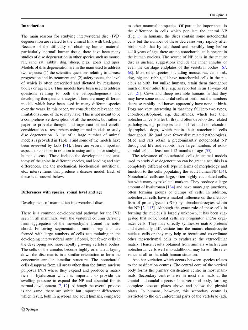
to other mammalian species. Of particular importance, is
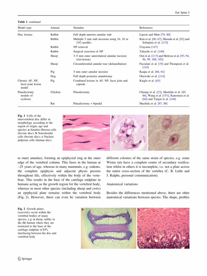
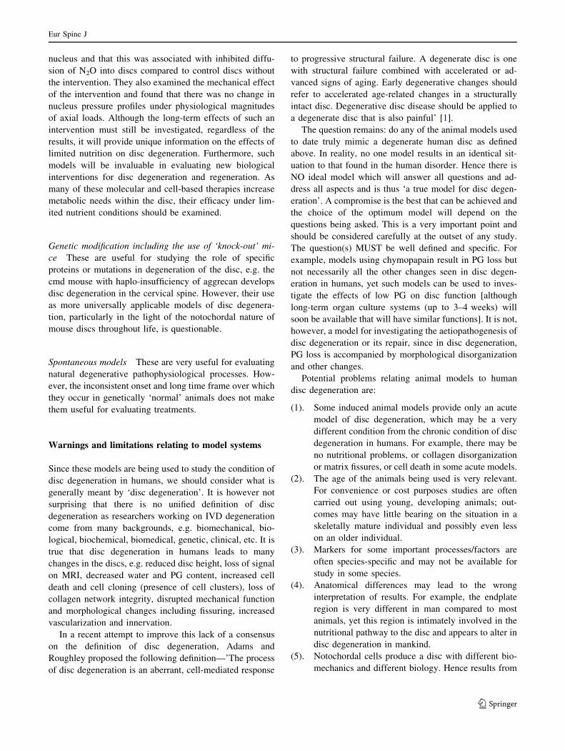
the difference in cells which populate the central NP
(Fig. 1): in humans, the discs contain some notochordal
cells but the number of these decreases very rapidly after
birth, such that by adulthood and possibly long before
4–10 years of age, there are no notochordal cells present in
the human nucleus. The source of NP cells in the mature
disc is unclear, suggestions include the inner annulus or
even the cartilage endplates of the vertebral bodies [67,
68]. Most other species, including mouse, rat, cat, mink,
dog, pig and rabbit, all have notochordal cells in the nu-
cleus at birth, but unlike humans, retain them throughout
much of their adult life, e.g. as reported in an 18-year-old
cat [21]. Cows and sheep resemble humans in that they
may have some notochordal cells at birth, but the numbers
decrease rapidly and horses apparently have none at birth.
Dogs are very interesting in that they fall into two types,
chondrodystrophoid, e.g. dachshunds, which lose their
notochordal cells after birth (and often develop disc related
pathologies, e.g. prolapses later in life) and non-chondro-
dystrophoid dogs, which retain their notochordal cells
throughout life (and have fewer disc related pathologies).
Mice and rats retain a predominantly notochordal NP
throughout life and rabbits have large numbers of noto-
chordal cells at least until 12 months of age [55].
The relevance of notochordal cells in animal models
used to study disc degeneration can be great since this is a
completely different cell type in terms of morphology and
function to the cells populating the adult human NP [54].
Notochordal cells are large, often highly vacuolated cells,
but with many cytoskeletal markers. They produce a large
amount of hyaluronan [134] and have many gap junctions,
often forming groups or clumps of cells. In addition,
notochordal cells have a marked influence on the metabo-
lism of proteoglycans (PGs) by fibrochondrocytes within
the NP [2, 113]. Although the exact role of these cells in
forming the nucleus is largely unknown, it has been sug-
gested that notochordal cells are progenitor and/or orga-
nizer cells. They may directly synthesize matrix proteins
and eventually differentiate into the mature chondrocytic
nucleus cells or they may help to recruit and co-ordinate
other mesenchymal cells to synthesize the extracellular
matrix. Hence results obtained from animals which retain
notochordal cells well into adulthood, may have little rele-
vance at all to the adult human situation.
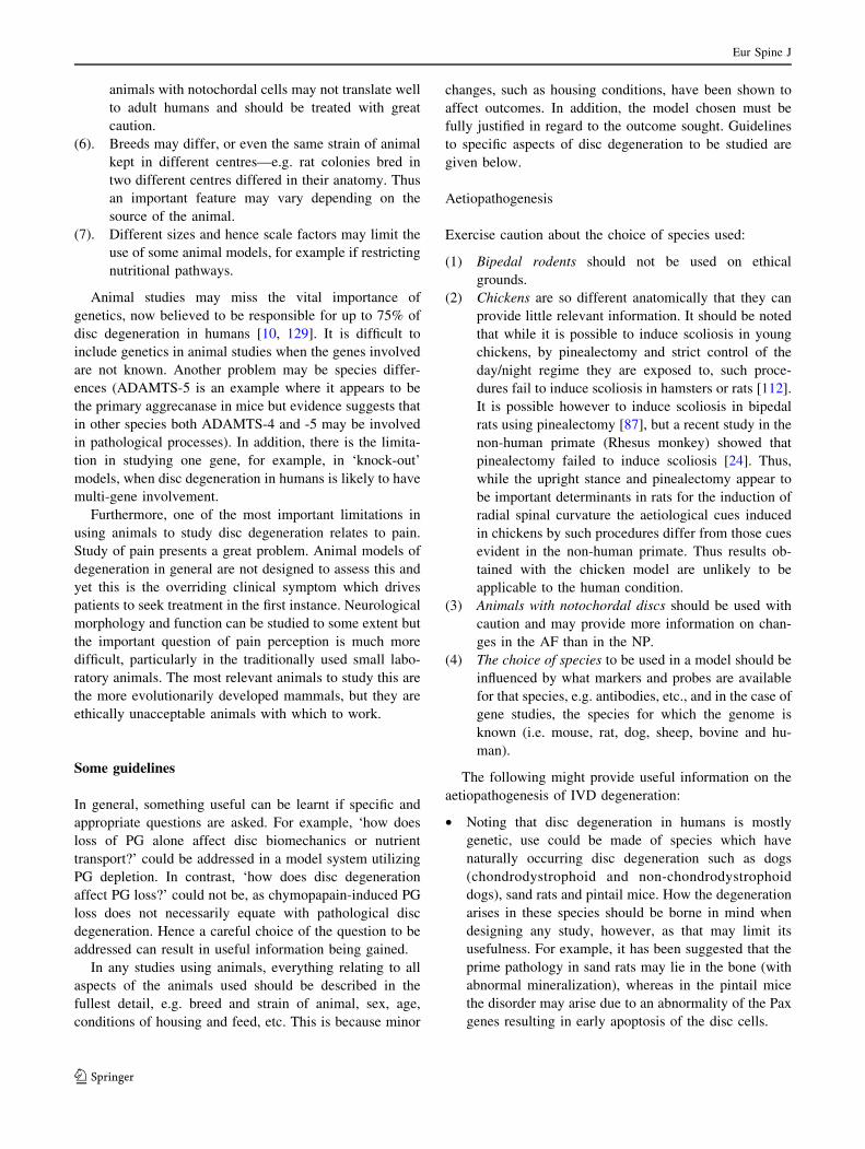
Another variation which occurs between species relates
to the ossification centres. The central core of the vertical
body forms the primary ossification centre in most mam-
mals. Secondary centres arise in most mammals at the
cranial and caudal aspects of the vertebral body, forming
complete osseous plates above and below the physial
plates. In humans, however, this secondary centre is
restricted to the circumferential parts of the vertebrae (adj.
Eur Spine J
123
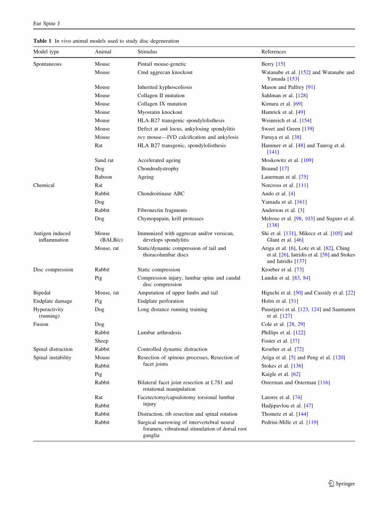
Table 1 In vivo animal models used to study disc degeneration
Model type Animal Stimulus References
Spontaneous Mouse Pintail mouse-genetic Berry [15]
Mouse Cmd aggrecan knockout Watanabe et al. [152] and Watanabe and
Yamada [153]
Mouse Inherited kyphoscoliosis Mason and Palfrey [91]
Mouse Collagen II mutation Sahlman et al. [128]
Mouse Collagen IX mutation Kimura et al. [69]
Mouse Myostatin knockout Hamrick et al. [49]
Mouse HLA-B27 transgenic spondylolisthesis Weinreich et al. [154]
Mouse Defect at ank locus, ankylosing spondylitis Sweet and Green [139]
Mouse twy mouse—IVD calcification and ankylosis Furuya et al. [38]
Rat HLA B27 transgenic, spondylolisthesis Hammer et al. [48] and Taurog et al.
[141]
Sand rat Accelerated ageing Moskowitz et al. [109]
Dog Chondrodystrophy Braund [17]
Baboon Ageing Lauerman et al. [75]
Chemical Rat Norcross et al. [111]
Rabbit Chondroitinase ABC Ando et al. [4]
Dog Yamada et al. [161]
Rabbit Fibronectin fragments Anderson et al. [3]
Dog Chymopapain, krill proteases Melrose et al. [98, 103] and Suguro et al.
[138]
Antigen induced
inflammation
Mouse
(BALB/c)
Immunized with aggrecan and/or versican,
develops spondylitis
Shi et al. [131], Mikecz et al. [105] and
Glant et al. [46]
Mouse, rat Static/dynamic compression of tail and
thoracolumbar discs
Ariga et al. [6], Lotz et al. [82], Ching
et al. [26], Iatridis et al. [58] and Stokes
and Iatridis [137]
Disc compression Rabbit Static compression Kroeber et al. [73]
Pig Compression injury, lumbar spine and caudal
disc compression
Lundin et al. [83, 84]
Bipedal Mouse, rat Amputation of upper limbs and tail Higuchi et al. [50] and Cassidy et al. [22]
Endplate damage Pig Endplate perforation Holm et al. [51]
Hyperactivity
(running)
Dog Long distance running training Puustjarvi et al. [123, 124] and Saamanen
et al. [127]
Fusion Dog Cole et al. [28, 29]
Rabbit Lumbar arthrodesis Phillips et al. [122]
Sheep Foster et al. [37]
Spinal distraction Rabbit Controlled dynamic distraction Kroeber et al. [72]
Spinal instability Mouse Resection of spinous processes, Resection of
facet joints
Ariga et al. [5] and Peng et al. [120]
Rabbit Stokes et al. [136]
Pig Kaigle et al. [62]
Rabbit Bilateral facet joint resection at L7S1 and
rotational manipulation
Osterman and Osterman [116]
Rat Facetectomy/capsulotomy torsional lumbar
injury
Latorre et al. [74]
Rabbit Hadjipavlou et al. [47]
Rabbit Distraction, rib resection and spinal rotation Thometz et al. [144]
Rabbit Surgical narrowing of intervertebral neural
foramen, vibrational stimulation of dorsal root
ganglia
Pedrini-Mille et al. [119]
Eur Spine J
123
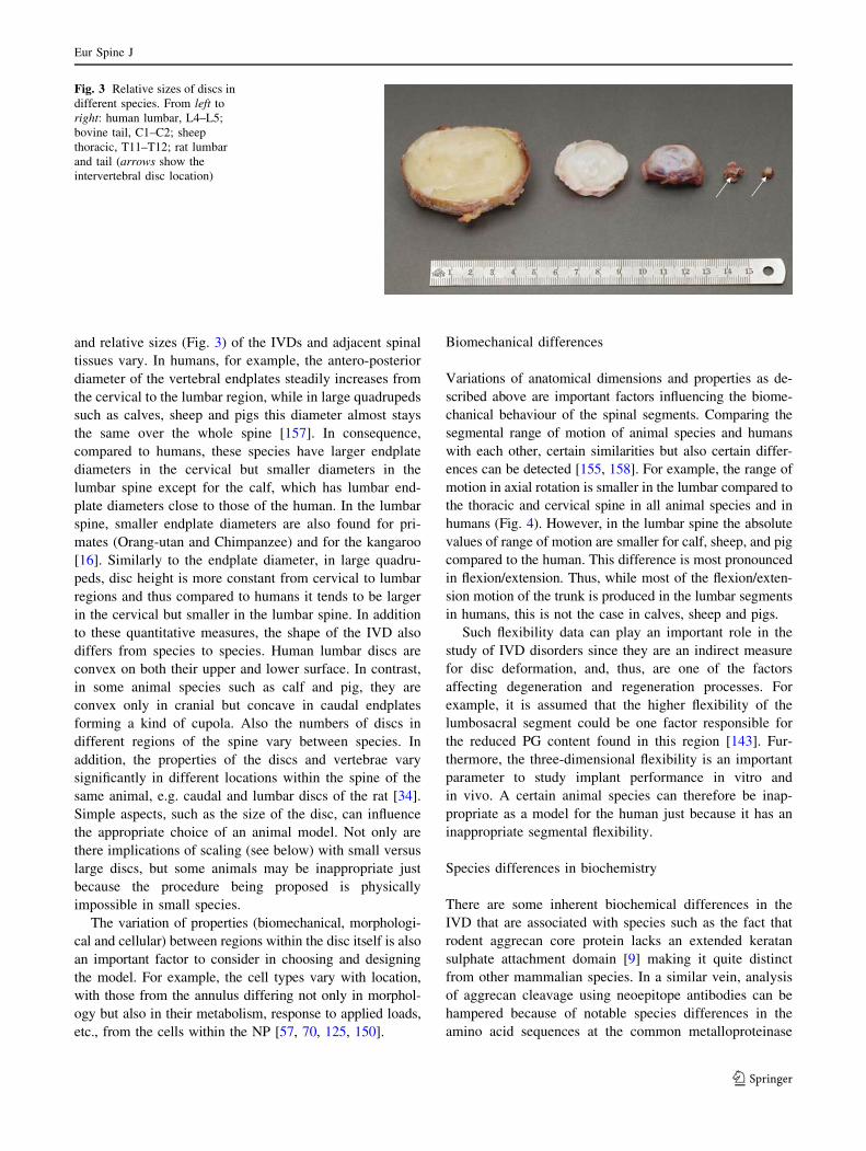
to outer annulus), forming an epiphysial ring at the outer
edge of the vertebral column. This fuses in the human at
~25 years of age, whereas in many mammals, e.g. rodents,
the complete epiphysis and adjacent physis persists
throughout life, effectively within the body of the verte-
brae. This results in the base of the cartilage endplate in
humans acting as the growth region for the vertebral body,
whereas in most other species (including sheep and cows)
an epiphysial plate remains within the vertebral body
(Fig. 2). However, there can even be variation between
different colonies of the same strain of species, e.g. some
Wistar rats have a complete centre of secondary ossifica-
tion whilst in others it is incomplete, i.e. not a plate across
the entire cross-section of the vertebra (C. B. Little and
J. Ralphs, personal communication).
Anatomical variations
Besides the differences mentioned above, there are other
anatomical variations between species. The shape, profiles
Table 1 continued
Model type Animal Stimulus References
Disc lesions Rabbit Full depth anterior annular stab Lipson and Muir [79, 80]
Rabbit Multiple 5 mm stab incisions using 16, 18 or
21G needles
Kim et al. [66, 67], Masuda et al. [92] and
Sobajima et al. [133]
Rabbit NP removal Urayama [147]
Rabbit Surgical resection of NP Takaishi et al. [140]
Sheep 3–5 mm outer anterolateral annular incision
(rim-lesion)
Osti et al. [117] and Melrose et al. [93, 94,
96, 99, 100, 102]
Sheep Circumferential annular tear (delamellation) Fazzalari et al. [35] and Thompson et al.
[145]
Pig 5 mm outer annular incision Kaapa et al. [60, 61]
Dog Full depth posterior annulotomy Olsewski et al. [114]
Chronic AF, NP,
facet joint lesion
model
Pig Combined lesions in AF, NP, facet joint and
capsule
Kaigle et al. [63]
Pinealectomy
models of
scoliosis
Chicken Pinealectomy Cheung et al. [23], Machida et al. [85,
86], Wang et al. [151], Kanemura et al.
[64] and Turgut et al. [146]
Rat Pinealectomy + bipedal Machida et al. [87, 88]
Fig. 2 Growth plates
(asterisks) occur within the
vertebral bodies of many
species, e.g. a sheep, unlike in
the (b) human where they are
restricted to the base of the
cartilage endplate (CEP),
interfacing between the disc and
vertebral body
Fig. 1 Cells of the
intervertebral disc differ in
morphology according to the
region of origin, age and
species: a Annulus fibrosus cells
(bovine disc); b Notochordal
cells (bovine disc); c Nucleus
pulposus cells (human disc)
Eur Spine J
123
and relative sizes (Fig. 3) of the IVDs and adjacent spinal
tissues vary. In humans, for example, the antero-posterior
diameter of the vertebral endplates steadily increases from
the cervical to the lumbar region, while in large quadrupeds
such as calves, sheep and pigs this diameter almost stays
the same over the whole spine [157]. In consequence,
compared to humans, these species have larger endplate
diameters in the cervical but smaller diameters in the
lumbar spine except for the calf, which has lumbar end-
plate diameters close to those of the human. In the lumbar
spine, smaller endplate diameters are also found for pri-
mates (Orang-utan and Chimpanzee) and for the kangaroo
[16]. Similarly to the endplate diameter, in large quadru-
peds, disc height is more constant from cervical to lumbar
regions and thus compared to humans it tends to be larger
in the cervical but smaller in the lumbar spine. In addition
to these quantitative measures, the shape of the IVD also
differs from species to species. Human lumbar discs are
convex on both their upper and lower surface. In contrast,
in some animal species such as calf and pig, they are
convex only in cranial but concave in caudal endplates
forming a kind of cupola. Also the numbers of discs in
different regions of the spine vary between species. In
addition, the properties of the discs and vertebrae vary
significantly in different locations within the spine of the
same animal, e.g. caudal and lumbar discs of the rat [34].
Simple aspects, such as the size of the disc, can influence
the appropriate choice of an animal model. Not only are
there implications of scaling (see below) with small versus
large discs, but some animals may be inappropriate just
because the procedure being proposed is physically
impossible in small species.
The variation of properties (biomechanical, morphologi-
cal and cellular) between regions within the disc itself is also
an important factor to consider in choosing and designing
the model. For example, the cell types vary with location,
with those from the annulus differing not only in morphol-
ogy but also in their metabolism, response to applied loads,
etc., from the cells within the NP [57, 70, 125, 150].
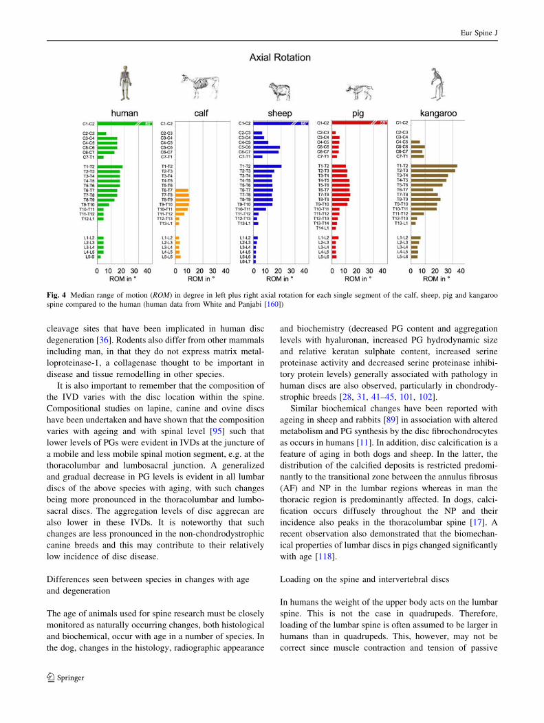
Biomechanical differences
Variations of anatomical dimensions and properties as de-
scribed above are important factors influencing the biome-
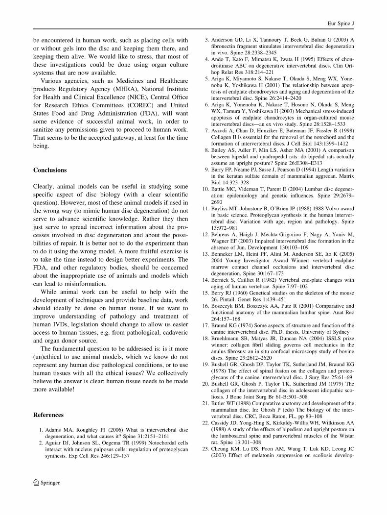
chanical behaviour of the spinal segments. Comparing the
segmental range of motion of animal species and humans
with each other, certain similarities but also certain differ-
ences can be detected [155, 158]. For example, the range of
motion in axial rotation is smaller in the lumbar compared to
the thoracic and cervical spine in all animal species and in
humans (Fig. 4). However, in the lumbar spine the absolute
values of range of motion are smaller for calf, sheep, and pig
compared to the human. This difference is most pronounced
in flexion/extension. Thus, while most of the flexion/exten-
sion motion of the trunk is produced in the lumbar segments
in humans, this is not the case in calves, sheep and pigs.
Such flexibility data can play an important role in the
study of IVD disorders since they are an indirect measure
for disc deformation, and, thus, are one of the factors
affecting degeneration and regeneration processes. For
example, it is assumed that the higher flexibility of the
lumbosacral segment could be one factor responsible for
the reduced PG content found in this region [143]. Fur-
thermore, the three-dimensional flexibility is an important
parameter to study implant performance in vitro and
in vivo. A certain animal species can therefore be inap-
propriate as a model for the human just because it has an
inappropriate segmental flexibility.
Species differences in biochemistry
There are some inherent biochemical differences in the
IVD that are associated with species such as the fact that
rodent aggrecan core protein lacks an extended keratan
sulphate attachment domain [9] making it quite distinct
from other mammalian species. In a similar vein, analysis
of aggrecan cleavage using neoepitope antibodies can be
hampered because of notable species differences in the
amino acid sequences at the common metalloproteinase
Fig. 3 Relative sizes of discs in
different species. From left to
right: human lumbar, L4–L5;
bovine tail, C1–C2; sheep
thoracic, T11–T12; rat lumbar
and tail (arrows show the
intervertebral disc location)
Eur Spine J
123
cleavage sites that have been implicated in human disc
degeneration [36]. Rodents also differ from other mammals
including man, in that they do not express matrix metal-
loproteinase-1, a collagenase thought to be important in
disease and tissue remodelling in other species.
It is also important to remember that the composition of
the IVD varies with the disc location within the spine.
Compositional studies on lapine, canine and ovine discs
have been undertaken and have shown that the composition
varies with ageing and with spinal level [95] such that
lower levels of PGs were evident in IVDs at the juncture of
a mobile and less mobile spinal motion segment, e.g. at the
thoracolumbar and lumbosacral junction. A generalized
and gradual decrease in PG levels is evident in all lumbar
discs of the above species with aging, with such changes
being more pronounced in the thoracolumbar and lumbo-
sacral discs. The aggregation levels of disc aggrecan are
also lower in these IVDs. It is noteworthy that such
changes are less pronounced in the non-chondrodystrophic
canine breeds and this may contribute to their relatively
low incidence of disc disease.
Differences seen between species in changes with age
and degeneration
The age of animals used for spine research must be closely
monitored as naturally occurring changes, both histological
and biochemical, occur with age in a number of species. In
the dog, changes in the histology, radiographic appearance
and biochemistry (decreased PG content and aggregation
levels with hyaluronan, increased PG hydrodynamic size
and relative keratan sulphate content, increased serine
proteinase activity and decreased serine proteinase inhibi-
tory protein levels) generally associated with pathology in
human discs are also observed, particularly in chondrody-
strophic breeds [28, 31, 41–45, 101, 102].
Similar biochemical changes have been reported with
ageing in sheep and rabbits [89] in association with altered
metabolism and PG synthesis by the disc fibrochondrocytes
as occurs in humans [11]. In addition, disc calcification is a
feature of aging in both dogs and sheep. In the latter, the
distribution of the calcified deposits is restricted predomi-
nantly to the transitional zone between the annulus fibrosus
(AF) and NP in the lumbar regions whereas in man the
thoracic region is predominantly affected. In dogs, calci-
fication occurs diffusely throughout the NP and their
incidence also peaks in the thoracolumbar spine [17]. A
recent observation also demonstrated that the biomechan-
ical properties of lumbar discs in pigs changed significantly
with age [118].
Loading on the spine and intervertebral discs
In humans the weight of the upper body acts on the lumbar
spine. This is not the case in quadrupeds. Therefore,
loading of the lumbar spine is often assumed to be larger in
humans than in quadrupeds. This, however, may not be
correct since muscle contraction and tension of passive
Fig. 4 Median range of motion (ROM) in degree in left plus right axial rotation for each single segment of the calf, sheep, pig and kangaroo
spine compared to the human (human data from White and Panjabi [160])
Eur Spine J
123
structures such as ligaments also adds substantially to the
loads acting on the spine [159]. This additional load may
be higher in large quadrupeds such as calves, sheep or pigs
than in humans since stabilizing a horizontally aligned
spine requires higher muscle forces and passive tension
than stabilizing an almost balanced vertically aligned
spine, e.g. suspenion bridge versus an inverted pendulum.
Hence, although there are no directly measured forces in
the literature, and therefore no consensus, loading of the
lumbar spine is probably not smaller but even larger for
large quadrupeds than for humans. This would also be
consistent with the up to fourfold higher bone mineral
density found in sheep, calf and pig lumbar vertebral
bodies compared to the human [156].
In small quadrupeds such as rabbit, mouse or rat, smaller
muscular and ligamentous forces are needed to stabilize the
spine. The spines of these animals are therefore probably
loaded by much lower forces than that of the human.
However, their intradiscal pressure might be similar to that
of the human since the diameter of their discs on which this
force is acting is much smaller.
Tail IVDs of animals mainly become loaded by muscle
contraction or tension of passive structures. The more the
muscles need to be contracted and the more the passive
structures become tensioned, the more the loading of the
tail discs will increase. Measurements of the in vivo
loading of the spine and tail of various animals, however,
are still lacking. Yet, this information would be crucial for
most in vivo studies since the mechanical environment is
known to influence the biological behaviour of the disc
cells. Also, the loading of the disc plays an important role
in in vivo or in vitro implant tests since it affects the per-
formance of the implant for example in terms of its sus-
ceptibility to destruction or dislocation.
The immobilization of a segment of the canine lumbar
spine induces compositional changes in the discs adjacent to
the immobilized segment relative to spinal level and age
matched discs of non-operated control animals [19, 29, 30,
142]. Alterations in spinal biomechanics due to scoliosis are
also important determinants of disc cell metabolism and
consequently also affect disc composition [20, 40, 97]. The
introduction of a controlled outer anterolateral annular
surgical defect also alters the intrinsic spinal mechanics at
this spinal level and changes in disc cell metabolism and
composition are measurable in the affected disc [93, 94, 96].
Scaling of specific parameters
Most non-human discs used in research are of a different
size (usually smaller) than their human counterparts.
Therefore, scaling is required in the interpretation of
experimental findings from animal models since many as-
pects of disc behaviour are size-dependent. This certainly
includes the mechanical behaviour of IVDs. Size (disc
height) also strongly affects solute diffusion in disc tissues
and thus solute transport in nutritional studies. Scaling
requires that we know the form of the relationship between
the size (geometrical parameters) and the behaviour being
tested. At the very least, one should know whether the
relationship is linear, quadratic, etc.
Either an empirical or an analytical approach can be used
to determine the form of this relationship. Empirically, a
series of discs having different sizes is studied and the rela-
tionship is found by linear or non-linear regression analysis.
Analytically, if known theoretical considerations define the
relationship between the behaviour of interest and the size
parameters, then the form of the scaling relationship can be
predicted. Ideally, both approaches should be used and the
theoretical relationship should be verified experimentally.
In the mechanical behaviour of a disc, the size of the
animal is also important in determining the magnitude of
loading that is habitually imposed in the disc. This factor
should also be taken into account in interpretation of
experimental findings in a model. Apart from size differ-
ences there may be other species-dependent differences in
disc behaviour, including structural variations. For in-
stance, the mechanical stiffness of a disc depends on the
annulus lamellar structure and also on its material proper-
ties (elastic moduli, etc.). The annulus structure has a dif-
ferent effect on the stiffness in each of the degrees of
freedom—compression, torsion, etc. Transport phenomena
depend on physical size (particularly diffusion pathways)
but also on anatomical differences between species in
relation to endplate structure and blood supply.
In principle, if the tissue properties do not vary between
discs, then only the geometrical dimensions need to be
taken into account in the scaling. Some basic structures and
the tissue material properties do not vary greatly between
mammalian species, or with anatomical level and age. For
example, there is (mostly anecdotal) evidence that the
number of lamellae, the lamellar fibre angle, and sizes of
trabeculae are relatively constant. Other variables such as
the thickness of lamellar layers vary with the disc size and
its level in the spinal column (cervical, thoracic, lumbar
and caudal).
Model systems
In vitro animal models for studying disc degeneration
and repair
Cell cultures
Growing disc cells in the laboratory is obviously one useful
technique for studying their behaviour and attempting to
Eur Spine J
123
mimic either different environments and insults or also the
effect of various interventions. Whilst useful information
can certainly be gained from such model systems, it is
important also to appreciate the intrinsic limitations of
these experimental systems. An advantage of cell culture is
that it is a ‘simple’ model, taking the cell down to literally
its bare minimum. This simplicity, however, can also be
considered a disadvantage, in that one is studying the cell
in a totally non-physiological situation; in vivo the disc
cells are surrounded by a large volume of extracellular
matrix, with negligible cell–cell contact. This is very dif-
ferent from cell culture systems, particularly when grown
in monolayer. The shape and morphology of cells is also
known to influence their behaviour enormously. Hence the
type of culture system used is capable of eliciting very
different responses from the cells, particularly monolayer
compared to three-dimensional culture system utilizing
some form of scaffold. Commonly used three-dimensional
culture systems include gels such as calcium alginate
(polyguluronic/mannuronic acid) in the form of micro-
beads [25, 90], agarose and collagen gels. Other scaffolds
have also been used in tissue engineering type applications
on IVDs such as gelatin/chondroitin-6-sulfate/hyaluronan
tri-copolymer, hyaluronan hydrogels, fibrin/collagen co-
polymer gels, polyglycolic acid or porous calcium poly-
phosphate. The choice of ‘support systems’ to be used
should be influenced, not only by what aspect will be
investigated, e.g. whether matrix production will be studied
or molecular biology (requiring easy dissociation of the
cells), but also on the type of cells being studied. Alginate,
for example, appears to be less than optimum for studying
cells from the outer annulus for which collagen gels may be
more suitable [52]. Despite these points, cell culture sys-
tems can be used to address certain issues, as long as the
question being asked and the interpretation of the results
are considered carefully, taking into account the intrinsic
limitations of each respective experimental system.
Explant cultures of whole discs
An important alternative to in vivo studies is an in vitro
explant model, in which controlled in vitro conditions can
be applied to a more physiologically relevant model. The
main advantage of in vitro explant systems compared to
cell models is that the cells are not removed from their
highly specialized extracellular matrix whose effect on
cell behaviour is not well understood. There are various
models in the literature, including small rodent discs ei-
ther alone, without bone, or as motion segments; the
disadvantage of these is that unless they are placed under
load they swell markedly and lose glycosaminoglycans
(GAGs) thus changing the extracellular environment.
Bovine caudal discs have been proposed as a suitable
biological and biomechanical model for the study of the
human lumbar disc. Bovine caudal discs (14–22 mm in
diameter and 5–10 mm thick) are close in size to the
human lumbar disc and the musculature of the bovine tail
maintains an in vivo pressure on the discs that is
approximately the same as in the human lumbar disc in
the prone position (0.1–0.3 MPa). However, caudal (tail)
discs in mice have been shown to have significantly dif-
ferent mechanical properties compared with lumbar discs
in the same animals [130]. As noted earlier, the bio-
chemical content and metabolic behaviour of discs from
different spinal levels vary markedly and this must be
borne in mind when extrapolating results from tail discs to
cervical or lumbar regions. Also, tail IVDs in most ani-
mals cannot be considered to be weight bearing in the
same context as discs above the sacrum; one exception to
this is the kangaroo which has caudal discs which are
weight bearing. This would be an interesting system to
evaluate but such experiments have yet to be undertaken.
Oshima et al. cultured bovine caudal discs under 5 kg
static load in order to maintain in vivo hydration levels
and measured changes in metabolic activity by varying
the load magnitude [115]. That study was limited to 12 h
and used discs without endplates, but such a system
appears to be promising, especially in terms of its appli-
cability for mechano-biology studies of the IVD. Further
studies have been conducted using bovine discs with and
without endplates [77] and they have shown that it is
possible to maintain cell viability and the biosynthetic
responsiveness of large discs for up to 1 week in vitro
when the discs are cultured under static load and without
vertebral endplates. Modifications of this method are
proposed, such as applying cyclic loading (and hence
motion), to influence convective transport, or reducing
clot formation without the vertebral endplate via the
administration of anticlotting agents ante-mortem [39].
Explant models, possibly simulating disc degeneration,
can be used by culturing bovine caudal motion segments
(with vertebral endplates attached) either with enzymes
injected into the nucleus or via serum deprivation. These
have been reported to be successfully cultured for up to
28 days [59].
The development of an entire IVD organ culture of large
dimensions will have a high impact on future research on
disc degeneration and regeneration. Such an organ culture
model will give us a unique opportunity to measure the
effect of growth factors, cytokines and protease inhibitors
introduced directly into the IVD by injection, on the meta-
bolic activity (synthesis and degradation) of disc cells in a
controlled environment nearly identical to the in vivo
conditions. This model could also be used to evaluate tis-
sue-engineering approaches designed to treat disc degene-
ration, such as the implantation of cells, either alone or
Eur Spine J
123
embedded in a biomatrix, as well as to assess transfection
efficiency (for gene therapy) in an environment similar to
the natural one. As with all in vitro models, the full
physiological interaction of systemic and local factors
cannot be controlled and any pathophysiological pathways
or therapeutic modalities identified would ultimately need
verification in vivo. However, once the ideal parameters
(i.e. optimal biological factors, which inhibit degeneration
or induce repair) have been identified with the model,
fewer animal experiments need to be performed to test
these conditions in vivo, reducing the use of live animals in
research to a minimum.
In vivo models for studying disc degeneration
and repair
Along with the cautionary notes relating to species and
spinal level discussed above, the actual mechanism for
inducing disc degeneration can influence the interpretation
of the results obtained from an experiment. In Table 1,
many of the different mechanisms for inducing or studying
disc disease and the species in which they have been
undertaken are summarized. The text following Table 1
discusses some of these models in more detail to highlight
potential deficiencies or strengths of individual models.
Mechanical models
The commonly used mechanical interventions involve in-
creased or decreased loading, increased or decreased mo-
tion, or injury. In models that impose altered loading, one
should distinguish between sustained (constant) forces, and
cyclic loading of different frequencies. Many of the inter-
ventions that are intended to alter just loading or motion in
fact unintentionally modify both (e.g. application of a
loading apparatus also reducing the intervertebral motion).
The relative influences of alterations of loading and motion
on disc degeneration were previously reviewed by Stokes
and Iatridis [137].
Tail models The tail provides an attractive model because
its discs are accessible to interventions, with minimal risk
of damage to surrounding structures, and minimal inter-
ference with normal physiological function. The IVDs in
the tails of mice, rats and cattle have been used. In vivo
mechanical interventions include an early forced ‘curved
tail’ model [78], external apparatus to apply axial com-
pression [27, 76, 149] or asymmetrical compression [32,
104]. Degeneration subsequent to injection of protein
digestive enzymes into caudal discs [111] has also been
reported. It should be noted that the biomechanical studies
with external apparatus normally impose some degree of
restricted motion, as well as altered loading. In vitro studies
of the tail discs include structural [121], cell mechanics
[18] and physiological studies.
The use of caudal discs as surrogates for the human
lumbar disc has been questioned at several levels:
1. Differing mechanical loading relative to human lumbar
spine. The tail has been suspected to have lesser load-
ing (especially compressive stress) than the human
spine. However, Oshima et al. reported that the swell-
ing pressure in bovine tail discs is similar to that in
human lumbar discs, indicating that the prevailing
compressive stress of the tail discs is probably of
similar magnitude to lumbar discs. Anaesthesia [110,
135] produces a similar increase in disc thickness as
that which results from applying distraction forces of
60% bodyweight (c. 0.25 MPa) to the tail, which sug-
gests that the ambient loading is of similar magnitude.
2. Different anatomy (absence of posterior elements), and
differing dimensions relative to human lumbar spine
(often being smaller, with anterioposterio-lateral pro-
portions being more circular and the cephalous-caudal
dimension being ‘thicker’ than lumbar discs). In fact,
Elliott and Sarver [34] and Sarver and Elliott [130] re-
ported substantial dimensional and mechanical behav-
iour similarity between the lumbar discs of mice, rats
and humans, after scale effects were taken into account.
Summarizing their dimensional data, together with
some anecdotal observations of radiographs by one of
the authors, the ‘aspect’ ratio (height/mean diameter) of
human lumbar discs averages 0.24, which is similar to
values for mouse and rabbit lumbar spines, whereas the
ratio for lumbar spines of rats, deer and pigs is as little
as half this value. The ratio for caudal discs is in the
range 0.23 (bovine and mice) to 0.35 (rat).
3. Composition and metabolism. Recently Demers et al.
[33] compared the composition and matrix synthesis in
bovine tail discs and human lumbar discs of differing
ages. They concluded that the bovine tail provided a
good model of the young human, but was less well
suited to studying age-related degeneration.
Hence, the tail has the advantage of being accessible for
manipulation, but the disadvantage that the discs within it
are distinctly different from lumbar discs in terms of bio-
mechanics and composition, at least in rodents. The use of
rats and mice must be questioned also because of the
presence of notochordal cell. These are likely to respond
very differently to mechanical loading compared with
normal NP cells.
Bipedal mice The strength of this model is that it is dri-
ven by overloading which some believe is involved in
Eur Spine J
123
human disc disease. However its use cannot be supported
on ethical grounds. Moreover, because of the notochordal
nature of the NP in rodents, its value in predicting re-
sponses in humans is doubtful. It is also questionable
whether laboratory housed animals with induced bipeda-
lism really experience different spinal loading [8].
Injury models In general these models are attractive in
that the exact timing of the insult can be precisely con-
trolled. In addition, damage to the AF and altered spinal
mechanics are believed to play a critical role in human IVD
degeneration.
Induction of spinal instability through various facetec-
tomies and resection of spinous processes have in general
had an uncertain time course and reliability in inducing
disc degeneration and may stabilize with fibrosis over time.
Fusion of a spinal segment results in induction of increased
motion and degeneration in adjacent discs, but using this as
a model of degeneration requires extensive surgery and is
limited to induction of disease in a single level. There may
also be an issue with the ability to readily transfer the same
surgical technique and method between laboratories with
extensive surgical procedures.
Perhaps the most extensively used surgical model has
been to damage the AF. In 1948, the ‘stabbing’ of the IVD
with a scalpel began to be used as a tool to induce disc
degeneration in animals [65, 132]. Stab models may be
classified into two types: the total annular stab model [80]
and the superficial AF injury model [117]. Total annular
injury induces NP avulsion and disc degeneration develops
relatively quickly. It may, therefore be the model of choice
for studying regeneration and the effect of therapy such as
growth factors [66, 92, 133]. Degeneration after a super-
ficial stab wound is much less rapid but involves actual
active degeneration and may be superior for studying the
pathophysiological processes [99, 100, 106–108]. In the
rabbit, puncturing the AF with needles of defined gauges
resulted in reproducible, degenerative changes within 2–
8 weeks that could be quantitatively assessed through
conventional radiography and magnetic resonance imaging
(MRI), as well as histology. In contrast, the changes in
endplate vascularity, facet joint osteoarthritis and meta-
bolic pathology in NP and AF cells following partial
thickness lesions in the sheep AF took 6–9 months to be-
come apparent. The NP changes in the sheep model are
much milder and less consistent compared with those in the
rabbit following needle puncture. Because the rabbit disc
contains notochordal cells that differ from human adult NP
cells, possible species-related differences in the response to
growth factors should be considered. However, in this
rabbit model, the histology of the disc after puncture
showed changes in the cell population from notochordal
cells to chondrocytic cells. Therefore, in pilot pre-clinical
studies, as a proof of concept, the use of the rabbit disc
model may still be relevant and cost effective.
Biological models, directly influencing cells
Injection of degradative enzymes One method which
might seem appropriate to attempt to mimic degenerative
disc disease in humans is proteolytic digestion or removal
of GAG chains. Enzymes such as chymopapain or chon-
droitinases could be used to this effect. However, whilst
this may result in a loss of GAG from the matrix, it would
not be an exact replication of what happens naturally in the
degenerating disc since there are no naturally occurring
mammalian extracellular chondroitinases. On the other
hand, depending on the question being asked of the study,
this may not matter as a loss of PGs is a classic feature of
disc degeneration. A further limitation of using enzyme
degradation in an in vivo model is that, depending on the
dose used, these models will heal with the cells synthe-
sizing and replenishing the lost GAGs; thus they do not
induce a true ‘degenerative’ process.
Altered nutrient supply Although in vitro cell culture
experiments [53] have demonstrated the effect of limited
nutrition on cell viability and several studies have demon-
strated the correlation between sclerosis of the endplate and
disc degeneration [13, 14, 126], limited nutrition as a cause
of disc degeneration has never been proven in intact discs.
For this purpose, a canine model of insufficient blood
supply to the disc was investigated by Hutton and col-
leagues [56]. To produce the blood supply disturbance,
bone cement was injected into the vertebra adjacent to a
single or to both endplates of experimental discs. After
70 weeks, they found no macroscopic degenerative chan-
ges in the experimental discs in comparison to control discs
within the same animal. Unfortunately, the magnitude of
nutrient transport inhibition within the disc created by the
intervention was never measured. A similar study was also
done in sheep to investigate the effect of multiple level
vertebroplasty on disc health [71]. After 12 months, the
authors observed histological changes consistent with a
reparative response in early disc degeneration. Again they
unfortunately did not assess nutrient transport alterations
induced by their vertebroplasty intervention. Alternatively,
another group is developing an ovine model for nutrient
insufficiency induced disc degeneration [148]. In this
model, the blood supply to the endplates is disrupted and a
titanium foil is inserted to prevent vascular regeneration.
To date, the group has been able to demonstrate that the
method of intervention decreased significantly the number
of perfused capillary buds in the endplate overlying the
Eur Spine J
123
nucleus and that this was associated with inhibited diffu-
sion of N2O into discs compared to control discs without
the intervention. They also examined the mechanical effect
of the intervention and found that there was no change in
nucleus pressure profiles under physiological magnitudes
of axial loads. Although the long-term effects of such an
intervention must still be investigated, regardless of the
results, it will provide unique information on the effects of
limited nutrition on disc degeneration. Furthermore, such
models will be invaluable in evaluating new biological
interventions for disc degeneration and regeneration. As
many of these molecular and cell-based therapies increase
metabolic needs within the disc, their efficacy under lim-
ited nutrient conditions should be examined.
Genetic modification including the use of ‘knock-out’ mi-
ce These are useful for studying the role of specific
proteins or mutations in degeneration of the disc, e.g. the
cmd mouse with haplo-insufficiency of aggrecan develops
disc degeneration in the cervical spine. However, their use
as more universally applicable models of disc degenera-
tion, particularly in the light of the notochordal nature of
mouse discs throughout life, is questionable.
Spontaneous models These are very useful for evaluating
natural degenerative pathophysiological processes. How-
ever, the inconsistent onset and long time frame over which
they occur in genetically ‘normal’ animals does not make
them useful for evaluating treatments.
Warnings and limitations relating to model systems
Since these models are being used to study the condition of
disc degeneration in humans, we should consider what is
generally meant by ‘disc degeneration’. It is however not
surprising that there is no unified definition of disc
degeneration as researchers working on IVD degeneration
come from many backgrounds, e.g. biomechanical, bio-
logical, biochemical, biomedical, genetic, clinical, etc. It is
true that disc degeneration in humans leads to many
changes in the discs, e.g. reduced disc height, loss of signal
on MRI, decreased water and PG content, increased cell
death and cell cloning (presence of cell clusters), loss of
collagen network integrity, disrupted mechanical function
and morphological changes including fissuring, increased
vascularization and innervation.
In a recent attempt to improve this lack of a consensus
on the definition of disc degeneration, Adams and
Roughley proposed the following definition—’The process
of disc degeneration is an aberrant, cell-mediated response
to progressive structural failure. A degenerate disc is one
with structural failure combined with accelerated or ad-
vanced signs of aging. Early degenerative changes should
refer to accelerated age-related changes in a structurally
intact disc. Degenerative disc disease should be applied to
a degenerate disc that is also painful’ [1].
The question remains: do any of the animal models used
to date truly mimic a degenerate human disc as defined
above. In reality, no one model results in an identical sit-
uation to that found in the human disorder. Hence there is
NO ideal model which will answer all questions and ad-
dress all aspects and is thus ‘a true model for disc degen-
eration’. A compromise is the best that can be achieved and
the choice of the optimum model will depend on the
questions being asked. This is a very important point and
should be considered carefully at the outset of any study.
The question(s) MUST be well defined and specific. For
example, models using chymopapain result in PG loss but
not necessarily all the other changes seen in disc degen-
eration in humans, yet such models can be used to inves-
tigate the effects of low PG on disc function [although
long-term organ culture systems (up to 3–4 weeks) will
soon be available that will have similar functions]. It is not,
however, a model for investigating the aetiopathogenesis of
disc degeneration or its repair, since in disc degeneration,
PG loss is accompanied by morphological disorganization
and other changes.
Potential problems relating animal models to human
disc degeneration are:
(1). Some induced animal models provide only an acute
model of disc degeneration, which may be a very
different condition from the chronic condition of disc
degeneration in humans. For example, there may be
no nutritional problems, or collagen disorganization
or matrix fissures, or cell death in some acute models.
(2). The age of the animals being used is very relevant.
For convenience or cost purposes studies are often
carried out using young, developing animals; out-
comes may have little bearing on the situation in a
skeletally mature individual and possibly even less
on an older individual.
(3). Markers for some important processes/factors are
often species-specific and may not be available for
study in some species.
(4). Anatomical differences may lead to the wrong
interpretation of results. For example, the endplate
region is very different in man compared to most
animals, yet this region is intimately involved in the
nutritional pathway to the disc and appears to alter in
disc degeneration in mankind.
(5). Notochordal cells produce a disc with different bio-
mechanics and different biology. Hence results from
Eur Spine J
123
animals with notochordal cells may not translate well
to adult humans and should be treated with great
caution.
(6). Breeds may differ, or even the same strain of animal
kept in different centres—e.g. rat colonies bred in
two different centres differed in their anatomy. Thus
an important feature may vary depending on the
source of the animal.
(7). Different sizes and hence scale factors may limit the
use of some animal models, for example if restricting
nutritional pathways.
Animal studies may miss the vital importance of
genetics, now believed to be responsible for up to 75% of
disc degeneration in humans [10, 129]. It is difficult to
include genetics in animal studies when the genes involved
are not known. Another problem may be species differ-
ences (ADAMTS-5 is an example where it appears to be
the primary aggrecanase in mice but evidence suggests that
in other species both ADAMTS-4 and -5 may be involved
in pathological processes). In addition, there is the limita-
tion in studying one gene, for example, in ‘knock-out’
models, when disc degeneration in humans is likely to have
multi-gene involvement.
Furthermore, one of the most important limitations in
using animals to study disc degeneration relates to pain.
Study of pain presents a great problem. Animal models of
degeneration in general are not designed to assess this and
yet this is the overriding clinical symptom which drives
patients to seek treatment in the first instance. Neurological
morphology and function can be studied to some extent but
the important question of pain perception is much more
difficult, particularly in the traditionally used small labo-
ratory animals. The most relevant animals to study this are
the more evolutionarily developed mammals, but they are
ethically unacceptable animals with which to work.
Some guidelines
In general, something useful can be learnt if specific and
appropriate questions are asked. For example, ‘how does
loss of PG alone affect disc biomechanics or nutrient
transport?’ could be addressed in a model system utilizing
PG depletion. In contrast, ‘how does disc degeneration
affect PG loss?’ could not be, as chymopapain-induced PG
loss does not necessarily equate with pathological disc
degeneration. Hence a careful choice of the question to be
addressed can result in useful information being gained.
In any studies using animals, everything relating to all
aspects of the animals used should be described in the
fullest detail, e.g. breed and strain of animal, sex, age,
conditions of housing and feed, etc. This is because minor
changes, such as housing conditions, have been shown to
affect outcomes. In addition, the model chosen must be
fully justified in regard to the outcome sought. Guidelines
to specific aspects of disc degeneration to be studied are
given below.
Aetiopathogenesis
Exercise caution about the choice of species used:
(1) Bipedal rodents should not be used on ethical
grounds.
(2) Chickens are so different anatomically that they can
provide little relevant information. It should be noted
that while it is possible to induce scoliosis in young
chickens, by pinealectomy and strict control of the
day/night regime they are exposed to, such proce-
dures fail to induce scoliosis in hamsters or rats [112].
It is possible however to induce scoliosis in bipedal
rats using pinealectomy [87], but a recent study in the
non-human primate (Rhesus monkey) showed that
pinealectomy failed to induce scoliosis [24]. Thus,
while the upright stance and pinealectomy appear to
be important determinants in rats for the induction of
radial spinal curvature the aetiological cues induced
in chickens by such procedures differ from those cues
evident in the non-human primate. Thus results ob-
tained with the chicken model are unlikely to be
applicable to the human condition.
(3) Animals with notochordal discs should be used with
caution and may provide more information on chan-
ges in the AF than in the NP.
(4) The choice of species to be used in a model should be
influenced by what markers and probes are available
for that species, e.g. antibodies, etc., and in the case of
gene studies, the species for which the genome is
known (i.e. mouse, rat, dog, sheep, bovine and hu-
man).
The following might provide useful information on the
aetiopathogenesis of IVD degeneration:
• Noting that disc degeneration in humans is mostly
genetic, use could be made of species which have
naturally occurring disc degeneration such as dogs
(chondrodystrophoid and non-chondrodystrophoid
dogs), sand rats and pintail mice. How the degeneration
arises in these species should be borne in mind when
designing any study, however, as that may limit its
usefulness. For example, it has been suggested that the
prime pathology in sand rats may lie in the bone (with
abnormal mineralization), whereas in the pintail mice
the disorder may arise due to an abnormality of the Pax
genes resulting in early apoptosis of the disc cells.
Eur Spine J
123
• Knock-out and transgenic mice to study specific
proteins or mutations (with caution because of the
notochordal nature of animals, species differences and
probable multi-gene involvement in humans).
• Naturally aged animals (sheep and rabbits), compared
to human could provide information.
Pathogenesis
Pathogenesis can be investigated by studies designed to
examine specific questions such as the influence of nutrient
occlusion, or altered mechanics or the effect of injury.
However, the specific pathological mechanism and its
elements must be similar between the animal model and
human disc, e.g. solute transport routes and mechanisms
(different in small rodents and humans but similar in larger
quadrapeds and humans).
Developing or testing surgical procedures
and prostheses
Since a minimum size is required to test procedures to be
used in mankind, the use of rodents and rabbits is inap-
propriate for this aspect. Dogs, sheep, goats and pigs can all
be useful for in vivo models of surgical procedures and
bovine (calf) for in vitro work. However, caution must be
exercised in relation to age, bone density, anatomy, disc
region and innervation of the species and individuals being
used.
Biological and pharmaceutical therapies
Animals are required for use in biocompatibility or toxicity
tests and they can provide baseline information in other
studies, for example, cell-based therapies (i.e. genetic and
tissue engineering and growth factor addition). However,
much work can be done in vitro in good explant culture
models; there remains a great need for a good long-term
explant model to be developed and validated. Some work
has begun in this direction, being carried out in different
species to date, including rat, rabbit, sheep and cow. The
use of in vivo testing of small animals, including rabbits,
could be useful to answer certain questions. However, these
should preferably have no notochordal cells, as the pres-
ence of notochordal cells could have many influences, as
discussed previously. Another property to consider in
models being used to test pharmacological aspects is
whether the animal is monogastric, since this will signifi-
cantly affect absorption pharmacokinetics of any prospec-
tive drug or therapeutic regimen if administered orally and
hence its applicability to man. As long as any limitations of
the chosen model are appreciated, the results can be
interpreted in a reliable fashion. Mechanical studies and the
effect of nutrient supply on biological therapies should be
tested on larger animals such as sheep or pig.
The clinical perspective
The overall aim of all involved in this area is to work
towards some sort of therapy against disc degeneration and
back pain. That therapy may be in the line of regeneration
or in the line of prevention. However, there is no inherent
good in succeeding against disc degeneration The only
good is the intention to prevent or reduce back pain when it
appears to be associated with disc degeneration. Pain is a
symptom, not a disease. Its presence probably has as much
to do with perception as with pathology. There is no direct
measurement of pain, only of the perceived disability it
causes. Not all disc degeneration is associated with back
pain. Disc degeneration with age probably does not qualify
as a disease in the first place, unless ageing itself is classed
as a disease. The experience of pain in animals can be
measured only in crude terms and only when severe.
There is therefore no means of measuring success or
failure of some new technique of disc salvage and back
pain control, through animal experimentation. Animal
experimentation in this context therefore serves a limited
purpose: proving the likely technical practicality of certain
minimally invasive techniques, with an eye to human
implementation. There is, in addition, the need to satisfy
certain statutory agencies which demand a certain level of
testing in animals before granting permission for a proce-
dure to be applied to humans.
Taking all the above into account would suggest that
pre-human animal work should involve some animal spine
as close as possible to the human spine in size and biology.
Only human work, however, will ultimately inform us
about success against pain. Many of us have spent much of
our disc research lives devoted to one animal model. Such
investment of time, trouble and money, makes it extremely
difficult to accept the need to change to a different animal
model for the sake of greater relevance to the problem at
hand. In theory, the most relevant animal model biologi-
cally for the next stage of experimentation should be a
primate. However, primates should not be used for obvious
ethical reasons.
In some sense it is probably quite immaterial which
animal model is used, from mouse to primate, because no
single animal model can sufficiently replicate the circum-
stances affecting the human adult lumbar disc sufficiently
to ensure that some specific technique will work as well in
humans; and no animals can explain how well they are
getting on with their back pain. The only benefit of animal
work really is to discover the practical problems likely to
Eur Spine J
123
be encountered in human work, such as placing cells with
or without gels into the disc and keeping them there, and
keeping them alive. We would like to stress, that most of
these investigations could be done using organ culture
systems that are now available.
Various agencies, such as Medicines and Healthcare
products Regulatory Agency (MHRA), National Institute
for Health and Clinical Excellence (NICE), Central Office
for Research Ethics Committees (COREC) and United
States Food and Drug Administration (FDA), will want
some evidence of successful animal work, in order to
sanitize any permissions given to proceed to human work.
That seems to be the accepted gateway, at least for the time
being.
Conclusions
Clearly, animal models can be useful in studying some
specific aspect of disc biology (with a clear scientific
question). However, most of these animal models if used in
the wrong way (to mimic human disc degeneration) do not
serve to advance scientific knowledge. Rather they then
just serve to spread incorrect information about the pro-
cesses involved in disc degeneration and about the possi-
bilities of repair. It is better not to do the experiment than
to do it using the wrong model. A more fruitful exercise is
to take the time instead to design better experiments. The
FDA, and other regulatory bodies, should be concerned
about the inappropriate use of animals and models which
can lead to misinformation.
While animal work can be useful to help with the
development of techniques and provide baseline data, work
should ideally be done on human tissue. If we want to
improve understanding of pathology and treatment of
human IVDs, legislation should change to allow us easier
access to human tissues, e.g. from pathological, cadaveric
and organ donor source.
The fundamental question to be addressed is: is it more
(un)ethical to use animal models, which we know do not
represent any human disc pathological conditions, or to use
human tissues with all the ethical issues? We collectively
believe the answer is clear: human tissue needs to be made
more available!
References
1. Adams MA, Roughley PJ (2006) What is intervertebral disc
degeneration, and what causes it? Spine 31:2151–2161
2. Aguiar DJ, Johnson SL, Oegema TR (1999) Notochordal cells
interact with nucleus pulposus cells: regulation of proteoglycan
synthesis. Exp Cell Res 246:129–137
3. Anderson GD, Li X, Tannoury T, Beck G, Balian G (2003) A
fibronectin fragment stimulates intervertebral disc degeneration
in vivo. Spine 28:2338–2345
4. Ando T, Kato F, Mimatsu K, Iwata H (1995) Effects of chon-
droitinase ABC on degenerative intervertebral discs. Clin Ort-
hop Relat Res 318:214–221
5. Ariga K, Miyamoto S, Nakase T, Okuda S, Meng WX, Yone-
nobu K, Yoshikawa H (2001) The relationship between apop-
tosis of endplate chondrocytes and aging and degeneration of the
intervertebral disc. Spine 26:2414–2420
6. Ariga K, Yonenobu K, Nakase T, Hosono N, Okuda S, Meng
WX, Tamura Y, Yoshikawa H (2003) Mechanical stress-induced
apoptosis of endplate chondrocytes in organ-cultured mouse
intervertebral discs—an ex vivo study. Spine 28:1528–1533
7. Aszodi A, Chan D, Hunziker E, Bateman JF, Fassler R (1998)
Collagen II is essential for the removal of the notochord and the
formation of intervertebral discs. J Cell Biol 143:1399–1412
8. Bailey AS, Adler F, Min LS, Asher MA (2001) A comparison
between bipedal and quadrupedal rats: do bipedal rats actually
assume an upright posture? Spine 26:E308–E313
9. Barry FP, Neame PJ, Sasse J, Pearson D (1994) Length variation
in the keratan sulfate domain of mammalian aggrecan. Matrix
Biol 14:323–328
10. Battie MC, Videman T, Parent E (2004) Lumbar disc degener-
ation: epidemiology and genetic influences. Spine 29:2679–
2690
11. Bayliss MT, Johnstone B, O’Brien JP (1988) 1988 Volvo award
in basic science. Proteoglycan synthesis in the human interver-
tebral disc. Variation with age, region and pathology. Spine
13:972–981
12. Behrens A, Haigh J, Mechta-Grigoriou F, Nagy A, Yaniv M,
Wagner EF (2003) Impaired intervertebral disc formation in the
absence of Jun. Development 130:103–109
13. Benneker LM, Heini PF, Alini M, Anderson SE, Ito K (2005)
2004 Young Investigator Award Winner: vertebral endplate
marrow contact channel occlusions and intervertebral disc
degeneration. Spine 30:167–173
14. Bernick S, Cailliet R (1982) Vertebral end-plate changes with
aging of human vertebrae. Spine 7:97–102
15. Berry RJ (1960) Genetical studies on the skeleton of the mouse
26. Pintail. Genet Res 1:439–451
16. Boszczyk BM, Boszczyk AA, Putz R (2001) Comparative and
functional anatomy of the mammalian lumbar spine. Anat Rec
264:157–168
17. Braund KG (1974) Some aspects of structure and function of the
canine intervertebral disc. Ph.D. thesis, University of Sydney
18. Bruehlmann SB, Matyas JR, Duncan NA (2004) ISSLS prize
winner: collagen fibril sliding governs cell mechanics in the
anulus fibrosus: an in situ confocal microscopy study of bovine
discs. Spine 29:2612–2620
19. Bushell GR, Ghosh DP, Taylor TK, Sutherland JM, Braund KG
(1978) The effect of spinal fusion on the collagen and proteo-
glycans of the canine intervertebral disc. J Surg Res 25:61–69
20. Bushell GR, Ghosh P, Taylor TK, Sutherland JM (1979) The
collagen of the intervertebral disc in adolescent idiopathic sco-
liosis. J Bone Joint Surg Br 61-B:501–508
21. Butler WF (1988) Comparative anatomy and development of the
mammalian disc. In: Ghosh P (eds) The biology of the inter-
vertebral disc. CRC, Boca Raton, FL, pp 83–108
22. Cassidy JD, Yong-Hing K, Kirkaldy-Willis WH, Wilkinson AA
(1988) A study of the effects of bipedism and upright posture on
the lumbosacral spine and paravertebral muscles of the Wistar
rat. Spine 13:301–308
23. Cheung KM, Lu DS, Poon AM, Wang T, Luk KD, Leong JC
(2003) Effect of melatonin suppression on scoliosis develop-
Eur Spine J
123
ment in chickens by either constant light or surgical pinealec-
tomy. Spine 28:1941–1944
24. Cheung KM, Wang T, Poon AM, Carl A, Tranmer B, Hu Y, Luk
KD, Leong JC (2005) The effect of pinealectomy on scoliosis
development in young nonhuman primates. Spine 30:2009–2013
25. Chiba K, Andersson GB, Masuda K, Momohara S, Williams JM,
Thonar EJ (1998) A new culture system to study the metabolism
of the intervertebral disc in vitro. Spine 23:1821–1827
26. Ching CT, Chow DH, Yao FY, Holmes AD (2003) The effect of
cyclic compression on the mechanical properties of the inter-
vertebral disc: an in vivo study in a rat tail model. Clin Biomech
18:182–189
27. Ching CT, Chow DH, Yao FY, Holmes AD (2004) Changes in
nuclear composition following cyclic compression of the inter-
vertebral disc in an in vivo rat-tail model. Med Eng Phys
26:587–594
28. Cole TC, Burkhardt D, Frost L, Ghosh P (1985) The proteo-
glycans of the canine intervertebral disc. Biochim Biophys Acta
839:127–138
29. Cole TC, Burkhardt D, Ghosh P, Ryan M, Taylor T (1985)
Effects of spinal fusion on the proteoglycans of the canine
intervertebral disc. J Orthop Res 3:277–291
30. Cole TC, Ghosh P, Hannan NJ, Taylor TK, Bellenger CR (1987)
The response of the canine intervertebral disc to immobilization
produced by spinal arthrodesis is dependent on constitutional
factors. J Orthop Res 5:337–347
31. Cole TC, Ghosh P, Taylor TK (1986) Variations of the prote-
oglycans of the canine intervertebral disc with ageing. Biochim
Biophys Acta 880:209–219
32. Court C, Colliou OK, Chin JR, Liebenberg E, Bradford DS, Lotz
JC (2001) The effect of static in vivo bending on the murine
intervertebral disc. Spine J 1:239–245
33. Demers CN, Antoniou J, Mwale F (2004) Value and limitations
of using the bovine tail as a model for the human lumbar spine.
Spine 29:2793–2799
34. Elliott DM, Sarver JJ (2004) Young investigator award winner:
validation of the mouse and rat disc as mechanical models of the
human lumbar disc. Spine 29:713–722
35. Fazzalari NL, Costi JJ, Hearn TC, Fraser RD, Vernon-Roberts
B, Hutchinson J, Manthey BA, Parkinson IH, Sinclair C (2001)
Mechanical and pathologic consequences of induced concentric
anular tears in an ovine model. Spine 26:2575–2581
36. Flannery CR, Little CB, Caterson B (1998) Molecular cloning
and sequence analysis of the aggrecan interglobular domain
from porcine, equine, bovine and ovine cartilage: comparison of
proteinase-susceptible regions and sites of keratan sulfate sub-
stitution. Matrix Biol 16:507–511
37. Foster MR, Allen MJ, Schoonmaker JE, Yuan HA, Kanazawa A,
Park SA, Liu B (2002) Characterization of a developing lumbar
arthrodesis in a sheep model with quantitative instability. Spine
J 2:244–250
38. Furuya S, Ohtsuki T, Yabe Y, Hosoda Y (2000) Ultrastructural
study on calcification of cartilage: comparing ICR and twy mice.
J Bone Miner Metab 18:140–147
39. Gantenbein B, Grunhagen T, Lee CR, van Donkelaar CC, Alini
M, Ito K (2006) An in vitro organ culturing system for inter-
vertebral disc explants with vertebral endplates: a feasibility
study with ovine caudal discs. Spine 31:2665–2673
40. Ghosh P, Bushell GR, Taylor TK, Pearce RH, Grimmer BJ
(1980) Distribution of glycosaminoglycans across the normal
and the scoliotic disc. Spine 5:310–317
41. Ghosh P, Melrose J, Cole TC, Taylor T (1992) A comparison of
the high buoyant density proteoglycans isolated from the inter-
vertebral discs of chondrodystrophoid and non-chondrodystro-
phoid dogs. Matrix 12:148–155
42. Ghosh P, Taylor TK, Braund KG (1977) The variation of the
glycosaminoglycans of the canine intervertebral disc with age-
ing. I. Chondrodystrophoid breed. Gerontology 23:87–98
43. Ghosh P, Taylor TK, Braund KG (1977) Variation of the gly-
cosaminoglycans of the intervertebral disc with ageing. II. Non-
chondrodystrophoid breed. Gerontology 23:99–109
44. Ghosh P, Taylor TK, Braund KG, Larsen LH (1976) The col-
lagenous and non-collagenous protein of the canine interverte-
bral disc and their variation with age, spinal level and breed.
Gerontology 22:124–134
45. Gillett NA, Gerlach R, Cassidy JJ, Brown SA (1988) Age-re-
lated changes in the beagle spine. Acta Orthop Scand 59:503–
507
46. Glant TT, Mikecz K, Arzoumanian A, Poole AR (1987) Pro-
teoglycan-induced arthritis in BALB/c mice. Clinical features
and histopathology. Arthritis Rheum 30:201–212
47. Hadjipavlou AG, Simmons JW, Yang JP, Bi LX, Ansari GA,
Kaphalia BS, Simmons DJ, Nicodemus CL, Necessary JT, Lane
R, Esch O (1998) Torsional injury resulting in disc degenera-
tion: I. An in vivo rabbit model. J Spinal Disord 11:312–317
48. Hammer RE, Maika SD, Richardson JA, Tang JP, Taurog JD
(1990) Spontaneous inflammatory disease in transgenic rats
expressing HLA-B27 and human beta 2m: an animal model of
HLA-B27-associated human disorders. Cell 63:1099–1112
49. Hamrick MW, Pennington C, Byron CD (2003) Bone architec-
ture and disc degeneration in the lumbar spine of mice lacking
GDF-8 (myostatin). J Orthop Res 21:1025–1032
50. Higuchi M, Abe K, Kaneda K (1983) Changes in the nucleus
pulposus of the intervertebral disc in bipedal mice. A light and
electron microscopic study. Clin Orthop Relat Res 175:251–257
51. Holm S, Holm AK, Ekstrom L, Karladani A, Hansson T (2004)
Experimental disc degeneration due to endplate injury. J Spinal
Disord Tech 17:64–71
52. Horner HA, Roberts S, Bielby RC, Menage J, Evans H, Urban
JPG (2002) Cells from different regions of the intervertebral
disc—effect of culture system on matrix expression and cell
phenotype. Spine 27:1018–1028
53. Horner HA, Urban JPG (2001) 2001 Volvo award winner in
basic science studies: effect of nutrient supply on the viability of
cells from the nucleus pulposus of the intervertebral disc. Spine
26:2543–2549
54. Hunter CJ, Matyas JR, Duncan NA (2003) The notochordal cell
in the nucleus pulposus: a review in the context of tissue engi-
neering. Tissue Eng 9:667–677
55. Hunter CJ, Matyas JR, Duncan NA (2004) Cytomorphology of
notochordal and chondrocytic cells from the nucleus pulposus: a
species comparison. J Anat 205:357–362
56. Hutton WC, Murakami H, Li A, Elmer WA, Yoon ST, Mina-
mide A, Akamaru T, Tomita K (2004) The effect of blocking a
nutritional pathway to the intervertebral disc in the dog model. J
Spinal Disord Tech 17:53–63
57. Iatridis JC, MacLean JJ, Roughley PJ, Alini M (2006) Effects of
mechanical loading on intervertebral disc metabolism in vivo. J
Bone Joint Surg Am 88(Suppl 2):41–46
58. Iatridis JC, Mente PL, Stokes IA, Aronsson DD, Alini M (1999)
Compression-induced changes in intervertebral disc properties
in a rat tail model. Spine 24:996–1002
59. Johnson WEB, Menage J, Evans H, Gaguilo B, Roberts S, Ei-
senstein SM (2004) Serum-deprivation is associated with de-
creased cellularity and a loss of proteoglycan in organ cultures
of bovine intervertebral discs. In: Proceedings of the 30th annual
meeting, 5-5-1930, Porto, p 223
60. Kaapa E, Han X, Holm S, Peltonen J, Takala T, Vanharanta H
(1995) Collagen synthesis and types I, III, IV, and VI collagens
in an animal model of disc degeneration. Spine 20:59–66
Eur Spine J
123
61. Kaapa E, Holm S, Han X, Takala T, Kovanen V, Vanharanta H
(1994) Collagens in the injured porcine intervertebral disc. J
Orthop Res 12:93–102
62. Kaigle AM, Holm SH, Hansson TH (1995) Experimental
instability in the lumbar spine. Spine 20:421–430
63. Kaigle AM, Holm SH, Hansson TH (1997) 1997 Volvo Award
winner in biomechanical studies. Kinematic behavior of the
porcine lumbar spine: a chronic lesion model. Spine 22:2796–
2806
64. Kanemura T, Kawakami N, Deguchi M, Mimatsu K, Iwata H
(1997) Natural course of experimental scoliosis in pinealec-
tomized chickens. Spine 22:1563–1567
65. Key JA, Ford LT (1948) Experimental intervertebral disc le-
sions. J Bone Joint Surg Am 30A:621
66. Kim KS, Yoon ST, Li J, Park JS, Hutton WC (2005) Disc
degeneration in the rabbit: a biochemical and radiological
comparison between four disc injury models. Spine 30:33–37
67. Kim KW, Ha KY, Park JB, Woo YK, Chung HN, An HS (2005)
Expressions of membrane-type I matrix metalloproteinase, Ki-
67 protein, and type II collagen by chondrocytes migrating from
cartilage endplate into nucleus pulposus in rat intervertebral
discs: a cartilage endplate-fracture model using an intervertebral
disc organ culture. Spine 30:1373–1378
68. Kim KW, Lim TH, Kim JG, Jeong ST, Masuda K, An HS (2003)
The origin of chondrocytes in the nucleus pulposus and histo-
logic findings associated with the transition of a notochordal
nucleus pulposus to a fibrocartilaginous nucleus pulposus in
intact rabbit intervertebral discs. Spine 28:982–990
69. Kimura T, Nakata K, Tsumaki N, Miyamoto S, Matsui Y, Ebara
S, Ochi T (1996) Progressive degeneration of articular cartilage
and intervertebral discs. An experimental study in transgenic
mice bearing a type IX collagen mutation. Int Orthop 20:177–
181
70. Kluba T, Niemeyer T, Gaissmaier C, Grunder T (2005) Human
anulus fibrosis and nucleus pulposus cells of the intervertebral
disc: effect of degeneration and culture system on cell pheno-
type. Spine 30:2743–2748
71. Krebs J, Ferguson SJ, Goss BG, Aebli N (2006) Degenerative
changes of intervertebral discs after vertebroplasty. In: Pro-
ceedings of the 33rd annual meeting of the international society
for the study of the lumbar spine, Bergen, p 31
72. Kroeber M, Unglaub F, Guehring T, Nerlich A, Hadi T, Lotz J,
Carstens C (2005) Effects of controlled dynamic disc distraction
on degenerated intervertebral discs: an in vivo study on the
rabbit lumbar spine model. Spine 30:181–187
73. Kroeber MW, Unglaub F, Wang HL, Schmid C, Thomsen M,
Nerlich A, Richter W (2002) New in vivo animal model to
create intervertebral disc degeneration and to investigate the
effects of therapeutic strategies to stimulate disc regeneration.
Spine 27:2684–2690
74. Latorre A, Albareda J, Castiella T, Lasierra JM, Seral F (1998)
Experimental model of multidirectional disc hernia in rats. Int
Orthop 22:44–48
75. Lauerman WC, Platenberg RC, Cain JE, Deeney VF (1992)
Age-related disk degeneration: preliminary report of a naturally
occurring baboon model. J Spinal Disord 5:170–174
76. Lee CR, Grad S, MacLean JJ, Iatridis JC, Alini M (2005) Effect
of mechanical loading on mRNA levels of common endogenous
controls in articular chondrocytes and intervertebral disk. Anal
Biochem 341:372–375
77. Lee CR, Iatridis JC, Poveda L, Alini M (2006) In vitro organ
culture of the bovine intervertebral disc: effects of vertebral
endplate and potential for mechanobiology studies. Spine
31:515–522
78. Lindblom K (1957) Intervertebral-disc degeneration considered
as a pressure atrophy. J Bone Joint Surg Am 39-A:933–945
79. Lipson SJ, Muir H (1980) Vertebral osteophyte formation in
experimental disc degeneration. Morphologic and proteoglycan
changes over time. Arthritis Rheum 23:319–324
80. Lipson SJ, Muir H (1981) 1980 Volvo award in basic science.
Proteoglycans in experimental intervertebral disc degeneration.
Spine 6:194–210
81. Lotz JC (2004) Animal models of intervertebral disc degener-
ation: lessons learned. Spine 29:2742–2750
82. Lotz JC, Colliou OK, Chin JR, Duncan NA, Liebenberg E
(1998) 1998 Volvo Award winner in biomechanical stud-
ies—compression- induced degeneration of the intervertebral
disc: an in vivo mouse model and finite-element study. Spine
23:2493–2506
83. Lundin O, Ekstrom L, Hellstrom M, Holm S, Sward L (1998)
Injuries in the adolescent porcine spine exposed to mechanical
compression. Spine 23:2574–2579
84. Lundin O, Ekstrom L, Hellstrom M, Holm S, Sward L (2000)
Exposure of the porcine spine to mechanical compression: dif-
ferences in injury pattern between adolescents and adults. Eur
Spine J 9:466–471
85. Machida M, Dubousset J, Imamura Y, Iwaya T, Yamada T,
Kimura J (1993) An experimental study in chickens for the
pathogenesis of idiopathic scoliosis. Spine 18:1609–1615
86. Machida M, Dubousset J, Imamura Y, Iwaya T, Yamada T,
Kimura J (1995) Role of melatonin deficiency in the develop-
ment of scoliosis in pinealectomised chickens. J Bone Joint Surg
Br 77:134–138
87. Machida M, Murai I, Miyashita Y, Dubousset J, Yamada T,
Kimura J (1999) Pathogenesis of idiopathic scoliosis. Experi-
mental study in rats. Spine 24:1985–1989
88. Machida M, Saito M, Dubousset J, Yamada T, Kimura J,
Shibasaki K (2005) Pathological mechanism of idiopathic sco-
liosis: experimental scoliosis in pinealectomized rats. Eur Spine
J 14:843–848
89. Maeda S, Kokubun S (2000) Changes with age in proteoglycan
synthesis in cells cultured in vitro from the inner and outer rabbit
annulus fibrosus. Responses to interleukin-1 and interleukin-1
receptor antagonist protein. Spine 25:166–169
90. Maldonado BA, Oegema TR Jr (1992) Initial characterization of
the metabolism of intervertebral disc cells encapsulated in
microspheres. J Orthop Res 10:677–690
91. Mason RM, Palfrey AJ (1984) Intervertebral disc degeneration
in adult mice with hereditary kyphoscoliosis. J Orthop Res
2:333–338
92. Masuda K, Aota Y, Muehleman C, Imai Y, Okuma M, Thonar
EJ, Andersson GB, An HS (2005) A novel rabbit model of mild,
reproducible disc degeneration by an anulus needle puncture:
correlation between the degree of disc injury and radiological
and histological appearances of disc degeneration. Spine
30:5–14
93. Melrose J, Ghosh P, Taylor TK, Hall A, Osti OL, Vernon-
Roberts B, Fraser RD (1992) A longitudinal study of the matrix
changes induced in the intervertebral disc by surgical damage to
the annulus fibrosus. J Orthop Res 10:665–676
94. Melrose J, Ghosh P, Taylor TK, Latham J, Moore R (1997)
Topographical variation in the catabolism of aggrecan in an
ovine annular lesion model of experimental disc degeneration. J
Spinal Disord 10:55–67
95. Melrose J, Ghosh P, Taylor TK, McAuley L (1994) Variation in
the composition of the ovine intervertebral disc with spinal level
and in its constituent proteoglycans. Vet Comp Orthop Trau-
matol 7:70–76
96. Melrose J, Ghosh P, Taylor TK, Vernon-Roberts B, Latham J,
Moore R (1997) Elevated synthesis of biglycan and decorin in
an ovine annular lesion model of experimental disc degenera-
tion. Eur Spine J 6:376–384
Eur Spine J
123
97. Melrose J, Gurr KR, Cole TC, Darvodelsky A, Ghosh P, Taylor
TK (1991) The influence of scoliosis and ageing on proteogly-
can heterogeneity in the human intervertebral disc. J Orthop Res
9:68–77
98. Melrose J, Hall A, Macpherson C, Bellenger CR, Ghosh P (1995)
Evaluation of digestive proteinases from the Antarctic krill Eu-
phasia superba as potential chemonucleolytic agents. In vitro and
in vivo studies. Arch Orthop Trauma Surg 114:145–152
99. Melrose J, Roberts S, Smith S, Menage J, Ghosh P (2002) In-
creased nerve and blood vessel ingrowth associated with pro-
teoglycan depletion in an ovine anular lesion model of
experimental disc degeneration. Spine 27:1278–1285
100. Melrose J, Smith S, Little CB, Kitson J, Hwa SY, Ghosh P
(2002) Spatial and temporal localization of transforming growth
factor-beta, fibroblast growth factor-2, and osteonectin, and
identification of cells expressing alpha-smooth muscle actin in
the injured anulus fibrosus: implications for extracellular matrix
repair. Spine 27:1756–1764
101. Melrose J, Taylor TK, Ghosh P (1996) Variation in interverte-
bral disc serine proteinase inhibitory proteins with ageing in a
chondrodystrophoid (beagle) and a non-chondrodystrophoid
(greyhound) canine breed. Gerontology 42:322–329
102. Melrose J, Taylor TK, Ghosh P (1997) The serine proteinase
inhibitory proteins of the chondrodystrophoid (beagle) and non-
chondrodystrophoid (greyhound) canine intervertebral disc.
Electrophoresis 18:1059–1063
103. Melrose J, Taylor TK, Ghosh P, Holbert C, Macpherson C,
Bellenger CR (1996) Intervertebral disc reconstitution after
chemonucleolysis with chymopapain is dependent on dosage.
Spine 21:9–17
104. Mente PL, Aronsson DD, Stokes IAF, Iatridis JC (1999)
Mechanical modulation of growth for the correction of vertebral
wedge deformities. J Orthop Res 17:518–524
105. Mikecz K, Glant TT, Poole AR (1987) Immunity to cartilage
proteoglycans in BALB/c mice with progressive polyarthritis
and ankylosing spondylitis induced by injection of human car-
tilage proteoglycan. Arthritis Rheum 30:306–318
106. Moore RJ, Crotti TN, Osti OL, Fraser RD, Vernon-Roberts B
(1999) Osteoarthrosis of the facet joints resulting from anular
rim lesions in sheep lumbar discs. Spine 24:519–525
107. Moore RJ, Osti OL, Vernon-Roberts B, Fraser RD (1992)
Changes in endplate vascularity after an outer anulus tear in the
sheep. Spine 17:874–878
108. Moore RJ, Vernon-Roberts B, Osti OL, Fraser RD (1996)
Remodeling of vertebral bone after outer anular injury in sheep.
Spine 21:936–940
109. Moskowitz RW, Ziv I, Denko CW, Boja B, Jones PK, Adler JH
(1990) Spondylosis in sand rats: a model of intervertebral disc
degeneration and hyperostosis. J Orthop Res 8:401–411
110. Neufeld JH, Machado T, Margolin L (1991) Variables affecting
disc size in the lumbar spine of rabbits: anesthesia, paralysis,
and disc injury. J Orthop Res 9:104–112
111. Norcross JP, Lester GE, Weinhold P, Dahners LE (2003) An
in vivo model of degenerative disc disease. J Orthop Res
21:183–188
112. O’Kelly C, Wang X, Raso J, Moreau M, Mahood J, Zhao J,
Bagnall K (1999) The production of scoliosis after pinealectomy
in young chickens, rats, and hamsters. Spine 24:35–43
113. Oegema TR, Johnson SL, Aguiar DJ, Ogilvie JW (2000)
Fibronectin and its fragments increase with degeneration in the
human intervertebral disc. Spine 25:2742–2747
114. Olsewski JM, Schendel MJ, Wallace LJ, Ogilvie JW, Gundry
CR (1996) Magnetic resonance imaging and biological changes
in injured intervertebral discs under normal and increased
mechanical demands. Spine 21:1945–1951
115. Oshima H, Ishihara H, Urban JP, Tsuji H (1993) The use of
coccygeal discs to study intervertebral disc metabolism. J Ort-
hop Res 11:332–338
116. Osterman K, Osterman H (1996) Experimental lumbar spond-
ylolisthesis in growing rabbits. Clin Orthop Relat Res 332:274–
280
117. Osti OL, Vernon-Roberts B, Fraser RD (1990) 1990 Volvo
Award in experimental studies. Anulus tears and intervertebral
disc degeneration. An experimental study using an animal
model. Spine 15:762–767
118. Park C, Kim YJ, Lee CS, An K, Shin HJ, Lee CH, Kim CH, Shin
JW (2005) An in vitro animal study of the biomechanical re-
sponses of anulus fibrosus with aging. Spine 30:E259–E265
119. Pedrini-Mille A, Weinstein JN, Found EM, Chung CB, Goel VK
(1990) Stimulation of dorsal root ganglia and degradation of
rabbit annulus fibrosus. Spine 15:1252–1256
120. Peng BG, Hou SX, Shi Q, Jia LS (2001) The relationship be-
tween cartilage end-plate calcification and disc degeneration: an
experimental study. Chin Med J 114:308–312
121. Pezowicz CA, Robertson PA, Broom ND (2005) Intralamellar
relationships within the collagenous architecture of the annulus
fibrosus imaged in its fully hydrated state. J Anat 207:299–312
122. Phillips FM, Reuben J, Wetzel FT (2002) Intervertebral disc
degeneration adjacent to a lumbar fusion—an experimental
rabbit model. J Bone Joint Surg Br 84B:289–294
123. Puustjarvi K, Lammi M, Helminen H, Inkinen R, Tammi M
(1994) Proteoglycans in the intervertebral disc of young dogs
following strenuous running exercise. Connect Tissue Res
30:225–240
124. Puustjarvi K, Lammi M, Kiviranta I, Helminen HJ, Tammi M
(1993) Proteoglycan synthesis in canine intervertebral discs after
long-distance running training. J Orthop Res 11:738–746
125. Rannou F, Poiraudeau S, Foltz V, Boiteux M, Corvol M, Revel
M (2000) Monolayer anulus fibrosus cell cultures in a
mechanically active environment: local culture condition adap-
tations and cell phenotype study. J Lab Clin Med 136:412–421
126. Roberts S, Menage J, Eisenstein SM (1993) The cartilage end-
plate and intervertebral disc in scoliosis: calcification and other
sequelae. J Orthop Res 11:747–757
127. Saamanen AM, Puustjarvi K, Ilves K, Lammi M, Kiviranta I,
Jurvelin J, Helminen HJ, Tammi M (1993) Effect of running
exercise on proteoglycans and collagen content in the interver-
tebral disc of young dogs. Int J Sports Med 14:48–51
128. Sahlman J, Inkinen R, Hirvonen T, Lammi MJ, Lammi PE,
Nieminen J, Lapvetelainen T, Prockop DJ, Arita M, Li SW,
Hyttinen MM, Helminen HJ, Puustjarvi K (2001) Premature
vertebral endplate ossification and mild disc degeneration in
mice after inactivation of one allele belonging to the Col2a1
gene for Type II collagen. Spine 26:2558–2565
129. Sambrook PN, MacGregor AJ, Spector TD (1999) Genetic
influences on cervical and lumbar disc degeneration: a magnetic
resonance imaging study in twins. Arthritis Rheum 42:366–372
130. Sarver JJ, Elliott DM (2005) Mechanical differences between
lumbar and tail discs in the mouse. J Orthop Res 23:150–155
131. Shi S, Ciurli C, Cartman A, Pidoux I, Poole AR, Zhang Y (2003)
Experimental immunity to the G1 domain of the proteoglycan
versican induces spondylitis and sacroiliitis, of a kind seen in
human spondylarthropathies. Arthritis Rheum 48:2903–2915
132. Smith JW, Walmsley R (1951) Experimental incision of the
intervertebral disc. J Bone Joint Surg Br 33-B:612–625
133. Sobajima S, Kompel JF, Kim JS, Wallach CJ, Robertson DD,
Vogt MT, Kang JD, Gilbertson LG (2005) A slowly progressive
and reproducible animal model of intervertebral disc degenera-
tion characterized by MRI, X-ray, and histology. Spine
30:15–24
Eur Spine J
123
134. Stevens JW, Kurriger GL, Carter AS, Maynard JA (2000) CD44
expression in the developing and growing rat intervertebral disc.
Dev Dyn 219:381–390
135. Stokes IA, Aronsson DD, Spence H, Iatridis JC (1998)
Mechanical modulation of intervertebral disc thickness in
growing rat tails. J Spinal Disord 11:261–265
136. Stokes IA, Counts DF, Frymoyer JW (1989) Experimental
instability in the rabbit lumbar spine. Spine 14:68–72
137. Stokes IA, Iatridis JC (2004) Mechanical conditions that
accelerate intervertebral disc degeneration: overload versus
immobilization. Spine 29:2724–2732
138. Suguro T, Oegema TR Jr, Bradford DS (1986) Ultrastructural
study of the short-term effects of chymopapain on the inter-
vertebral disc. J Orthop Res 4:281–287
139. Sweet HO, Green MC (1981) Progressive ankylosis, a new
skeletal mutation in the mouse. J Hered 72:87–93
140. Takaishi H, Nemoto O, Shiota M, Kikuchi T, Yamada H,
Yamagishi M, Yabe Y (1997) Type-II collagen gene expression
is transiently upregulated in experimentally induced degenera-
tion of rabbit intervertebral disc. J Orthop Res 15:528–538
141. Taurog JD, Maika SD, Satumtira N, Dorris ML, McLean IL,
Yanagisawa H, Sayad A, Stagg AJ, Fox GM, Le O’Brien A,
Rehman M, Zhou M, Weiner AL, Splawski JB, Richardson JA,
Hammer RE (1999) Inflammatory disease in HLA-B27 trans-
genic rats. Immunol Rev 169:209–223
142. Taylor TK, Ghosh P, Braund KG, Sutherland JM, Sherwood AA
(1976) The effect of spinal fusion on intervertebral disc com-
position: an experimental study. J Surg Res 21:91–104
143. Taylor TK, Melrose J, Burkhardt D, Ghosh P, Claes LE, Kettler
A, Wilke HJ (2000) Spinal biomechanics and aging are major
determinants of the proteoglycan metabolism of intervertebral
disc cells. Spine 25:3014–3020
144. Thometz JG, Liu XC, Lyon R (2000) Three-dimensional rota-
tions of the thoracic spine after distraction with and without rib
resection: a kinematic evaluation of the apical vertebra in rabbits
with induced scoliosis. J Spinal Disord 13:108–112
145. Thompson RE, Pearcy MJ, Barker TM (2004) The mechanical
effects of intervertebral disc lesions. Clin Biomech 19:448–455
146. Turgut M, Basaloglu HK, Yenisey C, Ozsunar Y (2006) Surgical
pinealectomy accelerates intervertebral disc degeneration pro-
cess in chicken. Eur Spine J 15:605–612
147. Urayama S (1986) Histological and ultrastructural study of
degeneration of the lumbar intervertebral disc in the rabbit fol-
lowing nucleotomy, with special reference to the topographical
distribution pattern of the degeneration. Nippon Seikeigeka
Gakkai Zasshi 60:649–662
148. van der Werf MJ, Lezuo P, Maissen O, Ito K (2006) Decreased
diffusion as a result of perfusion block in the ovine lumbar
spine: a future model for disc degeneration. In: Proceedings of
the 52nd annual meeting of the orthopaedic research society,
Chicago, IL, p 31
149. Walsh AJL, Lotz JC (2004) Biological response of the inter-
vertebral disc to dynamic loading. J Biomech 37:329–337
150. Wang JY, Baer AE, Kraus VB, Setton LA (2001) Intervertebral
disc cells exhibit differences in gene expression in alginate and
monolayer culture. Spine 26:1747–1751
151. Wang X, Jiang H, Raso J, Moreau M, Mahood J, Zhao J, Bagnall
K (1997) Characterization of the scoliosis that develops after
pinealectomy in the chicken and comparison with adolescent
idiopathic scoliosis in humans. Spine 22:2626–2635
152. Watanabe H, Nakata K, Kimata K, Nakanishi I, Yamada Y
(1997) Dwarfism and age-associated spinal degeneration of
heterozygote cmd mice defective in aggrecan. Proc Natl Acad
Sci USA 94:6943–6947
153. Watanabe H, Yamada Y (2002) Chondrodysplasia of gene
knockout mice for aggrecan and link protein. Glycoconj J
19:269–273
154. Weinreich S, Hoebe B, Ivanyi P (1995) Maternal age influences
risk for HLA-B27 associated ankylosing enthesopathy in trans-
genic mice. Ann Rheum Dis 54:754–756
155. Wilke HJ, Kettler A, Claes LE (1997) Are sheep spines a valid
biomechanical model for human spines? Spine 22:2365–2374
156. Wilke HJ, Kettler A, Gosh P, Claes L (1999) Is the lumbar sheep
spine an adequate model for the human lumbar spine?—a
comparison of biomechanical properties, macroscopic and
microscopic anatomy and bone mineral density. In: Proceedings
of the 26th annual meeting, Hawaii, p 124
157. Wilke HJ, Kettler A, Wenger KH, Claes LE (1997) Anatomy of
the sheep spine and its comparison to the human spine. Anat Rec
247:542–555
158. Wilke HJ, Krischak ST, Wenger KH, Claes LE (1997) Load-
displacement properties of the thoracolumbar calf spine:
experimental results and comparison to known human data. Eur
Spine J 6:129–137
159. Wilke HJ, Rohlmann A, Neller S, Graichen F, Claes L, Berg-
mann G (2003) ISSLS prize winner: a novel approach to
determine trunk muscle forces during flexion and extension: a
comparison of data from an in vitro experiment and in vivo
measurements. Spine 28:2585–2593
160. White AA, Panjabi MM (1990) Clinical Biomechanics of the
Spine. 2nd edn. JB Lippincott Company
161. Yamada K, Tanabe S, Eueno H, Oinuma A, Ytakahashi T,
Mityauchi S, Shigeno S, Hirose T, Miyahara K, Sato M (2001)
Investigation of the short term effect of chemonucleolysis with
chondroitinase ABC. J Vet Med Sci 63: 521–525
Eur Spine J
123