Embed Size (px)
Citation preview
7
BIOLOGY
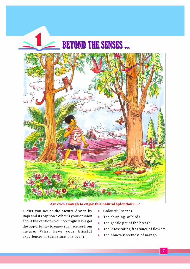
Didn't you notice the picture drawn byRaju and its caption? What is your opinionabout the caption? You too might have gotthe opportunity to enjoy such scenes fromnature. What have your blissfulexperiences in such situations been?
Are eyes enough to enjoy this natural splendour ...?
� Colourful scenes� The chirping of birds� The gentle pat of the breeze� The intoxicating fragrance of flowers� The honey-sweetness of mango

8
BIOLOGY
Though these are different experiences,can't they be felt at the same time? Can’twe hear, touch, smell and taste whileseeing?
Have you ever thought how this happens?
You know that we get information aboutthe changes in the environment throughthe sense organs. What are the senseorgans we have? What is the function ofeach? Prepare a note on it.
.....................................................................
EyeEyes enable us to see things. How are theyprotected? List the various means for theprotection of eyes.
� Position of the eyes – sockets in theskull
� Tears�
�
Lysozyme, the enzyme present in tearsdestroys germs that enter the eyes to acertain extent.
Let us examine the parts of the eye. Makea list of those you know.
� Pupil� Retina�
Don't you want to know how these partsare arranged in the eye? Collect the eye ofan animal from the butcher shop. Identifyits morphological characteristics byobserving and feeling it. Prepare a notebased on the indicators.
.....................................................................
Indicators
� Colour� Shape
� Firmness
With the help of your teacher take a crosssection of the eye that has been collectedby you. Making use of the features youobserved and the following descriptionwrite a note on the structure of the eye.Fill up the blanks in Figure 1.1.
Eye – The Window opening to Nature
The eye ball has three layers. The outermost layer is the sclera. It imparts firmness to theeye ball. The transparent front portion of the sclera is the cornea. The anterior part of theeye except the cornea is protected by a membrane called conjunctiva.
The middle layer, choroid contains many blood capillaries. The tissues in the eye receiveoxygen and nutrients from the blood that flows through these capillaries. In the anteriorpart of the choroid behind the cornea there is a circular dark screen named iris. Pupil isthe aperture at the centre of the iris. The pupil contracts when the intensity of light increasesand dilates when it decreases. In this way it regulates the amount of light entering theeye. This is facilitated by the muscles in the iris. The convex lens is placed behind thepupil. It is connected to the ciliary muscles by ligaments.

9
BIOLOGY
scleraciliary muscles
Retina is the innermost layer of the eye where the image is formed. Cells that arestimulated by light are seen here. The part of the retina with greatest vision is called theyellow spot and that with no vision is called the blind spot. The optic nerve carryingimpulses to the brain starts from the blind spot.
Inside the eye there are two chambers. The chamber between the lens and the cornea iscalled aqueous chamber. It is filled with a watery fluid, called aqueous humour. This fluidsupplies nutrients and oxygen to the cells of the cornea and the lens. The aqueous humourformed from the blood is reabsorbed into the blood itself. The large chamber seenbetween the lens and the retina is the vitreous chamber. It is filled with the jelly like vitreoushumour which helps to maintain the shape of the eyeball.
Couldn't you identify the parts you had seen in the cross section of the eye in thefigure? Now illustrate each part of the eye, their peculiarities and functions in aninterrelated manner in your Science diary. A model is given below.
CorneaTransparentanterior part
Allows light raysto enter
function
Outermostlayer
iris
???
ligaments
??
?
Fig 1.1 Structure of the eye
vitreous chamber?
yellow spot
optic nerve
peculiarity

10
BIOLOGY
� Ciliary muscles relax� Ligaments contract� Curvature of lens decreases� Focal length increases
� Ciliary muscles contract� Ligaments relax� Curvature of lens increases� Focal length decreases
You have understood the structure and function of the different parts of the eye. Ofthem the peculiarities of the lens and the retina deserve special mention.
Lens and Vision
You have seen the position and peculiarities of the lens of the eye.In order to understand how the lens helps to view near and distant objects, analyseFig.1.2 A, B and the following description and prepare notes.
Formation of ImageHow is the image formed in the retina? Analyse Figure 1.3. Note down the peculiaritiesof the image.
Characteristics of the Image� Real�
�
imageobject
Fig - 1.3. Formation of image in the retina.
Fig - 1.2.
A B
ciliary muscles
ligaments
lens
Viewing distant objects Viewing near objects
11
BIOLOGY
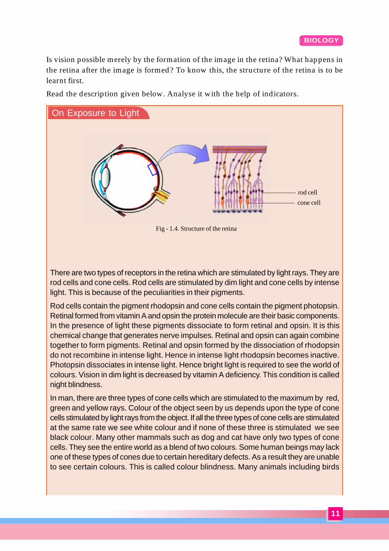
Is vision possible merely by the formation of the image in the retina? What happens inthe retina after the image is formed? To know this, the structure of the retina is to belearnt first.
Read the description given below. Analyse it with the help of indicators.
There are two types of receptors in the retina which are stimulated by light rays. They arerod cells and cone cells. Rod cells are stimulated by dim light and cone cells by intenselight. This is because of the peculiarities in their pigments.
Rod cells contain the pigment rhodopsin and cone cells contain the pigment photopsin.Retinal formed from vitamin A and opsin the protein molecule are their basic components.In the presence of light these pigments dissociate to form retinal and opsin. It is thischemical change that generates nerve impulses. Retinal and opsin can again combinetogether to form pigments. Retinal and opsin formed by the dissociation of rhodopsindo not recombine in intense light. Hence in intense light rhodopsin becomes inactive.Photopsin dissociates in intense light. Hence bright light is required to see the world ofcolours. Vision in dim light is decreased by vitamin A deficiency. This condition is callednight blindness.
In man, there are three types of cone cells which are stimulated to the maximum by red,green and yellow rays. Colour of the object seen by us depends upon the type of conecells stimulated by light rays from the object. If all the three types of cone cells are stimulatedat the same rate we see white colour and if none of these three is stimulated we seeblack colour. Many other mammals such as dog and cat have only two types of conecells. They see the entire world as a blend of two colours. Some human beings may lackone of these types of cones due to certain hereditary defects. As a result they are unableto see certain colours. This is called colour blindness. Many animals including birds
On Exposure to Light
Fig - 1.4. Structure of the retina
rod cell
cone cell
12
BIOLOGY
Indicators« What are the changes taking place in the rod cells in dim light and intense light?« How is vitamin A related to vision?« How do rod cells differ from cone cells physiologically?« What is the difference between night blindness and colour blindness?« What is the reason for the absence of vision at the blind spot?Let us try an activity to examine the power of vision in the blind spot?
How does the information received by thesense organs reach the brain? Look at thefollowing facts related to this.
� It is the nervous system that controlsand co-ordinates the functions of thebody.
� The basic unit of the nervous systemis neuron.
� Brain, spinal cord, nerves and sensoryreceptors constitute the nervoussystem.
Now analyse the following figures anddescription based on indicators andrecord your inferences.
.....................................................................
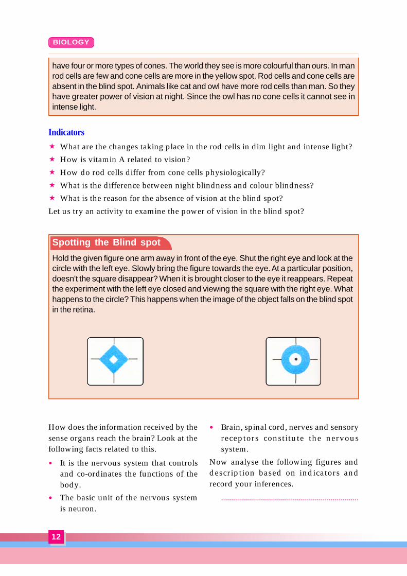
Spotting the Blind spot
Hold the given figure one arm away in front of the eye. Shut the right eye and look at thecircle with the left eye. Slowly bring the figure towards the eye. At a particular position,doesn't the square disappear? When it is brought closer to the eye it reappears. Repeatthe experiment with the left eye closed and viewing the square with the right eye. Whathappens to the circle? This happens when the image of the object falls on the blind spotin the retina.
have four or more types of cones. The world they see is more colourful than ours. In manrod cells are few and cone cells are more in the yellow spot. Rod cells and cone cells areabsent in the blind spot. Animals like cat and owl have more rod cells than man. So theyhave greater power of vision at night. Since the owl has no cone cells it cannot see inintense light.
13
BIOLOGY
Neuron and Nerve
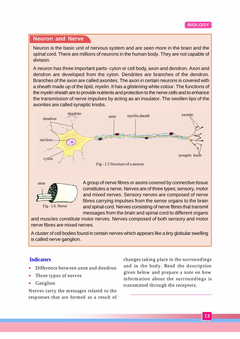
Neuron is the basic unit of nervous system and are seen more in the brain and thespinal cord. There are millions of neurons in the human body. They are not capable ofdivision.
A neuron has three important parts- cyton or cell body, axon and dendron. Axon anddendron are developed from the cyton. Dendrites are branches of the dendron.Branches of the axon are called axonites. The axon in certain neurons is covered witha sheath made up of the lipid, myelin. It has a glistening white colour. The functions ofthe myelin sheath are to provide nutrients and protection to the nerve cells and to enhancethe transmission of nerve impulses by acting as an insulator. The swollen tips of theaxonites are called synaptic knobs.
A group of nerve fibres or axons covered by connective tissueconstitutes a nerve. Nerves are of three types; sensory, motorand mixed nerves. Sensory nerves are composed of nervefibres carrying impulses from the sense organs to the brainand spinal cord. Nerves consisting of nerve fibres that transmitmessages from the brain and spinal cord to different organs
and muscles constitute motor nerves. Nerves composed of both sensory and motornerve fibres are mixed nerves.
A cluster of cell bodies found in certain nerves which appears like a tiny globular swellingis called nerve ganglion.
Indicators� Difference between axon and dendron� Three types of nerves� GanglionNerves carry the messages related to theresponses that are formed as a result of
changes taking place in the surroundingsand in the body. Read the descriptiongiven below and prepare a note on howinformation about the surroundings istransmitted through the receptors.
.....................................................................
axon
Fig - 1.6. Nerve
dendrite
Fig - 1.5 Structure of a neuron
dendron
cyton
myelin sheathaxon axonite
synaptic knob
nucleus
14
BIOLOGY
Stimulus, Receptors, Impulse, Response
Nerve cells or receptors that are capable of receiving stimuli from within the body andexternal environment are located in sense organs and in other different organs. Receptorsare modified neurons. They are of different types. Rods and cones in the eye, soundreceptors in the ear, taste receptors on the tongue, olfactory receptors in the nose andreceptors on the skin are examples.
As in all cells, opposite electric charges exist on either side of the plasma membraneof the receptor cells also. Changes in the internal and external environment of the bodycause variations in the electrical equilibrium existing on either side of the plasmamembrane. Such changes are known as stimuli. eg:- light, heat, cold etc. The flow ofelectric charges resulting from variations in equilibrium are called impulses. When suchimpulses reach the brain or the spinal cord through nerves corresponding changes takeplace in the body. These changes are termed responses.
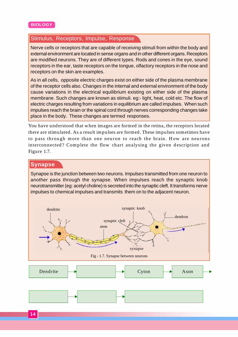
You have understood that when images are formed in the retina, the receptors locatedthere are stimulated. As a result impulses are formed. These impulses sometimes haveto pass through more than one neuron to reach the brain. How are neuronsinterconnected? Complete the flow chart analysing the given description andFigure 1.7.
Synapse
Synapse is the junction between two neurons. Impulses transmitted from one neuron toanother pass through the synapse. When impulses reach the synaptic knobneurotransmitter (eg: acetyl choline) is secreted into the synaptic cleft. It transforms nerveimpulses to chemical impulses and transmits them on to the adjacent neuron.
ASpØ \yqtdmWns‚ sU≥ss{U‰v
Cyton AxonDendrite
synapse
synaptic knob
dendron
axon
dendrite
synaptic cleft
Fig - 1.7. Synapse between neurons
15
BIOLOGY
Fig - 1.8. Synapse between a nerve cell and a muscle cell
synaptic knob
muscle cellsynaptic cleft
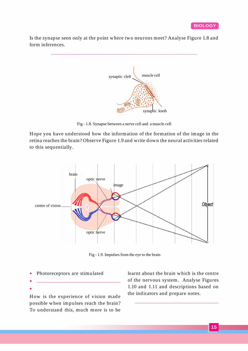
Is the synapse seen only at the point where two neurons meet? Analyse Figure 1.8 andform inferences.
.....................................................................................................................
Hope you have understood how the information of the formation of the image in theretina reaches the brain? Observe Figure 1.9 and write down the neural activities relatedto this sequentially.
� Photoreceptors are stimulated� ...................................................................�
How is the experience of vision madepossible when impulses reach the brain?To understand this, much more is to be
learnt about the brain which is the centreof the nervous system. Analyse Figures1.10 and 1.11 and descriptions based onthe indicators and prepare notes.
...................................................................
Fig - 1.9. Impulses from the eye to the brain
brain
centre of vision
optic nerve
optic nerve
image

16
BIOLOGY
Brain – The Centre of Wonders
The brain is the most complex organ in the human body. It is the centre of all the peculiarcharacteristics which make man unique. The brain is protected inside a hard case calledskull. The three layered covering of the brain is called meninges. The cerebrospinal fluid(CSF) fills between the inner layers. The CSF which is formed from the blood isreabsorbed into the blood. CSF helps to provide nutrients and oxygen to the tissues inthe brain, and also ensures the protection of the brain. CSF is also contained in thecerebral ventricles, the cavities in the brain.
Cerebrum is the largest part of the brain. Cytons (cell bodies) of neurons are seen crowdedtogether in this part. The outer part of the cerebrum is grey coloured due to the absenceof myelin. This is called grey matter. The folds and grooves in the grey matter areadaptations to accommodate more number of neurons. The interior of the cerebrum iscalled white matter. Nerve fibres covered with white coloured myelin are seen in thispart.
The cerebrum is the centre of qualities like intelligence, memory, thought and imagination.Senses like sight, hearing and taste are made possible by this part. Impulses from senseorgans reach specific centers of the cerebrum through nerves. Here impulses areinterpreted and integrated to give us precise sensations. The cerebrum controls allvoluntary movements.
Cerebellum, the part that lies below the cerebrum, is the second largest part of the brain.Here too, grooves and folds are present. The cerebellum maintains the equilibrium ofthe body by coordinating muscular activities.
Medulla oblongata is a stem-like part seen close to the cerebellum below the cerebrum.Medulla oblongata controls involuntary actions like breathing, heartbeat, etc. It continuesas the spinal cord which extends through the vertebral column till its posterior end. In themedulla oblongata and spinal cord, white matter is seen outside and grey matter inside.
The thalamus seen at the interior of the brain is the relay station of impulses to and fromthe cerebrum. Impulses reaching the thalamus are analysed before being relayed. Atthe base of the thalamus is the hypothalamus which plays an important role in maintaininghomeostasis.
Fig - 1.11. Parts of the brain that control variousactivities in man
movement touch
thought
speechsmell
taste
hearing maintenanceof equilibrium
vision
Fig - 1.10. Structure of the brain
cerebrumgrey matterwhite matter
thalamus
cerebellum
hypothalamus
medullaoblongata
spinal cord
pituitarygland

17
BIOLOGY
Indicators« How is the brain protected?« What are the main parts of the brain?« How do the cerebrum and medulla
oblongata differ in structure andfunction?
« What is the role of the brain inproviding sensory experiences?
« Which part of the brain enabled Rajuto draw the scenery?
Don't you doubt why two eyes are neededto see? One can see even with a single eye.Try the given activity.
Remove the cap of a pen and hand it overto your friend. Ask your friend to replacethe cap on the pen in your hand, with oneeye closed, standing at a distance ofatleast half a metre.
What happens? Find the reason and writeit down.
.....................................................................
Man has the power to focus both eyes atthe same time on objects in front of him.This is binocular vision. As a result, a threedimensional vision, giving a correctunderstanding of the distance from theobject, thickness of the object etc. ispossible.
Behind HearingYou have understood how vision is experienced. Is it in the same manner that hearingalso is effected? In order to understand the structure and function of the ear, analysethe following figure and description with the help of indicators.
Fig - 1.12. Structure of the ear
External ear Internal earEar drum or tympanum
Membrane at the innermost endof the ear canal. Vibrate inaccordance with sound waves.
CochleaPart withthree fluid-filled sacs.Auditoryreceptorsare situatedin this part.
PinnaDirects thesoundwavestowards theear canal.
Oval windowVibrates inaccordance with thevibration of stapes.
Ear canalDirects the sound waves to theear drum. Small hairs and earwax seen in this area preventdust and pathogens.
Ear ossiclesDirect the vibrationsthat occur in the eardrum towards theoval window.
Eustachian tubeConnects middleear with thepharynx. Regulatesthe pressure in themiddle ear.
incus
malleus stapes
semicircular canals
auditorynerve
Middle Ear

18
BIOLOGY
Don’t turn roundand round my
child... you will feelgiddy!
Auditory centre of the brain
Sound wave Pinna
Cochlea
Cochlea and Hearing
The part of the inner ear appears like the shell of a snail is the cochlea. It has threeinternal chambers. The middle chamber is filled with a fluid called endolymph and theother two chambers with the fluid perilymph. Auditory receptors are seen in the Organ ofCorti located in the middle chamber. Vibration of the oval window causes vibration inthe perilymph. This is transmitted to the endolymph and the auditory receptors arestimulated. Impulses thus formed reach the auditory centre of the brain through the auditorynerve.
Is hearing the only function of the ear?
Look at the following picture.
Indicators
� Parts of the external ear, middle ear, and internal ear and the functions of each.� The way through which the vibrations of the tympanic membrane reach the interior
of cochlea.� The mode of formation of impulses.� Part to which impulses reach through the auditory nerve.Discuss how the sensation of hearing is enabled. Based on your inferences fill up theflow chart.

19
BIOLOGY
Why do you feel giddy when you turn round and round?
Based on indicators analyse the given description, Figure 1.13 and the flow chart andform inferences.
.....................................................................................................................
Ear and Equilibrium of the Body
The semicircular canals and vestibule that are parts of the inner ear help to maintain theequilibrium of the body. These parts are filled with endolymph.
Semicircular canals – They are three in number, seen adjacent to the utricle. Theirswollen end is called ampulla. Receptors in the ampulla are stimulated in accordancewith the movement of the head.
Vestibule – It lies adjacent to the semicircular canals. It has two sacs – utricle and saccule.Receptors in these are stimulated according to the movements of the head.
Indicators« Where are the receptors related to equilibrium of body located?« What are the changes brought about by body movements in the parts related to
equilibrium?« Why is giddiness felt when you turn round and round?
Creates movements inthe endolymph of
semicircular canals andvestibule
Movement ofthe head andother parts of
the body
Receptors arestimulated
Impulse Auditorynerve
Cerebellum Coordinates muscular activities andmaintains equilibrium of the body
Fig - 1.13. Parts of the inner ear
ampulla
utriclesaccule
cochlea
} vestibule
semicircular canals

20
BIOLOGY
taste receptors
taste buds
minute pore to taste bud
bitterness
sournesssaltiness
Fig - 1.14. Taste buds in the tongue
sweetness
Food particlesdissolve in the
saliva
Taste receptors inthe taste buds are
stimulated
Till now we have been discussing the structure and function of the eye and the ear, andthe control of the brain over their functions. How are the other senses experienced?
The Sense of TasteThe taste buds located on the tongue, cheek and throat enable us to detect taste. Primarytastes such as sweetness, bitterness, sourness and saltiness can be detected by differenttaste buds. Analyse Figure 1.14 and understand how taste is detected and completethe flow chart.
To detect SmellOlfactory receptors seen on the walls of the nasal cavity are stimulated by olfactoryparticles which enter the nose through air. Analyse the illustration given below andenlist the activities related to the formation of olfactory sensation sequentially.
impulses to thecentre of taste inthe brain
Nerve
The sense oftaste
olfactory receptor
Olfactory NerveImpulses reach theolfactory centre ofthe brain.
olfactory receptor
Fig - 1.15. Olfactory receptors
mucus

21
BIOLOGY
heattouch
paincold
pressure
Fig - 1.16. Receptors in the skin
� Olfactory particles enter the nose along with air.� Olfactory particles dissolve in the mucus and reach the olfactory receptors.� Olfactory receptors are stimulated and impulses are formed.� ……………………..................................................................�
Skin – the Largest Sense OrganWe recognise several stimuli through the skin. What are they? Analyse Figure 1.16 andidentify the receptors in the skin.
When receptors are stimulated impulsesare formed. Impulses reach the brainthrough respective nerves and becomesenses. The largest number of receptors isseen in fingers and palm. You have nowunderstood the significant role of the senseorgans in recognizing changes in oursurroundings and responding to them
specifically. The sense organs may havedisorders or diseases due to manyreasons. Myopia (short sight),hypermetropia (long sight) etc. are eyedisorders. You might have studied aboutit. Find the causes and remedies for theseand complete the following table.

22
BIOLOGY
Condition Cause Remedy
Myopia
Hypermetropia
Note the indications given about otherdisorders of the eye and the ear. Collectmore information about them and recordit in the Science diary.
� Squint eye: The condition where botheyes are not able to focus on the sameobject. This defect can be rectifiedthrough surgery.
� Glaucoma: As reabsorption of aqueoushumour is hindered, pressureincreases in the eye. The optic nerveand photoreceptors in the retina mayget damaged due to glaucoma. Earlytreatment may prevent the onset ofblindness.
� Presbyopia: This visual disorder isusually seen in middle-aged persons.This is due to the loss of elasticity ofthe lens. In this condition nearbyobjects are not clearly seen. Use ofconvex lens is the remedial measure.
� Cataract: Aged people are affected by
this. Since the eye lens becomesopaque, the power of vision graduallydiminishes. Surgical replacement ofthe lens is the remedial measure.
� Deafness: This is the condition inwhich hearing ability is lost. It can becaused by several reasons.
Many habits of modern life adverselyaffect the health of our sense organs. Findsuch habits and their ill effects andorganize a seminar on the topic.
Not only man, all living beings havesensory experiences. However, man cantransform mere sensations into blissfulexperiences. While enjoying a scenerywhat we experience is an integrated blendof sight, hearing, touch, etc., a freshexperience beyond absolute sensation.This experience is rendered by the brain.The painting by Raju is the outcome ofsuch an experience. It is the brain thatprovides us the ability to enjoy nature.
23
BIOLOGY
Sense organs Receptors Stimuli Experience
Eye Rod Cells Light VisionCone cells
Ear
Nose
Tongue Tastes like saltiness,sourness, sweetness, Taste
bitterness
Skin � Touch� Pain��
Stimulus Dendrite Cyton
Synapse Axonite Adjacent Neuron
Axon
Follow up Activities1) An ordinary situation is given below. Analyse it and answer the questions.
� Two friends meet after a long time and have a chat.(a) What are the sensory functions performed here?(b) What are the nervous activities that enabled them to recognize each other?(c) How do the ear and the brain help in their communication? Analyse it.(d) Write an example of another situation in which sense organs and brain function
in collaboration.2) Enlist the nervous functions that take place in relation to the following situations
sequentially.(a) Sweetness is experienced on chewing toffee.(b) Enjoying the fragrance of flowers.(c ) The circus performer does not fall while walking over a wire.
3) Correct the mistakes, if any, in the flow chart and redraw.
4) Complete the table
Dendron