Embed Size (px)
Citation preview

A Role for AQP5 in Activation of TRPV4 by HypotonicityCONCERTED INVOLVEMENT OF AQP5 AND TRPV4 IN REGULATION OF CELLVOLUME RECOVERY*□S
Received for publication, January 19, 2006, and in revised form, March 21, 2006 Published, JBC Papers in Press, March 29, 2006, DOI 10.1074/jbc.M600549200
Xibao Liu‡, Bidhan Bandyopadhyay‡, Tetsuji Nakamoto§, Brij Singh¶, Wolfgang Liedtke�, James E. Melvin§,and Indu Ambudkar‡1
From the ‡Secretory Physiology Section, Gene Therapy and Therapeutics Branch, NIDCR, National Institutes of Health,Bethesda, Maryland 20892, the §Center for Oral Biology, University of Rochester, Rochester, New York 14642,the ¶Department of Biochemistry and Molecular Biology, University of North Dakota, Grand Forks, North Dakota 58203,and the �Center for Translational Neuroscience, Duke University Medical Center, Durham, North Carolina 27710
Regulation of cell volume in response to changes in osmolarityis critical for cell function and survival. However, the molecularbasis of osmosensation and regulation of cell volume are notclearly understood. We have examined the mechanism of regu-latory volume decrease (RVD) in salivary gland cells and report anovel association between osmosensing TRPV4 (transient recep-tor potential vanalloid 4) and AQP5 (aquaporin 5), which isrequired for regulating water permeability and cell volume.Exposure of salivary gland cells and acini to hypotonicity elicitedan increase in cell volume and activation of RVD. Hypotonicityalso activated Ca2� entry, which was required for subsequentRVD. Ca2� entry was associated with a distinct nonselective cat-ion current that was activated by 4�PDD and inhibited by ruthe-nium red, suggesting involvement of TRPV4. Consistent withthis, endogenous TRPV4 was detected in cells and in the apicalregion of acini along AQP5. Importantly, acinar cells from micelacking either TRPV4 or AQP5 displayed greatly reduced Ca2�
entry and loss of RVD in response to hypotonicity, although theextent of cell swelling was similar. Expression of N terminus-deleted AQP5 suppressed TRPV4 activation and RVD but notcell swelling. Furthermore, hypotonicity increased the associa-tion and surface expression of AQP5 and TRPV4. Both theseeffects and RVD were reduced by actin depolymerization. Thesedata demonstrate that (i) activation of TRPV4 by hypotonicitydepends on AQP5, not on cell swelling per se, and (ii) TRPV4 andAQP5 concertedly control regulatory volume decrease. Thesedata suggest a potentially important role for TRPV4 in salivarygland function.
The ability of cells to regulate their volume is essential for mainte-nance of cellular homeostasis under anisotonic environmental condi-tions (1–3). Changes in osmolarity of the extracellular medium inducewater fluxes that result in swelling or shrinkage of cells, depending onthe osmotic gradient. Most cells respond to changes in tonicity and cellvolume by initiating mechanisms that allow them to recover their orig-
inal volume in the continued presence of the osmotic stress. Such reg-ulatory volume changes depend on the activation of cation and anionpermeabilties that reverse the osmotic gradient and direction of waterflow (1–4). Emerging studies demonstrate that regulatory volumechanges are critical for cell survival and also for regulation of cellularprocesses such as gene transcription and proliferation (1). In addition, avariety of cells, such as exocrine gland cells, utilize the mechanism ofregulatory volume change to drive fluid secretion (5). Several monova-lent cation and anion channels as well as intracellular Ca2� changescontribute to cell volume regulation. For example, hypo-osmolarity-induced cell swelling has been associated with a rise in cytosolic Ca2�
concentration ([Ca2�]i) in different cell types, which is due to hypoto-nicity-activated Ca2� entry pathways. It has also been clearly demon-strated that this Ca2� entry is critical for regulating the ion fluxes thatdrive volume decrease (1–4). However, the underlying mechanism(s)that senses the change in osmolarity and/or cell volume to initiate vol-ume regulation is poorly understood (5).Regulation of transepithelial osmotic forces as well as cell volume
critically impacts salivary gland fluid secretion induced by neurotrans-mitter stimulation of the gland (5). The water channel, AQP5 (aqua-porin 5) provides a regulated water permeability across the apical mem-brane of salivary gland acinar cells that is important not only for fluidsecretion but also for regulatory volume changes (5–7).Aqp5�/�mousesalivary glands show decreased salivary secretion in response to musca-rinic receptor stimulation. Additionally, dispersed salivary gland acinifrom these mice have reduced ability to control volume changes inresponse to hyper- or hyposmotic solutions (7). Thus, fluid secretionand cell volume regulation converge at the level of AQP5, which medi-ates the final step in both processes (i.e.water efflux). However, it is notclear how cells sense the change in osmolarity and how this signal istransduced to achieve volume regulation.As discussed above, hypotonic and hypertonic conditions induce
[Ca2�]i increases indifferent cell types, andCa2� entryhasbeenreported tobe critical for the volume recovery process (1–4). Neurotransmitter-stimulated Ca2� mobilization events, including intracellular Ca2�
release and Ca2� entry as well as Ca2�-dependent activation of cationand anion channels, have been quite extensively studied in salivary andother exocrine gland cells (5, 8). Further, increases in cytosolic Ca2� dueto Ca2� entry have been correlated with ion channel activation and fluidsecretion. However, the Ca2� entry mechanism(s) involved in the cellularresponse to anisosmotic conditions has not yet been identified (5). TRPV4(transient receptor potential vanilloid 4), a member of the TRP super-family of cation channels (9) has been shown to be activated by hypoto-nicity and a variety of other stimuli (10–15). This channel is expressed
* This work was supported in part by National Institutes of Health Grant DE013823 (toAnil Menon, University of Cincinnati, and J. E. M.). The costs of publication of thisarticle were defrayed in part by the payment of page charges. This article must there-fore be hereby marked “advertisement” in accordance with 18 U.S.C. Section 1734solely to indicate this fact.
□S The on-line version of this article (available at http://www.jbc.org) contains supple-mental Figs. 1 and 2.
1 To whom correspondence should be addressed: Secretory Physiology Section,Gene Therapy and Therapeutics Branch, NIDCR, Bldg. 10, Rm. 1N-113, NationalInstitutes of Health, Bethesda, MD 20892. Tel.: 301-496-1478; Fax: 301-402-1228;E-mail: [email protected].
THE JOURNAL OF BIOLOGICAL CHEMISTRY VOL. 281, NO. 22, pp. 15485–15495, June 2, 2006Printed in the U.S.A.
JUNE 2, 2006 • VOLUME 281 • NUMBER 22 JOURNAL OF BIOLOGICAL CHEMISTRY 15485
by guest on July 6, 2018http://w
ww
.jbc.org/D
ownloaded from
by guest on July 6, 2018
http://ww
w.jbc.org/
Dow
nloaded from
by guest on July 6, 2018http://w
ww
.jbc.org/D
ownloaded from

in several cell types and has been reported to be involved in RVD2 inairway epithelial and keratinocyte cell lines (16, 17). TrpV4�/� micedisplay impaired regulation of systemic tonicity (18), although exactlyhow TRPV4 activity leads to regulation of cell volume has not yet beendirectly demonstrated. The molecular mechanism by which tonicitychanges regulate TRPV4 has also not yet been established (11, 12, 14,16–18).This study was directed toward defining the molecular basis of RVD.
Toward this end, we have measured the response of salivary gland celllines, primary cultures, and dispersed salivary gland acini to hypotonic-ity. These cells have been previously shown to display robust volumechanges as well as efficient volume recovery in response to anisos-motic conditions (5, 7). The exact molecular events involved in theseresponses are not yet known. The data presented here demonstrate anovel association between TRPV4 and AQP5, which controls RVD insalivary gland cells.We show that AQP5 is required for the activation ofTRPV4 by hypotonicity. Further, AQP5 and TRPV4 are concertedlyinvolved in regulating recovery of cell volume.
EXPERIMENTAL PROCEDURES
Cell Culture and Transfection—Cells were cultured in Dulbecco’smodified Eagle’s medium (rat basophilic leukemia cells; RBL-2H3),Earle’s minimal essential medium (human submandibular gland (HSG)and human parotid gland (HSY) cells) supplemented with 10% fetal calfserum, 2mM glutamine, 1% penicillin/streptomycin at 37 °C in 5% CO2.HSG cells were transiently transfected with 1 �g of required plasmidsand 0.2 �g of green fluorescent protein-encoding plasmid or only withgreen fluorescent protein plasmid using Lipofectamine reagent 2000(Invitrogen).
Dispersed Cell Preparation from Mouse Parotid and SubmandibularGland Cells—All mice were maintained according to guidelines approvedby the NIDCR, National Institutes of Health, Animal Care and Use Com-mittee. Submandibular glands were removed, cleaned, minced, anddigested in standard external solution containing 145mMNaCl, 5mMKCl,1 mMMgCl2, 1 mMCaCl2, 10 mMHepes, 10 mM glucose, pH 7.4 (NaOH),with 0.02% soybean trypsin inhibitor and 0.1% bovine serum albumin con-taining collagenase P (2.5 mg/8 ml) (7). After a 15–20-min incubation at37 °C, the digest was washed twice with the normal external solution andresuspended in external solution.
Electrophysiological Recording—The patch pipette had resistancesbetween 3 and 5 megaohms after filling with the standard intracellularsolution that contained 145mM cesiummethane-sulfonate, 8mMNaCl,10 mM MgCl2, 10 mM HEPES, 10 mM EGTA, pH 7.2 (CsOH). Externalsolutions were composed as follows: 145 mM NaCl, 5 mM CsCl, 1 mM
MgCl2, 10 mM CaCl2, 10 mM HEPES, 10 mM glucose, pH 7.4 (NaOH)(Ca2� and Na� solution); 170 mMNMDG, 5 mMCsCl, 1 mMMgCl2, 10mM HEPES, 10 mM glucose, pH 7.4 (HCl) (NMDG solution). Osmolar-ity for all solutions was adjusted with D-mannitol to 305 � 5 mmol/kgusing a vapor pressure osmometer (Wescor). For measuring swelling-activated currents, an isotonic solution containing 75 mM NaCl, 6 mM
CsCl, 5mMCaCl2, 1mMMgCl2, 10mMHepes, 150mMD-mannitol, and10 mM glucose, pH 7.4, with NaOH (305 � 5 mmol/kg) was used. Cellswelling was induced by omitting mannitol from this solution to reachproper osmolarity. In some experiments, the reduction of osmolaritywas achieved by reduction ofNaCl fromnormal external solutionwhereindicated.
Patch clamp experiments were performed in the tight seal whole-cellconfiguration at room temperature (22–25 °C) using an Axopatch 200Bamplifier (Axon Instruments) as described earlier (19–21). Voltageramps ranging from �90 to 90 mV over a period of 1 s were imposedevery 4 s from a holding potential of 0 mV and digitized at a rate of 1kHz. A liquid-junction potential of less than 8 mV was not corrected,and capacitative currents and series resistance were determined andminimized. For analysis, the first ramp was used for leak subtraction forthe subsequent current records. There was no significant increase in thecurrent under these conditions unless cells were exposed to hypotonicexternal solution (HTS).
Measurement of Intracellular Ca2� Concentration—Freshly isolatedsalivary gland cells were loaded with fura-2 for 45–60 min at 30 °C andallowed to attach to a glass bottomdish (MatekCorp.). Ducts and acinarcells were morphologically identified (7). Other cells were culturedovernight in Matek dishes. Fluorescence measurements were madeusing a Till Photonics-Polychrome IV spectrofluorimeter attached to aOlympus X51 microscope and Metafluor imaging system (UniversalImaging Corp.) (21). Fluorescence traces shown represent [Ca2�]i (val-ues are averages from�50 cells and representative of results obtained inat least 3–5 individual experiments).
Measurement of Cell Volume—Cells were loaded with the fluoro-probe calcein (Molecular Probes, Inc., Eugene, OR) and excited at 490nm. Emitted fluorescencewasmeasured at 510 nm. In situ calibration ofthe dye was performed. The relationship between dye fluorescence andthe volume change was linear over a volume range from �35 to �35%.In some experiments, cell volume was estimated using anOlympus X51microscope interfaced with Universal Imaging MetaMorph software.Data are presented as mean � S.E. Origin 7.5 (OriginLab, Northamp-ton, MA) was used for data analysis and display. Significant differencebetween individual groups was tested by using analysis of variance.
Immunoprecipitation and Immunoblotting—Immunoprecipitationwas done using solubilized crude membranes or from cell lysates aspreviously described (21). Precleared lysates were incubated with therequired antibody (1:200 dilution of anti-TRPV4 or anti-AQP5). Inter-acting proteins bound to Sepharose beads were separated, released withSDS-PAGE sample buffer, and detected by Western blotting asdescribed previously using anti-TRPV4 (1:500 dilution) or anti-AQP5antibody (1:1000 dilution).
Immunocytochemistry—Parotid glands were excised from the ani-mals and fixed in 10% formalin solution, embedded in paraffin, and usedto prepare 5–10-�m sections (AmericanHistolabs, Gaithersburg,MD).Sections were dewaxed, rehydrated, and permeabilized with 0.5% Tri-ton in PBS, pH 7.5. Streptavidin-peroxidase reactions using the DABHistostain kit (Zymed Laboratories, San Francisco, CA) were used todetect specific proteins (anti-TRPV4 at 1:70 and anti-AQP5 at 1:100dilutions). In control sections, rabbit IgG was used instead of primaryantibody.
Surface Biotinylation—Cells were treated as required and incubatedwith 0.5 mg/ml Sulfo-NHS-Biotin (Pierce) on ice (21), washed withbuffer containing 0.1 M glycine, and solubilized with 2 ml of cell lysisbuffer containing Nonidet P-40, 0.1% SDS, and proteolytic inhibitors.Biotinylated proteins were pulled down with Neutr-Avidin-linkedbeads (Pierce). The bound fraction was washed and released with SDS-PAGE sample buffer and analyzed by Western blotting.
RESULTS
Hypotonicity Stimulates Changes in Cell Volume and Ca2� Entry viaa TRPV4-like Channel in SalivaryGlandCells—Fig. 1A shows thatHTSinduced a sustained increase in [Ca2�]i in HSG (human submandibular
2 The abbreviations used are: RVD, regulatory volume decrease; HTS, hypotonic solution;NMDG, N-methyl-D-glucamine, 4�PDD, 4�-phorbol-12,13-didecanoate; IP, immuno-precipitation; cyt-D, cytochalasin D; RuR, ruthenium red.
TRPV4-AQP5 Signaling Complex Regulates RVD
15486 JOURNAL OF BIOLOGICAL CHEMISTRY VOLUME 281 • NUMBER 22 • JUNE 2, 2006
by guest on July 6, 2018http://w
ww
.jbc.org/D
ownloaded from

gland cell line) cells, which was dependent on the osmolarity. Averagedata representing peak increase in fluorescence from these experimentsare shown in Fig. 1B (significant increases above resting [Ca2�]i areindicated). Primary cultures of human submandibular gland cells (22)but not rat basophilic leukemia (RBL) cells displayed a similar responsetoHTS (Fig. 1C; 150mmol/kg solutionwas used here and in subsequentexperiments). The sustained [Ca2�]i increase in HSG cells was blockedby 100�M but not 1�MGd3� (Fig. 1D), indicating that HTS-stimulatedCa2� entry is unlike store-operated Ca2� entry, which is blocked by 1�M Gd3� in these cells (19). This was further confirmed by whole cellcurrent recordings. HTS increased HSG cell membrane conductancewith a 25–40 s delay (Fig. 1E) and generated a weakly outwardly recti-fying current that reversed at �6 � 3 mV (Fig. 1F; HTS-stimulated[Ca2�]i increase, and cation currents were seen in 91% (20 of 22) of
cells). With NMDG as the cation in the external solution (Fig. 1, G andH), inward current was greatly reduced, outward current remained rel-atively unchanged, and reversal potential showed a left shift (this effectwas seen in all of the cells that displayed a response to HTS). Together,these data demonstrate that HTS stimulates a nonselective Ca2�-per-meable cation conductance that is distinct from the relatively inwardlyrectifying store-operated Ca2� current ISOC previously described inHSG cells (19, 20). However, the characteristics of the HTS-stimulatedcation current in HSG cells are similar to those of HTS-activatedTRPV4 currents in other cell types (14, 23, 24).We therefore examined the possibility that TRPV4 contributes to
HTS-induced Ca2� entry. 1 �M ruthenium red (RuR), a concentrationused for maximum inhibition of TRPV4 currents (23, 25), substantiallyblocked both inward and outward currents stimulated byHTS (Fig. 2A).
FIGURE 1. HTS induces volume changes andCa2� entry in human salivary gland cells. A andB, HTS-induced [Ca2�]i increase (increase in the340/380 nm fluorescence ratio) measured in fura-2-loaded HSG cells. Effects of varying osmolarityare shown in A, and average data from theseexperiments are presented in B. *, values that aresignificantly different from those in control condi-tions, p � 0.01, n � 100 –200. HTS at 150 mmol/kgwas used in all subsequent experiments. C, effectof HTS on [Ca2�]i in human primary submandibu-lar gland (SMG) and rat basophilic leukemia (RBL)cells to HTS. D, Gd3� sensitivity of HTS-inducedsustained [Ca2�]i increase in HSG cells. 1 or 100 �M
Gd3� was added to the external solution whereindicated by arrows, and a control trace is shownby the dashed line. The decrease in fluorescence inthe presence of 1 �M Gd3� was not different fromthat in control cells; 100 �M Gd3� rapidly decreasedfluorescence to that in resting cells. E, activation of anonselective cation conductance by HTS in HSGcells. Time courses of the current measured at 80 and�80 mV in cells perfused with Ca2� � Na�-HTS areshown (see “Experimental Procedures” for details). F,I-V relationship of the current at the time indicatedby the arrow in E. G, cation permeability of the HTS-stimulated channel in HSG cells. External Ca2� �Na�-HTS was replaced by NMDG-HTS solution forthe period indicated. H, I-V curves of the current atthe time points indicated in G. Current traces are rep-resentative of results obtained with a minimum ofthree cells in each case.
TRPV4-AQP5 Signaling Complex Regulates RVD
JUNE 2, 2006 • VOLUME 281 • NUMBER 22 JOURNAL OF BIOLOGICAL CHEMISTRY 15487
by guest on July 6, 2018http://w
ww
.jbc.org/D
ownloaded from
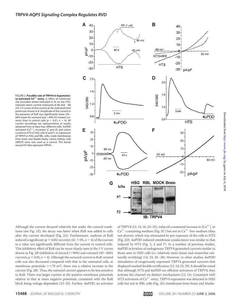
Although the current decayed relatively fast under the control condi-tions (see Fig. 1E), the decay was faster when RuR was added to cellsafter the current developed (Fig. 2A). Furthermore, washout of RuRinduced a significant (p� 0.05) recovery (41� 6%, n� 4) of the currentto a value not significantly different from the current in control cells.This inhibitory effect of RuR can be more clearly seen in the I-V curvesshown in Fig. 2B (inhibition of inward (�90%) and outward (50–60%)currents, p� 0.05, n� 4). Although the outward current in RuR-treatedcells was also decreased compared with that in the untreated cells, atmembrane potentials ��70 mV, there was a relative increase in thecurrent (Fig. 2B). Thus, the outward current appears to be less sensitiveto RuR. There was larger current at the positive membrane potentialsrelative to that at more negative potentials, consistent with the RuRblock being voltage-dependent (23–25). Further, 4�PDD, an activator
of TRPV4 (12, 14, 16, 23–25), induced a sustained increase in [Ca2�]i inCa2�-containing medium (Fig. 2C ) but not in Ca2�-free medium (datanot shown), which was attenuated by pre-exposure of the cells to HTS(Fig. 2D). 4�PDD-induced membrane conductance was similar to thatinduced by HTS (Fig. 2, E and F ). In a number of previous studies,4�PDD activation of endogenous TRPV4 generated currents similar tothose seen in HSG cells (i.e. relatively more linear and somewhat out-wardly rectifying) (14, 23, 26–28). However, in other studies, 4�PDDstimulation of exogenously expressed TRPV4 generated currents thatdisplayedmarked double rectification (12, 14, 23, 29). It should be notedthat although HTS and 4�PDD are efficient activators of TRPV4, theyactivate the channel via distinct mechanisms (12, 14). Consistent withHTS activation of Ca2� entry, TRPV4 expression was detected in HSGcells but not in RBL cells (Fig. 2G; membranes from brain and Madin-
FIGURE 2. Possible role of TRPV4 in hypotonici-ty-activated Ca2� entry. A, effect of rutheniumred (included where indicated in A) on the HTS-induced cation current measured at 80 and �80mV. I-V curves of the current at the indicated timepoints are shown in B. Amplitude of the current inthe presence of RuR was significantly lower (50 –60% lower for outward and �90% for inward cur-rents) than in control cells (p � 0.01, n � 4). Allcurrent recordings are representative of resultsobtained from at least four different cells. 4�PDDactivated [Ca2�]i increases (C and D) and cationcurrent as HTS in HSG cells (E and F). G, expressionof TRPV4 in HSG and RBL cells; crude membranesfrom brain and Madin-Darby canine kidney cells(MDCK) were was used as a control. The bandsaround 97 kDa represent TRPV4.
TRPV4-AQP5 Signaling Complex Regulates RVD
15488 JOURNAL OF BIOLOGICAL CHEMISTRY VOLUME 281 • NUMBER 22 • JUNE 2, 2006
by guest on July 6, 2018http://w
ww
.jbc.org/D
ownloaded from
Darby canine kidney cells were used as controls). In aggregate, the datain Fig. 2 suggest that TRPV4 might be involved in HTS-induced Ca2�
entry in salivary epithelial cells.Ca2� Entry Is Required for Regulatory Volume Decrease—The role of
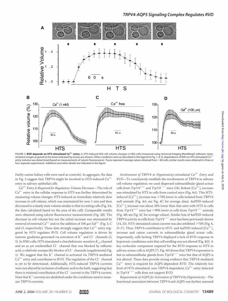
Ca2� entry in the cellular response to HTS was further determined bymeasuring volume changes. HTS induced an immediate relatively slowincrease in cell volume, which was maintained for over 1 min and thendecreased to a steady state volume similar to that in resting cells (Fig. 3A;the data calculated based on the area of the cell). Comparable resultswere obtained using calcein fluorescence measurements (Fig. 3B). Thedecrease in cell volume but not the initial increase was attenuated byremoval of external Ca2� and/or the addition of 100 �MGd3� (Fig. 3, Cand D, respectively). These data strongly suggest that Ca2� entry trig-gered by HTS regulates RVD. Cell volume regulation is driven byosmotic gradients generated via activation of K� and Cl� channels (3,5). In HSG cells, HTS stimulated a charybdotoxin-sensitiveKCa channeland an as yet unidentified Cl� channel that was blocked by niflumicacid, a relatively nonspecific blocker of Cl� channels (supplemental Fig.1). We suggest that the K� channel is activated via TRPV4-mediatedCa2� entry and contributes to RVD. The regulation of the Cl� channelis yet to be determined. Additionally, HTS-induced TRPV4 currentswere not altered by inclusion of niflumic acid in the bath, suggesting thatthere is minimal contribution of the Cl� current to the TRPV4 current.Note that K� currents are abolished under the conditions used tomeas-ure TRPV4 currents.
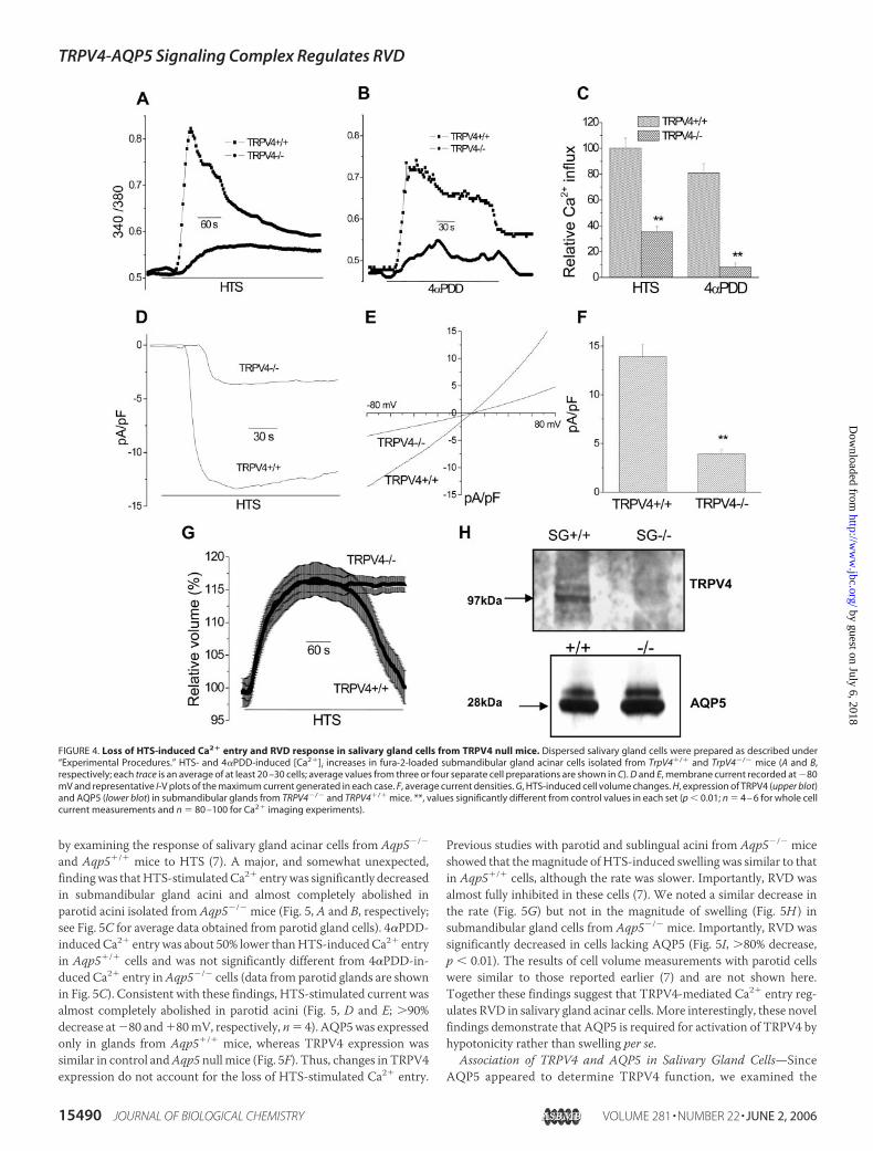
Involvement of TRPV4 in Hypotonicity-stimulated Ca2� Entry andRVD—To conclusively establish the involvement of TRPV4 in salivarycell volume regulation, we used dispersed submandibular gland acinarcells from TrpV4�/� and TrpV4�/� mice (18). Robust [Ca2�]i increasewas stimulated by HTS in cells from control mice (Fig. 4A). This HTS-induced [Ca2�]i increase was �70% lower in cells isolated from TRPV4null animals (Fig. 4A; see Fig. 4C for average data). 4�PDD-induced[Ca2�]i increase was about 20% lower than that seen with HTS in cellsfrom TrpV4�/� mice but �90% lower in cells from TrpV4�/� animals(Fig. 4B; see Fig. 4C for average values). Similar loss of 4�PDD-inducedTRPV4 activity in cells fromTrpV4�/� mice has been previously shown(14, 23). HTS-stimulated cation current was also inhibited�70% (Fig. 4,D–F ). Thus, TRPV4 contributes to HTS- and 4�PDD-induced [Ca2�]iincrease and cation currents in submandibular gland acinar cells.Importantly, cells lacking TRPV4 displayed a lack of RVD response inhypotonic conditions; note that cell swelling was not altered (Fig. 4G). Akey molecular component required for the RVD response to HTS insalivary acinar cells is AQP5 (7). Fig. 4H shows that TRPV4 expression islost in submandibular glands from TrpV4�/� mice but that of AQP5 isnot altered. These data provide strong evidence that TRPV4-mediatedCa2� entry is required for AQP5-dependent RVD. The relatively lowlevel of HTS-stimulated, non-TRPV4-dependent, Ca2� entry detectedin TrpV4�/� cells does not support RVD.
Requirement of AQP5 for Activation of TRPV4 by Hypotonicity—Thefunctional association between TRPV4 and AQP5 was further assessed
FIGURE 3. RVD depends on HTS-stimulated Ca2� entry. A, HTS-induced HSG cell volume changes in HSG cells (measured using Universal Imaging MetaMorph software; repre-sentative images acquired at the times indicated by arrows are shown). Other conditions were as described in the legend to Fig. 1. B–D, dependence of RVD on HTS-stimulated Ca2�
entry (volume was determined based on measurements of calcein fluorescence). Traces represent average values obtained from �80 cells; similar results were obtained in three orfour separate experiments. Additions and other details are indicated in the figure.
TRPV4-AQP5 Signaling Complex Regulates RVD
JUNE 2, 2006 • VOLUME 281 • NUMBER 22 JOURNAL OF BIOLOGICAL CHEMISTRY 15489
by guest on July 6, 2018http://w
ww
.jbc.org/D
ownloaded from
by examining the response of salivary gland acinar cells from Aqp5�/�
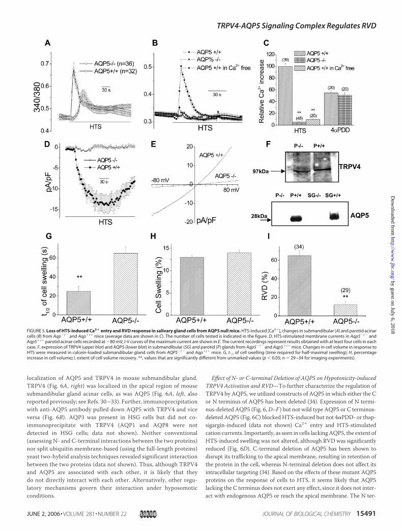
and Aqp5�/� mice to HTS (7). A major, and somewhat unexpected,findingwas thatHTS-stimulatedCa2� entrywas significantly decreasedin submandibular gland acini and almost completely abolished inparotid acini isolated from Aqp5�/� mice (Fig. 5, A and B, respectively;see Fig. 5C for average data obtained from parotid gland cells). 4�PDD-inducedCa2� entry was about 50% lower thanHTS-inducedCa2� entryin Aqp5�/� cells and was not significantly different from 4�PDD-in-duced Ca2� entry inAqp5�/� cells (data from parotid glands are shownin Fig. 5C). Consistent with these findings, HTS-stimulated current wasalmost completely abolished in parotid acini (Fig. 5, D and E; �90%decrease at�80 and�80mV, respectively, n� 4). AQP5was expressedonly in glands from Aqp5�/� mice, whereas TRPV4 expression wassimilar in control andAqp5 null mice (Fig. 5F). Thus, changes in TRPV4expression do not account for the loss of HTS-stimulated Ca2� entry.
Previous studies with parotid and sublingual acini from Aqp5�/� miceshowed that themagnitude of HTS-induced swelling was similar to thatin Aqp5�/� cells, although the rate was slower. Importantly, RVD wasalmost fully inhibited in these cells (7). We noted a similar decrease inthe rate (Fig. 5G) but not in the magnitude of swelling (Fig. 5H ) insubmandibular gland cells from Aqp5�/� mice. Importantly, RVD wassignificantly decreased in cells lacking AQP5 (Fig. 5I, �80% decrease,p � 0.01). The results of cell volume measurements with parotid cellswere similar to those reported earlier (7) and are not shown here.Together these findings suggest that TRPV4-mediated Ca2� entry reg-ulates RVD in salivary gland acinar cells.More interestingly, these novelfindings demonstrate that AQP5 is required for activation of TRPV4 byhypotonicity rather than swelling per se.
Association of TRPV4 and AQP5 in Salivary Gland Cells—SinceAQP5 appeared to determine TRPV4 function, we examined the
FIGURE 4. Loss of HTS-induced Ca2� entry and RVD response in salivary gland cells from TRPV4 null mice. Dispersed salivary gland cells were prepared as described under“Experimental Procedures.” HTS- and 4�PDD-induced [Ca2�]i increases in fura-2-loaded submandibular gland acinar cells isolated from TrpV4�/� and TrpV4�/� mice (A and B,respectively; each trace is an average of at least 20 –30 cells; average values from three or four separate cell preparations are shown in C). D and E, membrane current recorded at �80mV and representative I-V plots of the maximum current generated in each case. F, average current densities. G, HTS-induced cell volume changes. H, expression of TRPV4 (upper blot)and AQP5 (lower blot) in submandibular glands from TRPV4�/� and TRPV4�/� mice. **, values significantly different from control values in each set (p � 0.01; n � 4 – 6 for whole cellcurrent measurements and n � 80 –100 for Ca2� imaging experiments).
TRPV4-AQP5 Signaling Complex Regulates RVD
15490 JOURNAL OF BIOLOGICAL CHEMISTRY VOLUME 281 • NUMBER 22 • JUNE 2, 2006
by guest on July 6, 2018http://w
ww
.jbc.org/D
ownloaded from
localization of AQP5 and TRPV4 in mouse submandibular gland.TRPV4 (Fig. 6A, right) was localized in the apical region of mousesubmandibular gland acinar cells, as was AQP5 (Fig. 6A, left, alsoreported previously; see Refs. 30–33). Further, immunoprecipitationwith anti-AQP5 antibody pulled down AQP5 with TRPV4 and viceversa (Fig. 6B). AQP3 was present in HSG cells but did not co-immunoprecipitate with TRPV4 (AQP1 and AQP8 were notdetected in HSG cells; data not shown). Neither conventional(assessing N- and C-terminal interactions between the two proteins)nor split ubiquitin membrane-based (using the full-length proteins)yeast two-hybrid analysis techniques revealed significant interactionbetween the two proteins (data not shown). Thus, although TRPV4and AQP5 are associated with each other, it is likely that theydo not directly interact with each other. Alternatively, other regu-latory mechanisms govern their interaction under hyposomoticconditions.
Effect of N- or C-terminal Deletion of AQP5 on Hypotonicity-inducedTRPV4 Activation and RVD—To further characterize the regulation ofTRPV4 by AQP5, we utilized constructs of AQP5 in which either the Cor N terminus of AQP5 has been deleted (34). Expression of N termi-nus-deleted AQP5 (Fig. 6,D–F ) but not wild type AQP5 or C terminus-deleted AQP5 (Fig. 6C) blocked HTS-induced but not 4�PDD- or thap-sigargin-induced (data not shown) Ca2� entry and HTS-stimulatedcation currents. Importantly, as seen in cells lackingAQP5, the extent ofHTS-induced swelling was not altered, although RVD was significantlyreduced (Fig. 6D). C-terminal deletion of AQP5 has been shown todisrupt its trafficking to the apical membrane, resulting in retention ofthe protein in the cell, whereas N-terminal deletion does not affect itsintracellular targeting (34). Based on the effects of these mutant AQP5proteins on the response of cells to HTS, it seems likely that AQP5lacking the C terminus does not exert any effect, since it does not inter-act with endogenous AQP5 or reach the apical membrane. The N ter-
FIGURE 5. Loss of HTS-induced Ca2� entry and RVD response in salivary gland cells from AQP5 null mice. HTS-induced [Ca2�]i changes in submandibular (A) and parotid acinarcells (B) from Aqp�/� and Aqp�/� mice (average data are shown in C). The number of cells tested is indicated in the figure. D, HTS-stimulated membrane currents in Aqp5�/� andAqp5�/� parotid acinar cells recorded at �80 mV; I-V curves of the maximum current are shown in E. The current recordings represent results obtained with at least four cells in eachcase. F, expression of TRPV4 (upper blot) and AQP5 (lower blot) in submandibular (SG) and parotid (P) glands from Aqp5�/� and Aqp5�/� mice. Changes in cell volume in response toHTS were measured in calcein-loaded submandibular gland cells from AQP5�/� and Aqp�/� mice. G, t1⁄2 of cell swelling (time required for half-maximal swelling); H, percentageincrease in cell volume; I, extent of cell volume recovery. **, values that are significantly different from unmarked values (p � 0.05; n � 29 –34 for imaging experiments).
TRPV4-AQP5 Signaling Complex Regulates RVD
JUNE 2, 2006 • VOLUME 281 • NUMBER 22 JOURNAL OF BIOLOGICAL CHEMISTRY 15491
by guest on July 6, 2018http://w
ww
.jbc.org/D
ownloaded from

minus-deleted AQP5, on the other hand, induces a dominant negativeeffect. We suggest that this protein can interact with the endogenousAQP5 but does not form a functional channel. This further indicatesthat the N terminus of AQP5 might not be involved in homomericinteractions of AQP5. Additionally, the AQP5 N terminus might beinvolved in the HTS regulation of TRPV4. Whereas the mechanism bywhich the aquaporin mutant suppresses HTS responses has to be fur-ther established, these data strongly suggest that TRPV4 and AQP5 areassociated functionally and physically (although probably via indirectinteractions). These data also reveal a novel regulation of TRPV4 (i.e.that channel activation by hypotonicity is dependent on the presence offunctional AQP5). These data are also consistent with previous sugges-tions that TRPV4 is not activated by cell swelling per se (16).
Involvement of Cytoskeleton in Activation of TRPV4 and RegulatoryVolume Decrease—AQP5-mediated regulation of cellular volume dur-ing agonist stimulation of salivary gland cells has been associated with
translocation of the water channel protein to the plasmamembrane (31,32). This recruitment depends on [Ca2�]i increase and is blocked bydepolymerization of actin with cytochalasin D (cyto-D). Additionally,TRPV4 has also been shown to interact with the cytoskeleton (35).Thus, we examined the effect of depolymerization of the cytoskeletonon the responses of HSG cells toHTS. HTS- but not 4�PDD-stimulatedCa2� entry in HSG cells was blocked by actin depolymerization (Fig.7A). Interestingly, 4�PDD induced a transient activation of Ca2� entry(compare Fig. 7A with Fig. 2C) and cation currents (compare Fig. 7Bwith Fig. 2E). Furthermore, HTS-induced swelling of HSG cells was notaffected by cyto-D, but RVD was blocked (Fig. 7C), and the addition of4�PDD to the swollen cells induced a small transient decrease in cellvolume consistent with the transient Ca2� entry.
Effect of Hypotonicity on Cell Surface Expression and Association ofTRPV4 and AQP5—Fig. 7,D–F, shows the effect of HTS on the surfaceexpression of TRPV4 and AQP5 in control and cyto-D-treated HSG
FIGURE 6. Association and functional interac-tion between TRPV4 with AQP5. A, localizationof AQP5 (left) and TRPV4 (right; magnified imageincluded) in serial sections of mouse submandib-ular glands (reactivity was not detected in theabsence of primary antibody; image not shown).B, IP with anti-TRPV4 antibody (second panel; IBantibodies indicated below the blots) or AQP5antibody (third panel; inputs shown in the firstpanel). IP with TRPV4 does not pull down AQP3(first lane, IP fraction; second lane, unboundfraction). Shown is the HTS-induced increaseof [Ca2�]i in HSG cells transiently expressingAQP5-wt or AQP5-�C (C) or AQP5-�N (D). Theeffect of AQP5-�N expression on HTS-activatedcation current (E) and on HTS-induced volumechanges (F) is shown (average trace measured in20 –30 cells). 4�PDD was included in the externalsolution where indicated.
TRPV4-AQP5 Signaling Complex Regulates RVD
15492 JOURNAL OF BIOLOGICAL CHEMISTRY VOLUME 281 • NUMBER 22 • JUNE 2, 2006
by guest on July 6, 2018http://w
ww
.jbc.org/D
ownloaded from

cells. HTS induced a marked increase in the level of AQP5 and TRPV4in the biotinylated fraction, which was attenuated in cells treated withcyto-D (Fig. 7D; plasma membrane Ca2� pump in this fraction was notchanged; Supplemental Fig. 2). Additionally, HTS increased co-immu-noprecipitation of TRPV4 with AQP5, which was also blocked bycyto-D pretreatment of cells (Fig. 7E, left upper blots). Since anti-AQP5was used to pull down AQP5, the level of AQP5 is similar in both sam-ples (right panels, which also indicate similar protein load; note thatAQP5 and TRPV4 were detected using the same blot). Input levels ofTRPV4 and AQP5 in lysates of control and cyto-D-treated cells areshown in Fig. 7F (note that these lysates were used for IP with eitheravidin or anti-AQP5 (i.e. blots shown in Fig. 7, D and E) and thus serveas a control for both). The increased association of TRPV4 and AQP5was verified using dispersed mouse submandibular gland cells. HTSinduced a similar increase in the co-immunoprecipitation of TRPV4with AQP5 in these cells (Fig. 7G; IP was done using anti-AQP5 anti-body; blots show TRPV4 and AQP5 in IP samples in the top panels andcell lysates, input, in the lower panels). An increase was seen only in thelevels of TRPV4 in HTS-treated cells. Thus, the effects of HTSand cyto-D on the association of TRPV4 with AQP5 are not due to dif-ferences in protein load. In aggregate, these data suggest that HTS(i) increases the association between TRPV4 and AQP5 and (ii) regu-lates plasma membrane trafficking/insertion of these channels.Cytoskeletal rearrangements appear to be involved in regulating both ofthese events. It is important to note that both AQP5 and TRPV4 havebeen suggested to be regulated via interactions with the cytoskeleton(31, 32, 34, 35). Further studies are required to determine whether thetwo proteins associate with each other prior to translocation to theplasma membrane or after they are individually trafficked to the sur-face membrane.
DISCUSSION
The data presented above provide evidence that TRPV4 and AQP5concertedly control RVD in salivary epithelial cells. Hypotonic condi-tions induce cell swelling and activation of Ca2� entry via TRPV4. Sub-sequent activation of RVD is dependent on this Ca2� entry as well asAQP5, which regulates water permeability in these cells. Importantly,we demonstrate that TRPV4 activation by HTS is dependent on thepresence of functional AQP5. TRPV4 is not activated by HTS in cellslacking AQP5, although cells undergo swelling of a similar magnitudeas, albeit at a slower rate than, that in control cells. The slower rate ofswelling probably reflects a lack of contribution by AQP5 to plasmamembrane water permeability. Further, we also show that HTS stimu-lates the association and plasma membrane trafficking of both of thesechannels. Conditions that block these processes also block RVD. Thus,our data reveal for the first time an important functional link betweenTRPV4 and the water channel AQP5. Despite a number of recent stud-ies that demonstrate the involvement of TRPV4 in cell volume regula-tion and mechanosensation (10–18), the molecular basis for the regu-lation of TRPV4 has not yet been established. Further, the exactmechanisms by which AQP5 is regulated are also unclear (5). Our dataprovide evidence of a novel association between TRPV4 and AQP5 thatis involved in activation of TRPV4 by hypotonicity and regulation ofcellular response to the osmotic stress. We suggest that TRPV4 andAQP5 are assembled in a signaling complex that responds to anisos-motic conditions and coordinates cellular volume regulation.Our data also suggest that TRPV4 can have a critical role in salivary
gland fluid secretion. A fundamental role for AQP5 in regulation ofsalivary gland fluid secretion has been shown earlier (7). Aqp5�/� micedisplay pronounced decreases in salivary fluid secretion, and salivarygland cells from thesemice exhibit diminishedRVD response toHTS (5,
FIGURE 7. HTS-induced, cytoskeleton-dependent association and surface expression of TRPV4 and AQP5 regulates RVD. Effect of cytochalasin D treatment (CytoD; 1 �M for 30min) on HTS-induced [Ca2�]i increase (A), cation current (B), and regulation of cell volume (C). Changes in the external medium and time (horizontal bar) are indicated in the figure.D–G, effect of HTS on surface expression and association of TRPV4 and AQP5 in HSG (D–F) and freshly dispersed submandibular gland cells (G). Cells were treated with cytochalasinD as described above. Control and cyto-D cells were then exposed to HTS and biotinylated on ice. Cell lysates from control and HTS-treated cells were incubated with either avidin(D) or anti-AQP5 (E), and the blots were probed with anti-TRPV4 or anti-AQP5 antibodies as indicated (TRPV4 and AQP5) in HSG cell lysates (F). G, effect of HTS on co-IP of TRPV4 andAQP5 from dispersed mouse submandibular glands. Cells were prepared as described under “Experimental Procedures.” Cyto-D treatment and other experimental conditions weresimilar to those used for HSG cells. Data are representative of results obtained in three independent experiments.
TRPV4-AQP5 Signaling Complex Regulates RVD
JUNE 2, 2006 • VOLUME 281 • NUMBER 22 JOURNAL OF BIOLOGICAL CHEMISTRY 15493
by guest on July 6, 2018http://w
ww
.jbc.org/D
ownloaded from

7). Thus, it was suggested that AQP5 regulates salivary secretion bycontrolling the water permeability across salivary acinar cell apicalmembrane. However, the molecular basis of RVD in salivary gland andother cell types is not yet clear. Although it has been long recognizedthat regulatory volume changes are determined via regulation of[Ca2�]i, the mechanisms involved in sensing and transducing signalsrelated to changes in the osmolarity of the cell medium have not yetbeen determined. Our data clearly demonstrate that, like AQP5, TRPV4has a central role in regulating RVD in salivary epithelial cells. TRPV4channels are activated in response to hypotonicity in freshly dispersedsalivary gland cells and salivary cell lines. Further, the Ca2� entry viaTRPV4 is required for activation of a Ca2�-activated K� channel andsubsequent RVD. Importantly, we show that dispersed salivary glandcells from TrpV4�/� mice (18) display decreased responses to HTS;Ca2� entry is greatly reduced, and the ability to regulate cell volumeafter swelling is lost. These data demonstrate that TRPV4 contributes toHTS-activated Ca2� entry and regulates RVD via activation of the K�
channel. Together with an as yet unknown Cl� channel, these K� andCl� fluxes generate the osmotic gradient that drives water flow, viaAQP5, to induce shrinkage of cells to their original volume. Thus,TRPV4 has a critical role in the regulation of RVD. Since regulation oftransepithelial osmotic forces as well as cell volume critically impactssalivary gland fluid secretion, TRPV4 is likely to have a central role inregulating salivary gland function. This remains to be confirmed, sincesalivary secretion could not bemeasured reliably inTrpV4�/� mice dueto the sensitivity of the mice to general anesthesia.3
A pivotal finding of the present study is that hypotonicity-inducedactivation of TRPV4 is dependent on the presence of functionalAQP5 and not on the osmolarity of the medium or magnitude of cellswelling per se. Further, we show that the association between AQP5and TRPV4, as well as their surface expression, is increased uponexposure of the cells to hypotonic conditions. Although the molec-ular scaffold mediating these events is not yet known, based on ourdata, we suggest that sustained RVD is mediated by a cytoskeleton-dependent increase in the association and cell surface expression ofAQP5 and TRPV4. This suggestion does not exclude the possibilitythat other HTS-activated signals, such as arachidonic acid metabo-lites or protein kinase (12, 36), could contribute to this channelregulation. Consistent with our findings, Arniges et al. (16) previ-ously reported that reduced cell swelling does not account for lack ofTRPV4 activation and loss of RVD in airway epithelial cells exog-enously expressing a mutant cystic fibrosis membrane regulator.Cystic fibrosis membrane regulator has been known to interact withseveral other ion transport proteins and regulate fluid secretion.Cystic fibrosis airway epithelia have defective swelling-activated K�
andCl� channel activities and therefore display loss of RVD response (16).Although the role of aquaporins in cystic fibrosis membrane regulator-de-pendent regulation of cell volume has not yet been described, it has beensuggested that alveolar aquaporins might be more involved in cell vol-ume regulation than transepithelial fluid flux (6). In this regard, it isinteresting that AQP5 and cystic fibrosis membrane regulator appear tobe colocalized apically in pancreatic ductal cells and salivary epithelialcells (37). TRPV4 is also found in the apical region of salivary gland cells(30–33). Recently, TRPV4 has been reported to form a functional sig-naling complex with large conductance Ca2�-activated K� channels thatare involved invasodilation (27).Additionally, andconsistentwithourdata,TRPV4 has been reported to interact with MAP-7 via its C terminus; theinteraction links the channel to the cytoskeleton and regulates surface
expression of the channel (35). Together our present data and theseprevious studies suggest that TRPV4 and AQP5 are components of alarger signaling platform that can not only sense osmotic andmechanical signals but also transduce these signals and coordinatethe regulation of key ion channels that are involved in controlling cellshape and volume recovery. It is also interesting that TRPV4, likeTRPV1 and TRPV2, is translocated to the membrane in response tothe stimulus. Thus, regulated trafficking appears to be a commonmechanism underlying regulation of TRPV channels by specific sen-sory signals (38–41).In conclusion, our data demonstrate a role for AQP5 in the activation
of TRPV4 by hypotonicity in salivary gland cells. Further, we suggestthat AQP5 and TRPV4 are concertedly involved in regulation of cellvolume and salivary gland fluid secretion. AQP5 also mediates fluidsecretion in mucosal glands and epithelial cells in the lung as well as incells from pancreas and sweat glands (6, 7). TRPV4 has been suggestedto regulate RVD in airway epithelial cells, although the functional inter-action between TRPV4 and AQP5-mediated volume changes in thesetissues remains to be clarified. Thus, based on the role of TRPV4 inosmosensation and cell volume regulation demonstrated here and pre-viously (16–18, 27), it can be suggested that TRPV4 can function as atransducer of osmotic stimuli and in concert with AQP5 contribute tothe regulation of cellular volume. Further studies are required to deter-mine the critical molecular components that are involved in detectionof these signals and coordination of the cellular responses.
Acknowledgments—We thank Drs. Michael Caterina and Jon Levine for gen-erously providing the TRPV4 antibody, Drs. Bruce Baum, RobertWellner, andAnil Menon for anti-AQP5 antibodies and AQP5 plasmids, and David Wangfor invaluable help with immunocytochemistry.
REFERENCES1. Okada, Y., and Maeno, E. (2001) Biochem. Physiol. A Mol. Integr. Physiol. 130,
377–3832. Jakab, M., Fuerst, J., Gschwentner, M., Botta, G., Garavaglia, M. L., Bazzini, C.,
Rodighiero, S., Meyer, G., Eichmueller, S., Woell, E. S., Chwatal, S., Ritter, M., andPaulmichl,M. (2002) Cell. Physiol. Biochem. 12, 235–258
3. Strange, K. (2004) Adv. Physiol. Educ. 28, 155–1594. Tinel, H., Kinne-Saffran, E., and Kinne, R. K. (2000) Cell. Physiol. Biochem. 10,
297–3025. Melvin, J. E., Yule, D., Shuttleworth, T., and Begenisich, T. (2005) Annu. Rev. Physiol.
67, 445–4696. King, L. S., Kozono, D., and Agre, P. (2004) Nat. Rev. Mol. Cell. Biol. 5, 687–6987. Krane, C. M., Melvin, J. E., Nguyen, H. V., Richardson, L., Towne, J. E., Doetschman,
T., and Menon, A. G. (2001) J. Biol. Chem. 276, 23413–234208. Ambudkar, I. S. (2000) Crit. Rev. Oral Biol. Med. 11, 4–259. Montell, C. (2005) Sci. STKE 2005, RE310. Liedtke, W. (2000) Cell 103, 525–53511. Liedtke, W. (2005) J. Physiol. 567, 53–5812. Nilius, B., Vriens, J., Prenen, J., Droogmans, G., and Voets, T. (2004) Am. J. Physiol.
286, C195–C20513. Strotmann, R., Harteneck, C., Nunnenmacher, K., Schultz, G., and Plant, T. D. (2000)
Nat. Cell Biol. 2, 695–70214. Vriens, J., Watanabe, H., Janssens, A., Droogmans, G., Voets, T., and Nilius, B. (2004)
Proc. Natl. Acad. Sci. U. S. A. 101, 396–40115. Wissenbach, U., Bodding, M., Freichel, M., and Flockerzi, V. (2000) FEBS Lett. 485,
127–13416. Arniges, M., Vazquez, E., Fernandez-Fernandez, J. M., and Valverde, M. A. (2004)
J. Biol. Chem. 279, 54062–5406817. Becker, D., Blase, C., Bereiter-Hahn, J., and Jendrach, M. (2005) J. Cell. Sci. 118,
2435–244018. Liedtke, W., and Friedman, J. M. (2003) Proc. Natl. Acad. Sci. U. S. A. 100,
13698–1370319. Liu, X., Groschner, K., and Ambudkar, I. S. (2004) J. Membr. Biol. 200, 93–10420. Liu, X., Singh, B. B., and Ambudkar, I. S. (2003) J. Biol. Chem. 278, 11337–1134321. Liu, X., Bandyopadhyay, B. C., Singh, B. B., Groschner, K., and Ambudkar, I. S. (2005)
J. Biol. Chem. 280, 21600–216063 W. Liedtke, unpublished observations.
TRPV4-AQP5 Signaling Complex Regulates RVD
15494 JOURNAL OF BIOLOGICAL CHEMISTRY VOLUME 281 • NUMBER 22 • JUNE 2, 2006
by guest on July 6, 2018http://w
ww
.jbc.org/D
ownloaded from

22. Tran, S. D., Wang, J., Bandyopadhyay, B. C., Redman, R. S., Dutra, A., Pak, E., Swaim,W. D., Gerstenhaber, J. A., Bryant, J. M., Zheng, C., Goldsmith, C. M., Kok, M. R.,Wellner, R. B., and Baum, B. J. (2005) Tissue Eng. 11, 172–181
23. Chung, M.-K., Lee, H., and Caterina, M. (2003) J. Biol. Chem. 278, 33037–3304624. Chung, M.-K., Lee, H., Mizuno, A., Suzuki, M., and Caterina, M. (2004) J. Biol. Chem.
279, 21569–2157525. Watanabe, H., Davis, J. B., Smart, D., Jerman, J. C., Smith, G. D., Hayes, P., Vriens, J.,
Cairns, W., Wissenbach, U., Prenen, J., Flockerzi, V., Droogmans, G., Benham, C. D.,and Nilius, B. (2002) J. Biol. Chem. 277, 13569–13577
26. Andrade, Y. N., Fernandes, J., Vazquez, E., Fernandez-Fernandez, J. M., Arniges,M., Sanchez, T. M., Villalon, M., and Valverde, M. A. (2005) J. Cell. Biol. 168,869–874
27. Earley, S., Heppner, T. J., Nelson, M. T., and Brayden, J. E. (2005) Circ. Res. 97,1270–1279
28. Vriens, J., Owsianik, G., Fisslthaler, B., Suzuki, M., Janssens, A., Voets, T., Morisseau,C., Hammock, B. D., Fleming, I., Busse, R., andNilius, B. (2005)Circ. Res. 97, 908–915
29. Voets, T., Prenen, J., Vriens, J., Watanabe, H., Janssens, A., Wissenbach, U., Bodding,M., Droogmans, G., and Nilius, B. (2002) J. Biol. Chem. 277, 33704–33710
30. He, X., Tse, C. M., Donowitz, M., Alper, S. L., Gabriel, S. E., and Baum, B. J. (1997)Pflugers Arch. 433, 260–268
31. Ishikawa, Y., Skowronski, M. T., Inoue, N., and Ishida, H. (1999) Biochem. Biophys.
Res. Commun. 265, 94–10032. Ishikawa, Y., Yuan, Z., Inoue, N., Skowronski, M. T., Nakae, Y., Shono, M., Cho, G.,
Yasui, M., Agre, P., and Nielsen, S. (2005) Am. J. Physiol. 289, C1303–C131133. Gresz, V., Kwon, T. H., Hurley, P. T., Varga, G., Zelles, T., Nielsen, S., Case, R.M., and
Steward, M. C. (2001) Am. J. Physiol. 281, G247–G25534. Wellner, R. B., Hong, S., Cotrim, A. P., Swaim, W. D., and Baum, B. J. (2005) Tissue
Eng. 11, 1449–145835. Suzuki, M., Hirao, A., and Mizuno, A. (2003) J. Biol. Chem. 278, 51448–5145336. Cohen, D. M. (2005) Am. J. Physiol. 288, C483–C49337. Zeng, W., Lee, M. G., Yan, M., Diaz, J., Benjamin, I., Marino, C. R., Kopito, R., Freed-
man, S., Cotton, C., Muallem, S., and Thomas, P. (1997) Am. J. Physiol. 273,C442–C455
38. Morenilla-Palao, C., Plaanells-cases, R., Garcia-Sanz,N., and Ferre-Monteil, A. (2004)J. Biol. Chem. 279, 25665–25672
39. Van Buren, J. J., Bhat, S., Rotello, R., Pauza, M. E., and Premkumar, L. S. (2005)Mol.Pain 1, 17–28
40. Boels, K., Glassmeier, G., Herrmann, D., Riedel, I. B., Hampe,W., Kojima, I., Schwarz,J. R., and Schaller, H. C. (2001) J. Cell. Sci. 114, 3599–3606
41. Kanzaki, M., Zhang, Y. Q., Mashima, H., Li, L., Shibata, H., and Kojima, I. (1999)Nat.Cell. Biol. 1, 165–170
TRPV4-AQP5 Signaling Complex Regulates RVD
JUNE 2, 2006 • VOLUME 281 • NUMBER 22 JOURNAL OF BIOLOGICAL CHEMISTRY 15495
by guest on July 6, 2018http://w
ww
.jbc.org/D
ownloaded from
James E. Melvin and Indu AmbudkarXibao Liu, Bidhan Bandyopadhyay, Tetsuji Nakamoto, Brij Singh, Wolfgang Liedtke,
VOLUME RECOVERYINVOLVEMENT OF AQP5 AND TRPV4 IN REGULATION OF CELL
A Role for AQP5 in Activation of TRPV4 by Hypotonicity: CONCERTED
doi: 10.1074/jbc.M600549200 originally published online March 29, 20062006, 281:15485-15495.J. Biol. Chem.
10.1074/jbc.M600549200Access the most updated version of this article at doi:
Alerts:
When a correction for this article is posted•
When this article is cited•
to choose from all of JBC's e-mail alertsClick here
Supplemental material:
http://www.jbc.org/content/suppl/2006/03/31/M600549200.DC1
http://www.jbc.org/content/281/22/15485.full.html#ref-list-1
This article cites 35 references, 15 of which can be accessed free at
by guest on July 6, 2018http://w
ww
.jbc.org/D
ownloaded from

VOLUME 282 (2007) PAGES 32222–32232
Myelin basic protein-primed T cells induceneurotrophins in glial cells via �v�3 integrin.Avik Roy, Xiaojuan Liu, and Kalipada Pahan
In the entire article, we havemistaken�v as�5. Therefore, we requestthe readers to consider �5�3 as �v�3 and the title of the article as“Myelin Basic Protein-primed T Cells Induce Neurotrophins in GlialCells via �v�3 Integrin.”
We used anti-�v antibody (clone RMV-7) from BD Biosciences (cat-alog no. 552299) and eBioscience (catalog no. 16-0512) for fluores-cence-activated cell sorting analysis (Fig. 5) and neutralization experi-ments (Fig. 6 and supplemental Figs. 1–3). The following primers wereused for reverse transcription-PCR analysis of �v (Fig. 5C): 5�-GTTGGG AGA TTA GAC AGA GGA-3� (sense) and 5�-CAA AAC AGCCAGTAGCAACAA-3� (antisense). The labeling sequence for Fig. 5 (Aand B, right panels) should be itgaL, itgaM, itgaV, itgaX, itgb1, etc.
VOLUME 281 (2006) PAGES 15485–15495
A role for AQP5 in activation of TRPV4 by hypotonicity:concerted involvement of AQP5 and TRPV4 in regulationof cell volume recovery.Xibao Liu, Bidhan C. Bandyopadhyay, Tetsuji Nakamoto, Brij B. Singh,Wolfgang Liedtke, James E. Melvin, and Indu S. Ambudkar
Dr. Bandyopadhyay’s middle initial was inadvertently omitted. Hiscorrect name is shown above.
THE JOURNAL OF BIOLOGICAL CHEMISTRY VOL. 283, NO. 6, p. 3688, February 8, 2008© 2008 by The American Society for Biochemistry and Molecular Biology, Inc. Printed in the U.S.A.
3688 JOURNAL OF BIOLOGICAL CHEMISTRY VOLUME 283 • NUMBER 6 • FEBRUARY 8, 2008
ADDITIONS AND CORRECTIONS This paper is available online at www.jbc.org
We suggest that subscribers photocopy these corrections and insert the photocopies in the original publication at the location of the originalarticle. Authors are urged to introduce these corrections into any reprints they distribute. Secondary (abstract) services are urged to carrynotice of these corrections as prominently as they carried the original abstracts.