Embed Size (px)
Citation preview
Redox Biology 9 (2016) 50–56
Contents lists available at ScienceDirect
Redox Biology
http://d2213-23
n CorrE-m
journal homepage: www.elsevier.com/locate/redox
Research paper
Artemisinin protects human retinal pigment epithelial cells fromhydrogen peroxide-induced oxidative damage through activation ofERK/CREB signaling
Cheong-Meng Chong, Wenhua Zheng n
Faculty of Health Science, University of Macau, Macau, China
a r t i c l e i n f o
Article history:Received 13 May 2016Received in revised form16 June 2016Accepted 17 June 2016Available online 20 June 2016
Keywords:Retinal pigment epithelial cellsAge-related macular degenerationArtemisininROSERK
x.doi.org/10.1016/j.redox.2016.06.00217/& 2016 The Authors. Published by Elsevier
esponding author.ail address: [email protected] (W. Zhe
a b s t r a c t
The pathological increase in the levels of reactive oxygen species (ROS) in the retinal pigment epithelium(RPE), is implicated in the development of age-related macular degeneration (AMD). The discovery ofdrug candidates to effectively protect RPE cells from oxidative damage is required to resolve the pa-thological aspects and modify the process of AMD. In this study, a FDA-approved anti-malaria drug,Artemisinin was found to suppress hydrogen peroxide (H2O2)-induced cell death in human RPE cell-D407 cells. Further study showed that Artemisinin significantly suppressed H2O2
� induced D407 celldeath by restoring abnormal changes in nuclear morphology, intracellular ROS, mitochondrial membranepotential and apoptotic biomarkers. Western blotting analysis showed that Artemisinin was able to ac-tivate extracellular regulated ERK/CREB survival signaling. Furthermore, Artemisinin failed to suppressH2O2-induced cytotoxicity and the increase of caspase 3/7 activity in the presence of the ERK inhibitorPD98059. Taken together, these results suggest that Artemisinin is a potential protectant with the pro-survival effects against H2O2 insult through activation of the ERK/CREB pathway.& 2016 The Authors. Published by Elsevier B.V. This is an open access article under the CC BY-NC-ND
license (http://creativecommons.org/licenses/by-nc-nd/4.0/).
1. Introduction
Age-related macular degeneration (AMD) is a progressive de-generative eye disorder which is mainly caused by a breakdown ofthe macula in the retina. At early stage of AMD, it does not lead toany symptoms or vision loss. However, late AMD seriously affectsvision and further influences daily activities such as driving, reading,face recognition and life quality [1]. In the developed world, AMD isthe common leading cause of blindness and its incidence keeps in-creasing in the people with age 65 or older [2]. AMD can be dividedinto two types: dry AMD 85–90%) and wet AMD 10–15%). At present,the current therapies such as laser photocoagulation, photodynamictherapy and anti-VEGF therapy are limited to wet AMD, but themore prevalent dry AMD still lacks effective therapies [3,4].
The clinical characteristic of dry AMD is a decrease in chorior-etinal blood flow [5]. Chorioretinal atrophy results in subsequentdegeneration of retinal pigment epithelium (RPE) cells. The RPE, apigmented monolayer, plays a critical role in maintaining retinalfunctions such as nutrient transport, formation of the outer bloodretinal barrier to block the passage of water and ions, the phago-cytosis of photoreceptor outer segment tips as well as the
B.V. This is an open access article u
ng).
regeneration of visual pigments etc [6–8]. Age-associated degen-eration of RPE eventually lead to loss of photoreceptor cells andvision loss in the affected area [9]. Thus, the identification of ef-fective drug candidates to protect RPE cells from death may be apotential strategy for reducing the process of dry AMD.
Although the etiology and pathogenesis of dry AMD are not fullyelucidated, age-related alterations such as increased oxidative stressand decreased cell density in the RPE were considered as mainchanges in dry AMD [10–15]. The adequate oxygen distribution andsupply are necessary for maintenance of retinal function [16]. Due tohigh-oxygen microenvironment in the retina, RPE cells are exposedto constant oxidative stress resulting from reactive oxygen species(ROS) caused by intense light exposure [17]. Light-induced retinaldamage leads to the increase of ROS such as superoxide radical( �O2
�), hydroxyl radical ( �OH) and hydrogen peroxide (H2O2). Highlevels of ROS damage the RPE cells, which have been linked with thepathogenesis of clinical visual diseases [17]. H2O2 is an oxidizingagent, commonly used to cause irreversible oxidative damage andactivation of the apoptotic cascade in various RPE cell models [18,19].Therefore, H2O2-induced oxidative injury in RPE cell model, is sui-table for investigating the effects of drug candidates for the treat-ment of dry AMD.
Recently, we perform H2O2-induced RPE cell injury model toidentify potential protective candidates from various Chinesemedicine. Artemisinin (the chemical structure is shown in Fig. 1A),
nder the CC BY-NC-ND license (http://creativecommons.org/licenses/by-nc-nd/4.0/).
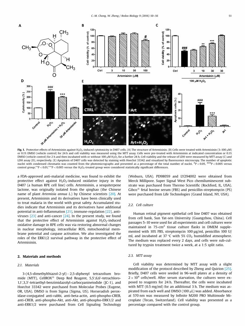
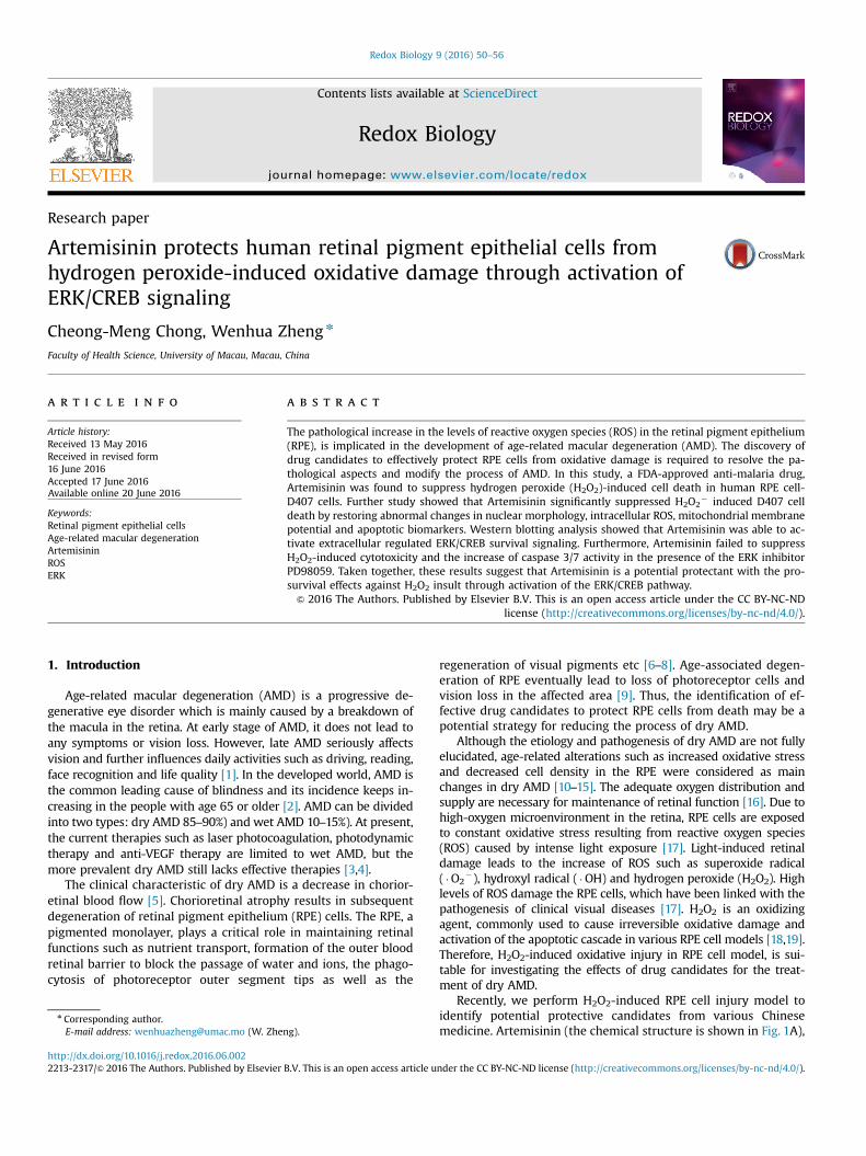
Fig. 1. Protective effects of Artemisinin against H2O2-induced cytotoxicity in D407 cells. (A) The structure of Artemisinin. (B) Cells were treated with Artemisinin (3–100 mM)or 0.1% DMSO (vehicle control) for 24 h and cell viability was measured using the MTT assay. Cells were pre-treated with Artemisinin at indicated concentration or 0.1%DMSO (vehicle control) for 2 h and then incubated with or without 100 mM H2O2 for a further 24 h. Cell viability and the release of LDH were measured by MTT assay (C) andLDH assay (D), respectively. (E) Apoptosis of D407 cells was detected by staining with Hoechst 33342 and visualized by fluorescence microscopy. The number of apoptoticnuclei with condensed chromatin was counted from the photomicrographs and presented as a percentage of the total number of nuclei. #Po0.05, ###Po0.005 versuscontrol group;**Po0.01,***Po0.005 versus the H2O2-treated group were considered statistically significant differences.
C.-M. Chong, W. Zheng / Redox Biology 9 (2016) 50–56 51
a FDA-approved anti-malarial medicine, was found to exhibit theprotective effect against H2O2-induced oxidative injury in theD407 (a human RPE cell line) cells. Artemisinin, a sesquiterpenelactone, was originally isolated from the qinghao (the Chinesename of plant Artemisia annua L.) by Chinese scientists [20]. Atpresent, Artemisinin and its derivatives have been clinically usedto treat malaria in the world with great safety. Accumulated stu-dies indicate that Artemisinin and its derivatives have additionalpotential in anti-Inflammation [21], immune-regulation [22], anti-viruses [23] and anti-cancer [24]. In the present study, we foundthat the protective effect of Artemisinin against H2O2-inducedoxidative damage in RPE cells was via restoring abnormal changesin nuclear morphology, intracellular ROS, mitochondrial mem-brane potential and caspase activation. We also investigated theroles of the ERK1/2 survival pathway in the protective effect ofArtemisinin.
2. Materials and methods
2.1. Materials
3-(4,5-dimethylthiazol-2-yl)�2,5-diphenyl tetrazolium bro-mide (MTT), CellROXs Deep Red Reagent, 5,5′,6,6′-tetrachloro-1,1′,3,3′-tetraethyl-benzimidazolyl-carbocyanineiodide (JC-1), andHoechst 33342 were purchased from Molecular Probes (Eugene,OR, USA). DMSO is from Sigma (Sigma, US). Horseradish perox-idase-conjugated anti-rabbi, anti-beta-actin, anti-phospho-CREB,anti-CREB, anti-phospho-Akt, anti-Akt, anti-phospho-ERK1/2 andanti-ERK1/2 were purchased from Cell Signaling Technology
(Woburn, USA). PD98059 and LY294002 were obtained fromMerck Millipore. Super Signal West Pico chemiluminescent sub-strate was purchased from Thermo Scientific (Rockford, IL, USA).Gibcos fetal bovine serum (FBS) and penicillin-streptomycin (PS)were purchased from Life Technologies (Grand Island, NY, USA).
2.2. Cell culture
Human retinal pigment epithelial cell line D407 was obtainedfrom cell bank, Sun Yat-sen University (Guangzhou, China). Cellpassages 5–10 were used for all experiments and cell cultures weremaintained in 75-cm2 tissue culture flasks in DMEM supple-mented with 10% FBS, streptomycin 100 μg/ml, penicillin 100 U/ml, and incubated at 37 °C with 5% CO2 humidified atmosphere.The medium was replaced every 2 days, and cells were sub-cul-tured by trypsin treatment twice a week, at a 1:5 split ratio.
2.3. MTT assay
Cell viability was determined by MTT assay with a slightmodification of the protocol described by Zheng and Quirion [25].Briefly, D407 cells were seeded in 96-well plates at a density of2�104 cells/well. After serum starvation, the cultures were ex-posed to reagents for 24 h. Thereafter, the cells were incubatedwith MTT (0.5 mg/ml) for an additional 3 h. The medium was as-pirated from each well and DMSO (100 mL) was added. Absorbanceat 570 nm was measured by Infinite M200 PRO Multimode Mi-croplate (Tecan, Switzerland). Cell viability was presented as apercentage compared with the control group.
C.-M. Chong, W. Zheng / Redox Biology 9 (2016) 50–5652
2.4. LDH assay
Cell cytotoxicity was determined by measuring the activity oflactate dehydrogenase (LDH) released into the incubation mediumwhen cellular membranes were damaged. D407 cells were seededinto 96-well plates (5�103 cells/well). After appropriate treat-ment, the activity of released LDH in the medium was determinedaccording to the instructions of CytoTox-ONE™ HomogeneousMembrane Integrity Assay (Promega, USA). The fluorescent in-tensity was measured using Infinite M200 PRO Multimode Mi-croplate at an excitation wavelength of 560 nm and emission at590 nm. All values of % LDH released were normalized to thecontrol group.
2.5. Hoechst 33342 staining
D407 cells were seeded into 12-well plates (8�104 cells/well).After appropriate treatment, these cells were washed with PBS,fixed with 4% formaldehyde (v/v in PBS) and then stained with10 μg/ml Hoechst 33342 for 15 min at room temperature. Afterwashing with PBS, the nuclei were visualized using EVOS FLImaging System (Thermo Fisher Scientific, USA).
2.6. Measurement of intracellular ROS levels
Intracellular ROS generation was evaluated using CellROXs
Deep Red Reagent (Thermo Fisher Scientific, USA). The cells wereincubated with CellROXs Deep Red Reagent (5 mM) in DMEM for1 h in the dark, rinsed twice with 1x PBS solution and the fluor-escence was observed and recorded using a fluorescent micro-scope at an excitation wavelength of 640 nm and an emissionwavelength of 665 nm. Semi-quantification of ROS level was as-sessed by using Image-J software. All values of % ROS level werenormalized to the control group.
2.7. Measurement of mitochondrial membrane potential (△ψm)
JC-1 dye was used to monitor mitochondrial integrity. In brief,D407 cells were seeded into black 96-well plates (1�104 cells/well). After appropriate treatment, the cells were incubated withJC-1 (10 μg/ml in medium) at 37 °C for 15 min and then washedtwice with PBS. For signal quantification, the intensity of redfluorescence (excitation 560 nm, emission 595 nm) and greenfluorescence (excitation 485 nm, emission 535 nm) were mea-sured using a Infinite M200 PRO Multimode Microplate. Mi-tochondrial membrane potential (△ψm) was calculated as the ratioof JC-1 red/green fluorescence intensity and the value was nor-malized to the control group. The fluorescent signal in the cellswas also observed and recorded with a fluorescent microscope.
3. Caspase 3/7 activity assay
After treatment, the activity of caspase 3/7 was measured usingthe commercially available Caspase-Glos 3/7 Assay (Invitrogen,USA) according to the manufacturer's protocol. Briefly, D407 cellswere lysed in lysis buffer and centrifuged at 12,500� g for 5 min15 mL of cell lysate was incubated with 50 mL of 2X substrateworking solution at room temperature for 30 min in 96-wellplates. The fluorescence intensity was then determined by InfiniteM200 PRO Multimode Microplate at an excitation wavelength of490 nm and emission at 520 nm. The fluorescence intensity ofeach sample was normalized to the protein concentration ofsample. All values of % caspase 3/7 activities were normalized tothe control group.
3.1. Western blotting
Western blotting was performed as previously described [25].Briefly, treated cells from different experimental conditions wererinsed once with ice-cold PBS and lysed in RIPA buffer [50 mMTris–HCl pH 8.0, 150 mM NaCl, 1 mM EDTA, 1% Igepal CA-630, 1%sodium dodecyl sulfate (SDS), 50 mM NaF, 1 mM NaVO3, 5 mMphenylmethysulfonyl fluoride, 10 μg/ml leupeptin (Sigma), and50 μg/ml aprotinin (Sigma) or 2� sample buffer (final con-centration of 62.5 mM Tris–HCl pH 6.8, 2% (w/v) SDS, 10% glycerol,50 mM dithiothreitol, and 0.1% (w/v) bromophenol blue)]. Sampleswith equal amounts of protein determined with a BCA proteinassay kit according to the manufacturer’s instructions, were se-parated by SDS polyacrylamide gel electrophoresis (PAGE) andtransferred to PVDF membranes. The phosphorylation of Akt andERK1/2 was determined by Western blotting with their respectivephospho-specific antibodies while β-actin was using as a totalprotein control. Blot was visualized using ECL kit according to themanufacturer's instructions. The blots were stripped, and reprobedwith anti-Akt or anti-ERK1/2, respectively. The intensity of thebands was quantified using ImageJ software.
3.2. Statistical analysis
Statistical analysis was performed using GraphPad Prism5.0 statistical software (GraphPad software, Inc., San Diego, CA,USA). All experiments were performed in triplicate. Data are ex-pressed as mean7standard deviation (SD). Statistical analysis wascarried out using one-way ANOVA followed by Tukey's multiplecomparison, with po0.05 considered statistically significant.
4. Results
4.1. Artemisinin attenuated H2O2-induced D407 cell death
To evaluate the cytotoxicity of Artemisinin, D407 cells wereincubated with various concentrations of Artemisinin for 24 h andthe cytotoxicity was determined by MTT assay. As shown in Fig. 1B,Artemisinin at between 3 and 30 μM did not cause any cytotoxi-city in D407 cells compared to the control group, and was used infurther experiments. To test the protective effects of Artemisinin,D407 cells were treated with Artemisinin for 2 h before beingexposed to H2O2 for 24 h. The result of MTT assay showed that thetreatment of 100 μM H2O2 resulted in dominant cell death (50%),whereas pre-treatment with 3, 10 and 30 μM of Artemisinin sig-nificantly attenuated H2O2-induced cell death in a concentration-dependent manner (54%, 65% and 75%, respectively) (Fig. 1C). Theprotective activity of Artemisinin was also confirmed by the lactatedehydrogenase (LDH) assay. As shown in Fig. 1D, pre-treated cellswith 30 μM of Artemisinin for 2 h significantly reduced H2O2-in-duced LDH leakage (from 251% to 199%). Nuclei condensation wasobserved in D407 cells after exposure to 100 μM H2O2 (26%) inHoechst 33342 staining assay. However, a pre-treatment of 30 μMArtemisinin decreased these changes (9%) (Fig. 1E). Artemisininalone did not lead to nuclear morphological change of D407 cells.
4.2. Artemisinin decreased H2O2-induced change of mitochondrialmembrane potential and caspase 3/7 activity
Mitochondrial inhibition causes the loss of mitochondrialmembrane potential (△ψm). To determine whether Artemisinincould reduce H2O2-induced △ψm loss, the △ψm in D407 cells wasassessed by analyzing the red/green fluorescent intensity ratio ofJC-1 staining. Exposure of D407 cells to 100 μM H2O2 resulted inan increase in green fluorescence intensity indicating △ψm
Fig. 2. Artemisinin attenuated H2O2-induced mitochondrial membrane potential (△ψm) loss and caspase 3/7 activity increase in D407 cells. After pre-treatment with 30 μMArtemisinin or 0.1% DMSO (vehicle control) for 2 h, D407 cells were incubated with or without 100 μM H2O2 for another 24 h. (A) △ψm was determined by the JC-1 assay.(B) Quantification of caspase 3/7 activity was determined by caspase 3/7 activity assay. ###Po0.005 versus control group;***Po0.005 versus H2O2-treated group wasconsidered significantly different.
C.-M. Chong, W. Zheng / Redox Biology 9 (2016) 50–56 53
dissipation (69%) (Fig. 2A). Pre-treatment with Artemisinin at30 μM for 2 h attenuated H2O2-induced △ψm loss (88%).
Caspase 3/7 is a main biomarker in the apoptosis of dopami-nergic neuronal cells. As shown in Fig. 2B, treatment of cells with100 μM H2O2 for 24 h increased caspase 3/7 activity by more than2-fold compared to the control group (219%). In contrast, pre-treatment with Artemisinin significantly reduced caspase 3/7 ac-tivation induced by H2O2 (160%).
4.3. Artemisinin suppressed H2O2-induced increase of intracellular ROS
The accumulation of excess ROS is considered to be one of themain causes of cell damage induced by H2O2. Intracellular ROS
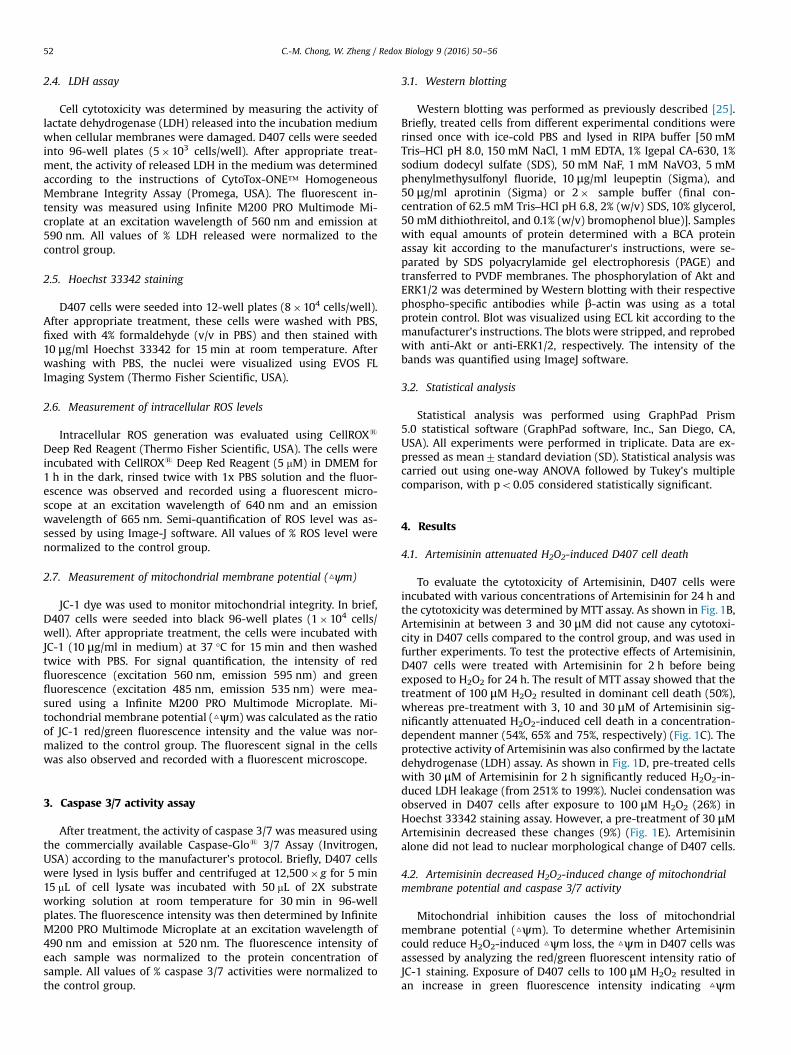
Fig. 3. Artemisinin reduced the increase of H2O2-induced oxidative stress in D407 cells.D407 cells were incubated with or without 100 μM H2O2 for another 24 h. Intracellularcontrol group;***Po0.005 versus H2O2-treated group was considered significantly diffe
were measured by staining with a fluorescent probe, CellROXs
Deep Red Reagent, in D407 cells. As shown in Fig. 3, 100 μM H2O2
caused a significant increase in fluorescent intensity in the cellscompared to the control group (177%). However, the increase inROS was significantly suppressed by pretreatment with Artemisi-nin at 30 μM (126%).
4.4. Artemisinin up-regulated ERK/CREB signaling
Previous studies indicated that Artemisinin was able to activateERK signaling in various cell models [26–28]. ERK was found to beinvolved in the inhibition of apoptosis. To determine whether theERK signaling pathway is regulated by Artemisinin in D407 cells,
After pre-treatment with 30 μM Artemisinin or 0.1% DMSO (vehicle control) for 2 h,ROS level was determined by the CellROXs Deep Red Reagent. ###Po0.005 versusrent.
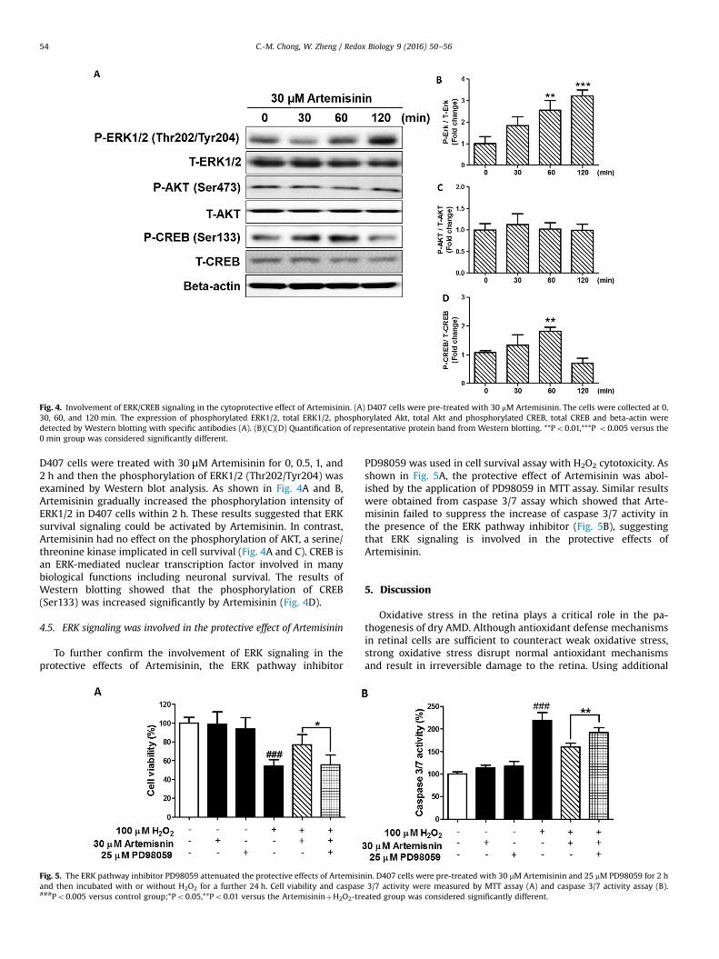
Fig. 4. Involvement of ERK/CREB signaling in the cytoprotective effect of Artemisinin. (A) D407 cells were pre-treated with 30 mM Artemisinin. The cells were collected at 0,30, 60, and 120 min. The expression of phosphorylated ERK1/2, total ERK1/2, phosphorylated Akt, total Akt and phosphorylated CREB, total CREB and beta-actin weredetected by Western blotting with specific antibodies (A). (B)(C)(D) Quantification of representative protein band from Western blotting. **Po0.01,***P o0.005 versus the0 min group was considered significantly different.
C.-M. Chong, W. Zheng / Redox Biology 9 (2016) 50–5654
D407 cells were treated with 30 μM Artemisinin for 0, 0.5, 1, and2 h and then the phosphorylation of ERK1/2 (Thr202/Tyr204) wasexamined by Western blot analysis. As shown in Fig. 4A and B,Artemisinin gradually increased the phosphorylation intensity ofERK1/2 in D407 cells within 2 h. These results suggested that ERKsurvival signaling could be activated by Artemisinin. In contrast,Artemisinin had no effect on the phosphorylation of AKT, a serine/threonine kinase implicated in cell survival (Fig. 4A and C). CREB isan ERK-mediated nuclear transcription factor involved in manybiological functions including neuronal survival. The results ofWestern blotting showed that the phosphorylation of CREB(Ser133) was increased significantly by Artemisinin (Fig. 4D).
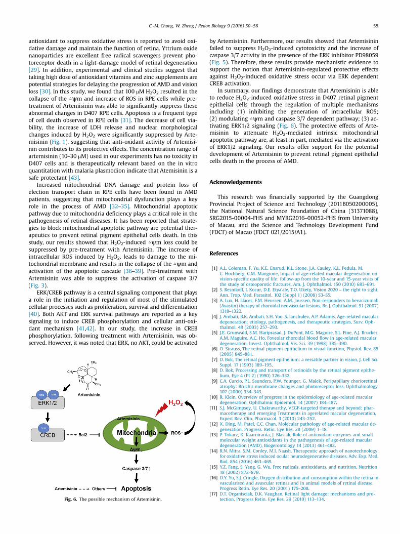
4.5. ERK signaling was involved in the protective effect of Artemisinin
To further confirm the involvement of ERK signaling in theprotective effects of Artemisinin, the ERK pathway inhibitor
Fig. 5. The ERK pathway inhibitor PD98059 attenuated the protective effects of Artemisinand then incubated with or without H2O2 for a further 24 h. Cell viability and caspase###Po0.005 versus control group;*Po0.05,**Po0.01 versus the ArtemisininþH2O2-tre
PD98059 was used in cell survival assay with H2O2 cytotoxicity. Asshown in Fig. 5A, the protective effect of Artemisinin was abol-ished by the application of PD98059 in MTT assay. Similar resultswere obtained from caspase 3/7 assay which showed that Arte-misinin failed to suppress the increase of caspase 3/7 activity inthe presence of the ERK pathway inhibitor (Fig. 5B), suggestingthat ERK signaling is involved in the protective effects ofArtemisinin.
5. Discussion
Oxidative stress in the retina plays a critical role in the pa-thogenesis of dry AMD. Although antioxidant defense mechanismsin retinal cells are sufficient to counteract weak oxidative stress,strong oxidative stress disrupt normal antioxidant mechanismsand result in irreversible damage to the retina. Using additional
in. D407 cells were pre-treated with 30 mM Artemisinin and 25 mM PD98059 for 2 h3/7 activity were measured by MTT assay (A) and caspase 3/7 activity assay (B).ated group was considered significantly different.
C.-M. Chong, W. Zheng / Redox Biology 9 (2016) 50–56 55
antioxidant to suppress oxidative stress is reported to avoid oxi-dative damage and maintain the function of retina. Yttrium oxidenanoparticles are excellent free radical scavengers prevent pho-toreceptor death in a light-damage model of retinal degeneration[29]. In addition, experimental and clinical studies suggest thattaking high dose of antioxidant vitamins and zinc supplements arepotential strategies for delaying the progression of AMD and visionloss [30]. In this study, we found that 100 mM H2O2 resulted in thecollapse of the △ψm and increase of ROS in RPE cells while pre-treatment of Artemisinin was able to significantly suppress theseabnormal changes in D407 RPE cells. Apoptosis is a frequent typeof cell death observed in RPE cells [31]. The decrease of cell via-bility, the increase of LDH release and nuclear morphologicalchanges induced by H2O2 were significantly suppressed by Arte-misinin (Fig. 1), suggesting that anti-oxidant activity of Artemisi-nin contributes to its protective effects. The concentration range ofartemisinin (10–30 mM) used in our experiments has no toxicity inD407 cells and is therapeutically relevant based on the in vitroquantitation with malaria plasmodion indicate that Atemisinin is asafe protectant [43].
Increased mitochondrial DNA damage and protein loss ofelection transport chain in RPE cells have been found in AMDpatients, suggesting that mitochondrial dysfunction plays a keyrole in the process of AMD [32–35]. Mitochondrial apoptoticpathway due to mitochondria deficiency plays a critical role in thepathogenesis of retinal diseases. It has been reported that strate-gies to block mitochondrial apoptotic pathway are potential ther-apeutics to prevent retinal pigment epithelial cells death. In thisstudy, our results showed that H2O2-induced △ψm loss could besuppressed by pre-treatment with Artemisinin. The increase ofintracellular ROS induced by H2O2, leads to damage to the mi-tochondrial membrane and results in the collapse of the △ψm andactivation of the apoptotic cascade [36–39]. Pre-treatment withArtemisinin was able to suppress the activation of caspase 3/7(Fig. 3).
ERK/CREB pathway is a central signaling component that playsa role in the initiation and regulation of most of the stimulatedcellular processes such as proliferation, survival and differentiation[40]. Both AKT and ERK survival pathways are reported as a keysignaling to induce CREB phosphorylation and cellular anti-oxi-dant mechanism [41,42]. In our study, the increase in CREBphosphorylation, following treatment with Artemisinin, was ob-served. However, it was noted that ERK, no AKT, could be activated
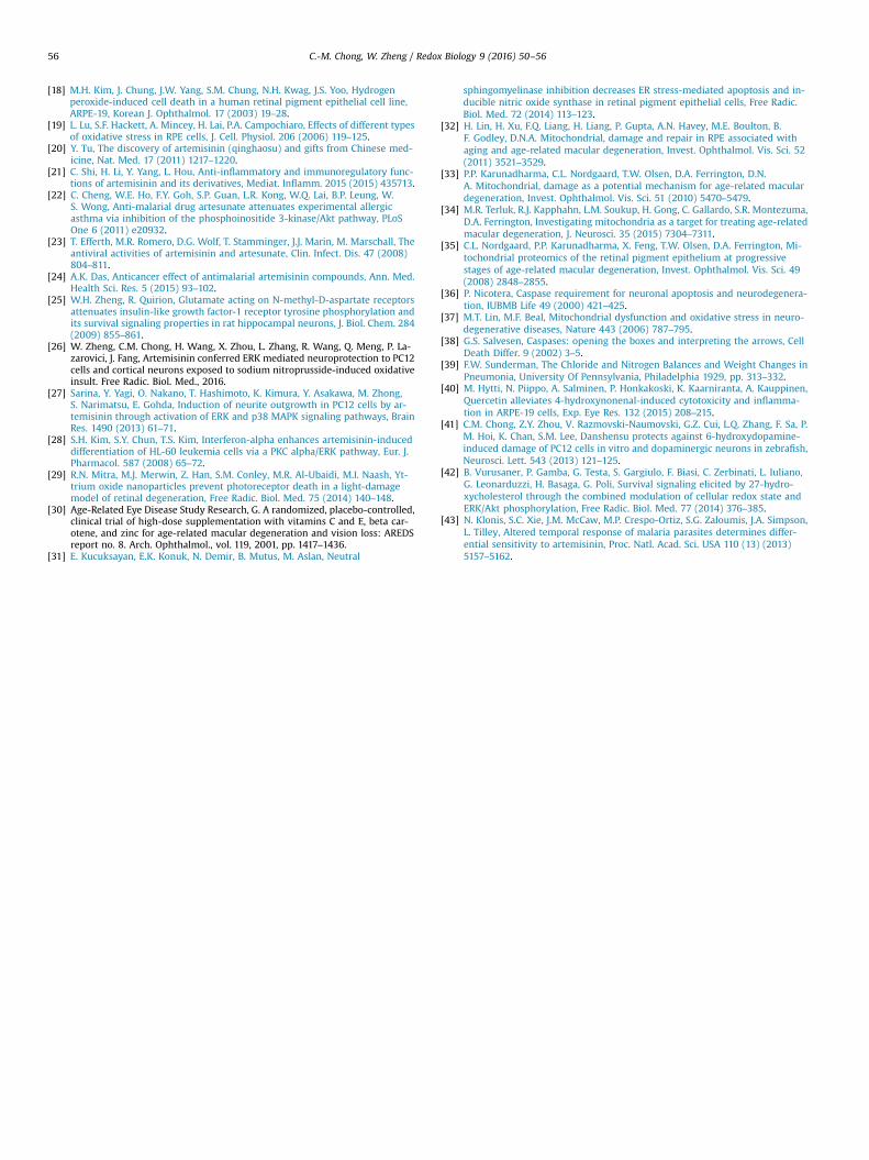
Fig. 6. The possible mechanism of Artemisinin.
by Artemisinin. Furthermore, our results showed that Artemisininfailed to suppress H2O2-induced cytotoxicity and the increase ofcaspase 3/7 activity in the presence of the ERK inhibitor PD98059(Fig. 5). Therefore, these results provide mechanistic evidence tosupport the notion that Artemisinin-regulated protective effectsagainst H2O2-induced oxidative stress occur via ERK dependentCREB activation.
In summary, our findings demonstrate that Artemisinin is ableto reduce H2O2-induced oxidative stress in D407 retinal pigmentepithelial cells through the regulation of multiple mechanismsincluding (1) inhibiting the generation of intracellular ROS;(2) modulating △ψm and caspase 3/7 dependent pathway; (3) ac-tivating ERK1/2 signaling (Fig. 6). The protective effects of Arte-misinin to attenuate H2O2-mediated intrinsic mitochondrialapoptotic pathway are, at least in part, mediated via the activationof ERK1/2 signaling. Our results offer support for the potentialdevelopment of Artemisinin to prevent retinal pigment epithelialcells death in the process of AMD.
Acknowledgements
This research was financially supported by the GuangdongProvincial Project of Science and Technology (2011B050200005),the National Natural Science Foundation of China (31371088),SRG2015-00004-FHS and MYRG2016–00052-FHS from Universityof Macau, and the Science and Technology Development Fund(FDCT) of Macao (FDCT 021/2015/A1).
References
[1] A.L. Coleman, F. Yu, K.E. Ensrud, K.L. Stone, J.A. Cauley, K.L. Pedula, M.C. Hochberg, C.M. Mangione, Impact of age-related macular degeneration onvision-specific quality of life: follow-up from the 10-year and 15-year visits ofthe study of osteoporotic fractures, Am. J. Ophthalmol. 150 (2010) 683–691.
[2] S. Resnikoff, I. Kocur, D.E. Etya’ale, T.O. Ukety, Vision 2020 – the right to sight,Ann. Trop. Med. Parasitol. 102 (Suppl 1) (2008) S3–S5.
[3] A. Lux, H. Llacer, F.M. Heussen, A.M. Joussen, Non-responders to bevacizumab(Avastin) therapy of choroidal neovascular lesions, Br. J. Ophthalmol. 91 (2007)1318–1322.
[4] J. Ambati, B.K. Ambati, S.H. Yoo, S. Ianchulev, A.P. Adamis, Age-related maculardegeneration: etiology, pathogenesis, and therapeutic strategies, Surv. Oph-thalmol. 48 (2003) 257–293.
[5] J.E. Grunwald, S.M. Hariprasad, J. DuPont, M.G. Maguire, S.L. Fine, A.J. Brucker,A.M. Maguire, A.C. Ho, Foveolar choroidal blood flow in age-related maculardegeneration, Invest. Ophthalmol. Vis. Sci. 39 (1998) 385–390.
[6] O. Strauss, The retinal pigment epithelium in visual function, Physiol. Rev. 85(2005) 845–881.
[7] D. Bok, The retinal pigment epithelium: a versatile partner in vision, J. Cell Sci.Suppl. 17 (1993) 189–195.
[8] D. Bok, Processing and transport of retinoids by the retinal pigment epithe-lium, Eye 4 (Pt 2) (1990) 326–332.
[9] C.A. Curcio, P.L. Saunders, P.W. Younger, G. Malek, Peripapillary chorioretinalatrophy: Bruch’s membrane changes and photoreceptor loss, Ophthalmology107 (2000) 334–343.
[10] R. Klein, Overview of progress in the epidemiology of age-related maculardegeneration, Ophthalmic Epidemiol. 14 (2007) 184–187.
[11] S.J. McGimpsey, U. Chakravarthy, VEGF-targeted therapy and beyond: phar-macotherapy and emerging Treatments in agerelated macular degeneration,Expert Rev. Clin. Pharmacol. 3 (2010) 243–252.
[12] X. Ding, M. Patel, C.C. Chan, Molecular pathology of age-related macular de-generation, Progress. Retin. Eye Res. 28 (2009) 1–18.
[13] P. Tokarz, K. Kaarniranta, J. Blasiak, Role of antioxidant enzymes and smallmolecular weight antioxidants in the pathogenesis of age-related maculardegeneration (AMD), Biogerontology 14 (2013) 461–482.
[14] R.N. Mitra, S.M. Conley, M.I. Naash, Therapeutic approach of nanotechnologyfor oxidative stress induced ocular neurodegenerative diseases, Adv. Exp. Med.Biol. 854 (2016) 463–469.
[15] Y.Z. Fang, S. Yang, G. Wu, Free radicals, antioxidants, and nutrition, Nutrition18 (2002) 872–879.
[16] D.Y. Yu, S.J. Cringle, Oxygen distribution and consumption within the retina invascularised and avascular retinas and in animal models of retinal disease,Progress Retin. Eye Res. 20 (2001) 175–208.
[17] D.T. Organisciak, D.K. Vaughan, Retinal light damage: mechanisms and pro-tection, Progress Retin. Eye Res. 29 (2010) 113–134.
C.-M. Chong, W. Zheng / Redox Biology 9 (2016) 50–5656
[18] M.H. Kim, J. Chung, J.W. Yang, S.M. Chung, N.H. Kwag, J.S. Yoo, Hydrogenperoxide-induced cell death in a human retinal pigment epithelial cell line,ARPE-19, Korean J. Ophthalmol. 17 (2003) 19–28.
[19] L. Lu, S.F. Hackett, A. Mincey, H. Lai, P.A. Campochiaro, Effects of different typesof oxidative stress in RPE cells, J. Cell. Physiol. 206 (2006) 119–125.
[20] Y. Tu, The discovery of artemisinin (qinghaosu) and gifts from Chinese med-icine, Nat. Med. 17 (2011) 1217–1220.
[21] C. Shi, H. Li, Y. Yang, L. Hou, Anti-inflammatory and immunoregulatory func-tions of artemisinin and its derivatives, Mediat. Inflamm. 2015 (2015) 435713.
[22] C. Cheng, W.E. Ho, F.Y. Goh, S.P. Guan, L.R. Kong, W.Q. Lai, B.P. Leung, W.S. Wong, Anti-malarial drug artesunate attenuates experimental allergicasthma via inhibition of the phosphoinositide 3-kinase/Akt pathway, PLoSOne 6 (2011) e20932.
[23] T. Efferth, M.R. Romero, D.G. Wolf, T. Stamminger, J.J. Marin, M. Marschall, Theantiviral activities of artemisinin and artesunate, Clin. Infect. Dis. 47 (2008)804–811.
[24] A.K. Das, Anticancer effect of antimalarial artemisinin compounds, Ann. Med.Health Sci. Res. 5 (2015) 93–102.
[25] W.H. Zheng, R. Quirion, Glutamate acting on N-methyl-D-aspartate receptorsattenuates insulin-like growth factor-1 receptor tyrosine phosphorylation andits survival signaling properties in rat hippocampal neurons, J. Biol. Chem. 284(2009) 855–861.
[26] W. Zheng, C.M. Chong, H. Wang, X. Zhou, L. Zhang, R. Wang, Q. Meng, P. La-zarovici, J. Fang, Artemisinin conferred ERK mediated neuroprotection to PC12cells and cortical neurons exposed to sodium nitroprusside-induced oxidativeinsult. Free Radic. Biol. Med., 2016.
[27] Sarina, Y. Yagi, O. Nakano, T. Hashimoto, K. Kimura, Y. Asakawa, M. Zhong,S. Narimatsu, E. Gohda, Induction of neurite outgrowth in PC12 cells by ar-temisinin through activation of ERK and p38 MAPK signaling pathways, BrainRes. 1490 (2013) 61–71.
[28] S.H. Kim, S.Y. Chun, T.S. Kim, Interferon-alpha enhances artemisinin-induceddifferentiation of HL-60 leukemia cells via a PKC alpha/ERK pathway, Eur. J.Pharmacol. 587 (2008) 65–72.
[29] R.N. Mitra, M.J. Merwin, Z. Han, S.M. Conley, M.R. Al-Ubaidi, M.I. Naash, Yt-trium oxide nanoparticles prevent photoreceptor death in a light-damagemodel of retinal degeneration, Free Radic. Biol. Med. 75 (2014) 140–148.
[30] Age-Related Eye Disease Study Research, G. A randomized, placebo-controlled,clinical trial of high-dose supplementation with vitamins C and E, beta car-otene, and zinc for age-related macular degeneration and vision loss: AREDSreport no. 8. Arch. Ophthalmol., vol. 119, 2001, pp. 1417–1436.
[31] E. Kucuksayan, E.K. Konuk, N. Demir, B. Mutus, M. Aslan, Neutral
sphingomyelinase inhibition decreases ER stress-mediated apoptosis and in-ducible nitric oxide synthase in retinal pigment epithelial cells, Free Radic.Biol. Med. 72 (2014) 113–123.
[32] H. Lin, H. Xu, F.Q. Liang, H. Liang, P. Gupta, A.N. Havey, M.E. Boulton, B.F. Godley, D.N.A. Mitochondrial, damage and repair in RPE associated withaging and age-related macular degeneration, Invest. Ophthalmol. Vis. Sci. 52(2011) 3521–3529.
[33] P.P. Karunadharma, C.L. Nordgaard, T.W. Olsen, D.A. Ferrington, D.N.A. Mitochondrial, damage as a potential mechanism for age-related maculardegeneration, Invest. Ophthalmol. Vis. Sci. 51 (2010) 5470–5479.
[34] M.R. Terluk, R.J. Kapphahn, L.M. Soukup, H. Gong, C. Gallardo, S.R. Montezuma,D.A. Ferrington, Investigating mitochondria as a target for treating age-relatedmacular degeneration, J. Neurosci. 35 (2015) 7304–7311.
[35] C.L. Nordgaard, P.P. Karunadharma, X. Feng, T.W. Olsen, D.A. Ferrington, Mi-tochondrial proteomics of the retinal pigment epithelium at progressivestages of age-related macular degeneration, Invest. Ophthalmol. Vis. Sci. 49(2008) 2848–2855.
[36] P. Nicotera, Caspase requirement for neuronal apoptosis and neurodegenera-tion, IUBMB Life 49 (2000) 421–425.
[37] M.T. Lin, M.F. Beal, Mitochondrial dysfunction and oxidative stress in neuro-degenerative diseases, Nature 443 (2006) 787–795.
[38] G.S. Salvesen, Caspases: opening the boxes and interpreting the arrows, CellDeath Differ. 9 (2002) 3–5.
[39] F.W. Sunderman, The Chloride and Nitrogen Balances and Weight Changes inPneumonia, University Of Pennsylvania, Philadelphia 1929, pp. 313–332.
[40] M. Hytti, N. Piippo, A. Salminen, P. Honkakoski, K. Kaarniranta, A. Kauppinen,Quercetin alleviates 4-hydroxynonenal-induced cytotoxicity and inflamma-tion in ARPE-19 cells, Exp. Eye Res. 132 (2015) 208–215.
[41] C.M. Chong, Z.Y. Zhou, V. Razmovski-Naumovski, G.Z. Cui, L.Q. Zhang, F. Sa, P.M. Hoi, K. Chan, S.M. Lee, Danshensu protects against 6-hydroxydopamine-induced damage of PC12 cells in vitro and dopaminergic neurons in zebrafish,Neurosci. Lett. 543 (2013) 121–125.
[42] B. Vurusaner, P. Gamba, G. Testa, S. Gargiulo, F. Biasi, C. Zerbinati, L. Iuliano,G. Leonarduzzi, H. Basaga, G. Poli, Survival signaling elicited by 27-hydro-xycholesterol through the combined modulation of cellular redox state andERK/Akt phosphorylation, Free Radic. Biol. Med. 77 (2014) 376–385.
[43] N. Klonis, S.C. Xie, J.M. McCaw, M.P. Crespo-Ortiz, S.G. Zaloumis, J.A. Simpson,L. Tilley, Altered temporal response of malaria parasites determines differ-ential sensitivity to artemisinin, Proc. Natl. Acad. Sci. USA 110 (13) (2013)5157–5162.